Embed Size (px)
Citation preview

High resolution manometry of pharyngeal swallow pressureevents associated with head turn and chin tuck
Timothy M. McCulloch, M.D., Matthew R. Hoffman, B.S., and Michelle R. Ciucci, Ph.D.Department of Surgery, Division of Otolaryngology – Head and Neck Surgery, University ofWisconsin School of Medicine and Public Health, Madison, WI, 53792-7375
AbstractObjectives—To quantify the effect of swallowing maneuvers on pharyngeal pressure eventsusing high resolution manometry (HRM).
Methods—Seven subjects swallowed multiple, five ml water boluses in three different posturalconditions: neutral, head turn, and chin tuck. Pressure and timing events were recorded with a 36-sensor HRM catheter. We analyzed the regions of the velopharynx and base of tongue for maximalpressure, rate of pressure increase, pressure gradient, and duration of pressure above baseline. Inthe region of the upper esophageal sphincter (UES), we analyzed duration of pressure declination,minimum pressure during opening, and maximum pressures before and after UES opening.
Results—Maneuvers did not have a significant effect on maximum pressure, rate of pressureincrease, or pressure gradients in the velopharyngeal or tongue base regions. Duration of pressureabove baseline was significantly longer in the velopharynx for head turn. Pre-swallow maximumUES pressure was significantly greater for neutral swallows compared to head turn and post-swallow maximum pressure was significantly lower for chin tuck. Both maneuvers appeared toprolong UES pressure declination duration, but neither reached significance.
Conclusions—HRM allows for optimal spatial and temporal resolution during recording ofpressure events along the length of the pharynx and revealed previously undetected task-dependentpressure and timing differences during chin tuck and head turn in healthy adults. These maneuversappear to influence the UES to a greater degree than the velopharynx and tongue base. Furtherstudies designed to quantify the effect of other maneuvers and bolus consistencies on thegeneration of pharyngeal pressure events in both normal and disordered subjects may lead tohypothesis-driven, optimal, individualized, swallowing therapies.
KeywordsPharyngeal pressure; swallowing maneuver; high resolution manometry; dysphagia
INTRODUCTIONThe pharyngeal swallow is an event that orchestrates a complex series of musclecontractions to close air spaces in the oral cavity, nasopharynx, and glottis whilesimultaneously generating pressure gradients to move a bolus from the posterior oral cavityto the cervical esophagus. That is, movement of the bolus is a pressure-driven event.Multiple investigations on pharyngeal pressures during the swallow have been conducted
Corresponding author: Timothy M. McCulloch, M.D., Address: Box 7375 Clinical Science Center – H4, 600 Highland Ave, Madison,WI 53792, Telephone number: 608-63-0192, [email protected] send reprint requests to Timothy M. McCulloch, M.D.
NIH Public AccessAuthor ManuscriptAnn Otol Rhinol Laryngol. Author manuscript; available in PMC 2011 March 1.
Published in final edited form as:Ann Otol Rhinol Laryngol. 2010 June ; 119(6): 369–376.
NIH
-PA Author Manuscript
NIH
-PA Author Manuscript
NIH
-PA Author Manuscript

using single, dual, or multi-array pressure sensors and have contributed to our understandingof pressure-modulated bolus movement (1–6). These studies employed catheters with one tofour unidirectional sensors, usually with a posterior orientation. Since the pharynx is arelatively long, asymmetric, and moving structure, traditional manometry with a smallnumber of unidirectional sensors cannot provide adequate spatial resolution, nor can itaccommodate anatomic variations such as the length of the pharynx. Coupling traditionalmanometry with videofluorography has been standardized to some degree (7) and thesecombined studies generated much of the theory on bolus propulsion and movement (8,9).However, due to the technological limitations of traditional manometry, we primarily relyon visual observations made from videofluorography and make assumptions about pressureevents during the pharyngeal swallow.
The advent of high resolution manometry (HRM) offers a drastically improved method toevaluate pressure during swallowing along the length of the entire pharynx and esophagus(10). Pioneering work with HRM in the pharyngoesophageal segment (PES) utilizing the“Dentsleeve”, a multi-channel, tightly spaced perfusion manometer (11), provided thebackground for this current study using modern, solid-state HRM through the entirepharynx. The most recent version of HRM (Sierra Scientific Instruments, Los Angeles CA),uses 36 sensor arrays spaced one centimeter apart and is capable of recording pressure inasymmetrical structures, offering the spatial and temporal resolution necessary to accuratelycapture rapidly changing pressures throughout the pharynx without concern for anatomicvariation or moving structures. This allows for a comprehensive evaluation of thepharyngeal swallow. Analyzing pressures across the entire length of the pharynx shouldreveal additional and perhaps subtle findings that were previously undetectable usingtraditional manometry.
We used HRM to measure pressure events from the velopharynx to the cervical esophagus,with emphasis on the regions of the velopharynx, tongue base, and upper esophagealsphincter (UES). This was done with the head in the neutral position as well as during twocompensatory maneuvers: head turn and chin tuck, as they are commonly employed clinicalstrategies (1,2,4,12,13) and have demonstrated alterations in pharyngeal and UES function(1,14,15). Head turn has been shown to redirect the bolus to the contralateral pyriform sinus(1,15) and chin tuck has been shown to widen the valleculae and narrow the aditus layrngis,reducing penetration depth into the larynx and trachea (16). These anatomic changes havebeen shown useful in many dysphagia patient populations (12,16,17). However, it remainsunclear how these maneuvers affect swallowing physiology (5) and more specifically,pharyngeal pressures. By changing the dimensions of the pharynx, these maneuvers possiblyalter pharyngeal pressure gradients, duration of pressure events, or both. We hypothesizedthat the chin tuck and head turn maneuvers would lead to changes in pressure-related events(maximum pressure, pressure gradients, duration of events). Specifically, we predictedaltering the relative position of the larynx, pharyngeal walls, and tongue may lead tochanges in tongue/pharyngeal driving force and UES opening, but little change at the levelof the velopharynx.
MATERIALS AND METHODSEquipment
A solid-state high resolution manometer was used for all data collection (ManoScan360 HighResolution Manometry System, Sierra Scientific Instruments, Los Angeles, CA). Themanometric catheter has an outer diameter of 4 mm and 36 circumferential pressure sensorsspaced 1 cm apart. Each sensor spans 2.5 mm and receives input from 12 circumferentialsectors. These inputs are averaged and a mean pressure is recorded as the pressure detectedby that individual sensor. The system is calibrated to record pressures between −20 and 600
McCulloch et al. Page 2
Ann Otol Rhinol Laryngol. Author manuscript; available in PMC 2011 March 1.
NIH
-PA Author Manuscript
NIH
-PA Author Manuscript
NIH
-PA Author Manuscript

mmHg with fidelity of 2 mmHg. Data were collected at a sampling rate of 50 Hz (ManoScanData Acquisition, Sierra Scientific Instruments). Prior to calibration, the catheter wascovered with a protective sheath to preserve sterility without the need to sterilize the catheterbetween uses (ManoShield, Sierra Scientific Instruments). The catheter was calibratedbefore each participant according to manufacturer specifications.
Data collectionFour males and three females, aged 20.9 ± 2.1 years (19 – 25), participated in this study withthe approval of the Institutional Review Board of the University of Wisconsin-Madison. Allsubjects were without swallowing, neurological, or gastrointestinal disorders. Participantswere instructed not to eat for four hours and not drink liquids for two hours prior to testingto avoid any potential confounding effect of satiety.
Topical 2% viscous lidocaine was applied to the nasal passages with a cotton swab andparticipants gargled a solution of 4% lidocaine (1 to 2 cc) for several seconds. Themanometric catheter was lubricated with 2% viscous lidocaine to ease passage of thecatheter through the pharynx. Once the catheter was positioned within the pharynx,participants sat quietly for 5–10 minutes to adjust to the catheter prior to performingswallowing tasks.
The following tasks were performed 5 times with a 5 milliliter (ml) water bolus: neutralswallow, head turn (to the side ipsilateral to catheter placement), and chin tuck. Each boluswas delivered to the oral cavity via syringe. Participants were trained on each swallow taskprior to the placement of the catheter. A total of 15 swallows were analyzed for eachparticipant. A 5 ml bolus was selected as it is commonly employed in research paradigmsand also represents a volume similar to the restricted volumes many dysphagic patients arelimited to as a component of aspiration management.
Data analysisPressure and timing data were extracted using ManoView software (Sierra ScientificInstruments). The regions of interest were defined manometrically (figure 1). Thevelopharynx was defined as the region of swallow-related pressure change just proximal tothe area of continuous nasal cavity quiescence and extending two centimeters. The tonguebase region was defined as the area of swallow related pressure change with a high pressurezone identified approximately midway between the nasopharynx and the UES, with itsepicenter at the high pressure point and extending two centimeters proximal and distal tothat point. The UES region was defined as the midpoint of stable high pressure just distal(rostral) to the baseline low esophageal pressure zone, extending to a point of lowesophageal pressure distally and low baseline pharyngeal pressure proximally. It isimportant to note that during swallowing, this anatomic area is mobile along the catheter,moving rostrally as much as 4 cm.
Mean and standard deviation values were recorded for maximum pressure, rate of pressureincrease, pressure gradient, and duration of pressure above baseline in the regions of thevelopharynx and tongue base. Rate of pressure increase was calculated by subtractingbaseline pressure from maximum pressure and dividing by the time lapse between thesepoints. Pressure gradients were measured by determining the sensor at which maximumpressure within a region occurred and then determining the pressure recorded in sensors oneand two cm downstream (toward the esophagus) at the same time-point. Duration ofpressure above baseline within a region was defined as the time duration between the onsetof pressure escalation and its return to or below baseline using the single senor wheremaximum pressure was recorded. Minimum pressure during UES opening as well as
McCulloch et al. Page 3
Ann Otol Rhinol Laryngol. Author manuscript; available in PMC 2011 March 1.
NIH
-PA Author Manuscript
NIH
-PA Author Manuscript
NIH
-PA Author Manuscript

maximum pressures proceeding and succeeding UES opening were also recorded. The timelapse between these pressure peaks is termed UES opening time (figure 2). Total swallowduration was defined as the time lapse between onset of velopharyngeal pressure rise and thepost-swallow UES pressure peak.
SigmaPlot 11.0 software was employed for statistical analyses. Mean values recorded duringhead turn and chin tuck were compared to those recorded for neutral swallows using two-tailed paired t-tests. Shapiro-Wilk and Levene’s tests were used to determine normality andequal variance, respectively. If data did not meet the statistical assumptions for parametrictesting, a Wilcoxon-Mann-Whitney rank sum test was performed. A significance level of α= 0.05 was determined a-priori.
RESULTSFigure 2 provides sample spatiotemporal plots for the three tasks from a single subject and agroup data summary is provided in table 1.
VelopharynxManeuvers did not have a significant effect on maximum velopharyngeal pressure (headturn: t = −0.113, df = 6, p = 0.914; chin tuck: t = 0.422, df = 6, p = 0.688). Rate ofvelopharyngeal pressure rise was not significantly different from neutral for either head turn(t = −0.398; df = 6; p = 0.705) or chin tuck (t = 1.220; df = 6; p = 0.268), though it wasdiscernibly lower for chin tuck. Duration of velopharyngeal pressure above baseline wassignificantly longer for the head turn (W = 28; T+ = 28; T− = −0; Z = 2.375; p = 0.016) anddiscernibly longer for the chin tuck (t = −1.511; df = 6; p = 0.182).
Tongue baseTongue base maximum pressure was highest in the neutral position and no significantdifference was found with head turn or chin tuck, (head turn: W = 2, T+ = 15, T− = − 13, Z= 0.169, p = 0.938; chin tuck: t = 0.922, df = 6, p = 0.392). Rate of pressure rise at thetongue base was not significantly different compared to neutral for either head turn (t =−0.761; df = 6; p = 0.476) or chin tuck (t = −0.079; df = 6; p = 0.946). And duration oftongue base pressure above baseline was longer for head turn (t = −1.198; df = 6; p = 0.276)and shorter for chin tuck (t = 0.906; df = 6; p = 0.4), though neither difference wassignificant.
UESPre-swallow peak UES pressure was significantly greater for neutral swallows compared tohead turn (t = 3.275; df = 6; p = 0.017). This pressure was also higher for neutral swallowscompared to chin tuck, but this difference was not significant (t = 1.384; df = 6; p = 0.216).Chin tuck exhibited significantly lower post-swallow peak UES pressure compared toneutral (t = 2.662; df = 6; p = 0.037), and there was no discernible trend between neutral andhead turn (t = −0.723; df = 6; p = 0.497). Minimum pressure during UES opening was lowerfor head turn (t = 1.401; df = 6; p = 0.211) and higher for chin tuck (t = −1.254; df = 6; p =0.256) (figure 3), but these differences were not significant. Both maneuvers appeared toincrease UES opening time, but neither increase was significant (head turn: t = −1.157, df =6, p = 0.18; chin tuck: t = −0.800, df = 6, p = 0.454) (figure 4).
Pressure gradientsNo differences were found in one centimeter pressure gradients at the level of thevelopharynx or tongue base when comparing neutral swallow to head turn (velopharynx: t =0.487, df = 6, p = 0.644; tongue base: t = 0.305, df = 6, p = 0.770) and chin tuck
McCulloch et al. Page 4
Ann Otol Rhinol Laryngol. Author manuscript; available in PMC 2011 March 1.
NIH
-PA Author Manuscript
NIH
-PA Author Manuscript
NIH
-PA Author Manuscript

(velopharynx: t = 0.740, df = 6, p = 0.487; tongue base: W = 2, T+ = 15, T− = −13, Z =0.169, df = 6, p = 0.938). There were also no differences for two centimeter gradients foreither the head turn (velopharynx: t = −0.044, df = 6, p = 0.966; tongue base: W = 2, T+ =15, T− = −13, Z = 0.169, df = 6, p = 0.938) or chin tuck (velopharynx: t = 0.271, df = 6, p =0.796; tongue base: W = 0, T+ = 14, T− = −14, Z = 0, df = 6, p = 1.000). There was,however, an interesting group mean shift of the tongue base gradients from neutral to thepostures with lower mean maximum pressure and lower caudal pressures preserving a stablegradient (figure 4, table 2).
Total swallow duration was not significantly affected by head turn (t = 0.122, df = 6, p =0.907) and chin tuck (t = 0.529, df = 6, p = 0.616).
DISCUSSIONManeuver-dependent differences in pharyngeal pressures were observed primarily in theUES, not in the velopharynx or tongue base. Thus, our hypotheses were partially supportedas we anticipated that altering the relative position of the larynx, pharyngeal walls, andtongue may lead to pressure changes in the pharynx at the level of the tongue base and UES,but little change to the velopharynx. However, the results do not completely support ourassumptions that the velopharynx is insulated from the changes in anatomic relationshipwhich occur with postural maneuvers, as durational changes were noted. Somewhat contraryto our predictions for the region of the tongue base, maximal tongue base pressures were thehighest for the neutral position, followed by head turn and the lowest for the chin tuck.There was no change in rate of pressure rise and there was no change in absolute pressuregradient, but an interesting shift in the entire gradient values toward lower pressures withboth of the maneuvers appeared.
The absence of more notable changes at the tongue base region should not be interpreted asa universal effect as it will be important to evaluate this feature in a disordered subjectpopulation. Further, we studied this in a small number of participants and a small samplesize can lead to beta error in results interpretation. However, in another study, a similar lackof effect during chin tuck was reported in hypopharyngeal intra-bolus pressure and durationin patients with moderate to severe dysphagia (5). In contrast, head rotation has also beenshown to increase hypopharyngeal (valleculae and pyriform sinus) pressure when the headis rotated toward the pyriform sinus occupied by the catheter, but decrease when the head isrotated away from the catheter (15). The bolus was directed away from the side of headrotation which would infer that the intra-bolus pressure was best recorded with head rotationaway from the catheter (15). It should be noted that this study used a unidirectional sensor,and thus their high pressure findings may be more related to catheter position rather thantrue intra-bolus pressures. In the current study, we recorded circumferential pressures, whicheliminates directionality of pressure recording and this may account for the discrepancy inhypopharyngeal pressures among studies.
Perhaps of most interest were our observed differences in the duration of pressure events.The duration of time above baseline pressures for the velopharyngeal region wassignificantly longer for head turn and discernibly longer for the chin tuck. These durationscoincide with longer time of minimum pressure (opening time) in the UES, which is notsurprising as the velopharyngeal port would usually remain sealed during UES opening tofacilitate bolus flow toward the esophagus. This infers either a passive or active interactionbetween these two distant pharyngeal sites during swallow. However, even as individualcomponents of the swallow showed durational change, the total time of swallow wasunaffected (table 1). Duration of tongue base pressure above baseline was longer for headturn, but actually shorter for chin tuck, although these differences were not significant. It
McCulloch et al. Page 5
Ann Otol Rhinol Laryngol. Author manuscript; available in PMC 2011 March 1.
NIH
-PA Author Manuscript
NIH
-PA Author Manuscript
NIH
-PA Author Manuscript

would be interesting to evaluate this in a disordered population, as we speculate that somechanges to the duration of tongue base activity may be vulnerable to disease processes thataffect tongue strength and timing.
Pressure and duration effects were observed in the UES region for both maneuvers. Pre-swallow maximum pressures were the greatest in the neutral position and a head turn led toa significant pressure decrease, with pre swallow mean maximum pressures averaging 227mmHg in the neutral position and 118 mmHg with head turn. This is consistent with thestudy of normal subjects by Logemann et al., where an 18 mmHg mean drop in UESpressure was noted with head rotation (1). Our post-swallow pressures were greater with thehead turn and significantly lower with the chin tuck as compared to neutral. Both maneuversincreased the duration of UES opening, though this measure did not reach statisticalsignificance. These findings are in accord with previous work using a sleeve catheter in theUES (1). According to radiographic studies, both head turn and chin tuck altercricopharyngeal position, facilitating UES opening and bolus passage into the esophagus(15,16). As such, these maneuvers may have caused passive changes in UES physiology viastructural change.
Our data roughly correspond with those presented in previous studies using conventionalmanometry (table 3), with a few key differences. Maximum pharyngeal pressure, defined inthis study as tongue base pressure, was higher than in previous studies (3,5,18). This couldbe attributed to HRM measuring pressure across a greater length of the pharynx thantraditional manometry, as more sensors allow for increased measurement precision.Additionally, HRM may detect higher pressures in asymmetric regions which areundetectable using unidirectional manometers. UES opening time, defined in this study asthe time between the pre-swallow pressure peak and post-swallow pressure peak, was longerthan UES relaxation time reported elsewhere (3,5). We chose to use a peak to peakdefinition as it reflects the entire period of sphincter opening including the times oftransition from closed to open and open to closed. Others have used the points of pressuredecrease and increase equal to half the UES baseline pressure (14), pressure curve analysisto identify transition points between bolus and contractile pressures (19), or the time of lowpressure between the high pressure peaks produced by a single recording catheter placed atthe level of the UES baseline high pressure zone, the ‘M’ wave (8,18). As we were focusingon intrasubject changes, we believe our method was simple, reliable, and appropriate for theresearch questions. Further, our ability to select a group of sensors covering the entire UESregion, as opposed to a single sensor somewhere within the region, most likely accounts forthese differences.
Intersubject variability probably partially accounts for some of the non-significant findingsas we identified a wide range of pressure events even in this small set of normal healthyswallowers. It is likely that this variability is a characteristic of human swallowing and willneed to be considered when working with dysphagia patients. Subtle disruptions in a uniqueswallow pattern will lead to a dysphagia which may be difficult to characterize with isolatedpressure recordings. It is possible that these maneuvers would indeed facilitate functionalpressure changes in disordered individuals with abnormal pressures.
Although topical anesthesia was used in this study to diminish participant discomfort, we donot believe it significantly altered swallow physiology with regard to our measurements.Omitting this in pilot experiments led to increased gagging and resting UES pressure,confounding data collection. As swallowing is a sensorimotor phenomenon, impairingafferent nerves in the pharynx could potentially alter swallow physiology. However,mechanoreceptors in the pharynx (deep to the mucosa) are largely responsible formodulating swallow physiology (20) and these fibers were probably not affected by our
McCulloch et al. Page 6
Ann Otol Rhinol Laryngol. Author manuscript; available in PMC 2011 March 1.
NIH
-PA Author Manuscript
NIH
-PA Author Manuscript
NIH
-PA Author Manuscript

topical anesthetic. Additionally, the oral mucosa was minimally affected, and afferentinformation from this area is also important to swallow modulation. We believe that thetrade-off for increased comfort at the expense of short-term pain/temperature afferentalteration improved the reliability of our data.
HRM provides the opportunity to evaluate pressure gradients during swallowing. However,even with high resolution capabilities, our definition of “pressure gradient” is simplified to ameasure of difference in pressure along the course of known fluid movement. In a strictersense, pressure gradient is defined as a vector quantity of a three dimensional scalar fieldwhere every point in the field has an assignable magnitude value, in this case pressure, and adeterminable vector influenced by pressure of adjacent points within the field. To a certainextent, we are able to assume information about the pressure in two of the three directionsdue to boundary conditions at the walls of the pharynx, leaving only one direction forchange to occur (the pharyngo-esophageal conduit where the HRM catheter is located). Thisassumption breaks down in the dysfunctional swallow where sphincteric failure at the oralcavity, nasopharynx, and larynx can modify these simplified boundary conditions.
It is also important to recognize that HRM fails to measure any of the pressure events thatoccur in the oral cavity during the swallow as the moving bolus and its surrounding pressureevents reach the catheter with established momentum. Thus, we must keep in mind that weare measuring many of the important swallow related pressures, but not all of them.
This study examined pressure and timing measures using a small water bolus (5 ml). Itwould be useful to examine these same parameters in different bolus volumes andconsistencies in a larger cohort of both normal and disordered subjects. With larger volumes,pressure changes may be found at the tongue base region; however, the identified changes atthe UES level appear to support the use of these maneuvers regardless of the absence of anobserved change in the “tongue driving force”. Though significant differences could beobserved for some parameters across swallowing tasks, increasing sample size in the futuremay reveal more evident trends. Trends may also be more apparent in patients withdysphagia, for whom maneuvers are often necessary to restore functional swallowing.Exploring the effects of other maneuvers such as effortful swallow, Mendelsohn’smaneuver, or supraglottic swallow may also be valuable, as these maneuvers will havedifferent effects on pharyngeal pressure patterns. Performing an objective, quantitativeevaluation of various maneuvers in a variety of bolus volumes and consistencies may allowfor selection of an optimal maneuver for use by individual dysphasic patients.
CONCLUSIONHigh resolution manometry allows us to collect pressure and timing data with optimalspatial and temporal resolution along the length of the pharynx and is a useful tool tomeasure variations associated with pharyngeal swallow physiology. Two swallowingmaneuvers, head turn and chin tuck, were evaluated using HRM with an emphasis on thepressure events in the regions of the velopharynx, base of tongue, and upper esophagealsphincter in healthy subjects. The measurement of new parameters and improved precisionof HRM revealed previously undetected, task-dependent manometric differences. Thesemaneuvers appear to influence the upper esophageal sphincter to a greater degree than thevelopharynx and tongue base. We are indentifying a considerable degree of individualvariation in swallow-related pressures even within our small and relatively uniform group ofsubjects. This uniqueness is most likely a normal and important quality of human swallowand will need to be better characterized with additional studies. In addition, further studiesdesigned to quantify the effect of other maneuvers, bolus consistencies and therapies on thegeneration of pharyngeal pressures, pressure gradients and timing of pressure events in
McCulloch et al. Page 7
Ann Otol Rhinol Laryngol. Author manuscript; available in PMC 2011 March 1.
NIH
-PA Author Manuscript
NIH
-PA Author Manuscript
NIH
-PA Author Manuscript

dysphasic patients may lead to the development of optimal, individualized, hypothesis-basedswallow rehabilitation strategies.
AcknowledgmentsThis research was supported by a grant from the Department of Surgery of the University of Wisconsin School ofMedicine and Public Health. Thank you to Dr. Glen Leverson for statistical consulting and the laboratory of Dr.Jack Jiang for personnel and technical support.
Grant Support: This research was supported by a grant from the University of Wisconsin School of Medicine andPublic Health, Department of Surgery
REFERENCES1. Logemann JA, Kahrilas PJ, Kobara M, Vakil NB. The benefit of head rotation on
pharyngoesophageal dysphagia. Arch Phys Med Rehabil 1989 Oct;70(10):767–771. [PubMed:2802957]
2. Lazarus C, Logemann JA, Song CW, Rademaker AW, Kahrilas PJ. Effects of voluntary maneuverson tongue base function for swallowing. Folia Phoniatr Logop 2002 Jul–Aug;54(4):171–176.[PubMed: 12169803]
3. Boden K, Hallgren A, Witt Hedstrom H. Effects of three different swallow maneuvers analyzed byvideomanometry. Acta Radiol 2006 Sep;47(7):628–633. [PubMed: 16950694]
4. Hind JA, Nicosia MA, Roecker EB, Carnes ML, Robbins J. Comparison of effortful andnoneffortful swallows in healthy middle-aged and older adults. Arch Phys Med Rehabil 2001 Dec;82(12):1661–1665. [PubMed: 11733879]
5. Bulow M, Olsson R, Ekkberg O. Supraglottic swallow, effortful swallow, and chin tuck did not alterhypopharyngeal intrabolus pressure in patients with pharyngeal dysfunction. Dysphagia2002;17:197–201. [PubMed: 12140645]
6. Bulow M, Olsson R, Ekkberg O. Videomanometric analysis of supraglottic swallow, effortfulswallow, and chin tuck in healthy volunteers. Dysphagia 1999;14:67–72. [PubMed: 10028035]
7. Salassa JR, DeVault KR, McConnel FMS. Proposed catheter standards for pharyngealmanofluorography (videomanometry). Dysphagia 1998;13:105–110. [PubMed: 9513306]
8. Olsson R, Nilsson H, Ekberg O. Simultaneous videoradiography and computerized pharyngealmanometry-videomanometry. Acta Radiol 1994 Jan;35(1):30–34. [PubMed: 8305269]
9. Olsson R, Nilsson H, Ekberg O. Simultaneous videoradiography and pharyngeal solid statemanometry (videomanometry) in 25 nondysphagic volunteers. Dysphagia 1995;10(1):36–41.[PubMed: 7859531]
10. Fox M, Bredenoord A. Oesophageal high-resolution manometry: moving from research intoclinical practice. Gut 2008;57:405–423. [PubMed: 17895358]
11. Williams RB, Pal A, Brasseur JG, Cook IJ. Space-time pressure structure of pharyngoesophagealsegment during swallowing. Am J Physiol Gastrointest Liver Physiol 2001;281:G1290–G1300.[PubMed: 11668038]
12. Lewin JS, Herbert TM, Putnam JB Jr, DuBrow RA. Experience with the chin tuck maneuver inpostesophagectomy aspirators. Dysphagia 2001 Summer;16(3):216–219. [PubMed: 11453570]
13. Kahrilas PJ, Logemann JA, Lin S, Ergun GA. Pharyngeal clearance during swallowing: acombined manometric and videofluroscopic study. Gastroenterology 1992 Jul;103(1):128–136.[PubMed: 1612322]
14. Castell JA, Castell DO. Modern solid state computerized manometry of the pharyngoesophagealsegment. Dysphagia 1993;8:270–275. [PubMed: 8359050]
15. Ohmae Y, Ogura M, Kitahara S, Karaho T, Inouye T. Effects of head rotation on pharyngealfunction during normal swallow. Ann Otol Rhinol Laryngol 1998;107(4):344–348. [PubMed:9557771]
16. Bulow M, Olsson R, Ekkberg O. Videomanometric analysis of supraglottic swallow, effortfulswallow, and chin tuck in patients with pharyngeal dysfunction. Dysphagia 2001;16:190–195.[PubMed: 11453566]
McCulloch et al. Page 8
Ann Otol Rhinol Laryngol. Author manuscript; available in PMC 2011 March 1.
NIH
-PA Author Manuscript
NIH
-PA Author Manuscript
NIH
-PA Author Manuscript

17. Logemann, JA. Evaluation and Treatment of Swallowing Disorders. 2nd edition. Pro-Ed Austin,TX: 1998.
18. Butler SG, Stuart A, Castell D, Russel GB. Effects of age, gender, bolus condition, viscosity, andvolume on pharyngeal and upper esophageal sphincter pressure and temporal measurementsduring swallowing. J Speech Lang Hear Res 2009 Feb;52:240–253. [PubMed: 19064903]
19. Ghosh SK, Pandolfino JE, Zhang Q, Jarosz A, Kahrilas PJ. Deglutitive upper esophageal sphincterrelaxation: a study of 75 volunteer subjects using solid-state high-resolution manometry. Am JPhysiol Gastrointest Liver Physiol 2006 Sep;291(3):G525–G531. [PubMed: 16645162]
20. Ali GN, Cook IJ, Laundl TM, Wallace KL, De Carle DJ. Influence of altered tongue contour andposition on deglutitive pharyngeal and UES function. Am J Physiol 1997 Nov;273(5 Pt 1):G1071–G1076. [PubMed: 9374704]
21. Takasaki K, Umeki H, Enatsu K, Tanaka F, et al. Investigation of pharyngeal swallowing functionusing high-resolution manometry. Laryngoscope 2008 Oct;118(10):1729–1732. [PubMed:18641532]
McCulloch et al. Page 9
Ann Otol Rhinol Laryngol. Author manuscript; available in PMC 2011 March 1.
NIH
-PA Author Manuscript
NIH
-PA Author Manuscript
NIH
-PA Author Manuscript

Figure 1.Spatiotemporal plot of 5 ml swallow in the head turn position. A. Total catheter lengthwithin pharynx (rostral boundary is the nasopharynx and caudal boundary is the upperesophageal sphincter (UES) (12 cm in this subject).
McCulloch et al. Page 10
Ann Otol Rhinol Laryngol. Author manuscript; available in PMC 2011 March 1.
NIH
-PA Author Manuscript
NIH
-PA Author Manuscript
NIH
-PA Author Manuscript

Figure 2.Spatiotemporal plots of neutral (left), head turn (middle), and chin tuck (right) swallowsfrom one subject. Catheter position is on the y-axis, time is on the x-axis, and pressure isindicated by the color scale. A – velopharyngeal pressure; B – tongue base pressure; C –pre-swallow UES pressure peak; D – UES relaxation pressure; E – post-swallow UESpressure peak; (UES opening time is the time duration between D and E); F – increasedduration of velopharyngeal pressure above baseline during head turn; G – decreased pre-swallow UES pressure during head turn; H – further subatmospheric UES relaxationpressure during head turn; I – increased duration of velopharyngeal pressure above baselineduring chin tuck; J – decreased post-swallow UES pressure peak during chin tuck.
McCulloch et al. Page 11
Ann Otol Rhinol Laryngol. Author manuscript; available in PMC 2011 March 1.
NIH
-PA Author Manuscript
NIH
-PA Author Manuscript
NIH
-PA Author Manuscript

Figure 3.Maximum and minimum pressures at the UES region associated with 5 ml water swallowduring each swallow task. Mean and Standard deviations with * identifying significantdifference relative to Neutral head position (α = 0.05).
McCulloch et al. Page 12
Ann Otol Rhinol Laryngol. Author manuscript; available in PMC 2011 March 1.
NIH
-PA Author Manuscript
NIH
-PA Author Manuscript
NIH
-PA Author Manuscript
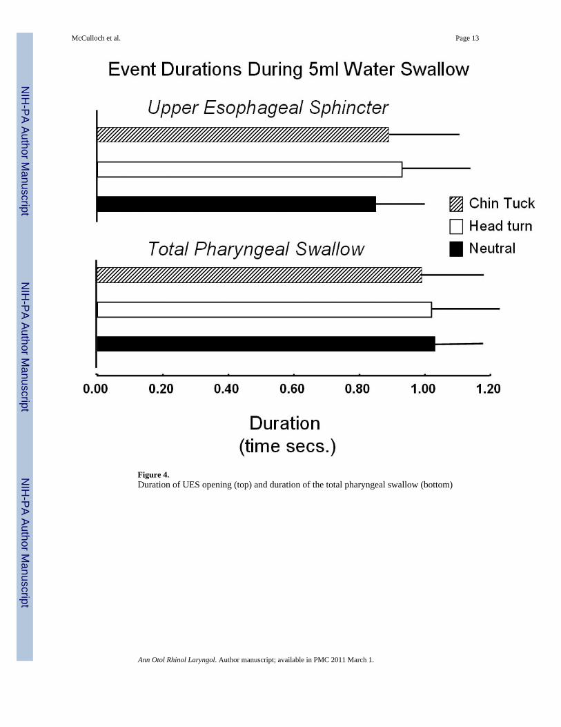
Figure 4.Duration of UES opening (top) and duration of the total pharyngeal swallow (bottom)
McCulloch et al. Page 13
Ann Otol Rhinol Laryngol. Author manuscript; available in PMC 2011 March 1.
NIH
-PA Author Manuscript
NIH
-PA Author Manuscript
NIH
-PA Author Manuscript

Figure 5.Gradients in velopharynx (A) and tongue base (B) for each swallowing task. Measurementswere recorded at the maximum pressure within a region (max), and the pressure at the sametime point one (1 cm) and two (2 cm) sensors downstream. Min represents the baselinepressure recorded at the same sensor as the maximum pressure.
McCulloch et al. Page 14
Ann Otol Rhinol Laryngol. Author manuscript; available in PMC 2011 March 1.
NIH
-PA Author Manuscript
NIH
-PA Author Manuscript
NIH
-PA Author Manuscript

NIH
-PA Author Manuscript
NIH
-PA Author Manuscript
NIH
-PA Author Manuscript
McCulloch et al. Page 15
Tabl
e 1
Sum
mar
y da
ta. V
alue
s are
pre
sent
ed a
s mea
n ±
stan
dard
dev
iatio
n.
Neu
tral
Hea
d tu
rnCh
in tu
ck
Regi
onPa
ram
eter
Mea
nM
ean
P-va
lue
Mea
nP-
valu
e
Vel
opha
rynx
P max
(mm
Hg)
169
± 50
170
± 41
0.91
416
3 ±
570.
688
Clo
sure
Dur
atio
n (s
)0.
79 ±
0.1
30.
91 ±
0.2
20.
016*
0.85
± 0
.15
0.18
2
Ris
e ra
te (m
mH
g/s)
865
± 20
284
8 ±
241
0.88
980
5 ±
247
0.54
5
Tong
ue b
ase
P max
(mm
Hg)
306
± 16
327
6 ±
940.
938
252
± 77
0.39
2
Hig
h Pr
essu
re D
urat
ion
(s)
0.67
± 0
.16
0.75
± 0
.25
0.27
60.
62 ±
0.1
90.
4
Ris
e R
ate
(mm
Hg/
s)15
76 ±
598
1793
± 1
856
0.75
712
39 ±
438
0.29
7
UES
P pre
(mm
Hg)
227
± 10
011
8 ±
600.
017*
154
± 11
80.
216
P pos
t(mm
Hg)
239
± 78
256
± 72
0.49
719
6 ±
560.
037*
Ope
ning
Dur
atio
n (s
)0.
85 ±
0.1
60.
93 ±
0.2
10.
180.
89 ±
0.2
20.
454
P min
(mm
Hg)
−5
± 9
−10
± 1
40.
211
0 ±
80.
256
Phar
ynx
Tota
l Sw
allo
w D
urat
ion
(s)
1.03
± 0
.16
1.02
± 0
.20
0.90
70.
99 ±
0.1
80.
616
Ast
eris
ks d
enot
e si
gnifi
cant
p-v
alue
s
Ann Otol Rhinol Laryngol. Author manuscript; available in PMC 2011 March 1.

NIH
-PA Author Manuscript
NIH
-PA Author Manuscript
NIH
-PA Author Manuscript
McCulloch et al. Page 16
Tabl
e 2
Pres
sure
gra
dien
ts in
vel
opha
rynx
and
tong
ue b
ase
at 1
and
2 c
m. M
ax –
1 c
m is
equ
al to
max
imum
pre
ssur
e in
the
regi
on m
inus
the
pres
sure
one
sens
ordo
wns
tream
at t
he sa
me
time.
Max
– 2
cm
is e
qual
to m
axim
um p
ress
ure
min
us th
e pr
essu
re tw
o se
nsor
s dow
nstre
am.
Neu
tral
Hea
d tu
rnCh
in tu
ck
Regi
onG
radi
ent
Mea
nM
ean
P-va
lue
Mea
nP-
valu
e
Vel
opha
rynx
Max
– 1
cm
121
± 58
114
± 56
0.64
410
8 ±
600.
487
Max
– 2
cm
149
± 48
150
± 43
0.96
614
6 ±
550.
796
Tong
ue b
ase
Max
– 1
cm
242
± 17
722
3 ±
109
0.77
020
6 ±
860.
938
Max
– 2
cm
293
± 16
626
0 ±
102
0.93
823
3 ±
821.
000
Ann Otol Rhinol Laryngol. Author manuscript; available in PMC 2011 March 1.

NIH
-PA Author Manuscript
NIH
-PA Author Manuscript
NIH
-PA Author Manuscript
McCulloch et al. Page 17
Tabl
e 3
Dat
a pr
esen
ted
by o
ther
man
omet
ric st
udie
s.
Stud
yP m
ax(m
mH
g)D
urat
ion
(ms)
UE
S re
laxa
tion
(ms)
UE
S P m
ax(m
mH
g)U
ES
P rel
axed
(mm
Hg)
VP
P max
(mm
Hg)
Bod
en, n
eutra
l317
8 ±
1865
6 ±
5950
9 ±
2450
9 ±
24-
-
Bul
ow, n
eutra
l625
5 ±
2448
7 ±
22-
--
-
Bul
ow, c
hin
tuck
619
3 ±
1642
1 ±
2561
0 ±
2923
3 ±
210
± 1
-
Taka
saki
, neu
tral,
mal
e21
183
± 84
--
236
± 79
-16
3 ±
95
Taka
saki
, neu
tral,
fem
ale2
116
7 ±
65-
-24
3 ±
87-
125
± 43
But
ler,
neut
ral m
ale1
814
0-
−5
--
-
But
ler,
neut
ral f
emal
e18
152
-−2
--
-
P max
= p
eak
phar
ynge
al c
ontra
ctio
n pr
essu
re; D
urat
ion
= du
ratio
n of
pha
ryng
eal c
ontra
ctio
n; U
ES P
max
= p
eak
uppe
r eso
phag
eal s
phin
cter
pre
ssur
e; U
ES re
laxa
tion
= du
ratio
n of
upp
er e
soph
agea
lsp
hinc
ter o
peni
ng; U
ES P
rela
xed
= up
per e
soph
agea
l sph
inct
er p
ress
ure
durin
g op
enin
g; V
P P m
ax =
pea
k ve
loph
aryn
geal
pre
ssur
e.
Ann Otol Rhinol Laryngol. Author manuscript; available in PMC 2011 March 1.


