Embed Size (px)
Citation preview

CINERADIOGRAPHYOF THE
LIQUID BOLUS SWALLOW
A study of theSpeed of the Bolus and Peristaltic Wave
and of Movement of the Hyoid Bone,Larynx, and Epiglottis
- ÅÖOÅ- A-
Pia Sundgren
Malmö 1991

o
S55
SI
OrganizationLUND UNIVERSITY
Department of Diagnostic RadiologyAllmanna sjukhuset214 01 Malmd, Sverige
Authors)
Pia Sundgren
Document nameDOCTORAL DISSERTATION
Dite of issue,99j n* 0 7
CODEN: LUMEDW/(MEXM-1OO1)/1-126 /(199I
Sponsoring orguuutton
Title Mid subtitleCineradiography of the liquid barium swallow. A study of the speed o f the bolus and peristalticand of movement of the hyoid bone, larynx and epiglottis.
Abstract
In the evaluation of the dysphagic patient, radiology is crucial as a technique for monitoring morpho-logy and function. In particular, high-speed cineradiography can reveal a variety of pharyngeal dys-functions. However, in the literature and in practice the difference between normal and abrormal func-tion is not always clear. This monography is based on high-speed cineradiographies of swallowing in 75non-dysphagic volunteers and in 189 dysphagic patients.The purpose was to study whether differences in bolus volumes, patient position, age and gender had anyeffects on the following parameters: the speed of the peristaltic wave and apex of the liquid bariumbolus, the length of movement and the movement pattern of the hyoid bone and larynx, and epiglotticfunction. Also differences in these parameters between non-dysphagic and dysphagic individuals wereanalyzed.The study disclosed that the speed of the bolus, the anterior-superior movement and net movement of thehyoid bone increased significantly with larger bolus volumes. The position of the individual in rela-tion to gravity significantly influenced the speed of peristalsis. In most of the measured parametersthere were no differences between non-dysphagic and dysphagic individuals except for differences inthe intrapersonal variations and in the anterior-superior movement of the hyoid bone. In patients withpharyngeal dysfunction the initial suge of the elevation of the larynx was significantly lower than inpatients without dysfunction. The approximation of the thyroid cartilage to the hyoid bone was signifi-cantly greater in individuals with normal epiglottic function than in those with epiglottic dysmobili
ty-It is suggested that abnormal speed of peristalsis may be a mild form of dysfunction. Measurements ofthe aforementioned speeds and movements can be done if bolus volume, age and position of the patient,film speed and magnification factors are known. Hypotheses concerning epiglottic function and centralcontrol of swallowing are proposed.
Keywords
Dysphagia; Pharyngeal dysfunction; Cineradiography; Pharynx
Classification system and/or index terms (if any)
Supplementary bibliographical information
ISSN and key tide
Recipient's notes Number of pages \2(,
Language English
ISBN
Price
Security classification
Distribution by (name and address)
Pia Sundgren, MD, Department of Diagnostic Radiology, Malmö General Hospital,1,'tne undensjpM0!1DVmgSi^<copyright owner of the abstract of the above-mentioned dissertation, hereby grant to all referencesources permission to. publish and disseminate the abstract of the above-mentioned dissertation.
Signature 9/03//

Cineradiography of the liquid bolus swallow.
A study of the speed of the bolus and peristaltic wave
and of movement of the byoid bone, larynx, and epiglottis
AKADEMISK AVHANDLING
./ r. med vederbörligt tillstånd av Medicinska fakulteten
v d Lunds universitet för avläggande av doktorsexamen i
•'idicinsk vetenskap kommer att offentligen försvaras å
liversitetsklinikernas aula, Allmänna sjukhuset i Malmö,
fredagen den 7 juni 1991 kl 09.15
av
Pia Sundgren
leg läk, Lunds nation
Malmö 1991

CINERADIOGRAPHYOF THE
LIQUID BOLUS SWALLOW
A Study of theSpeed of the Bolus and Peristaltic Waveand of Movement of the Hyoid Bone,
Larynx, and Epiglottis
Pia Sundgren
From the Department of Diagnostic Radiology,University of Lund,
Malmö General Hospital,Malmö, Sweden, 1991

© Pia Sundgren, 1991
Layout and typography by Kanelbullen - datakonsult H3Printed in Sweden, Studentl'ieratur, Lund 1991

Contents
PREFACE 7
INTRODUCTION 8
Definitions 8
Anatomic and Functional Aspects of Swallowing 9
General Considerations 9
Pharyngeal Constrictor Musculature 9
The Cricopharyngeal Muscle 10
Pharyngeal Elevator Musculature 11
TheHyoid Bone 12
Laryngeal Vestibule 13
The epiglottis 15
Neuroanatomic and Neurophysiologic Aspects of Swallowing 16
The Normal Act of Swallowing 18
General Introduction 21
Purpose of Investigation 25
Material 27
Rad iographic Methods 31
Single Film Technique 31
Cineradiography 31
Observed Events and Measured Parameters on Cineradiograms . . . . 33
Analytic and Statistical Methods 38
INVESTIGATIONS 41
1. Speed of the Peristaltic Wave 41
introduction 41

4 Cineradiography of the Liquid Bolus Swallow
Material and Method 42
Results 43
Discussion 49
Conclusions 53
2. Speed of the Apex of the Bolus 55
Introduction 55
Material and Method 56
Results 57
Discussion 62
Conclusions 65
3. Relation Between the Speed of the Peristaltic Wave and
the Speed of the Apex of the Bolus 67
Introduction 67
Material and Method 67
Results 68
Discussion 71
Conclusions 73
4. Movement of the Hyoid Bone during Swallow 75
Introduction 75
Material and Method 76
Results 77
First Movement of the Hyoid Bone 77
Second Movement of the Hyoid Bone 79
Net Movement of the Hyoid Bone 82
Discussion 84
Conclusions 87
5. Elevation of the Larynx 89
Introduction 89

Contents
Material and Methods 91
Results 93
Elevation of the Larynx when the Bolus Reached
the Valleculae 93
Maximal Elevation of the Larynx 95
Percentage Elevation of the Larynx when the BolusReached the Valleculae 97Frequency of Pharyngeal Dysfunction 97
Elevation of the Larynx in Individuals with PharyngealDysfunction 98
Discussion 100
Conclusions 103
6. Epiglottic Dysmobility 105
Introduction 105
Material and Method 107
Results 108
Discussion 109
Conclusion 115
SUMMARY OF THE RESULTS 117
CONCLUSIONS 120
HYPOTHESIS 122
ACKNOWLEDGEMENTS 123
REFERENCES 124

Preface
The swallowing act can be studied with various radiographic methods suchas conventional radiography including plain films with and without bariumcontrast medium, cineradiography, and videofluorography. In addition, theanatomy of the swallowing apparatus, including the mouth, larynx andpharynx can be delineated by CT or MRI.
The pharyngeal swallow is a complex process of contractions and relaxationsof several muscles which is regulated by the central nervous system. Thedetailed mechanisms controlling normal swallowing are not completelyunderstood. Thereby the pathophysiology behind abnormal pharyngealfunction is not always clear. This monograph focuses on certain functionalaspects of the swallowing process as they appear on a cineradiogram usingliquid barium contrast medium. The monograph is based on material which,in part, has been previously published [1,2,3,4].

u
Definitions
In the present investigation, the terms dysphagia, pharyngeal dysfunction, anddysphagic patient respectively are used according to the following definitions:
Dysphagia: A subjective feeling of disturbances in the course of swallowing.The term includes a long list of sensations such as a feeling of obstruction, afeeling of residual bolus material in the pharynx after the completion of aswallow, a feeling of misdirected swallowing into the airway, a feeling of alump in the throat between swallowing, or a feeling of inability to swallow.
Pharyngeal dysfunction: Presence of functional changes such as paresis of anyof the pharyngeal muscles, incoordination of the cricopharyngeal muscle,dysmobility of the epiglottis, passage of bolus into the airways, or incompleteclearing of the pharynx of bolus material — pharyngeal dysfunction may befound in patients with dysphagia as well as in patients without dysphagia.
Morphologic changes include tumours, strictures, webs, diverticula or otherstructural obstacles to the proper passage of the bolus.
Note, that dysphagic patients may or may not have pharyngeal dysfunctionand /or morphological changes, and that pharyngeal dysfunction and/ormorphological changes can occur in individuals without dysphagia.
Significantdy): In the present investigation the expression significant(ly)refers exclusively to a statistically proven significance - i.e. p < 0.05.
Correlation: In the present investigation the term correlation refers to statis-tical correlation and describes a measure of association.

Anatomic and Functional Aspects ofSwallowing
General Considerations
The pharynx consists of a muscular tube connected with the nasal and oralcavities, and with the laryngeal vestibule and esophageal opening.
The pharynx consists of three parts: the epi-, meso- and hypopharynx,respectively. The epipharynx (nasopharynx) extends from the base of theskull to the level of the hard palate. The mesopharynx (oropharynx) extendsfrom the level of the hard palate to the level of the valleculae. The hy-popharynx extends from the valleculae to the lower border of the cri-copharyngeal muscle.
The superior part of the epipharynx is attached to the base of the skull andis immobile during swallow. The meso- and hypopharynx are attached tothe surrounding structures by muscles and ligaments and are mobile duringswallow. Both the mesopharynx and the hypopharynx are a common path-way for the respiratory and alimentary systems.
The pharyngeal musculature consists of two functionally different groups ofmuscles — the constrictors and the elevators.
Pharyngeal Constrictor Musculature
The pharyngeal constrictors are composed of three portions, i.e. the superior,middle and inferior (Fig 1). These striated muscles overlap each other andinsert into a dense collagenous sheet of multidirectional fibres, the buc-copharyngeal aponeurosis. The superior portion of the constrictor muscle isattached to the base of the skull by the pharyngobasilar fascia, to thepterygomandibular raphe, to the mandible, and to the tongue. The middleconstrictor emerges from the hyoid bone and the stylohyoid ligament. Theinferior constrictor muscle emerges from the thyroid and cricoid cartilageand from the lateral thyrohyoid ligament. The pharyngeal constrictormuscles surround parts of the pharynx and unite posteriorly into the pharyn-

10 Cineradiography of the Liquid Bolus Swallow
Sup. pharyngealconstrictor muscle(4 portions)
Middle pharyngealconstrictor muscle
Inf. pharyngealconstrictor muscle
Cricopharyngealmuscte
Figure 1 . Schematic drawing of the pharyngeal constrictor musculature.
geal raphe. Functioning as a unit, the striated muscles contract sequentiallypropelling an uninterrupted peristaltic-like wave through the pharynx.
The Cricopharyngeal Muscle
The cricopharyngeal muscle consists of three portions, i.e. (1) the oblique, (2)the transverse, and (3) the longitudinal. The superior portion is called theoblique portion and its muscle bundles emerge from the lateral aspect of thecricoid cartilage and project in an oblique craniodorsal direction to join themuscle of the contralateral side in the pharyngeal raphe of the posterior wallof the pharynx. This superior portion can also be considered the caudal partof the inferior pharyngeal constrictor muscle (Fig 2).
The middle portion of the cricopharyngeal muscle, the transverse portion, isattached to the posterior lateral aspect of the cricoid cartilage from which itsurrounds the pharynx in a semicircular arrangement. Frequently, themiddle portion is separated from the oblique superior portion by a triangulararea of fibrous tissue, known as Killian's dehiscence (Fig 2).

Anatomic and Functional Aspects of Swallowing 11
The inferior portion of the cricopharyngeal muscle, the longitudinal portion,is attached to the inferior posterior aspect of the cricoid cartilage and runsinferiorly where it mingles with the longitudinal muscle bundles of theesophagus (Fig 2).
Figure 2. Schematic draw-ing of the cricopharyngertmuscle with its three por-tions (a) the oblique portion,(b) the transverse portion,(c) the longitudinal portion,(d) inferior pharyngeal con-strictor, (th) thyrcid cartilage,(cr) cricoid cartilage, (t) tra-chea.
Pharyngeal Elevator Musculature
The elevator musculature of the pharynx consists of two main muscles: Thestylopharyngeal and palatopharyngeal muscles. The stylopharyngealmuscle emerges from the styloid process and the base of the skull andattaches to the thyroid cartilage and along the sides of the epiglottis. A minorportion of the stylopharyngeal muscle intermixes with the superior andmiddle pharyngeal constrictor musculature. The palatopharyngeal muscleemerges from the aponeurosis of the soft palate, the eustachian tube car-tilage and the pterygoid process. One portion attaches to the interior surface
12 Cineradiography of the Liquid Bolus Swallow
of the thyroid cartilage and the other portion mingles with the constrictormuscles. The palatopharyngeal muscle is the main elevator muscle of thepharynx. The mylohyoid and the geniohyoid muscles may, also, be con-sidered as additional elevator muscles. The function of the elevator musclesof the pharynx is elevation of the pharynx and consequently of the larynxduring swallowing. In addition, the pharyngeal constrictor musculature actsas a minor pharyngeal elevator due to its oblique muscle fiber arrangement.
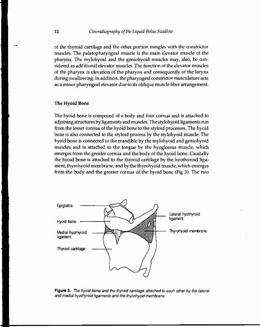
The Hyoid Bone
The hyoid bone is composed of a body and four cornua and is attached toadjoining structures by ligaments and muscles. The stylohyoid ligaments runfrom the lesser cornua of the hyoid bone to the styloid processes. The hyoidbone is also connected to the styloid process by the stylohyoid muscle. Thehyoid bone is connected to the mandible by the mylohyoid and geniohyoidmuscles and is attached to the tongue by the hyoglossus muscle, whichemerges from the greater cornua and the body of the hyoid bone. Caudallythe hyoid bone is attached to the thyroid cartilage by the hyothyroid liga-ment, thyrohyoid membrane, and by the thyrohyoid muscle, which emergesfrom the body and the greater cornua of the hyoid bone (Fig 3). The two
Epiglottis
Hyoid bone
Medial hyothyroidligament
Thyroid cartilage
Lateral hyothyroidligament
Thyrohyoid membrane
Figure 3. The hyoid bone and the thyroid cartilage attached to each other by the lateraland medial hyothyroid ligaments and the thyrohyoid membrane.

Anatomic and Functional Aspects of Swallowing 13
greater cornua of the hyoid bone and the hyothyroid ligament are part of thelateral pharyngeal wall in the mesopharynx. The mylohyoid and hyoglossusmuscles work in synchrony to raise the hyoid bone. Also the stylohyoidmuscles elevate the hyoid bone whereas the geniohyoid and mylohyoidmuscles work simultaneously to move the hyoid bone anteriorly. The twobellies of the digastric muscle participate in both movements.
Laryngeal Vestibule
The laryngeal vestibule constitutes the air filled cavity between the vocalcords and the pharynx and can be subdivided according to function asproposed by Ekberg [5]. This subdivision is made by an arbitrary planeextending from the inter-arytenoid incisure to the superior notch of thethyroid cartilage, -.hereby dividing the vestibule into a superior and aninferior space. The superior space is called, according to Ekberg, the sub-epiglottic space and is demarcated by the aryepiglottic folds and the namedplane. The inferior space is called the supraglottic space and is demarcated bythe named plane and the vocal cords (Fig 4).
Figure 4. The laryngeal vestibule: Epiglottis - E, Subepiglottic space - SE, Supraglotticspace - SG, Laryngeal ventricle • L V

14 Citieradiography of the Liquid Bolus Sxvallow
The closure of the laryngeal vestibule during swallowing is performed in asequence of events as follows [5].
The supraglottic space closes first in cranial direction starting with apposi-tion of vocal cords, false vocal cords and then lateral walls. At the time ofcompletion of the supraglottic space closure, the downfolding epiglottis hasreached a transverse position with its tip against the posterior pharyngealwall. The subepiglottic space is then closed from above in caudal directionby further tilting of the epiglottis. In a closed vestibulum the compressedlumen of the supraglottic segment is oriented in a sagittal plane and thelumen of the subepiglottic segment is oriented in a slightly tilted axial plane.
Figure 5. The thyroid car-tilage, the epiglottis and thethyroepiglottic ligament. Thyroepiglottic
ligament
Anatomic atni Functional Aspects of Swallowing 15
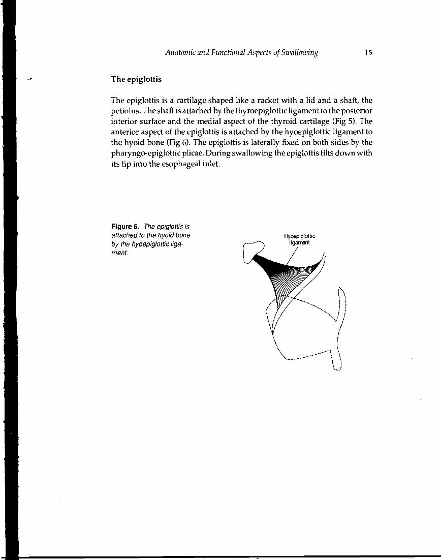
The epiglottis
The epiglottis is a cartilage shaped like a racket with a lid and a shaft, thepetiolus. The shaft is attached by the thyroepiglottic ligament to the posteriorinterior surface and the medial aspect of the thyroid cartilage (Fig 5). Theanterior aspect of the epiglottis is attached by the hyoepiglottic ligament tothe hyoid bone (Fig 6). The epiglottis is laterally fixed on both sides by thepharyngo-epiglottic plicae. During swallowing the epiglottis tilts down withits tip into the esophageal inlet.
Figure 6. The epiglottis isattached to the hyoid boneby the hyoepiglottic liga-ment.
Hyoepiglotticligament

Neuroanatomic andNeurophysiologic Aspects ofSwallowing
Deglutition is brought about by a functional interaction of the mouth,pharynx and esophagus. The neurophysiological control of deglutition in-volves three separate but interacting phases [6,7,8,9,10].
The oral phase is the initial part of the swallow, in which food, by masticationand intraoral manipulation, is formed into a bolus of the size suitable fortransport through the pharynx and esophagus.
During the pharyngeal phase the bolus passes without voluntary control to theesophagus. During this phase the respiratory tract is protected by a coordi-nated closure of the palatopharyngeal isthmus, the vocal cords and laryngealvestibule. The closure of the laryngeal vestibule is also achieved by elevationof the larynx and tilting down of the epiglottis.
The autonomically controlled esophageal phase, propels the bolus to thestomach.
The different muscles participating in the swallowing act are innervated bycranial nerves V, IX, X and XII. The muscles of mastication are controlled bythe third division of the trigeminal nerve (V3), while the tongue movementis controlled by the hypoglossal nerve (XII) and ansa cervicalis. The en-dolaryngeal muscles are innervated by the vagus nerve (X) which alsoinnervates the muscles of the palate, except for the tensor veli palati whichis innervated by V3. The pharyngeal muscles are also innervated by the vagusnerve, except for the stylopharyngeal muscle which is innervated by theglossopharyngeal nerve (IX). The movements of the hyoid bone and thelarynx during swallowing are regulated by multiple nerves (V3, VII, and
The cortical representation for swallowing and mastication is localizedimmediately anterior to the precentral cerebral cortex. From this regionpathways descend through the internal capsule and the subthalamic regionsto the level of the substantia nigra and the mesencephalic reticular formationof the upper brainstem. Stimulation of this pathway evokes mastication withswallowing [7].

and JS-'europhysiologic Aspect* of Swallowing 17
The paired swallowing 'renter: reside in the hindbrain. The swallow centersconsist of ill-defined broad zones located lateral to the midline and ventralto the caudal portion of tlie fourth ventricle, which incorporate the nucleustractus solitarii and tht* \ entrornedial reticular formation around the nucleusambiguus.
Sensory afferent neurons from receptors in the mouth, pharynx, and larynxpass in the fifth, seventh, ninth and tenth cranial nerves to the swallowingcentres in the brainstem. The input fibres from these cranial nerves andhigher cerebral centers synapse with;n the nucleus tractus solitarii or thereticular formation.
The efferent control to the musculature of the neck is transmitted via axonswhose cell bodies reside in the central nuclei of the trigeminal (V), facial (Vil),and hypoglossal (XII) cranial nerves [6,9]. The nucleus ambiguus, which islocated laterally to the floor of the fourth ventricle, is the somatic motornucleus of the ninth, tenth, and eleventh cranial nerves. The axons of thesenerves originate at different levels within the nucleus ambiguus without anywell-defined boundary between the different nuclei of the nerves [6,9].
Two major theories have been proposed to describe the neural mechanismthat executes the oral and pharyngeal phases of swallowing: the reflex chainhypothesis [8] and the central pattern generator hypothesis [6). However, theexact details of the control systems for the different phases in the swallowingact have not been determined. The reflex chain hypothesis by Doty [8]proposes that swallowing proceeds as a chain of linked reflexes caused by abolus moving through the mouth and pharynx stimulating sensory receptorsthat sequentially trigger the next step in the swallow sequence. The secondhypothesis, the central pattern generator hypothesis by Miller [6], suggeststhat once swallowing is initiated, the ensuing swallowing sequence is pro-grammed by a network of neurons in the swallowing centers in the medullaoblongata that function independently of sensory feedback. Thus, this hy-pothesis proposes that a central pattern generator for swallowing is notinfluenced by or dependent on peripheral feedback.
Some evidence exists to support both the reflex chain and the patterngenerator hypotheses of swallowing [8,11,12,13] and it might be assumedthat both types of mechanism are operative.

18 Cineradiography of the Liquid Bolus Swallow
The Normal Act of Swallowing
The normal act of swallowing occurs step by step and can be separated intothree functionally distinct phases, which involve a long series of eventsduring a very short period of time.
In the first phase, the oral phase, the oral content is formed by mastication andintraoral manipulation by the tongue into a bolus of proper size for swallow-ing. The tongue moves the bolus around in the mouth and finally the bolusis placed in the tongue groove to be moved posteriorly over the tongue baseand pharyngeal isthmus into the mesopharynx initiating the swallowingreflex [14]. The exact nature of the complex stimulus or stimuli that triggerthe pharyngeal swallow has not been clearly defined [6, 7]. However, it isbelieved that the swallowing reflex can be triggered by contact of bolusmaterial with the posterior portion of the tongue and the pharyngeal isthmus[ 15]. At this time the epipharynx is already closed from the rest of the pharynxby a superior movement of the soft palate. The hyoid bone is elevated in asuperior and slightly posterior direction and simultaneously the larynx iseievated.
The pharyngeal phase begins when the bolus reaches the mesopharynx. Thebolus is pressed by the back of the tongue against the posterior pharyngealwall and is thrust into the hypopharynx by a powerful tongue motion.Closure of the supraglottic space of the laryngeal vestibule starts as the bolusis pushed through the pharyngeal isthmus [16, 17, 18]. The simultaneouselevation of the hyoid bone and the larynx leads to a tilting of the epiglottisfrom an upright to a transverse position, closing the subepiglottic space ofthe laryngeal vestibule. Pharyngeal peristalsis is a cranial to caudal muscularcontraction that begins in the epipharynx and progresses to the hypopharynx[19, 20, 21]. The peristaltic contractions of the pharyngeal constrictors helpto propel the tail of the bolus through the hypopharynx and to cleanse thepharynx after the swallow. At the same time the epiglottis assumes a trans-verse position and then tilts down with its tip into the esophageal inlet tocover the laryngeal inlet and to obliterate the subepiglottic space of thelaryngeal vestibule [5, 22]. The more detailed mechanisms by which theepiglottis is tilted down from its upright position to an inverted position withits tip in the esophageal inlet are not yet fully understood.
The intrabolus forces mediated by an oncoming bolus together with thetraction caused by superior and anterior elevation of the larynx [23] and therelaxation of the cricopharyngeus muscle lead to an opening of the upper

Neuroanatomic and Neurophysiologic Aspects of Swallowing 19
esophageal segment including the cricopharyngeal region [23,24]. The bolusis propelled into the esophagus.
The autonomically controlled esophageal phase propels the bolus to thestomach. During this phase the muscles of the tongue relax and the tonguereturns to a resting position in the mouth. During relaxation of the pharyn-geal constrictor and levator muscles, the hyoid bone and the larynx returnto their resting positions and the epiglottis returns to an upright position.The cricopharyngeal muscle, as a part of the upper esophageal sphincter,assumes a tonic contraction.

?/ : / .
General Introduction
Normal swallowing requires normal morphology and neuromuscular func-tion of the mouth and pharynx. Abnormality of morphology and functionmay result in impaired oral and pharyngeal function, which may vary froma difficulty in initiating the swallow to a total inability to swallow. Impair-ment of pharyngeal function may appear as a subjective perception ofdisturbances — dysphagia (p. 8) — or functional changes that may bedetected by diagnostic methods — pharyngeal dysfunction (p. 8).
Pathologic conditions underlying impaired pharyngeal function can bedifferentiated into two main categories: morphological changes in thepharynx, and disturbances of the neuromuscular activity. The first category,morphological changes in the pharynx, includes stenoses, diverticula,rumours, inflammatory processes and webs in the esophageal inlet. Thesecond category, disturbances of the neuromuscular activity, includes boththe diseases affecting the central nervous system, e.g. cerebrovascular insults,tumours, inflammatory diseases, Parkinsons disease etc., and the diseasesthat affect the motor neurons, such as ALS, dermatomyositis, myastheniagravis, polio, etc. [25].
In many patients, dysphagia and/or the pharyngeal dysfunction result infeeding problems, disease and psychosocial problems. The patients try torelieve their symptoms by altering their diet, avoiding foods that are difficultto swallow, such as solids, including crisp fruits, vegetables, meat, and drybread products [26], and this may result in malnutrition. The patients oftentry to change the preparation of the food and change their eating habits.Solids are cut into small pieces and carefully chewed. Liquids are sippedslowly and carefully, and double swallowing may be used to clear thepharynx from retained bolus material. These measures prolong the feedingtime and the patients often fail to finish meals together with their tablemates[26]. All the secondary effects of the impairment of the swallowing may havea psychosocial impact both on the patients and their families.
Complaints related to swallowing — dysphagia — are relatively frequentand were, for instance, in a recent study found in 25% of individuals aged50 to 79 years [27]. The frequency of dysphagia is high in individuals withpharyngeal dysfunction but dysphagia may also occur in patients withoutany detected pharyngeal dysfunction [21, 28]. Dysphagia is also more com-

22 Cmeradiography of the Liquid Bolus Swallow
mon among elderly than among younger individuals [29,30,31]. Althoughpharyngeal dysfunction may be asymptomatic, it usually evokes symptomsincluding dysphagia. However, there is poor correlation between the typeof symptoms and the type of pharyngeal dysfunction [32].
Patients with complaints related to swallowing can be differentiated into twogroups, according to the results of the clinical and radiological examination.The first group consists of those who, besides dysphagia, also have roentgendiagnostic signs of pharyngeal dysfunction and/or morphological changes.The second group includes dysphagic patients with complaints only: thosepatients in whom the radiologic examination failed to demonstrate anypathological changes, according to the currently accepted diagnostic criteria.
In the past, the radiological examination has focused on morphologicalchanges such as stenoses, diverticula, tumors or webs. The introduction ofhigh speed cineradiography and videoradiography has made it possible tobetter evaluate coordination of various movements of structures such as thetongue, pharyngeal walls, hyoid bone, epiglottis, larynx, etc., which im-proves the evaluation of functional changes such as pareses of pharyngealconstrictors, incoordination of the cricopharyngeal muscle, defective tiltingof the epiglottis, defective closure of the laryngeal vestibule, aspiration ofbolus material into the trachea, etc. [33].
Despite the use of these techniques, there remains a group of dysphagicpatients in whom no pharyngeal dysfunction or morphological change canbe detected, which could explain the patients symptoms. It is, however,conceivable that these dysphagic individuals may have some, undiscovered,underlying morphological or functional disturbance that evokes their symp-toms. Disclosing such a disturbance would make it possible to explain to thepatient the cause of his/her symptoms, which is often of great psychologicimpact, and, possibly, to offer him/her special training or other therapy inthe future.
It is possible that the cineradiogram contains information that could help todisclose such, to date occult, disturbances of the swallowing act. Informationthat has not been utilized in clinical evaluation is the quantitative informa-tion represented by the lengths and speeds of movements of various struc-tures that are visible on the rineradiogram.
The interest in measuring the lengths and speeds of movements of variouslaryngeal and pharyngeal structures or of the bolus during swallowing datesback to approximately the 1930s. The data obtained from these measure-ments complement our knowledge about the pharyngeal phase of swallow-

General Introduction 23
ing and serve as a basis for various hypotheses about the swallowingmechanisms. To measure these parameters the following methods or theircombinations have been and are used: cineradiography and videoradiogra-phy [19,20,34,35,36] with frame rate varying from 6 frames/s [37] to 100frames/s [35]; manometric studies, sometimes, in combination with cine- orvideoradiography [12, 13, 37, 38, 39]; and studies employing electrical im-pedance [40]. The obtained information has been presented in a variety ofpoorly comparable units such as frame/s, msec, cm/s, cm, and also as lessprecise assessments [41] such as one and a half vertebral body. Differencesin the size and composition of patient material, employed techniques, exami-nation conditions (such as size, consistency of the bolus and patient position)and in the hardly comparable measurement units may have contributed tothe controversy on the physiological and palhophysiological mechanisms ofthe swallowing act. One of the objectives in the present investigation was toperform a systematic analysis of the various speeds and lengths of move-ments, as seen on the cineradiogram, in the same individuals.
The present investigation dealing with measurements of the speeds of theperistaltic wave, the apex of the bolus, of the lengths of movement of thehyoid bone, elevation of the larynx and approximation of the thyroid car-tilage to the hyoid bone, is in principle based on two hypothetical presump-tions of the author.
The first presumption is that the lengths of movements of various structures,and the speed of the peristaltic wave and the speed of the apex of the bolusare due to contractions and relaxations of muscles participating in theswallowing act, under CNS control. Consequently, valuable information onthe neuromuscular function of the swallowing apparatus can be gained bymeasuring these movements and speeds. The information obtained fromnormal subjects could enrich and possibly improve our understanding of thephysiology of swallowing. The information obtained from individuals withswallowing disturbances may yield data for creating new hypotheses toexplain mechanisms underlying the respective dysfunction.
The second presumption is that even very mild, and hereto undefined,pharyngeal dysfunction, may cause dysphagia. Therefore, a group of dys-phagic patients without radioiogically detected pharyngeal dysfunctionunderwent the same tests as a group of non-dysphagic volunteers. This wasdone in order to reveal if an abnormality in some of the measured parametersmay signal the presence of some latent dysfunction.
The information included in the measured parameters would not be of usefor possible future clinical or research application unless the influence of

24 Cxneradiography of the Liquid Bolus Swallow
factors that may potentially change these parameters is known. Therefore,the influence on these parameters of factors related to the examinationtechnique, such as the volume of the bolus or the examination position of thepatient, and factors related to the individuals, such as gender and age, wasalso investigated.

Purpose of Investigation
The main purpose of this investigation was to study the act of swallowingas reflected in cineradiography, in non-dysphagic healthy volunteers and inpatients with dysphagia, both without pharyngeal dysfunction and to com-pare some well-definable and measurable swallowing parameters in thesetwo groups of individuals.
The parameters studied were: speed of pharyngeal peristalsis, speed of theapex of contrast medium bolus at swallowing, and the lengths of movementand the movement patterns of the hyoid bone and larynx at swallowing.
The specific aims of this investigation were:
• to study if the size of the contrast medium bolus used at cineradiographyhas any measurable effect upon pharyngeal peristaltic speed or speed ofbolus.
• to study if the size of the contrast medium bolus used at cineradiographyhas any measurable effect upon elevation of the larynx and movement ofthe hyoid bone.
• to study if patient positioning has any effect on the speed of the peristalticwave or the apex of the bolus.
• to study if there are any differences in the inter- and intrapersonal variationsin the measurable swallowing parameters among the individuals includedin the investigation.
• to investigate if the two-step fashion movement of the hyoid bone changeswith the posture of the individual.
• to investigate if there are differences between dysphagic and non-dys-phagic individuals in any of the aforementioned parameters. This is anattempt to find an undisclosed dysfunction underlying the patients symp-toms.
Further, it was decided to test the usefulness of measuring the movementsthat are visible on the cineradiograph in both non-dysphagic and dysphagicindividuals with or without pharyngeal dysfunction. These measurementswere performed in an attempt to demonstrate any relationship, if present,
26 Cineradiography of the Liquid Bolus Swallow
between abnormal movement and some well-defined types of pharyngealdysfunction. Thus the subsequent aims of the investigation were:
• to study possible differences in the amount of elevation of the larynxbetween individuals with and without pharyngeal dysfunction (Chapter5).
• to study the spatial relationship between the hyoid bone and the thyroidcartilage during swallowing in patients with and without epiglottic dys-function in order to better understand the morphodynamic background ofepiglottic dysmobility (Chapter 6).

Material
A total of 264 individuals examined at the Department of Diagnostic Radi-ology, Malmö General Hospital, Sweden, have been included in the presentinvestigation. Individuals who had undergone surgery of the head and neckor who had pharyngeal neoplasia were excluded in the present investigation.The proper material consists of several cineradiograms obtained from eachone of these 264 individuals. One individual could participate in more thanone group as later described.
Of the 264 individuals, 75 served as normal controls while the remaining 189were referred to the X-ray department due to dysphagia.
The individuals who served as normal controls consisted of two categoriesof volunteers:
15 volunteers and 60 patients referred to the X-ray department for examina-tions other than of the pharynx and esophagus.
All the individuals who served as normal controls were asked about past orpresent dysphagia and were accepted only if such symptoms were denied.
The 189 dysphagic patients were differentiated in two categories of individu-als according to the results of the radiographic examination:
• patients with dysphagia but without detected pharyngeal dysfunction ormorphological changes such as stenoses, diverticula, webs etc., and
• dysphagic patients with detected pharyngeal dysfunction.
Al! individuals included in the present investigation are separated into ninegroups according to the special problems dealt with in the different chaptersof this presentation.
In this chapter the nine groups are described in detail and are assignedArabian numerals. In the following chapters the pertinent groups are de-scribed only briefly.
Group 1:15 non-dysphagic volunteers, 7 women and 8 men with a mean ageof 29.7 (range 21—41). They were examined in upright position, right lateraldecubitus horizontal and right lateral decubitus position with the table tiltedhead-down 30° with a bolus volume of 10 ml. The cineradiograms were

28 Cineradiography of the Liquid Bolus Swallow
analyzed for speed of the peristaltic wave, speed of the apex of the bolus,and pattern of the movement of the hyoid bone (ch 1, 2, 3, 4, 5). Ten of thenon-dysphagic volunteers also participated in another experimental settingin which they are named as non-dysphagic volunteers, group 2.
Group 2:10 non-dysphagic volunteers, 6 women and 4 men with a mean ageof 29.9 (range 21—41). They were examined in upright sitting position, witha bolus volume of 2.5, 5, 10, and 20 ml swallowed in increasing order ofvolume. The cineradiograms were analyzed for speed of the peristaltic wave,speed of the apex of the bolus, movement of the hyoid bone, and movementof the larynx (ch 1,2,3,4,5). These 10 non-dysphagic volunteers are the sameindividuals who in another experimental setting are named non-dysphagicvolunteers of group 1.
Group 3:60 consecutive non-dysphagic patients, 25 women and 35 men witha mean age of 46.6 (range 19—74) who were examined in upright sittingposition, swallowing a mouthful of contrast medium. The cineradiogramswere analyzed for movement of the larynx and presence of pharyngealdysfunction (ch 5).
Group 4:10 consecutive dysphagic patients without pharyngeal dysfunctionon the cineradiogram, 3 women and 7 men, with a mean age of 51.9 (range17—78). They were examined in upright sitting position, with a bolus volumeof 2.5, 5, 10, and 20 ml swallowed in increasing order of volume. Thecineradiograms were analyzed for speed of the peristaltic wave, speed of theapex of the bolus, movement of the hyoid bone, and movement of the larynx(ch 1,2,3,4,5).
Group 5:10 consecutive dysphagic patients, 4 with pharyngeal dysfunction(2 with mild retention of bolus material in the pharynx, 1 with PlummerVinson membrane and 1 with delayed tilting of the epiglottis) and 6 withoutany pharyngeal dysfunction on cineradiograms, 5 women and 5 men, witha mean age of 67.1 (range 41—87). They were examined in upright sittingposition swallowing a mouthful of contrast medium. The patients wereinstructed to complete the swallow of the bolus and then without delayswallow the next bolus of contrast medium. The cineradiograms were ana-lyzed for speed of the peristaltic wave in all 10 patients and in 7 of the 10patients the speed of the apex of the bolus was also analyzed. In the remain-ing three patients the speed of the apex of the bolus could not be measuredwith satisfactory accuracy, because of head movements during swallowing(chl,2).

Material 29
Group 6: 60 dysphagic patients without pharyngeal dysfunction on ciner-adiograms. In order to obtain more even distribution of patients with respectto age, patients in this group were selected as follows: (1) 20 consecutivepatients within age range 20—40 years, 14 women and 6 men, with a meanage of 31.6 (range 22—39), (2) 20 consecutive patients within age range50—60 years, 6 women and 14 men, with a mean age of 56.6 (range 51—59),(3) 20 consecutive patients aged 75 years or older, 17 women and 3 men, witha mean age of 81.2 (range 75—86). They were examined in upright sittingposition, swallowing a mouthful of contrast medium. The cineradiogramswere analyzed for speed of the peristaltic wave (ch 1).
Group 7:75 consecutive dysphagic patients, with or without roentgendiag-nostic signs of pharyngeal dysfunction, 48 women and 27 men, with a meanage of 64.7 (range 18—94). They were examined in upright sitting position,swallowing a mouthful of contrast medium. The cineradiograms were ana-lyzed for movement of the larynx and presence of pharyngeal dysfunction(ch 5).
Group 8: 19 consecutive dysphagic patients with normal mobility of theepiglottis, 6 women and 13 men, with a mean age of 58.9 (range 43—83). Theywere examined in an upright sitting position, swallowing a mouthful ofcontrast medium. Their cineradiograms were all normal. The cineradio-grams were analyzed for the approximation of the thyroid cartilage towardsthe hyoid bone during swallowing (ch 6).
Group 9:15 consecutive dysphagic patients with dysmobility of the epiglot-tis, 4 women and 11 men, with a mean age of 60.7 (range 30—85). They wereexamined in an upright sitting position, swallowing a mouthful of contrastmedium. Ten of the patients showed only dysmobility of the epiglottis whilethe remaining 5 patients showed one or two additional pharyngeal dysfunc-tions such as paresis in one of the pharyngeal constrictor muscles (4 patients),and incoordination of the cricopharyngeal muscle (2 patients). The ciner-adiograms were analyzed for the approximation of the thyroid cartilagetowards the hyoid bone during swallowing (ch 6).
The patient material in groups 8 and 9 was obtained from a survey of 90consecutive pa tients showing normal mobility of the epiglottis at swallowingand 90 consecutive patients showing dysmobility of the epiglottis at ciner-adiography, respectively. The criterion for including the patients into thegroups 8 and 9 was radiographic opacification of both the hyoid bone andthe thyroid cartilage.
A survey of the total material is also given in Tables I and II.

30 Cineradiography of the Liquid Bolus Swallow
Table I. Subjects included in the following chapters.
No. ofGroup patients Definition, characteristics Chapter
1 15 Non-dysphagic volunteers swallowing 10 ml boluses in sitting, 1,2,3,4,5horizontal and head tilted down 30° positions.
2 10 Non-dysphagic volunteers swallowing 2.5, 5,10, and 20 ml 1, 2 ,3 ,4 ,5
boluses in increasing order.
3 60 Non-dysphagic volunteers swallowing a mouthful of bolus 5
4 10 Dysphagic patients swallowing 2.5, 5,10, and 20 ml boluses in 1,2,3,4,5increasing order.
5 10 Dysphagic patients swallowing a mouthful of bolus with no 1,2interruption between swallows.
6 60 Dysphagic patients swallowing a mouthful of bolus, aged 20—40 1
years, 50—60 years, > 75 years.
7 75 Dysphagic patients swallowing a mouthful of bolus. 5
8 19 Dysphagic patients with normal tilting of the epiglottis swallowing 6a mouthful of bolus.
9 15 Dysphagic patients with dysmobility of the epiglottis swallowing a 6mouthful of bolus.
Table II. Group(s) of individuals included in the different chapters.
Chapter
1
2
3
4
5
6
Investigation
Speed of the peristaltic wave
Speed of the apex of the bolus
Relation between the speed of the peristaltic wave and thespeed of the apex of the bolus
Movement of the hyoid bone during swallow
Elevation of the larynx
Epiglottic dysmobility
Group(s) of subjects(no. of individuals)
1 (n=15)4(n=10)6 (n=60)
1 (n=15)4(n=1C)
1 (n=15)4(n=10)
1 (n=15)4(n=10)
1 (n=15)3 (n=60)7(n=75)
B(n=19)
2(n=10)5(n=10)
2(n=10)5(n=7)
2(n=10)
2(n=10)
2(n=10)4(n=10)
9(n=15)

Radiographic Methods
Single Film Technique
The patients referred for X-ray examination due to dysphagia were firstsubjected to a conventional examination of the pharynx and esophagus usinga single-film technique. The pharynx was examined with plain single filmsand films taken during and after swallowing a liquid barium sulphatesuspension. The plain films and the films during swallowing barium sul-phate were obtained in upright position in the posterior-anterior (PA) andleft lateral projections. The films obtained after swallowing barium sulphatewere taken in lateral, PA, left anterior oblique (LAO) and right anterioroblique (RAO) projections. Furthermore, PA, LAO, RAO and lateral films ofthe pharynx were obtained when the pharynx was inflated (puffed up)against closed mouth and nostrils. The esophagus was examined in LAO andRAO projections during and after swallowing barium sulphate.
A barium sulphate suspension (113% weight/volume Barytgen ®, FushimiPharmaceutical Co Ltd, Kagawa, Japan) was used as contrast medium. Thepatients were asked to take a mouthful of contrast medium and to swallowon command.
The plain radiograms were analyzed for:
• Passage of bolus material into the airways, incomplete cleansing of thepharynx from bolus material.
• Presence of morphologic changes such as tumours, strictures, diverticula,webs.
Henceforth, the radiograms performed with single film technique will bereferred to as pharyngograms and esophagograms, respectively.
Cineradiography
The cineradiograms were obtained with the aid of a high-speed film camera(Arriflex 35 mm XG 9210) mounted on a 9"/5" X-ray intensifier (Philips XG1450). The exposures were made with the aid of a cinepulse unit (Philips XG7002). The distance between the X-ray tube and the input screen of the

32 Cineradiography of the Liquid Bolus Swallow
intensifier was 115 cm. The exposures were made at between 70 and 80 KVwith a pulse time of 5 msec and with a maximum tube current of 130 mA.The film speed was 50 frames/s. Scopix RP IC (AGFA) negative film wasused. The mean radiation dosages to main structures in the area of a completecineradiogram were as follows: thyroid gland (27 mGy), eye (0.2 mGy) andbone marrow (0.44 mGy) [33].
The cineradiograms during barium swallowing were all obtained in anupright sitting position in anterior-posterior (AP) and left lateral projections.Volunteers in group I were also examined in other positions as describedearlier in this chapter. If not otherwise stated the patients were asked to takea mouthful of contrast medium and hold it in their mouth and swallow oncommand. The swallow of contrast medium was followed by several dryswallows. This procedure was repeated three times in each projection. In theprospective study (Chapters 1,2,3,4,5) the size of the bolus varied accordingto special schedules described in the different chapters.
The same type of contrast medium (Barytgen® 113% weight/volume) wasused at cineradiography as for the single film pharyngograms. The amountof contrast medium swallowed each time will subsequently be designatedthe bolus.
The cineradiograms obtained were analyzed on a Tagarno 35 CX analectorpermitting display at varying speeds including a frame by frame analysis.

Observed Events and MeasuredParameters on Cineradiograms
The cineradiograms were analyzed and several swallowing parameters andevents were registered, i.a.:
• epiglottic mobility, i.e. the first movement of the epiglottis from the uprightto the transverse position and its second movement from the transverse tothe inverted position with its tip in the esophageal inlet.
• closure of the subepiglottic and supraglottic segments of the laryngealvestibule as well as the vocal cords.
• peristalsis in the pharyngeal constrictor muscles in AP and lateral projec-tions.
• incoordinated opening of the cricopharyngeal muscle seen as an indenta-tion in the posterior wall of the pharyngoesophageal junction in the lateralprojection.
The cineradiographic analysis included the following measurements:
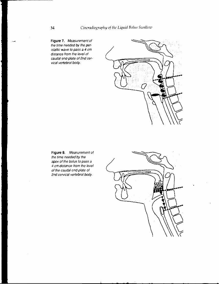
• speed of the pharyngeal peristaltic wave.
The peristalsis in the posterior pharyngeal wall was clearly outlined as aperistaltic wave, which in the lateral projection was seen as a descendentbulging of the posterior pharyngeal wall into the pharyngeal lumen. Thetime taken for the peristaltic wave to pass a 4 cm distance from the level ofthe caudal end-plate of the 2nd cervical vertebral body to approximately thelevel of the cranial end-plate of the 5th cervical vertebral body was measured(Fig 7). The procedure of measuring the time by frame counting and calcu-lation of the speed is described in detail in Analytic methods, p. 38.
• speed of the apex of the bolus
Time for the apex of the bolus to pass a 4 cm distance from the level of thecaudal end-plate of the 2nd cervical vertebral body to approximately thelevel of the cranial end-plate of the 5th cervical vertebral body was measured(Fig 8). The procedure of measuring the time by frame counting and calcu-lation of the speed is described in detail in Analytic methods, p. 38.
34 Cineradiography of the Liquid Bolus Swallozv
Figure 7. Measurement ofthe time needed by the per-istaltic wave to pass a 4 cmdistance from the level ofcaudal end-plate of 2nd cer-vical vertebral body.
Figure 8. Measurement ofthe time needed by theapex of the bolus to pass a4 cm distance from the levelof the caudal end-plate of2nd cervical vertebral body.

Observed Events and Measured Parameters on Cineradiograms 35
• movement of the hyoid bone
The movements of the hyoid bone during swallowing were measured in theleft lateral position using the most anterior aspect of the body of the hyoidbone as a reference point. The initial movement in superior and slightlyposterior direction was measured from the resting position to the positionof maximal elevation.
The second movement in anterior direction was measured from the positionat maximal elevation to the maximal anterior position.
The net movement was measured from the resting position to the maximallyelevated anterior position (Fig 9).
The procedure of measuring the length of the movements is described indetail in Analytic methods, p. 38.
Figure 9. Measurement ofthe movement of the hyoidbone. 1: First movement: su-periorly and slightly posteri-orly, 2: Second movement:anteriorly, 3: Net movement.
• elevation of the larynx
This parameter was measured in the left lateral position with the lowersurface of the vocal cords as a reference point. Measurements were per-formed at two stages during the act of swallowing, 1) when the apex of thebolus had reached the level of the valleculae, and 2) at the maximal elevation.Both distances of larvngeal movement at the two different stages were

36 Cineradiography of the Liquid Bolus Swallow
Figure 10. Measurement ofthe distance oflaryngealelevation when the apex ofthe bolus had reached thelevel of the valleculae (1),and at the maximal elevation(2) with the lower surface ofthe vocal cords as a refer-ence point.
valeculae
the lower surface of(he vocal cords in theresting position
maximum elevation — - ^ ! J
the lower surface ofthe vocal cords in theresting position
measured from the same caudal point in the resting position (Fig 10). Theprocedure of measuring the distances is described in detail in Analyticmethods, p. 38.
• approximation of the thyroid cartilage to the hyoid bone
The parameter shows the changes in distance between the thyroid cartilageand the hyoid bone during swallowing. This parameter was measured in theleft lateral position with the opacifications of the anterior superior portion of

Observed Events and Measured Parameters on Cineradiograms 37
Figure 11. Measurementof the approximation of thethyroid cartilage to the hyoidbone. (1) The maximal dis-tance between the thyroidcartilage and the hyoidbone. (2) The minimal dis-tance between the thyroidcartilage and the hyoidbone.
the thyroid cartilage and the ossifications of the most anterior portion of thehyoid bone as the reference points. Measurements were performed at twostages in the act of swallowing, 1) the maximal distance between the refer-ence points of the thyroid cartilage and the hyoid bone at rest betweenswallows, and 2) the minimal distance between the reference points of thethyroid cartilage and the hyoid bone during swallowing (Fig 11). The pro-cedure of measuring the distance is described in detail in Analytic methods, p.38.

Analytic Methods
All cineradiograms were projected on the screen of a Tagarno 35 CX projec-tor. The magnification factor of the resultant image on the screen has beenestimated by measuring the projected image of a calibration lead rod ofknown length in 15 volunteers. The lead rod had been placed in the midlineon the necks of volunteers during cineradiograms in the left lateral projec-tion. The magnification factor on the screen for the cineradiograms in the leftlateral projection, which was the only projection used for all the measure-ments presented in the monograph, was calculated to be 2.0.
The speed of the peristaltic wave and the apex of the bolus were calculatedfrom the time it took for the peristaltic wave or apex of the bolus to passthrough a distance of 4 cm. A distance of 8 cm was indicated by placing an8 cm long paper strip on the screen which corresponds to a distance of 4 cmin the patient. The paper strip was placed parallel to the long axis of thepharynx with the caudal end immediately above the cricopharyngealmuscle, thus being positioned between the caudal end-plate of the C2 verte-bral body and the cranial end-plate of the C5 vertebral body. The time it tookfor the peristaltic wave or the apex of the bolus to pass through the measureddistance was calculated from the known film speed and the number offrames obtained from an electronic automatic frame counter. This framecounting was repeated three times for each swallow and a mean (roundedto a whole number of frames) was estimated. The mean of these means from3 different swallows in each individual, was, again, rounded to a wholenumber, and served as the number of frames that was used to calculate thespeed of the peristaltic wave or the apex of the bolus in each individual. Thespeed is given in cm/s.
The lengths of the movements of the hyoid bone, the larynx, and the distancesbetween the thyroid cartilage and the hyoid bone were measured as follows.A paper sheet was placed on the screen of the Tagarno analector, the relevantpositions of the structures studied were marked with a pencil and thedistance between the marked points was measured with a ruler and cor-rected for the known magnification factor of 2.0. The lengths and distancesare given in mm.
All measurements concerning speed of the peristaltic wave, speed of bolus,movement of the hyoid bone, elevation of the larynx and approximation of

Analytic Methods 39
Table III. Calculated speed (cm/s) of the peristaltic wave or the apex of the bolus movingover a 4 cm distance using a film speed of 50 frames/s.
No. cf frames
1
2
3
4
5
6
7
8
9
10
11
12
13
14
15
16
17
18
19
20
21
22
25
24
25
Speed (cm/s)
2-102
1-102
66.7
50.0
40.0
33.3
28.6
25.0
22.2
20.0
18.2
16.7
15.4
14.3
13.3
12.5
11.8
11.1
10.5
10.0
9.5
9.1
8.7
8.3
8.0
No. of frames
26
27
28
29
30
31
32
33
34
35
36
37
38
39
40
41
42
43
44
45
46
47
48
49
50
Speed (cm/s)
7.7
7.4
7.2
6.9
6.7
6.5
6.3
6.1
5.88
5.71
5.56
5.41
5.26
5.13
5.00
4.89
4.76
4.65
4.55
4.44
4.35
4.26
4.17
4.08
4.00
the thyroid cartilage to the hyoid bone (ch 1, 2, 3,4, 5,6) were made by thesame observer. The measurements of the elevation of the larynx and theevaluation of pharyngeal dysfunction (groups 3 and 7) in chapter 5 weremade by another observer. Measurements made by different observers werenever matched with each other.
The precision of estimating the speed from sequential frames using framecounting decreases with increasing measured value of the speed, a fact which

40 Cineradiography of the Liquid Bolus Swallow
appears clearly from Table III. The higher the speed the fewer the number offrames obtained. Consequently, the risk for potential error in absolute valuesof calculated speed increases with increasing speed. For example, the differ-ence of 1 frame may be as great as 100 cm/s (between 100 and 200 cm/s) oras small as 0.08 cm/s (between 4.00 and 4.08 cm/s).
In the present investigation, three independent time calculations were per-formed for each separate bolus swallow. Very little variation in the timecalculations was observed. The maximum variation was 1/50 of a second (1frame) in a few cases. This confirmed the accuracy of the calculation and theindividual observer reproducibility. This high reproducibility is in goodagreement with earlier investigations emloying the same calculation tech-nique [36].
All cineradiograms were obtained and analyzed with the same equipment.
Statistical Methods
The statistical techniques used were analysis of variance and covarianceincluding repeated measurement [42]. Adjustment for age was performedby including age as a covariant. Correlation was estimated by Pearsonproductmomentcoefficient.
Statistical analysis disclosed that most of the parameters measured on ciner-adiograms decreased with increasing age of the subjects, either significantlysuch as speed of the apex of the bolus (p - 0.046) and second movement ofthe hyoid bone (p = 0.01), or showed a tendency to such a dependence suchas speed of peristalsis (p = 0.15), movement of the hyoid bone (first p = 0.28and net p = 0.14), and movement of the larynx (p = 0.46 and p = 0.40). Toavoid this bias in the further statistical evaluation of other factors that mayinfluence the speeds and movements analyzed in the present investigation,all measured data were adjusted for age. In the separate chapters, themeasured data are presented first without adjustment for age and then afteradjustment for age. All statistical evaluation of differences and correlationsis based on data adjusted for age with the only exception being the correlationbetween the age and the speed of the peristaltic wave in chapter 1, where themeasured speeds were used without adjustment for age.
L. . j .

1. Speed of the Peristaltic Wave
Introduction
Pharyngeal peristalsis is a crucial component of the pharyngeal swallowingact and is essential for transporting portions of the bolus through thepharynx. Especially important is the cleansing function of peristalsis whichremoves bolus material from the pharynx and prevents aspiration. Pharyn-geal peristalsis is a product of the pharyngeal constrictor muscle activitywhich results in a coherent wave of contractions that moves caudally throughthe muscles, transporting the bolus into the esophagus [15,43]. The continu-ous contractions narrow the pharyngeal lumen and increase the intraluminalpressure, resulting in a peristaltic wave moving from the epi- to the hy-popharynx. The peristaltic wave is visible on the cineradiogram, in lateralprojection, as a propulsive wave in the posterior wall of the pharynx. Thedifferent constrictor muscles act as a common functional unit and cannot bedifferentiated from each other during cineradiography of the swallowing act.
Dysfunction in the pharyngeal constrictor muscles may result in retention ofbolus material in the hypopharynx after completion of the swallow. Thisretention of bolus material can be seen on a cineradiogram. Such dysfunctionof the pharyngeal constrictor muscle activity is detected in some of thepatients with a feeling of residual bolus material in the pharynx after swal-lowing. However, in some patients with this feeling of bolus retention nosign of dysfunction can be detected. Among these patients a subtle, heretoundefined, pharyngeal dysfunction may cause symptoms. The diagnosticcriteria currently used in evaluation of the cineradiogram do not utilize thequantitative information included in the cineradiogram such as the speed orlength of movements of structures that are visible on the cineradiogram.Peristalsis is one of the basic manifestations of pharyngeal constrictor activ-ity, and therefore, theoretically, it cannot be excluded that an abnormal speedof the peristaltic wave could represent a mild or early dysfunction of thepharyngeal constrictor muscles or a sign of incoordination in the centralcontrol of the swallowing act. Such a mild pharyngeal dysfunction couldonly be detected by measuring th" speed of the peristaltic wave. The incite-ment to the study presented in this chapter is the proposed hypothesis, that

42 Cineradiography of the Liquid Bolus Swallow
the change in the speed of the peristaltic wave would be a valuable parameterfor detection of functional impairment of the swallowing act.
If measurements of the speed of the peristaltic wave by cineradiography areto be used to detect early pharyngeal dysfunction, then the question arises:is the peristaltic speed influenced by factors other than a suspected disease?Such factors are the volume of the bolus, the position of the patient, the sexor age of the patient, and the time interval between consecutive swallows.This chapter is, therefore, focused on investigation of the possible influenceof these factors on the speed of the peristaltic wave.
Material and Method
The material consisted of cineradiograms obtained from five groups ofindividuals.
Group 1:
Fifteen non-dysphagic volunteers without pharyngeal dysfunction swallow-ing boluses of 10 ml of contrast medium. They were examined in threedifferent positions; upright, right decubitus horizontal and right decubituswith the table tilted head-down 30°
Group 2:
Ten non-dysphagic volunteers without pharyngeal dysfunction swallowingboluses of 2.5, 5, 10 and 20 ml of contrast medium in increasing order ofvolume.
Group 4:
Ten dysphagic patients without pharyngeal dysfunction swallowing bolusesof 2.5,5,10 and 20 ml of contrast medium in increasing order of volume.
Group 5:
Ten dysphagic patients, 4 with and 6 without pharyngeal dysfunction,swallowing a mouthful of contrast medium. The patients were instructed tocomplete the swallow and without interruption swallow an additional fourboluses of contrast medium, thus swallowing five consecutive boluses.

1. Sveed of the Peristaltic Wave 43
Group 6:
Sixty dysphagic patients without pharyngeal dysfunction, divided into threeage groups, swallowing a mouthful of contrast medium.
The groups are described in further details in Material, p. 27. The peristalticwave was observed from the beginning of the peristaltic wave in the poste-rior pharyngeal wall and its speed was calculated over a distance of 4 cm,from the level of the caudal end-plate of the second cervical vertebral bodyto approximately the level of the cranial end-plate of the fifth cervicalvertebral body. The procedure of measuring the time by frame counting andthe calculation of the speed is described in detail in Analytic methods, p. 38.
Results
The speed of the peristaltic wave in the individual subjects in groups 2 and4 is graphically presented in Figures 1:1a and b. Mean values and SD for eachgroup with respect to swallowed bolus volume are given in Table 1:1 withoutadjustment for age (see p. 40). The speed of the peristaltic wave variedbetween 10—17 cm/s in group 2, and between 7—25.0 cm/s in group 4. Inthe remaining text all results are presented after adjustment for age.
Table 1 :l. Speed of the peristaltic wave in pharyngeal constrictors in 20 individuals whoswallowed different bolus volumes. Speed is given in cm/s; mean (SD). 10 non-dysphagicvolunteers (group 2) and 10 dysphagic patients (group 4), all without pharyngeal dysfunc-tion. Not adjusted for age.
Bolus volume
Group 2
Group 4
15 ml
13.0(1.7)
11.7(4.2)
5 ml
13.2(2.4)
12.4 (4.9)
10 ml
12,9 (2,0)
12.2 (3.9)
20 ml
12.8(1.8)
11,6(4.1)
The volume of the bolus did not influence the speed of the peristaltic waveeither in the non-dysphagic volunteers (group 2) or in the dysphagic patients(group 4) who both swallowed the boluses in increasing order of volume.There were no significant differences in the speed of the peristaltic wave atfour different bolus volumes within the groups (Table 1 :II). The differencesin interpersonal variation between groups were not significant (fig 1:1 a andb). The intrapcrsonal variations in the speed of the peristaltic wave were

44 Cineradiography of the Liquid Bolus Swallow
A cm/s" 30
2.5 20.0
ml
B cm/s
2.5 5.0 10.0ml
20.0
Figure 1:1. The speed of the peristaltic wave in individual subjects at different bolusvolumes, a) non-dysphagic volunteers (group 2), b) dysphagic patients (group 4).

1. Speed of the Peristaltic Wave 45
Table 1 :ll. Speed of the peristaltic wave in pharyngeal constrictors in 20 individuals whoswallowed different bolus volumes. Speed is given in cm/s; mean (SD). 10 non-dysphagicvolunteers (group 2) and 10 dysphagic patients (group 4), all without pharyngeal dysfunc-tion. Speed adjusted for age.
Bolus volume 2.5 ml 5 ml 10ml 20 ml
Group 2
Group 4
12.2(1.7)
12.5(4.2)
12.4(2.4)
13.2 (4.9)
12.1 (2.0)
13.0(3.9)
12.0(1.8)
12.4(4.1)
significantly (p = 0.05) higher among dysphagic patients (group 4) comparedwith non-dysphagic volunteers (group 2) (fig 1:1 a and b).
There was no statistically significant difference in the speed of the peristalticwave between the non-dysphagic volunteers (group 2) and the dysphagicpatients (group 4) at any bolus volume. However, the speed of the peristalticwave in one dysphagic patient (group 4) was higher, and in one lower, thanthe ± 2 SD interval of speeds in the non-dysphagic volunteers (group 2), atall bolus volumes (Fig 1:2).
cm/s
2.5 5.0 10.0 20.0
ml
Figure 1:2. The speed of the peristaltic wave in dysphagic patients (group 4) at differentbolus volumes. Superimposed is + 2 SD interval of the speeds of the peristaltic wave inthe nondysphagic volunteers (group 2).

46 Cinematography of the Liquid Bolus Swallow
cm/s
Erect Horizontal Head tilted
Figure 1:3. The speed of the peristaltic wave in the 15 individual non-dysphagic subjects(group 1) swallowing boluses in three different positions. 'Note that some of the individualsubjects had the same values and therefore are overprojected.
The speed of the peristaltic wave in the 15 individual non-dysphagic subjects(group 1) swallowing boluses in three different positions is graphicallypresented in Fig 1:3. The orientation of the pharyngeal axis with respect togravity significantly influenced the speed of the peristaltic wave. The speedof peristaltic wave decreased when the position was changed from erect tohorizontal and further to head-down position (Fig 1:4, Table 1:111). Thechange in the speed of the peristaltic wave, statistically evaluated as onefunction, and not as separate differences between the three different posi-tions, was significant (p = 0.001). The inter- and intrapcrsonal variations weresimilar for all three positions (Fig 1:3).
The speed of the peristaltic wave showed a slight tendency (p = 0.15) toreduction with age. The speed seemed to decrease slightly with increasingage, however, the correlations between speed of the peristaltic wave and agewere not statistically significant among the 20 individuals in groups 2 and 4or among the 60 dysphagic patients (group 6). The speed of the peristalticwave at three boluses swallowed by 60 dysphagic patients without pharyn-

2. Speed of the Peristaltic Wave 47
cm/s A
12.0
11.0
erect horzontal head down
position
Figure 1:4. The mean speed of the peristaltic wave decreased significantly (p=0.001)when changing position of the 15 non-dysphagic volunteers in group 1.
Table 1 :lll. Speed of the peristaltic wave in pharyngeal constrictors in 15 non-dysphagicvolunteers (group 1) swallowing 10 ml bolus in erect, horizontal and 30° head tilted downpositions. Speed is given in cm/s; mean (SD)
Position: Erect Horizontal Head tilted 30° down
Group 1 12.5(1.9) 11.5(1.7) 11.3 {1.7)
geal dysfunction (groups 6) is given in Table 1:IV. Correlation between ageand the mean speed of the peristaltic wave in each of the 60 individualdysphagic subjects (group 6) is graphically presented in Fig 1:5. The boluseswere swallowed as a separate bolus at approximately 30 seconds interval.
The speed of the peristaltic wave in the 10 individual subjects in group 5 whoswallowed five boluses without interruption between swallows is graphi-cally presented in Fig 1:6 and its mean values and SD are given in Table 1:V.

48 Cineradiography of the Liquid Bolus Swallow
Speed of peristalsis (cm/s)
24 -
20 80
Age (years)
100 120
Figure 1:5. Correlation between age and the mean speed of the peristaltic wave in 60individual dysphagic subjects (group 6) who swallowed three separate boluses.
Table 1 :IV. Speed of the peristaltic wave in pharyngeal constrictors in 60 dysphagicpatients without pharyngeal dysfunction (group 6) who swallowed three separate boluses.Speed is given in cm/s; mean and SD.
Bolus
Mean
SD
1
12.7
3.0
2
12.5
2.9
3
12.5
2.7
Mean of 1 - 3
12.6
The time interval between different swallows did not influence the speed ofthe peristaltic wave. There was no statistically significant difference in thespeed of the peristaltic wave between the three different swallows withapproximately 30-second intervals in the 60 individual dysphagic patients(group 6) (Table 1 :IV) or between the five different swallows without inter-ruption in the 10 individual dysphagic patients (group 5) (Table 1:V).

1. Speed of the Peristaltic Wave 49
cm/s
Figure 1:6. The speed of the peristaltic wave in the 10 individual dysphagic subjects withand without pharyngeal dysfunction (group 5) who swallowed five boluses without inter-ruption between swallows.
Table 1 :V. Speed of the peristaltic wave in 10 dysphagic patients (group 5) who swallowedfive boluses without interruption between swallows. Speed is given in cm/s; mean and SD.
Bolus
Mean
SD
1
13.2
2.9
2
13.7
2.7
3
13.2
2.4
4
13.1
2.8
5
13.4
3.1
Mean of 1-5
13.3
No statistical difference was found in the speed of the peristaltic wavebetween men and women.
Discussion
The results showed that the mean speed of peristalsis was almost the samein the group of healthy volunteers (group 2) and in both groups of dysphagic

50 Cinemdiography of the Liquid Bolus Swalhnv
patients without pharyngeal dysfunction (groups 4 and 6). Furthermore,there was no significant difference in the speed of the peristaltic wave atdifferent bolus volumes despite the fact that the volume varied by a factorof 8 (from 2.5 ml to 20 ml). The speed was approximately 12 cm/s which isin accordance with other investigations of the speed of pharyngeal peristalsisusing cineradiography and/or intraluminal manometry [37, 44, 45].
According to Buchholz et al. [46] the pharynx has a great ability to adapt tochanges in the bolus such as consistency, viscosity, elasticity, volume, andtemperature and also to changes in head and neck posture. It is known thatpatients with pharyngeal dysfunction show a spontaneous tendency todiminish the volume of the bolus in order to achieve a safe swallow [46].Furthermore, in the treatment of patients with detected pharyngeal dysfunc-tion successively increasing the volume of the bolus, beginning with a smallbolus, as well as varying the viscosity and the consistency of the bolus areused in an attempt to establish a safe swallow [47]. The therapeutic successthereby achieved suggests that some compensatory mechanism may existwhich may be activated by a successive increase in the volume of the bolus.The results of the present study suggests that the increase in the speed of theperistaltic wave is not responsible for such a therapeutic effect. The presentstudy suggests that there must be another compensatory mechanism re-sponsible for adaptation to an 8-fold increase in the bolus volume. A similarconclusion can also be drawn from a previous study by Kahrilas and co-workers [12], which was performed on 8 normal subjects with manometryand videofluoroscopy in which the velocity of the pharyngeal peristalsis was15 cm/s and did not change significantly with a 10-fold increase in bolusvolume.
The following conclusion of clinical importance can be drawn from theresults presented here — it is not necessary to know the exact volume of theswallowed bolus when the speed of the peristaltic wave is measured oncineradiograms or videorecordings.
Among the non-dysphagic volunteers in the present study, the pharyngealfunction, as reflected by the speed of the peristaltic wave, appeared relativelyconstant and showed small, non-significant differences despite 8-fold varia-tion of the bolus volumes. This suggests that (1) the "pacemaker" located inthe medullary swallowing centers which synchronizes contractions in thepharyngeal musculature has, in normal individuals, a constant rhythm and,also, (2) that the "pacemaker" does not respond to sensory impulses fromthe mouth or hypopharynx which relate to bolus volume. Such a fixedpattern of swallowing pre-programmed in a network of neurons in the

2. Speed of the Peristaltic Wave 51
swallowing centers has earlier been proposed by Miller [6] as one of twocurrent theories on control mechanisms of the swallowing act. However, thehere suggested unresponsiveness of the coordination center to sensory im-pulses from bolus volume does not necessarily mean that the center cannotbe influenced by other sensory feedback. The significant decrease of thespeed of the peristaltic wave with changes in posture, found in non-dys-phagic volunteers (group 1) makes the existence of some sensory feedbacklikely. Therefore, a hypothesis is proposed that the speed of the peristalticwave is controlled from the swallowing centers in a pre-p grammed patternwhich may, however, respond to certain afferent impulses such as thoseassociated with changes of posture. The nature of such impulses in thisparticular case is unknown including possible origins from the pharynx orvestibular centers.
The speed of the peristaltic wave varied significantly more with the bolusvolume in dysphagic patients (group 4) than in non-dysphagic volunteers(group 2) and, in addition, in one patient speeds at all bolus volumes werehigher and in one lower than ± 2 SD interval from the group of non-dys-phagic volunteers. If the presumption that the speed of peristalsis is control-led centrally is correct, then these results may suggest, that, in some dy-sphagic patients, the underlying cause of their dysphagia originates in adisturbance of central control. Such a central control disturbance may becineradiographically manifested merely as an aberrant speed of the peristal-tic wave. The dyscoordinated contractions in the pharynx musculature maythen result in aberrant afferent sensory impulses perceived as differentsymptoms of dysphagia.
Even though there was no statistically significant difference in the speed ofthe peristaltic wave between the groups of non-dysphagic volunteers anddysphagic patients, 2 out of 10 dysphagic patients had a speed which couldbe considered pathologically low —9 cm/s — and high — 25cm/s(Fig 1:2).The group of the dysphagic patients is heterogeneous with respect to thesymptoms and likely also with respect to the underlying process. It isconceivable that, in two patients included in this small material, the abnor-mally high or low speed of the peristaltic wave may be associated with thepathologic process underlying their dysphagia. An abnormal speed of theperistaltic wave may therefore be potentially considered a new sign ofpharyngeal dysfunction. \ lowever, todefine normal speed limits a dedicatedstudy of a large number of individuals is needed.
Pharyngeal dysfunction implies failure of one or several components of theswallowing act and includes paresis of the pharyngeal constrictor muscles,

52 Cineradiography of the Liquid Bolus Szcalloiv
incoordination of the cricopharyngeal muscle and dysmobility of the epiglot-tis. The frequency of pharyngeal dysfunction has been shown to increasewith age [31,48]. Pharyngeal '"sfunction, occurring with increasedfrequency among elderly patients includes paresis of any or all pharyngealconstrictor muscles, resulting in bolus retention and thereby an increasedrisk for aspiration. However, no attention has been paid to a potential mildabnormal pharyngeal function in terms of altered speed of the peristalticwave. If the speed of the peristaltic wave was dependent upon age it couldbe that the change in the speed with increasing age could contribute topharyngeal dysfunction. However, this investigation has shown that one ofthe basic components of the swallowing act — the speed of the peristalticwave did not significantly change with age in all 80 individuals investigated(groups 2,4,6).
In daily clinical work some of the patients referred for cineradiography arefor various reasons bedridden and have to be examined in a recumbentposition. If a reduced speed of the peristaltic wave is found in these patients,then this finding has to be evaluated critically and should not be considereda definite sign of pharyngeal dysfunction because a decrease of the speedalso occurs with changes in posture in normal individuals. As shown in thepresent investigation a significant (p = 0.001) decrease of the speed of theperistaltic wave was registered at swallowing in the horizontal position ascompared with swallowing in the erect position and even more when thesubject was examined in a head-down tilted position.
A possible criticism of the results in the present investigation is the fact thatno specified time interval was used between swallows. Theoretically, a longtime interval between the swallows may imply that the pharyngeal muscleshad completely recovered between the swallows and were ready to respondto the next nerve impulses at each separate swallow. Therefore, the pharyn-geal muscles would act as if every swallow occurred in a state of completerecovery and possible changes in speed related to the interval in time wouldnot be apparent. As shown in the present investigation no significant changeoccurred in the speed of the peristaltic wave between different swallowswhen the boluses were swallowed either with approximately 30 secondsinterval (group 6) or without interruption (group 5). This suggests that thetime interval between swallows does not influence the speed of the peristalticwave.
The following conclusion of clinical importance can be drawn from theaforementioned statement: correct measurements of the speed of the peristal-tic wave by means of cineradiography or videorecording are obtained from

1. Sfved of the Peristaltic Wave 53
any swallow; even retrospectively without knowledge of the exact timeinterval between swallows. Consequently, there is no need for strictly stand-ardized time intervals between swallows which, in addition, could poten-tially stress impaired patients.
Conclusions
Speed of the peristaltic wave is constant in individuals without pharyngealdysfunction and is not significantly influenced by the volume of the bolus,the interval in time between different boluses swallowed, the presence ofdysphagia, or the age or sex of the individual.
Speed of the peristaltic wave decreases significantly when changing posi-tion from erect to horizontal and when the table is tilted 30° head-down.
Intrapersonal variations of the speed of the peristaltic wave is higher indysphagic patients than in non-dysphagic individuals.
Measurements of the speed of the peristaltic wave can be carried outretrospectively from previously recorded cineradiographies and videore-cordings, provided that magnification factor and film speed are known, asno knowledge of the time interval between swallows or of the bolusvolumes is required.
Abnormally high or low speed of the peristaltic wave may be the first mildor early sign of pharyngeal dysfunction.
Therefore, measurement of the speed of the peristaltic wave may be usefulin the clinical evaluation of pharyngeal function.

2. Speed of the Apex of the Bolus
Introduction
The bolus is transported from the mouth into the pharynx by the tonguewhich successively presses against the hard palate in an aboral direction. Theexact mechanism for the transport of the bolus through the pharynx into theesophagus is not completely understood and is the subject of controversy inthe literature. Various mechanisms and their combinations such as positiveor negative pressure in the pharynx, subatmospheric pressure in theesophagus, peristaltic wave, and gravity have been postulated to be re-sponsible for the transport of the bolus through pharynx [19, 20, 37, 39, 49,50,51,521.
Apart from gravity, the force which moves the bolus is produced by muscu-lar contraction. While the speed of the peristaltic wave gives informationregarding the timing of the sequential contractions of the pharyngeal con-strictor musculature, the speed of the bolus provides additional informationabout contractile force of the pharyngeal constrictors as well as the musclesresponsible for the movements of the tongue and soft palate and, possibly,other muscles which create the subatmospheric esophageal pressure. It isconceivable that dysfunction of the muscles, either an altered contractileforce or a defective timing of their contractions, may result in an abnorma1
speed of the bolus. An abnormal speed of the bolus may therefore indicatethe presence of muscular dysfunction or the presence of incoordination ofthe central control of swallowing in individuals in which cineradiographyfails to demonstrate any pharyngeal dysfunction, according to the currentlyknown criteria of dysfunction.
In order to use the sp>ed of the apex of the bolus as a potential clinicalcriterion to disclose pharyngeal dysfunction, it is essential to analyze factorsthat might potentially alter the spe^d of the bolus in individuals withoutpharyngeal dysfunction. Factors that potentially may influence the speed ofthe bolus on cineradiograms performed with liquid barium include: thebolus volume, the interval between swallows, the position of the individualat swallowing, and age or sex of the individual. Therefore, the speed of thebest defined part of a moving bolus, its apex, was studied in various experi-

56 Cineradiography of the Liquid Bolus Swallow
mental settings in non-dysphagic and dysphagic individuals with normalpharyngeal function.
Material and Method
The material consists of cineradiographs obtained from four groups ofindividuals:
Group 1:
Fifteen non-dysphagic volunteers without pharyngeal dysfunction swallow-ing boluses of 10 ml of contrast medium. They were examined in threedifferent positions: upright, nght decubitus horizontal and right decubituswith the table tilted head-down 30°.
Group 2:
Ten non-dysphagic volunteers without pharyngeal dysfunction swallowingboluses of 2.5, 5, 10, and 20 ml of contrast medium in increasing order ofvolume.
Group 4:
Ten dysphagic patients without pharyngeal dysfunction swallowing bolusesof 2.5,5,10, and 20 ml of contrast medium in increasing order of volume.
Group 5:
Ten dysphagic patients, 4 with and 6 without pharyngeal dysfunction,swallowing a mouthful of contrast medium. The patients were instructed tocomplete a swallow and then without delay swallow four additional bolusesof contrast medium, thus swallowing five consecutive boluses. In three ofthe 10 patients it was impossible to measure the speed of the apex of the bolusdue to head movement artefacts. In the remaining seven dysphagic patientsfive boluses were analyzed.
The groups are described in further detail in Material, p. 27.
The time for the apex of the bolus to pass over a distance of 4 cm from thelevel of the caudal nd-plate of the 2nd cervical vertebral body to approxi-mately the level of t^e cranial end-plate of the 5th cervical vertebral body

2. Speed of the Apex of the Bolus 57
was measured. The procedure for measuring the time by frame counting andthe calculation of the speed are described in detail in Analytic methods, p. 38.
Results
The measured speed of the apex of the bolus in the individual subjects isgraphically presented in Figures 2:1a and b. Mean values of the measuredspeeds and SD for each group with respect to swallowed bolus volume aregiven in Table 2:1 without adjustment for age (see p. 40). The speed of the apexof the bolus varied between 17 and 67 cm/s in group 2, and between 11 and67 cm/s in group 4. In the further text, all results are presented afteradjustment for age.
Table 2:1. Speed of the apex of the bolus at different bolus volumes. Speed is given incm/s; mean (SD). 10 non-dysphagic volunteers (group 2) and 10 dysphagic patients (group4). All without pharyngeal dysfunction. Not adjusted for age.
Bolus volume Z5ml 5 ml 10 ml 20 ml
Group 2
Group 4
35.5(12.0)
23.2(7.1)
37.4 (9.2)
24.8 (9.5)
45.4(13.9)
25.0(11.8)
44.4(12.6)
31.6(17.2)
The speed of the apex of the bolus increased significantly (p = 0.012) withincreasing bolus volume in non-dysphagic volunteers (group 2) and had atendency (p = 0.09) to increase in dysphagic patients (group 4) (Fig 2:2)(Table 2:11). The trend of increasing the speed with increasing bolus volumewas significantly parallel (p = 0.01) between the two groups (Fig 2:2).
Table 2:11. Speed of the apex of the bolus at different bolus volumes. Speed is given incm/s; mean (SD). 10 non-dysphagic volunteers (group 2) and 10 dysphagic patients (group4). All without pharyngeal dysfunction. Adjusted for age.
Bolus volume 25 ml 5 ml 10 ml 20 ml
Group 2
Group 4
32.1 (12.0)
26.6(7.1)
33.9 (9.2)
28.2 (9.5)
42.0(13.9)
28.5(11.8)
40.9 («2.6)
35.0(17.2)

58 Cineradiography of the Liquid Bolus Swallow
cm/s
2.5 20.0
B cm/s
25 -
2.5 5.0 10.0
ml20.0
Figure 2:1. The speed of the apex of the bolus in individual subjects at different bolusvolumes, a) 10 non-dysphagic volunteers (group 2), b) 10 dysphagic patients (group 4).

2. Speed of the Afiex of the Bolus 59
cm/s
40.0
30.0
20.0
- • Group 2
Group 4
2.5 5.0 10.0ml
20.0
Figure 2:2. Illustrating the significant increase of the speed of the apex of the bolus (cm/s)(mean per group) in 10 non-dysphagic volunteers (group 2) (p=0.012) and the tendencyto such an increase in 10 dysphagic patients (group 4, p=0.09) at different bolus volumes.The parallel nature of the increase in both groups is significant (p=0.01).
The interpersonal variations within the groups in speed of the apex of thebolus were large in both groups and there were no significant differencesbetween the two groups in this respect.
The speed of the apex of the bolus in the individual subjects (group 1)swallowing boluses in the three different positions is graphically presentedin Fig 2:3, and its mean values and SD are given in Table 2:111.
Table 2:111. Speed of the apex of the bolus in 15 non-dysphagic volunteers (group 1)swallowing 10 ml bolus in erect, horizontal and head down tilted 30° positions. Speed isgiven in cm/s; mean (SD)
Position:
Mean
Erect
45.2(14.8)
Horizontal
48.6(14.7)
Head tilted 30° down
51.8(21.6)

60 Cineradiography of the Liquid Bolus Swallow
cm/s
20 :
Erect Horizontal Head tilted
Figure 2:3. The speed of the apex of the bolus in the 15 individual subjects (group 1)swallowing boluses in three different positions. ' Note that the speed in some of theindividual subjects was identical and the values are therefore overprojected.
The orientation of the pharynx with respect to gravity did not significantlyinfluence the speed of the apex of the bolus. A statistical tendency to increasethe speed of the bolus occurred when the pharyngeal axis became perpen-dicular to gravity (horizontal position of the individual) and even more whenthe bolus moved against gravity (object tilted 30° head down) (Table 2:111).The increase in speed of bolus when changing positions in 15 non-dysphagicvolunteers (group 1) swallowing 10 ml bolus was, however, not significant,mainly because of the large inter- and intmpersonal variations. The inter- andintrapersonal variations which can be seen in Fig 2:3, were similar in erect andhorizontal positions and insignificantly higher in head down tilted 30°position.
The highest speed of the apex of the bolus —100 cm/s — was noted in 1 casewhen changing the position from erect to horizontal and head down tiltedpositions (group 1), while the highest measured speeds occurred whenchanging the bolus volumes were 66.7 cm/s at 10 or 20 ml boluses (groups2, and 4).
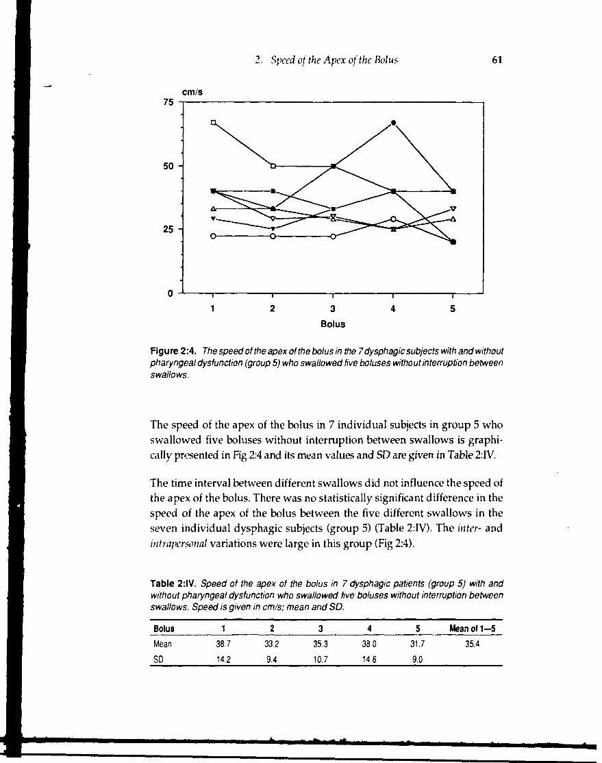
2. Speed of the Apex of the Bolus 61
cm/s
25 -
Figure 2:4. The speed of the apex of the bolus in the 7 dysphagic subjects with and withoutpharyngeal dysfunction (group 5) who swallowed five boluses without interruption betweenswallows.
The speed of the apex of the bolus in 7 individual subjects in group 5 whoswallowed five boluses without interruption between swallows is graphi-cally presented in Fig 2:4 and its mean values and SD are given in Table 2:IV.
The time interval between different swallows did not influence the speed ofthe apex of the bolus. There was no statistically significant difference in thespeed of the apex of the bolus between the five different swallows in theseven individual dysphagic subjects (group 5) (Table 2:IV). The inter- andintrapersonal variations were large in this group (Fig 2:4).
Table 2:1V. Speed of the apex of the bolus in 7 dysphagic patients (group 5) with andwithout pharyngeal dysfunction who swallowed five boluses without interruption betweenswallows. Speed is given in cm/s; mean and SD.
Bolus
Mean
SD
1
38.7
14.2
2
33.2
9.4
3
35.3
10.7
4
38,0
14.6
5
31.7
9.0
Mean of 1—5
35.4

62 Cineradiogmphy of the Liquid Bolus Swallow
The speed of the apex of the bolus was significantly influenced (p = 0.046)by the age of the individuals. The speed of the apex of the bolus decreasedwith increasing age.
There was no statistical difference in the speed of the apex of the bolusbetween men and women.
Discussion
There is an accordance between the speed of the apex of the bolus measuredin this investigation and the speed found by other investigators whomeasured the speed (cm/s) using different techniques [37, 40, 49]. In thepresent investigation the speed of the apex of the bolus for different groupsand bolus volumes varied between 11—67 cm/s in the upright position.Fischer and co-workers [40] found that the bolus was propelled from thepharynx into the esophagus with a speed of 10—70 cm/s in a study of thechanges of intraluminal electric impedance in 9 subjects swallowing bolusesof different volume. Atkinson and co-workers [37] registered a speed of40—50 cm/s in a simultaneous manometric and cineradiographic study,using a film speed of 6—12 frames, in 8 patients in sitting and supine position,swallowing bolus of different volume and nature.
The transport of the bolus through the pharynx is believed to be accom-plished or aided by several mechanisms or their combinations. Some authorsconsider the peristaltic wave to be the main force that propels the bolusthrough the pharynx [19,20,37]. According to Ramsey et al. [20], the activityin the pharyngeal constrictor muscles creates a "pharyngeal stripping wave"which is the major propulsive force in all normal swallowing of semiliquidand solid material. This theory is supported by Saunders et al. [19] andAtkinson et al. [37]. It is also believed that gravity aids the passage of theliquid bolus in the upright position [20,37, 50].
Rushmer and Hendron [49] have suggested that the propelling force duringdeglutition depends "upon the action of the tongue and the pharyngealconstrictor muscles down to the level of the clavicle." In addition, a negativeintraluminal pressure in the hypopharynx and upper esophagus has beenproposed to have some effect on the passage of the bolus [39, 50].
It appears unlikely that the peristaltic wave itself could have any significantimpact on propulsion of the majority of a liquid bolus as it moves 2—4 timesmore slowly than the apex of the barium bolus, as shown in the present

2. Speed of the Apex of the Bolus 63
examination. Also, the contribution of gravity to the speed of the bolus seemto be minimal because no decrease of the speed of the apex of the bolus wasnoted in the 15 volunteers (group 1) when they were tilted to horizontal, and30° head down positions, ie, when the gravitational force become perpen-dicular or slightly opposite to the direction of bolus movement. On thecontrary, a statistical tendency to increase the speed of the apex of the boluswas noted when the bolus moved without the aid of the gravity or againstit.
The results obtained here are most consistent with the theory suggestingtongue movement to be the major force propelling the liquid bolus throughthe pharynx, as proposed by McConnel et al. [38,39,53, 54]. The statisticallysignificant increase of the speed of the apex of the bolus with increasing bolusvolume in non-dysphagic volunteers (group 2, p = 0.012), the tendency tosuch an increase in dysphagic patients (group 4, p = 0.09), and the significantparallel nature of this trend (p = 0.01) strongly indicate that the speed of theapex of the bolus increases with larger bolus volumes. An increase in thespeed of the bolus with increasing bolus size has been observed by Fischerand co-workers [40] who discussed whether the higher velocity seen with alarge bolus is simply "the result of changes in the initial length of thepharyngeal muscles or depends on an increased discharge of nerve impulsesinto the pharyngeal muscles." However, according to McConnel et al. [38]and our own observations, the onset of the contraction of the pharyngealconstrictors occurs too late to be a major force in propelling the bolus throughthe pharynx. During barium swallow, the pressure generated by the tonguein the oropharynx is transmitted throughout the entire fluid, equally in alldirections. Conceptually, this pressure generation is analogous to the actionof a pump in which the tongue acts as a piston, and the pharyngeal wallssupported by the tension of the constrictors represent the chamber of thepump. The pump model and the decisive role of the tongue in generatingthe pharyngeal pressure is supported by recent manofluorographic studiesby McConnel et al. [38,39, 53,54].
In the present study, not only the speed of the apex of the bolus increasedsignificantly with larger bolus volumes but also, in the same individuals, thelength of the antero-superior movement of the hyoid bone (Chapter 4). Thissecond movement of the hyoid bone is closely related to the anterior move-ment of the tongue base away from the posterior wall of the mesopharynx[54]. It is conceivable that the extent and possibly also the force of the tonguemovement increases, with larger bolus, therewith increasing the intralumi-nal pressure with resultant acceleration of the apex of a liquid bolus in thedirection of lowest resistance — to the esophagus.
64 Cincradiography of the liquid Bolus Swallow
The significant increase of the speed of the apex of the bolus suggests thatthe forces propelling the bolus through the pharynx are regulated by neuralcontrol mechanism based on the sensory feedback from the receptors in theoropharynx. This control mechanism seems to be exact and stable, becauseapproximately the same speeds of the apex of the bolus were obtained at 5consecutive swallows of boluses of identical size (group 5).
Speculation, as to whether the significant decrease of the speed of the apexof the bolus with increasing age observed here depends on a possibledecrease of muscular power or on a decrease of receptor sensitivity orefficiency of the central neuron, lies outside the limits of this study.
The increase of the speed of the apex of the bolus when changing the postureof the individuals from erect to horizontal and head-down tilted positions,which appears from Fig 2.3 and Table 2:111, was not statistically significantbecause of large intrapersonal variations. It is conceivable that the largenumerical values of the variations depend on the inherent low exactness ofthe measuring method in the highest range of the speed as discussed on p.39. Most of the speed measurements, in group 1, varied between 33.3 and66.7 cm/s which means, that the movement of the bolus over the shortdistance of 4 cm was imaged on 3 to 6 frames (Table III, p. 39). Possible errorsthat may have resulted from measuring a real speed in this manner may beexemplified as follows. In a hypothetical, extreme situation, a measureddifference of 18 cm/s — between 50 and 66.7 cm/s (i.e. 4 and 3 frames) —could correspond to either a real speed difference of 2 cm/s, between realspeeds of 66 and 68 cm/s, or to a real speed difference of 49 cm/s, between51 and 99 cm/s. Theoretically, to increase the exactness of the measurementsand, consequently, to diminish the variations, the high speed of the apex ofthe bolus could be measured over a longer distance. This is, however, notexpedient because it is known that the speed decreases inferior to thecricopharyngeal muscle [37, 49] and the measurement would, in that case,also include the proximal esophagus. The most appropriate way of increas-ing the precision of the measurements would be to use a higher frame rate.However, attempts to increase the frame rate with the equipment used in thepresent investigation were unsucces 4ue to the thermal overload of theX-ray tube. The benefit of increasing the frame rate also has to be weightedagainst the risks of increased radiation dose to the patient.

2. Speed of the Apex of the Bolus 65
Conclusions
• The speed of the apex of the bolus increases significantly with increasingbolus volume in individuals without pharyngeal dysfunction.
• The speed of the apex of the bolus has a tendency to increase whenchanging position from erect to horizontal and when the table is tilted 30°head-down.
• The speed of the apex of the bolus is not significantly influenced by thetime interval between different boluses swallowed or by patient gender.
• The speed of the apex of the bolus is significantly influenced by the age ofthe individual.
• Measurements of the speed of the apex of the bolus can be carried outretrospectively from previously recorded cineradiographs and videore-cordings, provided that magnification factor and film speed are known.The significance of these measurements cannot be determined unless thebolus volume, patient age and position of the patient at the time of theexamination are known.

3. Relation Between the Speed of thePeristaltic Wave and the Speedof the Apex of the Bolus
Introduction
Both the peristaltic wave and the movement of the bolus reflect the activityof the oral and pharyngeal muscles, and their speed can be considered asdirect or indirect measures of the activity of these muscles. On the cineradio-gram, the visible peristaltic wave directly demonstrates the successivesequence of contractions of the pharyngeal constrictor muscle fibers, whilethe movement of the bolus is due to a more complex course of events. Asboth the speed of the peristaltic wave and the speed of the apex of the bolusreflect, to some extent, the activity of the same muscles, it would be interest-ing to see the extent to which the speed of the apex of a liquid bolus dependson the speed of the peristaltic wave. This knowledge may increase the, todate somewhat controversial understanding of the mechanisms responsiblefor transport of liquid bolus through the pharynx.
The objective of this chapter is to compare the results of chapters 1 and 2,which deal with the speeds of the peristaltic wave and the apex of the bolus,in order to determine if there is a correlation between these two parametersin different examination positions and at different volumes of the bolusswallowed.
Material and Method
The material consists of cineradiographies obtained from three groups ofindividuals.
Group 1:
Fifteen non-dysphagic volunteers without pharyngeal dysfunction ex-amined in three different positions; upright, right lateral decubitus horizon-
68 Cineradiography of the Liquid Bolus Swalloiv
tal, and right lateral decubitus position with the table tilted head-down 30°swallowing 10 ml of contrast medium.
Group 2:
Ten non-dysphagic volunteers without pharyngeal dysfunction swallowingboluses of 2.5, 5, 10, and 20 ml of contrast medium in increasing order ofvolume.
Group 4:
Ten dysphagic patients without pharyngeal dysfunction swallowing bolusesof 2.5,5,10 and 20 ml of contrast medium in increasing order of volume.
The groups are described in further detail in Material, p. 27
The time taken by the peristaltic wave and the time for the apex of the bolusto pass over a distance of 4 cm, from the level of caudal end-plate of the 2ndcervical vertebral body to approximately the level of the cranial end-plate ofthe 5th cervical vertebral body was measured. The procedure of measuringthe time by frame counting and the calculation of the speed are described indetail in Analytic methods, p. 38.
Results
In non-dysphagic and dysphagic individuals without pharyngeal dysfunc-tion (groups 2 and 4), who swallowed 2.5—20 ml boluses in erect positionthe measured speed of the peristaltic wave varied between 9.0 and 25 cm/sand the measured speed of the apex of the bolus varied between 11.0 and 67cm/s. Mean values and SD for both groups, adjusted for age, are given inTables 1:11 and 2:11 (groups 2 and 4).
The speed of the peristaltic wave did not change while the speed of the apexof the bolus increased significantly (p = 0.012) with larger bolus volumes innon-dysphagic volunteers (group 2) (Fig 3:1) and had a similar tendency (p= 0.09) to increase in dysphagic patients (group 4) (Fig 3:2).
The speed of the peristaltic wave in 15 non-dysphagic volunteers (group 1)varied in erect position between 9.0 and 15.0 cm/s and in head-down tiltedposition between 9.1 and 15.4 cm/s. The speed of the apex of the bolus inthese individuals varied in erect position between 17 and 67 cm/s, and inhead-down tilted position between 14 and 100 cm/s.

3. Speed Relation between Peristaltic Wave and Bolus 69
cm/s k
50 - f
40 -
30 -
20 -
10 -
2 5 5 10
Speed of the apex of the bolus
Speed of the peristaltic wave
20 ml
Figure 3:1. The mean speeds of the peristaltic wave and the apex of the bolus withdifferent bolus volumes in 10 non-dysphagic volunteers (group 2).
cm/s j
50 -
40 -
30 -
20 -
10 -
Speed of the apex of the bolus
Speed of the peristaltic wave
I T
2 5 5 10 20 ml
Figure 3:2. The mean speeds of the peristaltic wave and the apex of the bolus withdifferent bolus volumes in 10 dysphagic patients (group 4).

70 Cinematography of the Liquid Bolus Swallow
Mean values and SD of the speed of the peristaltic wave and the speed of theapex of the bolus in erect, horizontal, and in 30° head-down tilted positionsare given in Tables 1:111 and 2:111 (group 1).
The speed of the peristaltic wave decreased significantly (p = 0.001) whilethe speed of the apex of the bolus showed a tendency to increase whenchanging the position in the 15 non-dysphagic volunteers (group 1) fromupright to horizontal and head-down tilted positions (Fig 3:3).
cm/s
60 -
50 -
40 -
30 -
20 -
10 -
Speed of the apex of the bolus
Speed of the peristaltic wave
erect horizontal head-down position
Figure 3:3. The mean speeds of the peristaltic wave and the apex of the bolus in 15non-dysphagic volunteers (group 1) with respect to the posture.
No statistically significant correlation between the speed of the peristalticwave and the speed of the apex of the bolus was found in any group at anybolus volume (groups 2 and 4) or in any examining position (group 1). Thecorrelation coefficients varied between —0.11 and —-0.37. The speeds of theperistaltic wave and of the apex of the bolus in individual non-dysphagicvolunteers (group 1) are plotted with respect to posture in Figure 3:4.

3. Speed Relation betivcen Peristaltic Wave and Bolus 71
16 -
14 -
•
12 -
•
10 -
-
Speed of peristalsis
HO
O
O
D V
0
© V
•V
0 v
V
Q
D
V Ö
0
0
o
u
0
•V
V
aD
20
i
40
i
60 80
Speed of the bolus
100 120
Figure 3:4. There was no significant correlation between the speed of the peristaltic waveand the speed of the apex of the bolus at any examination posture in 15 non-dysphagicvolunteers (group 1). (upright*, horizontal V, head tilted down M).
Discussion
The speed of the peristaltic wave decreased significantly (p = 0.001) whileswallowing against gravity, while the speed of the apex of the bolus showeda tendency to increase in the same individuals (group 1). The speed of theperistaltic wave was not influenced by the increase in the bolus volume

71 Cincradiography of the Liquid Bolus Swallow
(group 2 and 4) while the speed of the apex of the bolus increased signifi-cantly (p = 0.012) with larger bolus volumes (group 2) and had a tendency(p = 0.09) to such an increase (group 4). Although the direction of movementof the peristaltic wave and of the apex of the bolus are the same, the resultspresented here suggest the absence of a relation between these two param-eters which are measurable on the cineradiogram. The speed of the peristalticwave reflects the speed of propulsion of the contraction wave '" ~ough thepharyngeal constrictors. In contrast to this organic nature of the peristalticwave, the speed of the apex of a liquid bolus reflects the results of the actionof various muscular groups on a foreign body—the bolus — that is propelledthrough the pharynx. Although the tone of the pharyngeal constrictors islikely essential for maintaining the pharyngeal intraluminal pressure that iscreated by the movement of the tongue, the contractions of the constrictorsdo not seem to substantially contribute to the forces that propel the majorportion of a liquid bolus through the pharynx. The strongest evidence of theindependence between the speed of the peristaltic wave and the speed of theapex of the bolus is the fact that no correlation between these speeds wasdemonstrated at any of the bolus volumes swallowed in erect position, or at10 ml bolus volume swallowed in three different positions. Another reasonwhy the peristaltic wave cannot be responsible for the transport of the firstportion of the liquid bolus is the fact that the peristaltic wave becomes visiblein the posterior wall of the pharynx when the bolus has already reached thelevel of the valleculae (38] or more aboral parts of the pharynx and moves2—4 times more slowly than the bolus as shown by the present investigation.
One possible explanation for the demonstrated increase in the speed of theapex of the bolus with increased bolus volume here may be the increase inthe intraluminal pressure which is created by the tongue motion and by thepharyngeal muscles in order to handle a larger bolus volume or to propelthe bolus against gravity. The mechanism of this increase in intraluminalpressure is discussed in the previous chapter. Therefore, the pressuregenerated in the oropharynx by the tongue movement is, probably, the mainforce propelling the bolus through the pharynx, as suggested in the previouschapter and also by McConnel el al. [38,39,53, 54].
The main function of the peristaltic wave, when swallowing a liquid bolus,appears to be the transport of its terminal portion, a clearing function whichhas also been suggested from studies by other authors employing othertechniques for studying the swallowing act [38,39, 50,53].

3. Speed Relation between Peristaltic Wave and Bolus 73
Conclusions
• It is concluded from the present investigation that the peristaltic wave isnot the major force that propels the initial part of a liquid bolus throughthe pharynx.
• It is suggested that the tongue movement has a dominating role and throwsthe first portion of a liquid bolus through the pharynx.

4. Movement of the Hyoid Boneduring Swallow
Introduction
The movement pattern of the hyoid bone as well as the timing of its move-ments during different phases of the swallowing act have been investigatedin several studies [11,15,19,20,34,36,41,50,55,56]. Movement of the hyoidbone is said to most often occur in a two-step fashion, first an upward andthen a forward movement, and is thought to be important for the closure ofthe airways, including the tilting down of the epiglottis [34]. Some abhorshave claimed that the movement pattern of the hyoid bone differs dependingon the size of the bolus [20]. The movements of the hyoid bone can be studiedon most cineradiograms because of the radioopacity of the hyoid bone. Asno defined dysfunction has been attributed to an isolated disturbance in themovement of the hyoid bone, little attention is paid to its movements in theroutine evaluation of cineradiograms.
Theoretically, changes in the normal two-step movement pattern and/or ofthe normal length of the different movements might reflect dysfunction ofseveral muscles that are attached to the hyoid bone and that participate inthe swallowing act. These muscles are: the stylohyoid and hypoglossusmuscles which elevate the hyoid bone, the geniohyoid muscle which movesit anteriorly and also the mylohyoid and digastric muscles which participatein both movements. An impaired function of these muscles or an incoordi-nation of their contraction may, conceivable, also produce abnormal sensoryimpulses perceived by the patient as dysphagia. Detection of such a dysfunc-tion, in dysphagic patients without any other pharyngeal dysfunction, mighthelp explain their symptoms and provide a better understanding of dyspha-gia. In order to disclose such a dysfunction of these muscles by studying themovement of the hyoid bone on a cineradiogram, it is essential to know theextent to which the movement is affected by variables such as bolus volume,position of the individual during swallow, sex and age of the individuals.
Such systematic analysis has not been performed to the author's knowledge,and quantitative knowledge of hyoid bone movement is ..lited. Ekberg [34]measured the lengths of both movements in 50 non-dysphagic volunteers

76 Cineradiography of the Liquid Bolus Swallow
following a swallow of a mouthful of barium contrast medium by means ofcineradiography. Dodds and co-workers [11] measured the "maximal supe-rior and anterior excursion of the hyoid bone" at different bolus volumes in15 subjects by means of videoradiography.
The objective was to obtain systematic data of the movement of the hyoidbone, which was studied under different conditions in an attempt to reveal;
• if there is any difference in movement patterns or length of movementsbetween non-dysphagic and dysphagic individuals.
• if the length of the first or second or net movements are influenced by thebolus volume.
• if bolus volume or gravity affects the movement pattern of the hyoid bone.
• if the special movement patterns are influenced by age or sex of theindividual.
Material and Method
The material consisted of cineradiographies obtained from three groups ofindividuals.
Group 1:
Fifteen non-dysphagic volunteers without pharyngeal dysfunction ex-amined in upright, right lateral decubital position nnd with the table tiltedhead-down 30°, swallowing 10 ml of contrast medium.
Group 2:
Ten non-dysphagic volunteers without pharyngeal dysfunction swallowing2.5,5,10, and 20 ml of contrast medium in increasing order of volume.
Group 4:
Ten dysphagic patients without pharyngeal dysfunction swallowing 2.5, 5,10 and 20 ml of contrast medium in increasing order of volume.
The groups are described in further details in Material, p. 27.
The movements of the hyoid bone during swallowing were measured in leftlateral projection using the most anterior aspect of the body of the hyoid bone

4. Movement of the Hyoid Bone during Swallow 77
as a reference point as described on p. 33 and shown in Fig 9, p. 35. The patternof movement was recorded in all three groups. In group 2 and 4 the lengthsof the first, the second, and the net movements were measured. The pro-cedure of measuring the length of the movements is described in Analyticmethods, p. 38.
Results
The hyoid bone initally moved cranially and slightly posteriorly (first move-ment) and then anteriorly (second movement). During the second movementthe hyoid bone also performed a slight (10—15°) tilt with the cornua movingin a cephallad direction. This two-step movement was observed in allindividuals (group 1, 2, 4), regardless of their sex or age and regardless ofwhether or not they were dysphagic. In addition, this pattern of two-stepmovement of the hyoid bone did not change when the bolus volume or theposition of the individual was changed.
First Movement of the Hyoid Bone
The measured lengths of the first movement of the hyoid bone in 10 non-dys-phagic volunteers (group 2) varied between 4 and 24 mmand in 10 dysphagicpatients (group 4) between 4 and 16 mm (not adjusted for age). The lengthsof the first movement in the individual subjects of groups 2 and 4 aregraphically presented, after adjustment for age, in Figures 4:1 a and b. Meanvalues and SD, after adjustment for age, for these groups with respect toswallowed bolus volume are given in Table 4:1.
Table 4:1. First movement of the hyoid bone in 20 individuals who swallowed different bolusvolumes. Movement measured in mm; mean (SD). 10 non-dysphagic volunteers (group 2)and 10 dysphagic patients (group 4), all without pharyngeal dysfunction. Adjusted forage.
Bolus volume 2.5 ml 5 ml 10ml 20 mlGroup 2
Group 3
9.5(4.4)
9.0(3.5)
8.8(4.4)
9.2 (3.5)
9.0 (6.6)
10.6(3.7)
9.8 (5.7)
9.5(4.4)

78 Citieradiography of the Liquid Bolus Swallow
mm
B mm
2.5 5.0 10.0 20.0
ml
2.5 5.0 10.0 20.0
ml
Figure 4:1. The length of the first movement in the individual subjects at different bolusvolumes, a) 10 non-dysphagic volunteers (group 2), b) 10 dysphagic patients (group 4).' Note, that the lengths of the first movement in some of the individuals was identical andthe values are therefore overprojected.

4. Movemen t of the Hyoid Bone during Swallow 79
The size of the bolus did not significantly influence the first movement of thehyoid bone in any group of individuals. As seen from Table 4:1, the firstmovement of the hyoid bone did not change with larger boluses when theboluses were swallowed in increasing order of volume (group 2 and 4).
There was no significant difference in the length of the first movementbetween the non-dysphagic volunteers (group 2) and the dysphagic patients(group 4) at any bolus volume (Table 4:1). The intrapersonal variations in thefirst movement were significantly lower in the dysphagic patients (group 4)than in the non-dysphagic volunteers (group 2) (p < 0.001) (Fig 4:1 a and b).The interpersonal variations within the groups in the first movement of thehyoid bone were not significantly different. However, the dysphagic patients(group 4) showed a tendency (p = 0.09) to smaller interpersonal variationsthan the non-dysphagic volunteers (group 2) (Fig 4:1 a and b).
The length of the first movement had a statistical tendency (p = 0.28) todecrease with increasing age.
Second Movement of the Hyoid Bone
The measured lengths of the second movement of the hyoid bone in 10non-dysphagic volunteers (group 2) varied between 9 and 22 mm and in 10dysphagic patients (group 4) between 7 and 23 mm (not adjusted for age).
The lengths of the second movement in the individual subjects of groups 2and 4 are graphically presented, after adjustment for age, in Figures 4:2 a andb. Mean values and SD, after adjustment for age, for each group with respectto swallowed bolus volume are given in Table 4:11.
Table 4:11. Second movement of the hyoid bone in 20 individuals who swallowed differentbolus volumes. Movement measured in mm; mean (SD). 10 non-dysphagic volunteers(group 2) and 10 dysphagic patients (group 4), all without pharyngeal dysfunction. Adjustedfor age.
Bolus volume 2.5 ml 5 ml 10 ml 20 ml
Group 2
Group 4
10.3(1.8)
15.3(4.0)
11.9(2.9)
14.0(3.7)
12.1 (3.4)
16.3(4.4)
14.4(3.8)
17.9(4.2)

80 Cineradiography of the Liquid Bolus Swallow
A mm
2.5 20.0
B mm
2.5 5.0 10.0 20.0
ml
Figure 4:2. The length of the second movement of the hyoid bone in the individualsubjects at different bolus volumes, a) 10 non-dysphagic volunteers (group 2), b) 10dysphagic patients (group 4). 'Note that the length of the second movement in some ofthe individuals was identical and the values are therefore overprojected.

4. Movement of the Hyoid Bone during Swallow 81
mm
17
16
15
14
13
12
11
10
- • Group 4
Group 2
2.5 5 .0 10.0ml
20.0
Figure 4:3. Illustrating the significant increase (p=0.009, p =0.006) of the length of thesecond movement in 10 non-dysphagic volunteers (group 2) and in 10 dysphagic patients(group 4) with larger bolus volumes.
The second movement of the hyoid bone increased significantly with increas-ing bolus volumes in non-dysphagic volunteers (p = 0.009) (group 2) and indysphagic patients (p = 0.006) (group 4) (Table 4:11) (Fig 4:3). The trend toincrease the second movement of the hyoid bone was significantly (p = 0.001)parallel between the two groups (Table 4:11, Fig 4:3). Dysphagic patients(group 4) showed a significantly larger (p = 0.02) second movement of thehyoid bone at all volumes as compared with non-dysphagic volunteers(group 2).
There were no differences in intrapersonal variations between the groups(Fig 4:2a and b). The interpersonal variations were slightly larger in thedysphagic patients (group 4) compared with the non-dysphagic volunteers(group 2) (Table 4:11) (Fig 4:2 a and b), but the difference was not statisticallysignificant.
The length of the second movement decreased significantly with increasingage (p = 0.01).

82 Cineradiography of the Liquid Bolus Szvalloiv
Net Movement of the Hyoid Bone
I he measured lengths of the net movement of the hyoid bone in 10 non-dys-phagic volunteers (group 2) varied between 5 and 27 mm and in 10 dysphagicpatients (group 4) between 9 and 27 mm, not adjusted for age. The length ofthe net movement in individual subjects is graphically presented, afteradjustment for age, in Figures 4:4 a and b. Mean values and SD, afteradjustment for age, for each group with respect to swallowed bolus volumeare given in Table 4:l!'I.
Table 4:111. Net movement of the hyoid bone in 20 individual? who swallowed differentbolus volumes. Movement measured in mm; mean (SD). 10 non-dysphagic volunteers(group2) and 10 dysphagic patients (group A), all without pharyngeal dysfunction. Adjustedfor age.
Bolus volume
Group 2
Group 4
15.2(6.7)
17.1(5.1)
5jnl _
16.7(6.0)
17.2(3.6)
10 rnl
17.9(7.0)
18.9(6.1)
20ml
16.9(6.1)
20.1 (5.1)
The net movement of the hyoid bone increased with larger volumes in boththe non-dysphagic volunteers (group 2) and in the dysphagic patients (group4). I fowever, the increase was only statistically significant (p - 0.03) in thedysphagic patients (group 4)(Fig 4:5).
The mean value of net movements of the hyoid bone was slightly smaller innon-dysphagic volunteers (group 2) than in dysphagic patients (group 4) atall bolus volumes, but the differences were not statistically significant (Table4:111). The intrapersonal variations in the net movement of the hyoid bonewere significantly smaller (p < 0.05) in the dysphagic patients (group 4) thanin non-dysphagic volunteers (group 2) (figures 4:4 a and b). The interper-sonal variations of the net movement were very small in both groups (2 and4) and there was no significant difference between the groups.
The length of the net movement had a statistical tendency (p-0.15) todecrease with increasing age.
There was no statistically significant difference between men and women inthe length of the different movements at any bolus volume examined.

4. Movement of the Hyoid Bone during Swallow 83
mm
2.5 20.0
2.5 20.0
Figure 4:4. The length of the net movement in the individual subjects at different bolusvolumes, a) 10 non-dysphagic volunteers (group 2), b) 10 dysphagic patients (group 4).
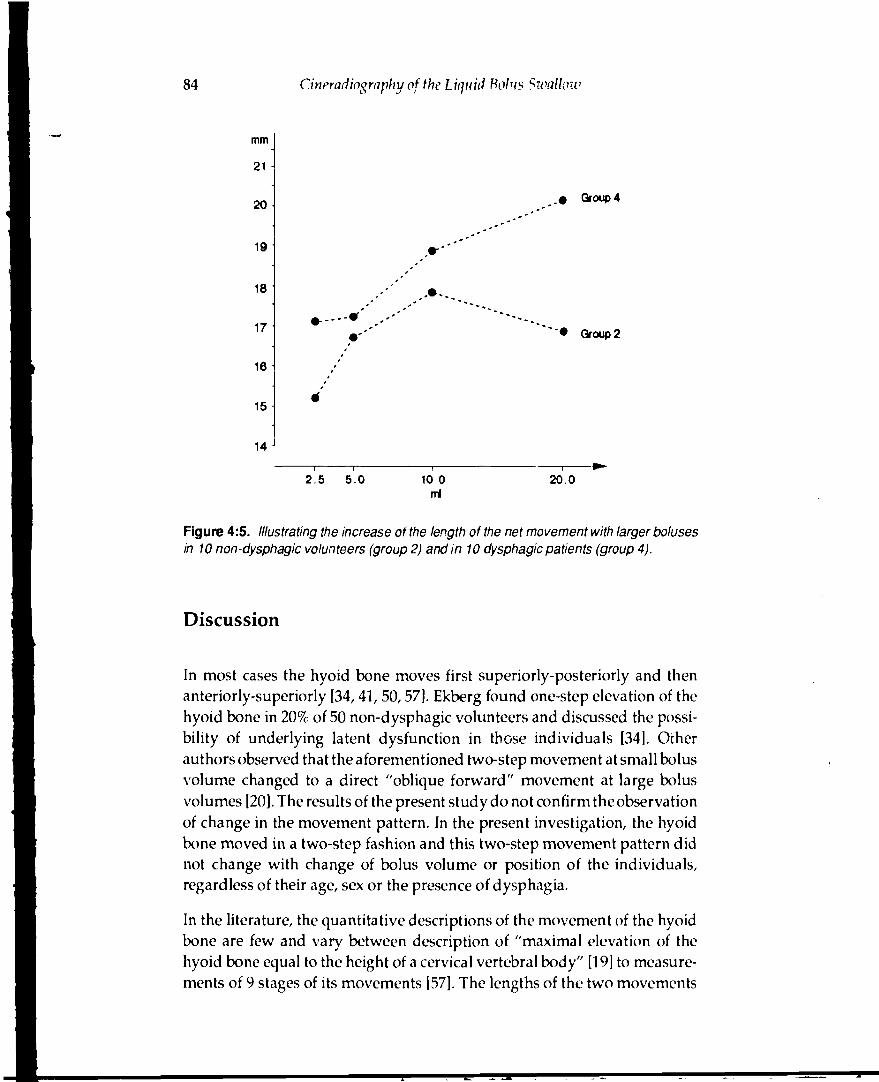
84 Cineradiography of the Liquid Bolus Swallow
mm
21 -I
20
19
18
17 1
16
15
14
Group 4
" • Group 2
2.5 5.0 10.0ml
20.0
Figure 4:5. Illustrating the increase of the length of the net movement with larger bolusesin 10 non-dysphagic volunteers (group 2) and in 10 dysphagic patients (group 4).
Discussion
In most cases the hyoid bone moves first superiorly-posteriorly and thenanteriorly-superiorly [34,41, 50,57]. Ekberg found one-step elevation of thehyoid bone in 20% of 50 non-dysphagic volunteers and discussed the possi-bility of underlying latent dysfunction in those individuals [34]. Otherauthors observed that the aforementioned two-step movement at small bolusvolume changed to a direct "oblique forward" movement at large bolusvolumes [20]. The results of the present study do not confirm the observationof change in the movement pattern. In the present investigation, the hyoidbone moved in a two-step fashion and this two-step movement pattern didnot change with change of bolus volume or position of the individuals,regardless of their age, sex or the presence of dysphagia.
In the literature, the quantitative descriptions of the movement of the hyoidbone are few and vary between description of "maximal elevation of thehyoid bone equal to the height of a cervical vertebral body" [19] to measure-ments of 9 stages of its movements [57]. The lengths of the two movements

4. Movement of the Hyoid Bone during Swalluiv 85
of the hyoid bone found in the present investigation are in agreement withobservations of Ekberg [34] who measured the same two movements usingthe lower margin of the mandible and the cervical spine as the referencepoints.
In the present investigation, the length of the first movement of the hyoidbone did not significantly change with increasing bolus volumes, in contrastto ihe length of the second movement which increased significantly withlarger bolus volumes in both groups (p = 0.009 and p = 0.006 respectively).In addition, the trend to increase the second movement with larger boluseswas highly significantly (p =0.001) parallel between the two groups. Thehere found difference between the two movements in response to changesin bolus volume gives rise to a hypothesis that there are different neuralcontrol mechanisms for each movement.
Two principal theories have been proposed to describe the neural mecha-nisms that control the oral and pharyngeal phases of swallowing. The centralpattern generator hypothesis [6] explains the control of swallowingsequences to be effected from neurons in the medulla in a stereotype pre-programmed manner, not influenced by any sensory feed-back. The reflexchain hypothesis [8] explains the swallowing sequence as a chain of linkedreflexes evoked by the bolus moving through the pharynx. This secondhypothesis presumes, consequently, a sensory feedback. Some authors com-bine both theories and suggest that "although swallowing is a centrallyprogrammed motor sequence, the inputs from the peripheral receptors cancontrol the complete motor sequence through sensory feedback" [58].
Concerning the movement of the hyoid bone, the present results support thelatter combined theory by suggesting the existence of a pre-programmedcontrol for its first movement and a regulated control for its second move-ment. The non-responsiveness found here of the first movement to changesin boids volume suggests that the first movement is controlled from themedulla in a pre-programmed way. Functionally, the first movement maybe seen as a preparatory stage of swallowing when the hyoid bone elevatestogether with larynx. A normally completed first movement prepares ana-tomical conditions for the subsequent increase in the width of thepharvngeallumen as well as for the folding down of the epiglottis. It is conceivable thatthis preparatory stage does not require adaptation to the bolus, but isinitiated and exerted in a stereotype manner. However, when the bolusenters the pharynx, the pharynx must adapt to its size. It is thereforeconceivable that the second movement which occurs during the passage ofthe bolus is the movement that adapts to the size or consistency of the bolus

86 Cineradiography of the Liquid Bolus Sicallow
[11,59]. It is the second movement that increases the anteroposterior diame-ter of the pharynx, thereby making it possible to accommodate boluses ofdifferent size.
The hypothesis proposed here of the regulatory nature cf the second move-ment is supported by previous observations of an increase of the AP diameterof the pharyngeal lumen with larger boluses [20] as well as results showingthat the extent of anterior movement of the larynx appears to be related tothe size and consistency of the bolus swallowed [16]. Also Dodds andco-workers [11] have found an increase of the superior an • anterior hyoidbone movements with increasing bolus volumes. However, they [11]measured the two perpendicular components of the entire movement andnot the two true partial movements with diffferent directions — the first andthe second. Therefore, they were unable to observe that the initial part of themovement is unaffected by the increase in bolus volume, in contrast to thesignificant increase of the second movement of the hyoid bone. Consequentlyihe possibility of two different neural control mechanisms responsible forthe entire movement could not be suggested from their results. Similarly,measurements of only the net movement, as measured in the present inves-tigation, could not result in this hypothesis.
The net movement increased in both the non-dysphagic volunteers (group2) and the dysphagic patients (group 4) but the increase was only statisticallysignificant in the group of dysphagic patients (p = 0.03). The ne movementis not a true movement but the result of two movements with differentdirections. The lower significance (p = 0.03) of dependence on the bolusvolume of the net movement as compared with that of the second movement(p = 0.009 and p = 0.006 respectively) demonstrates lower sensitivity of thisparameter in the evaluation of possible disturbances in function of themuscles attached to the hyoid bone. It also demonstrates the necessity ofmeasuring the true movements of the hyoid bone, the first and the second,
The present investigation c i not disclose any differences in the length of thefirst movement of the hyoid bone between the two groups (2 and 4) ofsubjects examined. The fact that the lengths of the first movement weresimilar in both groups further supports the here advanced hypothesis thatthe first movement is a stereotyped movement, pre-programmed in the CNS.However, although the length of the second movement increased signifi-cantly in both the non-dysphagic volunteers (group 2, p = 0.009) and dy-sphagic patients (group 4, p = 0.006), the length of the second movement wassignificantly (p = 0.002) larger at all bolus volumes in the dysphagic patients

4. Moi'emcnt of the llyoid Bone during Sivallozv 87
(group 4). The explanation for the differences between the two groups in thelength of the second movement could, theoretically, be that the dysphagicpatients may have had some underlying, as yet undetected, disturbance intheir act of swallowing, which caused their dyspahgia. If these patients hada defect or delayed elevation and/or anterior movement of the larynx andpharynx, their anteroposterior diameter of the pharyngeal lumen might havebeen diminished. This, conceivably, might produce a feeling of obstructionor, maybe, even a painful unpleasant swallow, especially when larger bolusvolumes had to be transported through the pharynx., The possibility of sucha delayed or defective movement of the larynx in the initial slage of swal-lowing is suggested by a non-significantly (p = 0.07) lower ekvation of thelarynx at the moment when the bolus reached the level of valleculae in thesame individuals (group 4) compared with the non-dysphagic volunteers(group 2) (chapter 5). Further support for this possibility may be seen in thesignificantly (p = O.(X)l) lower elevation at the same moment in dysphagicthan in non-dysphagic patients both with and without pharyngeal dysfunc-tion (chapter 5). An increase, above that normally expected, in the anteriormovement of the hyoid bone would result in an additional increase in theAP diameter of the pharyngeal lumen and could conceivably compensatefor a defect or delay in elevation of the larynx and pharynx. It is thereforepossible that the dysphagic patients (group 4) voluntarily or involuntarilyincreased the contractions of their muscles including those attached to thehyoid bone. This would increase the anterior movement of the hyoid bonein an attempt to increase the diameter of the pharyngeal lumen and facilitatetransport of the bolus through the pharynx.
Conclusions
A hypothesis is advanced suggesting that the first posterior-superiormovement o. the hyoid bone is controlled from the CNS in a pre-pro-grammed stereotype manner without sensory feedback in contrast to thesecond anterosuperior movement which is regulated by sensory feedbackand adapts the pharyngeal lumen to the size of the bolus.
Measurement of the first posterosuperior movement and the second an-terosuperior movement is a more sensitive way of evaluating hyoid bonemovement compared with measurement of the net movement eitherdirectly or as its inferosuperior and posteroanterior components.

5. Elevation of the Larynx
Introduction
During the pharyngeal passage of the bolus the larynx moves superiorly andanteriorly [8,15,19, 20]. The superior movement shortens the craniocaudalpharyngeal dimension and plays a part in the important displacement of thelarynx away from the passing bolus, thereby protecting the laryngeal inlet.The anteriorly directed movement of the larynx increases the antero-poste-rior diameter of the pharyngeal lumen [20]. The extent of this latter move-ment has been reported to be related to the bolus size [16]. In addition, it isassumed that elevation of the larynx and pharynx is important in openingthe pharyngo-esophageal segment during swallowing [6, 8,15,60, 61]. Theanteriorly directed movement starts when the bolus reaches the level of thevalleculae, and maximal elevation is completed later in the pharyngeal phaseof swallowing [20]. After the completion of the swallow, the larynx andpharynx return to their resting positions. With repetitive swallowing ofliquids the larynx and pharynx are only slightly lowered between swallowsand do not return to the resting position [20]. The elevators of the pharynxinclude: the stylohyoid, stylopharyngeus, geniohyoid, mylohyoid and thy-rohyoid muscles, as well as both bellies of the digastric muscle, and also themiddle and inferior pharyngeal constrictors [62].
Mendelsohn and McConnel have stressed that an incomplete laryngealelevation is directly related to impaired passage of the bolus through thepharyngoesophageal segment and aspiration [63]. Adrian and Kemp [17]have stated that the closure of the laryngeal vestibule and of the vocal cordsis independent of the degree of the laryngeal elevation. This statement maypossibly apply to the lower part of the laryngeal vestibule and the vocalcords, which are closed by the intralaryngeal muscles. The upper part of thelaryngeal vestibule is closed from above by the base of epiglottis during itstilting, and this, in part, is executed by extralaryngeal muscles. In thisinvestigation, a hypothesis has been proposed that an adequate approxima-tion of the larynx and the hyoid bone is an important condition for propertilting of the epiglottis (Chapter 6). As elevation of the larynx is an integralcomponent of this approximation, it is conceivable that defective elevationof the larynx alone may contribute to a defective downfolding of the epiglot-

90 Cincradiography of the Liquid Bolus Swallow
tis. In this way the upper, subepiglottic part of the laryngeal vestibule wouldremain open to intrusion of the passing bolus material A possible connectionbetween the tilting of the epiglottis and laryngeal movement has also beensuggested earlier [46].
Among pharyngeal dysfunctions, those resulting in aspiration of food andliquids into the respiratory tract are the most dangerous as they may lead toserious pulmonary complications. These, fortunately, are rare althoughminor sequelae of such dysfunctions are not uncommon. For example, inpatients with pharyngeal dysfunction accompanied by aspiration, Ekbergand Hilderfors found bronchopneumonia and chronic pulmonary disease in13% and 10% of patients, respectively [64].
Buchholz and co-workers [46] suggested that increased upward and forward"displacement" of the larynx may compensate for a deficiency of epiglottictilting or of glottic closure. If the length of laryngeal elevation were depend-ent on the volume of a liquid bolus, then it would be possible to search foran optimal bolus volume that could be recommended to the patients toprevent aspiration or, at least, decrease its frequency. The investigation intowhether such a dependence exists has been one of the aims of this study.Another aim was the investigation of possible differences in the length oflaryngeal movement in patients with dysphagia alone, compared with thosewith cineradiographically demonstrable pharyngeal dysfunction. This wasin order to examine the question of whether there may be any underlyingneuromuscular dysfunction of the laryngeal elevators, which may explainswallowing complaints or cineradiographically demonstrated pharyngealdysfunction in these patients.
Therefore, a thorough study of the movement pattern of the larynx duringswallowing, under different conditions and in different groups of patients,with and without swallowing complaints, was conducted in an attempt toreveal:
• whether the bolus volume influences the elevation of the larynx in non-dysphagic volunteers and in dysphagic patients, respectively.
• whether the elevation of the larynx differs between individuals with andwithout pharyngeal dysfunction.
• whether there is a special time-relation between the onset of laryngeal andhyoidal elevation during swallowing in normal volunteers.

5. Elevation of the Larynx 91
Material and Methods
The material consisted of cineradiograms obtained from five groups ofindividuals.
Group 1:
Fifteen non-dysphagic volunteers without pharyngeal dysfunction swallow-ing boluses of 10 ml of contras* medium in upright, right decubitus horizon-tal and right decubitus with the table tilted head-down 30°, respectively.Their cineradiograms were evaluated foi time-relation of movements of thelarynx and of the hyoid bone.
Group 2:
Ten non-dysphagic volunteers without pharyngeal dysfunction swallowingboluses of 2.5, 5, 10 and 20 ml oi contrast medium in increasing order ofvolume. Their cineradiograms were evaluated for elevation of the larynx.
Group 3:
Sixty non-dysphagic patients swallowing a mouthful of contrast medium.Their cineradiograms were evaluated for elevation of the larynx, and pre-sence or absence of pharyngeal dysfunction.
Group 4:
Ten dysphagic patients without pharyngeal dysfunction swallowing bolusesof 2.5, 5.. 10, and 20 ml of contrast medium in increasing order of volume.Their cineradiograms were also evaluated for elevation of the larynx.
Group 7:
Seventy-five dysphagic patients swallowing a mouthful of contrast medium.Their cineradiograms were evaluated for elevation of the larynx, and pre-sence or absence of pharyngeal dysfunction.
The groups are described in further details in Material, p. 27.
Elevation of the larynx can be measured on the lateral view. The location ofthe vocal cords is visible on the cineradiogram as a difference in radiodensitybetween the soft tissue of the vocal cords and the air in the subglottic space.The movement of the larynx on swallowing follows a two-step pattern. The

92 Cineradiography of the Liquid Bolus Swallow
first step is an elevation. The second step is an anterosuperiorly directedmovement and it starts when the bolus has reached the level of the valleculae.
The laryngeal elevation was measured in the left lateral position with thelower surface of the vocal cords as the reference point. Measurements wereperformed at two stages in the act of swallowing: when the apex of the bolushad reached the level of the valleculae and at maximal elevation. Bothmeasurements were made with reference to the same caudal point in theresting position as shown in Figure 10, p. 36. The procedure of measuring thedistances is described in detail in Analytic methods, p. 38.
In the following text, the following three terms are used to denote twomeasured lengths of laryngeal elevation, and a calculated one: "elevation(vail)", "maximal elevation", and "percentage elevation (vail)".
The first, "elevation (val!)", denotes the length of laryngeal elevation fromthe resting position to the position the larynx had reached at the momentwhen the bolus reached the vallecular level.
The second, "maximal elevation", denotes the length of laryngeal elevationfrom the resting position to the maximum cephalad position attained duringswallowing.
The third term, "percentage elevation (vail)", expresses the percentage of thetotal laryngeal elevation that was achieved at the moment when the bolushad reached the valleculae.
The "percentage elevation (vail)" was calculated according to the formula:
% elevation{vall) -elevation{vall) x 100maximal elevation
and denoted: percentage elevation (vail).
Pharyngeal dysfunction was evaluated with respect to the established cri-teria and included registration of the following major dysfunctions: incoordi-nation of the cricopharyngeal muscle, paresis of the pharyngeal constrictormusculature, webs, and Zenker's diverticula. In order to investigate putativerelationships between possible defective movement of the larynx and therisk of aspiration of bolus material, pharyngo-laryngeal dysfunction wasdivided into the following two categories: (1) defective closure of the laryn-geal vestibule, which was represented by intrusion of bolus material into thelaryngeal vestibule and (2) aspiration of bolus material into the trachea. Thelatter represents the greater degree of insufficiency of the mechanismsprotecting the airways — the proper aspiration. Defective tilting of the

5. Elei'ation of the Larynx 93
epiglottis, which was present to a greater or lesser degree in each categoryof pharyngo-laryngeal dysfunction, was considered as an integral com-ponent of the dysfunction and was, therefore, not statistically evaluatedseparately.
The cineradiograms of the 15 non-dysphagic volunteers (group 1) werereviewed in order to reveal whether the elevation of the larynx started andended prior to, synchronously with, or after the beginning or end of the hyoidbone movement.
Results
Elevation of the Larynx when the Bolus Re? hed the Valleculae
The mec sured lengths of elevation (mean values and SD) for the two groups(groups 2 and 4) with respect to swallowed bolus volume are given in Table5:1, without adjustment for age.
The measured lengths of elevation of the larynx when the bolus reached thevallecula in 10 non-dysphagic volunteers (group 2) varied between 4 and 25mm and in 10 dysphagic patients (group 4) between 6 and 28 mm (notadjusted for age). In the further text, all results are presented after adjustmentfor age (sec p. 40). The laryngeal elevation in individual subjects is graphicallypresented in Figures 5:1a and lb.
Table 5:1. Elevation of the larynx (vallecula; maximal) in 20 individuals who swalloweddifferent bolus volumes. Elevation measured in mm; mean (SD). 10 non-dysphagicvolunteers (group 2) and 10 dysphagic patients (group 4), all without pharyngeal dysfunc-tion. Not adjusted for age.
Bolus volume
Group 2
Group 4
2.5
Vallecmean(SD)
15.2(6.1)
18.7(4.6)
ml
Maxmean(SD)
22.4(4.8)
24.0(5.9)
5 ml
Vallecmean(SD)
18.8(6.1)
15.8(4.4)
Maxmean(SD)
26.9(4.5)
25.3(4.4)
10ml
Vallecmean(SO)
16.4(6.1)
16.2(7.2)
Maxmean(SD)
26.4
(4-t)
26.1(8.1)
20
Vallecmean(SD)
14.0(5.1)
15.5(6.1)
ml
Maxmean(SD)
4.8(6.5)
27.3(9.9)

94 Cineradiography of the Liquid Bolus Swallow
mm
B mm
30
25 -
20 -
15 -
10 -
2.5
2.5
ml
5.0 10.0
20.0
20.0
ml
Figure 5:1. The elevation of the larynx when the bolus reaches the valleculae in individualsubjects at different bolus volumes, a) 10 non-dysphagic volunteers (group 2), b) 10dysphagic patients (group 4).

5. Elevation of the Larynx 95
Table 5:11. Elevation of the larynx whnn the bolus had reached the valleculae in 20individuals who swallowed different bolus volumes. Elevation measured in mm; mean (SD).10 non-dysphagic volunteers (group 2) and 10 dysphagic patients (group 4), all withoutpharyngeal dysfunction. Adjusted for age.
Bolus volume 2.5 ml 5 ml 10ml 20 ml
Group 2
Group 4
14.6(6.1)
19.3(4 6)
18.2(6.1)
16.4(4.4)
15.8(6.1)
16.8(7.2)
13.4(5.1)
16.1(6.1)
The volume of the bcius did not significantly influence the laryngeal eleva-tion when the bolus had reached the valleculae in the non-dysphagic volun-teers (group 2) or in the dysphagic patients (group 4), both of which swal-lowed the boluses in increasing order of volume (Table 5:11). The elevationof the larynx was not influenced by the increasing bolus volume in non-dys-phagic volunteers (group 2) and had a tendency to decrease with increasingbolus volume in dysphagic patients (group 4) (Table 5:11). This differencebetween healthy volunteers and dysphagic patients in response to an in-creased bolus volume was nearly significant (p - 0.07).
Intrapersonal variations in the laryngeal elevation had a tendency to besmaller in dysphagic patients (group 4) compared with non-dysphagicvolunteers (group 2) (Fig 5:1 a and b). The interpersonal variations withinthe groups with regard to the laryngeal elevation were similar.
Maximal Elevation of the Larynx
The measured maximal lengths of laryngeal elevation in 10 non-dysphagicvolunteers (group 2) varied between 12 and 33 mm and in 10 dysphagicpatients (group 4) between 11 and 40 mm (not adjusted for age). The meanvalues of the maximum elevation of the larynx are given in Table 5:1 (notadjusted for age) and in Table 5:111 (adjusted for age). The maximal elevation
Table 5:111. Maximal elevation of the larynx in 20 individuals who swallowed different bolusvolumes. Elevation measured in mm; mean (SD). 10 nondysphagic volunteers (group 2)and 10 dysphagic patients (group 4), all without pharyngeal dysfunction. Adjusted forage.
Bolus volume
Group 2
Group 4
2.5 ml
21.6(4.8)
24.9 (5.9)
5 ml
26.1(4.5)
26.1 (4.4)
10 ml
25.5(4.2)
26.9(8.1)
20 ml
24.0 (6.5)
28.1 (9.9)

96 Cineradiography of the Liquid Bolus Swallow
mm
B
10 -
mm
10 -
2.5
5.0 10.0
ml
5.0 10.0
20.0
20.0
ml
Figure 5:2. The maximal elevation of the larynx in individual subjects at different bolusvolumes, a) 10non-dysphagic volunteers (group 2), b) 10 dysphagic patients (group 4).

5. Elevation of the Larynx 97
of the larynx in individual subjects is graphically presented in Figures 5:2aand b. The volume of the bolus did not significantly influence the maximallaryngeal elevation in either of the groups which swallowed different bolusvolumes (groups 2 and 4). There was no significant difference in the maximallaryngeal elevation between non-dysphagic volunteers and dysphagicpatients (groups 2 and 4), both without pharyngeal dysfunction (Table 5:111).There were no significant differences in intra- or interpersonal variationsamong these groups (group 2 and 4) (Fig 5:2 a and b).
Percentage Elevation of the Larynx when the Bolus Reached the Valleculae
No significant difference in percentage elevation (vail) was found betweenthe non-dysphagic volunteers (group 2) and the dysphagic patients (group4) (Table 5:IV). The percentage elevat'on (vail) did not significantly changewith changes in bolus volume.
There was no statistically significant difference between men and women inthe length of the different movements at any bolus volume examined.
Table 5:1V. Percentage elevation (vail). The figures express, in per cent, the proportion ofmaximum laryngeal elevation achieved at the moment when the bolus reached thevalleculae (mean SD). 10 non-dysphagic volunteers (group 2) and 10 dysphagic patients(group 4), all without pharyngeal dysfunction. Adjusted for age.
Bolus volume 2.5 ml 5 ml 10 ml 20 ml
Group 2
Group 4
65.7(20.9)
79.1 (14.6)
68.9(18.4)
62.3(12.1)
60.9(18.2)
61.0(15.2)
57.7(21.1)
58.0(13.5)
Frequency of Pharyngeal Dysfunction
Dysfunction, except for defective closure of the laryngeal vestibule andparesis of the pharyngeal constrictor muscles, was significantly (p = 0.001)more frequent in dysphagic patients (group 7) than in non-dysphagicpatients (group 3) (Table 5:V). Aspiration of bolus material into the tracheaoccurred significantly more frequently in dysphagic patients —16% — thanin non-dysphagic patients — 0%. Incoordination of the cricopharyngealmuscle occurred in a significantly higher percent c( dysphagic patients —37% — than in the respective group of non-dysphagic patients —15%. The

98 Cineradiography of the Liquid Bolus Swallow
Table 5: V. Number and frequency (in per cent) of various types of pharyngeal dysfunctionsamong 60 non-dysphagic and 75 dysphagic patients (groups 3 and 7). P-value ofdifferences between the two groups.
Type of dysfunction
Any
Defective closure of thelaryngeal vestibule
Aspiration of bolus materialinto the trachea
Incoordination of thecricopharyngeal muscle
Paresis of the pharyngealconstrictor muscles
Others
Non-dysphagic patients(Group 3)
No.
18
10
0
9
0
0
%
30
17
0
15
0
0
Dysphagic patients(Group 4)
No.
45
22
12
28
7
4
%
60
29
16
37
9
5
p-value
0.001
(n.s.)
0.0001
0.006
0.08
(n.s.)
percent of individuals with any type of dysfunction was significantly higherin dysphagic patients — 607c — than in the respective group of non-dy-sphagic patients — 30%.
Elevation of the Larynx in Individuals with Pharyngeal Dysfunction
The values given in Table 5:VI show the laryngcal elevation separately forindividuals swallowing without signs of pharyngeal dysfunction and that ofpatients with defective closure of the laryngeal vestibule, aspiration of bolusmaterial into the trachea, incoordination of the cricopharyngeal muscle andparesis of the pharyngeal constrictor muscles, respectively.
From the given figures it can be seen that the elevation was less in patientswith all the mentioned forms of pharyngeal dysfunction than in the 72patients with normal swallowing.
The percentage difference between laryngeal elevation in dysphagic andnon-dysphagic individuals with a normal swallowing act and that of patientswith defective closure of the laryngeal vestibule, with aspiration of bolusmaterial into the trachea, and wkh cricopharyngeal incoordination, respec-tively, is given in Table 5:VI1. In this table also the p-values for the registereddifferences are given.

5. Elevation of the Larynx 99
Table 5:VI. Elevation of the larynx in individuals without dysfunction and in those withdysfunction without respect to the presence or absence of dysphagia (groups 3 and 7pooled).
Elevation
valleculae (mm) max (mm)
Type of dysfunction
percentageelevation (vail)
mean SD mean SD mean SD
None
Defective closure of thelaryngeal vestbule
Aspiration of bolus materialinto the trachea
Incoordination of thecricopharyngeal muscle
Paresis of the pharyngealconstrictor muscles
72 13.1 5.3 21.6 5.5 59.5
12
16.9
32 10.2 5.8 19.7 5.9 48.9 22.9
9.1 6.0 20.2 6.9 43.3 23.4
37 11.0 6.3 20.0 7.1 52.0 21.2
8.6 7.0 16.1 5.6 45.9 29.0
Individuals with aspiration of bolus material into the larynx had lowerlaryngeal elevation than individuals without dysfunction. The laryngealelevation when the bolus had reached the valleculae was 29% lower (p =0.03) when expressed in mm and 28% lower (p = 0.003) when expressed inper cent of maximal elevation (Table 5:VII). Also, in individuals with in-
•v
4 Table 5:VII. Differences and their p-values in elevation of the larynx between individualswith pharyngeal a/sfunction and those without dysfunction (groups 3 and 7). Differencesare expressed in per cent of the mean value for individuals without dysfunction (-signmeans lower).
Elevation
Type of dysfunction
PercentageValleculae (mm) Maximum (mm) elevation (vail)
N ditf% p-value diff% p-value diff% p-value
Defective closure of thelaryngeal vestibule 32
Aspiration of the bolus intothe trachea 12
Incoordination of.thecricopharyngeal muscle 37
-23 0.007
-29 0.03
-16 0.06 -7
ns
ns
-19 0.01
-28 0.003
i
ns -12.5 0.04

100 Cineradiography of the Liquid Bolus Swallow
coordination of the cricopharyngeal muscle, the laryngeal elevation whenthe bolus had reached the valleculae was 16% lower (p = 0.06), when ex-pressed in mm, than in individuals without dysfunction, and 12.5% lower(p = 0.04) v hen expressed in per cent of maximal elevation (Table 5:VII). Inindividuals with defective closure of the laryngeal vestibule the laryngealelevation when the bolus had reached the valleculae was 23% lower (p =0.007), when expressed in mm, than in individuals without dysfunction and19% lower (p = 0.01) when expressed in per cent of maximal elevation (Table
The differences in maximal laryngeal elevation between any group of thethree types of dysfunction, as compared with those without dysfunction,were not significant (Table 5.VII).
In all 12 individuals in group 7 with aspiration of bolus material into thetrachea, the elevation of the larynx occurred simultaneously with the firstmovement of the hyoid bone. In 10 of them, the maximum elevation of thelarynx was completed synchronously with the completion of the secondmovement of the hyoid bone, while in the remaining two elevation of thelarynx continued for a short instance.
The elevation of the larynx occurred simultaneously with the first movementof the hyoid bone in 14 of the 15 non-dysphagic volunteers (group 1). In theremaining volunteer elevation of the larynx occurred slightly before the firstmovement of the hyoid bone. Elevation of the larynx continued after the endof the second movement of the hyoid bone in six of the 15 non-dysphagicvolunteers (group 1). In the remaining nine non-dysphagic volunteers themaximal elevation of the larynx was achieved by the end of the secondmovement of the hyoid bone.
Discussion
The mean length of elevation of the larynx was approximately 2.5 cm (TableI) which is in good accordance with 2.0—2.5 cm found in a fluoroscopicanalysis of the upper esophageal sphincter during swallowing [12]. It is alsoroughly in accordance with the less precise assessments described in theliterature as "one and a half vertebral body" [41], "two cervical vertebralbodies" [62], and "the height of one vertebral body and disc space" [63]).
According to some authors, the elevation of the larynx and pharynx beginsas the bolus starts to move [19, 50]. Others found that the elevation of the
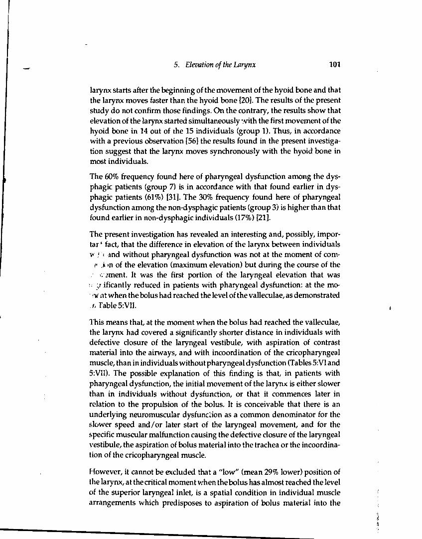
5. Elevation of the Larynx 101
larynx starts after the beginning of the movement of the hyoid bone and thatthe larynx moves faster than the hyoid bone [20]. The results of the presentstudy do not confirm those findings. On the contrary, the results show thatelevation of the larynx started simultaneously with the first movement of thehyoid bone in 14 out of the 15 individuals (group 1). Thus, in accordancewith a previous observation [56] the results found in the present investiga-tion suggest that the larynx moves synchronously with the hyoid bone inmost individuals.
The 60% frequency found here of pharyngeal dysfunction among the dys-phagic patients (group 7) is in accordance with that found earlier in dys-phagic patients (61%) [31]. The 30% frequency found here of pharyngealdysfunction among the non-dysphagic patients (group 3) is higher than thatfound earlier in non-dysphagic individuals (17%) [21].
The present investigation has revealed an interesting and, possibly, impor-tar*• fact, that the difference in elevation of the larynx between individualsv J i and without pharyngeal dysfunction was not at the moment of com-
/• i »n of the elevation (maximum elevation) but during the course of the•.\?ment. It was the first portion of the laryngeal elevation that was
) ;; ificantly reduced in patients with pharyngeal dysfunction: at the mo-<ye nt when the bolus had reached the level of the valleculae, as demonstrated./. Table 5:VII.
This means that, at the moment when the bolus had reached the valleculae,the larynx had covered a significantly shorter distance in individuals withdefective closure of the laryngeal vestibule, with aspiration of contrastmaterial into the airways, and with incoordination of the cricopharyngealmuscle, than in individuals without pharyngeal dysfunction (Tables 5:VI and5:VII). The possible explanation of this finding is that, in patients withpharyngeal dysfunction, the initial movement of the larynx is either slowerthan in individuals without dysfunction, or that it commences later inrelation to the propulsion of the bolus. It is conceivable that there is anunderlying neuromuscular dysfunction as a common denominator for theslower speed and/or later start of the laryngeal movement, and for thespecific muscular malfunction causing the defective closure of the laryngealvestibule, the aspiration of bolus material into the trachea or the incoordina-tion of the cricopharyngeal muscle.
However, it cannot be excluded that a "low" (mean 29% lower) position ofthe larynx, at the critical moment when the bolus has almost reached the levelof the superior laryngeal inlet, is a spatial condition in individual musclearrangements which predisposes to aspiration of bolus material into the

102 Cineradiography of the Liquid Bolus Swallow
airway. At that moment, the mechanisms protecting the airway have notaccomplished their function, i.e. closure of the laryngeal vestibule and tiltingof the epiglottis. This assumption could be supported by the hypothesisproposed in Chapter 6, which suggest that certain spatial conditions arerequired for correct tilting of the epiglottis during swallowing. Approxima-tion of the thyroid cartilage of the larynx to the hyoid bone is effected bysome of the muscles which are involved in laryngeal elevation. Therefore, itis possible that approximation of the thyroid cartilage to the hyoid bone isan important factor for proper epiglottic function and vestibular closing.
An alternative explanation for the observed lesser elevation of the larynx atthe moment when the bolus reaches the level of the valleculae could be thattht bolus reaches the level of the valleculae earlier in patients with dysfunc-tion, while the movement of the larynx itself— both its starting synchroni-zation and speed of elevation — is similar to that of individuals withoutdysfunction. This would imply that, in patients with pharyngeal dysfunc-tion, either the initial speed of the bolus is higher or that there is dyscoordi-nation of the swallowing act with precocious propulsion of the bolus.
It has been suggested that the elevation of the larynx, particularly its antero-superior movement, depends on the size of the bolus [12,16]. Results of thepresent study do not confirm such a dependence as neither the maximumelevation of the larynx nor its elevation at the moment when the bolusreaches the valleculae changed significantly with changes in bolus volume.However, in the present study the length of elevation was measured in theresultant direction and not as separate superior and anterior components ofthis movement. The movement of the larynx is closely linked with themovement of the hyoid bone. As the second movement of the hyoid bone,anteriorly and slightly superiorly, significantly increases with larger bolusvolume, as shown in the present investigation (Chapter 4), it is possible thatthe anterior component of the laryngeal movement may also increase withincreasing bolus volume.
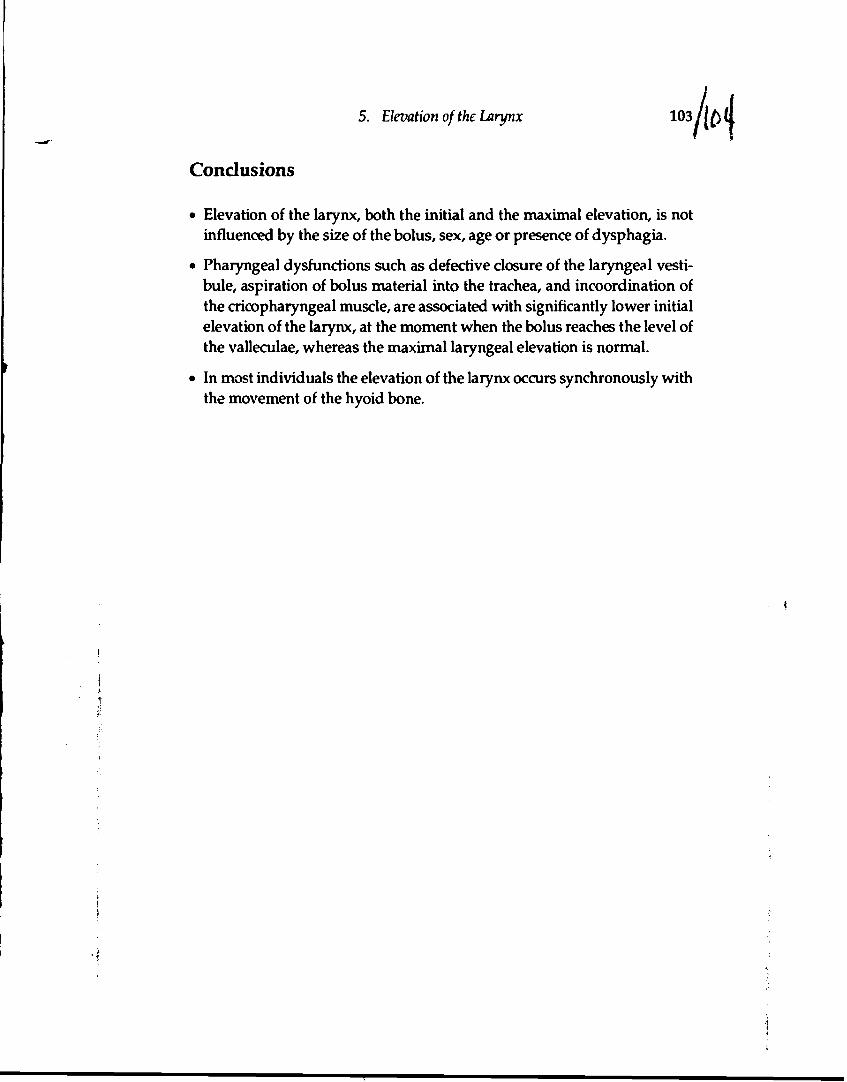
5. Elevation of the Larynx 103
Conclusions
• Elevation of the larynx, both the initial and the maximal elevation, is notinfluenced by the size of the bolus, sex, age or presence of dysphagia.
• Pharyngeal dysfunctions such as defective closure of the laryngeal vesti-bule, aspiration of bolus material into the trachea, and incoordination ofthe cricopharyngeal muscle, are associated with significantly lower initialelevation of the larynx, at the moment when the bolus reaches the level ofthe valleculae, whereas the maximal laryngeal elevation is normal.
• In most individuals the elevation of the larynx occurs synchronously withthe movement of the hyoid bone.
M

6. Epiglottic Dysmobility
Introduction
During swallowing the airways are protected against aspiration by a com-plex series of events. Initially in the act of swallowing the vocal cords areforced tightly together. This happens synchronously with the first swallow-ing movements of the tongue and the elevation of the soft palate.
When the vocal cords have been closed together the muscles of the false cords(lateral thyroarytenoid muscles) are contracted, and the supraglottic portionof the laryngeal vestibule and the laryngeal ventricular sinuses are dosed.The remaining portion of the laryngeal vestibule—the subepiglottic portion— stays open until the bolus has reached the level of the valleculae (Fig 4, p.13). At this moment the epiglottis is tilted down to an inverted position withits tip in the esophageal inlet. The subepiglottic portion of the laryngealvestibule is thereby compressed and obliterated by the epiglottic lid.
The epiglottic movement during swallowing is performed in a two-stepfashion. At first the epiglottis is moved from its upright resting position to atransverse position in the hypopharynx, perpendicular to the longitudinalaxis of the pharynx. During this phase of the movement the epiglottis retainsits form, with a convexity which is initially posteriorly-directed, but which,on assumption of the transverse position, becomes caudally-directed.
After a very short moment in this position, and synchros ~ily with initiationof the peristalsis in the pharyngeal constrictors, the epiglottis is tilted furtherdown with its tip in the esophageal inlet. During this second phase ofepiglottic movement the form of the epiglottis is changed. Its convexity isreshaped to a concavity and this caudally-directed concavity fits closely theary-region. This results in a watertight closure of the laryngeal vestibule andno portion of the bolus can enter the laryngeal inlet.
Thus, at swallowing the larynx and the laryngeal vestibule are successivelyclosed from below. The vocal cords and the supraglottic portion of thelaryngeal vestibule are compressed from side to side. The subepiglotticportion of the laryngeal vestibule is compressed in the transverse plane bythe inverted and down-tilted epiglottis.

106 Cineradiography of the Liquid Bolus Swallow
After the act of swallowing and the passage of the bolus the vocal cords andthe laryngeal vestibule are reopened more or less synchronously. Theepiglottis is very quickly tilted back to its resting position.
An impaired function of the epiglottis — an absent, delayed or incompletetilting down at swallowing — is in this report defined as "epiglottic dysmobil-ity". In colloquial medical terminology epiglottic dysmobility is termed"pseudobulbar palsy". (The reason for this term is the very common episodesof aspiration of bolus material into the airway in this condition).
Epiglottic dysmobility is very common. Ekberg and co-workers [28] reportedthat it is found in 33% of all patients who are referred for a radiologicalexamination because of dysphagia. They also reported that epiglottic dys-mobility was found in 9% of non-dysphagic individuals subjected to acineradiography [21].
The more detailed patho-physiological mechanisms underlying this dis-order are still unclear and have baffled researchers [16,20,22,65,66].
In epiglottic dysmobility the subepiglottic portion of the laryngeal vestibuleremains open, or is closed too late during the act of swallowing. As aconsequence of this defect, bolus material will enter the laryngeal vestibuledown to the level of the false cords. When the larynx — the vocal cords andthe false cords—reopens after the act of swallowing, this bolus material willbe aspirated down into the airway.
An epiglottic dysmobility may be slight or severe, i.e. a smaller or largerportion of the bolus may enter the laryngeal vestibule during swallowing,and a smaller or larger part of this material may then be aspirated into thelarynx and the trachea.
Furthermore, an epiglottic dysmobility will imply that bolus remnants,instead of being cleared out and collected in the valleculae by the retilting ofthe epiglottis will remain in the hypopharynx after swallowing. Fragmentsof this bolus material may also be aspirated into the airway after a swallow-ing act.
Defective swallowing movements of the hyoid bone and /or defective eleva-tion of the larynx during the act of swallowing are factors that in severalreports have been suggested as responsible for epiglottic dysmobility [20,30,34,46],
A careful comparative study of the spatial relationships between the hyoidbone and the thyroid cartilage during swallowing, in individuals with and t

6. Epiglottic Dysmobility 107
without epiglottic dysmobility, may yield valuable information about thepatho-physiological mechanisms underlying this disorder.
Therefore, the purpose of this study was to measure the distance betweenthe hyoid bone and the thyroid cartilage during swallowing, in a group ofdysphagic patients with normal epiglottic mobility and in another groupwith epiglottic dysmobility. This would allow assessment of the degree ofapproximation of the thyroid cartilage to the hyoid bone during swallowingand thus determination of whether any differences in these measurementscould help to explain the background of epiglottic dysmobility.
Material and Method
The material consisted of cineradiograms obtained from two groups ofindividuals.
Group 8:
Nineteen dysphagic patients, with normal tilting of the epiglottis, swallow-ing a mouthful of contrast medium.
Group 9:
Fifteen dysphagic patients, with dysmobility of the epiglottis, swallowing amouthful of contrast medium.
Epigiottic dysmobility was defined as any defective tilting of the epiglottisranging from unchanged upright position (no tilting at all) to horizontalposition (perpendicular to the long axis of the pharynx). Downfolding of theepiglottis caudal to the horizontal position was considered normal tilting.
The groups are described in further details in Material, p. 27.
The approximation of the thyroid cartilage towards the hyoid bone wasmeasured in the left lateral projection using the anterior superior portion ofthe thyroid cartilage and the most anterior portion of the hyoid bone as thereference points. The measurements were performed at two stages in the actof swallowing, as described on p. 36 and as shown in Fig 11.
Maximum and minimum distance between the thyroid cartilage and thehyoid bone during swallowing was measured, in mm, as described inAnalytic methods, p. 38. The size of body parts and also the distance between

108 Cineradiography of the Liquid Bolus Swallow
the thyroid cartilage and the hyoid bone vary among individuals; it wastherefore decided to eliminate the influence of such a variation by using arelative measure of approximation between the two structures.
For that purpose the percentage approximation between the thyroid car-tilage and the hyoid bone during swallowing was calculated according tothe formula:
„ . maximum distance - minimum distance ^nn
% Approx = : T7- x 100rr maximum distance
Results
The maximum distance in resting position between the thyroid cartilage andthe hyoid bone varied between 12 and 40 mm in 19 dysphagic patients withnormal tilting of the epiglottis (group 8) and between 20 and 38 mm in 15dysphagic patients with defective tilting of the epiglottis (group 9). Min-imum distance (at maximal approximation) between the thyroid cartilageand the hyoid bone varied between 1.5 mm and 16.5 mm in patients withnormal tilting of the epiglottis (group 8), and between 5.5 and 29 mm inpatients with epiglottic dysmobility (group 9). The percentage approxima-tion of the thyroid cartilage to the hyoid bone in individual subjects in groups8 and 9 is graphically presented in Fig 6:1.
The mean values of percentage approximation for each group are given inTable 6:1. From this table it is clear that the percentage approximation of thethyroid cartilage to the hyoid bone was significantly (p = 0.05) higher inpatients with normal tilting of the epiglottis than in those with defectivetilting of the epiglottis — 72% and 54% respectively.
Table 6:1. The percentage approximation (range, mean) and its p-value in dysphagicpatients with normal tilting of the epiglottis (group 8) and in dysphagic patients withdysmobility of the epiglottis (group 9)
Group
8
9
No. patients
19
15
Percentage approximation
range
48.5-93.3
23.9-74.4
mean
72.0
54.4
P-value
>0.05
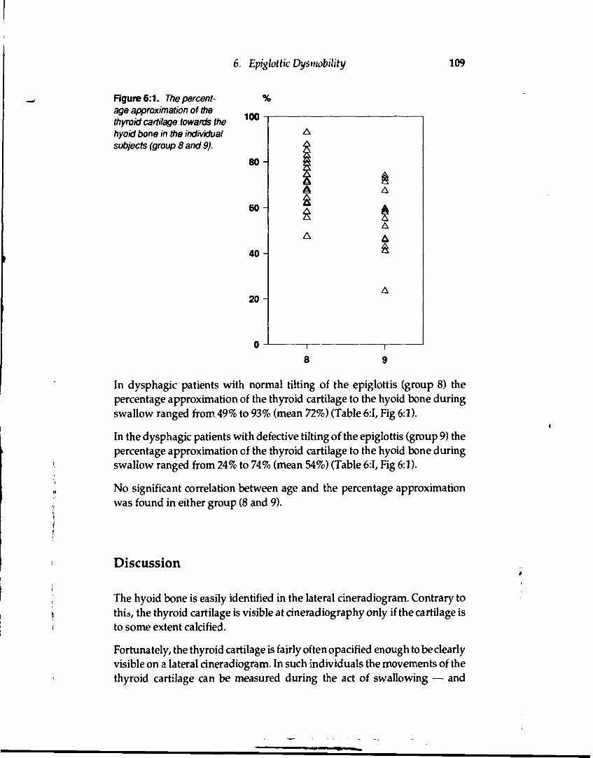
6. Epiglottic Dysmobility 109
Figure 6:1. The percent-age approximation of thethyroid cartilage towards thehyoid bone in the individualsubjects (group 8 and 9).
8
In dysphagic patients with normal tilting of the epiglottis (group 8) thepercentage approximation of the thyroid cartilage to the hyoid bone duringswallow ranged from 49% to 93% (mean 72%) (Table 6:1, Fig 6:1).
In the dysphagic patients with defective tilting of the epiglottis (group 9) thepercentage approximation of the thyroid cartilage to the hyoid bone duringswallow ranged from 24% to 74% (mean 54%) (Table 6:1, Fig 6:1).
No significant correlation between age and the percentage approximationwas found in either group (8 and 9).
Discussion
The hyoid bone is easily identified in the lateral cineradiogram. Contrary tothia, the thyroid cartilage is visible at cineradiography only if the cartilage isto some extent calcified.
Fortunately, the thyroid cartilage is fairly often opacified enough to be clearlyvisible on a lateral cineradiogram. In such individuals the movements of thethyroid cartilage can be measured during the act of swallowing — and

110 Cineradiography of the Liquid Bolus Swallow
variations of the distance between the hyoid bone and the thyroid cartilagecan also be measured.
The forces and movements responsible for proper tilting of the epiglottis arenot fully understood. The few existing descriptions and theories of epiglotticfunction do not offer a complete explanation for the entire cycle of movementand reshaping of the epiglottis.
The initiation of epiglottic tilting is, in the opinion of some authors, causedby the motion of the tongue [19,21,35] whereas, according to others, by theelevation of the larynx [22, 50, 65, 66]. Elongation of the hyoepiglotticligament and compression of the median thyrohyoid fold secondary toextreme thyrohyoid approximation have been suggested as alternatives forthe mechanisms usually invoked in the downfolding of the epiglottis, suchas pressure by the base of the tongue, swallowed bolus, or tension from thearyepiglottic muscles [22].
A short survey of the pertinent anatomy is necessary for a proper under-standing of the dynamics of epiglottic dysfunction.
The epiglottic cartilage has the shape of a tennis racket. The handle of theracket — the petiolus — is firmly fixed to the posterior aspect of the thyroidcartilage by the thyro-epiglottic ligament (Fig 6:2)
The epiglottis is also connected to the hyoid bone. The fan-shaped hyo-epiglottic ligament extends posteriorly from the hyoid bone, attaching to theanterior aspect of the epiglottic lid (Fig 6:2). Furthermore, the epiglottis isattached to the tongue by the medial glosso-epiglottic plica.
Only two small paired muscles are attached to the epiglottis. The ary-epiglot-tic muscles run cranially in the margins of the aryepiglottic folds, from theary-region of both sides, to attach to the lateral margins of the epiglottic lid.A contraction of these muscles will pull the epiglottis caudally down overthe ary region.
The thyro-epiglottic muscles are thin muscles running from the internalsurface of the thyroid cartilage in a cranio-dorsal direction to attach to themargins of the epiglottic lid. According to anatomical text-books the actionof the thyro-epiglottic muscles is thought to be downward-pulling of theepiglottis. However, if the epiglottis is in its upright resting position acontraction of these muscles is more likely to retain the epiglottis in thisposition.
However, the muscles attached to the epiglottis would seem too small andtoo weak to execute the very fast and precise movements the epiglottis

6. Epiglottic Dysmobility 111
Figure 6:2. The epiglottis isattached to the thyroid car-tilage by the thyroepiglotticligament and to the hyoidbone by the hyoepiglotticligament.
Hyoepiglotticligament
Thyroepiglotticligament
Figure 6:3. (A) the thyroidcartilage, (B) the hyoidbone, (C) the tip of theepiglottis. An approximationof the thyroid cartilage tothe hyoid bone resulting in achange of the position of theepiglottis.
exhibits during the act of swallowing. This conclusion is in agreement withthe opinion expressed in other reports in the literature [22]. Therefore, itseems reasonable to assume that the swallowing movements of the epiglottisare secondary to those of anatomical structures to which the epiglottis isattached. According to such a theory the function of the ary-epiglottic andthyro-epiglottic muscles would rather be to reshape the epiglottis to anappropriate form for its different positions at rest and during swallowing.
Movement of the epiglottis is dependent on its anatomical connection to thehyoid bone and the thyroid cartilage and, consequently movement of these

112 Cineradiography of the Liquid Bolus Swallow
structures is transmitted to the epiglottis. Also, changes in the mutual spatialpositions of the hyoid bone and the thyroid cartilage may also change theposition of the epiglottis. Theoretically, an approximation of the thyroidcartilage to the hyoid bone would change the position of the epiglottis froman upright position to a transverse one (Fig 6:3).
If this hypothesis is true then a defective mutual movement pattern of thehyoid bone and the thyroid cartilage during swallowing would result inepiglottic dysmobility.
From the results obtained in the present investigation it is clear that theapproximation of the thyroid cartilage to the hyoid bone during swallowingwas much larger in patients with normal epiglottic function than in thosewith epiglottic dysmobility. The difference was significant (p = 0.05) andsuggests that an approximation less than 49% is inconsistent with normalfunction of the epiglottis during swallowing. However, it is also clear thatepiglottic dysmobility may appear in patients with an approximation as highas 74%.
The net conclusion that can be drawn from the obtained results is that a 49%approximation of the thyroid cartilage to the hyoid bone during swallowingis a prerequisite for a normal epiglottic function but that other factors alsomay be involved to produce epiglottic dysmobility.
Elevation of the thyroid cartPage towards the hyoid bone is the result ofcontraction of the thyrohyoi. uscle, together with synergistic contractionsof the inferior pharyngeal constrictor and stylopharyngeal muscles. Since thestylopharyngeal muscle attaches to the margins of the thyroid cartilage aswell as to the margins of the base of the epiglottis, it may help to elevate theepiglottis. This muscle helps to elevate the epiglottis by pulling the base ofthe epiglottis backwards simultaneously with the elevation of the thyroidcartilage. Such an elevation of the base of the epiglottis, at the moment of itstransverse position, would change the geometric orientation of the directionof contractile force of the thyroepiglottic muscle. Earlier in this report it hasbeen stressed that this muscle has the function of keeping the epiglottisupright in resting position between swallows. When the thyroid cartilagehas been approximated to the hyoid bone and the epiglottis thereby hasattained a transverse position in the hypopharynx, the action of the musclemust be changed. In the transverse position of the epiglottis, contraction ofthe thyroepiglottic muscle will pull the epiglottis further downwards to aninverted position in the esophageal inlet (Fig 6:4a— c).

6. Epiglottic Dysmobility 113
B
Figure 6:4. Schematic drawing, il-lustrating that an approximation of thethyroid cartilage (1) to the hyoidbone(2) will change the direction of contrac-tile force of the thyroepiglottic muscle(3) resulting in a further downfoldingof the epiglottis (black).
At the same time there is a remodelling of the epiglottic lid from a cranialconcave to a cranial convex shape. In this context it is important to stress thatthe epiglottic lid consists of elastic cartilage with a preformed shallowcup-shape. Such structures have two steady states. This remodelling mayconceivably be performed by contraction of the same muscle.
This above-proposed hypothetical mechanism may be involved in the poorlyunderstood final stage of epiglottic downfolding and reshaping, as it addsthe force of the thyroepiglottic muscle to the force of the aryepiglottic muscle,which by itself is considered incapable of downfolding the epiglottis.

114 Cineradiography of the Liquid Bolus Swallow
The high values of percentage thyro-hyoid approximation obtained inseveral patients with epiglottic dysmobility (Fig 6:1) do not necessarilycontradict the necessity of high grade approximation for proper epiglottictilting. These patients may have been suffering from essentially isolateddysfunction of muscles tilting the epiglottis, including paresis or dyscoordi-nation, while the function of muscles elevating the thyroid cartilage waspreserved.
Theoretically, there may be a variety of patho-physiological and morpho-logic changes underlying an epiglottic dysmobility. Some of the patients withlow values may have had a general impairment of the pharyngeal andlaryngeal -teuromuscular function including both the muscles elevating thethyroid cardlage and those actively tilting the epiglottis.
This theoretical assumption could be supported by earlier findings presentedin this thesis, namely that the degree of elevation of the larynx, at the instantwhen the bolus reaches the level of the valleculae, was significantly lower (p= 0.05) among patients with pharyngeal dysfunction when compared withthose without pharyngeal dysfunction (chapter 5).
It could, theoretically, be that since the larynx may elevate later in patientswith epiglottic dysmobility, as discussed in chapter 5, the bolus has alreadyreached beyond the level of the laryngeal vestibule at the time of attainmentof the transverse position by the epiglottis. In such a situation, an alternativeor supportive mechanism for downfolding of the epiglottis, such as pressurefrom the base of the tongue and from the swallowed bolus, would act toolate to help in the tilting of the epiglottis over the laryngeal introitus. Thus,the result may be that the epiglottis will remain in a transverse position,regardless of the outcome of the final approximation of the thyroid cartilagetowards the hyoid bone.
Such a theoretical assumption could explain the fact that epiglottic dysmo-bility may even occur in patients with high grade approximation of thethyroid cartilage towards the hyoid bone. However, no patient in the mate-rial with an approximation of more than 74% had any signs of epiglotticdysmobility.
In conclusion, a high grade approximation between the thyroid cartilage andthe hyoid bone occurs during normal swallowing with a normal tilting of theepiglottis. This high grade approximation is the basis for a hypothesis of anactive role of the thyroepiglottic muscle together with the aryepiglotticmuscle in the end-stage of downfolding and reshaping of the epiglottisduring swallowing.
6. Epiglottic Dysmobility
Conclusion
• The proper downfolding of the epiglottis during swallowing is, in part,dependent on the degree of approximation of the thyroid cartilage to thehyoid bone.

Summary of the Results
Differences, correlations and dependences given in the further text are allstatistically significant
With increasing volume of the liquid bolus
• the speed of the peristaltic wave
- was unchanged in both r.on-dysphagic and dysphagic individualswithout pharyngeal dysfunction (Chapter 1)
• the speed of the apex of the bolus
- increased in non-dysphagic individuals
- had a tendency (p = 0.09) to increase in dysphagic individuals (Chap-ter 2)
• the first, posterosuperior movement of the hyoid bone
- was unchanged in non-dysphagic and dysphagic individuals (Chap-ter^
? • the second, anterosuperior movement of the hyoid bonei-
j - increased in non-dysphagic and dysphagic individuals (Chapter 4)
• the net movement of the hyoid bone
- increased in dysphagic individuals
- was unchanged in non-dysphagic individuals (Chapter 4)
• the movement of the hyoid bone
- did not change its two-step movement pattern in non-dysphagic o:dysphagic individuals (Chapter 4)
• the elevation of the larynx
- was unchanged in non-dysphagic and dysphagic individuals (Chap-; ter 5)

118 Cineradiography of the Liquid Bolus Swallow
With change in posture from erect to horizontal, and to 30° head downpositions
• the speed of the peristaltic wave decreased in non-dysphagic individuals(Chapter 1)
• the speed of the apex of the bolus was unchanged in non-dysphagicindividuals (Chapter 2)
• the movement of the hyoid bone did not change its two-step movementpattern in non-dysphagic individuals (Chapter 4)
The time interval between swallows
• did not influence the speed of the peristaltic wave or the speed of the apexof the bolus in dysphagic individuals (Chapter 1 and 2)
There was no correlation between the speed of the peristaltic wave and the speed ofthe apex of the bolus at any bolus volume in non-dysphagic and dysphagicindividuals or at any posture in non-dysphagic individuals (Chapter 3)
Between non-dysphagic and dysphagic individuals, both without pharyngealdysfunction, there were no differences:
• in the speed of the peristaltic wave (Chapter 1)
• in the speed of the apex of the bolus (Chapter 2)
• in the first and net movement of the hyoid bone (Chapter 4)
• in the elevation of the larynx (Chapter 5)
except for
• the second, anterosuperior movement of the hyoid bone which was largerin the dysphagic than in the non-dysphagic individuals (Chapter 4).
• the intrapersonal variations in the speed of the peristaltic wave which weresignificantly higher in the dysphagic than in the non-dysphagic individu-als (Chapter 1).
The frequency of pharyngeal dysfunction, such as aspiration of bolus materialinto the trachea or incoordination of the cricopharyngeal muscle, was higherin dysphagic than non-dysphagic individuals (Chapter 5).

Summary of the Results 119
When individuals were pooled without respect to the presence or absenceof dysphagia, the elevation of the larynx, at the moment when the bolus reachedthe level of valleculae, was lower in individuals with defective closure of thelaryngeal vestibule, in those with aspiration of bolus material into thetrachea, or those with incoordination of the cricopharyngeal muscle than inindividuals without pharyngeal dysfunction. There was no correspondingdifference in the net, total laryngeal elevation (Chapter 5).
The approximation between the thyroid cartilage and the hyoid bone was greater inindividuals with normal tilting of the epiglottis than in those with defectivetilting of the epiglottis (Chapter 6).
With increasing age of the individuals
• the speed of the apex of the bolus decreased (Chapter 2)
• the second movement of the hyoid bone decreased (Chapter 4)
• the speed of the peristaltic wave was unchanged (Chapter 1)
• the first and the net movement of the hyoid bone were unchanged (Chap-ter^
• the elevation of the larynx was unchanged (Chapter 5)
There was no difference between males and females in any of the measuredor calculated parameters.

Conclusions
Based on the results of the present investigation, the following conclu-sions can be drawn
Measurements of the speed of the peristaltic wave can be carried outprovided that magnification factor and film speed are known (Chapter 1)
Measurement of the speed of the peristaltic wave can be useful in theclinical evaluation of pharyngeal function as it potentially may disclose apharyngeal dysfunction (Chapter 1).
Measurements of the speed of the apex of the bolus can be carried out,provided that magnification factor and film speed are known. The signif-icance of these measurements cannot be determined unless the bolusvolume, patient age and position of the patient at the time of the examina-tion are known (Chapter 2).
The peristaltic wave is not the major force that propels the initial part of aliquid bolus through the pharynx (Chapter 3).
Measurement of the first posterosuperior movement and the second an-terosuperior movement is a more sensitive way of evaluating hyoid bonemovement compared with measurement of the net movement eitherdirectly or as its inferosuperior and posteroanterior components (Chapter4).
A short initial elevation of the larynx seem to be a predisposing factor fordefective closure of the laryngeal vestibule with risk for subsequent aspira-tion of bolus material into the airways (Chapter 5).
The proper downfolding of the epiglottis during swallowing is, in part,dependent on the degree of approximation of the thyroid cartilage to thehyoid bone (Chapter 6).

Conclusions 121
Based on the results in the present investigation, the following conclu-sions for further research can be drawn.
• An abnormally high or low speed of the peristaltic wave can be the earlysign of pharyngeal dysfunction. However, to define normal speed limits adedicated study of a large number of individuals is needed (Chapter 1).
• When selecting patient material for further research, consideration shouldbe given to the fact that the age of the patient may influence the resultsobtained. If selection is not feasible, then the results should be correctedfor age (Chapter 1—5).

Hypothesis
Based on the results in the present investigation, the following hypotheseshave been proposed:
• The speed of the peristaltic wave is controlled from the swallowing centersin a preprogrammed pattern which may, however, respond to certainafferent impulses such as those associated with changes in posture (Chap-ter 1).
• The speed with which the apex of a liquid bolus is propelled through thepharynx is partly the result of pressure from the base of the tongue,whereas the speed of the tail of the bolus through the pharynx is likely theresult of the peristaltic wave (Chapter 2).
• The first posterosuperior movement of the hyoid bone is controlled by theCNS in a pre-programmed stereotype manner without sensory feedbackin contrast with the second anterosuperior movement which is regulatedby sensory feedback and adapts the pharyngeal lumen to the size of thebolus (Chapter 4).

Acknowledgements
I wish to express my thanks to Docent Olle Ekberg, the initiator of thisresearch, to Professor Torsten Almén for his encouraging support, to DocentGöran Nyländer for valuable counsel and support, to Miss Eva Prahl forexcellent secretarial and administrative assistance as well as for her con-tagious encouraging optimism and to Gregory McGinn, MD, and JeffreyCrabbe, MB, Ch B, both at University of Michigan, Ann Arbor, for theirrevision of the English text.

References
1 Ekberg O, Olsson R, Sundgren-Borg-ström P: Relation between bolus sizeand pharyngeal swallow. Dysphagia3:69-72,1988.
2 Borgström PS, Ekberg O: Peristalsis inpharyngeal constrictor musculature inrelation to positioning and gravity.Acta Radiologica 30:183-185,1989.
3 Borgström PS, Ekberg O: Speed of per-istalsis in pharyngeal constrictormusculature; Correlated to age. Dy-sphagia 2:140-144,1988.
4 Birch-Iensen M, Borgström PS, EkbergO: Cineradiography in closed and openpharyngeal swallow. Acta RadiolDiagn 29:407-410,1989.
5 Ekberg G Closure of the laryngeal ves-tibule during deglutition. ActaOtolaryngol 93:123-129,1982.
6 Miller AJ: Deglutition. Physiol Rev62:129-184,1982.
7 Miller AJ: Neurophysiological basis ofswallowing. Dysphagia 1:91-100,1986.
8 Doty RW: Neural organization of de-glutition. In: Code CF (ed) Handbookof physiology. Alimentary Canal, sect6, vol 4, Washington DC: AmericanPhysiological Society 4:1861-1902,1968.
9 Dodds WJ: The physiology of swallow-ing. Dysphagia 3:171-178,1989.
10 Dodds WJ, Stewart ST, Logeman JA:Physiology and radiology of the nor-mal oral and pharyngeal phases ofswallowing. Am J Radiol 154:953-963,1990.
11 Dodds W, Man KM, Cook IJ, KahrilasPJ, Stewart ET, Kemp FH: Influence ofbolus volume on swallow-inducedhyoid movement in normal subjects.AJR 150:1307-1309,1988.
12 Kahrilas PJ, Dodds WJ, Dent J, Lo-geman J A, Shaker R: Upper esophageal
sphincter function during deglutition.Gastroenterology 95:52-62,1988.
13 Cook IJ, Dodds WJ, Dantas RO, KernMK, Massey BT, Shaker R, Hogan WJ:Timing of videofluoroscopic, mano-metric events, and bolus transit duringthe oral and pharyngeal phases ofswallowing. Dysphagia 4:8-15,1989.
14 Shedd D, Kirchner J, Scatliff J: Oral andpharyngeal components of deglutition.ArchSurg 82:371-380,1961.
15 Bosma JF: Deglutition: Pharyngealstage. Physiol Rev 37:275-300,1957.
16 Ardran GM, Kemp FH: The protectionof the laryngeal airway during swal-lowing. Br J Radiol 25:406-416,1952.
17 Ardran GM, Kemp FH: Closure andopening of the larynx during swallow-ing. Br J Radiol 29:205-208,1956.
18 Ardran GM, Kemp FH: The mecha-nism of the larynx. II. The epiglottis andclosure of the larynx. Br J Radiol40:372-389,1967.
19 Saunders JB, Davis C, Miller ER: Themechanism of deglutition (2nd stage)as revealed by cineradiography. AnnOto-Rhino-Laryngol 60:897-916,1951.
20 Ramsey GH, Watson JS, Cramiak R,Weinberg SA: Cinefluorographicanalysis of the mechanism of swallow-ing. Radiology 64:498-518,1955.
21 Ekberg O, Nyländer G: Cineradiogra-phy on the pharyngeal stage of deglu-tition in 150 individuals without dy-sphagia. Br J Radiol 55:253-257,1982.
22 Fink BR, Martin RW, Rohrmann CA:Biomechanics of the human epiglottis.Acta Otolaryngol 87:554-559,1979.
23 Kahrilas PJ, Dodds WJ, Dent J, HoganWJ, Tarbis SL: The dynamics ofpharyngeal and upper esophagealsphincter function during deglutition(abst). Gastroenterology 92:1458,1987.
References 125
24 Cook IJ, Dodds WJ, Dantas R, MasseyB, Kem MK, Lang IM, Brasseur JG,Högan WJ: Opening mechanisms of thehuman upper esophageal sphincter.Am J Physiol 257 (Gastrointest LiverPhysiol 20)748-759,1989.
25 Buchholz D: Neurologic causes of dy-sphagia. Dysphagia 1:152-156,1987.
26 Buchholz D: Neurologic evaluation ofdysphagia. Dysphagia 1:187-192,1987.
27 Lindgren S, Janzon L: The prevalenceof swallowing complaints and clinicalfindings among 50-79 year old menand women in an urban population.Dysphagia. To be published.
28 Ekberg O, Nyländer G. Cineradiogra-phy of the pharyngeal stage of degluti-tion in 250 patients with dysphagia. BrJ Radiol 55:258-262,1982.
29 Borgström PS, Ekherg O: Pharyngealdysfunction in the elderly. J Diagnhnag 2:74- 81,1988.
30 Borgström PS, Birch-Iensen M, EkbergO: Radiology of the pharynx and oe-sophagus in young adults. Br J Radiol61:909-913,1988.
31 Ekberg O, Wahlgren L: Dysfunction ofpharyngeal swallow. A cineradio-graphic study in 854 dysphagialpatients. Acta Radiol Diagn 26:389-395,1985.
32 Edwards DAW: Discriminatory valueof symptoms in differential diagnosisof dysphagia. Clin Gastroenterol 5: 49-57,1976.
33 Ekberg O: Cineradiography in normaland abnormal pharyngo-esophagealdeglutition. Thesis, Malmö 1981.
34 Ekberg O: The normal movements ofthe hyoid bone during swallow. InvestRadiol 21.408-410,1986.
35 Christrup J: Normal swallowing offoodstuffs of pasty consistence. A cine-fluorographic investigation of a normalmaterial. Dan Med Bull 11:79-91,1964.
36 Curtis DJ, Cruess DF, Dachman AH,Maso E: Timing in the normal pharyn-geal swallow. Invest Radiol 19:523-529,1984.
37 Atkinson M, Kramer P, Wyman SM,Ingelfinger FJ: The dynamics of swal-lowing. I. Normal pharyngeal mecha-nisms. J Clin Invest 36:581-588,1957.
38 McConnel FMS, Cerenko D, JacksonRT, Guffin Jr TN: Timing of majorevents of pharyngeal swallowing. ArchOtolaryngol Head Neck Surg114:1413-1418,1988.
39 McConnel FMS, Cerenko D, Mendel-sohn MS: Manofluorographic analysisof swallowing. Otolaryngol Clin NorthAm 21:625-634,1988.
40 Fischer MA, Hendrix TR, Hunt JN,Murrills AJ: Relation between volumeswallowed and velocity of the bolusejected from the pharynx into theesophagus. Gastroenterology 74:1238-1240,1978.
41 Roberts RI: A cineradiographic inves-tigation of pharyngeal deglutition. Br JRadiol 30:449^160,1957.
42 Winer BJ, Statistical principles for ex-perimental design. New York,McGraw-Hill, 1971.
43 Doty RW, Bosma J, : An electromyo-graphic analysis of reflex deglutation. JNeurophysiol 19:44-60,1956.
44 Hellemans J, Pe'.emans W, VantrappenG: Pharyngoesophageal swallowingdisorders and the pharyngoesophagealsphincter. Med Clin North Am65:1149-1171,1981.
45 Dodds WJ, Högan WJ, Lydon SB,Stewert ET, Steef JJ, Amdorfer RC:Quantitation of pharyngeal motorfunction in normal human subjects. JAppl Physiol 39:692-696,1975.
46 Buchholz DW, Bosma JF, Donner MW:Adaptation, compensation and decom-pensation of the pharyngeal swallow.Gastrointest Radiol 10:235-239,1985.
47 Herz Silverman E, Elfant IL: Dyspha-gia: An evaluation and treatment pro-gram for the adult. Am J Occup Ther33:382-390,1979.
48 Ekberg O, Wahlgren L: Pharyngealdysfunctions and their interrelation-ship in patients with dysphagia. ActaRadiol Diagn 26:659-664,1985.

126 Cineradiography of the Liquid Bolus Swallow
49 Rushmer RF, Hendron JA: The act ofdeglutition: A cinefluorographicstudy. J Appl Physiol 3:622-630,1951.
50 Curtis DJ, Cruess DF, Dachman AH:Normal erect swallowing. Normalfunction and incidence of variations.Invest Radiol 20:717-726,1985.
51 Palmer JB, Tanaka E, Siebens AA: Mo-tions of the posterior pharyngeal wallin swallowing. Laryngoscope 98:414-417,1988.
52 Johnson LH, Lin C, Hong SK: Gastroe-sophageal dynamics during immer-sion in water to the neck. J Appl Physiol38:449-454,1975.
53 Cerenko D, McConnel FMS, JacksonRT: Quantitative assessment ofpharyngeal bolus driving forces.Otolaryngol Head Neck Surg 100:57-63,1989.
54 McConnel FMS: Analysis of pressuregeneration and bolus transit duringpharyngeal swallowing. Laryngoscope98:71-78,1988.
55 Ingervall B, Bratt CM, Carlsson GE,Helkimo M, Lantz B: Positions andmovements of mandible and hyoidbone during swallowing. Acta OdontScand 29:549-562,1979.
56 Shelton RL, Bosma JF, Sheets BV:Tongue, hyoid and larynx displace-ment in swallow and phonation. J ApplPhysiol 15:283-288,1960.
57 YotsuyaH,NonakaK, Ide Y: Studies onpositional relationships of the move-ment of the pharyngeal organs duringdeglutition in relation to the cervicalvertebrae by X- ray TV cinematogra-
phy. Bull Tokyo Dent Coll 22:159-170,1981.
58 Jean A: Control of the central swallow-ing program by inputs from the periph-eral receptors. J Auto Nerv System10:225-233,1984.
59 Dantes RO, Dodds WJ, Massey BT,Kem MK: The effect of high vs lowdensity barium preparations on thequantitative features of swallowing.Am J Radiol 153:1191-1195,1989.
60 Goyal RK: Disorders of the cri-copharyngeus muscle. Gin North Am17:115-130,1984.
61 Ekberg O: Elevation of the pharynx andthe width of the pharyngo-esophageaalsegment during swallow. Acta RadiolDiagn 27:293-295,1986.
62 Curtis DJ: Laryngeal dynamics. CRCCrit Rev Diagn Imaging 18:29-80,1982
63 Mendelsohn MS, McConnel FMS:Function in the pharyngeal oe-sophageal segment. Laryngoscope97:483-489,1987.
64 Ekberg O, Hilderfors H: Defective clo-sure of the laryngeal vestibule:Frequency of pulmonary complica-tions. Am JRadiol 145:1159-1164,1985.
65 Curtis DJ: Epiglottic motion: Video re-cording of muscular dysfunction. Radi-ology 148:473-477,1983.
66 Fink BR: Folding mechanism of thehuman larynx. Acta Otolaryngol78:124-128,1974.

Abstract
In the evaluation of the dysphagic patient, radiology is crucial as a techniquefor monitoring morphology and function. In particular, high-speed cine-radiography can reveal a variety of pharyngal dysfunctions. However, in theliterature and in practice the difference between normal and abnormalfunction is not always clear. This monograph is based on high-speed cine-radiographies of swallowing in 75 non-dysphagic volunteers and in 189dysphagic patients.
The purpose was to study whether differences in bolus volumes, patientposition, age, and gender had any effects on the following parameters: thespeed of the peristaltic wave and apex of the liquid barium bolus; the lengthof movement and the movement pattern of the hyoid bone and larynx; andepiglottic function. Also differences in these parameters between non-dys-phagic and dysphagic individuals were analyzed.
The study disclosed that the speed of the bolus, as well as the anterior-supe-rior movement and net movement of the hyoid bone increased significantlywith larger bolus volumes. The position of the individual in relation togravity significantly influenced the speed of peristalsis. In most of themeasured parameters there were no differences between non-dysphagic anddysphagic individuals except for differences in the intrapersonal variationsand in the anterior-superior movement of the hyoid bone. In patients withpharyngeal dysfunction the initial stage of the elevation of the larynx wassignificantly slower than in patients without dysfunction. The approxima-tion of the thyroid cartilage to the hyoid bone was significantly greater inindividuals with normal epiglottic function than in those with epiglotticdysmobility.
It is suggested that abnormal speed of peristalsis may be a mild form ofdysfunction. Measurements of the aforementioned speeds and movementscan be done if bolus volume, age and position of the patient, film speed, andmagnification factors are known.
Hypotheses concerning epiglottic function and central control of swallowingare proposed.
![Kinetic analysis of the metabotropic glutamate subtype 5 tracer [18F]FPEB in bolus and bolus-plus-constant-infusion studies in humans](https://img.pdfslide.net/doc/110x75/6345f1b338eecfb33a06ca2e/kinetic-analysis-of-the-metabotropic-glutamate-subtype-5-tracer-18ffpeb-in-bolus.jpg)

![Effect of amphetamine on [18F]fallypride in vivo binding to D2 receptors in striatal and extrastriatal regions of the primate brain: Single bolus and bolus plus constant infusion studies](https://img.pdfslide.net/doc/110x75/6345ca5edf19c083b10839ea/effect-of-amphetamine-on-18ffallypride-in-vivo-binding-to-d2-receptors-in-striatal.jpg)


