Embed Size (px)
Citation preview
Vascular Biology, Atherosclerosis and Endothelium Biology
Histidine-Rich Glycoprotein Modulates theAnti-Angiogenic Effects of Vasculostatin
Philip A. Klenotic,* Ping Huang,† Juan Palomo,‡
Balveen Kaur,§ Erwin G. Van Meir,¶
Michael A. Vogelbaum,‡ Maria Febbraio,*Candece L. Gladson,† and Roy L. Silverstein*From the Departments of Cell Biology, * and Cancer Biology,†
and the Brain Tumor and Neuro-Oncology Center,‡ Lerner
Research Institute, Cleveland Clinic, Cleveland, Ohio; the
Dardinger Laboratory for Neuro-Oncology and Neurosciences,§
the Ohio State University, Columbus, Ohio; and the Department
of Neurosurgery, Hematology/Medical Oncology and Winship
Cancer Institute,¶ Emory University, Atlanta, Georgia
Brain angiogenesis inhibitor 1 (BAI1) is a transmem-brane protein expressed on glial cells within thebrain. Its expression is dramatically down-regulatedin many glioblastomas, consistent with its functionalability to inhibit angiogenesis and tumor growth invivo. We have shown that the soluble anti-angiogenicdomain of BAI1 (termed Vstat120) requires CD36, acell surface glycoprotein expressed on microvascularendothelial cells (MVECs), for it to elicit an anti-an-giogenic response. We now report that Vstat120 bind-ing to CD36 on MVECs activates a caspase-mediatedpro-apoptotic pathway, and this effect is abrogated byhistidine-rich glycoprotein (HRGP). HRGP is a circu-lating glycoprotein previously shown to function as aCD36 decoy to promote angiogenesis in the presenceof thrombospondin-1 or �2. Data here show thatVstat120 specifically binds HRGP. Under favorableMVEC growth conditions this interaction allows che-motactic-directed migration as well as endothelialtube formation to persist in in vitro cellular systems,and increased tumor growth in vivo as demonstratedin both subcutaneous and orthotopic brain tumormodels, concomitant with an increase in tumor vas-cularity. Finally, we show that HRGP expression isincreased in human brain cancers, with the proteinheavily localized to the basement membrane of thetumors. These data help define a novel angiogenicaxis that could be exploited for the treatment of hu-man cancers and other diseases where excess angio-genesis occurs. (Am J Pathol 2010, 176:2039–2050; DOI:10.2353/ajpath.2010.090782)
Angiogenesis is the process through which new bloodvessels are formed from pre-existing capillaries. In adult-hood the vasculature is normally quiescent, a homeo-static state that is maintained by a precise balance ofpro-angiogenic inducers such as vascular endothelialgrowth factor and anti-angiogenic inhibitors such asthrombospondin (TSP)�1 and �2. This angiostatic bal-ance can be physiologically regulated in favor of newblood vessel formation during processes such as men-struation and wound healing allowing normal tissue re-generation; while pathological disruption of this balanceis associated with many disease states, including dia-betic retinopathy, atherosclerosis-induced tissue ische-mia, chronic inflammation, tumor growth and metastasis,obesity, asthma, and several autoimmune diseases.1
Therefore, it is extremely important to identify and dissectthe pathways involved in vessel growth in both normaland aberrant conditions.
Neovascularization is a major component of malignanttumor growth and many therapeutic strategies have beendeveloped to inhibit tumor angiogenesis, including anti-bodies or decoys that bind and neutralize vascular en-dothelial growth factor,2,3 small molecule inhibitors ofgrowth factor signaling pathways,4 and peptides basedon the anti-angiogenic type I repeat domains (TSR) ofTSP-1.5–7 Studies on the mechanisms of TSP-mediatedanti-angiogenesis revealed that the TSR domains play anessential role8 and that the type B scavenger receptorCD36 functions as the critical endothelial cell surfacereceptor.9,10 Using mouse corneal pocket angiogenesisassays we recently demonstrated that CD36 also func-tions as the receptor for a 120 kDa anti-angiogenic frag-ment derived from an unrelated TSR-containing protein,Brain Angiogenesis Inhibitor 1 (BAI1). This fragment,known as vasculostatin or Vstat120, suppressed neoves-sel formation in corneas from wild-type mice yet no effectwas observed in CD36 null animals, showing for the first
Supported by National Institutes of Health grants HL67839 (to R.L.S.) andCA86335 (to E.G.V.M.). P.A.K. was supported by the American HeartAssociation grant 0525334B.
Accepted for publication December 14, 2009.
Address reprint requests to Roy L. Silverstein, M.D., Department of CellBiology, Lerner Research Institute, Cleveland Clinic, Cleveland, OH44195. E-mail: [email protected].
The American Journal of Pathology, Vol. 176, No. 4, April 2010
Copyright © American Society for Investigative Pathology
DOI: 10.2353/ajpath.2010.090782
2039
time that a TSR-containing protein distinct from TSP-1and �2 mediates its anti-angiogenic functions throughinteractions with CD36.11
BAI1 is a 1584-aa brain-specific protein predicted tohave seven transmembrane segments and a large extra-cellular domain. The extracellular domain contains anRGD integrin recognition motif, a putative hormone re-ceptor (HomR) domain, and five TSR domains. Its ex-pression is down-regulated in glioblastomas12 and in-versely correlated with vascularity and metastasis incolorectal cancer,13 consistent with an anti-angiogenicrole. Kaur et al14 have shown that the TSR-containingfragment, Vstat120, was released from the cell mem-brane via proteolytic cleavage at a G protein–coupledreceptor cleavage site and that this fragment inhibitedmicrovascular endothelial cell (MVEC) proliferation, mi-gration, and tube formation equivalent to that seen withfull-length BAI1. Strikingly, restoration of Vstat120 expres-sion in human glioma cells suppressed tumorigenicity andvascularity and enhanced animal survival in subcutaneousand orthotopic tumor implantation models in nude mice.11
The binding site on CD36 for the TSR domains of TSP-1and �2 and Vstat120 has been localized to amino acids93 to 12011,15,16 within a highly conserved region termedthe CLESH domain (CD36, LIMP2, EMP structural homol-ogy – see Figure 1A). CLESH domains are also found inproteins genetically distinct from the CD36 family,17 in-cluding some, such as HIV gp120 and histidine-rich gly-coprotein (HRGP) which are known to bind TSP-1. HRGP,
a 75-kDa glycoprotein synthesized in the liver, circulatesin the blood at moderately high concentrations (approx-imately 100 to 150 �g/ml), and is secreted from activatedplatelets. In addition to TSP-1 and �2, it binds a widerange of ligands including divalent metal cations andseveral components of the provisional matrix laid downby healing wounds and tumors where angiogenesis isprevalent (for review see18). It has been implicated in thecoagulation and fibrinolytic systems, and high levelshave been associated with thrombotic disorders.19,20
Work in our lab has shown that HRGP acts as a CD36decoy to promote angiogenesis by binding TSP-1 and�2 and thereby preventing them from binding to CD36on the endothelial cell surface.21,22 Because both HRGPand CD36 compete for the identical binding region inTSP-1 and �2, we hypothesized that the relative concen-trations of ligands, receptor, and decoy within a givenmicroenvironment would ultimately determine the degreeof angiogenesis. Within tumors and other areas of chronicinflammation, HRGP can be released from activatedplatelets as well as deposited from plasma that gainedaccess via poorly organized hyperpermeable vessels.The angiogenic balance would then be tipped to favorangiogenesis by HRGP binding to TSPs as well as po-tentially other TSR containing anti-angiogenic proteins.
In this article we show that the CLESH domain of HRGPbinds Vstat120 and suppresses its anti-angiogenic activ-ity by reversing inhibition of endothelial cell migration andtube formation. Furthermore, we show in both subcuta-neous and orthotopic brain tumor models that HRGPexacerbates glioblastoma tumor growth and enhancestumor vascularity. We also show that the amount of HRGPpresent in human brain is increased in patients with pri-mary tumors with the protein localized predominantlywithin the basement membrane. Finally, we offer someinsight into the mechanism of action of Vstat120 by show-ing caspase-3 activation and endothelial cell apoptosison Vstat120 addition. Together these results suggest thatdeposition of HRGP into angiogenic microenvironments,perhaps as the result of the inherent “leakiness” of theneo-vasculature and/or of platelet granule release, canmodulate the anti-angiogenic processes mediated by thegeneral family of TSR-containing proteins, and may shedlight on the mechanism of angiogenesis regulation in thebrain.
Materials and Methods
Antibodies and Reagents
Mouse anti–glutathione S-transferase (GST) monoclonalantibody (MAB3317) was purchased from Chemicon In-ternational (Millipore – Billerica, MA). Anti-human HRGPmonoclonal antibody (MAB1869) was purchased fromR&D Systems (Minneapolis, MN) and anti-human N-termi-nal BAI1 polyclonal antibody was previously described.14
Rabbit anti-human caspase-3 antibody (#9662) was pur-chased from Cell Signaling Technology (Beverly, MA).Horseradish peroxidase (HRP)-conjugated anti-rabbitF(ab�)2 from donkey, HRP anti-mouse IgG-linked whole
HRGP1 100 200 300 400 507
CLESH
CLESH
Cystatin
HRR
CLESH
CD36
GST-HRGP(155-213)
GST-HRGP(330-389)
GST-HRGP(428-502)
GST-
GST--
1 100 200 300 400 471
93 120
GST
-155
-213
GST
-330
-389
GST
-428
-502
GST
MW49
3425
8549
34
25
Vstat120 -
-+ Vstat120 in CM+ + + +
GST
-428
-502
GST
-330
-389
GST
-155
-213
GS
T
GST
-428
-502
GST
A
CB
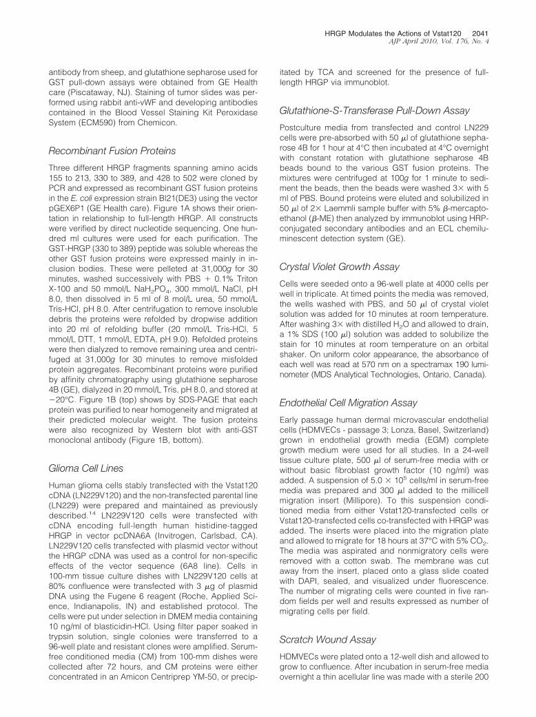
Figure 1. HRGP CLESH domain fusion protein specifically precipitatesVstat120 from tumor cell postculture media. A: HRGP/GST fusion proteins.Three recombinant GST fusion proteins were designed using fragments ofthe HRGP protein spanning amino acids 155 to 213 (within the secondcystatin domain), amino acids 330 to 389 (the HRR), and amino acids 428 to502 (CLESH domain). The linear structure of CD36 with the CLESH domainhighlighted is shown below for reference. B: Coomassie stained gel (toppanel) showing the purified GST-HRGP protein fragments after glutathionesepharose chromatography. The bottom panel shows Western blot analysisof each fusion protein probed with anti-GST antibody. C: Western blotanalysis of a GST pull-down assay. The three GST-HRGP fusion proteins andGST alone were bound to glutathione sepharose beads, and CM fromLN229V120 glioma cells were added to each sample. Each precipitate wasthen probed using the Vstat120/BAI1-specific antibody. Vstat120 was onlydetected when the CLESH domain (HRGP 428 to 502) was used in thepull-down and not the cystatin (HRGP 155 to 213) or the HRR (HRGP 330 to389) domains. HRGP 428 to 502 only precipitated Vstat120 when CM fromVstat120-transfected LN229 cells (� lanes) was used. No protein was de-tected when CM from the nontransfected parent cells (� lane) was used. Thisis a representative image from n � 3 experiments.
2040 Klenotic et alAJP April 2010, Vol. 176, No. 4
antibody from sheep, and glutathione sepharose used forGST pull-down assays were obtained from GE Healthcare (Piscataway, NJ). Staining of tumor slides was per-formed using rabbit anti-vWF and developing antibodiescontained in the Blood Vessel Staining Kit PeroxidaseSystem (ECM590) from Chemicon.
Recombinant Fusion Proteins
Three different HRGP fragments spanning amino acids155 to 213, 330 to 389, and 428 to 502 were cloned byPCR and expressed as recombinant GST fusion proteinsin the E. coli expression strain Bl21(DE3) using the vectorpGEX6P1 (GE Health care). Figure 1A shows their orien-tation in relationship to full-length HRGP. All constructswere verified by direct nucleotide sequencing. One hun-dred ml cultures were used for each purification. TheGST-HRGP (330 to 389) peptide was soluble whereas theother GST fusion proteins were expressed mainly in in-clusion bodies. These were pelleted at 31,000g for 30minutes, washed successively with PBS � 0.1% TritonX-100 and 50 mmol/L NaH2PO4, 300 mmol/L NaCl, pH8.0, then dissolved in 5 ml of 8 mol/L urea, 50 mmol/LTris-HCl, pH 8.0. After centrifugation to remove insolubledebris the proteins were refolded by dropwise additioninto 20 ml of refolding buffer (20 mmol/L Tris-HCl, 5mmol/L DTT, 1 mmol/L EDTA, pH 9.0). Refolded proteinswere then dialyzed to remove remaining urea and centri-fuged at 31,000g for 30 minutes to remove misfoldedprotein aggregates. Recombinant proteins were purifiedby affinity chromatography using glutathione sepharose4B (GE), dialyzed in 20 mmol/L Tris, pH 8.0, and stored at�20°C. Figure 1B (top) shows by SDS-PAGE that eachprotein was purified to near homogeneity and migrated attheir predicted molecular weight. The fusion proteinswere also recognized by Western blot with anti-GSTmonoclonal antibody (Figure 1B, bottom).
Glioma Cell Lines
Human glioma cells stably transfected with the Vstat120cDNA (LN229V120) and the non-transfected parental line(LN229) were prepared and maintained as previouslydescribed.14 LN229V120 cells were transfected withcDNA encoding full-length human histidine-taggedHRGP in vector pcDNA6A (Invitrogen, Carlsbad, CA).LN229V120 cells transfected with plasmid vector withoutthe HRGP cDNA was used as a control for non-specificeffects of the vector sequence (6A8 line). Cells in100-mm tissue culture dishes with LN229V120 cells at80% confluence were transfected with 3 �g of plasmidDNA using the Fugene 6 reagent (Roche, Applied Sci-ence, Indianapolis, IN) and established protocol. Thecells were put under selection in DMEM media containing10 ng/ml of blasticidin-HCl. Using filter paper soaked intrypsin solution, single colonies were transferred to a96-well plate and resistant clones were amplified. Serum-free conditioned media (CM) from 100-mm dishes werecollected after 72 hours, and CM proteins were eitherconcentrated in an Amicon Centriprep YM-50, or precip-
itated by TCA and screened for the presence of full-length HRGP via immunoblot.
Glutathione-S-Transferase Pull-Down Assay
Postculture media from transfected and control LN229cells were pre-absorbed with 50 �l of glutathione sepha-rose 4B for 1 hour at 4°C then incubated at 4°C overnightwith constant rotation with glutathione sepharose 4Bbeads bound to the various GST fusion proteins. Themixtures were centrifuged at 100g for 1 minute to sedi-ment the beads, then the beads were washed 3� with 5ml of PBS. Bound proteins were eluted and solubilized in50 �l of 2� Laemmli sample buffer with 5% �-mercapto-ethanol (�-ME) then analyzed by immunoblot using HRP-conjugated secondary antibodies and an ECL chemilu-minescent detection system (GE).
Crystal Violet Growth Assay
Cells were seeded onto a 96-well plate at 4000 cells perwell in triplicate. At timed points the media was removed,the wells washed with PBS, and 50 �l of crystal violetsolution was added for 10 minutes at room temperature.After washing 3� with distilled H2O and allowed to drain,a 1% SDS (100 �l) solution was added to solubilize thestain for 10 minutes at room temperature on an orbitalshaker. On uniform color appearance, the absorbance ofeach well was read at 570 nm on a spectramax 190 lumi-nometer (MDS Analytical Technologies, Ontario, Canada).
Endothelial Cell Migration Assay
Early passage human dermal microvascular endothelialcells (HDMVECs - passage 3; Lonza, Basel, Switzerland)grown in endothelial growth media (EGM) completegrowth medium were used for all studies. In a 24-welltissue culture plate, 500 �l of serum-free media with orwithout basic fibroblast growth factor (10 ng/ml) wasadded. A suspension of 5.0 � 105 cells/ml in serum-freemedia was prepared and 300 �l added to the millicellmigration insert (Millipore). To this suspension condi-tioned media from either Vstat120-transfected cells orVstat120-transfected cells co-transfected with HRGP wasadded. The inserts were placed into the migration plateand allowed to migrate for 18 hours at 37°C with 5% CO2.The media was aspirated and nonmigratory cells wereremoved with a cotton swab. The membrane was cutaway from the insert, placed onto a glass slide coatedwith DAPI, sealed, and visualized under fluorescence.The number of migrating cells were counted in five ran-dom fields per well and results expressed as number ofmigrating cells per field.
Scratch Wound Assay
HDMVECs were plated onto a 12-well dish and allowed togrow to confluence. After incubation in serum-free mediaovernight a thin acellular line was made with a sterile 200
HRGP Modulates the Actions of Vstat120 2041AJP April 2010, Vol. 176, No. 4
�l pipet tip in the middle of the well and the mediachanged to a 1:1 mix of complete media plus CM fromthe various transfected LN229 cells (prepared in serum-free DMEM as previously described). The HDMVECswere allowed to incubate overnight at 37°C before cellu-lar migration into the denuded area was assessed byphase contrast microscopy.
Caspase-3 Assay
Confluent monolayers of HDMVECs in six-well disheswere synchronized by incubation overnight in low-serumgrowth medium (2 ml serum-free EGM � 0.5 ml of com-plete EGM – Clontech, Mountain View, CA). After washingwith PBS, CMs from various LN229 lines were added andincubated at 37°C for 8 hours. The CM-EC supernatantswere centrifuged at 1000g to pellet the dead cells andcell debris. All but 1 ml of the supernatant was removed.The dead cells were resuspended and the remainingliquid was transferred to a 1.5 ml Eppendorf tube andcentrifuged at 17,000g for 5 minutes. The dead cells anddebris were washed with 750 �l of PBS and centrifugedagain at 17,000g. The pellet was resuspended in 20 �l ofLaemmli sample buffer plus �-ME. The remaining cells onthe dish were washed 1� with PBS, the liquid removed,and the cells scraped into 100 �l of sample buffer plus�-ME. Twenty �l was combined with the previous deadcell pellet and loaded onto a 15% SDS-PAGE gel. Both totaland cleaved caspase-3 was detected using anti–caspase-3 antibody (Cell Signaling).
Tube Formation Assay
In a 24-well dish, 320 �l of Matrigel (Becton DickinsonLabware, Franklin Lakes, NJ) was added per well andallowed to polymerize for 30 minutes at 37°C. To eachwell 3 � 104 HDMVECs in complete EGM media mixed1:1 with serum-free conditioned media from the trans-fected LN229 cells was added. After 24 hours tube for-mation was assessed by number of branch pointscounted using phase contrast microscopy.
Tumor Assays
All animal procedures were conducted in accordancewith the Cleveland Clinic Institutional Animal Care andUse Committee.
Subcutaneous Tumor Model
Male athymic nude mice (n � 4 mice, 2 tumors permouse) were injected subcutaneously with 1.5 � 106 ofVstat120-expressing, or Vstat120 and HRGP-expressingLN229 glioma cells. Tumor growth was measured with acaliper 3� per week and tumor volume calculated usingthe formula V � 1/2 � A � B2 with A as the tumor lengthand B the width. Animals were constantly monitored foroverall health and euthanized if adverse effects attribut-able to tumor burden were noticed. After day 50 mice
were euthanized and the tumors harvested for immuno-histochemical analysis.
Orthotopic Intracranial Tumor Model
3 � 105 glioma cells were injected into the brains ofathymic nude mice (six animals per group). These micewere anesthetized with ketamine and xylazine and se-cured to a stereotactic frame. A sagittal midline incisionwas made from 5 mm anterior of the bregma to theocciput. A 2-mm drill was used to make a hole 2.5 mm tothe right and 0.5 mm anterior of the bregma. A Hamiltonsyringe was then lowered 3.5 mm through the skull andraised 0.5 mm to create a pocket for cell implantation.Three �l of the LN229 glioma cells (100,000 cells/�l) wereslowly injected into the brain at a rate of one �l perminute. The syringe was kept in place for 5 minutes afterinjection then slowly retracted to minimize backflow. Thesurface of the skulls was washed with sterile water andeach incision closed with a surgical staple. The micewere monitored daily for adverse effects, and one wassacrificed before the end date due to morbidity.
Histological Analysis
After 9 weeks, the mice were sacrificed and the brainswere excised, fixed in formalin, dehydrated in 20% su-crose in PBS, and embedded in OCT compound. Coronalsections were cut at 10 �m and frozen at �80°C. Onthawing for 30 minutes at room temperature, sectionsbetween 1 mm anterior to 1 mm posterior to the bregmawere washed 3� in PBS, then either stained with anti-vWF and anti-rabbit Alexa Fluor 488 antibody (Invitrogen– Carlesbad, CA) then mounted with ProLong Gold anti-fade reagent with DAPI, or stained with Mayer Hematox-ylin (Dako Cytomation – Carpinteria, CA) for 3 minutes.The hematoxylin sections were then washed with H2O for15 minutes and stained with Accustain Eosin Y solution(Sigma – Aldrich, St. Louis, MO) for 3 minutes. After aquick rinse in H2O the sections were dehydrated andmounted with VectaMount permanent mounting medium(Vector Laboratories – Burlingame, CA). Pictures weretaken with a camera attached to a Leica DM5000 micro-scope. The sections with the largest tumor area within 2mm of the bregma were used for percent area determi-nation and the peripheral regions where there was con-tact with non-tumorous tissue were assayed for vesselanalysis.
Immunohistochemistry of Paraffin Sections
Subcutaneous tumors were fixed in formalin, processed,embedded in paraffin, and sectioned. Transverse sec-tions starting 1 mm from the tumor edge were used forsubsequent staining experiments. Sections were depar-affinized, hydrated, and incubated for 20 minutes at 37°Cin 0.2 N HCl with 0.4% pepsin. Sections were then stainedwith anti-vWF IgG or control serum and developed accord-ing to the manufactures’ instructions (Chemicon Blood Ves-sel Staining Kit). For studies of human nontumorous brain
2042 Klenotic et alAJP April 2010, Vol. 176, No. 4
and glioblastoma (GBM), 20 normal brain and 18 GBMbiopsy samples were stained for HRGP. Formalin-fixedand paraffin-embedded samples were deparaffinizedand subjected to antigen retrieval (10 mmol/L sodiumcitrate, pH 6.0, boiled for 8 minutes followed by a 5minutes cool down). The samples were blocked for non-specific peroxidase activity and then blocked for nonspe-cific avidin activity with the Avidin-Biotin blocking reagent(Vector Laboratories, Burlingame, CA). The tissues wereincubated with 0.7 �g/ml anti-HRGP IgG or control IgG(4°C, overnight), reacted with 6 �g/ml biotinylated anti-mouse IgG (2 hours, 22°C), and then reacted with theAvidin-Biotin-Complex (Vector Laboratories). The tissueslides were developed with the DAB substrate, counter-stained with hematoxylin, and photographed at 40�magnification using a Leica DMR microscope. Positivestaining was graded either wk�, weak intensity; 1�,strong intensity; and 2�, very strong intensity when com-pared with the IgG control.
Results
Vstat120 Binds to the CLESH Domain of HRGP
To test the hypothesis that the CLESH domain of HRGPcan bind specifically to nonthrombospondin family TSR-containing proteins we developed a pull-down assay us-ing recombinant GST fusion proteins bound to sepharosebeads as bait and postculture media from Vstat120cDNA-transfected glioma cells as target. GST fusion pep-tides containing the three major HRGP structural domains(CLESH, histidine and proline rich regions (HRR), andcystatin) and the CD36 CLESH domain are shown inFigure 1, A and B. Figure 1C shows by immunoblot thatthe C-terminal HRGP CLESH domain (428 to 502)-con-taining fusion protein specifically precipitated Vstat120from the CM, whereas GST alone or fusion proteins con-taining the HRR domain (330 to 389) or the cystatindomain (155 to 213) did not. Additionally, no Vstat120was detected in the precipitates from glioma cells trans-fected with the control plasmid, showing specificity.
Construction of HRGP-Expressing Glioma CellLines
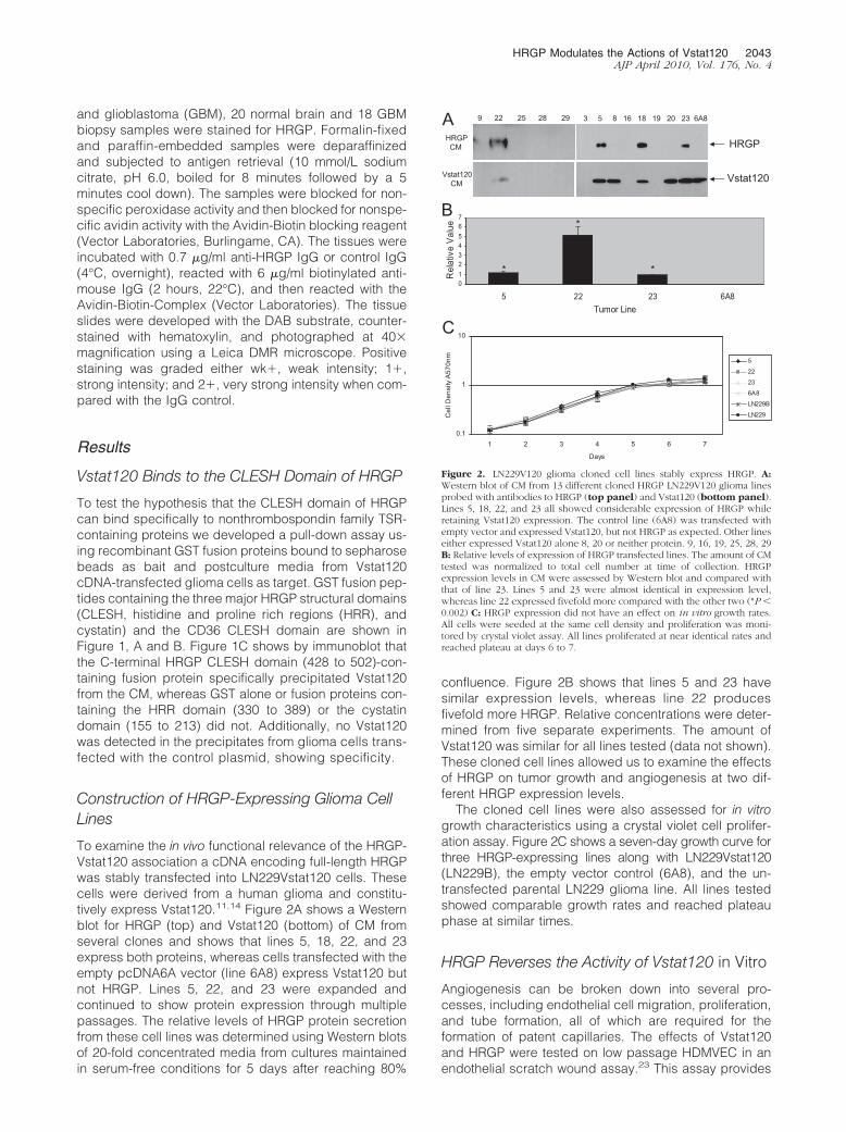
To examine the in vivo functional relevance of the HRGP-Vstat120 association a cDNA encoding full-length HRGPwas stably transfected into LN229Vstat120 cells. Thesecells were derived from a human glioma and constitu-tively express Vstat120.11,14 Figure 2A shows a Westernblot for HRGP (top) and Vstat120 (bottom) of CM fromseveral clones and shows that lines 5, 18, 22, and 23express both proteins, whereas cells transfected with theempty pcDNA6A vector (line 6A8) express Vstat120 butnot HRGP. Lines 5, 22, and 23 were expanded andcontinued to show protein expression through multiplepassages. The relative levels of HRGP protein secretionfrom these cell lines was determined using Western blotsof 20-fold concentrated media from cultures maintainedin serum-free conditions for 5 days after reaching 80%
confluence. Figure 2B shows that lines 5 and 23 havesimilar expression levels, whereas line 22 producesfivefold more HRGP. Relative concentrations were deter-mined from five separate experiments. The amount ofVstat120 was similar for all lines tested (data not shown).These cloned cell lines allowed us to examine the effectsof HRGP on tumor growth and angiogenesis at two dif-ferent HRGP expression levels.
The cloned cell lines were also assessed for in vitrogrowth characteristics using a crystal violet cell prolifer-ation assay. Figure 2C shows a seven-day growth curve forthree HRGP-expressing lines along with LN229Vstat120(LN229B), the empty vector control (6A8), and the un-transfected parental LN229 glioma line. All lines testedshowed comparable growth rates and reached plateauphase at similar times.
HRGP Reverses the Activity of Vstat120 in Vitro
Angiogenesis can be broken down into several pro-cesses, including endothelial cell migration, proliferation,and tube formation, all of which are required for theformation of patent capillaries. The effects of Vstat120and HRGP were tested on low passage HDMVEC in anendothelial scratch wound assay.23 This assay provides
9 22 25 28 29 3 5 8 16 18 19 20 23 6A8
Vstat120CM
HRGPCM
Vstat120
HRGP
A
B
C
01234567
5 22 23 6A8Tumor Line
Rel
ativ
e V
alue
0.1
1
10
1 2 3 4 5 6 7
DaysC
ell D
ensi
ty A
570n
m
5
22
23
6A8
LN229B
LN229
**
*
Figure 2. LN229V120 glioma cloned cell lines stably express HRGP. A:Western blot of CM from 13 different cloned HRGP LN229V120 glioma linesprobed with antibodies to HRGP (top panel) and Vstat120 (bottom panel).Lines 5, 18, 22, and 23 all showed considerable expression of HRGP whileretaining Vstat120 expression. The control line (6A8) was transfected withempty vector and expressed Vstat120, but not HRGP as expected. Other lineseither expressed Vstat120 alone 8, 20 or neither protein. 9, 16, 19, 25, 28, 29B: Relative levels of expression of HRGP transfected lines. The amount of CMtested was normalized to total cell number at time of collection. HRGPexpression levels in CM were assessed by Western blot and compared withthat of line 23. Lines 5 and 23 were almost identical in expression level,whereas line 22 expressed fivefold more compared with the other two (*P �0.002) C: HRGP expression did not have an effect on in vitro growth rates.All cells were seeded at the same cell density and proliferation was moni-tored by crystal violet assay. All lines proliferated at near identical rates andreached plateau at days 6 to 7.
HRGP Modulates the Actions of Vstat120 2043AJP April 2010, Vol. 176, No. 4
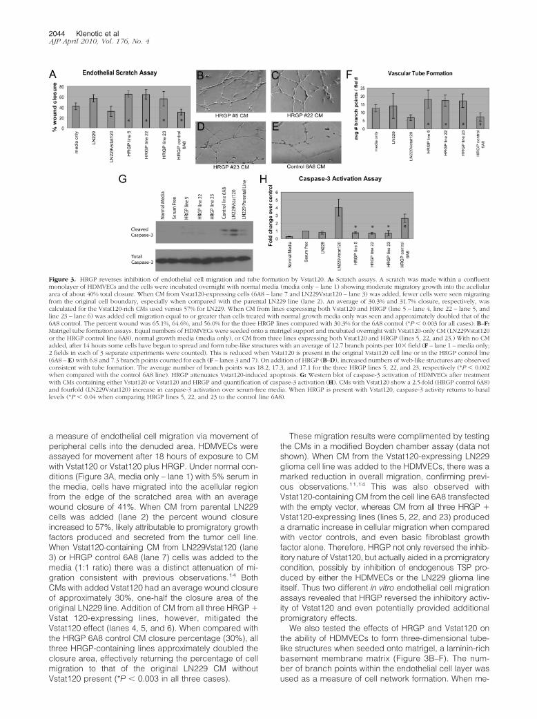
a measure of endothelial cell migration via movement ofperipheral cells into the denuded area. HDMVECs wereassayed for movement after 18 hours of exposure to CMwith Vstat120 or Vstat120 plus HRGP. Under normal con-ditions (Figure 3A, media only – lane 1) with 5% serum inthe media, cells have migrated into the acellular regionfrom the edge of the scratched area with an averagewound closure of 41%. When CM from parental LN229cells was added (lane 2) the percent wound closureincreased to 57%, likely attributable to promigratory growthfactors produced and secreted from the tumor cell line.When Vstat120-containing CM from LN229Vstat120 (lane3) or HRGP control 6A8 (lane 7) cells was added to themedia (1:1 ratio) there was a distinct attenuation of mi-gration consistent with previous observations.14 BothCMs with added Vstat120 had an average wound closureof approximately 30%, one-half the closure area of theoriginal LN229 line. Addition of CM from all three HRGP �Vstat 120-expressing lines, however, mitigated theVstat120 effect (lanes 4, 5, and 6). When compared withthe HRGP 6A8 control CM closure percentage (30%), allthree HRGP-containing lines approximately doubled theclosure area, effectively returning the percentage of cellmigration to that of the original LN229 CM withoutVstat120 present (*P � 0.003 in all three cases).
These migration results were complimented by testingthe CMs in a modified Boyden chamber assay (data notshown). When CM from the Vstat120-expressing LN229glioma cell line was added to the HDMVECs, there was amarked reduction in overall migration, confirming previ-ous observations.11,14 This was also observed withVstat120-containing CM from the cell line 6A8 transfectedwith the empty vector, whereas CM from all three HRGP �Vstat120-expressing lines (lines 5, 22, and 23) produceda dramatic increase in cellular migration when comparedwith vector controls, and even basic fibroblast growthfactor alone. Therefore, HRGP not only reversed the inhib-itory nature of Vstat120, but actually aided in a promigratorycondition, possibly by inhibition of endogenous TSP pro-duced by either the HDMVECs or the LN229 glioma lineitself. Thus two different in vitro endothelial cell migrationassays revealed that HRGP reversed the inhibitory activ-ity of Vstat120 and even potentially provided additionalpromigratory effects.
We also tested the effects of HRGP and Vstat120 onthe ability of HDMVECs to form three-dimensional tube-like structures when seeded onto matrigel, a laminin-richbasement membrane matrix (Figure 3B–F). The num-ber of branch points within the endothelial cell layer wasused as a measure of cell network formation. When me-
Figure 3. HRGP reverses inhibition of endothelial cell migration and tube formation by Vstat120. A: Scratch assays. A scratch was made within a confluentmonolayer of HDMVECs and the cells were incubated overnight with normal media (media only – lane 1) showing moderate migratory growth into the acellulararea of about 40% total closure. When CM from Vstat120-expressing cells (6A8 – lane 7 and LN229Vstat120 – lane 3) was added, fewer cells were seen migratingfrom the original cell boundary, especially when compared with the parental LN229 line (lane 2). An average of 30.3% and 31.7% closure, respectively, wascalculated for the Vstat120-rich CMs used versus 57% for LN229. When CM from lines expressing both Vstat120 and HRGP (line 5 – lane 4, line 22 – lane 5, andline 23 – lane 6) was added cell migration equal to or greater than cells treated with normal growth media only was seen and approximately doubled that of the6A8 control. The percent wound was 65.1%, 64.6%, and 56.0% for the three HRGP lines compared with 30.3% for the 6A8 control (*P � 0.003 for all cases). B–F:Matrigel tube formation assays. Equal numbers of HDMVECs were seeded onto a matrigel support and incubated overnight with Vstat120-only CM (LN229Vstat120or the HRGP control line 6A8), normal growth media (media only), or CM from three lines expressing both Vstat120 and HRGP (lines 5, 22, and 23.) With no CMadded, after 14 hours some cells have begun to spread and form tube-like structures with an average of 12.7 branch points per 10� field (F – lane 1 – media only;2 fields in each of 3 separate experiments were counted). This is reduced when Vstat120 is present in the original Vstat120 cell line or in the HRGP control line(6A8 – E) with 6.8 and 7.3 branch points counted for each (F – lanes 3 and 7). On addition of HRGP (B–D), increased numbers of web-like structures are observedconsistent with tube formation. The average number of branch points was 18.2, 17.3, and 17.1 for the three HRGP lines 5, 22, and 23, respectively (*P � 0.002when compared with the control 6A8 line). HRGP attenuates Vstat120-induced apoptosis. G: Western blot of caspase-3 activation of HDMVECs after treatmentwith CMs containing either Vstat120 or Vstat120 and HRGP and quantification of caspase-3 activation (H). CMs with Vstat120 show a 2.5-fold (HRGP control 6A8)and fourfold (LN229Vstat120) increase in caspase-3 activation over serum-free media. When HRGP is present with Vstat120, caspase-3 activity returns to basallevels (*P � 0.04 when comparing HRGP lines 5, 22, and 23 to the control line 6A8).
2044 Klenotic et alAJP April 2010, Vol. 176, No. 4
dia alone was added to the HDMVECs and incubated for18 hours, significant tube formation was observed and anaverage of approximately 13 branches per field wascounted (Figure 3F, media only – lane 1). An almostequivalent effect was seen with the LN229 parental line(LN229 – lane 2) with an average branch formation of 14 perfield. When the Vstat120-rich CM from LN229Vstat120 (Fig-ure 3F, lane 3) or from HRGP control 6A8 (Figures 3E,and 3F, HRGP control 6A8 – lane 7) cells was used, tubeformation was significantly inhibited and the averagebranch numbers diminished to approximately seven perfield. In contrast, CM from the three HRGP � Vstat120-expressing lines (Figure 3, B, C, D, and F - lanes 4, 5, and6) induced increased tube formation and cellular networkformation. The average branch number for these threelines was 18.1, 17.3, and 17.1, respectively – over 2.5times greater than the HRGP 6A8 control (Compare –Figure 3F; *P � 0.002 for all three cases) and evengreater than the LN229 CM absent of inhibitor. Takentogether these studies demonstrate that HRGP can po-tently reverse the effects of Vstat120 in vitro.
Caspase-3 Activation Is Attenuated by HRGPInhibition of Vstat120
Evidence suggests that TSP-1 and Vstat120 both engageCD36 via their TSR domains and that TSP-1 activatescaspase-3 mediated apoptosis of endothelial cells.9,11,24
Therefore, we tested whether Vstat120 could also induceapoptosis in a caspase-3–dependent manner andwhether HRGP could attenuate this apoptotic signal. Bothnormal media with 10% serum as well as serum-free mediaadded to HDMVECs induced little, if any, caspase-3cleavage products (see Figure 3, G and H; normalmedia – lane 1 and serum-free – lane 2). The parentalLN229 line showed minimal cleaved caspase-3 as well,approximately equal to the amount seen by serum-freemedia alone. When Vstat120 was produced in the CM ofthe HRGP control 6A8 line or the LN229Vstat120 line, a2.5- to 4-fold increase in the canonical doublet of cleavedcaspase-3 was observed (quantified in Figure 3H). Ad-dition of HRGP to the CM, in all three cases, effectivelyattenuated the amount of cleaved caspase-3 back tobasal levels (*P � 0.04 when comparing the three HRGP-producing lines to the control 6A8 line), suggesting thatHRGP prevents Vstat120 from anti-angiogenic signaling,in part, by blocking caspase-3 activation.
HRGP Reverses the Anti-Angiogenic Activity ofVstat120 in Vivo
Previous studies showed that Vstat120 suppressed invivo tumor growth in a dose-dependant manner via inhi-bition of tumor angiogenesis.11,14 Because we have nowshown that the HRGP CLESH domain physically interactswith Vstat120 and that addition of HRGP restores endo-thelial cell migration and tube formation in the presenceof Vstat120, we hypothesized that HRGP would reversethe tumor growth inhibition and diminished tumor angio-
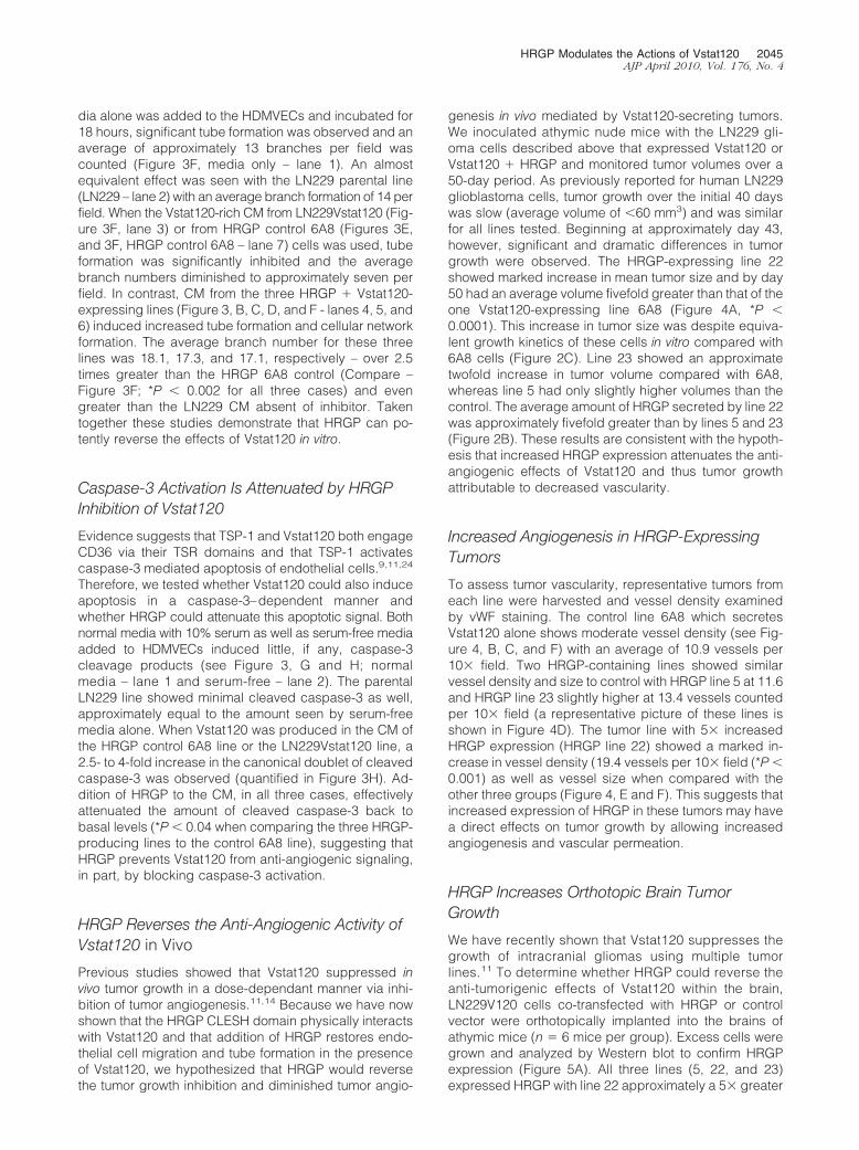
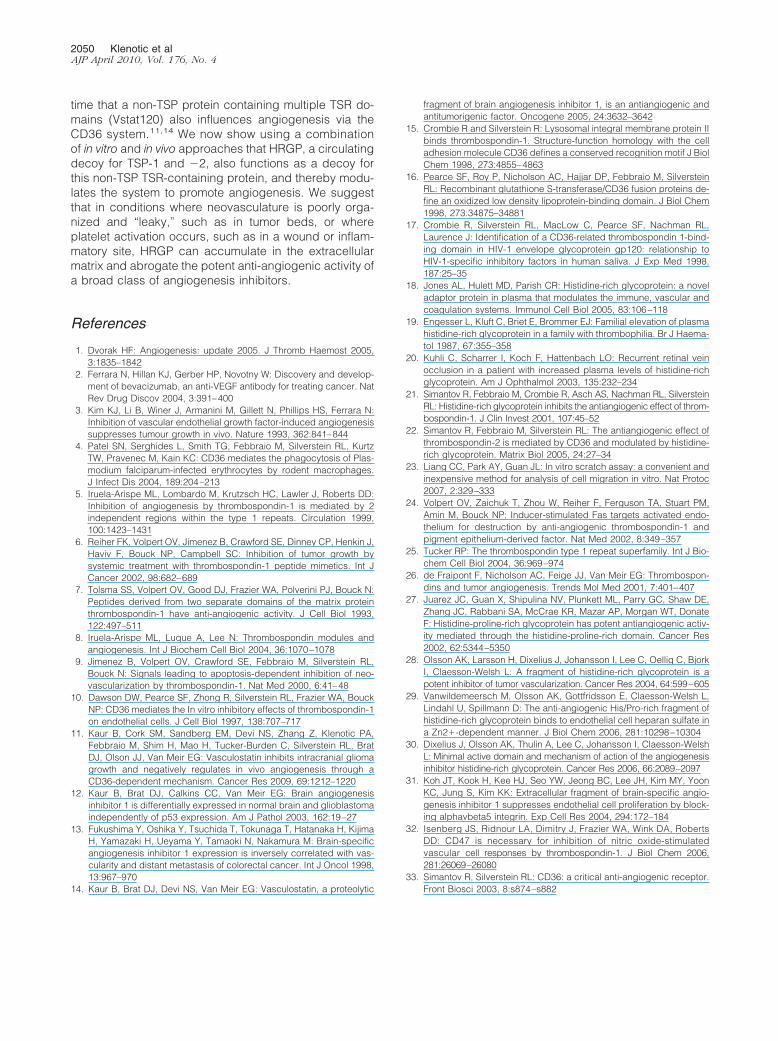
genesis in vivo mediated by Vstat120-secreting tumors.We inoculated athymic nude mice with the LN229 gli-oma cells described above that expressed Vstat120 orVstat120 � HRGP and monitored tumor volumes over a50-day period. As previously reported for human LN229glioblastoma cells, tumor growth over the initial 40 dayswas slow (average volume of �60 mm3) and was similarfor all lines tested. Beginning at approximately day 43,however, significant and dramatic differences in tumorgrowth were observed. The HRGP-expressing line 22showed marked increase in mean tumor size and by day50 had an average volume fivefold greater than that of theone Vstat120-expressing line 6A8 (Figure 4A, *P �0.0001). This increase in tumor size was despite equiva-lent growth kinetics of these cells in vitro compared with6A8 cells (Figure 2C). Line 23 showed an approximatetwofold increase in tumor volume compared with 6A8,whereas line 5 had only slightly higher volumes than thecontrol. The average amount of HRGP secreted by line 22was approximately fivefold greater than by lines 5 and 23(Figure 2B). These results are consistent with the hypoth-esis that increased HRGP expression attenuates the anti-angiogenic effects of Vstat120 and thus tumor growthattributable to decreased vascularity.
Increased Angiogenesis in HRGP-ExpressingTumors
To assess tumor vascularity, representative tumors fromeach line were harvested and vessel density examinedby vWF staining. The control line 6A8 which secretesVstat120 alone shows moderate vessel density (see Fig-ure 4, B, C, and F) with an average of 10.9 vessels per10� field. Two HRGP-containing lines showed similarvessel density and size to control with HRGP line 5 at 11.6and HRGP line 23 slightly higher at 13.4 vessels countedper 10� field (a representative picture of these lines isshown in Figure 4D). The tumor line with 5� increasedHRGP expression (HRGP line 22) showed a marked in-crease in vessel density (19.4 vessels per 10� field (*P �0.001) as well as vessel size when compared with theother three groups (Figure 4, E and F). This suggests thatincreased expression of HRGP in these tumors may havea direct effects on tumor growth by allowing increasedangiogenesis and vascular permeation.
HRGP Increases Orthotopic Brain TumorGrowth
We have recently shown that Vstat120 suppresses thegrowth of intracranial gliomas using multiple tumorlines.11 To determine whether HRGP could reverse theanti-tumorigenic effects of Vstat120 within the brain,LN229V120 cells co-transfected with HRGP or controlvector were orthotopically implanted into the brains ofathymic mice (n � 6 mice per group). Excess cells weregrown and analyzed by Western blot to confirm HRGPexpression (Figure 5A). All three lines (5, 22, and 23)expressed HRGP with line 22 approximately a 5� greater
HRGP Modulates the Actions of Vstat120 2045AJP April 2010, Vol. 176, No. 4
amount, consistent with protein analysis from previouscell passages. After 9 weeks, the animals were sacrificedand the brains removed. Ten-�m sections were cut rang-ing from 1 mm anterior to 1 mm posterior to the tumor
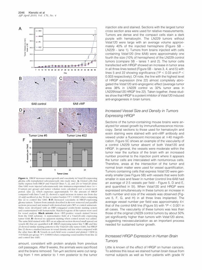
injection site and stained. Sections with the largest tumorcross section area were used for relative measurements.Tumors are dense and the compact cells stain a darkpurple with hematoxylin. The LN229 tumors withoutVstat120 were large with an average volume approxi-mately 40% of the injected hemisphere (Figure 5B –LN229 – lane 1). Tumors from brains injected with cellscontaining Vstat120 (line 6A8) were approximately onefourth the size (10% of hemisphere) of the LN229 controltumors (compare 5B – lanes 1 and 2). The tumor cellstransfected with HRGP showed an increase in tumor areain all three lines tested (Figure 5B – lanes 3, 4, and 5) withlines 5 and 22 showing significance (*P � 0.03 and P �0.003 respectively). Of note, the line with the highest levelof HRGP expression (line 22) almost completely abro-gated the Vstat120 anti-angiogenic effect (average tumorarea 38% in LN229 control vs 32% tumor area inLN229Vstat120 HRGP line 22). Taken together, these stud-ies show that HRGP is a potent inhibitor of Vstat120 inducedanti-angiogenesis in brain tumors.
Increased Vessel Size and Density in TumorsExpressing HRGP
Sections of the tumor-containing mouse brains were an-alyzed for vessel growth by immunofluorescence micros-copy. Serial sections to those used for hematoxylin andeosin staining were stained with anti-vWF antibody andviewed under a fluorescent microscope at �40 magnifi-cation. Figure 5C shows an example of the vascularity ofa control LN229 tumor absent of both Vstat120 andHRGP. In general, the vessels were moderate within thetumor near the surface of the brain with an increasednumber proximal to the injection point where it appearsthe tumor cells are intercalated with nontumorous cells.Therefore, areas at the intersection of the tumor andnormal brain matter were used for vessel quantification.Tumors containing cells that express Vstat120 were gen-erally smaller (see Figure 5B) with vessels that were bothsmaller in size and fewer in number (control line 6A8 hadan average of 2.5 vessels per field – Figure 5, D and E,and quantified in 5I). When Vstat120 and HRGP wereexpressed simultaneously in these tumors an increase inboth number and size of the vessels was observed (Fig-ure 5, F, G, and H) in all three lines implanted. Theaverage vessel number per field was approximately 4�that of the control 6A8 line (Figure 5I) with *P � 0.001 inall cases. The vascularity of these tumors was less thanthose of the original LN229 control tumors by about 50%yet significantly higher than tumors with Vstat120 alone,suggesting neovascularization as an important processneeded for sustained tumor growth.
Increased HRGP Expression in Human BrainTumors
Little is known of the effect of HRGP on human cancers.To address this issue we stained human brain tissue fromnormal subjects as well as from patients with grade IV
6A8 control 6A8 vWF
Tumor Line #5 Tumor Line #22
A
CB
D E
0.0
200.0
400.0
600.0
800.0
1000.0
1200.0
7 11 13 15 18 20 22 25 27 29 32 34 36 39 41 43 46 48 50
Day
Tum
or V
olum
e (c
u m
m)
mea
n ±
SE
#23
#6A8
#5
#22
0
5
10
15
20
6A8 5 22 23
Tumor Lines
# ve
ssel
s pe
r fie
ld
* *
F
*
Figure 4. HRGP increases tumor growth and vascularity in Vstat120-expressingglioma cells transplanted subcutaneously into nude mice. A: Cloned cells thatstably express both HRGP and Vstat120 (lines 5, 22, and 23) or Vstat120 alone(line 6A8) were injected subcutaneously into immunocompromised mice (n �8 tumors per group) and tumor volumes were calculated over a seven-weekperiod. Line 22, which expresses approximately 5� the amount of HRGPcompared with lines 5 and 23, showed a rapid increase in tumor size from day43 until sacrificed at day 50 due to tumor burden (*P � 0.0001 when comparingline 22 to control line 6A8). B–F: Increased vascularity in HRGP-expressingglioma tumors. Tumors from animals described in A were removed and paraffinsections processed and stained with monoclonal anti-vWF or control antiserum.Slides were developed with an HRP-conjugated avidin-biotin staining systemand counterstained with hematoxylin. Matched regions of the tumors were usedfor vessel analysis. Black arrows show vWF-positive vessels stained brownfrom the DAB substrate. A representative field of a Vstat120-only expressingtumor (6A8, B) showed no apparent staining when control serum was added.The same field stained with vWF on an adjacent section showed small spread outvessels (C) that are quantified in F. HRGP-expressing tumor line 5 (D) and line23 showed similar staining patterns to the Vstat120-only tumor (6A8), but HRGPline 22 shows a marked increase in vessel density and size when compared withthe other lines (E). F: Vessels in each field were counted and plotted �SEM (n�10 fields per group; *P � 0.00003 when comparing vessel number from line 22and control line 6A8).
2046 Klenotic et alAJP April 2010, Vol. 176, No. 4
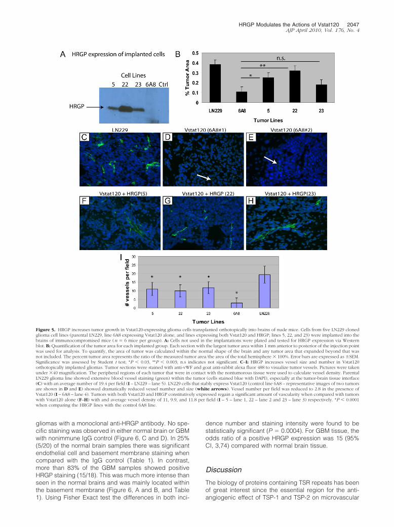
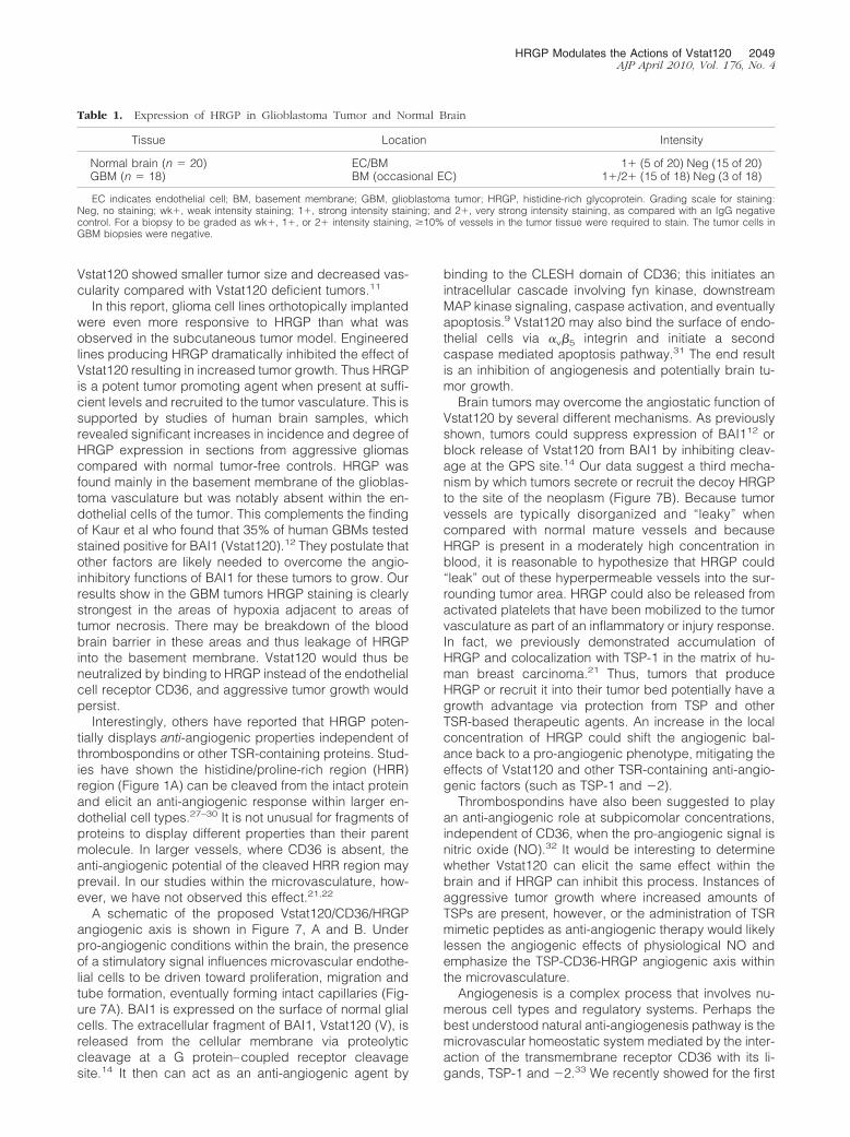
gliomas with a monoclonal anti-HRGP antibody. No spe-cific staining was observed in either normal brain or GBMwith nonimmune IgG control (Figure 6, C and D). In 25%(5/20) of the normal brain samples there was significantendothelial cell and basement membrane staining whencompared with the IgG control (Table 1). In contrast,more than 83% of the GBM samples showed positiveHRGP staining (15/18). This was much more intense thanseen in the normal brains and was mainly located withinthe basement membrane (Figure 6, A and B, and Table1). Using Fisher Exact test the differences in both inci-
dence number and staining intensity were found to bestatistically significant (P � 0.0004). For GBM tissue, theodds ratio of a positive HRGP expression was 15 (95%CI, 3,74) compared with normal brain tissue.
Discussion
The biology of proteins containing TSR repeats has beenof great interest since the essential region for the anti-angiogenic effect of TSP-1 and TSP-2 on microvascular
Figure 5. HRGP increases tumor growth in Vstat120-expressing glioma cells transplanted orthotopically into brains of nude mice. Cells from five LN229 clonedglioma cell lines (parental LN229, line 6A8 expressing Vstat120 alone, and lines expressing both Vstat120 and HRGP; lines 5, 22, and 23) were implanted into thebrains of immunocompromised mice (n � 6 mice per group). A: Cells not used in the implantations were plated and tested for HRGP expression via Westernblot. B: Quantification of the tumor area for each implanted group. Each section with the largest tumor area within 1 mm anterior to posterior of the injection pointwas used for analysis. To quantify, the area of tumor was calculated within the normal shape of the brain and any tumor area that expanded beyond that wasnot included. The percent tumor area represents the ratio of the measured tumor area/the area of the total hemisphere � 100%. Error bars are expressed as �SEM.Significance was assessed by Student t test; *P � 0.03, **P � 0.003; n.s indicates not significant. C–I: HRGP increases vessel size and number in Vstat120orthotopically implanted gliomas. Tumor sections were stained with anti-vWF and goat anti-rabbit alexa fluor 488 to visualize tumor vessels. Pictures were takenunder �40 magnification. The peripheral regions of each tumor that were in contact with the nontumorous tissue were used to calculate vessel density. ParentalLN229 glioma line showed extensive blood vessel staining (green) within the tumor (cells stained blue with DAPI), especially at the tumor-brain tissue interface(C) with an average number of 19.4 per field (I – LN229 – lane 5). LN229 cells that stably express Vstat120 (control line 6A8 – representative images of two tumorsare shown in D and E) showed dramatically reduced vessel number and size (white arrows). Vessel number per field was reduced to 2.8 in the presence ofVstat120 (I – 6A8 – lane 4). Tumors with both Vstat120 and HRGP constitutively expressed regain a significant amount of vascularity when compared with tumorswith Vstat120 alone (F–H) with and average vessel density of 11, 9.9, and 11.8 per field (I – 5 – lane 1, 22 – lane 2 and 23 – lane 3) respectively. *P � 0.0001when comparing the HRGP lines with the control 6A8 line.
HRGP Modulates the Actions of Vstat120 2047AJP April 2010, Vol. 176, No. 4
endothelial cells was mapped to these domains. Over 70human TSR-containing proteins have been identi-fied.25,26 Interestingly, the TSR domains have only beenmapped to extracellular regions of integral membraneproteins or in proteins secreted into the extracellularspace, with many being deposited into the matrix. Thiswould suggest potential roles for the TSRs in cell–celland cell–matrix interactions. A subset of TSR-containingproteins, including TSP-1, TSP-2, BAI1 (Vstat120), andADAMTS-20 are potent inhibitors of angiogenesis, withthe mechanism of at least three being dependent oninteraction with CD36.
In our studies of CD36 structure/function relationshipswe discovered protein sequences homologous to theCD36 CLESH domain in several unrelated proteins knownto bind TSP-1, including HIV gp12017 and HRGP. Wereport here that the C-terminal CLESH domain of HRGPbound to Vstat120, whereas other HRGP domains, in-cluding an N-terminal domain with a lower degree ofCLESH homology, did not. In multiple functional assayswe also showed that HRGP reversed the actions ofVstat120 when both proteins were expressed concomi-tantly. These results are the first demonstration thatHRGP can bind TSR-containing proteins other thanthrombospondins, and suggest that HRGP may modulateactivity of an entire class of angiogenesis inhibitors.
In vivo, HRGP exacerbated subcutaneous tumorgrowth when produced at a high enough concentration(Figure 4A). For tumors to rapidly increase in size, adisruption of the angiogenic balance to favor a pro-an-giogenic phenotype is likely. In this instance, it is possiblethat increased tumor growth was supported when athreshold concentration of HRGP was reached, activatingan angiogenic switch toward increased endothelial cellrecruitment, enhanced angiogenesis, and rapid cellgrowth. As we reported previously, the average survivaltime of mice implanted in the brain with tumor cells se-creting Vstat120 was significantly increased comparedwith animals injected with control cells, whereas tumorsamples excised at equivalent time points expressing
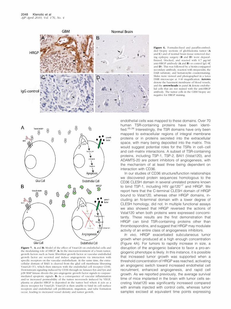
Figure 7. A and B: Model of the effect of Vstat120 on endothelial cells andthe modulating role of HRGP. A: In the microenvironment of a brain tumor,growth factors such as basic fibroblast growth factor or vascular endothelialgrowth factor are secreted and induce angiogenesis via interaction withspecific receptors on the vascular endothelium. At the same time, the extra-cellular domain of BAI1 is cleaved from the glial cell membrane liberatingVstat120 (V), which then interacts with the endothelial cell receptor CD36.Downstream signaling induced by CD36 through src kinases fyn and lyn andp38 MAP kinase diverts the pro-angiogenic growth factor signals to caspase-mediated apoptotic signals. B: As a consequence of vascular inflammationand/or increased permeability of the tumor neovessels induced by VEGF,plasma or platelet HRGP is deposited in the tumor bed where it acts as adecoy receptor for Vstat120. Vstat120 is then unable to bind its cell surfacereceptors and endothelial cell proliferation, migration, and tube formationoccur, leading to increased vessel density and tumor growth.
Figure 6. Formalin-fixed and paraffin-embed-ded biopsy sections of glioblastoma tumor (Aand C) and of normal brain tissue removed dur-ing epilepsy surgery (B and D) were deparaf-finized, blocked, and reacted with 0.7 �g/mlanti-HRGP antibody (A and B) or control IgG (Cand D). This was followed by a biotin-conjugatedsecondary antibody, reaction with strepavidin, theDAB substrate, and hematoxylin counterstaining.Slides were viewed and photographed in a LeicaDMR microscope at �40 magnification. Arrowsdenote the basement membrane of blood vessels,and the arrowheads in panel A denote endothe-lial cells that are not stained with the anti-HRGPantibody. The tumor cells in the GBM biopsy arenegative for HRGP staining.
2048 Klenotic et alAJP April 2010, Vol. 176, No. 4
Vstat120 showed smaller tumor size and decreased vas-cularity compared with Vstat120 deficient tumors.11
In this report, glioma cell lines orthotopically implantedwere even more responsive to HRGP than what wasobserved in the subcutaneous tumor model. Engineeredlines producing HRGP dramatically inhibited the effect ofVstat120 resulting in increased tumor growth. Thus HRGPis a potent tumor promoting agent when present at suffi-cient levels and recruited to the tumor vasculature. This issupported by studies of human brain samples, whichrevealed significant increases in incidence and degree ofHRGP expression in sections from aggressive gliomascompared with normal tumor-free controls. HRGP wasfound mainly in the basement membrane of the glioblas-toma vasculature but was notably absent within the en-dothelial cells of the tumor. This complements the findingof Kaur et al who found that 35% of human GBMs testedstained positive for BAI1 (Vstat120).12 They postulate thatother factors are likely needed to overcome the angio-inhibitory functions of BAI1 for these tumors to grow. Ourresults show in the GBM tumors HRGP staining is clearlystrongest in the areas of hypoxia adjacent to areas oftumor necrosis. There may be breakdown of the bloodbrain barrier in these areas and thus leakage of HRGPinto the basement membrane. Vstat120 would thus beneutralized by binding to HRGP instead of the endothelialcell receptor CD36, and aggressive tumor growth wouldpersist.
Interestingly, others have reported that HRGP poten-tially displays anti-angiogenic properties independent ofthrombospondins or other TSR-containing proteins. Stud-ies have shown the histidine/proline-rich region (HRR)region (Figure 1A) can be cleaved from the intact proteinand elicit an anti-angiogenic response within larger en-dothelial cell types.27–30 It is not unusual for fragments ofproteins to display different properties than their parentmolecule. In larger vessels, where CD36 is absent, theanti-angiogenic potential of the cleaved HRR region mayprevail. In our studies within the microvasculature, how-ever, we have not observed this effect.21,22
A schematic of the proposed Vstat120/CD36/HRGPangiogenic axis is shown in Figure 7, A and B. Underpro-angiogenic conditions within the brain, the presenceof a stimulatory signal influences microvascular endothe-lial cells to be driven toward proliferation, migration andtube formation, eventually forming intact capillaries (Fig-ure 7A). BAI1 is expressed on the surface of normal glialcells. The extracellular fragment of BAI1, Vstat120 (V), isreleased from the cellular membrane via proteolyticcleavage at a G protein–coupled receptor cleavagesite.14 It then can act as an anti-angiogenic agent by
binding to the CLESH domain of CD36; this initiates anintracellular cascade involving fyn kinase, downstreamMAP kinase signaling, caspase activation, and eventuallyapoptosis.9 Vstat120 may also bind the surface of endo-thelial cells via �v�5 integrin and initiate a secondcaspase mediated apoptosis pathway.31 The end resultis an inhibition of angiogenesis and potentially brain tu-mor growth.
Brain tumors may overcome the angiostatic function ofVstat120 by several different mechanisms. As previouslyshown, tumors could suppress expression of BAI112 orblock release of Vstat120 from BAI1 by inhibiting cleav-age at the GPS site.14 Our data suggest a third mecha-nism by which tumors secrete or recruit the decoy HRGPto the site of the neoplasm (Figure 7B). Because tumorvessels are typically disorganized and “leaky” whencompared with normal mature vessels and becauseHRGP is present in a moderately high concentration inblood, it is reasonable to hypothesize that HRGP could“leak” out of these hyperpermeable vessels into the sur-rounding tumor area. HRGP could also be released fromactivated platelets that have been mobilized to the tumorvasculature as part of an inflammatory or injury response.In fact, we previously demonstrated accumulation ofHRGP and colocalization with TSP-1 in the matrix of hu-man breast carcinoma.21 Thus, tumors that produceHRGP or recruit it into their tumor bed potentially have agrowth advantage via protection from TSP and otherTSR-based therapeutic agents. An increase in the localconcentration of HRGP could shift the angiogenic bal-ance back to a pro-angiogenic phenotype, mitigating theeffects of Vstat120 and other TSR-containing anti-angio-genic factors (such as TSP-1 and �2).
Thrombospondins have also been suggested to playan anti-angiogenic role at subpicomolar concentrations,independent of CD36, when the pro-angiogenic signal isnitric oxide (NO).32 It would be interesting to determinewhether Vstat120 can elicit the same effect within thebrain and if HRGP can inhibit this process. Instances ofaggressive tumor growth where increased amounts ofTSPs are present, however, or the administration of TSRmimetic peptides as anti-angiogenic therapy would likelylessen the angiogenic effects of physiological NO andemphasize the TSP-CD36-HRGP angiogenic axis withinthe microvasculature.
Angiogenesis is a complex process that involves nu-merous cell types and regulatory systems. Perhaps thebest understood natural anti-angiogenesis pathway is themicrovascular homeostatic system mediated by the inter-action of the transmembrane receptor CD36 with its li-gands, TSP-1 and �2.33 We recently showed for the first
Table 1. Expression of HRGP in Glioblastoma Tumor and Normal Brain
Tissue Location Intensity
Normal brain (n � 20) EC/BM 1� (5 of 20) Neg (15 of 20)GBM (n � 18) BM (occasional EC) 1�/2� (15 of 18) Neg (3 of 18)
EC indicates endothelial cell; BM, basement membrane; GBM, glioblastoma tumor; HRGP, histidine-rich glycoprotein. Grading scale for staining:Neg, no staining; wk�, weak intensity staining; 1�, strong intensity staining; and 2�, very strong intensity staining, as compared with an IgG negativecontrol. For a biopsy to be graded as wk�, 1�, or 2� intensity staining, �10% of vessels in the tumor tissue were required to stain. The tumor cells inGBM biopsies were negative.
HRGP Modulates the Actions of Vstat120 2049AJP April 2010, Vol. 176, No. 4
time that a non-TSP protein containing multiple TSR do-mains (Vstat120) also influences angiogenesis via theCD36 system.11,14 We now show using a combinationof in vitro and in vivo approaches that HRGP, a circulatingdecoy for TSP-1 and �2, also functions as a decoy forthis non-TSP TSR-containing protein, and thereby modu-lates the system to promote angiogenesis. We suggestthat in conditions where neovasculature is poorly orga-nized and “leaky,” such as in tumor beds, or whereplatelet activation occurs, such as in a wound or inflam-matory site, HRGP can accumulate in the extracellularmatrix and abrogate the potent anti-angiogenic activity ofa broad class of angiogenesis inhibitors.
References
1. Dvorak HF: Angiogenesis: update 2005. J Thromb Haemost 2005,3:1835–1842
2. Ferrara N, Hillan KJ, Gerber HP, Novotny W: Discovery and develop-ment of bevacizumab, an anti-VEGF antibody for treating cancer. NatRev Drug Discov 2004, 3:391–400
3. Kim KJ, Li B, Winer J, Armanini M, Gillett N, Phillips HS, Ferrara N:Inhibition of vascular endothelial growth factor-induced angiogenesissuppresses tumour growth in vivo. Nature 1993, 362:841–844
4. Patel SN, Serghides L, Smith TG, Febbraio M, Silverstein RL, KurtzTW, Pravenec M, Kain KC: CD36 mediates the phagocytosis of Plas-modium falciparum-infected erythrocytes by rodent macrophages.J Infect Dis 2004, 189:204–213
5. Iruela-Arispe ML, Lombardo M, Krutzsch HC, Lawler J, Roberts DD:Inhibition of angiogenesis by thrombospondin-1 is mediated by 2independent regions within the type 1 repeats. Circulation 1999,100:1423–1431
6. Reiher FK, Volpert OV, Jimenez B, Crawford SE, Dinney CP, Henkin J,Haviv F, Bouck NP, Campbell SC: Inhibition of tumor growth bysystemic treatment with thrombospondin-1 peptide mimetics. Int JCancer 2002, 98:682–689
7. Tolsma SS, Volpert OV, Good DJ, Frazier WA, Polverini PJ, Bouck N:Peptides derived from two separate domains of the matrix proteinthrombospondin-1 have anti-angiogenic activity. J Cell Biol 1993,122:497–511
8. Iruela-Arispe ML, Luque A, Lee N: Thrombospondin modules andangiogenesis. Int J Biochem Cell Biol 2004, 36:1070–1078
9. Jimenez B, Volpert OV, Crawford SE, Febbraio M, Silverstein RL,Bouck N: Signals leading to apoptosis-dependent inhibition of neo-vascularization by thrombospondin-1. Nat Med 2000, 6:41–48
10. Dawson DW, Pearce SF, Zhong R, Silverstein RL, Frazier WA, BouckNP: CD36 mediates the In vitro inhibitory effects of thrombospondin-1on endothelial cells. J Cell Biol 1997, 138:707–717
11. Kaur B, Cork SM, Sandberg EM, Devi NS, Zhang Z, Klenotic PA,Febbraio M, Shim H, Mao H, Tucker-Burden C, Silverstein RL, BratDJ, Olson JJ, Van Meir EG: Vasculostatin inhibits intracranial gliomagrowth and negatively regulates in vivo angiogenesis through aCD36-dependent mechanism. Cancer Res 2009, 69:1212–1220
12. Kaur B, Brat DJ, Calkins CC, Van Meir EG: Brain angiogenesisinhibitor 1 is differentially expressed in normal brain and glioblastomaindependently of p53 expression. Am J Pathol 2003, 162:19–27
13. Fukushima Y, Oshika Y, Tsuchida T, Tokunaga T, Hatanaka H, KijimaH, Yamazaki H, Ueyama Y, Tamaoki N, Nakamura M: Brain-specificangiogenesis inhibitor 1 expression is inversely correlated with vas-cularity and distant metastasis of colorectal cancer. Int J Oncol 1998,13:967–970
14. Kaur B, Brat DJ, Devi NS, Van Meir EG: Vasculostatin, a proteolytic
fragment of brain angiogenesis inhibitor 1, is an antiangiogenic andantitumorigenic factor. Oncogene 2005, 24:3632–3642
15. Crombie R and Silverstein R: Lysosomal integral membrane protein IIbinds thrombospondin-1. Structure-function homology with the celladhesion molecule CD36 defines a conserved recognition motif J BiolChem 1998, 273:4855–4863
16. Pearce SF, Roy P, Nicholson AC, Hajjar DP, Febbraio M, SilversteinRL: Recombinant glutathione S-transferase/CD36 fusion proteins de-fine an oxidized low density lipoprotein-binding domain. J Biol Chem1998, 273:34875–34881
17. Crombie R, Silverstein RL, MacLow C, Pearce SF, Nachman RL,Laurence J: Identification of a CD36-related thrombospondin 1-bind-ing domain in HIV-1 envelope glycoprotein gp120: relationship toHIV-1-specific inhibitory factors in human saliva. J Exp Med 1998,187:25–35
18. Jones AL, Hulett MD, Parish CR: Histidine-rich glycoprotein: a noveladaptor protein in plasma that modulates the immune, vascular andcoagulation systems. Immunol Cell Biol 2005, 83:106–118
19. Engesser L, Kluft C, Briet E, Brommer EJ: Familial elevation of plasmahistidine-rich glycoprotein in a family with thrombophilia. Br J Haema-tol 1987, 67:355–358
20. Kuhli C, Scharrer I, Koch F, Hattenbach LO: Recurrent retinal veinocclusion in a patient with increased plasma levels of histidine-richglycoprotein. Am J Ophthalmol 2003, 135:232–234
21. Simantov R, Febbraio M, Crombie R, Asch AS, Nachman RL, SilversteinRL: Histidine-rich glycoprotein inhibits the antiangiogenic effect of throm-bospondin-1. J Clin Invest 2001, 107:45–52
22. Simantov R, Febbraio M, Silverstein RL: The antiangiogenic effect ofthrombospondin-2 is mediated by CD36 and modulated by histidine-rich glycoprotein. Matrix Biol 2005, 24:27–34
23. Liang CC, Park AY, Guan JL: In vitro scratch assay: a convenient andinexpensive method for analysis of cell migration in vitro. Nat Protoc2007, 2:329–333
24. Volpert OV, Zaichuk T, Zhou W, Reiher F, Ferguson TA, Stuart PM,Amin M, Bouck NP: Inducer-stimulated Fas targets activated endo-thelium for destruction by anti-angiogenic thrombospondin-1 andpigment epithelium-derived factor. Nat Med 2002, 8:349–357
25. Tucker RP: The thrombospondin type 1 repeat superfamily. Int J Bio-chem Cell Biol 2004, 36:969–974
26. de Fraipont F, Nicholson AC, Feige JJ, Van Meir EG: Thrombospon-dins and tumor angiogenesis. Trends Mol Med 2001, 7:401–407
27. Juarez JC, Guan X, Shipulina NV, Plunkett ML, Parry GC, Shaw DE,Zhang JC, Rabbani SA, McCrae KR, Mazar AP, Morgan WT, DonateF: Histidine-proline-rich glycoprotein has potent antiangiogenic activ-ity mediated through the histidine-proline-rich domain. Cancer Res2002, 62:5344–5350
28. Olsson AK, Larsson H, Dixelius J, Johansson I, Lee C, Oellig C, BjorkI, Claesson-Welsh L: A fragment of histidine-rich glycoprotein is apotent inhibitor of tumor vascularization. Cancer Res 2004, 64:599–605
29. Vanwildemeersch M, Olsson AK, Gottfridsson E, Claesson-Welsh L,Lindahl U, Spillmann D: The anti-angiogenic His/Pro-rich fragment ofhistidine-rich glycoprotein binds to endothelial cell heparan sulfate ina Zn2�-dependent manner. J Biol Chem 2006, 281:10298–10304
30. Dixelius J, Olsson AK, Thulin A, Lee C, Johansson I, Claesson-WelshL: Minimal active domain and mechanism of action of the angiogenesisinhibitor histidine-rich glycoprotein. Cancer Res 2006, 66:2089–2097
31. Koh JT, Kook H, Kee HJ, Seo YW, Jeong BC, Lee JH, Kim MY, YoonKC, Jung S, Kim KK: Extracellular fragment of brain-specific angio-genesis inhibitor 1 suppresses endothelial cell proliferation by block-ing alphavbeta5 integrin. Exp Cell Res 2004, 294:172–184
32. Isenberg JS, Ridnour LA, Dimitry J, Frazier WA, Wink DA, RobertsDD: CD47 is necessary for inhibition of nitric oxide-stimulatedvascular cell responses by thrombospondin-1. J Biol Chem 2006,281:26069–26080
33. Simantov R, Silverstein RL: CD36: a critical anti-angiogenic receptor.Front Biosci 2003, 8:s874–s882
2050 Klenotic et alAJP April 2010, Vol. 176, No. 4


