Embed Size (px)
Citation preview

Histone Deacetylases (HDAC)-Induced Histone Modifications inthe Amygdala: A Role in Rapid Tolerance to the AnxiolyticEffects of Ethanol
Amul J Sakharkar, Huaibo Zhang, Lei Tang, Guangbin Shi, and Subhash C PandeyDepartment of Psychiatry (AJS, HZ, LT, GS, SCP), Department of Anatomy and Cell Biology(SCP), University of Illinois at Chicago, Chicago, Illinois; Jesse Brown VA Medical Center (AJS,HZ, LT, GS, SCP), Chicago, Illinois, 60612 USA
AbstractBackground—Rapid tolerance to the anxiolytic effects of ethanol appears to be an importantfactor in the development of alcoholism. Here, we investigated the involvement of amygdaloidhistone deacetylases (HDAC)-induced epigenetic changes in rapid ethanol tolerance (RET).
Methods—RET in rats was induced by two ethanol injections administered 24 hrs apart. Bothethanol-tolerant and control rats were treated with the HDAC inhibitor, trichostatin A (TSA), andanxiety-like behaviors were measured. HDAC activity, histone (H3 & H4) acetylation, andneuropeptide Y (NPY) expression in the amygdala of these rats were also measured.
Results—A single ethanol exposure was able to produce an anxiolytic response, inhibitamygdaloid HDAC activity, and increase both histone acetylation and NPY expression (mRNAand protein levels) in the central nucleus of amygdala (CeA) and medial nucleus of amygdala(MeA) of rats. In contrast, two exposures of the same dose of ethanol (24 hrs apart) neither eliciteda similar anxiolytic response nor modulated HDAC activity, histone acetylation, or NPYexpression in the amygdala. However, exposure to a higher dose of ethanol on the second day wasable to produce an anxiolytic response and also inhibit amygdaloid HDAC activity. TSA treatmentcaused the reversal of RET by inhibiting HDAC activity thereby increasing histone acetylationand NPY expression in the CeA and MeA.
Conclusions—Cellular tolerance to the initial acute ethanol-induced inhibition of HDACactivity and the subsequent up-regulation of histone acetylation and NPY expression in theamygdala may be involved in the mechanisms underlying rapid tolerance to the anxiolytic effectsof ethanol.
KeywordsAmygdala; anxiety; HDAC; histone acetylation; neuropeptide Y; rapid ethanol tolerance
Alcohol tolerance is a functional phenomenon that can be defined as the need for a higherdose of alcohol to achieve the initial responses of alcohol exposure and may thereby playimportant roles in the development of dependence and in promoting alcohol drinkingbehaviors (Kalant 1998; Tabakoff et al., 1986). Alcohol tolerance is an important criterionused to clinically diagnose alcohol dependence, according to the Diagnostic and StatisticalManual of Mental Disorders IV (DSM IV) guidelines (American Psychiatric Association,1994). Various forms of alcohol tolerance have been identified, such as acute functional
Reprint requests: Dr. Subhash C. Pandey, Department of Psychiatry, University of Illinois at Chicago and Jesse Brown VA MedicalCenter (M/C 151), 820 South Damen Avenue, Chicago, IL 60612, USA; Fax: 312-569-8114; [email protected].
NIH Public AccessAuthor ManuscriptAlcohol Clin Exp Res. Author manuscript; available in PMC 2013 January 1.
Published in final edited form as:Alcohol Clin Exp Res. 2012 January ; 36(1): 61–71. doi:10.1111/j.1530-0277.2011.01581.x.
NIH
-PA Author Manuscript
NIH
-PA Author Manuscript
NIH
-PA Author Manuscript

tolerance (AFT), rapid ethanol tolerance (RET), and chronic ethanol tolerance (CET)(Kalant 1998; Tabakoff et al., 1986). AFT can develop during a single session of alcoholconsumption (Mellanby 1919), whereas CET has been observed after long-term ethanolexposure (Kalant 1998; Tabakoff et al., 1986). RET develops between 8 and 24 hrs after thefirst ethanol exposure (Crabbe et al., 1979; Khanna et al., 1991, 1996; Koob et al., 1987) andcan serve as a good predictor of CET (Khanna et al., 1991).
Tolerance to some of the behavioral effects of ethanol, such as hypothermia, ataxia,sedation, hypnosis, and related neurobiological mechanisms has been studied (Chandler etal., 1998; Crabbe et al., 1979; Gill and Deitrich, 1998; Kalant 1998; Khanna et al., 1991;Kurtz et al., 1996). However, few studies have been conducted in relation to rapid toleranceto the anxiolytic effects of ethanol in animal models (Debatin and Barbosa, 2006; Koob etal., 1987). Alcohol produces anxiolytic effects in humans and rapid tolerance to theseanxiolytic effects may be involved in promoting alcohol drinking in alcoholics (Cloninger etal., 1988; Cooper et al., 1995; Debatin and Barbosa, 2006; Lipscomb et al., 1980; Mobergand Curtin, 2009). Thus, identifying the molecular mechanisms underlying rapid toleranceto the anxiolytic effects of ethanol is important in order to gain a better understanding of theprocesses of alcohol addiction.
Several studies indicate that epigenetic mechanisms, such as covalent histone modificationsvia acetylation, phosphorylation and methylation, and DNA methylation, appear to beinvolved in the pathophysiology of brain disorders including addictive behaviors (Feng andFan, 2009; Grayson et al., 2010; Kazantsev and Thompson, 2008; Renthal and Nestler,2008; Tsankova et al., 2007). Histone acetyltransferases and histone deacetylases (HDAC)can respectively add or remove acetyl groups from histones, thereby eliciting changes in thechromatin structure leading to altered gene expression (Kalkhoven 2004; Korzus et al.,2004; Smith 1991; Strahl and Allis, 2000). Amygdaloid structures, particularly the centralnucleus of amygdala (CeA) and medial nucleus of amygdala (MeA), serve as majorneuroanatomical substrates for anxiety and alcohol-drinking behaviors (Davis 1997; Koob2003; McBride 2002; Pandey et al., 2006). The neuropeptide Y (NPY) system in theseamygdaloid brain regions has been shown to be involved in anxiety and alcohol-drinkingbehaviors in various animal models (Gilpin et al., 2008; Heilig 1995; Heilig et al., 1989;Pandey 2004; Pandey et al., 2005, 2008; Primeaux et al., 2006; Thorsell et al., 2007; Zhanget al., 2010).
We have recently shown that attenuation of amygdaloid HDAC activity by trichostatin A(TSA), a potent HDAC inhibitor, was able to correct deficits in histone acetylation (H3-K9& H4-K8) and NPY expression in the CeA and MeA, while simultaneously preventinganxiety-like behaviors in rats under going withdrawal after chronic ethanol exposure(Pandey et al., 2008). However, the role of amygdaloid chromatin remodeling in thedevelopment of rapid tolerance to the anxiolytic effects of ethanol is currently unknown.Therefore, we investigated the role of amygdaloid HDAC-induced histone modifications andrelated changes in NPY expression in the development of RET.
MATERIALS AND METHODSAnimals and pharmacological manipulations
Adult male Sprague Dawley rats (290–325 g) were used in the present study. All procedureswere conducted in accordance with the NIH guidelines for the Care and Use of LaboratoryAnimals, and approved by the Institutional Animal Care and Use Committee. Ethanol wasdiluted to 200 mg/ml in n-saline, and was injected as 5 or 10 μl/g of body weight to achievea dose of 1 or 2 g/kg of body weight, respectively. Trichostatin A (TSA; Sigma, St. Louis)was dissolved in DMSO to a 5 mg/ml concentration and was further diluted in phosphate
Sakharkar et al. Page 2
Alcohol Clin Exp Res. Author manuscript; available in PMC 2013 January 1.
NIH
-PA Author Manuscript
NIH
-PA Author Manuscript
NIH
-PA Author Manuscript

buffered saline (PBS; 1:5), as described by us (Pandey et al., 2008) and other investigators(Korzus et al., 2004; Kumar et al., 2005). A TSA dose of 2 mg/kg was used and DMSOdiluted in PBS (1:5) was used as vehicle.
Rapid ethanol tolerance and dose-dependent effectsFor the development of RET, we employed a consecutive acute ethanol exposure paradigm,as reported earlier (Debatin and Barbosa, 2006). On the first day, rats were intraperitoneally(I.P.) injected with either n-saline (5 μl/g) or ethanol (1 g/kg) and were not subjected tobehavioral measurements. On the following day (24 hrs after the first injection), n-salinetreated rats were injected with either n-saline (Control group; n=7) or ethanol (1 g/kg)(Ethanol group; n=7), whereas ethanol-treated rats were injected with either 1 g/kg [Tolerant(1g) group; n=7] or 2 g/kg [Tolerant (2 g) group; n=7] doses of ethanol. One hour post-injection, rats were subjected to measurements of anxiety-like behaviors using the light/darkbox (LDB) exploration test, as described below. Immediately after the behavioralmeasurements, animals were anesthetized with pentobarbital (50 mg/kg) and decapitated.The brains were taken out to dissect the amygdaloid tissues, which were quickly frozen andstored at −80 °C until used to measure HDAC activity.
Effect of TSA on rapid tolerance to the anxiolytic effects of ethanolOn the first day, rats were I.P. injected with either n-saline (5 μl/g) or ethanol (1 g/kg) andwere not subjected to behavioral measurements. On the following day, n-saline injected rats(24 hrs after the first injection) were divided into: 1) control+ vehicle group [received I.P.injection with vehicle followed by n-saline injection after 1 hr; n=19], 2) control+ TSAgroup [received I.P. injection with TSA (2 mg/kg) followed by n-saline injection after 1 hr;n=16], and 3) ethanol+ vehicle group [received I.P. injection with vehicle followed byethanol injection (1 g/kg) after 1 hr; n=19]. The next two groups were derived from theethanol-injected rats (24 hrs after the first injection) and divided into: 4) tolerant+ vehiclegroup [received I.P. injection with vehicle followed by ethanol injection (1 g/kg) after 1 hr;n=19] and 5) tolerant+ TSA group [received I.P. injection with TSA (2 mg/kg) followed byethanol injection (1 g/kg) after 1 hr; n=16]. In another batch of rats, we examined the effectsof ethanol in TSA pre-treated rats. For this experiment, rats were treated on the first daywith n-saline (5 μl/g) and on the second day after 24 hr, one group of rats was injected withvehicle followed by n-saline after 1 hr (Control + vehicle group; n=6), a second group of ratswas injected with vehicle followed by ethanol (1g/kg) after 1 hr (Ethanol + vehicle group;n=6), and a third group of rats was first injected with TSA followed by ethanol (1g/kg) after1 hr (Ethanol + TSA group; n=6). In all experiments, on day 2, one hour after the n-saline orethanol injections and 2 hrs after the vehicle or TSA treatments, anxiety-like behaviors ofthe rats were measured using either the LDB or elevated plus maze (EPM) exploration tests,as described below.
Rats were anesthetized with pentobarbital (50 mg/kg) immediately after the behavioralmeasurement. Amygdaloid tissues of some rats, primarily containing CeA and MeA, weredissected out, quickly frozen and then stored at − 80 °C until used to measure HDACactivity. Some rats were perfused with n-saline followed by ice-cold 4% paraformaldehyde(PFA, pH 7.4) prepared in 0.1 M phosphate buffer (pH 7.4). After perfusion, brains weredissected out, postfixed overnight in 4% PFA, followed by a sucrose gradient (10, 20, and30%) prepared in 0.1M phosphate buffer. Brains were frozen and stored at −80 °C until usedfor gold immunolabeling and in situ reverse transcription (RT)-PCR. Just prior to braincollection, blood was obtained from all rats injected with ethanol for measurement of bloodethanol levels using the Analox Alcohol Analyzer (Lunenburg, MA).
Sakharkar et al. Page 3
Alcohol Clin Exp Res. Author manuscript; available in PMC 2013 January 1.
NIH
-PA Author Manuscript
NIH
-PA Author Manuscript
NIH
-PA Author Manuscript

Elevated plus maze exploration testAnxiety-like behavior was measured with EPM, as previously described by us and otherinvestigators (File 1993; Pandey et al., 2006, 2008). Briefly, each rat was placed on thecentral platform of the plus maze and the number of entries and the time spent in each arm(open or closed) during the 5-min test period was recorded. The results are represented aspercent open arm entries and the time spent in the open arm. The total number of closed armentries was used to represent general activity of rats (File 1993; Pandey et al., 2006).
Light dark box exploration testThe LDB exploration test for anxiety-like measurement was performed, as describedpreviously by us (Pandey et al., 2008; Zhang et al., 2010). The light and dark compartmentsof the LDB were connected to infrared beam and rat activity in each compartment wasmonitored by the computer for the 5-min test session. The percent time spent in either thelight or dark compartment by each rat was calculated. Total ambulations in the light anddark compartments were recorded as the general activity of the rat.
HDAC activity in the amygdalaHDAC activity in amygdaloid tissues was measured, as described by us previously (Pandeyet al., 2008). In brief, total cell lysates (cytosolic plus nuclear fractions) were prepared usinga nuclear extraction kit (Sigma, St. Louis, MO). HDAC activity (class I and II HDACs) wasassayed using the colorimetric HDAC activity assay kit (BioVision Research, MountainView, CA). Optical density (O.D.) was measured using an ELISA plate reader at 405 nm(Spectra MR; DynexTechnologies, Chantilly, VA). The results are represented as the meanpercent of controls.
Gold immunolabeling for acetylated histones H3 and H4, and NPYCoronal sections of brains (20 μm) were used for the gold immunolabeling histochemicalprocedure, as described previously by us (Pandey et al., 2008; Zhang et al., 2010). In brief,sections were washed and incubated in RPMI 1640 medium containing L-Glutamine (LifeTechnologies, Grand Island, NY) for 30 min. Sections were incubated with 10% normalgoat serum (NGS) diluted in PBS containing 0.25% Triton X-100 (PBST) for 30 min atroom temperature. After blocking the sections with 1% BSA in PBST, they were incubatedwith antibodies against acetylated histone H3-Lys 9 (1:500), acetylated histone H4-Lys8(1:500) (Millipore, Billerica, MA) or NPY (1:500; Immunostar, Hudson, WI) diluted in 1%BSA prepared in PBST for 18 h at room temperature. Sections were then washed with PBSfollowed with 1% BSA in PBS, followed by incubation with gold particle (1.4 nm)-conjugated anti-rabbit secondary antibody (1:200 dilution in 1% BSA in PBS; Nanoprobes,Inc., Yaphank, NY) for 1 hr at room temperature. After washing the sections with 1% BSAin PBS, they were washed with distilled water. Gold particles were then silver enhanced(Ted Pella Inc., Redding, CA) for 12 to 20 min and washed several times with tap water.The sections were then mounted on slides and examined under a light microscope.Quantification of the immunolabeled gold particles was performed by the computerizedImage Analyzer (Loats Associates, Westminster, MD), as described previously by us(Pandey et al., 2006, 2008; Zhang et al., 2010). The results are represented as the number ofimmunogold particles per 100 μm2 area.
In situ reverse transcription (RT)-PCR for NPY mRNA measurementIn situ RT-PCR of NPY mRNA was performed in 40 μm thick coronal brain sections, asdescribed by us previously (Pandey et al., 2008; Zhang et al., 2010). Briefly, brain sections(40 μm) were treated with proteinase K and then digested with DNase. Sections afterwashing with PBS, were transferred to PCR tubes containing 100 μl of PCR reaction
Sakharkar et al. Page 4
Alcohol Clin Exp Res. Author manuscript; available in PMC 2013 January 1.
NIH
-PA Author Manuscript
NIH
-PA Author Manuscript
NIH
-PA Author Manuscript
mixture (Applied Biosystems, Foster City, CA) to reverse transcribe for 1 h at 42 °C in thepresence of oligo d(T)16 using reverse transcriptase (RT) enzyme. Negative control sectionswere also reverse transcribed, however, without RT enzyme. PCR was performed (94°C for3 min; 94°C for 45 sec; 60°C for 45 sec; 72°C for 45 sec; for a total of 25 cycles and then72°C for 7 min) using Taq DNA polymerase enzyme and 100 pmol of each NPY primer(Primers 5′-TAGGTAACAAACGAATGGGG-3′ and 5′-AGGATGAGATGAGATGTGGG-3′) and 1 mM of each NTP (except that dTTP wasreplaced by digoxigenin (DIG)-11-dUTP) in 100 μl reaction mixture. Once the PCRamplification process was complete, sections were carefully mounted on slides and NPYmRNA-positive cells were detected by an alkaline phosphatase conjugated anti-DIGantibody employing NBT/BCIP as the specific substrate (Roche Molecular Biochemical,Mannheim, Germany). The optical density (O.D.) of NPY-positive cell bodies from threefields in the amygdaloid structures of each of the three brain sections from each rat wascalculated and values were then averaged. The results are represented as mean O.D./100pixels of area.
Confocal microscopy for the localization of acetylated histones H3 and H4, and NPY inneurons (NeuN) in amygdala
We used confocal microscopy to examine the cell-type specific [neuron-specific nuclearprotein (NeuN)] localization of acetylated histones H3 and H4, and NPY, according to theprocedure previously reported by us (Zhang et al., 2010). Coronal brain sections (20 μm)were incubated with antibodies against acetylated histones (H3-K9 or H4-K8) or NPY andco-incubated with NeuN (Millipore, Billerca) antibody. Negative control sections wereincubated in 2% normal goat serum diluted in PBST. Sections were then incubated withAlexaFluor-488 dye-conjugated goat anti-rabbit or AlexaFluor-568 dye-conjugated goatanti-mouse secondary antibodies (Molecular Probes Inc., Eugene, OR) to co-localize theacetylated histones H3 or H4, or NPY with NeuN, respectively. Sections were mounted onmicroscope slides and viewed using a confocal microscope (LSM 510, Zeiss, Thornwood).
Statistical analysisThe differences between the various groups were tested for significance using a one wayANOVA test. Tukey’s test was applied for Post hoc multiple comparisons. The p values lessthan 0.05 were considered to be significant.
RESULTSRapid tolerance to anxiolytic effects and HDAC activity: ethanol dose-response
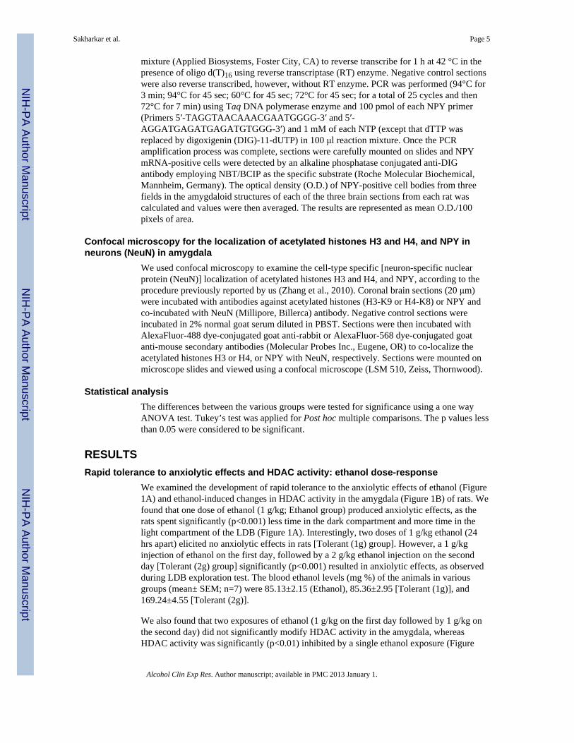
We examined the development of rapid tolerance to the anxiolytic effects of ethanol (Figure1A) and ethanol-induced changes in HDAC activity in the amygdala (Figure 1B) of rats. Wefound that one dose of ethanol (1 g/kg; Ethanol group) produced anxiolytic effects, as therats spent significantly (p<0.001) less time in the dark compartment and more time in thelight compartment of the LDB (Figure 1A). Interestingly, two doses of 1 g/kg ethanol (24hrs apart) elicited no anxiolytic effects in rats [Tolerant (1g) group]. However, a 1 g/kginjection of ethanol on the first day, followed by a 2 g/kg ethanol injection on the secondday [Tolerant (2g) group] significantly (p<0.001) resulted in anxiolytic effects, as observedduring LDB exploration test. The blood ethanol levels (mg %) of the animals in variousgroups (mean± SEM; n=7) were 85.13±2.15 (Ethanol), 85.36±2.95 [Tolerant (1g)], and169.24±4.55 [Tolerant (2g)].
We also found that two exposures of ethanol (1 g/kg on the first day followed by 1 g/kg onthe second day) did not significantly modify HDAC activity in the amygdala, whereasHDAC activity was significantly (p<0.01) inhibited by a single ethanol exposure (Figure
Sakharkar et al. Page 5
Alcohol Clin Exp Res. Author manuscript; available in PMC 2013 January 1.
NIH
-PA Author Manuscript
NIH
-PA Author Manuscript
NIH
-PA Author Manuscript
1B). In addition, tolerance to ethanol-induced HDAC inhibition in the amygdala wasreversed (p<0.05) by treatment with high doses of acute ethanol (1 g/kg ethanol on the firstday followed by 2 g/kg ethanol on the second day). These results suggest that rapidtolerance to the anxiolytic effects of acute ethanol may be related to the development ofcellular tolerance at the level of HDAC activity in the amygdala of rats. The second highdose (2 g/kg) but not low dose (1 g/kg) of ethanol exposure, elicited anxiolytic effects andinhibited HDAC activity, as observed after the first lower dose (1 g/kg) of ethanol exposure,suggesting that higher concentrations of ethanol are needed to achieve initial responses ofethanol exposure.
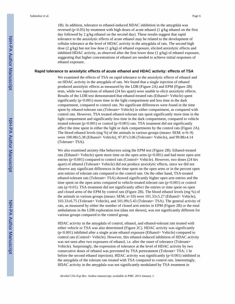
Rapid tolerance to anxiolytic effects of acute ethanol and HDAC activity: effects of TSAWe examined the effects of TSA on rapid tolerance to the anxiolytic effects of ethanol andon HDAC activity in the amygdala of rats. We found that a single injection of ethanolproduced anxiolytic effects as measured by the LDB (Figure 2A) and EPM (Figure 2B)tests, while two injections of ethanol (24 hrs apart) were unable to elicit anxiolytic effects.Results of the LDB test demonstrated that ethanol-treated rats (Ethanol+ Vehicle) spentsignificantly (p<0.001) more time in the light compartment and less time in the darkcompartment, compared to control rats. No significant differences were found in the timespent by ethanol-tolerant rats (Tolerant+ Vehicle) in either compartment, as compared withcontrol rats. However, TSA treated ethanol-tolerant rats spent significantly more time in thelight compartment and significantly less time in the dark compartment, compared to vehicletreated tolerant (p<0.001) or control (p<0.001) rats. TSA treatment did not significantlyaffect the time spent in either the light or dark compartments by the control rats (Figure 2A).The blood ethanol levels (mg %) of the animals in various groups (mean± SEM; n=6–9)were 100.08±5.38 (Ethanol+ Vehicle), 97.87±3.06 (Tolerant+ Vehicle), and 99.88±4.14(Tolerant+ TSA).
We also examined anxiety-like behaviors using the EPM test (Figure 2B). Ethanol-treatedrats (Ethanol+ Vehicle) spent more time on the open arms (p<0.001) and had more open armentries (p<0.001) compared to control rats (Control+ Vehicle). However, two doses (24 hrsapart) of ethanol (Tolerant+ Vehicle) did not produce anxiolytic effects, since we did notobserve any significant differences in the time spent on the open arms or in the percent openarm entries of tolerant rats compared to the control rats. On the other hand, TSA-treatedethanol-tolerant rats (Tolerant+ TSA) showed significantly higher open arm entries and thetime spent on the open arms compared to vehicle-treated tolerant rats (p<0.001) or controlrats (p<0.01). TSA treatment did not significantly affect the entries or time spent on openand closed arms of the EPM by control rats (Figure 2B). The blood ethanol levels (mg %) ofthe animals in various groups (mean± SEM; n=10) were 101.33±5.27 (Ethanol+ Vehicle),103.33±6.75 (Tolerant+ Vehicle), and 101.09±5.43 (Tolerant+ TSA). The general activity ofrats, as measured by either the number of closed arm entries in EPM (Figure 2B) or the totalambulations in the LDB exploration test (data not shown), was not significantly different forvarious groups compared to the control group.
HDAC activity in the amygdala of control, ethanol, and ethanol-tolerant rats treated witheither vehicle or TSA was also determined (Figure 2C). HDAC activity was significantly(p<0.001) inhibited after a single acute ethanol exposure (Ethanol+ Vehicle) compared tocontrol rats (Control+ Vehicle). However, this ethanol-induced inhibition of HDAC activitywas not seen after two exposures of ethanol, i.e. after the onset of tolerance (Tolerant+Vehicle). Surprisingly, the expression of tolerance at the level of HDAC activity by twoconsecutive doses of ethanol was prevented by TSA pretreatment (Tolerant+ TSA; 1 hrbefore the second ethanol injection). HDAC activity was significantly (p<0.001) inhibited inthe amygdala of the tolerant rats treated with TSA compared to control rats. Interestingly,HDAC activity in the amygdala was not significantly modulated by TSA treatment in
Sakharkar et al. Page 6
Alcohol Clin Exp Res. Author manuscript; available in PMC 2013 January 1.
NIH
-PA Author Manuscript
NIH
-PA Author Manuscript
NIH
-PA Author Manuscript
control rats (Figure 2C). These results suggest that rapid tolerance to the anxiolytic effects ofethanol may be due to development of tolerance to acute ethanol-induced inhibition ofHDAC activity in the amygdala of rats.
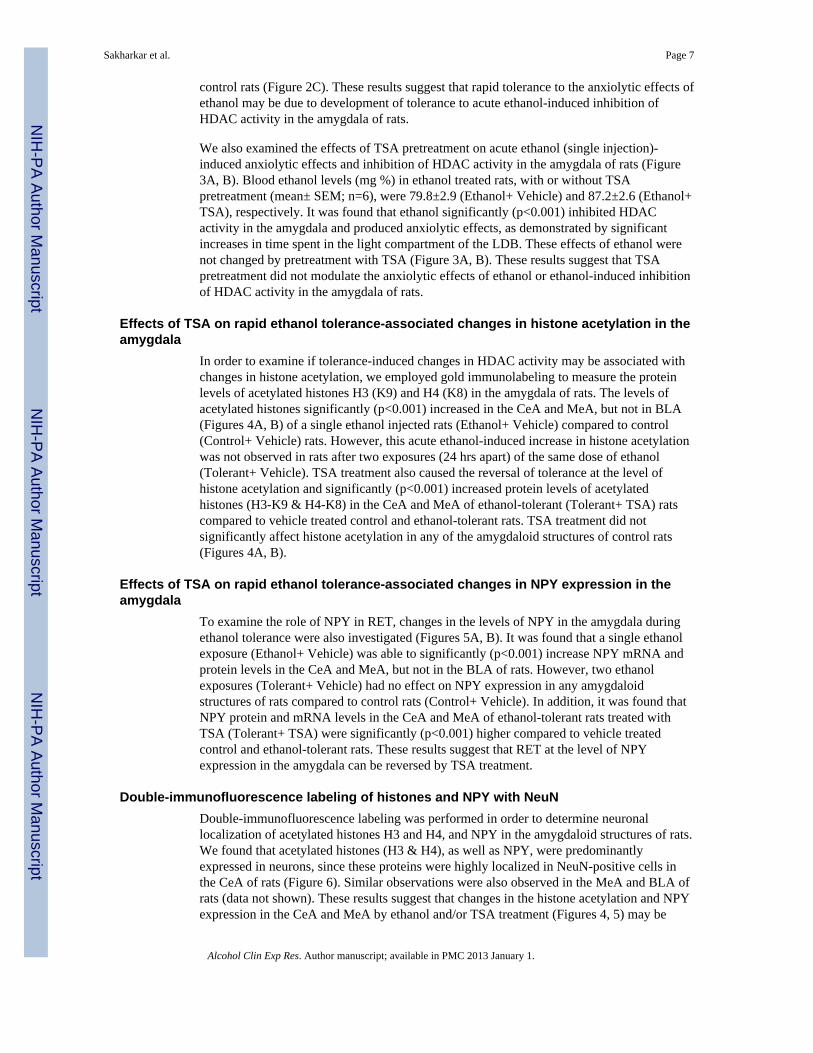
We also examined the effects of TSA pretreatment on acute ethanol (single injection)-induced anxiolytic effects and inhibition of HDAC activity in the amygdala of rats (Figure3A, B). Blood ethanol levels (mg %) in ethanol treated rats, with or without TSApretreatment (mean± SEM; n=6), were 79.8±2.9 (Ethanol+ Vehicle) and 87.2±2.6 (Ethanol+TSA), respectively. It was found that ethanol significantly (p<0.001) inhibited HDACactivity in the amygdala and produced anxiolytic effects, as demonstrated by significantincreases in time spent in the light compartment of the LDB. These effects of ethanol werenot changed by pretreatment with TSA (Figure 3A, B). These results suggest that TSApretreatment did not modulate the anxiolytic effects of ethanol or ethanol-induced inhibitionof HDAC activity in the amygdala of rats.
Effects of TSA on rapid ethanol tolerance-associated changes in histone acetylation in theamygdala
In order to examine if tolerance-induced changes in HDAC activity may be associated withchanges in histone acetylation, we employed gold immunolabeling to measure the proteinlevels of acetylated histones H3 (K9) and H4 (K8) in the amygdala of rats. The levels ofacetylated histones significantly (p<0.001) increased in the CeA and MeA, but not in BLA(Figures 4A, B) of a single ethanol injected rats (Ethanol+ Vehicle) compared to control(Control+ Vehicle) rats. However, this acute ethanol-induced increase in histone acetylationwas not observed in rats after two exposures (24 hrs apart) of the same dose of ethanol(Tolerant+ Vehicle). TSA treatment also caused the reversal of tolerance at the level ofhistone acetylation and significantly (p<0.001) increased protein levels of acetylatedhistones (H3-K9 & H4-K8) in the CeA and MeA of ethanol-tolerant (Tolerant+ TSA) ratscompared to vehicle treated control and ethanol-tolerant rats. TSA treatment did notsignificantly affect histone acetylation in any of the amygdaloid structures of control rats(Figures 4A, B).
Effects of TSA on rapid ethanol tolerance-associated changes in NPY expression in theamygdala
To examine the role of NPY in RET, changes in the levels of NPY in the amygdala duringethanol tolerance were also investigated (Figures 5A, B). It was found that a single ethanolexposure (Ethanol+ Vehicle) was able to significantly (p<0.001) increase NPY mRNA andprotein levels in the CeA and MeA, but not in the BLA of rats. However, two ethanolexposures (Tolerant+ Vehicle) had no effect on NPY expression in any amygdaloidstructures of rats compared to control rats (Control+ Vehicle). In addition, it was found thatNPY protein and mRNA levels in the CeA and MeA of ethanol-tolerant rats treated withTSA (Tolerant+ TSA) were significantly (p<0.001) higher compared to vehicle treatedcontrol and ethanol-tolerant rats. These results suggest that RET at the level of NPYexpression in the amygdala can be reversed by TSA treatment.
Double-immunofluorescence labeling of histones and NPY with NeuNDouble-immunofluorescence labeling was performed in order to determine neuronallocalization of acetylated histones H3 and H4, and NPY in the amygdaloid structures of rats.We found that acetylated histones (H3 & H4), as well as NPY, were predominantlyexpressed in neurons, since these proteins were highly localized in NeuN-positive cells inthe CeA of rats (Figure 6). Similar observations were also observed in the MeA and BLA ofrats (data not shown). These results suggest that changes in the histone acetylation and NPYexpression in the CeA and MeA by ethanol and/or TSA treatment (Figures 4, 5) may be
Sakharkar et al. Page 7
Alcohol Clin Exp Res. Author manuscript; available in PMC 2013 January 1.
NIH
-PA Author Manuscript
NIH
-PA Author Manuscript
NIH
-PA Author Manuscript

neuron-specific, since these proteins were observed to be predominantly localized inneuronal cells (Figure 6).
DISCUSSIONThe results of the present investigation show that cellular tolerance to the ethanol-inducedinhibition of HDAC and increased histone acetylation and NPY expression in the amygdalaof rats may be operative in the development of rapid tolerance to the anxiolytic effects ofethanol (Figure 7). Several clinical and preclinical studies indicate a relationship betweenanxiety and alcoholism, suggesting that the anxiolytic properties of ethanol may be crucialin promoting drinking in an attempt to self-medicate (Carrigan and Randall, 2003; Cloningeret al., 1988; Cooper et al., 1995; Conway et al., 2006; Debatin and Barbosa, 2006; Lipscombet al., 1980; Merikangas et al., 1998; Pandey et al., 2005; Primeaux et al., 2006; Schuckitand Hesselbrock, 1994; Zhang et al., 2010). The phenomenon of rapid tolerance to theanxiolytic effects of ethanol has been shown in both humans and animal models (Cloningeret al., 1988; Debatin and Barbosa, 2006; Koob et al., 1987; Libscomb et al., 1980). Here, weestablished a novel role for HDAC-induced histone modifications in the amygdala in themolecular mechanisms of RET. Tolerance to both the inhibitory properties of HDACs and tothe anxiolytic effects of ethanol were observed after two consecutive days of ethanol (1 g/kg) exposure. However, treatment with a higher dose (2 g/kg) of ethanol on the second dayproduced anxiolytic effects and inhibited HDAC activity in the amygdala. This findingsupports the notion that rapid tolerance to the anxiolytic effects of ethanol may promotedrinking in order to achieve a response similar to that obtained from an initial ethanolexposure. Using Drosophila melanogaster as a model, it was found that covalent histonemodifications and induction of BK-type Ca2+-activated K+ channel gene may be linked tobehavioral tolerance to the sedative effects of benzyl alcohol (Wang et al., 2007). Thus,covalent histone modifications represent an attractive mechanism that may regulate geneexpression during the process of alcohol tolerance.
Eleven different zinc-dependent HDAC isoforms have been identified and categorized aseither class I (HDAC 1–3, 8), class II (HDAC 4–7, 9, 10), or class IV (HDAC 11) HDACs.Class III HDACs are known as sirtuins and require NAD+ co-factor for activation (Abel andZukin, 2008; Chuang et al., 2009; Lee et al., 2008; Xu et al., 2007). It has been shown thatrepeated ethanol exposure has no effect on histone (H3 & H4) acetylation in several brainregions, such as the frontal cortex or nucleus accumbens (NAc) of rats (Pascual et al., 2009).Our previous studies (Pandey et al., 2008) showed that chronic ethanol exposure had nosignificant effect on HDAC activity, histone acetylation, or NPY expression in theamygdala. Chronic ethanol exposure also did not elicit anxiety-like behaviors in rats,whereas these behaviors appeared during withdrawal. HDAC activity significantly wasincreased in the amygdala during withdrawal after chronic ethanol exposure, whichcorrelated with decreased acetylation of histones (H3 & H4) and decreased expression ofNPY in the CeA and MeA, but not in BLA of rats. Treatment with a pan-HDAC inhibitor,TSA, attenuated the development of anxiety-like behaviors during ethanol withdrawal andcorrected ethanol withdrawal-induced deficits in both histone acetylation and NPYexpression in amygdaloid structures of rats (Pandey et al., 2008). Acute ethanol treatmentalso inhibited HDAC activity in the amygdala and produced anxiolytic effects in rats(Pandey et al., 2008). We extended these studies and found that HDAC-induced histonemodifications may be involved in the process of RET (Figure 7). Furthermore, the lack ofchanges in HDAC activity, histone acetylation, and NPY expression in the amygdala duringchronic ethanol exposure (Pandey et al., 2008) and RET suggest that similar epigeneticmechanisms may exist in the process of CET and RET. It is important to mention that themajority of acetylated histones H3 and H4, as well as NPY proteins, were co-localized withNeuN-positive neurons in amygdaloid structures. Therefore, it is possible that ethanol-
Sakharkar et al. Page 8
Alcohol Clin Exp Res. Author manuscript; available in PMC 2013 January 1.
NIH
-PA Author Manuscript
NIH
-PA Author Manuscript
NIH
-PA Author Manuscript

induced chromatin remodeling may orchestrate different patterns of gene expressionprofiling within neuronal networks in the amygdala that may contribute to the developmentof RET.
HDACs have been emerging as potential therapeutic targets in the treatment ofneurodegenerative diseases and psychiatric disorders, including addictive behaviors (Abeland Zukin, 2008; Kazantsev and Thompson, 2008; Kumar et al., 2005; Renthal and Nestler,2008; Romieu et al., 2008; Tsakova et al., 2007). Recently, studies involving manipulationof specific HDAC isoforms suggest the involvement of HDAC 2, HDAC 4, and HDAC 5 inthe pathogenesis of several brain disorders. For example, the hippocampal HDAC 5 isoformand HDAC 2 isoform in the NAc were both shown to play roles in depressive behaviors andin the mechanism of action of antidepressant drugs (Covington et al., 2009; Tsankova et al.,2006). In addition, the HDAC 2 isoform was found to negatively regulate synaptic plasticityrelated to memory formation (Guan et al., 2009), while the HDAC 4 isoform may beinvolved in neuronal cell death and cocaine addiction (Bolger and Yao, 2005; Kumar et al.,2005). These results clearly display distinct roles for specific HDAC isoforms in variouspsychiatric diseases. Future studies are needed to investigate the specific roles of HDACisoforms in chromatin remodeling during alcoholism. Nonetheless, perturbing HDACs (classI & II) by pharmacological means has revealed a critical role for HDAC-induced histonemodifications in the amygdala during the development of RET. It is important to point outthat TSA treatment had no effect on HDAC activity, histone acetylation, or NPY expressionin control rats, similar to findings from our previous study (Pandey et al., 2008). However,when HDACs were perturbed, as in ethanol tolerant and dependent rats, TSA was able toreverse RET and block the development of anxiety-like behaviors during withdrawal afterchronic ethanol exposure in rats (Pandey et al., 2008). These findings are also supported bythe observations that TSA pretreatment did not modulate the acute ethanol-inducedanxiolytic effects or inhibition of HDAC activity in the amygdala of rats. Similarly, otherinvestigators (Kumar et al., 2005) have shown that HDAC inhibitors alone have no effectson striatal histone phospho-acetylation or histone acetylation-mediated changes in c-fosexpression in control rats, but significantly modulated the cocaine-mediated responses onthese measures.
RET may be mediated by histone modification-induced gene expression in the amygdala orother brain regions. One such mechanism could involve changes in NPY expression in theCeA and MeA. NPY is an endogenous anxiolytic compound and higher levels of NPY in theamygdala, particularly in the CeA, have been shown to be anxiolytic, whereas lower levelsof NPY have been shown to be anxiogenic in various animal models (Gilpin et al., 2008;Heilig 1995; Heilig and Widerlov, 1995; Heilig et al., 1989; Pandey 2004; Pandey et al.,2005, 2008; Primeaux et al., 2006; Thorsell et al., 2007; Zhang et al., 2010). We found thatacute ethanol increased NPY expression in the CeA and MeA, whereas NPY levels were notaltered in ethanol-tolerant rats. However, TSA treatment was able to increase NPYexpression in the CeA and MeA of ethanol-tolerant, but not of control rats. These resultssuggest that rapid induction of NPY by acute ethanol and an observed lack of response inNPY expression during ethanol tolerance may be regulated via HDAC-induced epigeneticchanges in the amygdala. Several other mechanisms related to neurotransmitters such asglutamate, gamma aminobutyric acid, and arginine-vasopressin, and cyclic-AMP signalingsystems have been implicated in acute ethanol tolerance (Hoffman and Tabakoff, 1989;Kalant 1998; Kumar et al., 2009; Yang et al., 2003). A recent genomic study showed thatgenes involved in synaptic plasticity may also play a role in AFT (Hu et al., 2008). Futurestudies are needed to examine if HDAC-induced histone modifications in the amygdaladuring ethanol exposure is responsible for changes in the expression of genes, other thanNPY, and their possible roles in RET.
Sakharkar et al. Page 9
Alcohol Clin Exp Res. Author manuscript; available in PMC 2013 January 1.
NIH
-PA Author Manuscript
NIH
-PA Author Manuscript
NIH
-PA Author Manuscript

These results for the first time suggest that the treatment with HDAC inhibitors, such asTSA, was able to reverse rapid tolerance to the anxiolytic effects of ethanol via HDAC-induced epigenetic changes in the amygdala (Fig. 7). These findings, along with ourprevious report on alcohol dependence (Pandey et al., 2008), suggest that HDAC-inducedhistone modifications in the amygdala may be involved in the process of alcoholism.
AcknowledgmentsThis work was supported by National Institute on Alcohol Abuse and Alcoholism Grants AA-016690, AA-019971,AA-010005, and AA-013341 and by the Department of Veterans Affairs (Merit Review Grant; Research CareerScientist award) to S.C.P.
ReferencesAbel T, Zukin RS. Epigenetic targets of HDAC inhibition in neurodegenerative and psychiatric
disorders. Curr Opin Pharmacol. 2008; 8:57–64. [PubMed: 18206423]American Psychiatric Association. Diagnostic and Statistical Manual of Mental Disorders. 4.
American Psychiatric Press; Washington DC, USA: 1994.Bolger TA, Yao TP. Intracellular trafficking of histone deacetylase 4 regulates neuronal cell death. J
Neurosci. 2005; 25:9544–9553. [PubMed: 16221865]Carrigan MH, Randall CL. Self-medication in social phobia: a review of alcohol literature. Addict
Behav. 2003; 28:269–284. [PubMed: 12573678]Chandler LJ, Harris RA, Crews FT. Ethanol tolerance and synaptic plasticity. Trends Pharmacol Sci.
1998; 19:491–495. [PubMed: 9871410]Chuang DM, Leng Y, Marinova Z, Kim HJ, Chiu CT. Multiple roles of HDAC inhibition in
neurodegenerative conditions. Trends Neurosci. 2009; 32:591–601. [PubMed: 19775759]Cloninger CR, Sigvardsson S, Gilligan SB, von Knorring AL, Reich T, Bohman M. Genetic
heterogeneity and the classification of alcoholism. Adv Alcohol Subst Abuse. 1988; 7:3–16.[PubMed: 3066194]
Cooper ML, Frone MR, Russell M, Mudar P. Drinking to regulate positive and negative emotions: amotivational model of alcohol use. J Pers Soc Psychol. 1995; 69:990–1005. [PubMed: 7473043]
Conway KP, Compton W, Stinson FS, Grant BF. Lifetime comorbidity of DSM-IV mood and anxietydisorders and specific drug use disorders: results from the National Epidemiologic Survey onAlcohol and Related Conditions. J Clin Psychiatry. 2006; 67:247–257. [PubMed: 16566620]
Covington HE 3rd, Maze I, LaPlant QC, Vialou VF, Ohnishi YN, Berton O, Fass DM, Renthal W,Rush AJ 3rd, Wu EY, Ghose S, Krishnan V, Russo SJ, Tamminga C, Haggarty SJ, Nestler EJ.Antidepressant actions of histone deacetylase inhibitors. J Neurosci. 2009; 29:11451–11460.[PubMed: 19759294]
Crabbe JC, Rigter H, Uijlen J, Strijbos C. Rapid development of tolerance to the hypothermic effect ofethanol in mice. J Pharmacol Exp Ther. 1979; 208:128–133. [PubMed: 759607]
Davis M. Neurobiology of fear responses: the role of the amygdala. J Neuropsychiatry Clin Neurosci.1997; 9:382–402. [PubMed: 9276841]
Debatin T, Barbosa AD. Effect of isopregnanolone on rapid tolerance to the anxiolytic effect ofethanol. Rev Bras Psiquiatr. 2006; 28:18–23. [PubMed: 16612485]
Feng J, Fan G. The role of DNA methylation in the central nervous system and neuropsychiatricdisorders. Int Rev Neurobiol. 2009; 89:67–84. [PubMed: 19900616]
File SE. The interplay of learning and anxiety in the elevated plus-maze. Behav Brain Res. 1993;58:199–202. [PubMed: 8136046]
Gill K, Deitrich RA. Acute tolerance to the ataxic effects of ethanol in short-sleep (SS) and long-sleep(LS) mice. Psychopharmacology (Berl). 1998; 136:91–98. [PubMed: 9537687]
Gilpin NW, Misra K, Koob GF. Neuropeptide Y in the central nucleus of the amygdala suppressesdependence-induced increases in alcohol drinking. Pharmacol Biochem Behav. 2008; 90:475–480.[PubMed: 18501411]
Sakharkar et al. Page 10
Alcohol Clin Exp Res. Author manuscript; available in PMC 2013 January 1.
NIH
-PA Author Manuscript
NIH
-PA Author Manuscript
NIH
-PA Author Manuscript
Grayson DR, Kundakovic M, Sharma RP. Is there a future for histone deacetylase inhibitors in thepharmacotherapy of psychiatric disorders? Mol Pharmacol. 2010; 77:126–135. [PubMed:19917878]
Guan JS, Haggarty SJ, Giacometti E, Dannenberg JH, Joseph N, Gao J, Nieland TJ, Zhou Y, Wang X,Mazitschek R, Bradner JE, DePinho RA, Jaenisch R, Tsai LH. HDAC2 negatively regulatesmemory formation and synaptic plasticity. Nature. 2009; 459:55–60. [PubMed: 19424149]
Heilig M. Antisense inhibition of neuropeptide Y (NPY)-Y1 receptor expression blocks the anxiolytic-like action of NPY in amygdala and paradoxically increases feeding. Regul Pept. 1995; 59:201–205. [PubMed: 8584755]
Heilig M, Söderpalm B, Engel JA, Widerlöv E. Centrally administered neuropeptide Y (NPY)produces anxiolytic-like effects in animal anxiety models. Psychopharmacology (Berl). 1989;98:524–529. [PubMed: 2570434]
Heilig M, Widerlöv E. Neurobiology and clinical aspects of neuropeptide Y. Crit Rev Neurobiol.1995; 9:115–136. [PubMed: 8581979]
Hoffman PL, Tabakoff B. Mechanisms of alcohol tolerance. Alcohol Alcohol. 1989; 24:251–252.[PubMed: 2757699]
Hu W, Saba L, Kechris K, Bhave SV, Hoffman PL, Tabakoff B. Genomic insights into acute alcoholtolerance. J Pharmacol Exp Ther. 2008; 326:792–800. [PubMed: 18550690]
Kalant H. Research on tolerance: what can we learn from history? Alcohol Clin Exp Res. 1998; 22:67–76. [PubMed: 9514287]
Kalkhoven E. CBP and p300: HATs for different occasions. Biochem Pharmacol. 2004; 68:1145–1155. [PubMed: 15313412]
Kazantsev AG, Thompson LM. Therapeutic application of histone deacetylase inhibitors for centralnervous system disorders. Nat Rev Drug Discov. 2008; 7:854–868. [PubMed: 18827828]
Khanna JM, Chau A, Shah G. Characterization of the phenomenon of rapid tolerance to ethanol.Alcohol. 1996; 13:621–628. [PubMed: 8949959]
Khanna JM, Kalant H, Shah G, Weiner J. Rapid tolerance as an index of chronic tolerance. PharmacolBiochem Behav. 1991; 38:427–432. [PubMed: 2057511]
Koob GF. Alcoholism: allostasis and beyond. Alcohol Clin Exp Res. 2003; 27:232–243. [PubMed:12605072]
Koob GF, Wall TL, Schafer J. Rapid induction of tolerance to the antipunishment effects of ethanol.Alcohol. 1987; 4:481–484. [PubMed: 3435637]
Korzus E, Rosenfeld MG, Mayford M. CBP histone acetyltransferase activity is a critical componentof memory consolidation. Neuron. 2004; 42:961–972. [PubMed: 15207240]
Kumar A, Choi KH, Renthal W, Tsankova NM, Theobald DE, Truong HT, Russo SJ, Laplant Q,Sasaki TS, Whistler KN, Neve RL, Self DW, Nestler EJ. Chromatin remodeling is a keymechanism underlying cocaine induced plasticity in striatum. Neuron. 2005; 48:303–314.[PubMed: 16242410]
Kumar S, Porcu P, Werner DF, Matthews DB, Diaz-Granados JL, Helfand RS, Morrow AL. The roleof GABAA receptors in the acute and chronic effects of ethanol: a decade of progress.Psychopharmacol (Berl). 2009; 205:529–564.
Kurtz DL, Stewart RB, Zweifel M, Li TK, Froehlich JC. Genetic differences in tolerance andsensitization to the sedative/hypnotic effects of alcohol. Pharmacol Biochem Behav. 1996;53:585–591. [PubMed: 8866959]
Lee MJ, Kim YS, Kummar S, Giaccone G, Trepel JB. Histone deacetylase inhibitors in cancer therapy.Curr Opin Oncol. 2008; 20:639–649. [PubMed: 18841045]
Lipscomb TR, Nathan PE, Wilson GT, Abrams DB. Effects of tolerance on the anxiety-reducingfunction of alcohol. Arch Gen Psychiatry. 1980; 37:577–582. [PubMed: 7377915]
McBride WJ. Central nucleus of the amygdala and the effects of alcohol and alcohol-drinking behaviorin rodents. Pharmacol Biochem Behav. 2002; 71:509–515. [PubMed: 11830185]
Mellanby E. Alcohol: its absorption into and disappearance from blood under different conditions.Medical Research Committee Special Report. 1919; 31:1–48.
Sakharkar et al. Page 11
Alcohol Clin Exp Res. Author manuscript; available in PMC 2013 January 1.
NIH
-PA Author Manuscript
NIH
-PA Author Manuscript
NIH
-PA Author Manuscript

Merikangas KR, Stevens DE, Fenton B, Stolar M, O’Malley S, Woods SW, Risch N. Co-morbidityand familial aggregation of alcoholism and anxiety disorders. Psychol Med. 1998; 28:773–788.[PubMed: 9723135]
Moberg CA, Curtin JJ. Alcohol selectively reduces anxiety but not fear: startle response duringunpredictable vs. predictable threat. J Abnorm Psychol. 2009; 118:335–347. [PubMed: 19413408]
Pandey SC. The gene transcription factor cyclic AMP-responsive element binding protein: role inpositive and negative affective states of alcohol addiction. Pharmacol Ther. 2004; 104:47–58.[PubMed: 15500908]
Pandey SC, Ugale R, Zhang H, Tang L, Prakash A. Brain chromatin remodeling: a novel mechanismof alcoholism. J Neurosci. 2008; 28:3729–3737. [PubMed: 18385331]
Pandey SC, Zhang H, Roy A, Misra K. Central and medial amygdaloid brain-derived neurotrophicfactor signaling plays a critical role in alcohol-drinking and anxiety-like behaviors. J Neurosci.2006; 26:8320–8331. [PubMed: 16899727]
Pandey SC, Zhang H, Roy A, Xu T. Deficits in amygdaloid cAMP-responsive element-binding proteinsignaling play a role in genetic predisposition to anxiety and alcoholism. J Clin Invest. 2005;115:2762–2773. [PubMed: 16200210]
Pascual M, Boix J, Felipo V, Guerri C. Repeated alcohol administration during adolescence causeschanges in the mesolimbic dopaminergic and glutamatergic systems and promotes alcohol intakein the adult rat. J Neurochem. 2009; 108:920–931. [PubMed: 19077056]
Primeaux SD, Wilson SP, Bray GA, York DA, Wilson MA. Overexpression of neuropeptide Y in thecentral nucleus of the amygdala decreases ethanol self-administration in “anxious” rats. AlcoholClin Exp Res. 2006; 30:791–801. [PubMed: 16634847]
Renthal W, Nestler EJ. Epigenetic mechanisms in drug addiction. Trends Mol Med. 2008; 14:341–350.[PubMed: 18635399]
Romieu P, Host L, Gobaille S, Sandner G, Aunis D, Zwiller J. Histone deacetylase inhibitors decreasecocaine but not sucrose self-administration in Rats. J Neurosci. 2008; 28:9342–9348. [PubMed:18799668]
Schuckit MA, Hesselbrock V. Alcohol dependence and anxiety disorders: what is the relationship? AmJ Psychiatry. 1994; 151:1723–1734. [PubMed: 7977877]
Smith MM. Histone structure and function. Curr Opin Cell Biol. 1991; 3:429–437. [PubMed:1892654]
Strahl BD, Allis CD. The language of covalent histone modifications. Nature. 2000; 403:41–45.[PubMed: 10638745]
Tabakoff B, Cornell N, Hoffman PL. Alcohol tolerance. Ann Emerg Med. 1986; 15:1005–1012.[PubMed: 3526989]
Thorsell A, Repunte-Canonigo V, O’Dell LE, Chen SA, King AR, Lekic D, Koob GF, Sanna PP. Viralvector-induced amygdala NPY overexpression reverses increased alcohol intake caused byrepeated deprivations in Wistar rats. Brain. 2007; 130:1330–1337. [PubMed: 17405766]
Tsankova NM, Berton O, Renthal W, Kumar A, Neve RL, Nestler EJ. Sustained hippocampalchromatin regulation in a mouse model of depression and antidepressant action. Nat Neurosci.2006; 9:519–525. [PubMed: 16501568]
Tsankova N, Renthal W, Kumar A, Nestler EJ. Epigenetic regulation in psychiatric disorders. Nat RevNeurosci. 2007; 8:355–367. [PubMed: 17453016]
Wang Y, Krishnan HR, Ghezzi A, Yin JC, Atkinson NS. Drug-induced epigenetic changes producedrug tolerance. PLoS Biol. 2007; 5:2342–2353.
Xu WS, Parmigiani RB, Marks PA. Histone deacetylase inhibitors: molecular mechanisms of action.Oncogene. 2007; 26:5541–5552. [PubMed: 17694093]
Yang X, Oswald L, Wand G. The cyclic AMP/protein kinase A signal transduction pathway modulatestolerance to sedative and hypothermic effects of ethanol. Alcohol Clin Exp Res. 2003; 27:1220–1225. [PubMed: 12966313]
Zhang H, Sakharkar AJ, Shi G, Ugale R, Prakash A, Pandey SC. Neuropeptide Y signaling in thecentral nucleus of amygdala regulates alcohol-drinking and anxiety-like behaviors of alcohol-preferring rats. Alcohol Clin Exp Res. 2010; 34:451–461. [PubMed: 20028368]
Sakharkar et al. Page 12
Alcohol Clin Exp Res. Author manuscript; available in PMC 2013 January 1.
NIH
-PA Author Manuscript
NIH
-PA Author Manuscript
NIH
-PA Author Manuscript

Figure 1.The effects of acute ethanol exposure [Ethanol group (1 g/kg; I.P.)] and tolerance (1 g/kg or2 g/kg) [Tolerant (1g) group or Tolerant (2g) group], on the light/dark box (LDB)exploration test of anxiety-like behavior (A) and HDAC activity in the amygdala of rats (B).Values are the mean ± SEM of 7 rats in each group. *Significantly different from theirrespective control groups [p<0.05-0.001; ANOVA (F 3, 24 =112, p<0.001 for LDB;F3, 24=8.6, p<0.001 for HDAC activity) followed by Tukey’s test].
Sakharkar et al. Page 13
Alcohol Clin Exp Res. Author manuscript; available in PMC 2013 January 1.
NIH
-PA Author Manuscript
NIH
-PA Author Manuscript
NIH
-PA Author Manuscript

Figure 2.The effects of acute ethanol exposure [Ethanol group (1 g/kg; I.P.)] and tolerance (1 g/kg)with (Tolerant+ TSA) or without TSA (Tolerant+ Vehicle) treatment on anxiety-likebehavior in rats using the light/dark box exploration (LDB) test (A) and elevated plus maze(EPM) test (B), and HDAC activity in the amygdala (C). Values are the mean ± SEM of 6–9rats for the LDB exploration test and HDAC activity measurement, whereas 10 rats pergroup for the EPM test. *Significantly different from their respective control groups[p<0.01-0.001; ANOVA (F4, 34=29.9, p<0.001 for LDB; F4, 45=16.8, p<0.001 for EPM %open arm time spent; F4, 45=36.5, p<0.001 for EPM % open arm entries; F4, 34=13.9,p<0.001 for HDAC activity) followed by Tukey’s test].
Sakharkar et al. Page 14
Alcohol Clin Exp Res. Author manuscript; available in PMC 2013 January 1.
NIH
-PA Author Manuscript
NIH
-PA Author Manuscript
NIH
-PA Author Manuscript

Figure 3.The effects of TSA pretreatment on acute ethanol exposure (1g/kg; I.P.)-mediated anxiolyticeffects in rats using the light/dark box exploration (LDB) test (A) and inhibition of HDACactivity in the amygdala (B). Values are the mean ± SEM of 6 rats. *Significantly differentfrom control group [p<0.001; ANOVA (F 2, 15 =18.0, p<0.001 for LDB; F2, 15=49.9,p<0.001 for HDAC activity) followed by Tukey’s test].
Sakharkar et al. Page 15
Alcohol Clin Exp Res. Author manuscript; available in PMC 2013 January 1.
NIH
-PA Author Manuscript
NIH
-PA Author Manuscript
NIH
-PA Author Manuscript

Figure 4.A) Representative low-magnification photomicrographs of acetylated histones H3 (Lys 9)and H4 (Lys 8) gold-immunolabeling in central amygdaloid (CeA) structures of control ratswith (Control+ TSA) or without TSA (Control+ Vehicle) treatments, ethanol-treated rats(Ethanol+ Vehicle) or ethanol-tolerant rats with (Tolerant+ TSA) or without TSA (Tolerant+Vehicle) treatments (Scale bar = 40 μm). B) Changes in the acetylation of histones H3 andH4 in various amygdaloid (CeA, MeA, and BLA) structures of control rats with (Control+TSA) or without (Control+ Vehicle) TSA treatments, ethanol-treated rats (Ethanol+ Vehicle)and ethanol-tolerant rats with (Tolerant+ TSA) or without (Tolerant+ Vehicle) TSAtreatments. Values are the mean ± SEM of 5–6 rats per group. *Significantly different fromtheir respective control groups [p<0.001; ANOVA (acetylated histone H3: CeA,
Sakharkar et al. Page 16
Alcohol Clin Exp Res. Author manuscript; available in PMC 2013 January 1.
NIH
-PA Author Manuscript
NIH
-PA Author Manuscript
NIH
-PA Author Manuscript

F4, 25=117.7, p<0.001; MeA, F4, 25=95, p<0.001; Acetylated histone H4: CeA, F4, 20=104.8,p<0.001; MeA, F4, 20=98.8, p<0.001) followed by Tukey’s test)].
Sakharkar et al. Page 17
Alcohol Clin Exp Res. Author manuscript; available in PMC 2013 January 1.
NIH
-PA Author Manuscript
NIH
-PA Author Manuscript
NIH
-PA Author Manuscript

Figure 5.A) Representative low-magnification photomicrographs of NPY in situ RT-PCR (mRNAlevels) and NPY gold-immunolabeling (protein levels) in central amygdaloid (CeA)structures of control rats with (Control+ TSA) or without TSA (Control+ Vehicle)treatments, ethanol-treated (Ethanol+ Vehicle) or ethanol-tolerant rats with (Tolerant+ TSA)or without TSA (Tolerant+ Vehicle) treatments (Scale bar= 40 μm). B) Changes in mRNAand protein levels of NPY in CeA, MeA, and BLA of control rats with (Control+ TSA) orwithout TSA (Control+ Vehicle) treatments, ethanol-treated (Ethanol+ Vehicle) or ethanol-tolerant rats with (Tolerant+ TSA) or without TSA (Tolerant+ Vehicle) treatments. Valuesare the mean ± SEM of 5 rats in each group. *Significantly different from their respectivecontrol groups (p<0.001; ANOVA (NPY protein: CeA, F4, 20=122.1, p<0.001; MeA,F4, 20=159.8, p<0.001; NPY mRNA: CeA, F 4, 20=83.1, p<0.001; MeA, F4, 20=89, p<0.001)followed by Tukey’s test)].
Sakharkar et al. Page 18
Alcohol Clin Exp Res. Author manuscript; available in PMC 2013 January 1.
NIH
-PA Author Manuscript
NIH
-PA Author Manuscript
NIH
-PA Author Manuscript

Figure 6.Representative photomicrographs showing immunofluorescence staining of acetylatedhistones H3 or H4, or NPY (green), and NeuN (red) in the cells of the central nucleus ofamygdala (CeA). The yellow color represents localization of acetylated histones and NPY inNeuN-positive cells. Acetylated histones H3 and H4, and NPY are predominantly expressedin NeuN-positive cells. Scale bar = 50 μm.
Sakharkar et al. Page 19
Alcohol Clin Exp Res. Author manuscript; available in PMC 2013 January 1.
NIH
-PA Author Manuscript
NIH
-PA Author Manuscript
NIH
-PA Author Manuscript

Figure 7.A schematic model depicting the possible molecular mechanisms of rapid tolerance to theanxiolytic effects of ethanol, within the amygdaloid neurocircuitries (particularly central andmedial amygdaloid structures). First acute ethanol exposure (1 g/kg) inhibits histonedeacetylases (HDAC), thereby increasing histone acetylation (Ac). Increased histoneacetylation opens the chromatin structure (relaxed chromatin), making it more accessible totranscriptional machinery, thereby increasing neuropeptide Y (NPY) gene expression, andultimately eliciting an anxiolytic response. The second ethanol exposure using the same dose(1 g/kg) as the first ethanol exposure does not inhibit HDAC activity or increase histoneacetylation, maintaining normal chromatin structure and no change in NPY expression oranxiety levels, i.e. development of rapid tolerance. However, treatment with the HDACinhibitor, trichostatin A (TSA), prior to the second ethanol exposure (1 g/kg), prevents theexpression of rapid ethanol tolerance by inhibiting HDAC and increasing both histoneacetylation and NPY levels. On the other hand, a second ethanol exposure using a higherdose of ethanol (2 g/kg) elicited a similar response (inhibition of HDACs and anxiolyticeffects) observed after the first ethanol exposure using a lower dose (1 g/kg), suggesting thathigher concentrations of ethanol are needed during a second exposure to ethanol in order toovercome the onset of tolerance and experience the anxiolytic effects of ethanol. This maylead to development of alcohol addiction.
Sakharkar et al. Page 20
Alcohol Clin Exp Res. Author manuscript; available in PMC 2013 January 1.
NIH
-PA Author Manuscript
NIH
-PA Author Manuscript
NIH
-PA Author Manuscript


![[Procedural learning and anxiolytic effects: electroencephalographic, motor and attentional measures]](https://img.pdfslide.net/doc/110x75/635edc552dc9f5f89b06f4a2/procedural-learning-and-anxiolytic-effects-electroencephalographic-motor-and.jpg)


