Embed Size (px)
Citation preview

Progress Toward Complete Life Cycle Culturingof the Endangered Sunflower Star, Pycnopodia helianthoides
J. HODIN*, A. PEARSON-LUND
†, F. P. ANTEAU, P. KITAEFF, AND S. CEFALU
Friday Harbor Laboratories, University of Washington, Friday Harbor, Washington 98250
Abstract.q1 Until recently, the sunflower star,Pycnopodia he-lianthoides, was a dominant and common predator in a widevariety of benthic habitats in the NE Pacific. Then, in 2013, itspopulations began to plummet across its entire range as a re-sult of the spread of a phenomenon known as sea star wastingdisease, or sea star wasting. Although dozens of sea star spe-cies were impacted by this wasting event, P. helianthoidesseems to have suffered the greatest losses and is now listedby the International Union for the Conservation of Nature asthe first critically endangered sea star. In order to learn moreabout the life history of this endangered predator and to ex-plore the potential for its restoration, we have initiated a cap-tive rearing program to attempt complete life cycle (egg-to-egg) culture for P. helianthoides. We report our observationson holding and distinguishing individual adults, reproductiveseasonality, larval development, inducers of settlement, andearly juvenile growth and feeding. These efforts will promoteand help guide conservation interventions to protect remain-ing populations of this species in the wild and facilitate its ul-timate return.
Introduction
In 2013, a mass mortality of asteroids caused by sea starwasting disease, or sea star wasting (SSW), was first observedin the NE Pacific. By 2016, every known species of asteroidin the regionwas impacted, from obligatorily intertidal taxa todeep-sea specialists. Perhaps the species thatwasmost severely
affected by SSW was the sunflower star, Pycnopodia heli-anthoides (Brandt, 1835). Populations across its entire range,from the Alaskan Aleutian Islands to Baja California, were re-duced, in some cases quite severely (Harvell et al., 2019). In-deed, this formerly common intertidal and subtidal speciesmayhave been extirpated from the southern third of its range (Gra-vem, 2020). In December 2020, P. helianthoides was listed ascritically endangered by the International Union for Conserva-tion of Nature (IUCN, 2020; see also Gravem et al., 2020), thefirst endangered species listing for any sea star.
Among its unique features, P. helianthoides has numerousarms (up to 24 when full grown) and is the largest known as-teroid, with an arm tip-to-arm tip diameter of up to 65 cm(Feder, 1980). It is also a notably mobile sea star, travelingspeeds of up to 2 mmin21 under duress (N.McDaniel, SubseaEnterprises, pers. comm.). These features, along with its gen-eralist diet, make it a potent subtidal predator of a wide rangeof sessile and sedentary invertebrate taxa (Feder, 1980; Sloan,1980; Shivji et al., 1983), often eliciting dramatic escape re-sponses in potential prey species. For example, a brief contactwith a sunflower star causes the typically sedentary Californiasea cucumber, Apostichopus californicus, to immediately un-dertake an undulating, swimming, and crawling response thatlasts for over 30 min (Margolin, 1976).
Sunflower stars are major generalist predators in a varietyof ecosystems in the NE Pacific, including soft subtidal areas,rocky intertidal shoals, and kelp forests (Shivji et al., 1983).One notable type of prey of sunflower stars is sea urchinsof the genus Strongylocentrotus (Dayton, 1975; Feder, 1980;Duggins, 1983). In recent years, coincident with the enormousdie-offs of sunflower stars from SSW, there has been a tremen-dous increase in populations of the purple urchin, Strongy-locentrotus purpuratus, in waters off of central and northernCalifornia (Rogers-Bennett and Catton, 2019). Purple urchinsare, in turn, dominant grazers of the kelp forest-forming bullkelp, Nereocystis luetkeana, a species that has seen massive
* To whom correspondence should be addressed. Email: [email protected].† Present address: Deeley Research Centre, BC Cancer, Victoria, British
Columbia V8R 6V5, Canada.
Received 13 February 2021; Accepted 14 July 2021; Published onlineXX Month 2021.
Abbreviations: 1-MA, 1-methyladenine; dpf, days post-fertilization; FHL,Friday Harbor Laboratories; GVBD, germinal vesicle breakdown; MFSW,≤1-mm filtered seawater; pf, post-fertilization; SSW, sea star wasting.
Online enhancements: supplemental tables and figures, data supplement.
21018.p roof.3d 1 09/23/21 18:18Achorn International
The Biological Bulletin, December 2021, volume 241, number 3: 000–000. https://doi.org/10.1086/716552© 2021 The University of Chicago. All rights reserved. Published by The University of Chicago Press.
000

declines in the past decade (Rogers-Bennett and Catton, 2019;Finger et al., 2021). Together, these coincident populationshifts have raised the possibility that the disappearance of sun-flower stars as a result of SSW (and the absence of other pred-ators, such as otters;Kenyon, 1969) has released purple urchinsfrom predatory pressure, leading to or exacerbating the kelppopulation crashes (Burt et al., 2018). This recent transitionin parts of the NE Pacific from a kelp-dominant to an urchin-dominant benthos is a probable instance of an ecological phaseshift: a non-continuous alteration in the characteristics of anecosystem from one stable state to an alternate stable state(Done, 1992).
The restoration of an extirpated species, such as P. helian-thoides, might help restore the ecological state that precededthe phase shift. In the case of the NE Pacific kelp forest eco-system, recovery of ancestrally dominant sea otter and sun-flower star populations might reinstitute control of urchins,thus allowing for natural or human-assisted kelp recovery.This led us to undertake captive rearing of P. helianthoides,with dual goals of investigating basic questions involvingsunflower star life history and ecological functions and sensi-tivities, and also exploring the long-term possibility of rein-troductions to the wild.
Sea stars have been a widely studied taxon for embryonicand larval development, as well as larval ecology (reviewedin Chia andWalker, 1991;Metaxas, 2013; Hodin et al., 2019).A broad taxonomic array of species, including sunflower stars(Greer, 1962), have been reared from egg through metamor-phosis into newly settled juveniles. By contrast, few publica-tions have documented successful attempts to rear sea starsbeyond their early post-settlement stages as they grow intosub-adults. Indeed, we are aware of only one study document-ing successful lab rearing of a forcipulate sea star (Forcipula-tida, a diverse order of stars that includes P. helianthoides)with a feeding larva (planktotrophy): the New Zealand reefstar, Stichaster australis (Barker, 1979).
Settlement in most planktotrophic sea stars occurs at a di-ameter of about 0.5 mm (Emlet et al., 1987), which meansthat the juveniles of most taxa grow more than 2 orders ofmagnitude before attaining adult size. Clearly, the juvenilesand adults are feeding on quite different things. In the caseof S. australis, the juveniles graze on coralline algae and thenswitch to carnivory later in life (Barker, 1979), a phenome-non described for other sea star taxa as well (Martinez et al.,2017q2 ). Much less is known for species in which the post-settlement stages are carnivores.
Here we report on our initial progress toward realizing fulllife cycle (egg-to-egg) rearing of P. helianthoides in captiv-ity. We detail observations on collecting, holding, and distin-guishing individual adults, as well as spawning, fertilization,larval development, settlement, and early juvenile growth andsurvival in small-scale culture. Although, as previously stated,larval development in sea stars is well described (McEdwardand Miner, 2001), the endangered status of sunflower stars
merits detailed descriptions of their larval stages, and in partic-ular their approach to settlement and the transformation itself.We present our findings in the hope that they will spark furtherinterest and efforts in captive breeding of this iconic species,one that has a potentially pivotal function inmaintaining healthycoastal ecosystems in the NE Pacific.
Materials and Methods
Collecting, feeding, holding, and distinguishing adults
From March through September 2019, in and around SanJuan Island, Washington, we collected 35 adult Pycnopodiahelianthoides (Brandt, 1835) (>20 cm arm tip-to-arm tip di-ameter; henceforth, diameter), intertidally and off of dock pil-ings by hand and subtidally by scuba from 3- to 20-m depth.The director of Friday Harbor Laboratories (FHL) approvedcollections of sunflower stars in the San Juan Archipelago underthe auspices of state statute (House Bill 68, R.C.W.28.77.230,1969 Revision R.C.W.28B.20.320), with FHL as the manag-ing agency.
From nine adult P. helianthoides collected on a single divenear JuneauonJuly22,2019(S.Tamone,UniversityofAlaska–Southeast, Alaska Department of Fish and Game permit CF-19-031), we obtained gametes for fertilizations as describedbelow and in Results. For reproductive assessments, col-leagues at theWashington State Department of Fish andWild-life collected P. helianthoides adults for us on two occasionsfrom a sizable population in Hood Canal, Washington: August2019 (12 adults on a single dive) and January 2020 (6 adults ona single dive). We also assessed reproductive status in one in-dividual collected by colleagues in August 2020, which even-tually succumbed to wasting.
After collection, we transported adults in coolers filled withfreshly collected seawater in 1 hour or less to FHL, where wemaintained adults in 1-m-deep, partially covered outdooropaque flow-through natural seawater aquaria under highflow.We fed adults a diet consistingmainlyofmussels (Mytilustrossulus) every other day. A standard feeding consisted of1 large mussel (~70–120 g wet weight) or 2 medium mussels(~30–60 g wet weight). Water temperature in the aquariawherewe housed adults varied from~9 7C to 14 7C throughoutthe year. At the time of this writing (July 2021), we have held28 stars continually for 24 months with no mortality or signsof wasting.
The color patterns in P. helianthoides are remarkably var-iable (Fig. 1; Fig. S1, data supplement, available online), to thepoint where we can distinguish all 28 of these captive stars byappearance for tracking purposes. In addition to color and pat-terns, we also note the position of regenerating arms with re-spect to the madreporite as a distinguishing feature (Fig. S1F,data supplement, available online) and measured them overtime in all of our captive stars to assess regeneration rates.Additional details on specific characters that we use to posi-tively and consistently identify individual stars can be found
21018.p roof.3d 2 09/23/21 18:18Achorn International
000 J. HODIN ET AL.

in Figure S1. Further notes on collecting and holding adultscan be found in theq3 data supplement (available online).
Assessing reproductive status, obtaining gametes,maturation, and fertilization
Like most asteroids, P. helianthoides is dioecious; malesand females are not distinguishable by known external fea-tures. We assessed the reproductive status of our captiveadults by 1 of 5methods: (1) injectionwith 1mL of the spawn-ing and maturation hormone 1-methyladenine (1-MA) per100 mL of sea star volume (Strathmann, 1987; Adams et al.,2019); (2) making an incision of a small (1 cm ! 1 cm) flapin the aboral epidermis in the proximal portion of an arm;(3) arm amputation; (4) gonad examination in stars that hadself-autotomized or otherwise lost an arm (e.g., as a conse-quence of SSW) or otherwise suffered an aboral wound; or(5) direct observation of spontaneous spawning.
AdultP. helianthoides increased activity for 30–45min fol-lowing 1-MA injection (assessment method 1); but very fewspawned, despite repeated attempts at different times of year.In only one case did a female spawn by this method, releasinga limited quantity of eggs, though they were fertilizable. Weobserved no disease or lethality following any injection.
See the data supplement (available online) for more detailson our spawning attempts with 1-MA injection, as well as ournotes on assessment method 2, aboral incisions, which we donot recommend, and assessment method 3, arm autotomy bydissection, which has been our preferred method and has beenfollowed in all cases by full recovery of the stars and regener-ation of the missing arm.
We observed spontaneous spawnings on several occasionsin captive stars: May 2020 and March, April, and May 2021.Males spawned in all four of these cases, and females spawnedin two of them; we recovered fertilized eggs from both of thelatter events (seeq4 Table 1). Therefore, with careful observationof held stars, spontaneous spawnings could be a viablemethodof starting larval cultures without further interventions.
Oocytes are often oblong in shape; our reported diametersare averages of the longest diameter and the diameter perpen-dicular to that, both passing approximately through the cen-troid (n 5 10–20 oocytes measured per individual). See thedata supplement (available online) for notes on assessing theviability and maturity of dissected male and female gonadaltissue, including slide preparation techniques for measuringoocyte diameters, as well as final maturation of oocytes within vitro 1-MA treatments. See also Strathmann (1987) andAd-ams et al. (2019) for more detailed methods.
We accomplished successful in vitro fertilizations on fouroccasions, using dissected gonad from stars in which armshad been amputated: in July 2019 deriving from Alaskan (AK)P. helianthoides parents, and in November 2019, April 2020,and January 2021 deriving from Washington (WA) parents.We reared larvae from all of these fertilizations except April2020, in which the fertilizations were for reproductive assess-ment only. For the AK stars, the day after amputation, our col-leagues in Juneau sent us gonad by overnight mail; we set upfertilizations upon arrival the following day (thus, 2 days afteramputation). For the WA stars, we set up the fertilizations thesame day as the amputations. q5Table 1 summarizes the detailsof all successful fertilizations, including two deriving fromevents of spontaneous spawning in captive stars.
Embryogenesis and larval development
At 10–11 7C, early bipinnaria larvae are ready to feed at5 days post-fertilization (dpf). We cultured them initially at~1 larva mL21 and fed them a mixture of the live microalgaeDunaliella tertiolecta and Rhodomonas sp. at 3000 cells mL21
and 2500 cellsmL21, respectively.We cultured larvae in glassjars, using a motor-driven stirring apparatus (see M. Strath-mann, 1987; R. Strathmann, 2014). Every 2 days, we changed95% of the culture water by either gentle reverse or forwardfiltration (see Hodin et al., 2019) and then fed the larvae asabove.
Figure 1. Color variation in Pycnopodia helianthoides. We used features including disk and arm color, positionof regenerating arms (A), spine color and prominence (A, C), and arm stripe patterns (B, C) to positively identifyindividual stars in our colony and track them over time.
21018.p roof.3d 3 09/23/21 18:18Achorn International
CULTURING AN ENDANGERED SEA STAR 000
At ~25 dpf, the left and right coelomic sacs had fused an-teriorly (i.e., anterior to the mouth) and were within ~10 mmof fusing posteriorly (i.e., posterior to the stomach). At thispoint, we began to thin the cultures stepwise over 2 succes-sive water changes: initially to ~0.5 larva mL21, and 2 dayslater to ~0.25 larva mL21. We continued to culture them atthis latter density through settlement. Juvenile skeleton firstappeared in the rudiments of larvae at ~27 dpf. See Resultsfor further details on structures that we use to stage theselate-stage larvae as they approach competence.
Settlement and completion of metamorphosis
See the data supplement (available online) for our defini-tions of metamorphosis and settlement. Starting on ~50 dpf,larvae will settle spontaneouslyq6 (namely, in the absence of aspecific cue) on the sides of the culture vessels. Larval attach-ment with the brachiolar apparatus of P. helianthoides is re-markably firm, presumably employing a secreted cement pre-viously described in other asteroids (Barker, 1978). As such,settling larvae are resistant to mechanical dislodgement evenfrom a directed jet of seawater from a squirt bottle. Thus, werecommend scrubbing the walls of the vessels thoroughly ateach water change to remove the biofilm to which the compe-tent larvae are apparently attracted. Doing so greatly limits theaforementioned spontaneous settlement, allowing the investi-gator to elicit settlement in the containers in which the juve-niles will be reared.
Despite the rigid adhesion to the surface exhibited by set-tled larvae, they can be manipulated by gently tapping theside of the juvenile body with a glass Pasteur or plastic trans-
fer pipette. Doing so generally induces the juvenile to tran-siently release its attachment, at which point it can be suckedup into the pipette and quickly moved to a new location if de-sired (quick movement is necessary to prevent the juvenilefrom adhering to the inside of the pipette). This entire processis best done with care under a binocular microscope, so asnot to harm the juvenile in the process.
Competent brachiolaria larvae havewell-developed opaquehelmets (defined further in Results) of juvenile skeleton at theposterior margin, clearly visible in a binocular dissectingscope. We employ half-pint (0.24-L) wide-mouth glass can-ning jars for settlement assays involving exposure to naturalsettlement inducers (biofilms, coralline algae) and in somecases following turbulence exposures, as described below q7.These jars can be immersed in tanks to accumulate biofilm,contain sufficient volume (0.2 L) of seawater to allow larvaeto swim freely and at a comparable larval density to their larvalcultures, and can be viewed easily in a binocular dissectingscope.
To test for settlement induction following biofilm expo-sures, we first immersed replicate jars in flow-through aquariaat FHL containing adults of P. helianthoides, purple urchins(Strongylocentrotus purpuratus), mussels (Mytilus trossulus),or mixed invertebrates (a display tank) or with no macro-invertebrates present (general biofilm). We accumulated bio-film for 7 d unless otherwise noted and then maintained thosejars at 11 7Cwith ≤1-mm filtered seawater (MFSW) exchangesevery 2 d until conducting settlement assays. We used thesame style of replicate jars for settlement assays with the cor-alline alga Calliarthron tuberculosum, at an exposure densityof 0.65 g of algal fronds in 100 mL of settlement water.
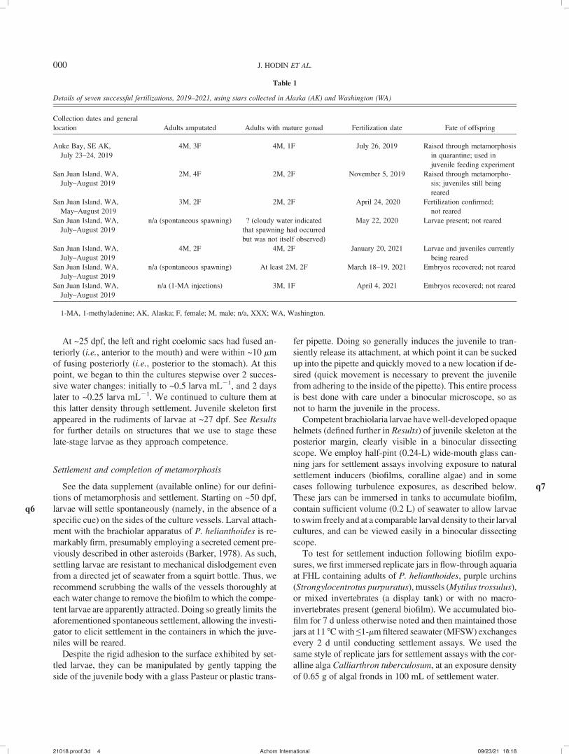
Table 1
Details of seven successful fertilizations, 2019–2021, using stars collected in Alaska (AK) and Washington (WA)
Collection dates and generallocation Adults amputated Adults with mature gonad Fertilization date Fate of offspring
Auke Bay, SE AK,July 23–24, 2019
4M, 3F 4M, 1F July 26, 2019 Raised through metamorphosisin quarantine; used injuvenile feeding experiment
San Juan Island, WA,July–August 2019
2M, 4F 2M, 2F November 5, 2019 Raised through metamorpho-sis; juveniles still beingreared
San Juan Island, WA,May–August 2019
3M, 2F 2M, 2F April 24, 2020 Fertilization confirmed;not reared
San Juan Island, WA,July–August 2019
n/a (spontaneous spawning) ? (cloudy water indicatedthat spawning had occurredbut was not itself observed)
May 22, 2020 Larvae present; not reared
San Juan Island, WA,July–August 2019
4M, 2F 4M, 2F January 20, 2021 Larvae and juveniles currentlybeing reared
San Juan Island, WA,July–August 2019
n/a (spontaneous spawning) At least 2M, 2F March 18–19, 2021 Embryos recovered; not reared
San Juan Island, WA,July–August 2019
n/a (1-MA injections) 3M, 1F April 4, 2021 Embryos recovered; not reared
1-MA, 1-methyladenine; AK, Alaska; F, female; M, male; n/a, XXX; WA, Washington.
21018.p roof.3d 4 09/23/21 18:18Achorn International
000 J. HODIN ET AL.

To investigate the modulation of larval settlement responsesby turbulence, we employed both a quantifiable method and amore qualitative method of generating turbulence and com-pared them (along with a control) in a single experiment. Webegan by selecting seemingly competent WA-derived larvaeat 113 dpf into beakers containing 100 mL of MFSW. We se-lected 20–25 larvae into each of 12 beakers, after which werandomly assigned the beakers to 1 of 3 treatments: (1) thequantifiable turbulence method, (2) the qualitative turbulencemethod, or (3) unexposed controls. We exposed larvae fromeach replicate run to turbulence or control conditions for3 min, after which we poured all recovered larvae from a runinto glassfinger bowls for observations of larvae swimming ver-sus those on the bottom (i.e., contacting the walls of the bowl)and then assessed settlement over 24 h by immediately transfer-ring all of the larvae to a jar—1 jar per run—containing biofilmexposed to adult P. helianthoides for 5 d (and then maintainedfor 5 additional days before the experiment; see aboveq8 ).
For thequantifiable turbulencemethod,weemployedaTaylor-Couette cell (Taylor, 1923; Karp-Boss et al., 1996; Denny et al.,2002), a standard device used to produce and study both laminarand turbulent flows in the laboratory. See the data supplement(available online) for details on this device and specific exposuresused.
Following the 3-min exposures in the device in 125 mL,we gently poured the larvae into a 1-L beaker containing~100 mL of MFSW and then rinsed the device 2 times withMFSW to recover any adhered larvae, concentrated the lar-vae by gentle reverse filtration, and then proceeded with set-tlement observations as above.We rinsed the device with dis-tilled water between runs to ensure that no larvae remainedbefore starting the next exposure.
For the qualitative method of generating turbulence, wesimilarly poured larvae from the selection beakers into 125 mLtotal MFSW in 250-mL Erlenmeyer flasks, covered the topswith parafilm, and then exposed them to 3 min of oscillatoryshaking (~240 repetitionsmin21) to simulate turbulence, keep-ing the flask vertical the entire time. After exposure, wepoured the entire contents of the flask into a glass finger bowland rinsed the beaker one time withMFSW to recover any ad-hered larvae. Controls were treated identically to the shakenlarvae, except that we left the flasks undisturbed for the 3-minduration.
Culturing juveniles post-settlement
In our initial efforts to culture the post-settlement stages,the juveniles experienced highmortality.We attribute thismor-tality to a combination of the following causes: (1) physicaldamage, (2) contaminants, (3) closed culture, and (4) cannibal-ism. See the data supplement (available online) for more de-tailed notes on these four causes of juvenile mortality. Anotherpossibility, but one that we were unable to evaluate, is thatsome of the juveniles succumbed to SSW.
Our subsequent, more successful efforts at juvenile rearing(i.e., with higher juvenile survival and growth rates) involvedculturing juveniles in flow-through cages (food-grade plastictubs with windows cut in the side covered by 100–200-mmNitex mesh; q9Genesee Scientific, San Diego, CA).We initiatedthis design inMarch 2019 (with 0–2-mo post-settlement juve-niles), using recirculating ~10-gal MFSW tanks, with waterchanges in the tanks every 4 d. We used 30 gal h21 fountainpumps (Danner Manufacturing, Islandia, NY) to enhancecirculation around the cages and fed water from additionalpumps through seawater manifolds to deliver positive waterflow through each tank.
In November 2019 (with 7–9-mo post-settlement juve-niles), we improved this design by using continuously flow-ing natural seawater filtered down to a nominal 1 mm beforeentering the holding tank in which our cages were situated.Juvenile survival stabilized under these conditions, and growthrates increased (see Results).
Feeding. The greatest challenge in culturing P. helian-thoides was identifying suitable food for the earliest juvenilestages. We tried a range of possible food sources for the earlyjuveniles, including natural multispecies biofilms, cultivatedbiofilms of benthic diatoms (B2042 Nitzschia frustulum andB2046 Navicula incerta; UTEX, Austin, TX), epiphytes andepizoites on coralline algae, small pieces of kelp, newly settledechinoid juveniles, a commercial fish diet (Otohime larvalfeed B1, 200–360-mmsize range; Pentair Aquatic Eco-Systems,Apopka, FL), and cultivated bivalve juveniles (the cockleLaevicardium elatum [from the Puget Sound Restoration Fund]and the Manila clam Venerupis philippinarum and the Pacificoyster Miyagi oyster [both from Taylor Shellfish Farms,Shelton, WA]). Either we offered P. helianthoides juvenilesintact juvenile bivalves or we crushed the juvenile bivalveshells with forceps to give young P. helianthoides juvenilesaccess to tissues. We provide feeding observations in Results.
Beginning about 35 d after settlement, we conducted a feed-ing experiment with AK-derived juveniles, using separatedhalves of plastic tea infusers (~200-mm mesh; Upton Tea Im-ports, Holliston, MA; henceforth, filter baskets). Once the fil-ter baskets had cured for several weeks in natural seawatercontainers to generate a biofilm, we placed two juveniles intoeach basket and set them in a shallow aquarium with rapidlyrecirculating flow (95 gal h21) and with the upper half of thebasket above the water level. We then haphazardly assignedbaskets to one of three test treatment groups or a control. Inthe treatment groups, juveniles were fed (a) 1–2 crushed juve-nile cockles Laevicardium elatum, (b) 10 early post-settlement(~300-mm test diameter) Dendraster excentricus juveniles, or(c) 10 grains of Otohime larval feed B1 (200–360-mm sizerange; Pentair Aquatic Eco-Systems). The control juvenileswere not fed (aside from the biofilm already present). We po-sitioned the filter baskets in a table of recirculatingUV-treatedMFSW such that water could not flow over the tops of thebaskets.
21018.p roof.3d 5 09/23/21 18:18Achorn International
CULTURING AN ENDANGERED SEA STAR 000

During the feeding experiment, we examined the juvenilesevery 2 d to watch for changes in gross morphology (normalor shriveled), coloration (colored or pale), and activity levels(active or sluggish) as indicators of overall health. We re-placed food on an as-needed basis, as available food ranlow, or as we observed fouling of uneaten food. We shiftedthe basket positions in the water table every 2 d to promotesimilar flow conditions for each basket. We ran the experi-ment for 50 d in December 2019 through January 2020.
Transport of different life stages
See the data supplement (available online) for detailedmethods on transporting gametes, larvae juveniles, and adultP. helianthoides.
Microscopy and measurements
We observed, measured, and photographed live embryosand larvae, gently immobilized under cover glass raised withmodeling clay (see Strathmann, 2014), at 40! to 400!mag-nification, using a variety of compound microscopes. We vi-sualized larval and incipient juvenile skeleton by using cross-polarized light and made measurements by using calibratedocular micrometers or from calibrated micrographs.
We measured and photographed juveniles in situ by usingbinocular microscopes furnished with an ocular micrometerand reflected (episcopic) light. At larger sizes (>1 cm), wealso used calipers for size measurements.
Statistics
We conducted all statistical analyses with R (ver. 3.5.2; RCore Team, 2017). For the majority of our settlement exper-iments and for the feeding experiment, we used a logistic(generalized linear) mixed-effects model, employing the lme4and emmeans packages (Bates et al., 2015; Lenth, 2018), to an-alyze data due to the binomial nature of our response variables(e.g., larvae settled or knocked down). In our tests, we treatedeach replicate exposure (jar, etc.) of a group of larvae as a ran-dom intercept. In two experiments (the temporal comparisonof settlement on P. helianthoides vs. general biofilm; and inthe analysis of juvenile size in the feeding experiment) we con-ducted an ANOVA. These two types of analyses can be distin-guished in Results by the types of statistics shown: we report Z-statistics for the logisticmixed-effectsmodels andF-statistics forANOVAs. All of our reported P-values are after employingBonferroni corrections for multiple comparisons.
Results
Adult arm regeneration rates
For the purpose of collecting gonad, we amputated 1 arm in22 of our captive stars over the course of 16 mo. In 12 of these
22 stars, we amputated an additional arm after a minimum6-mo recovery period. All amputated arms subsequently be-gan to regenerate (see, e.g., Fig. S1F, data supplement, avail-able online), with an average rate of regeneration of 0.11 ±0.05 (SD) mm d21, or 4.0 ± 1.8 cm yr21. The average armlength in the captive stars in November 2020 was 12.4 ±1.6 (SD) cm. Therefore, based on our observed regenerationrates, we predicted that it would take between 2.7 and 4.2 yr(95% confidence interval [CI]) for the sunflower stars to fullyregenerate their lost or amputated arms. Individual regenera-tion rateswere not strongly correlatedwith differences in bodysize (r5 0.144). Note that we were not attempting to feed thesea stars ad libitum during this study, for fear of overfeedingthem. Thus, it is possible that our data represent underesti-mates of maximum potential regeneration rates.
Oocyte sizes, reproductive maturity, and fertilizations
We measured oocyte diameters in amputated stars as wellas several others that lost an arm for another reason, in-cluding SSW. Table S1 (data supplement, available online)lists oocyte sizes for each of these stars and, if treated withthe maturation hormone 1-MA, whether the oocytes under-went germinal vesicle breakdown (GVBD) and were ulti-mately fertilized; Figure 2 plots these data over time forthe WA stars. Individuals in which the largest oocytes were~150 mm in diameter or less did not undergo GVBD and didnot fertilize (n 5 8). Based on the results listed in Figure 2and Table S1, we suggest that the reproductive season for fe-males of this species in WA begins in November–Januaryand ends in April–May. Mature and fertilizable oocytes fromP. helianthoides in SE Alaska were isolated by a colleague inJuly 2019. It is likely that the reproductive season for femalesis later in this northern part of the species range. Egg sizesacross all individuals that we examined ranged from 155 to170 mm (Table S1).
Table S1 lists the results for several females that we ampu-tated twice, with a minimum of 6 mo between amputations.Two of those females did not have mature oocytes on theirfirst amputations (late 2019) but did have mature oocytes16–18 mo later (early 2021), indicating that they completedtheir reproductive cycle in captivity.
In males, by contrast, we did not observe strong evidencefor reproductive seasonality.We successfully recovered testescontaining mature sperm from the amputated arms of malesthroughout the year (data not shown). Furthermore, we ob-served a testis completely devoid of sperm on only one oc-casion, yet we also had a successful spawning of a male sun-flower star by the standard technique of 1-MA injection that samemonth, May 2019. Note that we did not attempt to measure go-nad size as a proxy for reproductive cycling because recover-ing the entire gonad during amputation would have involvedincreased stress to the adults.
21018.p roof.3d 6 09/23/21 18:18Achorn International
000 J. HODIN ET AL.

Developmental table and staging
Pycnopodia helianthoides proceeds through embryogene-sis, bipinnaria, and brachiolaria stages according to the de-velopmental schedule shown in Table 2, a faster rate thanpreviously reported (Greer, 1962). Selected embryo and lar-val stages are shown in Figure 3. Brachiolariae have notablylong arms, especially the posterolateral arms, which can besignificantly longer than the larval body length. As is truefor many sea stars (reviewed in Allen et al., 2018), cloningin P. helianthoides larvae was commonplace: we observedclones in all larval cultures.
Juvenile skeleton first appeared in the rudiment of larvaefromWA parents at ~27 dpf (q10 Fig. 4A). Radial canals appearedsuccessively, starting at ~39 dpf (Fig. 4B). At ~43 dpf, the budsthat will form the brachiolar arms are visible in the most ad-vanced larvae (Fig. 4F); we consider this the beginning ofthe brachiolar stage. At ~51 dpf, larvae exhibit skeletal ringsaround their posterior periphery, resembling a helmet (Figs. 3I,4G). We refer to this as the helmet stage and use the promi-nence of the helmet as a way of tracking the approach of larvaeto competence (Fig. 4I). At around this same time, bona fidebrachiolar arms are present in most larvae (Fig. 4J), as are longposterolateral arms, often longer than the entire body length(see, e.g., Figs. 3I, 4G).
To further characterize late larval development in P.helianthoides, we developed a staging scheme based uponformation of the aforementioned and other skeletal structuresin the rudiments of advanced larvae, which, along with si-multaneous events in the maturation of the brachiolar appa-ratus, can be used to monitor the approach to metamorphiccompetence in a batch of larvae (Table 2; Fig. 4).
We reared larvae at 11 7C and 14 7C (WA parents); thosereared at 14 7C developed more rapidly (data not shown).Therefore, it should be noted that the timeline in Table 2 islikely very temperature dependent, and time to competenceand interim stages would likely be significantly reduced athigher temperatures.
Settlement observations and experiments,including turbulence exposures
Competent P. helianthoides larvae will settle spontane-ously on the sides of the culture vessels and will do so morereadily if a biofilm is allowed to form on the jars. We firstobserved such spontaneous settlement starting about 7 wkpost-fertilization (pf) in larvae reared at 10–11 7C. Peakspontaneous settlement appeared to have occurred earlierin larvae derived from AK parents when compared to WA-derived larvae (week 8 pf vs. week 11 pf; Fig. 5), possiblypointing to different temperature optima in populationsacross the species range. However, the later spontaneous set-tlement seen in larvae derived from WA parents in Figure 5may have been due to our more intense jar exchange andcleaning regime—resulting in a less well-developed bio-film—when culturing these latter larvae.
We identified four phases in the settlement process (Fig. 6):1. In the exploration phase (not shown), the larva will
swim freely and occasionally remain in contact with thewalls of the jar or a settlement surface, such as a frond of cor-alline alga, and then resume swimming.
2. In the attachment phase (Fig. 6A), the larva will adhereto a surface by using its brachiolar apparatus. Larvae in the
Figure 2. Oocyte sizes in Washington-collected Pycnopodia helianthoides. Filled circles indicate oocytesthat, when treated with 1-methyladenine (1-MA), did not undergo germinal vesicle breakdown (GVBD) and be-come fertilizable, that is, immature oocytes. Open circles indicate oocytes that underwent GVBD and became fer-tilizable after treatment with 1-MA, that is, mature oocytes. X indicates data from preserved ovaries, not treatedwith 1-MA. Error bars are 95% confidence intervals. There appears to be an annual cycle, with peak oocyte sizes inthe late autumn to mid-late spring. All mature oocytes were >155 mm on average.
21018.p roof.3d 7 09/23/21 18:18Achorn International
CULTURING AN ENDANGERED SEA STAR 000

Figure 3. Oocyte maturation, embryogenesis, and larval development in Pycnopodia helianthoides. (A) Ovaryreleasing (spawning) mature oocytes in response to 1-methyladenine (1-MA). (B) Just-released oocytes withintact germinal vesicles (clear areas within oocytes). (C) At 1.5 h later, after germinal vesicle breakdown (GVBD),these eggs are now fertilizable. (D) Two-cell stage, 5 hours post-fertilization. (E) Mid-gastrula, 3 days post-fertilization (dpf ). (F) Early-feeding bipinnaria larva, 7 dpf; gut red from Rhodomonas. (G) Mid-bipinnaria,17 dpf; enterocoels are just fusing anteriorly (black arrow). (H) Late bipinnaria, 26 dpf; enterocoels are almost fusedposteriorly (white arrow). (I) Swimming brachiolaria larva, 41 dpf. Scale bars in (A) and (I)5 1mm; all other panels50.1 mm.
Table 2
Developmental stages in Washington Pycnopodia helianthoides, 10–11 7C, including proposed rudiment staging scheme for late larval development(RS-1 to RS-5)
Stage Time Figure reference
Fertilization 0 hpfFirst cleavage 5 hpf Fig. 3DSecond cleavage 6 hpfHatching 44 hpfGastrulation 50 hpf Fig. 3EEarly bipinnaria (can feed) 6 dpf Fig. 3FMid-bipinnaria, anterior fusion of enterocoels 17 dpf Fig. 3GLate bipinnaria, posterior fusion of enterocoels; RS-1: first appearance of juvenile skeleton in rudiment;
wishbone spicules (incipient body skeletal plates) present in most advanced larvae27 dpf
Figs. 3H, 4ARS-2: first appearance of radial canals (pentamery beginning to develop through successive formation
of the five radial canals via hydrocoel bumps)39 dpf Fig. 4B
Early brachiolaria larva (brachiolar arm buds visible, adhesive disk forming); RS-3: skeletal elementsforming at lateral edges of radial canals; body skeletal plates now at snowflake stage
43 dpf Fig. 4D, 4E, 4F
Brachiolar adhesive disk developing, lateral papillae forming; RS-4: helmet forming at posteriorperiphery via peripheral spicules; stellar arrangement of spicules around snowflake body skeletal plates
51 dpf Figs. 3I, 4G, 4H
Competent larva; adhesive papillae on brachiolar arms well developed; RS-5: bumped appearanceof helmet from expanding peripheral skeletal spicules
55 dpf Fig. 4I, 4J
Alaska P. helianthoides larvae proceeded through these stages slightly more quickly at the same temperature (data not shown). dpf, days post-fertilization;hpf, hours post-fertilization; RS, rudiment stage.
21018.p roof.3d 8 09/23/21 18:18Achorn International

attachment stage can remain in this stage for extended peri-ods (24 h or more), can detach and return to the explorationphase, or can proceed to the settler phase.
3. In the settler phase (Fig. 6B), the larva has irreversi-bly committed to settling. The larval body shrinks along itsanterior-posterior axis, and the length of the larval arms (thisis particularly noticeable in the posterolateral arms) shrinksdramatically as well.
4. In the settled stage (Fig. 6C, D), which occurs ~24–48 hafter the larva commits to the settler phase, we consider it tobe a bona fide juvenile. The larval body has withdrawn com-pletely into the aboral surface of the juvenile. Soon, the juve-
nile begins to adhere to the surface with tube feet instead ofthe brachiolar apparatus and can move freely on the substra-tum. Settled juveniles will continue metamorphosis for sev-eral days until their mouths are open.
In addition to the spontaneous settlement described above,larvae will settle at low levels in response to a variety of nat-ural biofilms ( q11Fig. 7), including biofilms grown in the pres-ence of either of two P. helianthoides prey species, purple ur-chins (Strongylocentrotus purpuratus; Fig. 7C) or mussels(Mytilus spp.; Fig. 7B), or in the absence of any macro-invertebrates (general biofilm; Fig. 7A). By contrast, biofilmgrown in the presence of P. helianthoides adults results in
Figure 4. Characters used to define proposed rudiment stages (RS) and correlated late-stage larval charactersof Pycnopodia helianthoides. (A) Posterior right side of the larva, dorsal view. Incipient juvenile body skeletalplates initially form multi-branched spicules in the form of a wishbone (white arrowhead), characteristic of RS-1.(B) Posterior left side of the larva, ventral view. Radial canals form as out-pocketings of the hydrocoel in RS-2,the first signs of fivefold symmetry. White arrowheads denote two visible radial canal anlagen. (C) Posterior rightside of the larva, ventral view. Here the wishbone structure seen in (A) is further bifurcating as it grows (whitearrowhead). (D) Posterior left side of the larva, ventral view. Skeletal spicules (black arrowhead) are visible alongthe lateral edges of the extending radial canals, characteristic of RS-3. (E) Posterior end of the larva, dorsal view.The wishbone structure seen in (A) and (C) has further elaborated into what we term the snowflake form (whitearrowhead), also characteristic of RS-3. (F) Anterior end of the larva, ventral view. Coincident with RS-3, thebrachiolar arms are forming as buds but have not yet developed adhesive papillae (white arrow). The adhesive diskof the incipient attachment complex (see Haesaerts et al., 2005) is here coalescing (black arrowhead). Five anlagenof the lateral papillae of the attachment complex are visible in this larva as well (two indicated with white arrow-heads). (G) Ventral view, indicating that the helmet structure (white arrowhead), characteristic of RS-4 and com-prised of spicules at the posterior periphery, is visible in this binocular microscope image. (H) Posterior right sideof the larva, ventral view. White arrowheads indicate three elements of the stellar array of spicules that formsaround the further elaborated (snowflake) body skeletal plates in RS-4. (I) Posterior end of the larva, ventral view.In RS-5, the helmet takes on a clear, bumpy appearance, resulting from the growing spicules in the helmet.(J) Anterior end of the larva, ventral view. Coincident with RS-5, the brachiolar arms are now fully formed withadhesive papillae, the adhesive disk is mature and birefringent, and there are now eight lateral papillae visible (cf.,F). (A), (C–E), (I), and (J) employed cross-polarized light to highlight spicules. Scale bars 5 100 mm.
21018.p roof.3d 9 09/23/21 18:18Achorn International
CULTURING AN ENDANGERED SEA STAR 000

both a faster and more robust settlement response when com-pared to any of these other biofilms (Fig. 7A–C; see figurelegend for statistics). Exposure of larvae to live fronds of the ar-ticulated coralline alga, Calliarthron tuberculosum, results incomparably enhanced settlement (Z 5 8.751; P < 0.001;Fig. 7C). The potency of P. helianthoides-associated biofilmdegrades over time as the biofilm ages in MFSW in the ab-sence of the adult stars (Z 5 4.654; P < 0.001; Fig. 7D).
We also examined settlement behavior following turbu-lence exposure, a condition that may signal to larvae that theyare approaching the shoreline, where turbulence levels in-crease (for review see Hodin et al., 2018). We assessed threebehaviors in response to turbulence: a temporary knockdownof larvae to the substratum (see Hodin et al., 2020), attach-ment of larvae onto the substratum, and the rate of subsequentirreversible settlement. Following turbulence exposure, thequantifiable (5 W kg21 in a Taylor-Couette device) and qual-itative (shaking) treatments resulted in a threefold increasein larval knockdown when compared to controls (Z 5 6.111;P < 0.001; Fig. 8A); we did not detect a difference betweenthe two turbulence treatments (Z 5 1.606; P 5 0.44; Fig. 8A).Likewise, after 1 h in a strong settlement inducer (P. heli-anthoides biofilm), the larvae in both turbulence treatmentsexhibited an approximate 2-fold increase in irreversible settle-ment when compared to controls (Z 5 3.433; P < 0.005;Fig. 8B, open bars). Again, we detected no difference betweenthe 2 turbulence treatments in irreversible settlement at 1 h(Z 5 0.185; P > 0.5; Fig. 8B, open bars). We obtained com-parable results at 1 h for larval attachment (data not shown), areversible stage of the settlement process. By 24 h after expo-sure, we no longer detected a difference in settlement betweenany of the treatments (Z5 1.716; P > 0.5; Fig. 8B, filled bars).Together, these results suggest that turbulenceexposureheightenslarval responses to settlement inducers but does not change theproportion of larvae that will ultimately settle.
Juvenile survival, feeding, and growth
In early juvenile stages, P. helianthoides has a rounded,dome-like shape ( q12Fig. 9A). As it matures, it becomes dorsalventrally q13compressed and pentagonal in form, and distinct ra-dial arms begin to develop (Fig. 9B). Additional arms are addedperiodically by one of three methods (frommost to least com-mon): (1) a new arm emerging from an armpit (Fig. 9D, E);(2) sagittal bifurcation of an existing arm (Fig. 9D); or (3) lat-eral budding of a new arm (Fig. 9C; observed only once).Some juveniles initially developed fewer than five arms(Fig. 9E, H). We have observed several such juveniles even-tually form their fifth and add subsequent arms as they con-tinue to grow (e.g., Fig. 9E, H).
Juveniles lose their larval pigmentation and their colora-tion may fade or change in response to their diet; once larvalpigment fades, the juvenile gut appears clear. As juvenilessuccessfully feed and grow, color can be seen in the stomach
Figure 5. Spontaneous settlement in Pycnopodia helianthoides larvaederived from Alaska (AK) or Washington (WA) adults. Open circles indi-cate AK larvae (summer–autumn 2019); plus symbols indicate WA larvae(autumn–winter 2019–2020). Note that we cultured many fewer total AKlarvae than we did WA larvae.
Figure 6. Phases of settlement in Pycnopodia helianthoides. The firstphase is exploration (not shown). (A) Attachment phase, lateral view. Larvaadheres to a frond of coralline algae (Calliarthron tuberculosum) withbrachiolar arms and waves its posterolateral arms. (B) Settler phase, lateralview. Irreversible settlement commences with adhesive disk attachment andretraction (collapse) of larval body along the anterior-posterior axis. (C) Set-tled phase, lateral view. Larval body has been completely withdrawn. Juve-nile will soon move with tube feet. (D) Recently settled juvenile, aboralview, exhibiting first indications of arm development (note pentamery inthe incipient juvenile gut). Scale bars 5 250 mm.
21018.p roof.3d 10 09/23/21 18:18Achorn International
000 J. HODIN ET AL.

(presumably from digesting food); and gut development canthen be monitored as juveniles grow: the incipient pyloriccoeca can be seen growing down the arms of live juvenilesand then bifurcating (e.g., Fig. 9B, I, N).
When P. helianthoides juveniles were maintained on bio-film with or without coralline algae, they did appear to beconsuming something, as indicated by the slight darkeningof their guts after a few weeks. Nevertheless, they did not ex-hibit significant growth under these conditions, indicatingthat this minimal consumption may have simply been suffi-cient for metabolic maintenance rather than growth per se.
For juveniles derived from AK parents, starting at 35 d post-settlement we assessed growth and survival over 50 d, duringwhich time juveniles were fed 1 of 3 potential food sources: ju-venile sand dollars, Otohime (a commercial fish diet; PentairAquatic Eco-Systems), or crushed cockles. Controls did not re-
ceive any supplemental food aside from the biofilm present inthe treatment vessels at the onset of the experiment (seeMate-rials and Methods). Each treatment vessel had 2 P. helian-thoides juveniles at the beginning of the experiment, and byday 50 there were always either 1 or 2 juveniles remaining.In one case (a control vessel) we observed a cannibalism event,thus accounting for the disappearance of one juvenile. In theother cases of juvenile disappearance, we presume that the re-maining juvenile consumed its partner, but we are unsurewhether these other cases were due to cannibalism or scaveng-ing on an already dead juvenile. After 50 d, we detected nodifferences between treatments either in mortality (Z 5 0.800;P > 0.4) or in final juvenile size (F3, 8 5 1.436; P5 0.3). Nev-ertheless, when we grouped vessels regardless of treatment bynumbers of juveniles remaining (one or two), we detected a
Figure 8. Turbulence promotes rapid settlement behaviors in Pycnopodiahelianthoides. (A) Immediately following 3min of either 5W kg21 exposure ina Taylor-Couette device (quantitative turbulence) or shaking of larvae in a flask(qualitative turbulence), about 3-fold more larvae were knocked down to thesubstratum when compared to controls (4 replicates [reps] per treatment; n 525–30 larvae per rep). (B) Then, 1 h later, about 2-foldmore turbulence-exposedlarvae were in the process of irreversibly settling in response to conspecificbiofilm as compared to controls (open bars). By 24 h, similar proportions oflarvae were settling in all treatments (filled bars). Error bars5 SEM. **P < 0.01;***P < 0.001.
Figure 7. Settlement of Pycnopodia helianthoides on various substrata.(A) Alaska (AK) larvae. (B–D) Washington (WA) larvae. (A) Experiment 1(68 days post-fertilization [dpf ]; 18- and 96-h exposure): larvae settled bothmore quickly (open bar; F1, 8 5 9.263; P 5 0.016) and in higher numbers(filled bars; F1, 8 5 23.459; P 5 0.0013) when exposed to biofilm grownin the presence of conspecific adults, as compared to general biofilm (i.e.,no macroinvertebrates present). (B) Experiment 2 (86 dpf; 24-h exposure;4 replicates [reps] per treatment; 20 larvae per rep): this response to con-specific biofilm was not due to the confounding effect of the presence oftheir prey (Mytilus edulis mussels; Z 5 5.357; P < 0.001). (C) Experiment 3(134 dpf; 50-h exposure; 4 reps per treatment, n5 30 larvae per rep): larvae alsoresponded more readily to fronds of an articulated coralline alga (Calliarthrontuberculosum) or to conspecific biofilm (Z 5 5.250; P < 0.001) when com-pared to biofilm grown in the presence of another known adult prey species,the purple urchin, Strongylocentrotus purpuratus. (D) Experiment 4 (73 dpf;96-h exposure; 4 reps per treatment; 20 larvae per rep): the conspecific biofilmis much more active when freshly collected (tested 9 d after exposure toadults), compared to similar biofilm aged for 25 d after exposure to adults.“Settled” in all panels includes both settlers and settled phase larvae. Errorbars5 SEM. **P < 0.01; ***P < 0.001.
21018.p roof.3d 11 09/23/21 18:18Achorn International
CULTURING AN ENDANGERED SEA STAR 000

Figure 7. Settlement of Pycnopodia helianthoides on various substrata. (A) Alaska (AK) larvae. (B–D)
Washington (WA) larvae. (A) Experiment 1 (68 days post-fertilization [dpf ]; 18- and 96-h exposure):
larvae settled both more quickly (open bar; F1, 8 = 9.263; P = 0.016) and in higher numbers (filled bars;
F1, 8 = 23.459; P = 0.0013) when exposed to biofilm grown in the presence of conspecific adults, as
compared to general biofilm (i.e., no macroinvertebrates present). (B) Experiment 2 (86 dpf; 24-h
exposure; 4 replicates [reps] per treatment; n = 20 larvae per rep): this response to conspecific biofilm
was not due to the confounding effect of the presence of their prey (Mytilus edulis mussels; Z = 5.357;
P < 0.001). (C) Experiment 3 (134 dpf; 50-h exposure; 4 reps per treatment, n = 30 larvae per rep):
larvae also responded more readily to fronds of an articulated coralline alga (Calliarthron
tuberculosum) or to conspecific biofilm (Z = 5.250; P < 0.001) when compared to biofilm grown in the
presence of another known adult prey species, the purple urchin, Strongylocentrotus purpuratus. (D)
Experiment 4 (73 dpf; 96-h exposure; 4 reps per treatment; n = 20 larvae per rep): the conspecific
biofilm is much more active when freshly collected (tested 9 d after exposure to adults), compared to
similar biofilm aged for 25 d after exposure to adults. “Settled” in all panels includes both settlers and
settled phase larvae. Error bars = SEM. **P < 0.01; ***P < 0.001.

difference in juvenile size (F1, 105 8.498;P < 0.02): juveniles invessels with 1 survivor had a mean diameter ± SEM of 741 ±66 mm as compared to a mean diameter of 594 ± 28 mm whenboth juveniles survived. In other words, cannibalism or conspe-cific scavenging resulted in clearer evidence for juvenile growththan did feeding on any of the provided food sources. Qualita-tively, the juveniles fed cockles appeared generally healthierthan in any of the other treatments, and this was the only treat-ment exhibiting zero mortality (seeMaterials and Methods forcriteria employed to assess juvenile health).
Preliminary observations on juvenile feeding. Juvenilesunder flow conditions appeared more robust and more active.The juveniles consumed living and dead organisms, includ-ing echinoid juveniles (the Pacific sand dollar Dendraster
excentricus and the purple urchin, S. purpuratus), bivalve ju-veniles (mussels [Mytilus spp.], Manila clams, and Pacificoysters), and conspecific juveniles (P. helianthoides). Ourobservations indicate that P. helianthoides juveniles activelyprey or scavenge on the echinoderm juveniles earlier in juve-nile ontogeny than when they begin to prey upon the bi-valves. They also feed on bivalve tissues before they are ca-pable of opening the shells (we observed several juvenilesattempt unsuccessfully to open a mussel and only after manyweeks of trying finally succeed).
Once juveniles reach 1–2 mm in diameter, they will read-ily feed on tissue ofManila clams and Pacific oysters; at ~1 cmin diameter, they will begin to successfully open and feed onthese bivalve species (shell length5 1–2 mm) and will consume
Figure 9. Early juvenile ontogeny of Pycnopodia helianthoides. Unless indicated, all photos are of Washing-ton (WA)-derived juveniles and are aboral views. Brown areas are gut, and pyloric caeca are seen through the bodywall. Time in months (mo) is age post-settlement. (A) Cluster of newly settled juveniles on a dead piece of cor-alline algae. (B) Alaska (AK)-derived juvenile, 3 mo, with the typical 5 initial arms. (C–E) Three methods of newarm formation. (C) Lateral bud (arrow), 2 mo. This is the only star we observed forming a new arm in this manner.(D) This 7-mo juvenile exhibits the other 2 methods of new arm formation: sprouting of new arms from armpits(most common; arrowheads), and sagittal bifurcation of an existing arm (arrow). (E) Aberrant (though not veryrare) 4-arm juvenile, oral view, forming its fifth arm from armpit (arrow) at 4 mo. Note the red eyespots at the tipsof each arm, including the incipient fifth one. (F) Pigment cells outlining the nerve ring (arrowhead in magnifiedinset), oral view, 3 mo. (G–M) Juveniles feeding. (G) Cannibalism. Arrow indicates tube feet of living juvenilebeing cannibalized by the 2-mo AK-derived juvenile on top. (H) This 9-mo, 4-arm juvenile (note that broad fourtharm at upper left is preparing to bifurcate) is scavenging a dead conspecific. (I) This 5-mo juvenile under a frond ofcoralline algae is bringing in a dead purple urchin juvenile (arrowhead) with its tube feet. (J) Slowly growing 4-mojuvenile preparing to consume a live, precocious purple urchin juvenile (arrow) that settled without spines. (K) Un-successful attempt by 4-mo juvenile to consume a juvenile mussel (epizoite on coralline alga). (L) Oral view, 9 mo,successful predation underway on a live Manila clam juvenile. (M) Results of successful predation on a live Pacificoyster juvenile. (N) Juvenile cage mates, 8 mo, exhibiting typical gregarious behavior, similar to that seen in adults.Note the extensive migration of pyloric caeca down the arms of these stars relative to juveniles in other panels. Allscale bars 5 0.5 mm, except 5 mm in (D), (I), (K–N).
21018.p roof.3d 12 09/23/21 18:18Achorn International
000 J. HODIN ET AL.

other live invertebrates as well (e.g., juvenile limpets) if giventhe opportunity. The relationship between the diameter of P.helianthoides juveniles and the volume of bivalves consumedper day can be found inq14 Figure S4 (data supplement, availableonline). Pycnopodia helianthoides juveniles at >1 mm will alsoreadily feed on sand dollar and purple urchin juveniles.
Initial survival was poor, and growth was slow (q15 Fig. 10;Figs. S2, S3, data supplement, available online). We attributethese findings to the fact that during these early juvenile stages,we were investigating and adjusting vessel design, flow condi-tions, water filtration, and possible food sources, none ofwhichhave been optimized previously for any sea star of which weare aware.
Figure 10 summarizes our growth and survival data in theform of a model (akin to a Lefkovitch matrix; Lefkovitch,1965). The upper half of the figure indicates the daily likeli-hood of a given juvenile dying, remaining in the same sizeclass, or advancing to the next size class. Although we haveseen the rare individual juvenile shrinking slightly between
measurements, we never observed an individual regressingto a prior size class (data not shown). These data are consis-tent with the survival (Fig. S2, data supplement, available on-line) and growth (Fig. S3, data supplement, available online)figures, showing high mortality and slow growth early, withincreases in both juvenile survival and growth rates at largersizes.
The lower half of Figure 10 gives an approximate ontoge-netic growth schedule, summarizing the number of days post-settlement that it took for 50% of the surviving juveniles, 20%of the surviving juveniles, or the first juvenile to reach a givensize class. We present this range of growth schedules in lightof our uncertainty regarding the suitability of the initial growthconditions used.
Discussion
We report on successful culturing of the endangered sun-flower star, Pycnopodia helianthoides Brandt, from egg to 1 yr
Figure 10. Model of juvenile growth and survival in Pycnopodia helianthoides. We here summarize ourgrowth and survival data in the form of a modified Lefkovitch model (Lefkovitch, 1965). Put simply, we period-ically counted and measured every juvenile in our rearing chambers and from these data calculated the likelihoodof a given larva surviving and growing into a subsequent size class between sampling dates. We made the simpli-fying assumption that individual juveniles within a chamber maintained their relative rank order of sizes betweenmeasurements. We also assumed, unless there was specific evidence to the contrary, that if juveniles disappearedbetween observation dates, it was the smaller individuals that were most likely to have perished. The data in theupper half of the figure support the conclusion that mortality is highest at smaller sizes (with the caveat that theaforementioned survival-by-size assumption slightly biases this conclusion) and that growth accelerated at largersizes. Data in the bottom half of the figure resulted from the same analysis. The 95% confidence ranges are notavailable for the larger size classes because midway through our observation period, we separated juveniles bysize class to limit cannibalism. The result was that we no longer had replicate chambers of the different size classesat larger juvenile sizes.
21018.p roof.3d 13 09/23/21 18:18Achorn International
CULTURING AN ENDANGERED SEA STAR 000

post-settlement juvenile. Our goal is to establish egg-to-eggfull life cycle culturing in captivity, with three long-term ob-jectives in mind. First, captive rearing can promote furtherstudies in the ontogeny, physiology, and ecology of this keybenthic predator, while obviating the necessity of collectingthis endangered species from the wild. Second, by developingmethods to culture P. helianthoides juveniles for the first time,we now have access to these cryptic life stages for investiga-tions into their growth, feeding ecology, and behavior. And,third, because sunflower stars are thought to be extirpated,or nearly so, from almost half of their historical range in theNE Pacific, our captive rearing program represents the firststep in a collective exploration of the eventual feasibility oftheir reintroduction into the wild.
Weobtainedmature oocytes fromamputatedP. helianthoidesarms; the standard method to obtain eggs by injection of the seastar spawning hormone 1-MA had limited success. The stars re-covered from amputations, as evidenced by regenerating arms,completion of additional reproductive cycles the year afteramputation, and no observed mortality or other health issuesfollowing amputation. Because we can distinguish individualadult stars based on their color patterns (see Fig. 1; Fig. S1,data supplement, available online), we have been able to moreefficiently undertake amputations, because we now know thesex of 24 of our 28 captive stars.
Prior studies stated that mature eggs of P. helianthoideswere ~120 mm in diameter (Greer, 1962; Strathmann, 1987),but we could not find primary data to support this assertion.Our data for P. helianthoides from both AK and WA popula-tions show that the egg size inP. helianthoides is 155–170 mmin diameter, which is consistent with the onemeasurement of afertilized P. helianthoides egg of which we are aware (G. vonDassow, University of Oregon, pers. comm.) prior to the re-cent SSW event. Therefore, it does not seem that egg sizeshave changed in P. helianthoides populations in response tothe recent SSW event. We have observed the rare egg withina batch that was smaller than 155 mm and successfully ma-tured and fertilized. Nevertheless, when the maximum oocytesize for a given female was less than 150 mm, the oocytesfailed to mature and, thus, remained unfertilizable.
We also noted differences from the literature in the timingof reproductivematurity in sunflower stars in the San Juan Ar-chipelago. Strathmann (1987 and references therein) gave thetiming of mature oocytes as March–July, with peak spawningin thefield inMay–June. By contrast, we have seenmature oo-cytes as early as November and little evidence for mature oo-cytes after April–May.We observed four spontaneous spawn-ing events in captivity, all betweenMarch andMay, which is abit earlier than the peak spawning timing noted in Strathmann(1987). It is possible that the earlier reproductive maturity weobserved is a recent shift, possibly associated with SSW. Fur-thermore, we have noted that recently collected stars, evenwhen reproductively mature, tended to have small gonads (afew centimeters in length), relative to what is seen in other
sympatric asteroids (JH, pers. obs.). This may also be a conse-quence of SSW (e.g., a trade-off between survival and repro-duction in the face of the disorder). In sum, we advocate am-putations for reproductive studies in WA in the winter andearly spring. Our one data point on stars in AK suggests a laterreproductive season in those colder northern waters.
As stated above, our data indicate that captive adult starscompleted a reproductive cycle (and, thus, became reproduc-tive again) while in captivity. In response to 1-MA treatment,mature oocytes in isolated gonads underwent GVBD, eggswere successfully fertilized, and we reared larvae throughmetamorphosis and settlement following established proto-cols (Adams et al., 2019; Hodin et al., 2019). Progressionthrough embryonic and larval stages was as described byGreer (1962), though larval development proceeded morerapidly under our growth conditions. We first observed set-tlement beginning in cultures 7 wk pf at 10–11 7C, 2 wk ear-lier than reported by Greer (1962) at similar temperatures.Competent larvae will settle spontaneously and in responseto a variety of natural biofilms, but settlement proportionsand rates are greatly enhanced in response to a biofilm col-lected in the presence of adult sunflower stars or if larvae areexposed to fronds of the articulated coralline alga,Calliarthrontuberculosum. The combination of these two cues seems to en-hance settlement even further. We also found that turbulenceexposure induces a more rapid and synchronous settlement re-sponse to the aforementioned cues, which could be a usefultechnique in any large-scale rearing effort.
Our greatest challenges have occurred in developing suit-able protocols for juvenile rearing, a process that has beensuccessfully reported in the past for only a handful of aster-oid species with planktotrophic development (e.g., the NewZealand sea star Stichaster australis; Barker, 1979). Our ju-venile cultures experienced high initial mortality rates due toa variety of likely causes (seeMaterials andMethods,Results,and data supplement, available online, for details). With opti-mization of methods, we were able to stabilize this mortality,and the juveniles began to grow rapidly. We had the greatestsuccess when we cultured juveniles in micron-filtered natu-ral seawater in flow-through cages and fed them juvenilebivalves.
We note that the juvenile growth pattern that we observed—slow initial growth for the first months after settlement, fol-lowed by more rapid growth after (see Figs. S2, S3, data sup-plement, available online)—is reminiscent of observations onjuvenile growth in two other sea stars: the temperate NE At-lantic species Asterias rubens (Nauen 1978), and the tropicalS Pacific crown-of-thorns sea star, Acanthaster spp. (Deakeret al., 2020). In both cases, the juveniles seem to have the abil-ity to persist for long periods under suboptimal growth condi-tions as a waiting period until better conditions arise, such astheir encountering a prime prey species. We do not yet know,however, whether this waiting period for sunflower star juve-niles is facultative or obligate, since we have not yet had the
21018.p roof.3d 14 09/23/21 18:18Achorn International
000 J. HODIN ET AL.

opportunity to provide them optimal food and growth condi-tions from immediately after settlement. We plan to assessthese scenarios in the coming year.
Importantly, we observed for the first timeq16 young P. heli-anthoides juveniles (a few months post-settlement; <1-cm di-ameter) actively consuming living echinoid juveniles (sea ur-chins and sand dollars; <2-mm diameter). Juvenile and adultred (Mesocentrotus franciscanus), purple (Strongylocentrotuspurpuratus), and green (Strongylocentrotus droebachiensis)sea urchins are all known prey of adult sunflower stars(Mauzey et al., 1968; Dayton, 1975; Moitoza and Phillips, 1979;Duggins, 1983; Freeman, 2005; Nishizaki and Ackerman, 2007).Our observations raise the possibility that P. helianthoides couldexert top-down predatory control in the wild on these urchinspecies, starting at early juvenile stages in both predator andprey. This intriguing hypothesis remains to be tested.
We conclude by advocating that species reintroductionsshould truly be considered the solution of last resort when itcomes to ensuring the persistence of species in their naturalhabitats. Andwhile the long-term possibility of sunflower starreintroductions is a credible consideration, our primary objec-tive in this captive-rearing effort is to learnmore about the ba-sic biology and ecology of this endangered predator. We seethis approach as not only adding to our general knowledge ofbenthic biology and ecology but also informing other conser-vation interventions designed to protect this species in thewild and encourage its return. We further hope that our stud-ies highlight the importance of basic research into poorly un-derstood marine taxa so that critical information about suchspecies’ biological and ecological functions are known beforethe next SSW-like calamity strikes.
Acknowledgments
We are extremely grateful for the diverse assistance we havereceived from somany people in various aspects of the project.We first thank Sherry Tamone and lab for collecting Alaskanstars, isolating gonads, and shipping them to us for our firstsuccessful sunflower star fertilizations. We further thank JohnDorsett,WillemWeertman, JoeyUllman, Hank Carlson, Tay-lor Frierson, Jon Allen and team, Mo Turner, ZooBots 2019,Jim Murray, Sadie Youngstrom, Olivia Graham, Frank Hurd,Richard Emlet, Katie Dobkowski, Tim Dwyer, and DerekSmith for subtidal and intertidal collecting efforts in Wash-ington. The following people and organizations kindly pro-vided facilities access and related assistance: University ofWashington Biology (and especially Ron Killman and AlexHanson), Jim Truman, Lynn Riddiford, Merrill Hille, RoseAnn Cattolico, Evelyn Lessard, Mike Foy, Carolyn Friedman,Bryanda Wippel, Jacqueline Padilla-Gamiño, Jeremy Ax-worthy, Miranda Roethler, Julie Keister, the Port TownsendMarine Science Center, the Seattle Aquarium, Doug Engel,Tom Campbell, and Tommy Pieples. For conversations andother miscellaneous assistance, we thank Walter Heady, Re-
becca Guenther, Jon Allen, Ryan Crim, Sophie George, Rich-ard Strathmann, Mike Barker, Mary Sewell, Donald Greer,David Cohen, Kathy Foltz, Aidan Cox, Beatriz Velazquez,Chloe Deodato, Mo Turner, Wai-Pang Chan, Drew Harvell,Morgan Eisenlord, Megan Dethier, Julia Kobelt, and AaronGalloway. Nyle Taylor at Taylor Shellfish and Ryan Crimat the Puget Sound Restoration Fund delivered juvenile bi-valves at a moment’s notice. Rebecca Guenther, Matt Ferner,Richard Strathmann, and two anonymous reviewers kindlyprovided comments on an earlier draft. Brady Blake and theWashington Department of Fish andWildlife approved transferpermits, allowing us tomove our operation betweenFridayHar-bor and Seattle. Generous funding came from the Nature Con-servancy (to JH) and California SeaGrant (NA18OAR4170073to B. Gaylord, M. Baskett, A. Ricart, M. Edwards, M. Zippay,B. Hughes, S. Place, and JH). We dedicate this paper to JohnPearse, who in one of his last conversations with JH asked,“Do you think you can figure out the juvenile rearing prob-lem?” We’re getting there, John!
Data Accessibility
Upon publication, primary data will be publicly accessibleat the following link in the ResearchWorks archive at theUniversity of Washington: http://hdl.handle.net/1773/46681.
Literature Cited
Adams, N. L., A. Heyland, L. L. Rice, and K. R. Foltz. 2019. Procuringanimals and culturing of eggs and embryos. Methods Cell Biol. 150: 3–46.
Allen, J. D., A. M. Reitzel, and W. Jaeckle. 2018. Asexual reproductionof marine invertebrate embryos and larvae. Pp. 271–276 in EvolutionaryEcology of Marine Invertebrate Larvae, T. J. Carrier, A. M. Reitzel, andA. Heyland, eds. Oxford University Press, New York.
Barker, M. F. 1978. Structure of the organs of attachment of brachiolarialarvae of Stichaster australis (Verrill) and Coscinasterias calamaria(Gray) (Echinodermata: Asteroidea). J. Exp. Mar. Biol. Ecol. 33: 1–36.
Barker, M. F. 1979. Breeding and recruitment in a population of the NewZealand starfish Stichaster australis (Verrill). J. Exp. Mar. Biol. Ecol.41: 195–211.
Bates, D., M. Maechler, B. Bolker, and S. Walker. 2015. Fitting linearmixed-effects models using lme4. J. Stat. Softw. 67: 1–48.
Burt, J. M., M. T. Tinker, D. K. Okamoto, K. W. Demes, K. Holmes,and A. K. Salomon. 2018. Sudden collapse of a mesopredator revealsits complementary role in mediating rocky reef regime shifts. Proc. R.Soc. B Biol. Sci. 285: 20180553.
Chia, F. S., and C. W. Walker. 1991. Echinodermata: Asteroidea.Pp. 301–353 in Reproduction of Marine Invertebrates, Vol. VI, Echino-derms and Lophophorates, A. C. Giese, J. S. Pearse, and V. B. Pearse,eds. Boxwood Press, Pacific Grove, CA.
q17Cole, R. N., and W. W. Burggren. 1981. The contribution of respiratorypapulae and tube feet to oxygen uptake in the sea star Asterias forbesi(Desor). Mar. Biol. Lett. 2: 279–287.
Dayton, P. K. 1975. Experimental evaluation of ecological dominance ina rocky intertidal algal community. Ecol. Monogr. 45: 137–159.
Deaker, D. J., B. Mos, H.-A. Lin, C. Lawson, C. Budden, S. A.Dworjanyn, and M. Byrne. 2020. Diet flexibility and growth ofthe early herbivorous juvenile crown-of-thorns sea star, implicationsfor its boom-bust population dynamics. PLoS One 15: e0236142.
21018.p roof.3d 15 09/23/21 18:18Achorn International
CULTURING AN ENDANGERED SEA STAR 000

Denny, M. W., E. K. Nelson, and K. S. Mead. 2002. Revised estimatesof the effects of turbulence on fertilization in the purple sea urchin,Strongylocentrotus purpuratus. Biol. Bull. 203: 275–277.
Done, T. J. 1992. Phase shifts in coral reef communities and their ecolog-ical significance. Hydrobiologia 247: 121–132.
Duggins, D. O. 1983. Starfish predation and the creation of mosaic pat-terns in a kelp-dominated community. Ecology 64: 1610–1619.
Emlet, R. B., L. R. McEdward, and R. R. Strathmann. 1987. Echinodermlarval ecology viewed from the egg. Pp. 55–136 in Echinoderm Studies,Vol. 2, M. Jangoux and J. M. Lawrence, eds. Balkema, Rotterdam.
Feder, H. M. 1980. Asteroidea: the sea stars. Pp. 117–135 in Intertidal In-vertebrates of California, R. H. Morris, D. L. Abbott, and E. C. Haderlie,eds. Stanford University Press, Palo Alto, CA.
Finger, D. J. I., M. L. McPherson, H. F. Houskeeper, and R. M. Kudela.2021. Mapping bull kelp canopy in northern California using Landsatto enable long-term monitoring. Remote Sens. Environ. 254: 112243.
Freeman, A. 2005. Size-dependent trait-mediated indirect interactionsamong sea urchin herbivores. Behav. Ecol. 17: 182–187.
Gravem, S. A., W. N. Heady, V. R. Saccomanno, K. F. Alvstad, A. L. M.Gehman, T. N. Frierson, and S. L. Hamilton. 2020. Pycnopodiahelianthoides. [Online]. IUCN Red List of Threatened Species 2020,e.T178290276A178341498. Available: https://doi.org/10.2305/IUCN.UK.2020-3.RLTS.T178290276A178341498.en [2021, February 4].
Greer, D. L. 1962. Studies on the embryology of Pycnopodia helianthoides(Brandt) Stimpson. Pac. Sci. 16: 280–285.
Haesaerts, D., M. Jangoux, and P. Flammang. 2005. The attachmentcomplexofbrachiolaria larvaeof theseastarAsteriasrubens (Echinodermata):an ultrastructural and immunocytochemical study. Zoomorphology 124:67–78.
Harvell, C. D., D. Montecino-Latorre, J. M. Caldwell, J. M. Burt, K.Bosley, A. Keller, S. F. Heron, A. K. Salomon, L. Lee, O. Pontieret al. 2019. Disease epidemic and a marine heat wave are associatedwith the continental-scale collapse of a pivotal predator (Pycnopodiahelianthoides). Sci. Adv. 5: eaau7042.
Hodin, J., M. C. Ferner, A. Heyland, and B. Gaylord. 2018. I feel that!Fluid dynamics and sensory aspects of larval settlement across scales.Pp. 190–207 in Evolutionary Ecology of Marine Invertebrate Larvae,T. J. Carrier, A. M. Reitzel, and A. Heyland, eds. Oxford UniversityPress, New York.
Hodin, J., A. Heyland, A. Mercier, B. Pernet, D. L. Cohen, J.-F. Hamel,J. D. Allen, J. S. McAlister, M. Byrne, P. Cisternas et al. 2019. Cul-turing echinoderm larvae through metamorphosis. Methods Cell Biol.150: 125–169.
Hodin, J., M. C. Ferner, and B. Gaylord. 2020. Choosing the righthome: Settlement responses by larvae of six sea urchin species alignwith hydrodynamic traits of their contrasting adult habitats. Zool. J.Linn. Soc. 190: 737–756.
IUCN (International Union for Conservation of Nature). 2020. TheIUCN Red List of Threatened Species. Version 2020-3. [Online]. Avail-able: https://www.iucnredlist.org [2021, February 4].
Karp-Boss, L., E. Boss, and P. A. Jumars. 1996. Nutrient fluxes toplanktonic osmotrophs in the presence of fluid motion. Oceanogr. Mar.Biol. Annu. Rev. 34: 71–107.
Kenyon, K. W. 1969. The sea otter in the eastern Pacific Ocean. N. Am.Fauna 68: 1–352.
Lefkovitch, L. P. 1965. The study of population growth in organismsgrouped by stages. Biometrics 21: 1–18.
Lenth, R. 2018. emmeans: estimated marginal means, aka least-squaresmeans. [Online]. R package version 1.1.2. Available: https://CRAN.R-project.org/package5emmeans [2021, September 8]. q18
Margolin, A. S. 1976. Swimming of the sea cucumber Parastichopuscalifornicus (Stimpson) in response to sea stars. Ophelia 15: 105–114.
Metaxas, A. 2013. Larval ecology, settlement, and recruitment of Aster-oids. Pp. 59–66 in Starfish: Biology and Ecology of the Asteroidea,J. M. Lawrence, ed. Johns Hopkins University Press, Baltimore.
Martinez, A. S., M. Byrne, and R. A. Coleman. 2017. Filling in thegrazing puzzle: a synthesis of herbivory in starfish. Pp. 1–34 in Ocean-ography and Marine Biology: An Annual Review, Vol. 55, S. J. Haw-kins, A. J. Evans, A. C. Dale, L. B. Firth, D. J. Hughes, and I. P. Smith,eds. CRC Press/Taylor & Francis, Boca Raton, FL.
Mauzey, K. P., C. Birkeland, and P. K. Dayton. 1968. Feeding behaviorof asteroids and escape responses of their prey in the Puget Sound re-gion. Ecology 49: 603–619.
McEdward, L. R., and B. G. Miner. 2001. Larval and life-cycle patternsin echinoderms. Can. J. Zool. 79: 1125–1170.
Moitoza, D. J., and D. W. Phillips. 1979. Prey defense, predator prefer-ence, and nonrandom diet: the interactions between Pycnopodia heli-anthoides and two species of sea urchins. Mar. Biol. 53: 299–304.
q19Murphy, C. T., and M. B. Jones. 1987. Some factors affecting the respi-ration of intertidal Asterina gibbosa (Echinodermata: Asteroidea). J.Mar. Biol. Assoc. U.K. 67: 717–727.
Nishizaki, M. T., and J. D. Ackerman. 2007. Juvenile-adult associationsin sea urchins (Strongylocentrotus franciscanus and S. droebachiensis):protection from predation and hydrodynamics in S. franciscanus. Mar.Biol. 151: 135–145.
R Core Team. 2017. R: a language and environment for statistical com-puting. [Online]. R Foundation for Statistical Computing, Vienna. Avail-able: https://www.R-project.org/ [2021, September 8]. q20
Rogers-Bennett, L., and C. A. Catton. 2019. Marine heat wave andmultiple stressors tip bull kelp forest to sea urchin barrens. Sci. Rep. 9:15050.
Shivji, M., D. Parker, B. Hartwick, andM. J. Smith. 1983. Feeding anddistribution study of the sunflower sea star Pycnopodia helianthoides(Brandt, 1835). Pac. Sci. 37: 133–140.
Sloan, N. A. 1980. Aspects of the feeding biology of asteroids. Oceanogr.Mar. Biol. Annu. Rev. 18: 57–124.
Strathmann, M. F. 1987. Reproduction and Development of Marine In-vertebrates of the Northern Pacific Coast. University of WashingtonPress, Seattle.
Strathmann, R. R. 2014. Culturing larvae of marine invertebrates. Pp. 1–25 inMethods in Molecular Biology, Vol. 1128, Developmental Biologyof the Sea Urchin and Other Marine Invertebrates, Methods and Proto-cols, D. J. Carroll and S. A. Stricker, eds. Springer, New York.
Taylor, G. I. 1923. Stability of a viscous liquid contained between two ro-tating cylinders. Philos. Trans. R. Soc. AMath. Phys. Eng. Sci. 232: 289–343.
21018.p roof.3d 16 09/23/21 18:18Achorn International
000 J. HODIN ET AL.

1
DATA SUPPLEMENT for
Progress towards complete life-cycle culturing of the endangered sunfower star Pycnopodia helianthoides
Hodin J, Pearson-Lund A, Anteau FP, Kitaeff P and S Cefalu
The Biological Bulletin, Dec 2021, volume 241, number 3. https://doi.org/10.1086/716552
SUPPLEMENTARY MATERIALS & METHODS
1. Collecting, feeding, holding and distinguishing adultsIn addition to the stars collected in 2019 as described in the main text, we also received documentation during this period of sighting of three additional sunfower stars > 20 cm that were not collected. Our chosen collection locales were made up of sites that P. helianthoides had historically been observed pre-SSW, as well as some additional sites. We found that certain locations (such as subtidal areas around Reuben Tarte County Park; 48.612 N, 123.097 W) represented apparent 'hot spots' where we had repeated success fnding individuals of a range of sizes (including several <20 cm diameter that we did not collect), whereas other locations with formerly (pre-2013) robust populations (such as Point Caution; 48.563 N, 123.017 W) yielded zero observed individuals despite our repeated collecting attempts. All told, these collections and sightings of 38 total stars in 6 mo required 84 person-hours of searching (i.e., ~2.2 person-hours for each >20 cm sunfower star found). Note that our searches became more successful towards the end of this collecting period, as we focused our searches in 'hot spot' locales.
Adult P. helianthoides maintained in the same aquaria are gregarious (remaining in contact with one another for long periods despite room to separate) and only display aggressive interactions during feeding. During Spring 2019, a few of the captive adults exhibited signs of SSW, including arm curling behavior and arm autotomy. These animals and all of their aquarium mates (5 stars total) were then returned to the feld where we hoped that they would have a better chance of recovery. From June 2019 until June 2021 (the time of this writing), we have maintained 28 of 30 stars continually with no further instances of wasting symptoms observed. The 29th star was found dead in Nov 2020 on the ground near one of our tanks; we suspect it was a victim of a raccoon (Procyon lotor) predation event. The 30th star was released due to poor apparent condition in Spring 2021,at its site of collection 2 years earlier: the dock pilings at Friday Harbor Labs. In subsequent weeks, we encountered this star several times at a similar location, confrming that he had survived reintroduction and his condition had improved. His color pattern andthe position of his amputated arms allowed us to know for certainty that this was the samestar that we had released. For more on our methods for identifying individual stars, see Supplementary Figure S1 (below).

2
2. Assessing reproductive status, obtaining gametes, maturation and fertilization We prepared 1-MA stock solutions by frst making a 2.5% solution of 1-MA (Fisher Scientifc Acros 201310250) in dimethyl sulfoxide (DMSO). We then diluted that 1:1130 in 1 µm-fltered seawater (henceforth MFSW) to make a 100 µM 1-MA stock solution (fnalDMSO concentration: <0.1%), which we sterile-fltered through a 0.2 µm syringe flter and stored at 4°C.
We attempted 1-MA injections to induce spawnings on 15 different occassions over 24 months, and we were only successful three times: May 2019 (only 1 male spawned); Sept 2019 (only one male spawned) and April 2021 (3 males and 1 female spawned, with the female releasing few total eggs, but they did fertilize; see Table 1 in main text). At 11-12°C, these fve males began spawning between 55 and 110 min after injection; the one female began spawning 155 min after injection.
We attempted a aboral fap incisions of epidermis in three stars [Assessment Method (2); see the main text]. While this method did allow us easy access to sample the gonad, recovery from incision was poor: all three of these stars autotomized the adjacent arm within 2 d of dissection. We then released those stars due to SSW concerns, allowing them to recover in the wild. Note that the poor recovery from incision has been previouslyreported in the ochre sea star, Pisaster ochraceous (Sanford et al. 2009), so we do not recommend this technique.
By contrast, and consistent with the aforementioned fndings of Sanford and colleagues (2009) for P. ochraceous, arm amputation is an effective gonad sampling method in P. helianthoides, and results in excellent recovery. In the 16 mo between Sep 2019 and Jan 2021, we amputated an arm in 22 of the stars; all have recovered, the amputated arms areall regenerating (see Supplemental Results, below), and none of these stars has subsequently exhibited signs of wasting. Furthermore, 12 of these 22 stars have received two amputations (with amputations separated by at least 6 mo), again with no observed ill effects.
Finally, we note that if an adult star were to self-autotomize an arm, as often occurs duringadvanced stages of SSW, it is often possible to recover gonad for analysis. It is worth noting that stars that succumb to wasting would probably not be an appropriate choice forsiring offspring for captive breeding programs, though it could be of research interest (e.g.,in studying potential parent-offspring transmission). Sampling gonad in wasting stars couldprovide valuable information on reproductive seasonality, so we recommend doing so with appropriate precautions. Dissecting tools, dishes and other laboratory implements that come into contact with tissue from adults showing signs of wasting should be thoroughly decontaminated before use with healthy individuals or their gonadal tissues.

3
The viability of male gonadal tissue can be assessed by making a slide preparation in sea water and examining it under 100x or greater power. Mature, viable male gonad should contain highly active sperm. Female gonadal tissue can be assessed similarly, but in this case the cover glass should be raised slightly (with, e.g., clay "feet"; see Strathmann 2014) so that the oocytes remain uncompressed, allowing their diameters to be accurately measured to assess maturity. We measured oocyte diameters by one of two methods: (1) with a calibrated ocular micrometer; or (2) from micrographs with known pixel calibration.
If oocytes are obtained by natural or induced spawning from intact females, what will be recovered should be mainly fully mature oocytes (see Fig. 2B in the main text) which should then complete meiosis and germinal vesicle breakdown (GVBD) in situ over the following 60 min or more, at which point they are fertilizable (Strathmann 1987; Adams etal. 2019; see Fig. 2 in the main text). If mature oocytes are recovered by dissection [methods (2-4) above], then the pieces of dissected ovary must be treated with 1 µM 1-MAin MFSW for >60 min for them to release ("spawn") oocytes and then complete meiosis and GVBD (see Fig. 2A-C, in the main text).
Oocytes having undergone GVBD (Figure 2C in the main text) are fertilizable for the subsequent ~24 h or even more, using a freshly diluted sperm suspension, but the highest proportion of fertilized eggs occurs in the frst 6 h after GVBD. We conducted all fertilizations at 10-11°C in 100 ml beakers by adding 10-20 drops of a fresh, dilute sperm suspension (approximately 1:300) in MFSW, and then stirring the water in the beakers for 5 sec. After eggs settled, we decanted off the sperm-flled water, and replaced with fresh MFSW. P. helianthoides eggs have clearly visible fertilization membranes, raised approximately 10 µm from the egg surface (see Fig. 2D in the main text). See Strathmann (1987) and Adams et al. (2019) for more detailed methods.
3. Embryogenesis and larval developmentNo supplemental notes.
4. Settlement and completion of metamorphosisWe defne metamorphosis as a major morphological transformation from one life history stage to a subsequent one, often involving a change in habitat (Hodin 2006; for other defnitions see Bishop et al. 2006). In echinoderms, this transformation involves a radical change in symmetry from what is generally a bilaterally-symmetric planktonic larval form into a pentameral juvenile in the benthos. Still, in many echinoderms (including P. helianthoides), the shift from bilateral to pentameral begins within the feeding larval body itself. Therefore, we consider metamorphosis in P. helianthoides to begin at approximately27 dpf, when juvenile-structures structures appear in the rudiment and to end a few days after the transition to the sea foor, at which point the benthic juvenile can feed. Inherent in this conception is that the irreversible habitat shift from the plankton to the benthos –namely, settlement– is a key stage in the metamorphic process, but is not sensu stricto synonymous with metamorphosis. In summary, metamorphosis is the morphological change from larva to juvenile; settlement is the shift in habitat.
4
The Taylor-Couette cell that we used consisted of two concentrically nested cylinders separated by a 3.5 mm gap flled with seawater, into which we introduced larvae by gently pouring from their selection beaker through a funnel. Relative rotation of the two cylinders sheared the water in the gap between the cylinders, and did so strongly enough to generate turbulent fow. Turbulence generated by the Taylor–Couette cell recreates many of the features of natural turbulence produced beneath breaking waves, where kinetic energy is translated down through ever-smaller eddies to the smallest scales of fuid motion until that turbulent energy is dissipated by viscosity. More intense turbulence results in higher levels of energy dissipation, indexed in watts per kilogram (= m2 s−3), and a broader energy cascade that sustains eddies of tinier size, smaller than (hence detectableby) marine larvae.
The Couette device we used here is identical to one used in prior publications focused on echinoid larvae (see Hodin et al. 2020 and references therein), and we here tested a singleturbulence intensity of ~5 W kg-1 (which at 11°C in our device corresponds to a rotation rate of 500 rpm). We selected this level of exposure based on peak intensities of turbulence that have been measured in the feld on exposed rocky shores in the NE Pacifc(a historical habitat for some P. helianthoides populations), and which also elicited responses across echinoid taxa in previous studies (Gaylord 2008; Gaylord et al. 2013; Hodin et al. 2015; Hodin et al. 2020).
5. Culturing juveniles post-settlementIn our initial efforts to culture the post settlement stages, the cultures experienced high mortality. We attribute this mortality to a combination of the following causes:
a. Physical damage. As mentioned previously, the juveniles tend to adhere extremely strongly during the settlement process and are quite resistant to dislodgement. Using, for example, a high intensity water stream will ultimately dislodge them, butoften results in tissue damage to the juvenile. We suspect that damaged juveniles subsequently exhibit poor survival.
b. Contaminants. Due to the location of our initial juvenile culturing efforts, we did not have access to fowing sea water, so we used recirculating natural sea water tanks, with seawater water fltered to a nominal 20 µm. This resulted in the rapid accumulation of ciliates, benthic diatom fouling and other microbial contaminants that were associated with mortality of juveniles. Once such contaminants enter the cultures, they are exceedingly diffcult to remove. Even transferring individual juveniles out of contaminated containers into fresh ones can result in incidental transfer of contaminants, which will then infect the new container. We subsequently increased our fltration level down to a nominal 1 µm ("MFSW"), which likely decreased upstream sources of new contaminants. We did not have access to UV treatment except in one set of experiments with Alaskan stars. We recommend employing UV treatment if available to limit upstream contamination, while noting that it is possible that a certain complement of microbes might be benefcial to juvenile development.

5
c. Closed culture. Whereas the small jars we described previously were useful for settlement experiments, post-settlement observations and transport of cultures, juveniles maintained under these conditions eventually experienced high mortality,despite frequent (every other day) water exchanges. We attribute this high mortalityto a combination of causes, including the aforementioned contaminants, low oxygen levels, especially in the bioflm boundary layer, and, fnally, a positive feedback of juvenile deaths resulting in culture fouling, an increase in contaminant populations, and further death.
d. Cannibalism. In the absence of an alternate food source, the juveniles will readily resort to cannibalism, thus resulting in another signifcant source of mortality. Especially during our initial rearing efforts, during which time we had not identifeda quality food source for the young juveniles, we directly observed several instances of cannibalism, and inferred others from the disappearance of juveniles between observations. See also Brocco French and Allen (2021).
6. Transport of different life stagesGametes. Sperm collected 'dry' and dissected pieces of testis can be transported on ice, and stored for one week or longer in capped tubes at 4°C and remain viable. Pieces of dissected ovary (untreated with 1-MA) can be stored for at least 2 d at cold ambient temperatures (~8-10°C) in MFSW and remain viable. For transport of dissected ovary pieces, we placed them inside 125-250 ml tissue culture fasks, flled to the brim with MFSW, the tops sealed with paraflm (ideally with no air bubble), and then reinforced withplastic wrap (i.e., cling flm), affxed tightly to the fask neck with a rubber band, and kept cool during transport between two 4°C-equilibrated ice packs inside a polystyrene cooler.
Larvae. We transported larvae successfully over a 12-h period in the manner described above for ovary pieces, except that we used larger fasks and maintained the larvae no more than 3x their current culturing density. We did not attempt longer transport, but based on our success in doing so as described with a variety of other echinoderm larvae (see, e.g., Hodin et al. 2020), including asteroids, we suspect that even a 1-2 d transport period by this method would be successful. For longer transport periods, larval density is ideally maintained at or near the current culturing density.
Juveniles. We successfully transported juveniles over a 12-h period by two different methods: in canning jars with plastic leak-proof lids (Ball), or in their culturing cages flledto 50% capacity inside outer plastic containers. In both cases, we placed the containers incoolers with 4°C-equilibrated ice packs.
Adults. In our experience, adult Pycnopodia do not tolerate long periods of transport (>1h)in still water (e.g., large coolers with sea water). We are aware of two occasions in which adults that were transported over longer time periods from their collection to their holding locales then subsequently succumbed to sea star wasting (SSW). In holding, adults survive best in constantly-fowing, well-oxygenated seawater. It is possible that if one could mimicthese conditions during transport, then they might tolerate longer transport periods.

6
SUPPLEMENTARY RESULTS
Table S1. P. helianthoides oocyte sizes in Washington, 2019-2021, plus one observation from Alaska 2019. Abbreviations: 1-methyladenine, 1-MA; germinal vesicle breakdown, GVBD; oocytes with germinal vesicles, GV+, oocytes following GVBD, GV-; fertilized eggs, +FE (N.b., measured zygote diameter, not including FE).; seastar wasting (SSW).
SAMPLE NUMBER
DATE, LOCATION AND DEPTH COLLECTED
DATE OOCYTES MEASURED
DESCRIPTION
MEAN OOCYTE DIAMETER
95%C.I.
1 Mar 2019, Friday Harbor WA, from FHLdock piling by hand
May 2019 Ovary sampled by dissection, oocytes didnot undergo GVBD in response to 1-MA, measured GV+.
56.1 ± 3.3
2 Jul 2019, Auke Bay, Juneau AK, subtidal 9 m
Jul 2019 Ovary recovered by arm amputation, shipped to Friday Harbor, matured in 1-MA, oocytes fertilized,measured +FE.
165.3 ± 2.7
3 Jul 2019, arm autotomized during attempted intertidal collection, Beaverton Cove, Friday Harbor WA
Jul 2019 Gonad completely devoid of oocytes (recently spawned?).
n/a n/a
4 Jun 2019, Cattle Point, San Juan Island WA, intertidal by hand
Sep 2019 Ovary recovered by arm amputation, oocytes did not undergo GVBD in response to 1-MA, measured GV+.
77.6 ± 6.3
5 Jun 2019, off Brown Island, Friday Harbor WA, subtidal 12 m
Sep 2019 As above. 50.3 ± 8.8
6 Jun 2019, near FHL dock, Friday Harbor WA, subtidal 12 m
Nov 2019 Ovary recovered by arm amputation, ovarytreated with 1-MA, measured GV- subset with +FE following sperm addition.
164 ± 2.2
7

7 As above (Sample 6) Nov 2019 As above, here measured subset of oocytes that did not undergo GVBD (GV+).
139.5 ± 9.9
8 Jul 2019, Snug Harbor, San Juan Island WA, subtidal 10 m
Nov 2019 Ovary recovered by arm amputation, ovary treated with 1-MA, measured +FE.
157.8 ± 3.6
9 Aug 2019, O'Neill Island (off San Juan Island) WA, subtidal 13 m
Nov 2019 Ovary recovered by arm amputation, ovary treated with 1-MA, did not undergo GVBD, measured GV+.
95.4 ± 5.8
10 Aug 2019, off Brown Island, Friday HarborWA, subtidal, in crabpot, unknown depth
Nov 2019 As above. 101.3 ± 5.8
11 Jan 2020, Sund Rock,Hood Canal WA, subtidal 9-15 m
Jan 2020 Ovary recovered from arm dropped following SSW. Not treated with 1-MA; measured GV+.
157 ± 4.7
12 Jan 2020, Sund Rock,Hood Canal WA, subtidal 9-15 m
Feb 2020 As above. 167.1 ± 6.6
13 July 2019, near FHL dock, Friday Harbor WA, subtidal 8 m
Apr 2020 Ovary recovered by arm amputation, ovary treated with 1-MA, measured GV-.
168.2 ± 2.9
14 June 2019, Snug Harbor, San Juan Island WA, intertidal by hand
Apr 2020 As above. 155.8 ± 4.8
15 Aug 2020, Goose Island (Cattle Pass) WA, subtidal 10 m
Sep 2020 Ovary recovered from arm dropped following SSW. Not treated with 1-MA; measured GV+.
83.2 ± 5.9
16 June 2019, Snug Harbor, San Juan Island WA, intertidal by hand
(same star as in Sample 14)
Nov 2020 Ovary recovered by arm amputation, ovary treated with 1-MA, some oocytes appearedto undergo GVBD, measured GV-. No oocytes successfully fertilized.
126.2 ± 4.1
8

17 July 2019, near FHL dock, Friday Harbor WA, subtidal 8 m
(same star as in Sample 13)
Nov 2020 As above. 130.3 ± 4.7
18 Aug 2019, O'Neal Island (off San Juan Island) WA, subtidal 13 m
(same star as in Sample 9)
Nov 2020 As above. 137.9 ± 5.6
19 Aug 2019, off Brown Island, Friday HarborWA, subtidal, in crabpot, unknown depth.
(same star as in Sample 10)
Jan 2021 Ovary recovered by arm amputation, treated with 1-MA, underwent GVBD, and successfully fertilized. Measured GV+ before 1-MA addition.
159.9 ± 5.2
20 Jun 2019, Cattle Point, San Juan Island WA, intertidal by hand
(same star as in Sample 4)
Jan 2021 As above. 166.9 ± 6.3
9

Supplementary Figures and Explanatory Text
Adult Identifcation
Figure S1 provides a guide for how to identify and track individual stars. Although the color patterns have been quite consistent in the vast majority of our captive stars over time(Fig. S1E, F), there are subtle exceptions (Fig. S1C, D).
Juvenile survival and growth
As seen in Fig. 10 (see main text) and Figs S3 and S4 (below), we had increasing success with juvenile survival and growth once juveniles reached approximately 1 mm, at which point they began to consume juvenile bivalves. Initially, we crushed individual bivalves togive the sea star juveniles access to the fesh; older sea star juveniles began to open and consume live bivalves. Figure S2 shows the relationship between P. helianthoides juvenilediameter and volume of juvenile bivalves (crushed or intact) consumed per day.
Figure S3 shows juvenile survival over time, with each line representing numbers of surviving juveniles within an independent growth chamber. The clear trend is high attrition early in juvenile ontogeny with greater survival after a few months. We are unableto evaluate the extent to which this pattern is a function of normal attrition in the early juvenile stage of P. helianthoides as opposed to our aforementioned explorations into different growth conditions.
Figure S4 shows juvenile growth over time, in this case with each line representing a single individual. Note that in only a subset of cases were we able to defnitively identify individuals within a vessel in repeated measures –for example, by their numbers of arms or other distinguishing features. In most of the data shown here, we assumed for the purpose of this analysis that individual juveniles sharing a culture vessel maintained their relative rank order of sizes. As in the aforementioned survival data, the overall pattern here is slow initial growth with rapid increases in growth rates at larger sizes. And again, we are here unable to evaluate the extent to which this pattern is a refection of our offering quality food sources only to the larger individuals rather than it being a normal ontogenetic pattern for the species.

Figure S1
Figure S1. Individual identifcation in P. helianthoides, exemplifed in four of our captive stars. Contrasting two stars in (A) and (B). The star in A is more silver overall, is greener and fuzzier around the central disk [this is presumably from respiratory papulae (Cole & Burggren 1981); compare white arrowheads in main panels of A, B], and has a disorganized stripe pattern down each arm (marked at left in A by white dots); the arms of the star in B are not striped. Also, the star in A has prominent blue spines; those of the star in B are prominent and brown (compare white arrowheads in the insets in A, B). Both starsshare burgundy reticulations (white arrows in insets in A, B), a feature absent in most otherstars. (C, D) Change in a third star's color over 18 mo. In late 2019 (C) this star had an overall plum color, prominent blue spines, disorganized plum arm stripes and purple arm tips. In early 2021 (D) these features were still present, but had become obscured by a silvery-green fuzzy appearance (papulae, which can be retracted; Murphy & Jones 1987) overlaying the arms and central disk. This star's diagnostic arm stub (white arrows in C, D), the only non-regenerating arm that we have observed in P. helianthoides, was present upon collection in mid 2019, and unchanged since. The stub is 7 arms clockwise from themadreporite (white arrowhead in C, D). (E, F) Consistent coloration and patterns in a fourth star from late 2019 (E) to late 2020 (F). Note the bright orange coloration and regular arm stripes in both E and F [c.f., the less well-organized arm stripes in A and C, D].The white arrowhead in F indicates the regenerating arm, 11 mo after amputation. Scale bars are 5 cm.

Figure S2
Figure S2. Juvenile feeding. Strong linear relationship (R2=0.846) between juvenile size and volume of bivalves consumed each day. Our data on the largest juvenile stars (>2 cm diameter) is recent and thus limited, but might suggest an increased rate of consumption by body size later in juvenile ontogeny.

Figure S3
Figure S3. Juvenile mortality in WA stars in the frst year after settlement. Each line represents an independent vessel or set of vessels cultured in a similar manner. When the lines end, the remaining juveniles were moved to new vessels, locations or culturing conditions. The general pattern is high mortality early, with increased survival rates over time (see also Fig. 10 in the main text). Arrows and lowercase letters at the top indicate dates at which we made signifcant changes to culturing conditions as follows. a–juvenilesmoved from static to recirculating fow conditions. b– began feeding juveniles crushed Manila clams and Pacifc oysters. c–juveniles separated by size in an effort to limit cannibalism. d–juveniles moved into continuously fowing, micron-fltered sea water at FHL.

Figure S4
Figure S4. Juvenile growth in individual WA stars. Each line represents the growth trajectory of a single star from May 2020 until Jan 2021. Lines that end before Jan 2021 represent stars that died. Two trends from this graph are: (1) the high variation in growth rate among stars raised in similar conditions; and (2) growth rates generally appear to accelerate once stars reach approximately 10 mm in diameter. This is approximately the size at which juveniles are able to open and consume live juvenile bivalves; before that point, we crush the bivalve shells for the sea star juveniles. Note that for this analysis we made the simplifying assumption that individual stars within a cage maintain their relative rank order of sizes between measurements. Lowercase letters above arrows indicate changes in juvenile culturing conditions: b– began feeding juveniles crushed Manila clams and Pacifc oysters. c–juveniles separated by size in an effort to limit cannibalism. d–juveniles moved into continuously fowing, micron-fltered sea water at FHL.

SUPPLEMENTAL REFERENCES CITED
Adams, N. L., A. Heyland, L. L. Rice and K. R. Foltz. 2019. Procuring animals and culturing of eggs and embryos. Pp 3–46 in Methods in Cell Biology, K. R Foltz and A. Hamdoun, eds. Echinoderms – Part A. 150. Chapter 1. Elsevier, New York.
Bishop, C. D., D. F. Erezyilmaz, T. Flatt, C. D. Georgiou, M. G. Hadfeld, A. Heyland, J. Hodin, M. W. Jacobs, S. A. Maslakova, A. Pires, et al. 2006. What is metamorphosis? Integrative and Comparative Biology 46: 655–661.
Brocco French, K. I. and J. D. Allen. 2021. Cannibalism of newly metamorphosed juvenile sea stars. Ecology doi:10.1002/ecy.3352
Cole, R. N., and W. W. Burggren. 1981. The contribution of respiratory papulae and tube feet to oxygen uptake in the sea star Asterias forbesi (Desor). Mar. Biol. Lett. 2: 279–287.
Gaylord, B. 2008. Hydrodynamic context for considering turbulence impacts on external fertilization. The Biological Bulletin 214: 315–318.
Gaylord, B., J. Hodin and M. C. Ferner. 2013. Turbulent shear spurs settlement in larval sea urchins. Proceedings of the National Academy of Sciences, USA 110: 6901–6906.
Hodin, J. 2006. Expanding networks: Signaling components in and a hypothesis for the evolution of metamorphosis. Integrative and Comparative Biology 46: 719–742.
Hodin, J., M. C. Ferner, G. Ng, C. J. Lowe and B. Gaylord. 2015. Rethinking competence in marine life cycles: ontogenetic changes in the settlement response of sand dollar larvae exposed to turbulence. Royal Society Open Science 2: 150114.
Hodin J., M. C. Ferner and B. Gaylord. 2020. Choosing the right home: settlement responses by larvae of six sea urchin species align with hydrodynamic traits of their contrasting adult habitats. Zoological Journal of the Linnean Society 190: 737–756.
Murphy, C. T., and M. B. Jones. 1987. Some factors affecting the respiration of intertidal Asterina gibbosa (Echinodermata: Asteroidea). J. Mar. Biol. Assoc. U.K. 67: 717–727.
Sanford, E., M. E. Wood and K. J. Nielsen. 2009. A non-lethal method for estimation of gonad and pyloric caecum indices in sea stars. Invertebrate Biology 128: 372–380.
Strathmann, M. F. 1987. Reproduction and Development of Marine Invertebrates of the Northern Pacifc Coast. University of Washington Press: Seattle.


