Embed Size (px)
Citation preview

Homocysteine induces inflammatory transcriptional signaling inmonocytes
Shu Meng, Stephen Ciment, Michael Jan, Tran Tran, Hung Pham, Ramón Cueto, Xiao-FengYang*, and Hong Wang*
Department of Pharmacology and Cardiovascular Research Center, Temple University School ofMedicine, Philadelphia, PA 19140
AbstractHyperhomocysteinemia (HHcy) is an independent risk factor for cardiovascular disease. Thisstudy is to investigate transcriptional mechanism underlying homocysteine (Hcy)-induced andmonocytes (MC)-derived inflammatory response. We identified 11 Hcy-induced genes, 17 anti-inflammatory cytokine interleukin 10-induced, 8 pro-inflammatory cytokine interferon γ (IFNγ)-induced and 8 pro-inflammatory cytokine tumor necrosis factor α (TNFα)-induced genes throughliterature search. Binding frequency of 36 transcription factors (TFs) implicated in inflammationand MC differentiation were analyzed within core promoter regions of identified genes, andclassified into 3 classes based on the significant binding frequency to the promoter of Hcy-inducedgenes. Class 1 TFs exert high significant binding frequency in Hcy-induced genes. Class 2 and 3TFs have low and no significant binding frequency, respectively. Class 1 TF binding occurrence inHcy-induced genes is similar to that in IFNγ-induced genes, but not that in TNFα-induced. Weconclude that Hcy is a pro-inflammatory amino acid and induces inflammatory transcriptionalsignal pathways mediated by class 1 TF. We term class 1 TF, which includes heat shock factor,MC enhancer factor-2, nuclear factor of activated T-cells, nuclear factor kappa light chainenhancer of activated B cells and Krueppel-like factor 4, as putative Hcy-responsive TFs.
2. INTRODUCTIONHyperhomocysteinemia (HHcy) has been established as an independent risk factor forcardiovascular disease (CVD) (1, 2). Meta-analysis shows that an increase of 5 μmol/L inplasma homocysteine (Hcy) levels enhances the risk of CVD by 1.6- to 1.8-fold, which issimilar to the risk seen with an increase of 20 mg/dL (0.52 mmol/L) in cholesterolconcentration (3).
The causative role of HHcy in human CVD remains controversial. Several secondaryprevention trials of Hcy-lowering therapy were reported to have no effect on combinedendpoints of cardiovascular events (4). Hcy-lowering is found beneficial to reduce the riskof overall stroke in the HOPE 2 Trial. Indirect evidence for such a benefit was recentlyobtained a large population-based cohort study, which demonstrated that Hcy-lowering dueto folic acid fortification significantly (5, 6).
Reports from our laboratory and others have established that HHcy accelerates vascularinflammation and atherosclerosis in mice (7–9). It has been suggested that HHcy acceleratesatherosclerosis via inhibiting endothelial cell growth and impairing post-injury
Send correspondence to: Drs. Hong Wang and Xiaofeng Yang, Department of Pharmacology, Temple University School of Medicine,3420 North Broad Street, Philadelphia, PA 19140, USA, Telephone: 215-707-5986 (Wang), 215-707-5985 (Yang), Fax:215-707-7068, [email protected] (Wang), [email protected] (Yang).
NIH Public AccessAuthor ManuscriptFront Biosci. Author manuscript; available in PMC 2013 April 26.
Published in final edited form as:Front Biosci. ; 18: 685–695.
NIH
-PA Author Manuscript
NIH
-PA Author Manuscript
NIH
-PA Author Manuscript

endothelialization (2, 10, 11), promoting vascular smooth muscle proliferation (12) andinflammatory monocyte (MC) differentiation (13).
MC activation and its recruitment into the artery wall are key cellular events during thedevelopment of atherosclerosis. MC can differentiate into macrophages and become lipid-laden foam cells in the arterial walls. Recent notions indicate that MCs can differentiate intoinflammatory MC subsets and contribute to vessel wall inflammation (14). We recentlyreported that HHcy increases the differentiation of the inflammatory Ly6Chigh/middle MCsubsets, promotes the accumulation of inflammatory MCs/macrophages in the vessel wall,and accelerates atherosclerosis in a HHcy-enhanced atherosclerotic mouse model of HHcyand hyperlipidemia (13). However, how HHcy promotes the differentiation of non-inflammatory MCs into inflammatory subsets remains unknown; and transcriptionalmechanisms underlying HHcy-induced pro-inflammatory effects and MC differentiation hasnot been studied.
Several studies on the transcriptome (all the mRNAs being expressed) have showed that Hcy(≥100 μM), induces pro-inflammatory gene expression in endothelial cells (15, 16). Theeffect of Hcy on inflammatory gene expression has been investigated by several groups (17–21). These individual studies revealed that Hcy induces inflammatory gene expression inMC. However, a summarized analysis of Hcy’s effects on inflammatory gene expression inMCis missing.
Transcription factors (TFs) are master genes that regulate gene expression and impact theirassociated pathways (22). Research to identify TF binding profile has become emergingfrontier and fulfills an urgent need for the identification of therapeutic molecular targets.Transcriptional regulation of Hcy-modulated gene expression has not been studied in asystemic manner.
The complete human genome DNA sequence has been deposited in the National Institutes ofHealth (NIH) database, making the retrieval of experimentally-identified gene promotersequences possible. In addition, nearly 2,000 TFs and their binding sequences have beenexperimentally characterized and placed in the searchable web-based transcription elementsearch system (TESS) and other databases (23). These advances make the identification ofTF binding patterns in the promoters of genes feasible.
In this study, we summarized Hcy- and cytokine-induced genes in human MC throughextensive literature and data base search, analyzed their relevant TF binding profile,identified putative Hcy-responsive TFs and established a hypothetic model of Hcytranscriptional signaling (Figure 1). Our study is the first to use dynamic approaches tosystemically summarize gene regulation findings in the literature, combining with databasemining and bioinformatics analysis, leading to the identification of TF binding profiling andmodels of transcriptional signaling.
3. MATERIALS AND METHODS3.1. Identification of Hcy- and cytokine-induced genes in human MCs
Extensive literature search was performed using the NIH/PubMed database (http://www.ncbi.nlm.nih.gov/pubmed/) to identify Hcy- and cytokines-induced genes. All Genesinduced by Hcy, pro-inflammatory cytokine tumor necrosis factor α (TNFα), pro-inflammatory cytokine interferon γ (IFNγ), and anti-inflammatory cytokine interleukin 10(IL-10) in human MC were identified and listed in Figure 2. All selected genes werevalidated for their transcriptional regulation in their original publication. The Gene IDnumbers of the identified genes were obtained from the NIH/National Center of
Meng et al. Page 2
Front Biosci. Author manuscript; available in PMC 2013 April 26.
NIH
-PA Author Manuscript
NIH
-PA Author Manuscript
NIH
-PA Author Manuscript

Biotechnology Information (NCBI) gene database (http://www.ncbi.nlm.nih.gov/gene/). Thefold change of gene expression was obtained from the original publication if specified, orderived from the published graphs if not specified. CD36 levels were determined by flowcytometry (24). SOD1 levels were examined by western blot (21). All other identified geneswere examined by real-time PCR or northern blot (17–21). Further, in order to screen genesinduced by both Hcy and pro-inflammatory cytokines, we searched for genes (25–33)induced by 20 cytokines included 5 MC differentiation cytokines [granulocyte macrophagecolony stimulating factor (GM-CSF), CSF-1, TNFα, IL-4 and IFNγ], 11 pro-inflammatorycytokines [macrophage inflammatory protein-1α (MIP-1α), TNFβ, IL-1β, IL-2, IL-3, IL-6,IL-7, IL-8, IL-12, IL-15 and IL-18] and 4 anti-inflammatory cytokines [transforming growthfactor (TGFβ), IL-9, IL-10 and IL-11]. The overall database mining strategy is illustrated ina flowchart shown in Figure 1.
3.2. TF selection and classification36 TFs were selected based on their identified function implicated in inflammation and MCdifferentiation via literature search (Figure 3). The DNA sequences of 1,000 bp upstream ofthe transcription start site of the identified genes listed in Figure 2 were retrieved from theNIH/NCBI gene database and defined as putative core promoters. TF binding sites on theputative promoters were determined by using the publicly accessible TF database TESS(http://www.cbil.upenn.edu/cgi-bin/tess/tess) (34). Binding frequency denotes the number ofthe binding sites identified for the TF on the core promoter region. A confidence interval ofbinding frequency for each TF was established by evaluating the binding frequency on 4housekeeping genes, including β-actin (ACTB), fructose-bisphosphate aldolase A(ALDOA), rho GDI 1 (ARHGDIA) and glyceraldehyde-3-phosphate dehydrogenase(GAPDH) (35), and 17 IL-10-induced genes (Figure 2B). These four genes are the mostlyconsistently expressed gene cross tissues in human and mouse as described in our recentdatabase mining studies (35). An upper threshold of the confidence interval was set as mean+3× standard deviations (SD) of the TF binding frequencies in the promoters of 4housekeeping genes and 17 IL-10-induced genes, and marked as the dashed lines. Bindingfrequency higher than the upper threshold of the confidence interval (p < 0.01) was definedas significant binding frequency. TFs with identified corresponding binding element wereclassified into three groups, class 1, 2 and 3, based on the pattern of significant bindingfrequency on Hcy-induced genes.
3.3. TF occurrence in the promoter of Hcy- and pro-inflammatory cytokines-induced genesThe occurrence of class 1 TFs in the promoter regions of Hcy- and pro-inflammatorycytokines TNFα and IFNγ-induced genes were analyzed (Figure 4). TFs with at least onebinding site on the promoter of indicated genes were recorded as occurrence positive (+),whereas, TFs that did not have binding sites on the promoter of indicated genes wererecorded as occurrence negative (−). Ratio of occurrence positive on selected genes wascalculated. Independent t test was used to determine the p value of paired groups. Aprobability value p < 0.05 was considered to be significant.
4. RESULTS4.1. Hcy preferentially induced pro-inflammatory genes in human MC
Via extensive literature search, we identified 11 genes whose expressions are induced bypathogenic concentrations of Hcy (≥100μM) in cultured human PBMC or THP1 cells (ahuman monocytic leukemia cell line) (Figure 2A). Among these 11 genes, 8 are pro-inflammatory genes, including IL-8, MC chemoattractant protein-1 (MCP-1), IL-1β, IL-6,TNFα, IL-12β, chemokine (C-C motif) receptor 2 (CCR2) and cluster of differentiation 36(CD36). All selected genes were validated for their transcriptional regulation in their
Meng et al. Page 3
Front Biosci. Author manuscript; available in PMC 2013 April 26.
NIH
-PA Author Manuscript
NIH
-PA Author Manuscript
NIH
-PA Author Manuscript

original publication. The fold change of gene expression level is indicated in Figure 2A.Interestingly, Hcy also reduced the expression of 2 anti-inflammatory TFs peroxisomeproliferator-activator receptor (PPAR) α and PPARγ (data not shown).
4.2. Hcy shares features with pro-inflammatory cytokines in inducing inflammatory genesin human MC
To compare the feature of Hcy, pro- and anti-inflammatory cytokines in inducing geneexpression in human MC, we further examined genes induced by pro-inflammatorycytokines TNFα and IFNγ, and anti-inflammatory cytokine IL-10 in human MC throughliterature search. We identified 8 genes induced by TNFα (32), 8 genes by IFNγ (33, 36–41), and 17 genes induced by IL-10 (42) (Figure 2B). All selected genes were validated fortheir transcriptional regulation in their original publication. The fold change of geneexpression level is indicated in Figure 2B. We found that TNFα and IFNγ each inducedspecific inflammatory genes, respectively. Interestingly, we found that Hcy induced IFNγresponsive gene CCR2 and TNFα responsive genes IL-1β and IL-8.
Since both HHcy (7) and pro-inflammatory cytokines have been shown to promoteatherogenesis (43), we examined mRNA expression induced by 20 cytokines (three groups),including 5 MC differentiation cytokines, 11 pro-inflammatory cytokines and 4immunosuppressive cytokines. We found that 5 Hcy-induced genes (CCR2, IL-1β, IL-6,IL-8 and MCP-1) are also induced by pro-inflammatory cytokines (Figure 5), but not byanti-inflammatory cytokines.
4.3. Class 1 TFs exert high significant binding frequency on the promoter of Hcy-inducedgenes and are termed as putative Hcy-responsive TFs
To identify a MC relevant transcriptional profile of Hcy-induced genes, we selected 36 TFswhich are implicated in inflammation and MC differentiation (Figure 3). These include 6TFs with identified functions in facilitating MC differentiation, such as E-twenty six (ETS),hematopoietic transcription factor PU.1 (PU-1), IFN consensus sequence binding protein/IFN regulatory factor 8 (ICSBP/IRF-8), krueppel-like factor-4 (KLF4), Mafmusculoaponeurotic fibrosarcoma oncogene homolog B (MafB) and proto-oncogene c-maf(c-Maf) (44–46), and several TFs involved in modulating inflammation including NF-κB,nuclear factor of activated T-cells (NFAT), forkhead box O (FOXO) and heat shocktranscription factor (HSF), as detailed in Figure 3. Since most of the important TFs bind tothe promoter region 1,000 bp upstream of the transcription start site to fulfill their function(47), we defined putative promoter regions as 1,000 bp upstream of transcription start site.We examined the binding frequencies of these TFs on the putative promoter regions of 5groups of genes; (1) housekeeping genes as the controls; (2) anti-inflammatory cytokineIL-10-induced genes as negative controls (Figure 2B); (3) Hcy-induced genes (Figure 2A);(4) pro-inflammatory cytokine IFNγ-induced genes and (5) pro-inflammatory cytokineIFNγ- and TNFα-induced genes (Figure 2B). A binding frequency higher than the upperthreshold of the confidence interval (mean+3×SD of binding sites of 4 housekeeping genesand 17 IL-10-induced genes) was considered as the significant binding frequency. TFs weredivided into 3 classes based on the pattern of binding frequency in Hcy-induced genes(Figure 6). Class 1 TFs are defined as TFs having significant binding frequency on morethan 3 Hcy-induced genes, including HSF, myocyte enhancer factor-2 (MEF2), NFAT, NF-κB, and KLF4, which are termed as Hcy-responsive TF. Class 1 Hcy-responsive TFs havesimilar high significant binding frequency in the promoters of all IFNγ-induced genes, butonly on a few genes induced by TNFα (Figure 6A). Class 2 had 10 TFs which havesignificant binding frequency on less than three Hcy-induced genes, including androgenreceptor, CCAAT-enhancer-binding proteins (C-EBP), ETS, early growth response protein 1(EGR-1), glucocorticoid receptor, octamer-4 (Oct4), p53, signal transducer and activator of
Meng et al. Page 4
Front Biosci. Author manuscript; available in PMC 2013 April 26.
NIH
-PA Author Manuscript
NIH
-PA Author Manuscript
NIH
-PA Author Manuscript

transcription 3(STAT3), MafB and PU.1 (Figure 6B). Class 3 contained 8 TFs, includingactivator protein 1(AP-1), cAMP response element binding (CREB), estrogen receptor, ETSrelated gene 1 (Erg-1), PPAR, retinoic acid receptor, specificity protein 1 (SP1) and TCFtranscription factor/lymphoid enhancer-binding factor (TCF/LEF), which do not havesignificant binding frequency on Hcy-induced genes. The remaining 13 TFs, withoutidentified binding sites in the promoters of Hcy-induced genes, were not included in theclassification. Interestingly, class 1 TF binding sites are preferentially high in Hcy- andIFNγ-induced genes (Figure 6B), whereas classes 2 and 3 TF binding sites are similarlydistributed in the promoter of all 5 groups.
In addition, we analyze the binding site of these 36 TFs on 5,000 bp upstream oftranscription start site promoter regions of the identified genes. There is no significantdifference regarding binding frequency towards the interval identified between the 5,000 bpand 1000 bp promoter (data not shown). This finding supported the strategy to focus the TFanalysis on the 1,000 bp upstream of transcription start site.
4.4. Hcy-induced genes show similar TF binding profiles as IFNγ-induced genesBecause Hcy-induced genes were also induced by pro-inflammatory cytokines TNFα andIFNγ (Figure 5), we further analyzed the biding profile of class 1 TFs in the promoters ofgenes induced by Hcy, TNFα and IFNγ As shown in Figure 4, class 1 TFs have significantbinding frequency on Hcy-induced genes, except for TNFα. Among Hcy-induced genes, all5 class 1 TFs have significant binding frequency on the promoter of CD36, IL-6 and IL-12β,4 on IL-1β and IL-8, 3 on CCR2 and SOD1, 2 on MCP1, and 1 on other Hcy-induced genes.Similarly, class 1 TFs had significant binding frequencies in the promoters of all IFNγ-induced genes. In contrast, class 1 TFs with significant binding frequencies was onlyidentified in 5 of 8 genes induced by TNFα. The occurrence of class 1 TF’s significantbinding frequencies in the promoters of Hcy-induced genes was similar to that of IFNγ-induced genes (p=0.242), but significantly different from that in the promoters of TNFα-induced genes (p=0.032) (Figure 4).
4.5. Hcy transcriptional signaling resulted in inflammation and MC differentiationA working model of Hcy transcriptional signaling was established based on above results.As shown in Figure 7, Hcy induces pro-inflammatory gene expression (Figure 2) via class 1TFs (Figure 6), the putative Hcy-responsive TFs. The Class 1 TF are known to mediateTNFα-(MEF2 and NF-κB), IFNγ-(KLF4) and stress (HSF and NFAT)-inducedinflammation and MC differentiation (Figure 3 and Figure 7), and has high significantbinding frequency in Hcy-induced genes (Figure 6A). Hcy can directly trans-activate genesvia class 1 TFs and induce inflammation and MC differentiation. The classical signalingpathways of class 1 TFs related to inflammation and MC differentiation were also listed. Inaddition, we propose that Hcy promote inflammation via TNFα induction as indicated inFigure 2A & 4, which in turn leads to MEF2 and NF-κB transactivation and inflammatoryresponse (Figure 7) in MC.
5. DISCUSSIONIt has been reported that HHcy accelerates atherogenic process and that Hcy might causevascular inflammation by inducing MC-derived inflammatory gene expression (7–9). MCsplay critical roles in the development of atherosclerosis and can transmigrate acrossendothelial cells into the vessel wall contributing to vessel wall inflammation. Recentstudies have shown that Hcy increased gene expression in cultured human and mouse MC(17–21, 24). However, previous gene expression studies were largely based on mRNAassessment, limited by individual model system, and lacked integrative analysis and
Meng et al. Page 5
Front Biosci. Author manuscript; available in PMC 2013 April 26.
NIH
-PA Author Manuscript
NIH
-PA Author Manuscript
NIH
-PA Author Manuscript

mechanistic assessment. Hcy-relevant transcriptional signaling is unknown. Therefore, it isof important significance to profile Hcy-induced gene regulation and transcriptionalsignaling in MC.
In this study we developed a novel dynamic model system in combining intensive literaturesearching, database mining of experimental genomic data, pro-inflammatory and MCdifferentiation TF profiling and TF screening. We identified Hcy-relevant transcriptionalsignaling and reported four findings: 1) Hcy (≥100μM) preferentially induced 8 pro-inflammatory gene expression (Figure 2A), 2) Hcy induced 5 pro-inflammatory cytokines(CCR2, IL-1β, IL6, IL-8 and MCP1) which can also be induced by other pro-inflammatorycytokines (Figure 5), 3) class 1 TFs (HSF, MEF2, NF-AT, NF-κB, and KLF4) have highsignificant binding frequency in Hcy-induced genes and are putative Hcy-responsive TFs(Figure 6), and 4) HHcy may contribute to inflammation and MC differentiation via Class 1TF transcriptional signaling (Figure 7).
Our data support the notion that Hcy functions as a pro-inflammatory molecule and inducesMC-derived inflammation and MC differentiation. This conclusion is primarily based on theobservation that Hcy preferentially induced pro-inflammatory genes in human MC (Figure2A). In addition, we found that HHcy reduced the expression of anti-inflammatory genes,macrophage migration inhibitory factor (MIF) (48), PPARγ and PPARα in human MC (49).Since PPARγ and PPARα are TFs (50). these results suggest Hcy may promoteinflammatory reaction, in part, by suppressing PPARγ/α-associated anti-inflammatorysignaling. Interestingly, Hcy induced antioxidant enzyme superoxide dismutase 1 (SOD1)and thioredoxin (Figure 2A). Hcy-induced SOD and thioredoxin responses might becompensatory and may be overpowered by the pro-inflammatory response. We propose thatHcy induces MC-derived inflammation via increasing pro-inflammation gene expressionand suppressing anti-inflammatory gene expression.
Our study suggests that Hcy has a broader spectrum in cytokine induction than other pro-inflammatory cytokines, because five Hcy-induced genes, CCR2, IL-1β, IL-6, IL-8 andMCP-1 can also be induced by several other pro-inflammatory cytokines, but not by anti-inflammatory cytokines (Figure 5). Furthermore, we demonstrated that Hcy promotesinflammatory response not only via inducing pro-inflammatory gene expression as indicatedin Figure 2A, but also through facilitating MC differentiation. As indicated in Figure 4 & 7,Hcy induces MC differentiation via trans-activating signaling similar to that of IFNγ andstress (involving KLF4, HSF and NFAT). The Hcy-MC differentiation hypothesis isconsistent with our recent findings showing that HHcy promotes the differentiation ofinflammatory Ly6Chigh MC subset, increases the accumulation of inflammatory MCs/macrophages in atherosclerotic lesions, and accelerates atherosclerosis in mice (13).
TFs are master genes controlling gene expression. It is well-accepted that a greater numberof a TF binding sites in a given promoter will result in a greater likelihood for the actualbinding of this specific TF (51). Most important TFs bind to the putative core promoterregion (1,000 bp upstream of the transcription start site) to fulfill their functions (47). Weprofiled 36 TFs which were involved in inflammation and MC differentiation. We identified5 putative Hcy-responsive TFs (HSF, MEF2, NFAT, NF-κB, and KLF4, Figure 6) andestablished hypothetical Hcy transcriptional signaling (Figure 7). The characterization of TFbinding profile and transcriptional signaling is novel and provided integrative view of Hcy-MC response and critical insights into the identification of key mechanisms determiningMC-derived inflammation.
Our strategy to identify TF binding profile and transcriptional signaling is an importantadvance in merging bioinformatics information and experimental science. This study,
Meng et al. Page 6
Front Biosci. Author manuscript; available in PMC 2013 April 26.
NIH
-PA Author Manuscript
NIH
-PA Author Manuscript
NIH
-PA Author Manuscript

together with our previous database mining works (35, 48, 52, 53), presented novel modelsystems of database mining in identifying disease related signaling pathways. Our researchmodel is featured as; (1) hypothesis-driven, (2) intensively grounded in the literature, (3)summarized analysis and integrative for gene and TF regulation, (4) database mining on theNCBI databases, (5) well-characterized TFs in the searchable database TESS, (6)statistically rigorous analysis of available public databases (52).
In summary, we developed a working model of Hcy transcriptional signaling. As shown inFigure 3. Hcy can induce gene expression via 5 Hcy-responsive TFs, the class 1 TFs, whichmediate TNFα-signaling (MEF2 and NF-κB), IFNγ- and stress-signaling (KLF4, and HSFand NFAT). Hcy can directly trans-activate genes via class 1 TFs to induce the expression ofgenes involved in inflammation and MC differentiation.
In conclusion, our results demonstrate, for the first time, that Hcy induces pro-inflammatorygene expression via TF-dependent signaling pathways in MC, leading to MC differentiationand MC-mediated inflammation, thus contributing to vascular inflammation andatherosclerosis.
References1. Maron BA, Loscalzo J. The treatment of hyperhomocysteinemia. Annu Rev Med. 2009; 60:39–54.
[PubMed: 18729731]
2. Jamaluddin MD, Chen I, Yang F, Jiang X, Jan M, Liu X, Schafer AI, Durante W, Yang X, Wang H.Homocysteine inhibits endothelial cell growth via DNA hypomethylation of the cyclin A gene.Blood. 2007; 110(10):3648–55. [PubMed: 17698632]
3. Boushey CJ, Beresford SA, Omenn GS, Motulsky AG. A quantitative assessment of plasmahomocysteine as a risk factor for vascular disease. Probable benefits of increasing folic acid intakes.Jama. 1995; 274(13):1049–57. [PubMed: 7563456]
4. Joseph J, Handy DE, Loscalzo J. Quo vadis: whither homocysteine research? Cardiovasc Toxicol.2009; 9(2):53–63. [PubMed: 19484390]
5. Yang Q, Botto LD, Erickson JD, Berry RJ, Sambell C, Johansen H, Friedman JM. Improvement instroke mortality in Canada and the United States, 1990 to 2002. Circulation. 2006; 113(10):1335–43. [PubMed: 16534029]
6. Saposnik G, Ray JG, Sheridan P, McQueen M, Lonn E. Homocysteine-lowering therapy and strokerisk, severity, and disability: additional findings from the HOPE 2 trial. Stroke. 2009; 40(4):1365–72. [PubMed: 19228852]
7. Wang H, Jiang X, Yang F, Gaubatz JW, Ma L, Magera MJ, Yang X, Berger PB, Durante W,Pownall HJ, Schafer AI. Hyperhomocysteinemia accelerates atherosclerosis in cystathionine beta-synthase and apolipoprotein E double knock-out mice with and without dietary perturbation. Blood.2003; 101(10):3901–7. [PubMed: 12506016]
8. Hofmann MA, Lalla E, Lu Y, Gleason MR, Wolf BM, Tanji N, Ferran LJ Jr, Kohl B, Rao V, KisielW, Stern DM, Schmidt AM. Hyperhomocysteinemia enhances vascular inflammation andaccelerates atherosclerosis in a murine model. J Clin Invest. 2001; 107(6):675–83. [PubMed:11254667]
9. Zhou J, Moller J, Danielsen CC, Bentzon J, Ravn HB, Austin RC, Falk E. Dietary supplementationwith methionine and homocysteine promotes early atherosclerosis but not plaque rupture in ApoE-deficient mice. Arterioscler Thromb Vasc Biol. 2001; 21(9):1470–6. [PubMed: 11557674]
10. Wang H, Jiang X, Yang F, Chapman GB, Durante W, Sibinga NE, Schafer AI. Cyclin Atranscriptional suppression is the major mechanism mediating homocysteine-induced endothelialcell growth inhibition. Blood. 2002; 99(3):939–45. [PubMed: 11806997]
11. Tan H, Jiang X, Yang F, Li Z, Liao D, Trial J, Magera MJ, Durante W, Yang X, Wang H.Hyperhomocysteinemia inhibits post-injury reendothelialization in mice. Cardiovasc Res. 2006;69(1):253–62. [PubMed: 16226235]
Meng et al. Page 7
Front Biosci. Author manuscript; available in PMC 2013 April 26.
NIH
-PA Author Manuscript
NIH
-PA Author Manuscript
NIH
-PA Author Manuscript

12. Tsai JC, Wang H, Perrella MA, Yoshizumi M, Sibinga NE, Tan LC, Haber E, Chang TH, SchlegelR, Lee ME. Induction of cyclin A gene expression by homocysteine in vascular smooth musclecells. J Clin Invest. 1996; 97(1):146–53. [PubMed: 8550827]
13. Zhang D, Jiang X, Fang P, Yan Y, Song J, Gupta S, Schafer AI, Durante W, Kruger WD, Yang X,Wang H. Hyperhomocysteinemia promotes inflammatory monocyte generation and acceleratesatherosclerosis in transgenic cystathionine beta-synthase-deficient mice. Circulation. 2009;120(19):1893–902. [PubMed: 19858416]
14. Galkina E, Ley K. Immune and inflammatory mechanisms of atherosclerosis (*). Annu RevImmunol. 2009; 27:165–97. [PubMed: 19302038]
15. Outinen PA, Sood SK, Pfeifer SI, Pamidi S, Podor TJ, Li J, Weitz JI, Austin RC. Homocysteine-induced endoplasmic reticulum stress and growth arrest leads to specific changes in geneexpression in human vascular endothelial cells. Blood. 1999; 94(3):959–67. [PubMed: 10419887]
16. Li H, Goligorsky MS. Endothelial gene responses to homocysteine: relation to atherosclerosis. ExpNephrol. 2002; 10(2):164–9. [PubMed: 11937763]
17. Wang G, KO. Homocysteine stimulates the expression of monocyte chemoattractant protein-1receptor (CCR2) in human monocytes: possible involvement of oxygen free radicals. Biochem J.2001; 357(Pt 1):233–40. [PubMed: 11415454]
18. Su SJ, Huang LW, Pai LS, Liu HW, Chang KL. Homocysteine at pathophysiologic concentrationsactivates human monocyte and induces cytokine expression and inhibits macrophage migrationinhibitory factor expression. Nutrition. 2005; 21(10):994–1002. [PubMed: 16157236]
19. Zeng X, Dai J, Remick DG, Wang X. Homocysteine mediated expression and secretion ofmonocyte chemoattractant protein-1 and interleukin-8 in human monocytes. Circ Res. 2003; 93(4):311–20. [PubMed: 12881478]
20. Xiong YS, Zhou YH, Rong GH, Wu WL, Liang Y, Yang ZX, Geng HL, Zhong RQ. Siglec-1 onmonocytes is a potential risk marker for monitoring disease severity in coronary artery disease.Clin Biochem. 2009; 42(10–11):1057–63. [PubMed: 19285973]
21. Dai J, Wang X, Feng J, Kong W, Xu Q, Shen X, Wang X. Regulatory role of thioredoxin inhomocysteine-induced monocyte chemoattractant protein-1 secretion in monocytes/macrophages.FEBS Lett. 2008; 582(28):3893–8. [PubMed: 18976655]
22. Yang XF, Fang P, Meng S, Jan M, Xiong X, Yin Y, Wang H. The FOX transcription factorsregulate vascular pathology, diabetes and Tregs. Front Biosci (Schol Ed). 2009; 1:420–36.[PubMed: 19482711]
23. Matys V, Fricke E, Geffers R, Gossling E, Haubrock M, Hehl R, Hornischer K, Karas D, Kel AE,Kel-Margoulis OV, Kloos DU, Land S, Lewicki-Potapov B, Michael H, Munch R, Reuter I, RotertS, Saxel H, Scheer M, Thiele S, Wingender E. TRANSFAC: transcriptional regulation, frompatterns to profiles. Nucleic Acids Res. 2003; 31(1):374–8. [PubMed: 12520026]
24. Ide N, Keller C, Weiss N. Aged garlic extract inhibits homocysteine-induced CD36 expression andfoam cell formation in human macrophages. J Nutr. 2006; 136(3 Suppl):755S–758S. [PubMed:16484557]
25. Musso T, Espinoza-Delgado I, Pulkki K, Gusella GL, Longo DL, Varesio L. IL-2 induces IL-6production in human monocytes. J Immunol. 1992; 148(3):795–800. [PubMed: 1730872]
26. Gusella GL, Musso T, Bosco MC, Espinoza-Delgado I, Matsushima K, Varesio L. IL-2 up-regulates but IFN-gamma suppresses IL-8 expression in human monocytes. J Immunol. 1993;151(5):2725–32. [PubMed: 8360487]
27. Alderson MR, Tough TW, Ziegler SF, Grabstein KH. Interleukin 7 induces cytokine secretion andtumoricidal activity by human peripheral blood monocytes. J Exp Med. 1991; 173(4):923–30.[PubMed: 2007858]
28. Standiford TJ, Strieter RM, Allen RM, Burdick MD, Kunkel SL. IL-7 up-regulates the expressionof IL-8 from resting and stimulated human blood monocytes. J Immunol. 1992; 149(6):2035–9.[PubMed: 1517567]
29. Badolato R, Ponzi AN, Millesimo M, Notarangelo LD, Musso T. Interleukin-15 (IL-15) inducesIL-8 and monocyte chemotactic protein 1 production in human monocytes. Blood. 1997; 90(7):2804–9. [PubMed: 9326248]
Meng et al. Page 8
Front Biosci. Author manuscript; available in PMC 2013 April 26.
NIH
-PA Author Manuscript
NIH
-PA Author Manuscript
NIH
-PA Author Manuscript

30. Puren AJ, Fantuzzi G, Gu Y, Su MS, Dinarello CA. Interleukin-18 (IFNgamma-inducing factor)induces IL-8 and IL-1beta via TNFalpha production from non-CD14+ human blood mononuclearcells. J Clin Invest. 1998; 101(3):711–21. [PubMed: 9449707]
31. Jost MM, Ninci E, Meder B, Kempf C, Van Royen N, Hua J, Berger B, Hoefer I, Modolell M,Buschmann I. Divergent effects of GM-CSF and TGFbeta1 on bone marrow-derived macrophagearginase-1 activity, MCP-1 expression, and matrix metalloproteinase-12: a potential role duringarteriogenesis. Faseb J. 2003; 17(15):2281–3. [PubMed: 14525945]
32. Schulze HA, Hasler R, Mah N, Lu T, Nikolaus S, Costello CM, Schreiber S. From model cell lineto in vivo gene expression: disease-related intestinal gene expression in IBD. Genes Immun. 2008;9(3):240–8. [PubMed: 18340362]
33. Hu X, Park-Min KH, Ho HH, Ivashkiv LB. IFN-gamma-primed macrophages exhibit increasedCCR2-dependent migration and altered IFN-gamma responses mediated by Stat1. J Immunol.2005; 175(6):3637–47. [PubMed: 16148108]
34. Schug J. Using TESS to predict transcription factor binding sites in DNA sequence. Curr ProtocBioinformatics. 2008; Chapter 2(Unit 2):6. [PubMed: 18428685]
35. Chen NC, Yang F, Capecci LM, Gu Z, Schafer AI, Durante W, Yang XF, Wang H. Regulation ofhomocysteine metabolism and methylation in human and mouse tissues. Faseb J. 24(8):2804–17.[PubMed: 20305127]
36. Alderson MR, Armitage RJ, Tough TW, Strockbine L, Fanslow WC, Spriggs MK. CD40expression by human monocytes: regulation by cytokines and activation of monocytes by theligand for CD40. J Exp Med. 1993; 178(2):669–74. [PubMed: 7688031]
37. Curiel RE, Garcia CS, Rottschafer S, Bosco MC, Espinoza-Delgado I. Enhanced B7-2 geneexpression by interferon-gamma in human monocytic cells is controlled through transcriptionaland posttranscriptional mechanisms. Blood. 1999; 94(5):1782–9. [PubMed: 10477704]
38. Ouellet S, Muller E, Rola-Pleszczynski M. IFN-gamma up-regulates platelet-activating factorreceptor gene expression in human monocytes. J Immunol. 1994; 152(10):5092–9. [PubMed:8176225]
39. Strunk RC, Cole FS, Perlmutter DH, Colten HR. gamma-Interferon increases expression of classIII complement genes C2 and factor B in human monocytes and in murine fibroblasts transfectedwith human C2 and factor B genes. J Biol Chem. 1985; 260(28):15280–5. [PubMed: 2866182]
40. Newburger PE, Ezekowitz RA, Whitney C, Wright J, Orkin SH. Induction of phagocytecytochrome b heavy chain gene expression by interferon gamma. Proc Natl Acad Sci U S A. 1988;85(14):5215–9. [PubMed: 2839835]
41. Wilson KC, Finbloom DS. Interferon gamma rapidly induces in human monocytes a DNA-bindingfactor that recognizes the gamma response region within the promoter of the gene for the high-affinity Fc gamma receptor. Proc Natl Acad Sci U S A. 1992; 89(24):11964–8. [PubMed:1334553]
42. Williams L, Jarai G, Smith A, Finan P. IL-10 expression profiling in human monocytes. J LeukocBiol. 2002; 72(4):800–9. [PubMed: 12377950]
43. Kleemann R, Zadelaar S, Kooistra T. Cytokines and atherosclerosis: a comprehensive review ofstudies in mice. Cardiovasc Res. 2008; 79(3):360–76. [PubMed: 18487233]
44. Auffray C, Sieweke MH, Geissmann F. Blood monocytes: development, heterogeneity, andrelationship with dendritic cells. Annu Rev Immunol. 2009; 27:669–92. [PubMed: 19132917]
45. Varol C, Yona S, Jung S. Origins and tissue-context-dependent fates of blood monocytes. ImmunolCell Biol. 2009; 87(1):30–8. [PubMed: 19048016]
46. Gordon S, Taylor PR. Monocyte and macrophage heterogeneity. Nat Rev Immunol. 2005; 5(12):953–64. [PubMed: 16322748]
47. Zill P, Engel R, Baghai TC, Juckel G, Frodl T, Muller-Siecheneder F, Zwanzger P, Schule C,Minov C, Behrens S, Rupprecht R, Hegerl U, Moller HJ, Bondy B. Identification of a naturallyoccurring polymorphism in the promoter region of the norepinephrine transporter and analysis inmajor depression. Neuropsychopharmacology. 2002; 26(4):489–93. [PubMed: 11927173]
48. Noels H, Bernhagen J, Weber C. Macrophage migration inhibitory factor: a noncanonicalchemokine important in atherosclerosis. Trends Cardiovasc Med. 2009; 19(3):76–86. [PubMed:19679264]
Meng et al. Page 9
Front Biosci. Author manuscript; available in PMC 2013 April 26.
NIH
-PA Author Manuscript
NIH
-PA Author Manuscript
NIH
-PA Author Manuscript

49. Yideng J, Zhihong L, Jiantuan X, Jun C, Guizhong L, Shuren W. Homocysteine-mediatedPPARalpha, gamma DNA methylation and its potential pathogenic mechanism in monocytes.DNA Cell Biol. 2008; 27(3):143–50. [PubMed: 18004978]
50. Marx N, Duez H, Fruchart JC, Staels B. Peroxisome proliferator-activated receptors andatherogenesis: regulators of gene expression in vascular cells. Circ Res. 2004; 94(9):1168–78.[PubMed: 15142970]
51. Holloway DT, Kon M, DeLisi C. Integrating genomic data to predict transcription factor binding.Genome Inform. 2005; 16(1):83–94. [PubMed: 16362910]
52. Argyropoulos C, Nikiforidis GC, Theodoropoulou M, Adamopoulos P, Boubali S, GeorgakopoulosTN, Paliogianni F, Papavassiliou AG, Mouzaki A. Mining microarray data to identify transcriptionfactors expressed in naive resting but not activated T lymphocytes. Genes Immun. 2004; 5(1):16–25. [PubMed: 14735145]
53. Yang XF, Mirkovic D, Zhang S, Zhang QE, Yan Y, Xiong Z, Yang F, Chen IH, Li L, Wang H.Processing sites are different in the generation of HLA-A2.1-restricted, T cell reactive tumorantigen epitopes and viral epitopes. Int J Immunopathol Pharmacol. 2006; 19(4):853–70.[PubMed: 17166407]
Meng et al. Page 10
Front Biosci. Author manuscript; available in PMC 2013 April 26.
NIH
-PA Author Manuscript
NIH
-PA Author Manuscript
NIH
-PA Author Manuscript
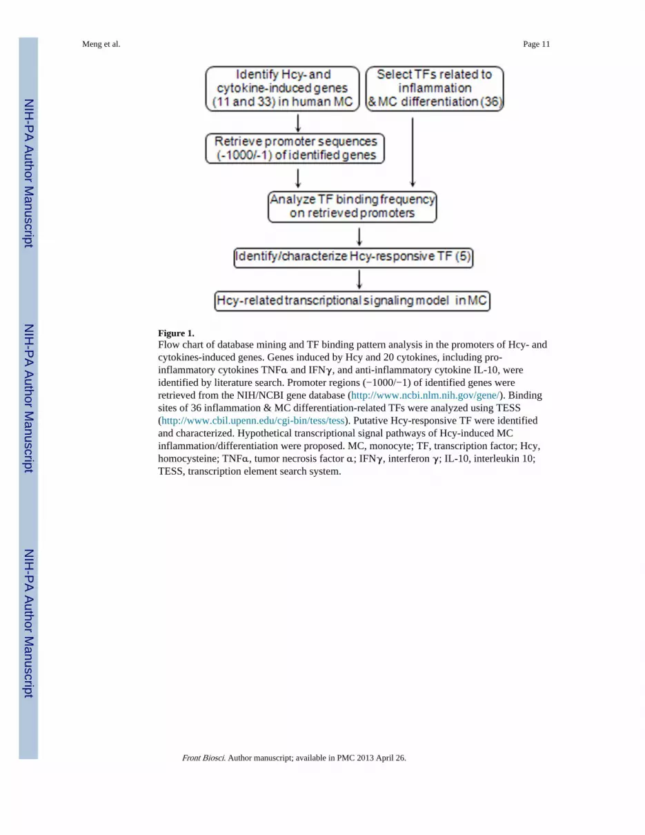
Figure 1.Flow chart of database mining and TF binding pattern analysis in the promoters of Hcy- andcytokines-induced genes. Genes induced by Hcy and 20 cytokines, including pro-inflammatory cytokines TNFα and IFNγ, and anti-inflammatory cytokine IL-10, wereidentified by literature search. Promoter regions (−1000/−1) of identified genes wereretrieved from the NIH/NCBI gene database (http://www.ncbi.nlm.nih.gov/gene/). Bindingsites of 36 inflammation & MC differentiation-related TFs were analyzed using TESS(http://www.cbil.upenn.edu/cgi-bin/tess/tess). Putative Hcy-responsive TF were identifiedand characterized. Hypothetical transcriptional signal pathways of Hcy-induced MCinflammation/differentiation were proposed. MC, monocyte; TF, transcription factor; Hcy,homocysteine; TNFα, tumor necrosis factor α; IFNγ, interferon γ; IL-10, interleukin 10;TESS, transcription element search system.
Meng et al. Page 11
Front Biosci. Author manuscript; available in PMC 2013 April 26.
NIH
-PA Author Manuscript
NIH
-PA Author Manuscript
NIH
-PA Author Manuscript

Figure 2.Identification of genes induced by Hcy and cytokines in human MC. Genes induced by Hcy,pro-inflammatory cytokines and anti-inflammatory cytokine in human MC were identifiedby literature search using NIH/PubMed database. A. Hcy-Induced genes. 11 Hcy-inducedgenes were identified. B. Cytokines-induced genes. 8 TNFα induced genes, 8 IFNγ-inducedgenes, and 17 IL-10-induced genes were identified. a, represents fold change quantified fromoriginal graphs; b, represents converted unit from original data; Reference, consistent withthe text and details in the “References” section; NS, not specified; MC, monocyte; TF,transcription factor; Hcy, homocysteine; TNFα, tumor necrosis factor α; IFNγ, interferonγ; IL-10, interleukin 10, PBMC, peripheral blood mononuclear cell; THP1, a humanmonocytic leukemia cell line.
Meng et al. Page 12
Front Biosci. Author manuscript; available in PMC 2013 April 26.
NIH
-PA Author Manuscript
NIH
-PA Author Manuscript
NIH
-PA Author Manuscript

Figure 3.TFs related to inflammation and MC differentiation. 36 TFs were selected based on theiridentified functions related to inflammation and MC differentiation. We used PubMed ID#to cite knowledge information regarding the function of 36 TF in inflammation and MCdifferentiation. MC, monocyte; TF, transcription factor.
Meng et al. Page 13
Front Biosci. Author manuscript; available in PMC 2013 April 26.
NIH
-PA Author Manuscript
NIH
-PA Author Manuscript
NIH
-PA Author Manuscript

Figure 4.Class 1 TF biding site distribution in the promoter regions of Hcy- and pro-inflammatorycytokine-induced genes. The occurrence (presence of class 1 TFs in the promoter regions ofHcy- and pro-inflammatory cytokines TNFα and IFNγ-induced genes) were listed. TFs withat least one binding site on the promoter of indicated genes were recorded as occurrencepositive (+), whereas, TFs that did not have binding sites on the promoter of indicated geneswere recorded as occurrence negative (−). Ratio of occurrence positive on selected geneswas calculated. Ratio of class 1 TFs in TNFα– and IFNγ–induced genes to Hcy–inducedgenes were compared and independent t -tests were performed to determine the p value ofpaired groups as indicated. MC, monocyte; TF, transcription factor; Hcy, homocysteine;TNFα, tumor necrosis factor α; IFNγ, interferon γ; HSF, heat shock factor; MEF2,monocyte enhancer factor-2; NFAT, nuclear factor of activated T-cells; NFκB, nuclearfactor kappa light chain enhancer of activated B cells; KLF4, krueppel-like factor 4.
Meng et al. Page 14
Front Biosci. Author manuscript; available in PMC 2013 April 26.
NIH
-PA Author Manuscript
NIH
-PA Author Manuscript
NIH
-PA Author Manuscript

Figure 5.Identification of genes induced by both Hcy and cytokines in human MC. Genes induced byHcy and 20 cytokines including 5 MC differentiation cytokines (GM-CSF, CSF-1, TNFα,IL-4 and IFNγ), 11 pro-inflammatory cytokines (MIP-1α, TNF-β, IL-1β, IL-2, IL-3, IL-6,IL-7, IL-8, IL-12, IL-15 and IL-18) and 4 anti-inflammatory cytokines (TGFβ, IL-9, IL-10and IL-11) were identified by literature search. Genes induced by both Hcy and cytokinesare listed. c, cytokines exerting both MC differentiation and pro-inflammatory function.Reference, consistent with the text and details in the “References” section; MC, monocyte;Hcy, homocysteine; TNFα, tumor necrosis factor α; IFNγ, interferon γ; GM-CSF,granulocyte macrophage colony stimulating factor; MIP-1α, macrophage inflammatoryprotein-1α; TGFβ, transforming growth factor β.
Meng et al. Page 15
Front Biosci. Author manuscript; available in PMC 2013 April 26.
NIH
-PA Author Manuscript
NIH
-PA Author Manuscript
NIH
-PA Author Manuscript

Figure 6.TF binding element analysis. Proximal promoter sequences (1,000 bp upstream of thetranscription start site) of Hcy- and cytokines- induced genes and four randomly-chosenhousekeeping genes were retrieved from the NIH/NCBI gene database, and analyzed forbinding sites of 36 inflammation- and MC differentiation-related TFs using TESS (http://www.cbil.upenn.edu/cgi-bin/tess/tess). A. TF binding frequency. Binding frequency denotesthe number of the binding sites for each TF on the promoter region. An upper threshold ofthe confidence interval was set as mean value+3SD of the TF binding frequencies in thepromoters of 4 housekeeping genes and 17 IL-10-induced genes, and marked as the dashedlines. The binding frequencies higher than the upper threshold of the confidence interval (p< 0.01) were considered as significant high frequency and directly labeled with number. TFswere divided into 3 classes based on the pattern of significant binding frequency in Hcy-induced genes. Class 1 TF is defined as TFs having high significant binding frequency onmore than 3 Hcy-induced genes, including HSF, MEF2, NFAT, NF-κB and KLF4, whichare termed as putative Hcy-responsive TF. Class 2 has 10 TFs which have significantbinding frequency on less than three Hcy-induced genes. Class 3 contained 8 TFs which donot have significant binding frequency on Hcy-induced genes. The remaining 13 TFs,
Meng et al. Page 16
Front Biosci. Author manuscript; available in PMC 2013 April 26.
NIH
-PA Author Manuscript
NIH
-PA Author Manuscript
NIH
-PA Author Manuscript

without identified binding sites in the promoters of Hcy-induced genes, were not included inthe classification. All class 1 TFs and one representative of class 2 and 3 TFs were shown.B. TF binding sites. Mean value and standard deviation of binding site of each TF on genesinduced by IL-10-, Hcy, IFNγ, TNFα were calculated. MC, monocyte; TF, transcriptionfactor; Hcy, homocysteine; TNFα, tumor necrosis factor α; IFNγ, interferon γ; IL-10,interleukin 10; HSF, heat shock factor; MEF2, monocyte enhancer factor-2; NFAT, nuclearfactor of activated T-cells; NF-κB, nuclear factor kappa light chain enhancer of activated Bcells; KLF4, krueppel-like factor 4; AR, androgen receptor; AP-1, activator protein 1.
Meng et al. Page 17
Front Biosci. Author manuscript; available in PMC 2013 April 26.
NIH
-PA Author Manuscript
NIH
-PA Author Manuscript
NIH
-PA Author Manuscript

Figure 7.Hypothetical Hcy transcriptional signaling pathways mediating Hcy-induced MCdifferentiation and MC-derived inflammation. Five class 1 TFs and their validated signalingpathways related to inflammation and MC differentiation were summarized. MC, monocyte;TF, transcription factor; Hcy, homocysteine; TNFα, tumor necrosis factor α; IFNγ,interferon γ; HSF, heat shock factor; MEF2, monocyte enhancer factor-2; NFAT, nuclearfactor of activated T-cells; NF-κB, nuclear factor kappa light chain enhancer of activated Bcells; KLF4, krueppel-like factor 4; MKK4, mitogen-activated protein kinase kinase 4; p38MAPK, p38 mitogen-activated protein kinases; IκB, nuclear factor of kappa lightpolypeptide gene enhancer in B-cells inhibitor; IKK, IκB kinase; p-IκB, phosphorylatedIκB; PLK1, polo-like kinase 1; CaM, calmodulin; PKC, protein kinase C; PU.1-P,phosphorylated PU.1 transcription factor; JAK, Janus kinase; Stat, signal transducer andactivator of transcription.
Meng et al. Page 18
Front Biosci. Author manuscript; available in PMC 2013 April 26.
NIH
-PA Author Manuscript
NIH
-PA Author Manuscript
NIH
-PA Author Manuscript


