Embed Size (px)
Citation preview

BioMed CentralBMC Evolutionary Biology
ss
Open AcceResearch articleHorizontal gene transfer in chromalveolatesTetyana Nosenko and Debashish Bhattacharya*Address: University of Iowa, Department of Biological Sciences and the Roy J. Carver Center for Comparative Genomics, 446 Biology Building, Iowa City, Iowa 52242, USA
Email: Tetyana Nosenko - [email protected]; Debashish Bhattacharya* - [email protected]
* Corresponding author
AbstractBackground: Horizontal gene transfer (HGT), the non-genealogical transfer of genetic materialbetween different organisms, is considered a potentially important mechanism of genome evolutionin eukaryotes. Using phylogenomic analyses of expressed sequence tag (EST) data generated froma clonal cell line of a free living dinoflagellate alga Karenia brevis, we investigated the impact of HGTon genome evolution in unicellular chromalveolate protists.
Results: We identified 16 proteins that have originated in chromalveolates through ancient HGTsbefore the divergence of the genera Karenia and Karlodinium and one protein that was derivedthrough a more recent HGT. Detailed analysis of the phylogeny and distribution of identifiedproteins demonstrates that eight have resulted from independent HGTs in several eukaryoticlineages.
Conclusion: Recurring intra- and interdomain gene exchange provides an important source ofgenetic novelty not only in parasitic taxa as previously demonstrated but as we show here, also infree-living protists. Investigating the tempo and mode of evolution of horizontally transferred genesin protists will therefore advance our understanding of mechanisms of adaptation in eukaryotes.
BackgroundHorizontal gene transfer (HGT) is the movement ofgenetic material between different species and is consid-ered to be one of the major driving forces of prokaryoticevolution [1-4]. Until recently, it was believed that thisphenomenon was largely restricted to the prokaryoticdomain. In eukaryotes, gene duplication has classicallybeen viewed as the major source of genetic novelty [5,6];this paradigm of eukaryotic evolution is based on genomestudies of model organisms such as multicellular plants,animals, and fungi. In the last decade, rapid accumulationof genome data from unicellular eukaryotes, protists, hasallowed researchers to reassess the role of HGT in eukary-otic evolution. The results of comparative analyses of
genomes of anaerobic parasitic protists provided a majorbreakthrough in our understanding of the impact of inter-domain HGT in eukaryotes. For example, 96 potentialcases of prokaryote-to-eukaryote HGT were identified inthe genome of an intestinal parasite of humans and ani-mals Entamoeba histolytica [7], 84 in the fish parasite Spiro-nucleus salmonicida [8], 152 in a sexually transmittedhuman pathogen Trichomonas vaginalis [9], 24 in Crypt-osporidium parvum [10], and 148 in anaerobic rumen cili-ates [11]. These numbers comprise up to 4% of genes inthe extremely reduced genomes of these anaerobic pro-tists. It is believed that the acquisition of bacterial genesby these eukaryotes accelerated their adaptation to anaer-obic environments and the transition to a parasitic life
Published: 25 September 2007
BMC Evolutionary Biology 2007, 7:173 doi:10.1186/1471-2148-7-173
Received: 30 March 2007Accepted: 25 September 2007
This article is available from: http://www.biomedcentral.com/1471-2148/7/173
© 2007 Nosenko and Bhattacharya; licensee BioMed Central Ltd. This is an Open Access article distributed under the terms of the Creative Commons Attribution License (http://creativecommons.org/licenses/by/2.0), which permits unrestricted use, distribution, and reproduction in any medium, provided the original work is properly cited.
Page 1 of 18(page number not for citation purposes)

BMC Evolutionary Biology 2007, 7:173 http://www.biomedcentral.com/1471-2148/7/173
style. Several recent reports indicate that HGT also plays arole in the genome evolution of free-living protists. Anal-ysis of the complete genome sequence of the soil amoebaDictyostelium discoideum led to the identification of 18genes derived from prokaryotes [12]. Several cases of HGThave been reported for dinoflagellate and chlorarachnio-phyte algae [13-16]. The fact that complete genomesequences are available now for a limited number of freeliving protists explains a significant disproportion in thestudy of HGT in different groups of protists. However,public databases also contain Expressed Sequence Tag(EST) libraries for over 50 species of free living unicellulareukaryotes [17,18] that can also be used to assess theimpact of HGT on genome evolution in protists.
Here we analyze EST and complete genome data to studyHGT in chromalveolate protists. Chromalveolates com-prise the six eukaryotic lineages, cryptophytes, hapto-phytes, stramenopiles, ciliates, apicomplexans, anddinoflagellates and have adapted to a wide variety of envi-ronments. They are characterized by a tremendous diver-sity of forms and modes of nutrition includingheterotrophy, parasitism, phototrophy, and mixotrophy.According to the chromalveolate hypothesis, the commonancestor of the six constituent lineages was a free livingphotosynthetic organism that derived its plastid via a redalgal secondary endosymbiosis [19]. Within-chromalveo-late taxon relationships and the monophyly of this groupare controversial [20]. Nuclear gene phylogenies supportthe monophyly of stramenopiles, ciliates, apicomplexans,and dinoflagellates and monophyly of cryptophytes andhaptophytes [21,22]. However, relationships between thetwo clades still remain unresolved.
Gene movement from the endosymbiont to the hostnucleus is a specific instance of HGT that is referred to asendosymbiotic gene transfer (EGT). The impact of EGT onthe evolution of chromalveolate genomes has been inten-sively studied in the last decade [23-27] and will not beconsidered here. We limited our research to gene transfersfrom non-organellar sources. To identify genes acquiredby chromalveolates through HGT at different time pointsin their evolutionary history, we performed a broad scalephylogenetic analysis of the EST data generated for a freeliving phototrophic dinoflagellate alga Karenia brevis thatis renowned as an agent of toxic algal blooms that annu-ally cause massive fish and marine mammal mortality inthe Gulf of Mexico [28]. Detailed analyses of the identi-fied genes presented in this paper suggest that recurringinter- and intradomain gene movement should be consid-ered as an important source of genetic novelty in chroma-lveolates.
ResultsIn this study, we used a combination of four differentapproaches to identify genes acquired by chromalveolatesthrough HGT (see Methods). The major goal of this studywas to discover genes uniquely present in chromalveo-lates and bacteria. This study is based on the assumptionthat HGT is the most plausible explanation for the occur-rence of bacterial genes in a single eukaryotic lineage. Analternative explanation is that these bacterial genes werederived via intracellular transfer from the mitochondrialprogenitor by the ancestral eukaryote and subsequentlylost from most taxa. Apart from invoking independentgene losses from potentially many eukaryotic lineages, thelatter scenario implies (improbably) that the genome sizeof the eukaryotic ancestor was far larger than in extanttaxa.
Our data screening approach was designed to retrieve rel-atively ancient cases of HGT that occurred before thedivergence of two closely related genera of dinoflagellatealgae, Karenia and Karlodinium. Using a sequence similar-ity search (BLAST; e-value ≤ 10-10) we identified 3,341genes shared by K. brevis and at least 1/5 dinoflagellatespecies for which EST data are available [18]. To identifygenes acquired by chromalveolates through interdomainHGT, we analyzed the restricted set of K. brevis genes usinga combination of a standard "Best Hit" approach and highthroughput automated and manual phylogenomic analy-ses (see Methods). These analyses yielded 80 unique genesencoding proteins from 45 different families putativelyderived from prokaryotes at different time points of chro-malveolate evolution. Throughout the text, we will use theterm "protein" to unite members of one protein familyencoded by distinct genes. Three proteins represented byfive unigenes resulted from an additional analysis aimedat detecting K. brevis homologs of bacterial proteinsinvolved in cell wall biogenesis (see Methods). Detailedphylogenetic analyses of the identified proteins providestrong support for the origin through HGT of 16 proteinsrepresented by 36 unique genes (Table 1). Six of these pro-teins are uniquely shared by dinoflagellates and prokary-otes (BLAST; e-value ≤ 10-20); two proteins, malate-quinone oxidoreductase and monomeric NADPH-dependent isocitrate dehydrogenase, are present only inseveral chromalveolate lineages and prokaryotes.Homologs of eight proteins (e-value ≤ 10-20) are presentin prokaryotes, chromalveolates, and at least one othereukaryotic lineage. Phylogenies of the remaining 32 pro-teins represented by 49 K. brevis EST contigs could not beclarified due to the presence of multiple, highly divergentsequences in different protist lineages that might haveresulted either from independent gene transfers or fromancient gene duplications.
Page 2 of 18(page number not for citation purposes)

BMC Evolutionary Biology 2007, 7:173 http://www.biomedcentral.com/1471-2148/7/173
Below we provide a detailed description of the most inter-esting cases of prokaryote-to-eukaryote HGTs. The direc-tion of interdomain HGTs was inferred based on therelative distribution of the gene among bacteria andeukaryotes. Genes widespread among prokaryotes andrare among eukaryotes were considered to be derivedfrom a prokaryotic donor. The identified proteins are clas-sified in various functional groups based on known func-tions of their bacterial homologs including plasmamembrane biogenesis and biosynthesis of secondarymetabolites, energy and amino acid metabolism, sub-strate transport, regulation of gene expression and DNArepair. In addition, we present results of phylogeneticanalyses of translation elongation factor EF2 that repre-sents the only identified case of a recent transfer thatoccurred after the Karenia and Karlodinium divergence andthe only example of HGT involving a gene of eukaryoticorigin.
Plasma membrane biogenesis and biosynthesis of secondary metabolitesDehydrogenase MVIM-sugar aminotransferase fusion proteinScreening of the K. brevis EST data resulted in the identifi-cation of two related proteins, sugar aminotransferase(WECE) and the fused dehydrogenase MVIM-sugar ami-notransferase (MVIV-WECE) that showed high similarityto bacterial proteins. Both proteins are encoded by multi-ple gene copies each represented by multiple transcripts inthe K. brevis EST data. To simplify matters, we namedallMVIV-WECE-encoding genes mviM/wecE-14 and allWECE-encoding genes wecE-17. The gene names werederived from the names of the corresponding proteindomains followed by the number of the contigs encodingthe full-length cDNA sequence of these proteins. Bacterialhomologs of the wecE-17 encode a sugar aminotransferasethat is classified in the DegT/DnrJ/EryC1/StrS aminotrans-ferase family in the Pfam database [29,30]. We identified
Table 1: Horizontal gene transfers from bacteria to chromalveolates
1 2 3 4 5 6Domain (Pfam) Protein family [Function] Accession
numberNumber unigenes
Best Hit
Phylogeny
MVIM Predicted dehydrogenase EF540335 7 1e-57 Fig. 2WECE Pyridoxal phosphate dependent aminotransferase [Cell
envelope biogenesis, outer membrane]EF540335EF540337
12 2e-100 Fig. 2
Epimerase NAD dependent epimerase/dehydratase [Cell envelope biogenesis, outer membrane]
EF540339 2 1e-56 Fig. 3
CAS-like Clavaminic acid synthetase [Biosynthesis of clavulanic acid]
EF540323EF540325
3 1e-53 Fig. 4
MQO Malate-quinone oxidoreductase [Energy metabolism] EF540331EF540333
2 1e-98 Fig. 5
NADP-IDH Monomeric NADP(+)-dependent isocitrate dehydrogenase. [Energy metabolism]
EF540327EF540328
2 1e-162 Fig. 6
Fe-ADH Iron-containing alcohol dehydrogenase [Energy metabolism]
EF540326 1 2e-97 Additional file 1
PutA NAD-dependent aldehyde dehydrogenases [Energy metabolism]
EF540338 2 2e-120 Additional file 1
PBPb Substrate-bound, membrane-associated, periplasmic binding protein [Substrate transport]
EF540334 1 2e-26 Additional file 1
SIR2 Silent information regulator 2 [Gene silencing, DNA repair]
EF540336 2 5e-39 Additional file 1
AslA Arylsulfatase A [Substrate transport] EF540322 1 1e-70 Additional file 1*
COG3129 SAM-dependent methyltransferase EF540332 1 1e-23 Additional file 1*
ATS1 Alpha-tubulin suppressor [Cell division and chromosome partitioning, cytoskeleton]
EF540324 5 8e-52 Additional file 1*
PdxA Pyridoxal phosphate biosynthetic protein PdxA [Amino acid metabolism]
EF540340 1 4e-54 Additional file 1*
COG3618 Metal-dependent hydrolase of the TIM-barrel fold EF540329 2 1e-55 Additional file 1*
COG3022 Hypothetical [unknown] EF540330 1 4e-26 Additional file 1*
1. The Pfam domain designation [29, 30]. 2. Confirmed or proposed function of the prokaryotic homologs is given. 3. GenBank accession number. 4. Numbers of unigenes encoding particular protein identified in the Karenia brevis EST data are given. Only one copy of gene was submitted into the GenBank. All gene copies can be found at [111]. 5. E-values for the best bacterial hit from the GenBank nonredundant database. 6. Phylogenetic trees for all proteins present in dinoflagellates and at least one other eukaryotic lineage (BLASTp e-value ≤ 10-10).*The protein was found only in dinoflagellates and prokaryotes.
Page 3 of 18(page number not for citation purposes)

BMC Evolutionary Biology 2007, 7:173 http://www.biomedcentral.com/1471-2148/7/173
five genes of the wecE-17 type in the K. brevis genome. ThewecE-17 genes share 93–99% amino acid sequence iden-tity over their protein coding regions and significant sim-ilarity of the 3' UTR sequences. The total length of theidentified wecE-17 sequences is 442 aa including 367 aa ofthe mature WECE protein and 74 aa of the incomplete N-terminal extension (Fig. 1). The N-terminus is highlyhydrophobic and, according to the protein topology pre-diction program TMHMM [31], contains a transmem-brane motif (P = 0.87%).
The mviM/wecE-14 genes have a bipartite structure (Fig.1). The N-terminal region of this sequence contains aNAD-binding Rossmann fold domain typical for theGFO/IDH/MocA oxidoreductase family and shows highsimilarity to the dehydrogenase MVIM found in Bacteriaand Archaea (55% amino acid sequence identity). The C-terminal domain of mviM/wecE-14 encodes WECE thatshares 81–84% amino acid sequence identity with proteinencoded by wecE-17 and about 67% amino acid sequenceidentity with its bacterial homologs. Using analyses ofnucleotide differences in protein coding regions andinsertion-deletions in 5' and 3' UTR, we identified at leastseven MVIM-WECE-encoding genes in K. brevis. Becausethese genes share 92–99% amino acid sequence identityand retain significant sequence similarity of their 5' and 3'UTRs their origin is likely through recent gene duplica-tions. The K. brevis culture that was used for the EST datacollection was a vegetative haploid clonal cell line there-fore all sequence variants were non-allelic gene copies.
The sequence structure is conserved between all mviM/wecE-14 copies: they are composed of 361 aa of MVIM,368 aa of WECE, and 10 aa of the spacer region separatedthe two domains. MviM/wecE-14 genes do not have an N-terminal extension and, presumably, encode cytosolicproteins. The comparison of the wecE-14 and wecE-17
sequences shows that the two types of transcripts do notresult from alternative splicing, but are encoded by differ-ent loci in the K. brevis genome. The number of aminoacid substitutions between the protein coding regions ofwecE-14 and wecE-17 is higher than the number of withingroup substitutions. Furthermore, the N-terminus and 3'UTRs are highly conserved between wecE-17 genes andshare no sequence similarity with mviM/wecE-14. Phylo-genetic analyses provide strong support for wecE-14 andwecE-17 monophyly (bootstrap proportions, maximumlikelihood, BPml = 100%; neighbor joining, BPnj = 100%;Bayesian posterior probability, BPP = 1.0; Fig. 2A).
Homologs of the K. brevis MVIM and WECE are broadlydistributed among Bacteria and Archaea. Interestingly, thetwo genes are located in one operon separated by 9–89 bpspacer regions in several proteobacteria (Fig. 1). Thisobservation suggests that dinoflagellates might havederived the mviM/wecE-14 fragment through a singleHGT. WecE-17 most likely resulted from a recombinationbetween MviM/wecE-14 and a DNA fragment that gave riseto the hydrophobic N-terminus of wecE-17. This eventoccurred after the Karenia divergence and was followed bymultiple duplications of wecE-17 and mviM/wecE-14.Homologs of the bacterial MVIM and WECE proteins havenot previously been reported for eukaryotes. Using aBLAST search against the GenBank dbEST database weidentified homologs of the K. brevis WECE proteins in freeliving heterotrophic species of excavates: jakobids Secula-monas ecuadoriensis and Jakoba bahamiensis and heterolo-bosean amoebae Sawyeria marylandensis (see Additionalfile 1). Homologs of the K. brevis MVIM have been identi-fied in Karlodinium micrum and two species of excavates, J.bahamiensis and S. marylandensis (see Additional file 1).The MVIM and WECE distribution and phylogeny indi-cate that these proteins have been acquired by excavatesbefore the divergence of jakobids and heteroloboseans
Structure of the mviM/wecE fragment in the dinoflagellate Karenia brevis and proteobacteriaFigure 1Structure of the mviM/wecE fragment in the dinoflagellate Karenia brevis and proteobacteria. Each arrow-shaped box represents an open reading frame (ORF). Arrows indicate the direction of transcription. Solid black boxes represent 5' untranslated regions (UTR) and protein spacer regions. Black lines connecting two boxes represent intergenic regions.
Karenia brevis mviM/wecE-14
100 bp
wecE
mviM wecE
100100 1000 2000(bp)
mviM wecEMagnetospirillum magneticum AMB-1
Geobacter sulfurreducens PCA
Karenia brevis wecE-17
wecEmviM
Page 4 of 18(page number not for citation purposes)

BMC Evolutionary Biology 2007, 7:173 http://www.biomedcentral.com/1471-2148/7/173
(Fig. 2). The observed monophyly of the K. brevis and K.micrum MVIM sequences and the absence of MVIM andWECE homologs in EST libraries from four species ofperidinin dinoflagellates that are ancestral to Karenia andKarlodinium [32], suggests that the mviM/wecE-14 frag-ment originated on the branch uniting Karenia and Kar-lodinium. The result of the MVIM and WECE nucleotidecomposition analysis also demonstrates that the acquisi-tion of these genes by dinoflagellates and excavates was arelatively ancient event(s). The nuclear gene nucleotidecomposition varies significantly among different speciesof excavates and dinoflagellates. However, the GC contentof the MVIM- and WECE-encoding sequences does notdeviate from the GC content range identified for protein-coding regions in the host organisms (Table 2). Phyloge-netic analyses of WECE and MVIM support the mono-phyly of these sequences in eukaryotes (BPml = 96%; BPnj= 85%; BPP = 1.0 for WECE and BPml = 76%; BPnj = 79%;BPP = 0.97 for MVIM) (Fig. 2A, B). Although we cannotexclude the possibility that dinoflagellates and excavatesindependently derived MVIM- and WECE-encoding genesfrom the same bacterial source, the most plausible inter-pretation of the similar phylogenies for the two genes andthe co-occurrence of both sequences in two distantlyrelated groups of eukaryotes is that the bacterial mviM/wecE DNA fragment was acquired by one of these eukary-otic lineages and passed to the next via HGT, perhapsthrough phagocytosis (Fig. 2C). The incongruence of theprokaryotic MVIM and WECE phylogenies with therespective species phylogenies and disagreement betweenthe MVIM and WECE tree topologies (Fig. 2A, B) do notallow us to unambiguously identify the prokaryotic donorof the mviM/wecE fragment. The disagreement betweenphylogenies may result from frequent transfers amongbacteria that has abolished a specific sister group relation-ship to the eukaryotic sequences (see Discussion fordetails).
Study of HGT in bacteria demonstrates that genes encod-ing physiologically coupled reactions are often co-trans-ferred, frequently in operons [33]. The fact that MVIM-and WECE-encoding genes are fused in K. brevis andlinked in several proteobacteria may indicate that proteinsencoded by these genes are functionally coupled. Func-tions of the dehydrogenase MVIM are poorly character-ized in bacteria. The bacterial homologs of WECE havebeen intensively studied for their involvement in the bio-synthesis of microlide antibiotics that belong to the largefamily of secondary metabolites known as polyketides[34-37], outer membrane liposaccharides [38-40], andsurface layer glycoproteins [41-43]. According to theresults of phylogenetic analyses, WECE proteinsidentifiedin eukaryotes form a monophyletic clade with bacterialproteins from two distinct groups (Fig. 2A). The firstgroup is represented by actinobacterial proteins involved
Origin of sugar aminotransferase WECE and dehydrogenase MVIM in eukaryotesFigure 2Origin of sugar aminotransferase WECE and dehy-drogenase MVIM in eukaryotes. A. ML tree of sugar ami-notransferase WECE. B. ML tree of dehydrogenase MVIM. The numbers above and below the branches are the results of ML and NJ bootstrap analyses, respectively. Only boot-strap values ≥ 60% are shown. The thick branches indicate ≥ 0.95 posterior probability from a Bayesian inference. Branch lengths are proportional to the number of substitutions per site (see scale bars). Numbers in bold indicate bootstrap sup-port for the monophyly of wecE-14 and wecE-17 sequences in Karenia brevis. CH indicates chromalveolates and EX indicates Excavata. Names of bacterial WECE-encoding genes that have been studied experimentally [34-36, 41, 43] are given in brackets. C. Origin and distribution of MVIM and WECE in eukaryotes through sequential HGTs.
0.1
Geobacter metallireducens GS-15
Geobacter sulfurreducens PCA
74
Methylococcus capsulatus str. Bath
100
Methanosarcina barkeri str. fusaro
Methanosphaera stadtmanae DSM 3091
100
100
Methanococcoides burtonii DSM 6242
89
Karenia brevis
Karlodinium micrum
100
Sawyeria marylandensis - Heterolobosea94
Jakoba bahamiensis - Jakobida
76
74
Syntrophobacter fumaroxidans MPOB
Pelobacter propionicus DSM 2379
100
Chloroflexus aurantiacus J-10-fl
Rhizobium etli CFN 42
Nitrobacter hamburgensis X14
Magnetospirillum magneticum AMB-1
Arthrobacter sp. FB24
92
Acidobacteria bacterium Ellin345
88
Roseiflexus sp. RS-1
Coxiella burnetii Dugway 7E9-12
100
Nostoc sp. PCC 7120
Trichodesmium erythraeum IMS101
Proteobacteria
Archaea
Dinoflagellates (CH)
Proteobacteria
- Actinobacteria- Acidobacteria- Chloroflexi- Proteobacteria
Cyanobacteria
B.
100
79
85100
94
9887
100
100100
0.1
Helicobacter hepaticus ATCC 51449Aneurinibacillus thermoaerophilus (fdtB)
100
Thermoanaerobacterium thermosaccharolyticum (gdtB)
100
Micromonospora griseorubida (mydD)Aeromicrobium erythreum (eryC1)
74
Streptomyces venezuelae (desV)
100
94
Jakoba bahamiensisSeculamonas ecuadoriensis
84
Sawyeria marylandensis - Heterolobosea81
Karenia brevis (wecE-14)Karenia brevis (wecE-17)
10096
62
Acidobacteria bacterium Ellin345Arthrobacter sp. FB24
Magnetospirillum magneticum AMB-1Nitrobacter hamburgensis X14
83
Rhizobium etli CFN 42Chloroflexus aurantiacus J-10-fl
84
Solibacter usitatus Ellin6076
78
Pelobacter propionicus DSM 2379Methanosphaera stadtmanae DSM 3091Methanothermobacter thermautotrophicus str. Delta H
66
Methanosarcina barkeri str. fusaro
100
Desulfitobacterium hafniense Y51Roseiflexus sp. RS-1
81
Trichodesmium erythraeum IMS101Nostoc sp. PCC 7120
99
Synechococcus elongatus PCC 6301
100
A.
Proteobacteria
Actinobacteria
-
Firmicutes
Jakobida
Dinoflagellates (CH)
- Actinobacteria- Acidobacteria
Proteobacteria
Archaea
- Firmicutes- Chloroflexi
Cyanobacteria
64100
10099
85
63
100 64
8979
100
100
-63
85
--
--
--
--
91
-
100
-
-
-----
--
--
C.
BACTERIUM EUKARYOTE I EUKARYOTE II
mviM/wecE
HGT1 HGT2
EX
EX
mviM/wecE
Page 5 of 18(page number not for citation purposes)
BMC Evolutionary Biology 2007, 7:173 http://www.biomedcentral.com/1471-2148/7/173
in the biosynthesis of microlide antibiotics such as narbo-mycin, erythromycin, pikromycin, and neomethymycin(Fig. 2A). These catalyze the biosynthesis of the deoxysugar D-desosamine, the addition of which to the actino-bacterial polyketides is crucial for their antibiotic activity[34]. The second group includes monofunctional sugaraminotransferases involved in the glycosylation of thesurface layer proteins in firmicutes Aneurinibacillus ther-moaerophilus (ftdB) and Thermoanaerobacterium thermosac-charolyticum (qdtB). FtdB and qdtB encode a key enzyme ofthe biosynthesis of thymidine diphosphate-activated 3-acetamido-3,6-dideoxy- D- galactose (dTDP-D-Fucp3NAc), which serves as a precursor for the assembly ofstructural polysaccharides in bacteria [41,43]. The pres-ence of multiple WECE-encoding genes and a high expres-sion level of these genes in K. brevis suggest that thisprotein is involved in physiologically important processesin this organism.
NAD dependent sugar nucleotide epimerase/dehydrataseBLAST searches using sequences of bacterial proteinsinvolved in outer membrane liposaccharide and S-layerglycoprotein biosyntheses [43,44] allowed us to identifytwo genes encoding NAD dependent sugar nucleotide epi-merase in the K. brevis EST data. Protein coding regions ofthe two genes share 98% amino acid sequence identityand significant similarity of their 3'UTR regions. This sug-
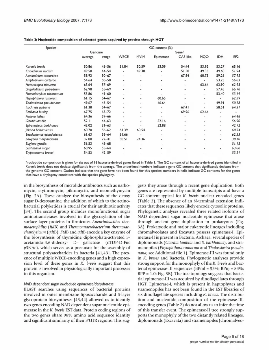
gests they arose through a recent gene duplication. Bothgenes are represented by multiple transcripts and have aGC content typical for K. brevis nuclear encoded genes(Table 2). The absence of an N-terminal extension indi-cates that these sequences likely encode cytosolic proteins.Phylogenetic analyses revealed three related isoforms ofNAD dependent sugar nucleotide epimerase that arosethrough ancient gene duplication in prokaryotes (Fig.3A). Prokaryotic and major eukaryotic lineages includingchromalveolates and Excavata possess epimerase-I. Epi-merase-II is present in Bacteria, Archaea, several species ofdiplomonads (Giardia lamblia and S. barkhanus), and stra-menopiles (Phytophthora ramorum and Thalassiosira pseudo-nana; see Additional file 1). Epimerase-III was found onlyin K. brevis and Bacteria. Phylogenetic analyses providestrong support for the monophyly of the K. brevis and bac-terial epimerase-III sequences (BPml = 93%; BPnj = 83%;BPP = 1.0; Fig. 3B). The tree topology suggests that bacte-rial epimerase-III was acquired by dinoflagellates throughHGT. Epimerase-I, which is present in haptophytes andstramenopiles has not been found in the EST libraries ofsix dinoflagellate species including K. brevis. The distribu-tion and nucleotide composition of the epimerase-III-encoding genes (Table 2) do not allow us to infer the timeof this transfer event. The epimerase-II tree strongly sup-ports the monophyly of the two distantly related lineages,diplomonads (Excavata) and stramenopiles (chromalveo-
Table 2: Nucleotide composition of selected genes acquired by protists through HGT
Species GC content (%)Genome Gene1
average range WECE MVIM Epimerase CAS-like MQO IDH EF2
Karenia brevis 50.86 45–56 51.84 50.59 53.09 54.44 53.92 53.27 65.16Karlodinium micrum 49.50 44–54 - 49.30 - 51.50 49.35 49.60 51.94Alexandrium tamarense 58.93 50–67 - - - 67.84 60.75 59.26 57.92Amphidinium carterae 54.64 50–58 - - - - - 53.75 56.03Heterocapsa triquetra 63.64 57–69 - - - - 63.64 63.90 62.93Lingulodinium polyedrum 62.98 55–69 - - - - - 57.45 66.78Phaeodactylum tricornutum 53.86 49–60 - - - - - 53.40 53.19Phytophthora ramorum 61.15 54–67 - - 60.65 - - - 62.59Thalassiosira pseudonana 49.67 45–54 - - 46.64 - - 49.91 50.78Isochrysis galbana 61.38 54–67 - - - 67.41 - 58.51 64.31Emiliania huxleyi 67.75 63–72 - - - 69.96 62.64 - -Pavlova lutheri 64.36 59–66 - - - - - - 64.48Giardia lamblia 52.11 44–63 - - 52.16 - - - 56.90Spironucleus barkhanus 43.02 31–63 - - 52.88 - - - 42.72Jakoba bahamiensis 60.70 56–62 61.39 60.54 - - - - 60.54Seculamonas ecuadoriensis 61.63 56–64 61.66 - - - - - 62.53Sawyeria marylandensis 32.00 22–41 30.51 24.36 - - - - 30.10Euglena gracilis 56.53 45–68 - - - - - - 51.12Leishmania major 60.95 55–64 - - - - - - 63.08Trypanosoma brucei 54.53 42–59 - - - - - - 53.21
Nucleotide composition is given for six out of 16 bacteria-derived genes listed in Table 1. The GC content of all bacteria-derived genes identified in Karenia brevis does not deviate significantly from the average. The underlined numbers indicate a gene GC content that significantly deviates from the genome GC content. Dashes indicate that the gene have not been found for this species; numbers in italic indicate GC contents for the genes that have a phylogeny consistent with the species phylogeny.
Page 6 of 18(page number not for citation purposes)

BMC Evolutionary Biology 2007, 7:173 http://www.biomedcentral.com/1471-2148/7/173
lates) (BPml = 100%; BPnj = 99%; BPP = 1.0) (Fig. 3B).This tree topology is most easily explained by serial HGTthat involved ancient prokaryote-to-eukaryote and morerecent eukaryote-to-eukaryote gene transfers (Fig. 2C).Similar to WECE and MVIM, the epimerase tree does notallow us to identify the prokaryotic lineages that contrib-uted these genes to eukaryotes.
The functions of these enzymes have not been studied inchromalveolates. In Bacteria, epimerase-I and III catalyzethe biosyntheses of dTDP-D-Fuc p3NAc and dTDP-L-ram-nose, compounds that serve as precursors for the assemblyof outer membrane structural polysaccharides [41-43,45].On the dTDP-D-Fuc p3NAc pathway, they functionupstream of the described earlier WECE proteins. We can-not exclude that WECE and epimerase-III represent a func-tionally coupled enzyme pair in K. brevis that might havebeen acquired by dinoflagellates in one HGT event (Figs.2, 3). Available experimental data suggest that epimerase-I and II are also involved in cell wall polysaccharide bio-synthesis in plants [46] and diplomonads [47]. Based onthese data we propose that epimerase-III performs similarfunction(s) in K. brevis.
Clavaminic acid synthetase-like proteinClavaminic acid synthetase (CAS) belongs to the largefamily of iron and 2-oxoacid-dependent dioxygenases, animportant class of enzymes that mediates a variety of oxi-dative reactions [48]. Most studies of CAS have been car-ried out using the Streptomyces isozymes [49]. InStreptomyces, CAS catalyzes three major steps of the clavu-lanic acid biosynthesis. Clavulanic acid is a natural inhib-itor of β-lactamases, enzymes that confer resistance to β-lactam antibiotics in bacteria.
We identified homologs of the bacterial CAS proteins infour eukaryotic lineages: Fungi (Opisthokonta), greenalgae (Plantae), dinoflagellates, and haptophytes (chro-malveolates; Fig. 4, see Additional file 1). Partialsequences of three genes encoding CAS-like protein wereidentified in the K. brevis EST data. To the best of ourknowledge, the functions of these proteins have not beenstudied in eukaryotes. According to TargetP and MitoProt,CAS-like proteins in K. brevis (P = 0.763 and P = 0.870respectively), K. micrum (P = 0.742 and P = 0.951), and agreen alga Ostreococcus tauri (P = 0.748 and P = 0.706) aremitochondrial targeted. CAS-like proteins in O. lucimari-nus and Chlamydomonas reinhardtii do not have an N-ter-minal extension. PSORT [50] predicts a peroxisomal
Phylogeny of NAD-dependent sugar nucleotide epimerase/dehydratase isoformsFigure 3Phylogeny of NAD-dependent sugar nucleotide epimerase/dehydratase isoforms. A. ML tree of three isoforms of NAD dependent sugar nucleotide epimerase/dehydratase. Light brown color represents eukaryotic clades; blue color repre-sents prokaryotic clades.B. A fragment of the ML tree from the Figure 3A representing the phylogeny of NAD dependent sugar nucleotide epimerase/dehydratase isoforms II (E-II) and III (E-III). The numbers above and below the branches are the results of ML and NJ bootstrap analyses, respectively. Only bootstrap values ≥ 60% are shown. The thick branches indicate ≥ 0.95 posterior probability from a Bayesian inference. Branch lengths are proportional to the number of substitutions per site (see the scale bar). CH indicates chromalveolates and EX indicates Excavata. Names of the epimerase-encoding genes that have been studied experimentally [44, 45, 47] are given in brackets.
0.1
Rhodobacter sphaeroides ATCC 17025Oceanicola granulosus HTCC2516
80
Rhizobium etli CFN 42
100
Geobacter sp. FRC-32Solibacter usitatus Ellin6076 - Acidobacterium
78
Anaeromyxobacter dehalogenans 2CP-C - Proteobacterium
84
65
Acidobacteria bacterium Ellin345 - Acidobacterium
82
marine actinobacterium PHSC20C1Frankia alni ACN14a
100
Karenia brevis -Rhodoferax ferrireducens T118
Burkholderia xenovorans LB40061
Mycobacterium avium (gepiA) - ActinobacteriumXanthomonas oryzae pv. Oryzae (wxoA) - Proteobacterium
10095
93
Acidobacteria bacterium Ellin345 - AcidobacteriumHerpetosiphon aurantiacus ATCC 23779 - Chloroflexi
86
Halobacterium sp. NRC-1
86
Methanosarcina barkeri str. Fusaro
64
Oceanospirillum sp. MED92Pseudomonas fluorescens Pf-5
77
Staphylococcus aureus subsp. aureus USA300Lactococcus lactis subsp. lactis Il1403
97
Giardia lamblia (uae)
Spironucleus barkhanus100
Phytophthora ramorumThalassiosira pseudonana
99100
100
79100
--
-
96
100
-67--
8393
69100
7879-
92
10099
88
EPIMERASE-I
Diplomonadida (EX)
Stramenopiles (CH)
100
-
-
-
72
-
-82
Proteobacteria
Actinobacteria
Proteobacteria
Archaea
Firmicutes
Proteobacteria
0.1
BACTERIA& ARCHAEA
BACTERIA
BACTERIA
CHROMALVEOLATA(Stramenopiles)
EXCAVATA(Diplomonadida)
OPISTHOKONTAPLANTAEAMOEBOZOAEXCAVATACHROMALVEOLATA
BACTERIA& ARCHAEA
A
Karenia brevis
B
Dinoflagellates (CH)
IIIII
I
E-III
E-II
Page 7 of 18(page number not for citation purposes)

BMC Evolutionary Biology 2007, 7:173 http://www.biomedcentral.com/1471-2148/7/173
localization for the fungal CAS-like proteins however,with low probability (P-value range = 0.500 – 0.660).Phylogenetic analyses of the CAS-like proteins (Fig. 4)support the monophyly of the two chromalveolate line-ages (BPml = 87%; BPnj = 87%; BPP = 0.99) and placeschromalveolates in one clade with a cyanobacterium Tri-chodesmium erythraeum IMS101 (BPml = 100%; BPnj =100%; BPP = 1.0). Most proteins of cyanobacterial originwere derived by chromalveolates from red or green algalprogenitors of their plastids via secondary endosymbiosis[25,26]. Absence of Plantae from the cyanobacterial-chro-malveolate clade likely indicates that this cyanobacterialprotein was acquired by chromalveolates not throughendosymbiotic but rather through horizontal gene trans-fer. Recently, Waller at al. [15] reported another case ofcyanobacterium-to-dinoflagellate HGT that involved aDNA fragment encoding the plastid targeted shikimate-O-methyltransferase junction protein. However, presence ofthe CAS-like proteins in haptophytes suggests a differentscenario for the occurrence of this enzyme in dinoflagel-lates, which might include an additional, haptophyte-to-
dinoflagellate HGT. Alternatively the CAS-like protein treetopology could be explained by the gene transfer fromcyanobacteria before the divergence of chromalveolatesand its subsequent loss from stramenopiles, ciliates, andapicomplexans.
Energy metabolismIron-containing alcohol dehydrogenase and NAD-dependent aldehyde dehydrogenaseIron-containing alcohol dehydrogenase (Fe-ADH) andNAD-dependent aldehyde dehydrogenase (PutA) areprobably the best-studied proteins from the perspective ofHGT in eukaryotes. Aldehyde-alcohol dehydrogenase pro-tein (AdhE) has arisen through the fusion of two proteindomains, PutA and Fe-ADH and is considered to be a keyenzyme in energy metabolism in parasitic amitochondri-ate protists [51]. Previous studies on parasitic protistsdemonstrated that AdhE-encoding genes have been sub-jects of multiple independent prokaryote-to-eukaryoteHGTs. For information about AdhE functions and phylog-eny in parasitic protists we would direct readers to refer-
Origin of clavaminic acid synthetase (CAS) -like protein in ChromalveolataFigure 4Origin of clavaminic acid synthetase (CAS) -like protein in Chromalveolata. ML tree of CAS and CAS-like proteins. The numbers above and below the branches are the results of ML and NJ bootstrap analyses, respectively. Only bootstrap val-ues ≥ 60% are shown. The thick branches indicate ≥ 0.95 posterior probability from Bayesian inference. Branch lengths are proportional to the number of substitutions per site (see the scale bar). CH indicates chromalveolates. The name of the CAS protein-encoding gene that has been studied experimentally [49] is given in brackets. * The position of Alexandrium tamarense within the haptophytes clade was inferred from the analysis of a short C-terminal sequence.
0.1
Isochrysis galbanaAlexandrium tamarense*
67Emiliania huxleyi
98
Karlodinium micrum
Karenia brevis
100
87
Trichodesmium erythraeum IMS101 -Cyanobacterium
100
Streptomyces rubellomurinus (FrbJ) - ActinobacteriumBurkholderia xenovorans LB400 - Proteobacterium
Frankia sp. CcI3 - ActinobacteriumPhotorhabdus luminescens subsp. laumondii TTO1
Parvibaculum lavamentivorans DS-1
Bordetella parapertussis 12822
Ostreococcus lucimarinus
Ostreococcus tauri
100
Chlamydomonas reinhardtii
Chaetomium globosum
Magnaporthe grisea
72
Neurospora crassa
99
Coccidioides immitis
Aspergillus fumigatus
89100
81
87
100
-
95
100
-
99
95
100
--
--
------ --
PLANTAE
(Green Algae)
OPISTHOKONTA
(Fungi)
Dinoflagellates
Haptophytes
Proteobacteria
CH
Page 8 of 18(page number not for citation purposes)

BMC Evolutionary Biology 2007, 7:173 http://www.biomedcentral.com/1471-2148/7/173
ences [51,52]. In addition to previous findings, weidentified Fe-ADH in free-living dinoflagellates and jako-bids (see Additional file 1). These sequences share over50% amino acid identity with bacterial homologs. Resultsof phylogenetic analyses suggest that the two lineagesacquired Fe-ADH-encoding genes independently fromclosely related prokaryotes. The Fe-ADH tree obtained inthis study is shown in Additional file 1. PutA has beenfound in free living dinoflagellates, stramenopiles, andjakobids (see Additional file 1). PutA sequences in thesethree lineages show over 50% amino acid identity to cor-responding bacterial proteins and according to the resultof phylogenetic analyses, originated through independentinterdomain transfers from distinct prokaryotic donors(see Additional file 1). Phylogenetic analyses stronglysupport the monophyly of dinoflagellate and bacterialsequences (BPml = 100%; BPnj = 100%; BPP = 1.0) andsuggest that dinoflagellates acquired PutA before thedivergence of Karenia and Karlodinium.
Malate-quinone oxidoreductaseMalate-quinone oxidoreductase (MQO) is a functionalanalog of the better-known NAD-dependent malate dehy-drogenase (MDH) that catalyses the conversion of malateto oxaloacetate in the tricarboxylic acid (TCA) cycle. Incontrast to MDH, bacterial MQO is a membrane-associ-ated enzyme that utilizes flavin adenine dinucleotide(FAD) as a cofactor and donates the electrons from malateoxidation to quinones instead of NAD [53-55]. MQO isprotein common in bacteria. Among eukaryotes, thisenzyme has been previously reported only for apicompl-exans [56]. Apicomplexans lack the mitochondrial formof MDH. It has been shown that MQO compensates formitochondrial MDH in the TCA cycle in this lineage [57].
We found two MQO-encoding genes in the K. brevis ESTdata. These share 60% amino acid sequence identity andabout 44% identity with their bacterial homologs. Bothhave nucleotide compositions typical for K. brevis nuclearencoded genes (Table 2). Using sequence similaritysearches, we identified homologs of the K. brevis MQO inseveral species of dinoflagellates and haptophytes (seeAdditional file 1). Phylogenetic analyses support themonophyly of the two genes identified in K. brevis (BPml= 87%; BPP = 1.0) suggesting that they originated via geneduplication after the Karenia and Karlodinium divergence.Phylogenetic analyses place haptophytes and dinoflagel-lates within one clade (BPml = 84%; BPnj = 99%; Fig. 5A).The most parsimonious explanation for the observedMQO-DH tree topology is a haptophyte-to-dinoflagellateHGT that occurred early in dinoflagellate evolution.
Surprisingly, comparison of the MQO sequences identi-fied in dinoflagellates and haptophytes (MQO-DH) withthe apicomplexan MQO (MQO-A) show that these pro-
teins share significant similarity only at the short N-termi-nal FAD-binding domains (22% overall amino acidsequence identity). The analysis of the protein distribu-tion and phylogeny showed that MQO-DH and MQO-Ahave been acquired by chromalveolates from differentbacterial donors through independent transfer events(Figs. 5A, 5B). The fact that MQO-A shows highest simi-larity to homologs in epsilon proteobacteria (all BLASThits with e-value ≤ 10-20) suggests that apicomplexansacquired MQO-A from this bacterial group. Homologs ofMQO-DH have been identified in multiple bacterial line-ages including firmicutes, actinobacteria, and three pro-teobacterial groups: alpha-, beta-, and gammaproteobacteria. Although the tree topology (Fig. 5A) doesnot allow us to identify the bacterial donor of the MQO-DH in chromalveolates, the presence of N-terminal exten-sion in both chromalveolate and proteobacterial MQO-DH sequences suggests a proteobacterial origin of thisprotein. Highly hydrophobic N-terminal extensions of theproteobacterial MQO sequences are likely responsible forthe protein interaction with bacterial membrane. The cor-responding regions of the dinoflagellate MQO sequenceshave a low hydrophobicity and according to the results ofanalyses with protein topology prediction programs donot encode mitochondrial-, plastid-, peroxisomal-target-ing or signal peptides (results not shown). Most likely,MQO-DH represents a cytosolic enzyme. To verify thishypothesis we assessed the presence/absence of MDH iso-forms in haptophytes and dinoflagellates. We found amitochondrial-targeted MDH in both lineages and acytosolic MDH in haptophytes (see Additional file 1). Thecytosolic isoform is absent from EST libraries of six dino-flagellate species that have been analyzed. This observa-tion suggests that cytosolic MDH was replaced by MQO indinoflagellates, because haptophytes retain both cytosolicenzymes. Analogous cases have been observed in prokary-otes. For example, Escherichia coli and Corynebacteriumglutamicum contain both MQO and MDH [54,55], andHelicobacter pylori has only MQO [53]. The study of bacte-rial MQO shows that reactions catalyzed by this enzymehave a very favorable standard free energy difference(ΔG°) in comparison with reactions catalyzed by MDH[53]. In addition, MQO uses carbon and energy sourcesdifferent from MDH. Therefore this enzyme may be bene-ficial for the cell under the conditions unfavorable forMDH activity.
Monomeric NADP-dependent isocitrate dehydrogenaseNADP-dependent isocitrate dehydrogenase (NADP-IDH)is an important enzyme of the intermediary metabolismthat controls the carbon flux within the TCA cycle andsupplies the cell with 2-oxoglutarate and NADPH for bio-synthesis [58]. There are several NADP-IDH isoforms inphotosynthetic organisms including cytosolic, mitochon-drial, plastid, and peroxisomal enzymes. These four
Page 9 of 18(page number not for citation purposes)

BMC Evolutionary Biology 2007, 7:173 http://www.biomedcentral.com/1471-2148/7/173
NADP-IDH isoforms have arisen in eukaryotes from a sin-gle progenitor enzyme [59]. Eukaryotic NADP-IDH pro-teins form a dimeric structure composed of identicalsubunits of 40–50 kDa and share about 40% identity tothe prokaryotic dimeric NADP-IDH (NADP-IDH-I)[58,59].
Analyses of the K. brevis EST data allowed us to identify anovel eukaryotic form of NADP-IDH similar to the mon-omeric NADP-IDH (NADP-IDH-II) found in some bacte-ria (60% amino acid sequence identity). Apart fromprokaryotes, we identified sequences homologous to theK. brevis NADP-IDH-II in photosynthetic algae from threechromalveolate lineages: dinoflagellates, stramenopiles,and haptophytes (Fig. 6; see Additional file 1). The struc-ture and distribution of chromalveolate NADP-IDH-II-encoding genes suggest this protein is plastid targeted.These proteins contain plastid targeting N-terminal exten-sions composed of a 22–23 aa signal peptide (P > 0.9) anda 48–65 aa plastid targeting peptide that are typical forchromalveolates [60,61]. The inventory of IDH isoformsin chromalveolates showed that plastid NADP-IDH-I isabsent from this lineage. Presumably, NADP-IDH-II wasacquired by the chromalveolate ancestor from an uniden-tified bacterial donor at the time of plastid establishment
and consequently lost from several chromalveolate line-ages including ciliates, apicomplexans, and non-photo-synthetic stramenopiles. The gene losses correlate with theloss of photosynthetic ability in these lineages, which sug-gests the involvement of the enzyme in photosynthesis-related processes. In bacteria, NADP-IDH-II performs thesame functions as NADP-IDH-I. Most extant bacteria haveonly one of these enzymes. Experimental study of IDHactivity in the marine bacterium, Colwellia maris, whichuniquely possesses both IDHs, showed that NADP-IDH-IIcontributes a molecular basis for cold adaptation in thisspecies [62]. NADP-IDH-II demonstrated maximum bio-chemical activity at 4°C and was completely inactivatedabove 20°C. Furthermore, this enzyme has been shown toenable E. coli mutants to grow at low temperature.
Substrate-bound periplasmic binding proteinBacterial substrate-bound periplasmic binding proteins(PBPb) are components of membrane-associated com-plexes that transport a wide variety of substrates, such as,amino acids, peptides, sugars, vitamins, and inorganicions [63]. We found homologs of a bacterial PBPb in threelineages of photosynthetic chromalveolates and a photo-synthetic excavate Euglena gracilis (see Additional file 1).Although PBPb has a restricted distribution similar to
Origins of two malate/quinone oxidoreductase (MQO) isoforms in ChromalveolataFigure 5Origins of two malate/quinone oxidoreductase (MQO) isoforms in Chromalveolata. A. ML tree of MQO isoform DH. B. ML tree of MQO isoform A. The numbers above and below the branches are the results of ML and NJ bootstrap anal-yses, respectively. Only bootstrap values ≥ 60% are shown. The thick branches indicate ≥ 0.95 posterior probability from a Bayesian inference. Branch lengths are proportional to the number of substitutions per site (see scale bars). CH indicates chro-malveolates. Only bacterial sequences that have a BLAST e-value ≤ 10-20 to homologs in eukaryotes are included in these trees. Names of the MQO-encoding genes that have been studied experimentally [54, 55, 57] are given in brackets.
0.1
Exiguobacterium sibiricum 255-15
Bacillus weihenstephanensis KBAB4
72
Bacillus halodurans C-125
96
Geobacillus kaustophilus HTA426
100
Kineococcus radiotolerans SRS30216
Arthrobacter sp. FB2468
Nocardia farcinica IFM 10152
Corynebacterium glutamicum ATCC (mqo)100
Karenia brevis 1
Karenia brevis 287
Karlodinium micrum
86
Alexandrium tamarense
Emiliania huxleyi -
84
Rhodopseudomonas palustris CGA009
Gluconobacter oxydans 621H
Bradyrhizobium japonicum USDA 110
100
Oceanospirillum sp. MED92
Hahella chejuensis KCTC 2396
92
Oceanobacter sp. RED65
100
98
Xylella fastidiosa Ann
Azotobacter vinelandii AvOP
97
Escherichia coli (mqo) F11
100
Agrobacterium tumefaciens str. C58
Pseudomonas entomophila L48
Ralstonia solanacearum UW551
Burkholderia sp. 383
88
66
Dinoflagellates
(CH)
Alpha-,
Gamma-,
&
Beta-
Proteobacteria
Actinobacteria
Firmicutes
-
-
-
-
99
93
-
100
69
96100
--
98
-
-
100--
97100
94
100
67
-
A
0.1
Plasmodium yoelii (mqo)
Plasmodium berghei
93
Plasmodium chabaudi
100
Plasmodium falciparum
100
Toxoplasma gondii
Eimeria tenella
100100
Thiomicrospira denitrificans ATCC 33889
Campylobacter fetus
Campylobacter upsaliensis RM3195
87
100
Helicobacter acinonychis
Helicobacter pylori 26695
--
89
100
100
100
100 100
97
Apicomplexans
(CH)
Epsilon-
Proteobacteria
B
Haptophytes (CH)
Page 10 of 18(page number not for citation purposes)
BMC Evolutionary Biology 2007, 7:173 http://www.biomedcentral.com/1471-2148/7/173
NADP-IDH-II in photosynthetic eukaryotes, analyses withprotein topology prediction programs do not support aplastid localization of PBPb. Phylogenetic analyses sup-port the monophyly of chromalveolate and E. gracilisPBPb sequences (see Additional file 1) suggesting that,like MVIM-WECE and epimerase-II, this protein spreadamong eukaryotes through intradomain HGT.
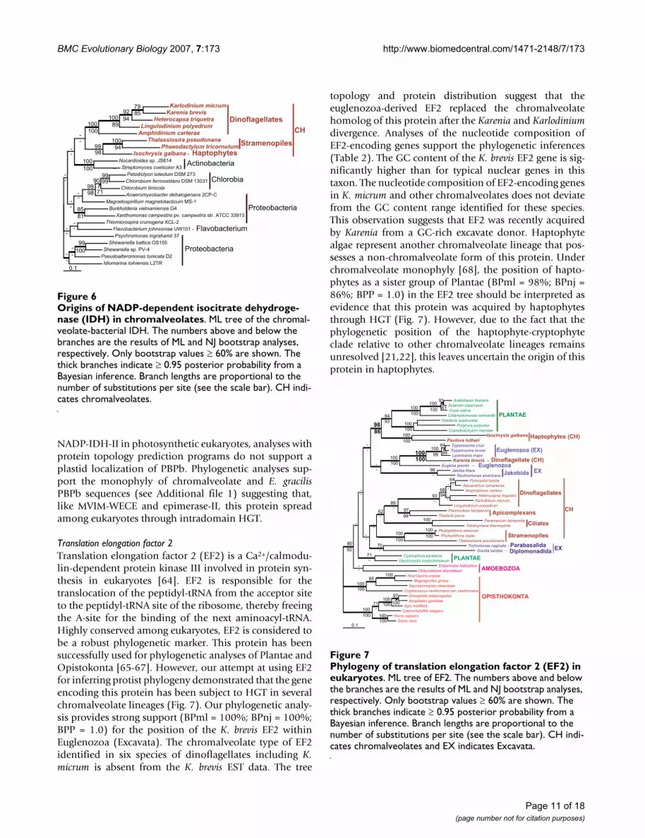
Translation elongation factor 2Translation elongation factor 2 (EF2) is a Ca2+/calmodu-lin-dependent protein kinase III involved in protein syn-thesis in eukaryotes [64]. EF2 is responsible for thetranslocation of the peptidyl-tRNA from the acceptor siteto the peptidyl-tRNA site of the ribosome, thereby freeingthe A-site for the binding of the next aminoacyl-tRNA.Highly conserved among eukaryotes, EF2 is considered tobe a robust phylogenetic marker. This protein has beensuccessfully used for phylogenetic analyses of Plantae andOpistokonta [65-67]. However, our attempt at using EF2for inferring protist phylogeny demonstrated that the geneencoding this protein has been subject to HGT in severalchromalveolate lineages (Fig. 7). Our phylogenetic analy-sis provides strong support (BPml = 100%; BPnj = 100%;BPP = 1.0) for the position of the K. brevis EF2 withinEuglenozoa (Excavata). The chromalveolate type of EF2identified in six species of dinoflagellates including K.micrum is absent from the K. brevis EST data. The tree
topology and protein distribution suggest that theeuglenozoa-derived EF2 replaced the chromalveolatehomolog of this protein after the Karenia and Karlodiniumdivergence. Analyses of the nucleotide composition ofEF2-encoding genes support the phylogenetic inferences(Table 2). The GC content of the K. brevis EF2 gene is sig-nificantly higher than for typical nuclear genes in thistaxon. The nucleotide composition of EF2-encoding genesin K. micrum and other chromalveolates does not deviatefrom the GC content range identified for these species.This observation suggests that EF2 was recently acquiredby Karenia from a GC-rich excavate donor. Haptophytealgae represent another chromalveolate lineage that pos-sesses a non-chromalveolate form of this protein. Underchromalveolate monophyly [68], the position of hapto-phytes as a sister group of Plantae (BPml = 98%; BPnj =86%; BPP = 1.0) in the EF2 tree should be interpreted asevidence that this protein was acquired by haptophytesthrough HGT (Fig. 7). However, due to the fact that thephylogenetic position of the haptophyte-cryptophyteclade relative to other chromalveolate lineages remainsunresolved [21,22], this leaves uncertain the origin of thisprotein in haptophytes.
Phylogeny of translation elongation factor 2 (EF2) in eukaryo-tesFigure 7Phylogeny of translation elongation factor 2 (EF2) in eukaryotes. ML tree of EF2. The numbers above and below the branches are the results of ML and NJ bootstrap analyses, respectively. Only bootstrap values ≥ 60% are shown. The thick branches indicate ≥ 0.95 posterior probability from a Bayesian inference. Branch lengths are proportional to the number of substitutions per site (see the scale bar). CH indi-cates chromalveolates and EX indicates Excavata.
0.1
Arabidopsis thaliana
Solanum tuberosum
91
Oryza sativa
100
Chlamydomonas reinhardtii
100
Galdieria sulphuraria
Porphyra purpurea
Cyanidioschyzon merolae
100
84
Isochrysis galbanaPavlova lutheri
100
98
Trypanosoma cruzi
Trypanosoma brucei
64
Leishmania major
100
Karenia brevis - Dinoflagellate (CH)
100
Euglena gracilis -
100
Jakoba libera
Reclinomonas americana
98
Pyrocystis lunula
Alexandrium tamarense
64
Amphidinium cartera
Heterocapsa triquetra
Karlodinium micrum
68
Lingulodinium polyedrum
93
Plasmodium falciparum
Theileria parva
97
86
Paramecium tetraurelia
Tetrahymena thermophila
100
62
Phytophthora ramorum
Phytophthora sojae
100
Thalassiosira pseudonana
100
Trichomonas vaginalis - ParabasalidaGiardia lamblia -
70
Cyanophora paradoxa
Glaucocystis nostochinearum
71
Entamoeba histolytica
Dictyostelium discoideum
80
Neurospora crassa
Magnaporthe grisea
100
Saccharomyces cerevisiae
85
Cryptococcus neoformans var. neoformans
100
Drosophila melanogaster
Anopheles gambiae
97
Apis mellifera
100
Caenorhabditis elegans
71
Homo sapiens
Danio rerio
100
100
100
100
-100 100
100
92
-
9999100
100
-
--
61100100
93
100
100
86
--
---
---
94
99
-
-
100100
-
--
--
--
--
--
Diplomonadida
Stramenopiles
Ciliates
Apicomplexans
Dinoflagellates
CH
Jakobida
Euglenozoa (EX)
Haptophytes (CH)
PLANTAE
OPISTHOKONTA
AMOEBOZOA
PLANTAE
EuglenozoaEX
EX
Origins of NADP-dependent isocitrate dehydrogenase (IDH) in chromalveolatesFigure 6Origins of NADP-dependent isocitrate dehydroge-nase (IDH) in chromalveolates. ML tree of the chromal-veolate-bacterial IDH. The numbers above and below the branches are the results of ML and NJ bootstrap analyses, respectively. Only bootstrap values ≥ 60% are shown. The thick branches indicate ≥ 0.95 posterior probability from a Bayesian inference. Branch lengths are proportional to the number of substitutions per site (see the scale bar). CH indi-cates chromalveolates.
0.1
Karlodinium micrumKarenia brevis
79
Heterocapsa triquetra
92
Lingulodinium polyedrum
100
Amphidinium carterae
100
Thalassiosira pseudonanaPhaeodactylum tricornutum
100
Isochrysis galbana - Haptophytes99
Nocardioides sp. JS614
Streptomyces coelicolor A3100
Pelodictyon luteolum DSM 273
Chlorobium ferrooxidans DSM 1303199
Chlorobium limicola
90
Anaeromyxobacter dehalogenans 2CP-C
99
Magnetospirillum magnetotacticum MS-1
Burkholderia vietnamiensis G4
Xanthomonas campestris pv. campestris str. ATCC 3391385
Thiomicrospira crunogena XCL-2
Flavobacterium johnsoniae UW101 -
Psychromonas ingrahamii 37
Shewanella baltica OS155
Shewanella sp. PV-4
99
Pseudoalteromonas tunicata D2
Idiomarina loihiensis L2TR
-
Actinobacteria
Chlorobia
Proteobacteria
Flavobacterium
Proteobacteria
CH
100
100
99
7198
81
--
--
--
--
--
8594
89100
9894
--
---
-
--
Dinoflagellates
Stramenopiles
Page 11 of 18(page number not for citation purposes)

BMC Evolutionary Biology 2007, 7:173 http://www.biomedcentral.com/1471-2148/7/173
Phylogenetic trees and sequence information for theremaining seven proteins putatively acquired by chroma-lveolates via HGT (see Table 1) are provided in Additionalfile 1.
DiscussionRecurrent HGT in protistsThe results of our study demonstrate that the recurrentnon-genealogical influx of genetic material from variousprokaryotic and eukaryotic donors is an important con-tributor to genome evolution in chromalveolates.Although our data screening approach was aimed atdetecting only chromalveolate-specific HGTs, detailedanalyses of the identified proteins revealed that many ofthem are present in several eukaryotic lineages. A co-occurrence of bacteria-derived genes encoding the sameenzyme in a limited number of species from distantlyrelated eukaryotic lineages has been reported previously[8,13-15,51,69-72] (Table 3).
Two features typical for phylogenetic trees resulting fromthe analysis of these proteins are: (1) the presence of sev-eral prokaryotic-eukaryotic clades within one tree (Fig. 3,Additional file 1) and (2) the presence of several speciesfrom distantly related eukaryotic lineages within oneclade (Figs. 2, 3, 4, 5, 7, Additional file 1). These treetopologies may be explained by multiple independentinter- and intradomain transfers of genes encoding thesame enzyme. The study of HGT in bacteria and parasiticprotists demonstrates that adaptation to specific environ-ments is the major force driving HGT [33,72]. Genes ben-eficial under certain environmental conditions canindependently be acquired by different eukaryotic line-ages that occupy different niches. Reconstructing the phy-logeny of proteins involved in anaerobic glycolysis in
parasitic protists provides an illustration of this scenario[69]. Here, the gene encoding fructose-bisphosphate aldo-lase class II, type B was acquired independently by Paraba-salida and the common ancestor of Oxymonadida andDiplomonadida. Three prokaryote-to-eukaryote transfersexplain the occurrence of pyruvate phosphate dikinase inParabasalida, parasitic Euglenozoa, and Oxymonadida-Diplomonadida lineage. The aerobe-to-anaerobe transi-tion occurred several times in the evolution of excavates.During this transition, different lineages of excavates inde-pendently acquired genes associated with anaerobic glyc-olysis from prokaryotes that had already inhabitedcorresponding niches.
The results of our study show that this scenario is applica-ble as well to free-living eukaryotes. Two isoforms of sugarepimerase, epimerase-II and epimerase-III that originatedvia an ancient gene duplication event in prokaryotes wereindependently acquired by dinoflagellates and strameno-piles (Fig. 3). Two independent interdomain HGTsexplain the occurrence of structurally distinct isoforms ofbacterial MQO in free living haptophytes and dinoflagel-lates and parasitic apicomplexans. The second feature ofphylogenetic trees resulted from the analysis of trans-ferred genes, the monophyly of distantly related eukaryo-tes, may be explained either by intradomain (eukaryote-to-eukaryote) gene transfer or by several interdomaintransfers from the same prokaryotic donor. This type ofphylogeny is more likely to reflect specific relationshipsbetween microorganisms that occupy (or occupied intheir evolutionary past) one ecological niche. SequentialHGTs that involved a prokaryote and two distantly relatedanaerobic protists have been previously proposed as anexplanation for the patchy distribution of alcohol dehy-drogenase, alanyl-tRNA synthetase, and fructose-bisphos-
Table 3: Gene distribution by multiple independent HGTs in eukaryotes
Protein Multiple Independent HGTs Referencemode eukaryotic lineages involved
Fructose-bisphosphate aldolase class II, type B p-eu, eu-eu Excavata, Amoebozoa [69]Pyruvate phosphate dikinase p-eu Excavata [69]Translation elongation factor-1 alpha-like protein eu-eu Excavata, Chromalveolata, Opisthokonta, Plantae [13]Shikimate biosynthetic enzyme AroB p-eu Chromalveolata, Opisthokonta (Fungi) [15]Hybrid-cluster protein p-eu, eu-eu Excavata, Chromalveolata, Amoebozoa, Plantae (Green algae) [51]A-type flavoprotein p-eu, eu-eu Excavata, Amoebozoa [51]Glucosamine-6-phosphate isomerase p-eu, eu-eu Excavata, Chromalveolata, Amoebozoa, Opisthokonta (Fungi) [51]Alcohol dehydrogenase p-eu, eu-eu Excavata, Chromalveolata, Amoebozoa, Plantae (Green algae),
Opisthokonta (Fungi)[51]
Glutamate dehydrogenase p-eu Excavata, Chromalveolata [70]Glyceraldehyde-3-phosphate p-eu, eu-eu Excavata, Chromalveolata [14]Alanyl-tRNA synthetase p-eu, eu-eu Excavata, Chromalveolata, Amoebozoa [71]Arginine deiminase p-eu, eu-eu Excavata, Amoebozoa [8]Rubrerythrin p-eu Excavata, Amoebozoa [8]Hypothetical protein p-eu, eu-eu Excavata, Amoebozoa [72]
The "p-eu" indicates interdomain prokaryote-to-eukaryote HGT. The "eu-eu" indicates intradomain eukaryote-to-eukaryote HGT.
Page 12 of 18(page number not for citation purposes)

BMC Evolutionary Biology 2007, 7:173 http://www.biomedcentral.com/1471-2148/7/173
phate aldolase class II, type B protein among eukaryotes[51,69,71] (Table 3). The bacteria-derived isoform of glyc-eraldehyde-3-phosphate that functions as a cytosolic pro-tein in free living dinoflagellates and Euglena and as aglycosomal protein in parasitic Euglenozoa providesanother example of a protein derived by eukaryotesthrough sequencial HGTs [14] (Table 3). Gene acquisitionthrough sequential HGTs is the most plausible scenariofor the distribution of MVIM-WECE, epimerase-II, PBPb,and, possibly, MQO-DH and CAS-like protein presentedin this paper. It is believed that HGT is more likely tooccur between closely related lineages [73]. Such transfersare hard to identify unless transferred genes have a limiteddistribution within the studied taxonomic group. CAS-like protein and MQO-CH identified in this study arepresent in two chromalveolate lineages, haptophytes anddinoflagellates (Figs. 4, 5). Possible interpretations of apatchy gene distribution between closely related lineagesinclude differential gene loss and gene transfer. The geneloss scenario would assume an independent gene lossfrom three chromalveolate lineages: stramenopiles, cili-ates, and apicomplexans. However, the fact that hapto-phytes provide not only a food source but also a uniquepool of temporary plastids (kleptoplastids) for severalspecies of extant dinoflagellates [74,75] and have contrib-uted the plastid to the common ancestor of Karenia andKarlodinium [32,76] demonstrates that these two relativelydistantly related algal lineages have been involved in spe-cific predator-prey interactions over millions of years. Thisfact makes sequential HGTs a more plausible scenario forthe occurrence of MQO-CH and CAS-like protein in dino-flagellates.
Genes encoding MVIM, WECE, epimerase-II, and PBPbproteins are shared by bacteria and several lineages ofchromalveolates and excavates. According to the results ofour phylogenetic analyses, genes encoding these proteinswere acquired by one eukaryotic lineage through anancient interdomain HGT and transferred to another viaintradomain HGT. Phagotrophy is widespread in chroma-lveolates and excavates therefore this feeding mode mayexplain an increased rate of HGT in these taxa[11,52,72,77]; i.e., many extant species of excavates andchromalveolates feed on bacterial and eukaryotic micro-organisms [78-82]. This dynamic process has made itimpossible to identify donors and recipients in theseeukaryote-to-eukaryote HGTs. An inconsistency betweenthe gene phylogeny and species phylogeny observed inthe prokaryotic region of the MVIM, WECE, and epime-rase trees suggests that genes encoding these proteins aresubjects of frequent HGTs in bacteria. Structural analysesof bacterial gene clusters that include close homologs ofthe K. brevis WECE (ftdB and qdtB) and epimerase-III(gepiA and wxoA) support this scenario [41,45]. FtdB andqdtB belong to the large cluster of genes involved in the
biosynthesis of surface layer glycoproteins (SLG) in firmi-cutes. It has been shown that the GC content of the SLGclusters deviates significantly in many bacteria from theGC content of genome as a whole [41]. This observationtogether with the fact that SLG clusters are typicallyflanked by several transposases or remnants thereof indi-cate that the entire SLG region may be a subject of HGT inbacteria. A similar conclusion resulted from the analysisof the bacterial lipopolysaccharide biosynthetic loci thatincludes gepiA and wxoA [45]. This observation completesthe proposed scenario of gene distribution by sequentialHGTs with an additional feature that is prokaryote-to-prokaryote HGT.
Measuring the contribution of HGT to eukaryotic genomesSeveral attempts to numerically estimate the contributionof HGT to eukaryotic genomes suggest a substantial inter-taxon variation in the number of horizontally derivedgenes [7,12,52,83,84]. Existing studies show thatalthough extremely rare in Plantae and multicellularOpisthokonta, HGT is a common phenomenon in Amoe-bozoa, Excavata, and chromalveolates. However, the vari-ation in the numbers of HGTs reported for differentspecies within the phagotrophic lineages (see Back-ground) reflects a difference in analytical approaches. Dif-ferences in stringency of data screening parameters, thetaxonomic composition of databases used for the compar-ative analysis, and methods of phylogenetic analyses cansignificantly affect the outcome of the study. Standardiza-tion of methods for estimating the contribution of HGT ineukaryotic genomes should be based on the knowledge oftempo and mode of evolution of horizontally transferredgenes. To our knowledge, these issues have never beenexhaustively studied in eukaryotes.
Studies of prokaryotic genome evolution demonstratethat many recently transferred genes have very large KA/KSratio that suggests directional selection [73]. In addition,the rate of duplications among genes derived throughHGTs is significantly higher than among indigenous onesin bacteria [85]. The proposed scenario for the fate oftransferred genes in bacteria based on these observationsincludes their uptake, duplication, rapid diversification ofgene copies by mutations, and consequent fixation of the"best" copies and elimination of other duplicates. Is thisscenario applicable for eukaryotes?
Analyses of proteins presented in these study show thatnine of them are encoded by at least 2–12 genes in the K.brevis genome (Table 1). Following the standard approachof phylogenomics, we excluded from the analysis all pro-teins represented by multiple paralogs in several eukaryo-tic lineages. Therefore we investigated only those paralogsthat arose from relatively recent duplications. The fact thatseveral dinoflagellate species contain multiple highly
Page 13 of 18(page number not for citation purposes)

BMC Evolutionary Biology 2007, 7:173 http://www.biomedcentral.com/1471-2148/7/173
divergent (< 50% amino acid identity) paralogs of ATS1(Additional file 1) suggests that duplication of genesencoding this protein occurred before the divergence ofdinoflagellate lineages. Phylogenetic analyses of ATS1support this statement (see Additional file 1). The aminoacid sequence identity of paralogs resulting from duplica-tions that, according to phylogenetic analyses, occurredafter the Karenia and Karlodinium divergence varies from60% (MQO) to 99% (WECE). The comparison of genesencoding WECE protein shows that the amino acidsequence identity of different copies varies from 81 to99%. Assuming that divergence between two paralogs isproportional to the time elapsed since gene duplication,the observed variation suggests that duplication of WECEgenes is a continuous process in K. brevis.
To summarize, the results of this analysis demonstratethat the impact of HGT on genome evolution in dinoflag-ellates is reinforced by continuous duplications of thetransferred genes and consequent diversification of theresulting paralogs. Additional studies are required to esti-mate relative duplication rates of foreign and indigenousgenes, rates of mutations, and paralog silencing in thisgroup of organisms.
ConclusionTaking into consideration that genomic data are availablefor only a minuscule fraction of bacteria and protists pop-ulating our planet and that we were able to identify mul-tiple cases of HGT of genes encoding the same proteinsleads us to one simple conclusion. Horizontal gene trans-fer contributes significantly to protist genomes. Webelieve that in niches where parasitism and phagotrophyare common, beneficial genes may spread rapidly fromprokaryotes to eukaryotes and provide a molecular basisfor niche-specific adaptations in the latter group. It is clearhowever, that all genes are not transferred with equal fre-quency in eukaryotes with the majority of HGT candidatesbeing involved in metabolic processes. However, giventhat foreign DNA fragments from eukaryotes frequentlyintegrate in protist chromosomes, it should not be sur-prising that occasionally genes encoding proteins of amore universally conserved function such as EF2 andpotentially EF-1 alpha-like [13] may also be co-trans-ferred. Apart from the exciting ramifications for post-HGTgene evolution in eukaryotes that includes gene familyevolution and selection for novel functions, our work alsounderlines the great care that needs to be taken when gen-erating eukaryote-wide trees of life that include manyphagotrophic or parasitic taxa.
MethodsKarenia brevis EST libraryIn this study, we used EST data generated from clonal Kbrevis Wilson cells grown under five different culture con-
ditions: 1) under nitrate depletion, 2) under phosphatedepletion, 3) in log phase under replete conditions, har-vested during the light phase, 4) in the presence of oxida-tive metals, and 5) undergoing heat stress. For completeinformation about generation, sequencing, and process-ing of the K. brevis EST library see reference [25]. Cluster-ing and assembly of the EST was done using defaultsettings of the TGICL computer program [86,87]. Theassembly resulted in 9,786 EST contigs; each representinga unique gene.
Identification of proteins acquired by chromalveolates through ancient HGTsTo identify ancient HGTs in chromalveolates, we analyzeda subset of genes present in the K. brevis EST data and inthe EST data of at least one other species of dinoflagellate.This approach allowed us to exclude from the analysispossible bacterial contaminants of the EST library. Genesshared by dinoflagellates have been detected using K.brevis ESTs as an input for the sequence similarity search(BLAST; e-value ≤ 10-10) against a local database thatincluded available data from the GenBank dbEST data-base [18] for five dinoflagellate species: Alexandrium tam-arense, Amphidinium carterae, Heterocapsa triquetra,Lingulodinium polyedrum, and K. micrum. This analysisyielded 3,341 EST contigs. To detect potential HGTs, weused the defined subset of K. brevis DNA sequences as aninput for the sequence similarity search (BLASTx; e-value≤ 10-20) against the GenBank non-redundant database(nr). Sequences that showed highest similarity to prokary-otic proteins (three top hits) or chromalveolate andprokaryotic proteins have been selected for further analy-ses. Using this approach, we identified 95 K. brevis uni-genes encoding proteins from 55 protein familiesputatively derived from prokaryotes at different timepoints of chromalveolate evolution.
In parallel, we performed a high throughput automatedanalysis of the subset of sequences shared by dinoflagel-lates. The 3,341 sequences were translated into the sixopen reading frames using the Transeq program in theEmboss package [88] and used as input for the analysiswith the PhyloGenie package of computer programs [89].PhyloGenie serves as an automated pipeline in which thefollowing analyses can be implemented: BLAST searchagainst a local database, extraction of homologoussequences from the BLAST results, generation of align-ments, phylogenetic tree reconstruction, and calculationof bootstrap support values for individual phylogenies.We created a local protein database for the PhyloGenieBLAST search by retrieving completed genome sequencesand EST data from the National Center for BiotechnologyInformation (NCBI) [90] genomic projects web site anddbEST [18], DOE Joint Genome Institute (JGI) [91], Cya-nidioschyzon merolae genome project [92], and The Galdie-
Page 14 of 18(page number not for citation purposes)

BMC Evolutionary Biology 2007, 7:173 http://www.biomedcentral.com/1471-2148/7/173
ria sulphuraria genome project [93] for species listedbelow. EST sequences have been translated into six openreading frames and combined with protein sequences.The final fasta file that included all of the data was format-ted using the formatdb program in the BLAST package[94]. Our local database included complete genome andEST data for following species: Oryza sativa, Drosophilamelanogaster, Saccharomyces cerevisiae, the green algaChlamydomonas reinhardtii, red algae C. merolae and G. sul-phuraria; chromalveolates Guillardia theta, Emiliania hux-leyi, Thalassiosira pseudonana, Plasmodium falciparum,Toxoplasma gondii, A. tamarense, A. carterae, H. triquetra, L.polyedrum, and K. micrum, excavates G. lamblia andTrypanosoma brucei, archaea Halobacterium sp. NRC-1,Methanothermobacter thermautotrophicus, and Sulfolobustokodaii, and eubacteria Clostridium acetobutylicumATCC824, Escherichia coli 536, Geobacter sulfurreducens,Oceanobacillus iheyensis, Synechococcus elongatus PCC 7942,Trichodesmium erythraeum, and Nostoc sp. PCC 7120.
PhyloGenie was run using default settings except that theminimum expect "e-value" for the BLAST search of thedata was set at 10. The hidden Markov model (hmm)alignments were built using all hits with an e-value below0.01. The program TreeView [95] was used to visualize theresulting trees. We selected sequences represented by treesthat contained only chromalveolates and bacteria andtrees that contained well-defined chromalveolate-bacte-rial clades (at least 50% bootstrap support). Using this cri-terion for the gene selection, we excluded from theanalyses genes of eukaryotic and mitochondrial originthat are shared by most eukaryotic organisms. In addition,this approach allowed us to exclude from the analysesgenes of red algal and green algal origin acquired by chro-malveolates from the genomes of plastid progenitors viaEGT (see [25,26] for detailed analyses of EGT in chromal-veolates). This analysis yielded 37 unigenes encoding pro-teins from 23 different protein families; 22 of themrepresented a subset of proteins identified using the "Besthit" approach. The necessity of using a combination oftwo described above methods for detecting putative HGTsresulted from the fact that neither the non-redundant (nr)nor our local database included all taxa of interest. Thelocal database for the PhyloGenie BLAST search comple-mented nr with complete genome and EST data for freeliving protists. In addition to the gene discovery, the Phy-loGenie output was used to verify the phylogeny of pro-teins identified using the "Best hit" approach. Based onthe results of PhyloGenie, eight proteins represented by12 unigenes have been excluded from the analysis asderived from the genome of plastid progenitor throughEGT. Three proteins represented by five unigenes wererejected as shared by multiple eukaryotic lineages. Theremaining set of 45 proteins represented by 80 unigenes
in the K. brevis EST data were subjects for detailed analy-ses.
Identification of bacteria-derived proteins in K. brevis involved in cell wall biogenesisIn bacteria, genes encoding physiologically coupled reac-tions are often transferred together, frequently in anoperon [33]. To test whether this scenario is applicable forinterdomain prokaryote-to-eukaryote transfers, wescreened the K. brevis EST data for the presence ofhomologs of bacterial proteins involved in cell envelopebiogenesis. This category of proteins was chosen to iden-tify genes that potentially may be co-transferred withgenes encoding MVIM-WECE. The latter represents a rarecase of gene fusion in interdomain HGT. Gene clustersinvolved in the surface layer protein biosynthesis in A.thermoaerophilus [43] and glycopeptidolipid biosynthesisin Mycobacterium avium [44] were retrieved from GenBankand used as an input in sequence similarity search(BLAST; e-value ≤ 10-20) against the non-redundant geneset generated from the K. brevis EST data. This analysisallowed us to identify three additional proteins repre-sented by five unigenes in the K. brevis EST data.
Identification of eukaryotic proteins acquired by chromalveolates through intradomain HGTGenes derived through intradomain eukaryote-to-eukary-ote HGT were extremely hard to identify using highthroughput phylogenomic analyses due to the limitednumber of taxa included in our local database. The onlycandidate for the transfer of a bona fide eukaryotic geneamong taxa is EF2 that was identified in the course ofanother study of potential markers for reconstructing theeukaryotic tree of life. The detailed description of methodsused for the EF2 analysis can be found in reference [21].
To assess the possibility that the EF2 sequence found inthe K. brevis data resulted from contamination of the ESTlibrary with kinetoplastid DNA, we used the complete K.brevis EST data set as an input for a sequence similaritysearch (BLASTx; e-value ≤ 10-10) against the GenBanknon-redundant database (nr). Sequences that showed thehighest similarity to kinetoplastid genes were subjected todetailed analyses. This work did not provide any supportfor the kinetoplastid origin of identified sequences in K.brevis with the exception of EF2 (results not shown).
Building the final alignmentsTo build the final alignments, we identified homologs ofthe candidate K. brevis sequences using BLAST searches (e-value ≤ 10-10) against GenBank nr, dbEST, and public pro-tist databases including the JGI database [91], the FrenchNational Sequencing Center, Genoscope [96], the ProtistEST Program database [17], C. merolae [92,97] and G. sul-phuraria [93,98] databases. The DNA sequences were
Page 15 of 18(page number not for citation purposes)

BMC Evolutionary Biology 2007, 7:173 http://www.biomedcentral.com/1471-2148/7/173
translated, and the amino acid data for each protein weremanually aligned with the identified bacterial and eukary-otic homologs using BioEdit [99]. Only regions that wereunambiguously aligned were retained for phylogeneticanalysis.
Analysis of sequence structure and phylogenyThe eukaryotic structure of the identified K. brevis tran-scripts has been verified by translating and aligning theresulting amino acid sequences with their bacterialhomologous. The subcellular localization of the studiedproteins was predicted using online analyses with proteintopology prediction programs SignalP [100,101], TargetP[102,103], MITOPROT [104], PSORT [50], and TMHMM[31]. Percent of amino acid sequence identity betweengene copies was inferred using an online tool for pair wisesequence alignment bl2seq [105]. GC content of nucle-otide sequences was identified using BioEdit. The averageand range of nucleotide composition of protist genomeswas inferred from the analysis of coding regions of 50sequences from each species.
We used the maximum likelihood (ML) method to recon-struct the gene phylogenies. The ML analysis was done inPHYML V2.4.3 [106,107] using the WAG + Γ + I evolu-tionary model and tree optimization. The alpha values forthe gamma distribution were calculated using eight ratecategories. To test the stability of monophyletic groups inthe ML trees, we calculated PHYML bootstrap (100 repli-cates) support values [108]. In addition, we calculatedbootstrap values (500 replications) using the neighborjoining (NJ) method with JTT+Γ distance matrices usingPHYLIP V3.63 [109]. The NJ analysis was done with rand-omized taxon addition. Bayesian posterior probabilitiesfor nodes in the ML tree were calculated using MrBayesV3.0b4 [110] and the WAG + Γ model. The Metropolis-coupled Markov chain Monte Carlo from a random start-ing tree was run for 1,000,000 generations with trees sam-pled each 1,000 cycles. The initial 20,000 cycles (200trees) were discarded as the "burn in." A consensus treewas made with the remaining 800 phylogenies to deter-mine the posterior probabilities at the different nodes.
The names of the K. brevis proteins are derived from thenames of corresponding protein domains according to thePfam [29,30] nomenclature. The K. brevis sequences listedin the Table 1 have been deposited in GenBank underaccession numbers EF540322–EF540340.
Competing interestsThe author(s) declare that there are no competing inter-ests.
Authors' contributionsTN did the phylogenetic and other bioinformatic analysesand prepared the manuscript draft. DB conceived of andsupervised this study and edited the manuscript. Allauthors read and approved the final manuscript.
Additional material
AcknowledgementsThis work was supported by grants from the National Science Foundation and the National Aeronautics and Space Administration awarded to D.B. (EF 04-31117, NNG04GM17G). T. N. was partially supported by an Avis E. Cone Fellowship from the University of Iowa. We are grateful to Frances M. Van Dolah (NOAA, Charleston, SC) for helpful advice and the US Department of Energy-Joint Genome Institute for having generated the K. brevis data for another project.
References1. Ochman H, Lawrence JG, Groisman EA: Lateral gene transfer and
the nature of bacterial innovation. Nature 2000, 405:299-304.2. Boucher Y, Douady CJ, Papke RT, Walsh DA, Boudreau ME, Nesbo
CL, Case RJ, Doolittle WF: Lateral gene transfer and the originsof prokaryotic groups. Annu Rev Genet 2003, 37:283-328.
3. Dagan T, Martin W: Ancestral genome sizes specify the mini-mum rate of lateral gene transfer during prokaryote evolu-tion. Proc Natl Acad Sci USA 2007, 104:870-875.
4. Goldenfeld N, Woese C: Biology's next revolution. Nature 2007,445:369.
5. Prince VE, Pickett FB: Splitting pairs: the diverging fates ofduplicated genes. Nat Rev Genet 2002, 3:827-837.
6. Ohno S: Evolution by Gene Duplication. Berlin: Springer; 1970. 7. Loftus B, Anderson I, Davies R, Alsmark UC, Samuelson J, Amedeo P,
Roncaglia P, Berriman M, Hirt RP, Mann BJ, et al.: The genome ofthe protist parasite Entamoeba histolytica. Nature 2005,433:865-868.
8. Andersson JO, Sjogren AM, Horner DS, Murphy CA, Dyal PL, SvardSG, Logsdon JM Jr, Ragan MA, Hirt RP, Roger AJ: A genomicsurveyof the fish parasite Spironucleus salmonicida indicatesgenomic plasticity among diplomonads and significant lat-eral gene transfer in eukaryote genome evolution. BMCGenomics 2007, 8:51.
9. Carlton JM, Hirt RP, Silva JC, Delcher AL, Schatz M, Zhao Q, Wort-man JR, Bidwell SL, Alsmark UC, Besteiro S, et al.: Draft genomesequence of the sexually transmitted pathogen Trichomonasvaginalis. Science 2007, 315:207-212.
10. Huang J, Mullapudi N, Lancto CA, Scott M, Abrahamsen MS, KissingerJC: Phylogenomic evidence supports past endosymbiosis,intracellular and horizontal gene transfer in Cryptosporidiumparvum. Genome Biol 2004, 5:R88.
11. Ricard G, McEwan NR, Dutilh BE, Jouany JP, Macheboeuf D, Mit-sumori M, McIntosh FM, Michalowski T, Nagamine T, Nelson N, et al.:Horizontal gene transfer from bacteria to rumen ciliates
Additional file 1Supporting materials for the study. The complete list of sequences used in this study is shown as well as three figures that show the origins of iron-containing alcohol dehydrogenase (Fe-ADH) and NAD-dependent alde-hyde dehydrogenase (PutA) in eukaryotes by multiple HGTs, the origins of substrate-bound periplasmic binding protein (PBPb) and silent infor-mation regulator 2 (SIR2) in protists, and examples of HGT events from bacteria to dinoflagellates.Click here for file[http://www.biomedcentral.com/content/supplementary/1471-2148-7-173-S1.pdf]
Page 16 of 18(page number not for citation purposes)

BMC Evolutionary Biology 2007, 7:173 http://www.biomedcentral.com/1471-2148/7/173
indicates adaptation to their anaerobic, carbohydrates-richenvironment. BMC Genomics 2006, 7:22.
12. Eichinger L, Pachebat JA, Glockner G, Rajandream MA, Sucgang R,Berriman M, Song J, Olsen R, Szafranski K, Xu Q, et al.: The genomeof the social amoeba Dictyostelium discoideum. Nature 2005,435:43-57.
13. Keeling PJ, Inagaki Y: A class of eukaryotic GTPase with a punc-tate distribution suggesting multiple functional replace-ments of translation elongation factor 1alpha. Proc Natl AcadSci USA 2004, 101:15380-15385.
14. Takishita K, Ishida K, Maruyama T: An enigmatic GAPDH gene inthe symbiotic dinoflagellate genus Symbiodinium and itsrelated species (the order Suessiales): possible lateral genetransfer between two eukaryotic algae, dinoflagellate andeuglenophyte. Protist 2003, 154:443-454.
15. Waller RF, Slamovits CH, Keeling PJ: Lateral gene transfer of amultigene region from cyanobacteria to dinoflagellatesresulting in a novel plastid-targeted fusion protein. Mol BiolEvol 2006, 23:1437-1443.
16. Archibald JM, Rogers MB, Toop M, Ishida K, Keeling PJ: Lateral genetransfer and the evolution of plastid-targeted proteins in thesecondary plastid-containing alga Bigelowiella natans. ProcNatl Acad Sci USA 2003, 100:7678-7683.
17. The Protist EST Program database (TBestDB) [http://megasun.bch.umontreal.ca/pepdb/pepdb.html]
18. NCBI. Expressed Sequence Tags database [http://www.ncbi.nlm.nih.gov/dbEST/index.html]
19. Cavalier-Smith T: Principles of protein and lipid targeting insecondary symbiogenesis: euglenoid, dinoflagellate, and spo-rozoan plastid originsand the eukaryote family tree. JEukaryot Microbiol 1999, 46:347-366.
20. Parfrey L, Barbero E, Lasser E, Dunthorn M, Bhattacharya D, Patter-son D, Katz L: Evaluating support for the current classificationof eukaryotic diversity. PLoS 2006, 2:e220.
21. Hackett JD, Yoon HS, Li S, Reyes-Prieto A, Rummele SE, BhattacharyaD: Phylogenomic analysis supports the monophyly of crypto-phytes and haptophytes and the association of 'Rhizaria' withchromalveolates. Mol Biol Evol 2007 in press.
22. Patron NJ, Inagaki Y, Keeling PJ: Multiple gene phylogenies sup-port the monophyly of cryptomonad and haptophyte hostlineages. Curr Biol 2007, 17:887-891.
23. Archibald JM, Keeling PJ: Recycled plastids: a 'green movement'in eukaryotic evolution. Trends Genet 2002, 18:577-584.
24. Hackett JD, Yoon HS, Soares MB, Bonaldo MF, Casavant TL, ScheetzTE, Nosenko T, Bhattacharya D: Migration of the plastid genometo the nucleus in a peridinin dinoflagellate. Curr Biol 2004,14:213-218.
25. Nosenko T, Lidie KL, Van Dolah FM, Lindquist E, Cheng JF, Bhattach-arya D: Chimeric plastid proteome in the Florida "redtide"dinoflagellate Karenia brevis. Mol Biol Evol 2006,23:2026-2038.
26. Li S, Nosenko T, Hackett JD, Bhattacharya D: Phylogenomic anal-ysis identifies red algal genes of endosymbiotic origin in thechromalveolates. Mol Biol Evol 2006, 23:663-674.
27. Patron NJ, Waller RF, Keeling PJ: A tertiary plastid uses genesfrom two endosymbionts. J Mol Biol 2006, 357:1373-1382.
28. Fleming LE, Backer LC, Baden DG: Overview of aerosolized Flor-ida red tide toxins: exposures and effects. Environ Health Per-spect 2005, 113:618-620.
29. Sonnhammer EL, Eddy SR, Durbin R: Pfam: a comprehensivedatabase of protein domain families based on seed align-ments. Proteins 1997, 28:405-420.
30. Bateman A, Birney E, Cerruti L, Durbin R, Etwiller L, Eddy SR, Grif-fiths-Jones S, Howe KL, Marshall M, Sonnhammer EL: The Pfamprotein families database. Nucleic Acids Res 2002, 30:276-280.
31. TMHMM Server v. 2.0. Prediction of transmembrane helicesin proteins [http://www.cbs.dtu.dk/services/TMHMM-2.0/]
32. Yoon HS, Hackett JD, Van Dolah FM, Nosenko T, Lidie KL, Bhattach-arya D: Tertiary endosymbiosis driven genome evolution indinoflagellate algae. Mol Biol Evol 2005, 22:1299-1308.
33. Pal C, Papp B, Lercher MJ: Adaptive evolution of bacterial met-abolic networks by horizontal gene transfer. Nat Genet 2005,37:1372-1375.
34. Xue Y, Zhao L, Liu HW, Sherman DH: A gene cluster for mac-rolide antibiotic biosynthesis in Streptomyces venezuelae:
architecture of metabolic diversity. Proc Natl Acad Sci USA 1998,95:12111-12116.
35. Xue Y, Wilson D, Zhao L, Liu H, Sherman DH: Hydroxylation ofmacrolactones YC-17 and narbomycin is mediated by thepikC-encoded cytochrome P450 in Streptomyces venezuelae.Chem Biol 1998, 5:661-667.
36. Anzai Y, Saito N, Tanaka M, Kinoshita K, Koyama Y, Kato F: Organ-ization of the biosynthetic gene cluster for the polyketidemacrolide mycinamicin in Micromonospora griseorubida.FEMS Microbiol Lett 2003, 218:135-141.
37. Brikun IA, Reeves AR, Cernota WH, Luu MB, Weber JM: The eryth-romycin biosynthetic gene cluster of Aeromicrobium eryth-reum. J Ind Microbiol Biotechnol 2004, 31:335-344.
38. Awram P, Smit J: Identification of lipopolysaccharide O antigensynthesis genes required for attachment of the S-layer ofCaulobacter crescentus. Microbiology 2001, 147:1451-1460.
39. Bastin DA, Reeves PR: Sequence and analysis of the Oantigengene (rfb) cluster of Escherichia coli O111. Gene 1995,164:17-23.
40. Nesper J, Kraiß A, Schild S, Blaß J, Klose KE, Bockemühl J, Reidl J:Role of Vibrio cholerae O139 surface polysaccharides in intes-tinal colonization. Infect Immun 2002, 70:2419-2433.
41. Novotny R, Pfoestl A, Messner P, Schaffer C: Genetic organizationof chromosomal S-layer glycan biosynthesis loci of Bacil-laceae. Glycoconj J 2004, 20:435-447.
42. Schaffer C, Messner P: Surface-layer glycoproteins: an examplefor the diversity of bacterial glycosylation with promisingimpacts on nanobiotechnology. Glycobiology 2004, 14:31R-42R.
43. Pfoestl A, Hofinger A, Kosma P, Messner P: Biosynthesisof dTDP-3-acetamido-3,6-dideoxy-alpha-D-galactose in Aneurinibacil-lus thermoaerophilus L420-91T. J Biol Chem 2003,278:26410-26417.
44. Eckstein TM, Belisle JT, Inamine JM: Proposed pathway for thebiosynthesis of serovar-specific glycopeptidolipids in Myco-bacterium avium serovar 2. Microbiology 2003, 149:2797-2807.
45. Patil PB, Sonti RV: Variation suggestive of horizontal genetransfer at a lipopolysaccharide (lps) biosynthetic locus inXanthomonas oryzae pv. oryzae, the bacterial leaf blight path-ogen of rice. BMC Microbiol 2004, 4:40.
46. Seifert GJ: Nucleotide sugar interconversions and cell wall bio-synthesis: how to bring the inside to the outside. Curr OpinPlant Biol 2004, 7:277-284.
47. Lopez AB, Sener K, Jarroll EL, van Keulen H: Transcription regu-lation is demonstrated for five key enzymes in Giardia intes-tinalis cyst wall polysaccharide biosynthesis. Mol BiochemParasitol 2003, 128:51-57.
48. Prescott AG, Lloyd MD: The iron(II) and 2-oxoacid-dependentdioxygenases and their role in metabolism. Nat Prod Rep 2000,17:367-383.
49. Baggaley KH, Brown AG, Schofield CJ: Chemistry and biosynthe-sis of clavulanic acid and other clavams. Nat Prod Rep 1997,14:309-333.
50. WoLF PSORT. Protein Subcellular Localization Prediction..
51. Andersson JO, Hirt RP, Foster PG, Roger AJ: Evolution of fourgene families with patchy phylogenetic distributions: influxof genes into protist genomes. BMC Evol Biol 2006, 6:27.
52. Andersson JO: Lateral gene transfer in eukaryotes. Cell Mol LifeSci 2005, 62:1182-1197.
53. Kather B, Stingl K, van der Rest ME, Altendorf K, Molenaar D:Another unusual type of citric acid cycle enzyme in Helico-bacter pylori: the malate:quinone oxidoreductase. J Bacteriol2000, 182:3204-3209.
54. Molenaar D, van der Rest ME, Drysch A, Yucel R: Functionsof themembrane-associated and cytoplasmic malate dehydroge-nases in the citric acid cycle of Corynebacterium glutamicum.J Bacteriol 2000, 182:6884-6891.
55. Van der Rest ME, Frank C, Molenaar D: Functions of themem-brane-associated and cytoplasmic malate dehydrogenases inthe citric acid cycle of Escherichia coli. J Bacteriol 2000,182:6892-6899.
56. Gardner MJ, Shallom SJ, Carlton JM, Salzberg SL, Nene V, Shoaibi A,Ciecko A, Lynn J, Rizzo M, Weaver B, et al.: Sequence of Plasmo-dium falciparum chromosomes 2, 10, 11 and 14. Nature 2002,419:531-534.
Page 17 of 18(page number not for citation purposes)

BMC Evolutionary Biology 2007, 7:173 http://www.biomedcentral.com/1471-2148/7/173
57. Uyemura SA, Luo S, Vieira M, Moreno SN, Docampo R: Oxidativephosphorylation and rotenone-insensitive malate- andNADH-quinone oxidoreductases in Plasmodium yoelii yoeliimitochondria in situ. J Biol Chem 2004, 279:385-393.
58. Chen RD, Gadal P: Structure, function and regulation of NADand NADP dependent isocitrate dehydrogenase in higherplants and in other organisms. Plant Physiol Biochem 1990,28:411-427.
59. Schnarrenberger C, Martin W: Evolution of the enzymes of thecitric acid cycle and the glyoxylate cycle of higher plants. Acase study of endosymbiotic gene transfer. Eur J Biochem 2002,269:868-883.
60. Lang M, Apt KE, Kroth PG: Protein transport into "complex"diatom plastids utilizes two different targeting signals. J BiolChem 1998, 273:30973-30978.
61. Patron NJ, Waller RF, Archibald JM, Keeling PJ: Complex proteintargeting to dinoflagellate plastids. J Mol Biol 2005,348:1015-1024.
62. Suzuki M, Sahara T, Tsuruha J, Takada Y, Fukunaga N: Differentialexpression in Escherichia coli of the Vibrio sp. strain ABE-1icdI and icdII genes encoding structurally different isocitratedehydrogenase isozymes. J Bacteriol 1995, 177:2138-2142.
63. Kang CH, Shin WC, Yamagata Y, Gokcen S, Ames GF, Kim SH: Crys-tal structure of the lysine-, arginine-, ornithine-binding pro-tein (LAO) from Salmonella typhimurium at 2.7-A resolution.J Biol Chem 1991, 266:23893-23899.
64. Jorgensen R, Carr-Schmid A, Ortiz PA, Kinzy TG, Andersen GR:Purification and crystallization of the yeast elongation factoreEF2. Acta Crystallogr D Biol Crystallogr 2002, 58:712-715.
65. Moreira D, Le Guyader H, Philippe H: The origin of red algae andthe evolution of chloroplasts. Nature 2000, 405:69-72.
66. Regier JC, Shultz JW: Elongation factor-2: a useful gene forarthropod phylogenetics. Mol Phylogenet Evol 2001, 20:136-148.
67. Kullnig-Gradinger CM, Szakacs G, Kubicek CP: Phylogenyand evo-lution of the genus Trichoderma: a multigene approach. MycolRes 2002, 106:757-767.
68. Bhattacharya D, Yoon HS, Hackett JD: Chromalveolates unite:endosymbiosis connects the dots. BioEssays 2004, 26:50-60.
69. Liapounova NA, Hampl V, Gordon PM, Sensen CW, Gedamu L,Dacks JB: Reconstructing the mosaic glycolytic pathway of theanaerobic eukaryote Monocercomonoides. Eukaryot Cell 2006,5:2138-2146.
70. Andersson JO, Roger AJ: Evolution of glutamate dehydrogenasegenes: evidence for lateral gene transfer within and betweenprokaryotes and eukaryotes. BMC Evol Biol 2003, 3:14.
71. Andersson JO, Sarchfield SW, Roger AJ: Gene transfers fromnanoarchaeota to an ancestor of diplomonads and parabasa-lids. Mol Biol Evol 2005, 22:85-90.
72. Andersson J: Genome evolution of anaerobic protists: meta-bolic adaptation via gene acquisition. In Genomics and Evolutionof Microbial Eukaryotes Edited by: Katz L, Bhattacharya D. Oxford Uni-versity Press; 2006:109-122.
73. Hao W, Golding GB: The fate of laterally transferred genes: lifein the fast lane to adaptation or death. Genome Res 2006,16:636-643.
74. Koike K, Sekiguchi H, Kobiyama A, Takishita K, Kawachi M, Koike K,Ogata T: A novel type of kleptoplastidy in Dinophysis (Dino-phyceae): presence of haptophyte-type plastid in Dinophysismitra. Protist 2005, 156:225-237.
75. Gast RJ, Moran DM, Dennett MR, Caron DA: Kleptoplasty in anAntarctic dinoflagellate: caught in evolutionary transition?Environ Microbiol 2007, 9:39-45.
76. Tengs T, Dahlberg OJ, Shalchian-Tabrizi K, Klaveness D, Rudi K, Del-wiche CF, Jakobsen KS: Phylogenetic analyses indicate that the19'Hexanoyloxy-fucoxanthin-containing dinoflagellates havetertiary plastids of haptophyte origin. Mol Biol Evol 2000,17:718-729.
77. Doolittle WF: You are what you eat: a gene transferratchetcould account for bacterial genes in eukaryotic nucleargenomes. Trends Genet 1998, 14:307-311.
78. Graham L, Wilcox L, (eds.): Algae. Upper Saddle River: PrenticeHall; 2000.
79. Sogayar MI, Gregorio EA: Uptake of bacteria by trophozoites ofGiardia duodenalis (Say). Ann Trop Med Parasitol 1989, 83:63-66.
80. Pereira-Neves A, Benchimol M: Phagocytosis by Trichomonasvaginalis: new insights. Biol Cell 2007, 99:87-101.
81. Flavin M, Nerad TA: Reclinomonas americana N. G., N. Sp., anew freshwater heterotrophic flagellate. J Eukaryot Microbiol1993, 40:172-179.
82. Cohen CJ, Bacon R, Clarke M, Joiner K, Mellman I: Dictyosteliumdiscoideum mutants with conditional defects in phagocytosis.J Cell Biol 1994, 126:955-966.
83. Watkins RF, Gray MW: The frequency of eubacterium-to-eukaryote lateral gene transfers shows significant cross-taxavariation within amoebozoa. J Mol Evol 2006, 63:801-814.
84. Hall C, Brachat S, Dietrich FS: Contribution ofhorizontal genetransfer to the evolution of Saccharomyces cerevisiae. EukaryotCell 2005, 4:1102-1115.
85. Hooper SD, Berg OG: Duplication is more common among lat-erally transferred genes than among indigenous genes.Genome Biol 2003, 4:R48.
86. DFCI Gene Indices Software Tools [http://compbio.dfci.harvard.edu/tgi/software/]
87. Pertea G, Huang X, Liang F, Antonescu V, Sultana R, Karamycheva S,Lee Y, White J, Cheung F, Parvizi B, et al.: TIGR Gene Indices clus-tering tools (TGICL): a software system for fast clustering oflarge EST datasets. Bioinformatics 2003, 19:651-652.
88. Emboss [http://emboss.sourceforge.net/]89. Lupas N, Frickey T: PhyloGenie: automated phylome genera-
tion and analysis. Nucleic Acids Res 2004, 32:5231-5238.90. National Center for Biotechnology Information [http://
www.ncbi.nlm.nih.gov/]91. JGI. DOE Joint Genome Institute [http://www.jgi.doe.gov/]92. Cyanidioschyzon merolae Genome Project [http://mero
lae.biol.s.u-tokyo.ac.jp/]93. The Galdieria sulphuraria Genome Project [http://genom
ics.msu.edu/galdieria/]94. The National Center for Biotechnology Information Basic
Local Alignment Search Tool (BLAST) [http://www.ncbi.nlm.nih.gov/BLAST/]
95. TreeView. Tree drawing software for Apple MacintoshandWindows [http://taxonomy.zoology.gla.ac.uk/rod/treeview.html]
96. Genoscope. The French National Sequencing Center [http://www.genoscope.cns.fr/externe/English/corps_anglais.html]
97. Matsuzaki M, Misumi O, Shin IT, Maruyama S, Takahara M, Miyag-ishima SY, Mori T, Nishida K, Yagisawa F, Nishida K, et al.: Genomesequence of the ultrasmall unicellular red alga Cyanidio-schyzon merolae 10D. Nature 2004, 428:653-657.
98. Weber AP, Oesterhelt C, Gross W, Brautigam A, Imboden LA,Krassovskaya I, Linka N, Truchina J, Schneidereit J, Voll H, et al.: EST-analysis of the thermo-acidophilic red microalga Galdieriasulphuraria reveals potential for lipid A biosynthesis andunveils the pathway of carbon export from rhodoplasts. PlantMol Biol 2004, 55:17-32.
99. BioEdit. Biological sequence alignment editor for Windows95/98/NT/2000/XP [http://www.mbio.ncsu.edu/BioEdit/bioedit.html]
100. SignalP 3.0 Server [http://www.cbs.dtu.dk/services/SignalP/]101. Bendtsen JD, Nielsen H, von Heijne G, Brunak S: Improved predic-
tion of signal peptides: SignalP 3.0. J Mol Biol 2004, 340:783-795.102. TargetP 1.1 Server [http://www.cbs.dtu.dk/services/TargetP/]103. Emanuelsson O, Nielsen H, Brunak S, von Heijne G: Predicting sub-
cellular localization of proteins based on their N-terminalamino acid sequence. J Mol Biol 2000, 300:1005-1016.
104. MITOPROT: Prediction of mitochondrial targetingsequences [http://ihg.gsf.de/ihg/mitoprot.html]
105. Blast 2 Sequences [http://www.ncbi.nlm.nih.gov/blast/bl2seq/wblast2.cgi]
106. Guindon S, Gascuel O: A simple, fast, and accurate algorithmto estimate large phylogenies by maximum likelihood. SystBiol 2003, 52:696-704.
107. Guindon S, Lethiec F, Duroux P, Gascuel O: PHYML Online – aweb server for fast maximum likelihood-based phylogeneticinference. Nucleic Acids Res 2005, 33:W557-559.
108. Felsenstein J: Confidence limits on phylogenies: an approachusing the bootstrap. Evolution 1985, 39:783-791.
109. PHYLIP V3.63 [http://evolution.genetics.washington.edu/phylip.html]
110. Huelsenbeck JP, Ronquist F: Bayesian inference of phylogenetictrees. Bioinformatics 2001, 17:754-755.
111. Bhattacharya lab downloads page [http://www.biology.uiowa.edu/debweb/downloads/]
Page 18 of 18(page number not for citation purposes)


