Embed Size (px)
Citation preview

Seediscussions,stats,andauthorprofilesforthispublicationat:https://www.researchgate.net/publication/7212087
Hormonalregulationofalveolarization:Structure-functioncorrelation
ARTICLEinRESPIRATORYRESEARCH·FEBRUARY2006
ImpactFactor:3.09·DOI:10.1186/1465-9921-7-47·Source:PubMed
CITATIONS
8
READS
49
9AUTHORS,INCLUDING:
SamuelJGarber
TheChildren'sHospitalofPhiladelphia
5PUBLICATIONS92CITATIONS
SEEPROFILE
HuayanZhang
TheChildren'sHospitalofPhiladelphia
16PUBLICATIONS460CITATIONS
SEEPROFILE
AndrewJGow
Rutgers,TheStateUniversityofNewJersey
182PUBLICATIONS7,065CITATIONS
SEEPROFILE
RashminCSavani
UniversityofTexasSouthwesternMedicalCe…
105PUBLICATIONS3,471CITATIONS
SEEPROFILE
Availablefrom:AndrewJGow
Retrievedon:04February2016

BioMed CentralRespiratory Research
ss
Open AcceResearchHormonal regulation of alveolarization: structure-function correlationSamuel J Garber†1, Huayan Zhang†1, Joseph P Foley1, Hengjiang Zhao1, Stephan J Butler1, Rodolfo I Godinez2, Marye H Godinez2, Andrew J Gow1 and Rashmin C Savani*3Address: 1Division of Neonatology, Department of Pediatrics, Children's Hospital of Philadelphia, University of Pennsylvania School of Medicine, Philadelphia, PA, USA, 2Department of Anesthesiology and Critical Care Medicine, Children's Hospital of Philadelphia, University of Pennsylvania School of Medicine, Philadelphia, PA, USA and 3Division of Neonatal-Perinatal Medicine, Division of Pulmonary and Vascular Biology, Room K4.224, University of Texas Southwestern at Dallas, Dallas, TX USA
Email: Samuel J Garber - [email protected]; Huayan Zhang - [email protected]; Joseph P Foley - [email protected]; Hengjiang Zhao - [email protected]; Stephan J Butler - [email protected]; Rodolfo I Godinez - [email protected]; Marye H Godinez - [email protected]; Andrew J Gow - [email protected]; Rashmin C Savani* - [email protected]
* Corresponding author †Equal contributors
AbstractBackground: Dexamethasone (Dex) limits and all-trans-retinoic acid (RA) promotes alveolarization. While structural changesresulting from such hormonal exposures are known, their functional consequences are unclear.
Methods: Neonatal rats were treated with Dex and/or RA during the first two weeks of life or were given RA after previousexposure to Dex. Morphology was assessed by light microscopy and radial alveolar counts. Function was evaluated byplethysmography at d13, pressure volume curves at d30, and exercise swim testing and arterial blood gases at both d15 and d30.
Results: Dex-treated animals had simplified lung architecture without secondary septation. Animals given RA alone had smaller,more numerous alveoli. Concomitant treatment with Dex + RA prevented the Dex-induced changes in septation. While theresults of exposure to Dex + RA were sustained, the effects of RA alone were reversed two weeks after treatment was stopped.At d13, Dex-treated animals had increased lung volume, respiratory rate, tidal volume, and minute ventilation. On d15, bothRA- and Dex-treated animals had hypercarbia and low arterial pH. By d30, the RA-treated animals resolved this respiratoryacidosis, but Dex-treated animals continued to demonstrate blood gas and lung volume abnormalities. Concomitant RAtreatment improved respiratory acidosis, but failed to normalize Dex-induced changes in pulmonary function and lung volumes.No differences in exercise tolerance were noted at either d15 or d30. RA treatment after the period of alveolarization alsocorrected the effects of earlier Dex exposure, but the structural changes due to RA alone were again lost two weeks aftertreatment.
Conclusion: We conclude that both RA- and corticosteroid-treatments are associated with respiratory acidosis at d15. WhileRA alone-induced changes in structure andrespiratory function are reversed, Dex-treated animals continue to demonstrateincreased respiratory rate, minute ventilation, tidal and total lung volumes at d30. Concomitant treatment with Dex + RAprevents decreased septation induced by Dex alone and results in correction of hypercarbia. However, these animals continueto have abnormal pulmonary function and lung volumes. Increased septation as a result of RA treatment alone is reversed upondiscontinuation of treatment. These data suggest that Dex + RA treatment results in improved gas exchange likely secondaryto normalized septation.
Published: 27 March 2006
Respiratory Research2006, 7:47 doi:10.1186/1465-9921-7-47
Received: 27 May 2005Accepted: 27 March 2006
This article is available from: http://respiratory-research.com/content/7/1/47
© 2006Garber et al; licensee BioMed Central Ltd.This is an Open Access article distributed under the terms of the Creative Commons Attribution License (http://creativecommons.org/licenses/by/2.0), which permits unrestricted use, distribution, and reproduction in any medium, provided the original work is properly cited.
Page 1 of 12(page number not for citation purposes)

Respiratory Research 2006, 7:47 http://respiratory-research.com/content/7/1/47
BackgroundBronchopulmonary Dysplasia (BPD) remains a signifi-cant cause of morbidity and mortality affecting as many as30–40% of infants born less than 30 weeks gestation [1].While the pathophysiology of BPD includes both inflam-matory and fibrotic processes, a critical component is anarrest of lung development at the saccular stage and fail-ure of alveolarization [1]. Alveolar hypoplasia has beendocumented in preterm humans [2] as well as in pretermbaboons born at 75% gestation and ventilated for the firsttwo to three weeks of life [3].
Lung development is a dynamic process consisting ofembryonic, pseudoglandular, canalicular, saccular, andalveolar stages marked by the progression from a rudi-mentary lung bud to a saccule with a completely devel-oped respiratory tree. In the human, alveolarizationbegins during the 36th week of gestation and continues forat least 3 years after birth [4]. The development of alveoliinvolves the formation of crests, or secondary septae, atprecise sites of the saccular wall. These crests protrude intothe saccular air space, include the inner layer of the capil-lary bilayer, and further subdivide the saccule into subsac-cules that later become mature alveoli. While not fullyunderstood, the regulation of this process involves severalcell types including endothelial cells, myofibroblasts, andepithelial cells as well as growth factors, hormones, andenvironmental conditions that either inhibit or stimulatealveolar growth [5].
The stages of lung development are the same in rodentsexcept that alveolar formation is an entirely postnatalevent occurring in the first three weeks of life [6,7]. Inter-estingly, in rodents, alveolarization is associated withdecreased plasma corticosteroid concentrations [8], andadministration of corticosteroids during this periodinhibits alveolarization [9]. Using a neonatal rat model,Massaro and others have demonstrated the effects of Dex-amethasone (Dex) and all-trans-retinoic acid (RA) treat-ment on alveolar development [10]. Dex-treated animalsdevelop a simplified architecture with large terminal sacs,whereas RA-treated animals develop smaller, morenumerous alveoli. Dex-induced changes are prevented inanimals that receive either concomitant Dex + RA admin-istration [10] or RA after earlier treatment with Dex alone,even though RA is given after the period known to be crit-ical for alveolar development [11].
While considerable information is available for hormo-nally mediated structural changes during alveolarization,there is a paucity of information on the impact of suchhormonal manipulations and the resultant architecturalalterations on pulmonary function. Srinivasan et al. [12]measured pulmonary function in rats treated with Dexand/or RA in the first two weeks of life. In their studies,
changes in lung volume and compliance resulting fromDex treatment alone were not reversed with simultaneousRA administration [12]. However, since Srinivasan's studywas done in sedated 30–39 day old animals, it is unclearif functional effects of altered alveolarization are evidentin normally breathing rats. In addition, no informationon arterial oxygen or carbon dioxide homeostasis or exer-cise tolerance was currently available for this model.
The goal of the current study was to determine the struc-ture-function relationships after glucocorticoid and retin-oid treatment in neonatal rat pups undergoingalveolarization. We report that both RA and Dex-inducedalteration of alveolarization was associated with hypercar-bia at two weeks. However, only Dex-treated animals hadlarger lung volumes with increased respiratory rate andtidal volume. Concomitant RA treatment prevented theDex-induced changes in secondary septation and cor-rected the respiratory acidosis. However, Dex + RA-treatedanimals continued to have increased respiratory rate, tidalvolume, minute ventilation, and larger lung volumes.Treatment with RA alone increased the number of alveolias measured by radial alveolar counts, but this responsewas reversed two weeks after stopping treatment, even ifthe RA treatment was given later, after the optimal timefor alveolarization.
MethodsAnimalsAll protocols were reviewed and approved by the Chil-dren's Hospital of Philadelphia (CHOP) InstitutionalAnimal Care and Use Committee in accordance with NIHguidelines. Timed pregnant Sprague-Dawley rats (CharlesRiver Breeding Laboratory, Wilmington, MA), were main-tained until parturition on a 12:12 h light:dark cycle withunlimited access to food (Purina Lab Diet, St. Louis, MO)and water in the Laboratory Animal Facility at CHOP.
Day 15/30 protocolAfter birth, litters were adjusted to 10 pups per litterwithin 12 h of birth and divided into the following treat-ment groups: (1) Dexamethasone (Dex, American RegentLaboratories, Inc., Shirley, NY) 0.1 µg in 20 µl 0.9%NaCI[saline]) or saline alone (20 µl) subcutaneously (SQ)daily from days 1–14; (2) all-trans-retinoic acid (RA,Sigma-Aldrich, St. Louis, MO) 500 µg/kg in 20 µl cotton-seed oil (CSO, Sigma-Aldrich, St. Louis, MO) or CSOalone (20 µl) via intraperitoneal (IP) injection daily days3–14; (3) Dex and RA at doses and days as above; (4)saline and CSO at doses and days above; and (5) control(no injections). The dose of Dex was based on previousliterature demonstrating only mild effects on somaticgrowth [9]. Because it was difficult to discern the genderof rats at birth, both males and females were studied atdays 1,5, 10, 15, and 30 as described below.
Page 2 of 12(page number not for citation purposes)
Respiratory Research 2006, 7:47 http://respiratory-research.com/content/7/1/47
Extended (Day 37/52) study protocolThis study was designed to evaluate the structural conse-quences of RA administered after the critical period foralveolar development in rats previously treated with Dexfrom days 1 to 14. Pups were normalized to 10 per littershortly after birth and, in addition to a control group (noinjections), were divided into the following groups receiv-ing either saline or Dex SQ daily on days 1–14 followedby either IP CSO or RA daily on days 24–36: (1) EarlySaline + Later CSO; (2) Early Saline + Later RA; (3) EarlyDex + Later CSO; (4) Later Dex + Later RA. Animals wereweaned from their mothers at d21 and divided by sex intoseparate cages (n = 2–3 per cage). The doses of saline, Dex,and CSO were as above. Males and females were studiedindependently as described below on d37 (after 2 weeksof RA treatment) and d52 (2 weeks after stopping RA treat-ment).
Lung harvestAnesthesia for all studies was attained using an intramus-cular injection of a Ketamine/Xylazine (87:13 µg/kg)cocktail. The right lung was removed, snap frozen in liq-uid nitrogen, and stored at -80°C for future analysis. Ashas been previously described [13], the left lung wasinflated to 25 cm H2O pressure with formalin and storedin formalin for 24 hours. Lungs were then processed toobtain 5-micron thick paraffin sections.
Structural analysesHistologyFor each time point, sections were stained with hematox-ylin and eosin in order to examine lung architectural dif-ferences using light microscopy. Both 40x and 100ximages were obtained using a Nikon TE 300 invertedmicroscope.
Radial alveolar counts (RAC)To quantify alveolarization, RAC were obtained asdescribed by Emery and Mithal [14] and validated byCooney and Thurlbeck [15]. These investigators con-firmed that forty measurements per lung were sufficient toestablish a reliable morphometric assessment of alveolari-zation. Briefly, a perpendicular line was drawn from thelast respiratory bronchiole to either the pleura or the near-est connective tissue septum. Using low power images,over 90% of all lines drawn were to the pleura. A mini-mum of forty lines for each lung were drawn and thenumber of septae intersected were counted for each line.In addition, at least three sections from several levelswithin the tissue block were used for each animal.
Functional analysesPlethysmographyOn d13, pups were placed in a dual chamber plethysmo-graph (Buxco Electronics Inc, Sharon, CT) for non-inva-
sive, non-sedated, real-time measurement of pulmonaryfunction. This airtight system, which separates the headfrom the body by a latex collar barrier, measures airflowacross a pneumotach plate and uses a flow transducer todetermine various parameters including respiratory rate(RR), tidal volume (TV), and minute ventilation (MV).Animals were acclimated to the chamber until consist-ently normal breathing patterns were noted. Thermal neu-trality was maintained throughout the study period foreach animal. Measurements were made twice, each fortwo minutes with only data that were consistently within5% variance of each other used for analysis. TV and MVwere normalized to body weight. We were unable toobtain measurements at d30 as the rats were too large forthe dual chamber plethysmograph.
Arterial blood gases (ABG)To evaluate the efficiency of gas exchange, an ABG wasobtained from the distal aorta at the time of harvest ford15 and d30 animals. While animals were spontaneouslybreathing under adequate anesthesia, the abdomen wasopened. With the diaphragm left intact, the distal aortawas identified, and a sample drawn using a heparinizedsyringe. The harvest then proceeded as described above.Samples were analyzed using an i-STAT Portable ClinicalAnalyzer (i-STAT Corporation, East Windsor, NJ).
Lung volume of displacementAt d15, lungs were inflated to a pressure of 25 cm H2Owith 10% formalin, harvested en bloc and fixed over-night. Lung volume was measured by waterdisplacementimmediately after inflation with maintenance of inflationconfirmed by repeat measurement 24 hours after fixation.
Pressure-Volume (PV) studiesSeparate animals were studied at d30 to obtain PV curves.After appropriate anesthesia, the trachea was cannulatedand the animals were placed on a Harvard rodent ventila-tor (Harvard Apparatus Inc., Holliston, MA). Animalswere ventilated with 100% O2 for 10 minutes after whichtime the cannula was sealed by closing the stopcock toallow the lungs to degas. PV curves were obtained with thechest closed. Inflation and deflation of the lungs was per-formed in 0.5 ml air increments and pressure was meas-ured by an HP Omni Care (Wolfpham, MA) using anAbbott pressure transducer (HP M1006B pressure modu-lator, North Chicago, IL). Maximum inflation wasachieved at 33 mmHg (25 cm H2O) and maximum defla-tion was achieved by the corresponding withdrawal of airto decrease pressure to 0 mmHg. Only lungs that did notleak were included for analysis.
Analysis of PV curves
Regression analysis using Sigma Plot 8.0 (Systat SoftwareInc., Port Richmond, CA) generated best-fit models for
Page 3 of 12(page number not for citation purposes)

Respiratory Research 2006, 7:47 http://respiratory-research.com/content/7/1/47
inflation and deflation curves using data for all animals ineach treatment group. For inflation, a sigmoidal 3 param-
eter model was utilized [ ] where
"y" is the lung volume, "a" is an estimate of the maximumlung volume (Vmax), "x" is the pressure at a given vol-ume, "x0" is the pressure at a volume of 0, and "b" is a con-
stant. The deflation model was based on an exponentialrise model [y = a(1-e-bx)]. The parameters within thismodel provided an estimate of Vmax and the standarderror of the estimate. First derivative curves were used todetermine maximum compliance and the pressure atwhich this was achieved, while second derivative curveswere used to calculate points of maximum accelerationand deceleration during inflation and deflation. Hystere-sis was defined as the area bound by the inflation anddeflation curves. To quantify differences between treat-ment groups, the area was obtained and averaged for eachtreatment group. All parameters were adjusted to bodyweight in kilograms.
Forced exercise swim testingSeparate groups of animals underwent forced swim test-ing to evaluate exercise tolerance at d15 and d30. Ratswere placed in a tank filled with water at 24°C at a levelhigh enough to prevent their tails from touching the bot-tom of the tank. They remained in the water until the firstsign of fatigue manifested by their entire body sinkingbelow the water level. They were then rescued and har-vested a day later as described above.
Statistical analysisFor all variables measured, values were expressed as mean± SE, using the number of animals rather than the numberof observations for calculations. ANOVA and two-tailed t-tests assuming unequal variances with Tukey correctionwere used to determine intergroup significance with a p-value <0.05 considered statistically significant for all anal-yses.
To analyze PV curves, z-scores were used for comparisonof Vmax between treatment groups with a score >1.96considered significant at p < 0.05. For analysis of maxi-mum compliance and pressure at which it was achieved,rate of maximum deflation, and hysteresis, an ANOVAand unpaired t-test with Tukey correction were used withp < 0.05 considered significant for all analyses.
ResultsDay 15/30 protocolNeonatal rats were exposed to either saline or Dex and/orCSO or RA for the first two weeks of life as described inMethods. We first sought to reproduce the structural alter-ations from hormonal treatments during the criticalperiod of alveolar development in rats [10]. Animals wereexamined at postnatal days 1, 5, 10, 15, and 30.
Weight gainAll animals were the same weight at the start of the exper-iment (6.7 ± 0.1 grams, n = 60), and litter sizes were nor-malized to 10 per litter to ensure equal access to nutrition.Table 1 shows the weights of rats given various treatmentsthroughout the first two weeks and at d30 of life. Dex- andDex + RA-treated pups had significantly less weight gaincompared to saline- or Saline + CSO-treated animals byday 5 (Table 1). At d 15, Dex and Dex + RA pups weighedapproximately 15% less than corresponding controls. Ratstreated with RA alone had weight gain comparable to con-trol animals at all time points. Despite stopping hormo-nal treatments at d14, the body weights of Dex- and Dex+ RA-treated animals continued to be significantly lower(about 20%) than controls at d30 (Table 1).
MorphologyHormonal treatment of rat pups during the period of alve-olar development resulted in alterations of lung architec-ture. At d15, Dex-treated animals appeared to have larger,simpler distal air spaces than saline controls. These struc-tural changes were evident as early as d5 (Figures 1 and 2)and, despite discontinuation of treatment at d14, per-sisted to d30. RA-treated pups, on the other hand,appeared to have smaller, more numerous alveoli than
y a e x x b= + − −/( )(( )/ )1 0
Table 1: Body weights in grams: Though no differences in body weight were noted at birth between groups (6.7 ± 0.1 grams, n = 60), the effect of Dex on weight gain was evident by day 5 and continued until d30 as both Dex and Dex + RA pups had significantly lower weights compared to saline controls. RA treatment alone did not affect weight. Values are expressed mean ± SE. *p < 0.05 vs. corresponding controls.
Treatment Group Day 5 Day 10 Day 15 Day 30
Saline/Saline + CSO (n = 8–15) 12.9 ± 0.4 17.1 ± 0.4 31.4 ± 0.8 164 ± 7Cottonseed oil (CSO) (n = 8–15) 13.2 ± 0.3 16.6 ± 0.6 29.1 ± 1.0 139 ± 13Retinoic Acid (RA) (n = 8–15) 12.9 ± 0.3 16.8 ± 0.4 28.8 ± 0.8 150 ± 8Dexamethasone (Dex) (n = 8–16) 11.4 ± 0.2* 15.0 ± 0.2* 25.8 ± 0.7* 126 ± 6*Dex + RA(n = 7–14) 11.7 ± 0.2* 15.4 ± 0.3* 26.0 ± 0.9* 125 ± 10*
Page 4 of 12(page number not for citation purposes)

Respiratory Research 2006, 7:47 http://respiratory-research.com/content/7/1/47
CSO controls as early as d5 and up to d15. Interestingly,rats treated with RA alone up to 14 days and examined atd30 had lung histology similar to that of control animals(Figure 2), demonstrating a loss of the RA effect withintwo weeks of stopping treatment. Meanwhile, Dex + RA-
exposed pups showed a simplified distal architecture sim-ilar to Dex alone pups at days 5 and 10. The corticoster-oid-induced changes in architecture were prevented bydays 10 to 15 with concomitant RA treatment such that, atd15, they displayed a distal lung structure similar to thatof controls. In contrast to animals treated with RA alone,the effect of concomitant Dex + RA treatment was sus-tained to d30 (Figure 2).
Radial alveolar counts (RAC)Morphometric evaluation of alveolarization was achievedusing RAC. (Figure 3). Compared to controls, and inaccordance with histological appearance, RAC were signif-icantly lower in Dex-treated and significantly higher inRA-treated pups at days 5, 10, and 15. Dex + RA animalshad lower RAC compared to saline controls at days 5 and10, but by day 15, rats treated with both hormones hadRAC that were similar to controls (Figure 3). At d30, whileRAC remained significantly lower in Dex-treated animalscompared to saline controls, the RAC for both RA- and
Changes in morphology during hormonal treatments at days 1, 5, 10, 15, and 30: Dex-induced changes in architecture were evident as early as d5 and persisted to d30Figure 1Changes in morphology during hormonal treatments at days 1, 5, 10, 15, and 30: Dex-induced changes in architecture were evident as early as d5 and persisted to d30. RA-induced changes were also evident at d5, continued to d15, but had reversed at d30. Concomitant Dex and RA administration resulted in septation similar to that of controls between d10 and d15 with con-tinued normal appearance at d30. Dex: Dexamethasone. RA: all-trans-retinoic acid, (all images 40× magnification)
Table 2: Radial alveolar counts (RAC) at d15 and d30: RAC at d15 were significantly lower in Dex-treated and higher in RA-treated pups while Dex + RA animals were similar to controls. At d30, RAC continued to be significantly lower in Dex-treated pups but RA alone increases were lost demonstrating reversal upon stopping treatment. Values are expressed mean ± SE. *p < 0.05 vs. saline-treated animals. †p < 0.01 vs. CSO-treated animals. ‡p < 0.01 vs. d15 RA animals.
Treatment Group Day 15 Day 30
Control (n = 3–4) 8.5 ± 0.2 9.0 ± 0.3Saline (n = 3–4) 8.7 ± 0.3 8.8 ± 0.3Cottonseed oil (CSO) (n = 3–4) 8.5 ± 0.2 8.8 ± 0.1Retinoic Acid (RA) (n = 3–4) 11.6 ± 0.2† 8.9 ± 0.1‡
Dexamethasone (Dex) (n = 3–4) 6.6 ± 0.2* 7.3 ± 0.4*Dex + RA (n = 3–4) 8.9 ± 0.3 8.5 ± 0.2
Page 5 of 12(page number not for citation purposes)

Respiratory Research 2006, 7:47 http://respiratory-research.com/content/7/1/47
Dex + RA-treated animals were similar to controls (Table2).
PlethysmographyIn order to determine the functional consequences of hor-monally altered lung architecture, a number of variablesof pulmonary function were examined at d13 (Table 3).In association with decreased RAC, Dex-treated animalshad a significantly increased RR, TV, and MV compared tosaline controls. While concomitant treatment with RA(Dex + RA) reversed RAC as compared to Dex alone, thistreatment had no effect on the increased RR, MV, or TVseen in association with Dex alone. RA alone, a treatmentthat increased RAC, had no effect on RR, MV, or TV (Table3).
ABGSince significantly increased RR, MV, and TV wereobserved on d13 in Dex- and Dex + RA-treated animals,we examined ABGs to evaluate gas exchange both at d15and d30 (Table 4). On d15, when RA alone and Dex alonetreatment altered distal lung architecture, both sets of ratpups had hypercarbia with respiratory acidosis. On d30,when the RA alone-treated animals had RAC similar tothose of controls, the RA-alone animals had normal pHand PCO2. Also at d30, Dex alone-treated animals, thathad persistentlylarger distal air spaces, continued to havehypercarbia with a respiratory acidosis. However, Dex +RA-treated animals at both d15 and d30 had pCO2 valuesno different from control despite continued increased RR,MV, and TV. Interestingly, oxygenation was lower in thed15 group given RA alone. Dex + RA animals at d15, how-ever, had PO2 values no different from those of controls(Table 4). These data suggest impaired gas exchange inDex alone-treated animals with a failure of secondary sep-tation, and, despite continued tachypnea and increasedminute ventilation, a correction of this abnormalityoccurred with concomitant RA treatment.
Lung volume of displacementAs hypercarbia could result from increased dead spacewith larger lung volumes, we determined the lung vol-umes of displacement of hormonally treated animals atd15 of life (Table 5). Both Dex- and Dex + RA-treated pupshad volumes of displacement that were significantlygreater than those of control, saline or RA-treated animals,suggesting that Dex treatment is associated with largerlung volumes and that concomitant RA-treatment doesnot prevent this.
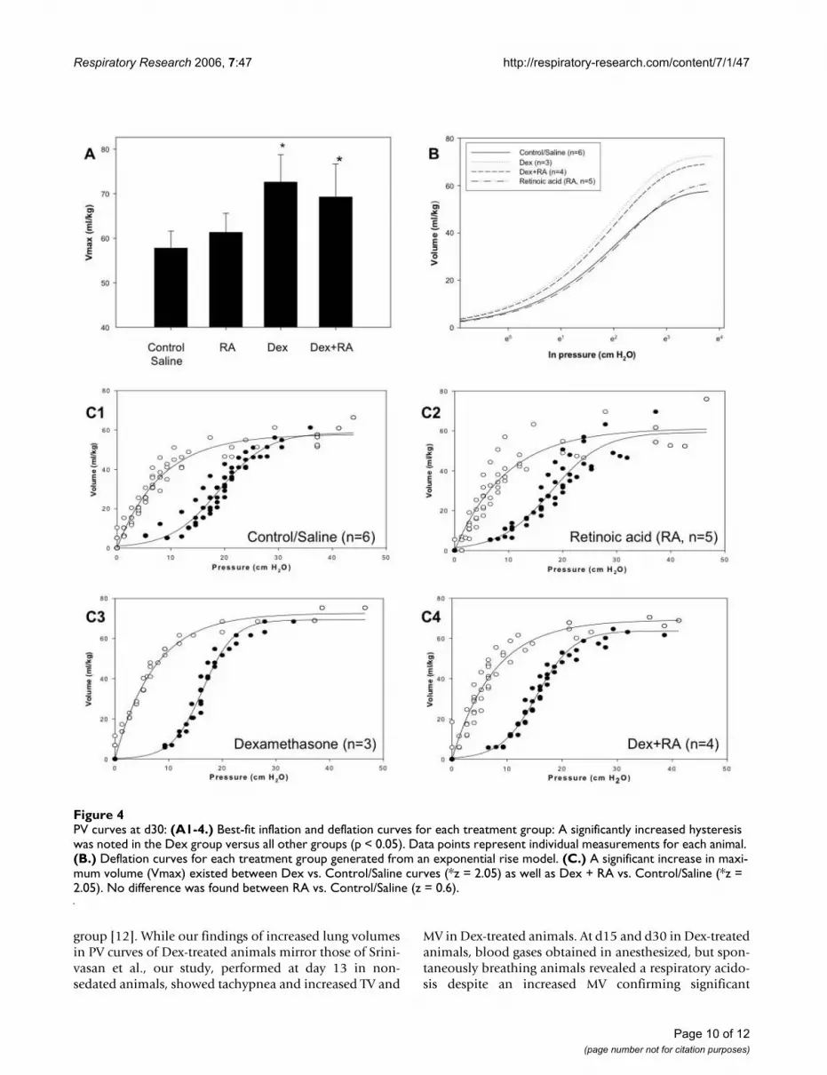
PV curvesIn order to confirm the lung volume of displacementmeasurements made at d15 and to evaluate lung volumesusing an independent method, PV curves were generatedon d30 as described in Methods. As shown in Figure 4A
and 4C, Vmax was significantly increased in both Dex andDex + RA compared to control/saline (z = 2.05). No dif-ference existed between Dex compared to Dex + RA curves(z = 0.13) and Control/Saline versus RA curves (z = 0.6).The deflation curve for each treatment group was based onan exponential rise to maximum model (Figure 4B). Thebest-fit inflation and deflation curves generated for eachtreatment group are shown in Figure 4C1-4, with dots rep-resenting individual measurements for each animal. A sig-nificantly increased hysteresis was noted in Dex versus allother groups (Dex: 739 ± 19; Control/Saline: 566 ± 41;RA: 572 ± 66; Dex + RA: 594 ± 24 (ml/kg)2, p < 0.05, n =3–6 per group). Collectively, these data suggest that Dextreatment resulted in larger lung volumes that concomi-tant RA treatment failed to abrogate.
The pressure required to reach the point of maximumcompliance during inflation was lower in Dex vs. Control/Saline (16.3 ± 0.3 vs. 18.4 ± 0.6 mm H2O/kg, p = 0.014, n= 3–6 per group) and between RA and Dex + RA curves(17.9 ± 0.3 vs. 15.3 ± 0.3 mm H2O/kg, p < 0.01, n = 3–6per group). In addition, the rate of maximal deflation wassignificantly greater in Dex vs. Control/Saline curves (9.6± 0.3 vs. 5.5 ± 0.4 mm H2O/kg/s, p < 0.05, n = 3–6 pergroup). The rate of maximal deflation tended to be lowerin Dex vs. Dex + RA curves but this did not reach signifi-cance (9.6 ± 0.3 vs. 7.2 ± 0.9 mm H2O/kg/s, p = 0.07, n =3–6 per group). This parameter was similar between RAand Dex + RA curves (7.4 ± 1.3 vs. 7.2 ± 0.9 mm H2O/kg/s, p = 0.88, n = 3–6 per group). These data suggest that Dextreatment resulted in lungs that had larger residual vol-umes requiring higher pressures to achieve the point ofmaximal compliance but were less stable during deflation.
Taken together, these physiologic data demonstratethat Dex + RA treatment failed to prevent larger lungvolumes, RR, MV, and TV seen with Dex treatmentalone. However, CO2 elimination improved, sug-gesting better gas exchange with increased septation.
Exercise swim testingNo difference in time to fatigue was noted on forced exer-cise swim testing for any group of rats (Saline/Control 45± 2, CSO/RA 45 ± 3, Dex, 45 ± 2, Dex + RA 39 ± 2 minutes,n = 6–8 per group). This suggests that, even in Dex-treatedanimals that demonstrated compromised pulmonaryfunction by other measures, exercise tolerance was notaffected by hormonal treatments.
Extended studySince Massaro and Massaro have previously demonstratedthat RA promotes septation after the period of normalalveolarization [11] and our data showed that early RAeffects were lost two weeks after stopping treatment, wenext sought to determine whether the effects of later
Page 6 of 12(page number not for citation purposes)

Respiratory Research 2006, 7:47 http://respiratory-research.com/content/7/1/47
administration of RA were also reversed. Rat pups werenormalized to 10 pups per litter and treated with eitherDex or saline from days 1–14 followed by either CSO orRA from days 24–36 (12 days of treatment). Animals werestudied at either day 37 (at the end of treatments) or day52 (2 weeks after stopping treatment).
Weight gainBirth weights were the same for all animals (6.9 ± 0.1, n =40). In untreated animals, growth velocities were similaruntil d24 after which males grew faster than females suchthat by d36 females weighed approximately 10% less thanmales (data not shown). Dex treatment affected bothmales and females equally with 8–9% lower weight at d14(p < 0.01, n = 8 per group) and a 6–8% lower weight atd36 as compared to sex-matched controls (p = 0.09, n = 8
per group). As with the earlier study, RA treatment alonehad no effect on weight (data not shown).
MorphologyAlterations of lung architecture were similar to those seenwith the Day 15/30 protocol (Fig. 5). At d37, Early Dex +Later CSO-treated animals had simplified distal air spacescompared to Early Saline + Later CSO controls. EarlySaline + Later RA-treated pups, on the other hand, hadsmaller, more numerous alveoli than Early Saline + LaterCSO controls. Normal architecture was restored in EarlyDex + Later RA-exposed rats (Figure 5). Changes seen inthe Early Dex + Later CSO group persisted at d52 whileanimals exposed to Early Dex + Later RA continued tohave architecture similar to that of controls. Interestingly,at d52, Early Saline + Later RA-treated rats had lung histol-
Days 15 (top) and 30 (bottom) histology: A simplified distal architecture was seen in Dex-treated animals at both daysFigure 2Days 15 (top) and 30 (bottom) histology: A simplified distal architecture was seen in Dex-treated animals at both days. At d15, RA-treated pups had smaller more numerous alveoli, but these changes were no longer seen at d30. Dex + RA treatment resulted in a restitution of septation to near that of saline controls at both days, (all images 100× magnification)
Table 3: Plethysmography at d13: Dex-treated animals showed an increased respiratory rate (RR), tidal volume, and minute ventilation compared to saline controls. Retinoic acid treatment alone did not alter RR but, when given with Dex, resulted in decreased RR similar to that of controls. Values are expressed mean ± SE. *p < 0.05 vs. saline; †p = 0.3 vs. Dex; ‡p = 0.5 vs. Dex; §p = 0.9 vs. Dex. bpm: breaths per minute
Treatment Group Respiratory Rate (bpm) Tidal Volume (ml/kg) Minute Ventilation (ml/kg)
Control (n = 8) 171 ± 20 5.2 ± 0.9 861 ± 145Saline (n = 9) 180 ± 5.0 4.8 ± 0.8 890 ± 163Cottonseed oil (CSO) (n = 14) 179 ± 12 6.5 ± 0.7 935 ± 109Retinoic acid (RA) (n = 15) 180 ± 6.0 5.2 ± 0.7 810 ± 103Dexamethasone (Dex) (n = 16) 211 ± 11* 8.1 ± 0.8* 2260 ± 150*Dex + RA (n = 14) 195 ± 9.0† 8.7 ± 1.0‡ 2131 ± 186§
Page 7 of 12(page number not for citation purposes)

Respiratory Research 2006, 7:47 http://respiratory-research.com/content/7/1/47
ogy that appeared similar to that of control animals (Fig-ure 5), again demonstrating a loss of the effects of RAalone two weeks after treatment was stopped.
Radial alveolar countsRAC were used to quantify the changes in alveolarizationin the extended study. In concordance with histologicalappearance, RAC were significantly lower in Early Dex +Later CSO-treated and significantly higher in Early Saline+ Later RA-treated animals at d37 (Table 6). Rats treatedwith both hormones had RAC no different from controls.At d52, RAC remained significantly lower in Early Dex +Later CSO-treated animals compared to Early Saline +Later CSO controls, but both Early Saline + Later RA- andEarly Dex + Later RA-treated animals were similar to con-trols thereby confirming the reversal of RA alone effectstwo weeks after stopping treatment (Table 6). The distri-bution of males and females in these studies was equaland no differences were noted between them with respectto the histology or RAC (data not shown).
DiscussionIn the present study, we confirm hormonally mediatedchanges in architecture during postnatal lung develop-ment in the rat. Respiratory acidosis, the most significant
functional abnormality, was noted on d15 in both RAalone-and Dex alone-treated rat pups and was resolved inDex + RA-treated animals. However, Dex + RA failed toresolve the increased tachypnea, MV, and TV seen in Dexalone-treated rats. Massaro and Massaro have previouslyshown that Dex + RA treatment results in an increasedbody mass-specific surface area available for gas exchangecompared to rats treated with Dex alone [10]. In the faceof persistently larger lung volumes and equivalent bodyweight in both Dex-and Dex + RA-treated animals, theimproved CO2 elimination in Dex + RA-treated animals islikely the effect of improved secondary septation and alarger surface area for gas exchange.
Interestingly, the increase in RAC on d15 in rats treatedwith RA alone was associated with hypercarbia, lowerPaO2 and acidosis, but without any effect on other pulmo-nary function parameters studied. The reason for thisabnormality in ABG is unclear, but suggests a defect in gasexchange. It is unlikely that this abnormality is due to theincreased number of alveoli as it has previously beenshown that RA treatment alone does not increase surfacearea [10]. However, ABG were normal in Dex + RA ani-mals at d15, as well as in RA alone-treated pups by d30when the RA alone-stimulated changes in distal lungstructure had also resolved. Indeed, while the effects ofDex alone and concomitant RA administration were sus-tained for at least 15 days after stopping the treatments,the effects of RA alone from either d4 to d14 or d24 to d36were reversed two weeks after stopping RA.
Alveolar development, the last phase of lung develop-ment, occurs either pre- or postnatally depending on thespecies. In the human, alveolarization begins in utero atabout 36 weeks of gestation and continues postnatally,whereas in the rodent, secondary septation is an entirelypostnatal event. Alveolarization appears to correlateinversely with changes in serum corticosteroid concentra-tions. It is likely that the normal timing of alveolar devel-opment reflects decreased corticosteroid levels leading toan increase in DNA synthesis and septation. For example,in the rat, corticosterone concentrations drop to a nadirbetween postnatal days 6 and 12, the time of maximumalveolar formation [8]. Conversely, administration of cor-ticosteroid during this critical period results in an inhibi-tion of alveolarization [9]. Indeed, exposure of fetalrhesus macaques to triamcinolone during the pseudog-landular and saccular phases of lung development resultsin an inhibition of septation [16]. The mechanisms ofDex-induced inhibition of alveolarization are likely mul-tifactorial, including inhibition of DNA synthesis, differ-ential regulation of matrix components, and changes ingene expression in the lung [17]. In addition, corticoster-oids cause a growth retardation that is in itself associatedwith a slowing of alveolar growth [18].
Radial alveolar counts (RAC) as a percentage of day 1: Changes in RAC were seen as early as day 5 with RA alone-treated animals having significantly higher counts at days 5, 10, and 15Figure 3Radial alveolar counts (RAC) as a percentage of day 1: Changes in RAC were seen as early as day 5 with RA alone-treated animals having significantly higher counts at days 5, 10, and 15. However, at d30, RA-treated animals had counts similar to controls. Dex alone-treated animals had signifi-cantly lower RAC at each time point studied. (*p < 0.05 vs. saline-treated animals; †p < 0.05 vs. CSO-treated animals, ‡p = 0.49 vs. saline-treated animals, §p = 0.58 vs. CSO-treated animals) CSO: cottonseed oil.
Page 8 of 12(page number not for citation purposes)

Respiratory Research 2006, 7:47 http://respiratory-research.com/content/7/1/47
Since the structural effects of Dex administration duringthe time of secondary septation are sustained to adult-hood, the concept of a "critical period" of alveolar devel-opment was proposed. However, several lines of evidencesupport the notion that alveolar growth occurs throughoutlife and can be manipulated past the immediate newbornperiod. For example, starvation-induced decreases in alve-olar formation are reversed upon refeeding [18]. In addi-tion, treatment of rats previously exposed to Dex from d3to d15 with RA from d24 to d36 results in a restitution ofDex-induced simplification of the distal lung [11]. Mostpromising for clinical practice, however, is the ability of RAto stimulate alveolar formation in adult rats after emphy-sema was induced by intratracheal instillation of elastase[19]. On the other hand, in the present study, the effects ofRA alone were not sustained after discontinuation of treat-ment.
The mechanisms of RA effects on alveolarization are likelyvia changes in the expression of epithelial (e.g. VEGF) andmesenchymal (e.g. PDGF and TGFβ) growth factors criti-
cal for cell proliferation and differentiation, angiogenesis,and matrix deposition during lung development [20,21].In addition, the regulation of free all-trans-RA by RA-bind-ing proteins and interactions with RA receptors (RAR andRXR) contribute to appropriate lung development. Forexample, RAR-α promotes epithelial cell differentiationduring the progression from pseudoglandular to canalicu-lar stages of lung development [20]. In addition, RAR-αalso promotes alveolar formation after the perinatalperiod [22]. Meanwhile, the expression of RAR-β increasestowards the end of the saccular stage corresponding to aninduction of both type 1 and type 2 epithelial cells [20].However, RAR-β knockout mice exhibit premature septa-tion and RAR-β agonist treatment of neonatal rats resultsin impaired septation, thereby identifying RAR-β as aninhibitor of alveolar formation [23]. On the other hand,impaired distal airspace formation during postnatal lungdevelopment has also been reported in RAR-β knockoutmice [24]. Finally, targeted deletion of RAR-γ in mice isassociated with a decrease in alveolar number, suggestingthe importance of this receptor in the development ofnormal alveoli [25]. In our study, while Dex + RA treat-ment prevented some structural effects seen with Dexalone, effects due to RA alone were reversible. This sug-gests that mechanisms are in place to normalize alveolarstructure, but these mechanism(s), in particular thoseleading to the reversal of RA effects, are currentlyunknown.
To date, there has been a paucity of literature on the effectsof hormone-induced structural changes on pulmonaryfunction. Srinivasan et al. examined several lung variablesincluding RR, MV, and TV in sedated animals at 30 to 39days of life and noted no differences in any treatment
Table 4: Arterial blood gases at d15 and d30: Dex- or RA-treated animals at d15 had a respiratory acidosis with hypercarbia (*p < 0.01 vs. saline/controls) and this was maintained in Dex-treated animals at d30 (*p < 0.01 vs. saline/controls). Day 15 animals given Dex + RA did not have respiratory acidosis compared to Dex alone pups (†p < 0.05 vs. Dex alone). Animals at d30 that had been given Dex + RA showed a correction of pH and pCO2 (†p < 0.05 vs. Dex). Only d15 RA-treated animals had significantly lower pO2 values compared to controls (**p < 0.05 vs. saline/controls). Values are expressed mean ± SE.
Treatment group pH pCO2 pO2
Day 15
Control/Saline (n = 6) 7.39 ± 0.03 41.9 ± 3.2 89.8 ± 6.4Retinoic acid (RA) (n = 4) 7.29 ± 0.03* 54.2 ± 1.6* 67.3 ± 7.1**Dexamethasone (Dex) (n = 7) 7.27 ± 0.01* 56.3 ± 3.4* 76.0 ± 9.4Dex + RA (n = 4) 7.31 ± 0.04† 50.1 ± 6.0† 79.3 ± 2.9
Day 30
Control/Saline (n = 12) 7.38 ± 0.01 47.7 ± 1.0 83.5 ± 2.8Retinoic acid (RA) (n = 10) 7.38 ± 0.01 50.0 ± 1.5 80.1 ± 3.7Dexamethasone (Dex) (n = 12) 7.33 ± 0.01* 55.5 ± 1.4* 80.9 ± 5.3Dex + RA (n = 6) 7.38 ± 0.02† 48.5 ± 2.0† 83.7 ± 3.9
Table 5: Volumes of displacement on day 15: Lungs were inflated to 25 cm H2O, dissected en bloc and fixed overnight. The displacement of water by these lungs was determined and normalized to body weight in grams. Both Dex- and Dex + RA-treated animals had increased volumes of displacement as compared to controls and RA-treated animals (* p < 0.01).
Treatment Group – d15 Vdisp/body weight (mL/g)
Control/Saline (n = 9) 43.7 ± 1.28Retinoic Acid (RA) (n = 7) 46.5 ± 1.28Dexamethasone (Dex) (n = 6) 53.1 ± 3.13*Dex + RA (n = 4) 55.5 ± 3.02*
Page 9 of 12(page number not for citation purposes)
Respiratory Research 2006, 7:47 http://respiratory-research.com/content/7/1/47
group [12]. While our findings of increased lung volumesin PV curves of Dex-treated animals mirror those of Srini-vasan et al., our study, performed at day 13 in non-sedated animals, showed tachypnea and increased TV and
MV in Dex-treated animals. At d15 and d30 in Dex-treatedanimals, blood gases obtained in anesthesized, but spon-taneously breathing animals revealed a respiratory acido-sis despite an increased MV confirming significant
PV curves at d30: (A1-4.)Figure 4PV curves at d30: (A1-4.) Best-fit inflation and deflation curves for each treatment group: A significantly increased hysteresis was noted in the Dex group versus all other groups (p < 0.05). Data points represent individual measurements for each animal. (B.) Deflation curves for each treatment group generated from an exponential rise model. (C.) A significant increase in maxi-mum volume (Vmax) existed between Dex vs. Control/Saline curves (*z = 2.05) as well as Dex + RA vs. Control/Saline (*z = 2.05). No difference was found between RA vs. Control/Saline (z = 0.6).
Page 10 of 12(page number not for citation purposes)

Respiratory Research 2006, 7:47 http://respiratory-research.com/content/7/1/47
compromise in pulmonary function. It is likely that therespiratory acidosis and consequent tachypnea are due toincreased dead space in affected animals. Interestingly,while lung volume differences were not resolved, RAC andblood gases were normalized with Dex + RA treatment,suggesting that correction of blood gas abnormalitieslikely resulted from an increase in the surface area availa-ble for CO2 elimination rather than full correction of lungvolumes. Further, the mechanisms leading to the respira-tory acidosis and lower PaO2 seen with RA alone treat-ment at d15 are unknown. However, these changes werenot evident in Dex + RA pups at d15 or at d30 when theRA alone-induced changes had reversed, suggesting thatthe altered architecture seen in RA alone-treated pups mayhave contributed to the blood gas abnormalities.
Varying degrees of exercise intolerance have beendescribed in patients with emphysema [26] and BPD [27].In our study, no differences in exercise tolerance werefound with any treatment. This suggests that factors otherthan altered alveolar structure, such as fibrosis and restric-tive/obstructive lung disease, may play a significant role inthe diminished exercise tolerance observed in affectedpatients.
Lastly, starvation and decreased body weight are associ-ated with decreased alveolarization [18]. However, in ourstudies, while weight was decreased in both Dex- and Dex
+ RA-treated animals, Dex + RA animals had a restitutionof secondary septation, suggesting that distal lung struc-ture can be manipulated independent of body weight.This is important in preterm neonates that have signifi-cantly compromised nutrition and poor weight gain inaddition to an arrest of alveolarization.
ConclusionIn summary, hormonal treatment of rat pups results inaltered lung architecture. This is associated with signifi-cant structure-function disturbances where Dex-induced
Table 6: Radial alveolar counts (RAC) at d37 and d52: RAC were significantly lower in Early Dex + Later CSO-treated and higher in Early Saline + Later RA-treated pups while Early Dex + Later RA animals were similar to controls. At d52, while RAC continued to be significantly lower in Early Dex + Later CSO-treated pups, Early Saline + Later RA effects were lost two weeks after stopping RA treatment. Values are expressed mean ± SE. *p < 0.01 vs. d37 Early Saline + Later CSO-treated animals; †p < 0.01 vs. Early Dex + Late CSO; ¶p < 0.01 vs. Early Saline + late RA. CSO: Cottonseed oil.
Treatment Group Day 37 Day 52
Control (n = 4) 9.1 ± 0.2 9.5 ± 0.2Early Saline + Late CSO (n = 4) 9.3 ± 0.1 9.4 ± 0.1Early Saline + Late RA (n = 4) 10.6 ± 0.1* 9.7 ± 0.1¶Early Dex + Late CSO (n = 4) 7.7 ± 0.2* 7.8 ± 0.1*Early Dex + Late RA (n = 4) 8.9 ± 0.3† 9.2 ± 0.1†
Effect of delayed RA treatment on alveolarization: Animals were treated with either Dex or saline from d1-14 followed by either CSO or RA from d24-36Figure 5Effect of delayed RA treatment on alveolarization: Animals were treated with either Dex or saline from d1-14 followed by either CSO or RA from d24-36. A simplified distal architecture was seen in Dex-treated animals at both d37 (top) and d52 (bottom). RA-treated pups had smaller more numerous alveoli at d37 with changes no longer seen at d52. Early Dex + Later RA treatment resulted in restored secondary septation that was similar to that of saline controls at d37, and this was sustained to d52. (all images 100× magnification)
Page 11 of 12(page number not for citation purposes)

Respiratory Research 2006, 7:47 http://respiratory-research.com/content/7/1/47
Publish with BioMed Central and every scientist can read your work free of charge
"BioMed Central will be the most significant development for disseminating the results of biomedical research in our lifetime."
Sir Paul Nurse, Cancer Research UK
Your research papers will be:
available free of charge to the entire biomedical community
peer reviewed and published immediately upon acceptance
cited in PubMed and archived on PubMed Central
yours — you keep the copyright
Submit your manuscript here:http://www.biomedcentral.com/info/publishing_adv.asp
BioMedcentral
decreases in alveolarization are associated with increasedlung volumes, CO2 retention, acidosis, and tachypnea.Our findings showing that the effects of RA on generationof alveoli and on gas exchange appear to be time-limitedand reversible may be relevant for the use of RA in treat-ment of diseases such as BPD or emphysema.
Competing interestsThe author(s) declare that they have no competing inter-ests.
Authors' contributionsSJG was responsible for injecting and harvesting all ani-mals, measuring radial alveolar counts, performing allfunctional and statistical analyses, and drafting the manu-script. HZ was responsible for generating all the blood gasand volume of displacement data at day 15, interpretationand revision of the manuscript. JPF assisted in animal har-vesting and injections as well as conducting functionalstudies. RIG and MHG assisted in obtaining the pressurevolume measurements. AJG assisted in the analysis of PVcurves. RCS conceived the study, participated in its designand coordination, and helped draft and revise the manu-script. All authors read and approved the final manu-script.
AcknowledgementsThe authors thank Dr. Phillip L. Ballard for his critical review of the manu-script. The experiments in this study were supported by NIH grants HL62858 and HL075930 to RCS. HZ is the recipient of the NIH Pediatric Scientist Development Award (HD00850).
References1. Jobe AH: The new BPD: an arrest of lung development. Pediatr
Res 1999, 46:641-643.2. Hislop AA: Bronchopulmonary dysplasia: pre- and postnatal
influences and outcome. Pediatr Pulmonol 1997, 23(2):71-75.3. Coalson JJ, Winter V, deLemos RA: Decreased alveolarization in
baboon survivors with bronchopulmonary dysplasia. Am JRespir Crit Care Med 1995, 152(2):640-646.
4. Burri PH: Structural aspects of prenatal and postnatal devel-opment and growth of the lung. In Lung Growth and DevelopmentVolume 100. Edited by: McDonald JA. New York: Marcel Dekker;1997:1-35.
5. Massaro GD, Massaro D: Formation of pulmonary alveoli andgas-exchange surface area: quantitation and regulation. AnnuRev Physiol 1996, 58:73-92.
6. Burri PH: The postnatal growth of the rat lung. 3. Morphol-ogy. Anat Rec 1974, 180(1):77-98.
7. Burri PH, Dbaly J, Weibel ER: The postnatal growth of the ratlung. I.Morphometry. Anat Rec 1974, 178(4):711-730.
8. Henning SJ: Plasma concentrations of total and free corticos-terone during development in the rat. Am J Physiol 1978,235(5):E451-456.
9. Massaro D, Teich N, Maxwell S, Massaro GD, Whitney P: Postnataldevelopment of alveoli: Regulation and evidence for a criticalperiod in rats. J Clin Invest 1985, 76:1297-1305.
10. Massaro GD, Massaro D: Postnatal treatment with retinoic acidincreases the number of pulmonary alveoli in rats. Am J Physiol1996, 270:L305-L310.
11. Massaro GD, Massaro D: Retinoic acid treatment partially res-cues failed septation in rats and in mice. Am J Physiol Lung CellMol Physiol 2000, 278(5):L955-960.
12. Srinivasan G, Bruce EN, Houtz PK, Bruce MC: Dexamethasone-induced changes in lung function are not prevented by con-
comitant treatment with retinoic acid. Am J Physiol Lung Cell MolPhysiol 2002, 283(2):L275-287.
13. Savani RC, Godinez Rl, Godinez MH, Wentz E, Zaman A, Cui Z,Pooler PM, Guttentag SH, Beers MF, Gonzales LW, et al.: Respira-tory distress after intratracheal bleomycin: selective defi-ciency of surfactant proteins B and C. Am J Physiol Lung Cell MolPhysiol 2001, 281:L685-L696.
14. Emery JL, Mithal A: The number of alveoli in the terminal res-piratory unit of man during late intrauterine life and child-hood. Arch Dis Child 1960, 35:544-547.
15. Cooney IP, Thurlbeck WM: The radial alveolar count method ofEmery and Mithal: a reappraisal 1 – postnatal lung growth.Thorax 1982, 37(8):572-579.
16. Bunton IE, Plopper CG: Triamcinolone-induced structuralalterations in the development of the lung of the fetal rhesusmacaque. Am J Obstet Gynecol 1984, 148(2):203-215.
17. Saklatvala J: Glucocorticoids: do we know how they work?Arthritis Res 2002, 4(3):146-150.
18. Massaro D, Massaro GD, Baras A, Hoffman EP, Clerch LB: Calorie-related rapid onset of alveolar loss, regeneration, andchanges in mouse lung gene expression. Am J Physiol Lung CellMol Physiol 2004, 286(5):L896-906.
19. Massaro GD, Massaro D: Retinoic acid treatment abrogateselastase- induced pulmonary emphysema in rats. Nature Med1997, 3(6):675-677.
20. Belloni PN, Garvin L, Mao CP, Bailey-Healy I, Leaffer D: Effects ofall-trans-retinoic acid in promoting alveolar repair. Chest2000, 117(5 Suppl 1):235S-241S.
21. Morrisey EE, Savani RC: Midkine: a potential bridge betweenglucocorticoid and retinoid effects on lung vascular develop-ment. Am J Respir Cell Mol Biol 2003, 28(1):5-8.
22. Massaro GD, Massaro D, Chambon P: Retinoic acid receptor-alpha regulates pulmonary alveolus formation in mice after,but not during, perinatal period. Am J Physiol Lung Cell Mol Physiol2003, 284(2):L431-433.
23. Massaro GD, Massaro D, Chan WY, Clerch LB, Ghyselinck N, Cham-bon P, Chandraratna RA: Retinoic acid receptor-beta: an endog-enous inhibitor of the perinatal formation of pulmonaryalveoli. Physiol Genomics 2000, 4(1):51-57.
24. Snyder JM, Jenkins-Moore M, Jackson SK, Goss KL, Dai HH, BangsundPJ, Giguere V, McGowan SE: Alveolarization in retinoic acidreceptor-beta- deficient mice. PediatrRes 2005, 57(3):384-391.
25. McGowan S, Jackson SK, Jenkins-Moore M, Dai HH, Chambon P, Sny-der JM: Mice bearing deletions of retinoic acid receptors dem-onstrate reduced lung elastin and alveolar numbers. Am JRespir Cell Mol Biol 2000, 23(2):162-167.
26. Ferguson GT, Fernandez E, Zamora MR, Pomerantz M, Buchholz J,Make BJ: Improved exercise performance following lung vol-ume reduction surgery for emphysema. Am J Respir Crit CareMed 1998, 157(4 Pt 1):1195-1203.
27. Parat S, Moriette G, Delaperche MF, Escourrou P, Denjean A, Gault-ier C: Long-term pulmonary functional outcome of bron-chopulmonary dysplasia and premature birth. Pediatr Pulmonol1995, 20(5):289-296.
Page 12 of 12(page number not for citation purposes)