Embed Size (px)
Citation preview

Hm
MIa
b
c
d
a
ARRAA
KFPBMLL
1
mWOlmbowdin
n
0h
Chemistry and Physics of Lipids 177 (2014) 19– 25
Contents lists available at ScienceDirect
Chemistry and Physics of Lipids
j ourna l h o mepa ge: www.elsev ier .com/ locate /chemphys l ip
ow to link pyrene to its host lipid to minimize the extent ofembrane perturbations and to optimize pyrene dimer formation
iroslava Dékány Franováa, Jarmila Repákováb, Juha M. Holopainenc,lpo Vattulainenb,d,∗
Department of Chemical Physics and Optics, Faculty of Mathematics and Physics, Charles University, Ke Karlovu 3, Prague 2 CZ-12116, Czech RepublicDepartment of Physics, Tampere University of Technology, P.O. Box 692, FI-33101 Tampere, FinlandHelsinki Eye Lab, Department of Ophthalmology, University of Helsinki, Helsinki, FinlandMEMPHYS – Center for Biomembrane Physics, University of Southern Denmark, Odense, Denmark
r t i c l e i n f o
rticle history:eceived 5 July 2013eceived in revised form 8 October 2013ccepted 11 October 2013vailable online 8 November 2013
eywords:luorescent probeyreneis-pyreneolecular dynamics simulation
ipid bilayer
a b s t r a c t
We study how lipid probes based on pyrene-labeling could be designed to minimize perturbations inlipid bilayers, and how the same design principles could be exploited to develop probes which gaugelipid dynamics primarily within a single lipid monolayer or between them. To this end, we use atomisticmolecular dynamics simulations to consider membranes where pyrene moieties are attached to lipidacyl chains in varying positions. We find that in a DOPC bilayer the conformational ordering of lipidsaround di-pyrenyl-PC probes is altered to a largely similar extent regardless of where the pyrene moietyis attached to the hydrocarbon chain. This is in contrast to saturated membranes, where pyrene-inducedperturbations have been observed to be more prominent. Meanwhile, the formation of pyrene dimersdepends on the linkage point between pyrene and its host lipid. Membrane-spanning dimers betweenlipids in different membrane leaflets are observed only if the pyrene moiety is attached to the latter halfof the acyl chain. A seemingly minor change to link pyrene to an acyl chain that is two carbons shorter
ipid membrane leads to a situation where membrane-spanning dimers are no longer observed. Further, simulationssuggest that formation of dimers is a slow process, where the rate is limited by both lateral diffusion andthe dimerization process once the two probes are neighbors to one another. Typical lifetimes of pyrenedimers turn out be of the order of nanoseconds. The results are expected to pave the way for designingways to consider experimentally topics such as intraleaflet lateral diffusion, motion of lipids within andbetween membrane domains, and membrane domain registration across bilayers.
. Introduction
Imaging has become one of the most useful and illustrativeeans to study lipid membrane behavior (Somerharju, 2002;ustner, 2007; Groves et al., 2008; de Almeida et al., 2009).
ver the years it has been particularly helpful in visualization ofarge-scale phenomena such as formation of membrane domains,
olecular traffic, and partitioning of proteins to different mem-rane environments. However, recent progress in the developmentf imaging techniques and probe molecules has also paved theay for very detailed descriptions of membrane structure and
ynamics, as super-resolution imaging techniques have renderedt possible to investigate membranes over scales as small as tens ofanometers (Eggeling et al., 2009).
∗ Corresponding author at: Department of Physics, Tampere University of Tech-ology, P.O. Box 692, FI-33101 Tampere, Finland.
E-mail address: [email protected] (I. Vattulainen).
009-3084/$ – see front matter © 2013 Elsevier Ireland Ltd. All rights reserved.ttp://dx.doi.org/10.1016/j.chemphyslip.2013.10.004
© 2013 Elsevier Ireland Ltd. All rights reserved.
For imaging, one needs probes. Numerous fluorescent markershave thus been developed to consider a variety of differ-ent lipid bilayer properties (Somerharju, 2002; Wustner, 2007;Chattopadhyay and London, 1987; Epand et al., 1996; Lakowicz,1983; Lentz, 1989, 1993; Maier et al., 2002; Holtta-Vuori et al.,2008; Sezgin et al., 2012). Free probes (not linked to lipids or othermolecules) allow one to explore how different membrane regionsare formed, as even seemingly minor changes in the structure ofprobes affect their partitioning to domains with different physi-cal properties. Meanwhile, the probes linked to lipids can provideone with highly useful insight of, e.g., lipid dynamics. However,how useful and reliable insight is given by imaging depends quitecritically on the probes’ ability to mimic native membrane behav-ior. Since free probes are usually not natural, and since the lipidslinked to probes have quite certainly properties that are different
from those of the corresponding native lipids, some perturbationsin membrane behavior are inevitable (Lentz, 1989, 1993; Koivusaloet al., 2004; Leonard-Latour et al., 1996; Parente and Lentz, 1985;Somerharju et al., 1985; Yashar et al., 1987; Repáková et al., 2005).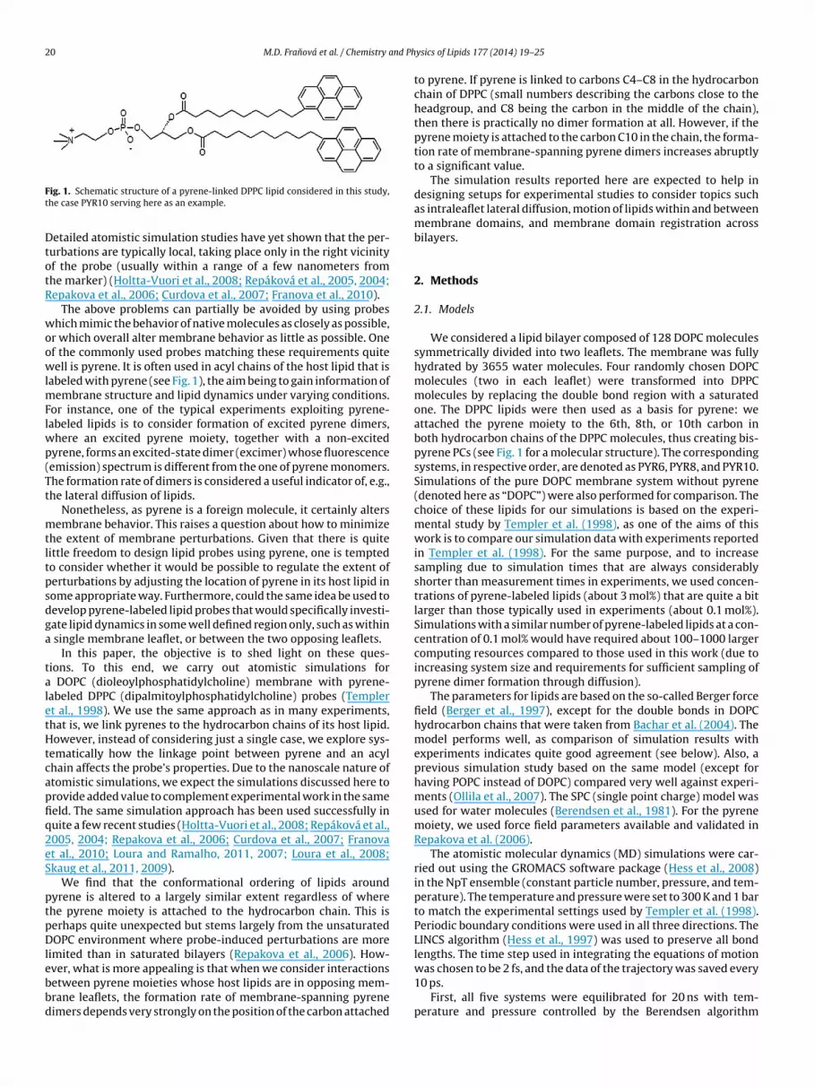
20 M.D. Franová et al. / Chemistry and Ph
Ft
DtotR
woowlmFlwp(Tt
mtltpsdga
taletHtcapfiq2eS
ptpDlebbd
ig. 1. Schematic structure of a pyrene-linked DPPC lipid considered in this study,he case PYR10 serving here as an example.
etailed atomistic simulation studies have yet shown that the per-urbations are typically local, taking place only in the right vicinityf the probe (usually within a range of a few nanometers fromhe marker) (Holtta-Vuori et al., 2008; Repáková et al., 2005, 2004;epakova et al., 2006; Curdova et al., 2007; Franova et al., 2010).
The above problems can partially be avoided by using probeshich mimic the behavior of native molecules as closely as possible,
r which overall alter membrane behavior as little as possible. Onef the commonly used probes matching these requirements quiteell is pyrene. It is often used in acyl chains of the host lipid that is
abeled with pyrene (see Fig. 1), the aim being to gain information ofembrane structure and lipid dynamics under varying conditions.
or instance, one of the typical experiments exploiting pyrene-abeled lipids is to consider formation of excited pyrene dimers,
here an excited pyrene moiety, together with a non-excitedyrene, forms an excited-state dimer (excimer) whose fluorescenceemission) spectrum is different from the one of pyrene monomers.he formation rate of dimers is considered a useful indicator of, e.g.,he lateral diffusion of lipids.
Nonetheless, as pyrene is a foreign molecule, it certainly altersembrane behavior. This raises a question about how to minimize
he extent of membrane perturbations. Given that there is quiteittle freedom to design lipid probes using pyrene, one is temptedo consider whether it would be possible to regulate the extent oferturbations by adjusting the location of pyrene in its host lipid inome appropriate way. Furthermore, could the same idea be used toevelop pyrene-labeled lipid probes that would specifically investi-ate lipid dynamics in some well defined region only, such as within
single membrane leaflet, or between the two opposing leaflets.In this paper, the objective is to shed light on these ques-
ions. To this end, we carry out atomistic simulations for DOPC (dioleoylphosphatidylcholine) membrane with pyrene-abeled DPPC (dipalmitoylphosphatidylcholine) probes (Templert al., 1998). We use the same approach as in many experiments,hat is, we link pyrenes to the hydrocarbon chains of its host lipid.owever, instead of considering just a single case, we explore sys-
ematically how the linkage point between pyrene and an acylhain affects the probe’s properties. Due to the nanoscale nature oftomistic simulations, we expect the simulations discussed here torovide added value to complement experimental work in the sameeld. The same simulation approach has been used successfully inuite a few recent studies (Holtta-Vuori et al., 2008; Repáková et al.,005, 2004; Repakova et al., 2006; Curdova et al., 2007; Franovat al., 2010; Loura and Ramalho, 2011, 2007; Loura et al., 2008;kaug et al., 2011, 2009).
We find that the conformational ordering of lipids aroundyrene is altered to a largely similar extent regardless of wherehe pyrene moiety is attached to the hydrocarbon chain. This iserhaps quite unexpected but stems largely from the unsaturatedOPC environment where probe-induced perturbations are more
imited than in saturated bilayers (Repakova et al., 2006). How-
ver, what is more appealing is that when we consider interactionsetween pyrene moieties whose host lipids are in opposing mem-rane leaflets, the formation rate of membrane-spanning pyreneimers depends very strongly on the position of the carbon attachedysics of Lipids 177 (2014) 19– 25
to pyrene. If pyrene is linked to carbons C4–C8 in the hydrocarbonchain of DPPC (small numbers describing the carbons close to theheadgroup, and C8 being the carbon in the middle of the chain),then there is practically no dimer formation at all. However, if thepyrene moiety is attached to the carbon C10 in the chain, the forma-tion rate of membrane-spanning pyrene dimers increases abruptlyto a significant value.
The simulation results reported here are expected to help indesigning setups for experimental studies to consider topics suchas intraleaflet lateral diffusion, motion of lipids within and betweenmembrane domains, and membrane domain registration acrossbilayers.
2. Methods
2.1. Models
We considered a lipid bilayer composed of 128 DOPC moleculessymmetrically divided into two leaflets. The membrane was fullyhydrated by 3655 water molecules. Four randomly chosen DOPCmolecules (two in each leaflet) were transformed into DPPCmolecules by replacing the double bond region with a saturatedone. The DPPC lipids were then used as a basis for pyrene: weattached the pyrene moiety to the 6th, 8th, or 10th carbon inboth hydrocarbon chains of the DPPC molecules, thus creating bis-pyrene PCs (see Fig. 1 for a molecular structure). The correspondingsystems, in respective order, are denoted as PYR6, PYR8, and PYR10.Simulations of the pure DOPC membrane system without pyrene(denoted here as “DOPC”) were also performed for comparison. Thechoice of these lipids for our simulations is based on the experi-mental study by Templer et al. (1998), as one of the aims of thiswork is to compare our simulation data with experiments reportedin Templer et al. (1998). For the same purpose, and to increasesampling due to simulation times that are always considerablyshorter than measurement times in experiments, we used concen-trations of pyrene-labeled lipids (about 3 mol%) that are quite a bitlarger than those typically used in experiments (about 0.1 mol%).Simulations with a similar number of pyrene-labeled lipids at a con-centration of 0.1 mol% would have required about 100–1000 largercomputing resources compared to those used in this work (due toincreasing system size and requirements for sufficient sampling ofpyrene dimer formation through diffusion).
The parameters for lipids are based on the so-called Berger forcefield (Berger et al., 1997), except for the double bonds in DOPChydrocarbon chains that were taken from Bachar et al. (2004). Themodel performs well, as comparison of simulation results withexperiments indicates quite good agreement (see below). Also, aprevious simulation study based on the same model (except forhaving POPC instead of DOPC) compared very well against experi-ments (Ollila et al., 2007). The SPC (single point charge) model wasused for water molecules (Berendsen et al., 1981). For the pyrenemoiety, we used force field parameters available and validated inRepakova et al. (2006).
The atomistic molecular dynamics (MD) simulations were car-ried out using the GROMACS software package (Hess et al., 2008)in the NpT ensemble (constant particle number, pressure, and tem-perature). The temperature and pressure were set to 300 K and 1 barto match the experimental settings used by Templer et al. (1998).Periodic boundary conditions were used in all three directions. TheLINCS algorithm (Hess et al., 1997) was used to preserve all bondlengths. The time step used in integrating the equations of motion
was chosen to be 2 fs, and the data of the trajectory was saved every10 ps.First, all five systems were equilibrated for 20 ns with tem-perature and pressure controlled by the Berendsen algorithm

nd Physics of Lipids 177 (2014) 19– 25 21
(riPtnwTs
2
oi
towpf
fib
iealRlDcl
odLccafot
csmswwnimsc
3
3a
Fi
0
0.1
0.2
0.3
|SC
D|
0
0.1
0.2
0.3
0
0.1
0.2
|SC
D|
0
0.1
0.2
42 86 10 12 14 160
0.1
0.2
0.3
0.4
0.5
0.6
|SC
D|
42 86 10 12 14 160
0.1
0.2
0.3
0.4
PYR10, sn-1 PYR10, sn-2
PYR8, sn-1 PYR8, sn-2
PYR6, sn-1 PYR6, sn-2
Carbon atom
same layeropposite layer
Fig. 2. Order parameter (|SCD|) profiles computed from 500 ns long simulations.Small carbon atom numbers correspond to those close to the lipid head group, whilelarge ones are close to the terminal carbons in membrane center. The left columnshows the order parameter profile for the sn − 1 chain and the right column for thesn − 2 chain. Interactions within the same leaflet are shown by full lines, and interac-tions with lipids in the opposing leaflet by dashed lines. Different colors correspondto different distances between center of mass positions of DOPC and the probe thatis the closest to it: R ≤ 0.5 nm (red), 0.5 < R ≤ 1.0 nm (green), 1.0 < R ≤ 1.5 nm (blue),1.5 nm <R (yellow), and an average over all lipids (black). Error bars are about thesize of the linewidth (full lines), or 10% (dashed lines, PYR10), or 10–20% (dashed
M.D. Franová et al. / Chemistry a
Berendsen et al., 1984) using time constants of 0.1 and 1.0 ps,espectively. In the subsequent production simulations, each last-ng for 500 ns, the pressure was controlled by the semi-isotropicarrinello–Rahman barostat (Parrinello and Rahman, 1981) and theemperature by the Nose-Hoover thermostat (Hoover, 1985) (witho change in time constants). Long-range interactions were dealtith by the particle mesh Ewald technique (Essman et al., 1995).
he total simulation time (covering all simulations reported in thistudy) was about 2 microseconds.
.2. Analysis
We determined the average area per lipid by dividing the areaf the bilayer in the membrane (xy) plane by the number of lipidsn a single monolayer.
For mass density profiles across the membranes, we computedhe profiles along the membrane normal (z) direction. As the centerf mass (CM) position of the bilayer may slightly fluctuate in time,e calculated the positions of all atoms with respect to the CMosition of the bilayer (in the z-direction), and did this separatelyor each system configuration.
Membrane thickness was determined from a mass density pro-le by considering the points where the profiles of water and theilayer coincided (Patra et al., 2003).
The order parameter SCD describing the conformational order-ng of DOPC acyl chains was computed in a standard manner (Ollilat al., 2007). In this work SCD was mainly used to quantify the extentnd the range of membrane perturbations around the pyrene-inked lipid. To this end, for each DOPC, we computed the distance,, from its CM to the CM of the closest pyrene probe in the same
eaflet. The SCD order parameter profiles were then determined forOPCs as a function of distance R. The same approach was used toonsider the SCD values of DOPCs close to the probe but in a differenteaflet.
The formation of pyrene–pyrene dimers was determined basedn two criteria (distance and geometry). First, we considered theistance between the center of mass positions of two pyrene rings,. Second, if L ≤ 0.6 nm, we defined the planes of the two rings andonsidered the angle between them, �. If | cos �| ≥ 0.9, we con-luded that the two pyrene rings were attached to one anothers a complex, thereby forming a dimer. The criteria above wereound to be robust since based on visual inspection a wide rangef values for L and � yielded essentially the same conclusions (seeext).
We consider it important to stress that the pyrene dimersonsidered in this simulation study are not identical to excimerstudied in fluorescence experiments. In excimers, one of theonomers in the excited-state pyrene dimer has to be in an excited
tate, and this is not possible in classical simulations used in thisork. What we consider here is the formation of pyrene dimershere the given monomer moieties comprising the dimer are or areot in an excited state. If the formation process of a true excimer and
ts properties were to be unlocked, one should resort to quantum-echanical simulations. The downside in that case is, however, the
imulated time scale which would be a tiny fraction of what onean do through classical MD simulations.
. Results
.1. Pyrene alters the order of hydrocarbon chains in its vicinitynd can also influence lipids in the opposing membrane leaflet
The results for |SCD| as a function of distance R are depicted inig. 2. The data show that pyrene probes alter membrane orderingn a largely similar fashion in all the systems (PYR6, PYR8, PYR10).
lines, PYR8). In the case of PYR6 (dashed lines), the number of samples is very lowand thereby the data shown has only marginal value due to major error bars.
First, let us consider the case where DOPCs are in the same leafletas the probes. Close to the glycerol region, hydrocarbon chains oflipids close to the probe (R ≤ 1.0 nm) become more ordered. Theeffect is somewhat stronger in PYR10 and PYR8 compared to PYR6,but the trend is largely the same in every case. Meanwhile, closeto the membrane center, pyrene probes slightly weaken confor-mational disorder in hydrocarbon chains that are in the vicinityof the probes (R ≤ 0.5 nm), while the next shell of lipids (0.5 nm<R ≤ 1.0 nm) becomes more ordered compared to lipids far fromthe probes. Nonetheless, the perturbations in acyl chain order arenot substantial, and the range of perturbations is about 1 nm, mean-ing that the conformational order of only the nearest neighbors ofpyrene-linked lipids is affected to a significant degree.
The results are in agreement with previous simulations(Repakova et al., 2006), where Repakova et al. considered pyrene-labeled DPPCs in a DPPC bilayer (pyrene attached to the carbon 10 ina palmitoyl chain; cf. PYR10 in the present work). The main differ-ences of our work compared to Repakova et al. (2006) are the DOPCmembrane used in our system, and the presence of bis-pyrene PCsin our systems, while Repakova et al. had only one pyrene moiety
in each host lipid. Despite these differences, Repakova et al. alsoobserved local ordering around the probe, with a range of about1.0–1.5 nm. However, in a saturated membrane the probe-induced
22 M.D. Franová et al. / Chemistry and Physics of Lipids 177 (2014) 19– 25
Fig. 3. Snapshot of a pyrene dimer in the center of the PYR10 membrane formed byt
owd
coRAciqpttidn
fPbeaa
amtwuspolc
TAl
0
0.25
0.5
0.75
1
1.25
1.5
DOPCPYR6PYR8PYR10
-3 -2 -1 10 32z [nm]
0
0.01
0.02
0.03
mas
s de
nsity
[g/
cm3 ]
PYR6PYR8PYR10
system
bilayer
pyrene moiety
A
BCM
Fig. 4. Mass density calculated from 500 ns long simulations. (A) Mass density pro-files of the bilayer and the whole system (including water) shown for all the studied
wo pyrene moieties in opposing membrane leaflets.
rdering of nearby acyl chains persisted over the length of thehole chain, while with DOPCs in our systems the ordering partlyecreased in the terminal side beyond the double bond region.
When pyrenes interact with lipids in the opposing leaflet, PYR6auses no noticeable effects (data not shown). In PYR8, the orderf DOPC chains close to the probe increases to some extent if
≤ 1.0 nm, and equally strong effects are observed with PYR10.s a matter of fact, the most pronounced perturbations in acylhain order emerge when PYR10 interacts with lipids in the oppos-ng membrane leaflet. Fig. 3 depicts an example of a situation inuestion. It highlights that in PYR10, when a membrane-spanningyrene dimer is being formed, the probe lipids orient themselveso a more vertical position, increasing the conformational order inheir right vicinity. While there are also other processes contribut-ng to the data in Fig. 2 (PYR10), events associated with pyreneimer formation shown in Fig. 3 play a role in this structural orga-ization.
Additional data for the average area per lipid show that it variesrom (0.689 ± 0.002) nm2 for PYR6 up to (0.693 ± 0.002) nm2 forYR10 (Table 1). The largest area was found for a pure DOPC mem-rane: (0.697 ± 0.002) nm2, which is quite well consistent with thexperimental findings that have given average areas of 0.674 nm2
t 303 K (Kucerka et al., 2008), 0.724 nm2 at 303 K (Pan et al., 2008),nd 0.691 nm2 at 288 K (Pan et al., 2008).
From the simulation data one can conclude that pyrenes have minor influence on membrane packing as they do compress theembrane, the effect being the larger the closer pyrenes are to
he lipid headgroup region. The smallest perturbations are foundhen pyrenes are close to membrane center, where the free vol-me of the membrane is the largest. Importantly, the results alsohow that pyrene-induced perturbations are less prominent in theresent DOPC based bilayer compared to a membrane comprisedf saturated phospholipids (Repakova et al., 2006). Yet the high-ight is that PYR10 seems to be different from the other systems
onsidered here.able 1verage area per lipid and membrane thickness calculated from 500 ns long simu-
ations. The error bars are ±0.002 nm2 for area and ±0.02 nm for thickness.
System 〈A〉 (nm2) Membranethickness (nm)
PYR6 0.689 4.10PYR8 0.690 4.10PYR10 0.693 4.09DOPC 0.697 4.10
systems and (B) mass density profiles for the center of mass (CM) of the pyrenemoiety.
3.2. PYR10 leads to interdigitation
The mass density profiles for all simulated cases are displayedin Fig. 4. The profiles readily show that the position of pyrene doesnot affect the average membrane thickness (Table 1 and Fig. 4A).
For a pure DOPC membrane, SANS experiments have given4.58 nm for the thickness of a DOPC membrane at 25 ◦C (Gallováet al., 2004), and Leonenko et al. (2004) measured the thickness at22 ◦C to be 4.0 nm. Since there is no unique way to define mem-brane thickness, the comparison is inevitably somewhat tricky, butdespite this issue the agreement is quite good since the value of4.10 nm we found from simulations is essentially consistent withexperimental observations.
More interesting is to elucidate the transmembrane distribu-tion of the pyrene moiety (Fig. 4B). The profiles depict how thedistribution of pyrene moves closer to membrane center as thelinkage point of pyrene moves from the headgroup region towardsthe terminal carbon in the hydrocarbon chain. This is no news.What is more appealing to realize is that while PYR6 and PYR8show no interdigitation across membrane center at all, the case ofPYR10 is very different. In PYR10, the pyrene moieties are locatedsubstantially in the opposing leaflet, indicating quite profoundinterdigitation. This highlights that in PYR10, the pyrenes from theopposite leaflets extend to the membrane center region and alsobeyond it, providing further support for the idea that with PYR10the pyrenes in different leaflets can interact with each other andalso form dimers.
3.3. PYR10 gives rise to membrane-spanning pyrene dimers
The above view about membrane-spanning pyrene dimers issupported by the snapshop shown in Fig. 3, which describes oneof the many cases where we observed dimer formation associatedwith PYR10. Fig. 5 further depicts the dimer formation process by
showing how L (distance between two pyrenes) and cos � (describ-ing the orientation of two dimers with respect to each other) evolvein time.
M.D. Franová et al. / Chemistry and Ph
100 120 140 160 180 200
Time (ns)
-1
0
1
2
3
4
5
Red
- D
ista
nce
(nm
), B
lack
- c
os(
angle
)
Fig. 5. Time dependence of the dimer formation process in the PYR10 system fortwo pyrene moieties whose host lipids are in different membrane leaflets. The dimerformation process in question takes place between 120 and 170 ns, and the dimeri(o
ftoo
i(utTPsb
tptabltdt
mswt
TNdwptF
alternative interpretations include, e.g., contributions of pyrenetrimers, whose formation rate is not expected to be frequent butwhich yet contribute to pyrene complex formation. More quantita-tive comparison of lifetimes between simulations and experiments
s established most of the time after about 173 ns. Shown here are curves for L(t)distance between two pyrenes) shown in red color, and cos �(t) (describing therientation of the two dimers with respect to each other) depicted in black.
When we analyzed the occurrences of this process in detail, weound that during the simulation time scale of 500 ns, formation ofransmembrane dimers took place only with PYR10 (Table 2). Withther pyrene-linked lipids, dimers across the membrane were notbserved.
Meanwhile, shorter pyrene-linked lipids (PYR6, PYR8) werenvolved in numerous intraleaflet dimer formation processesTable 2). Here, there are two possible events. Either the pyrenenits in the same molecule form a dimer, or the pyrene rings inwo different lipid probes get together to form a dimer complex.he results in Table 2 indicate that in all systems (PYR6, PYR8,YR10) there are numerous dimers when the host lipids are in theame leaflet, but the number of events fluctuates quite considerablyetween intralipid and interlipid dimers.
We stress that we prefer not to draw strong conclusions abouthe quantitative numbers, since the results in Table 2 depend, inart, on the initial configuration of the systems we prepared: duringhe simulation time of 500 ns, the lipids diffuse laterally on aver-ge by about 4.5 nm, while the linear system size of the simulationox in the membrane plane was about 6.7 nm. Thus, sampling of
ipid motion for the few pyrene probes is too limited for quantita-ive predictions. For adequate quantitative sampling of lateral lipidynamics and collisions of pyrene-labeled lipids, the simulationime scale should be larger by a factor of at least 10.
Despite this limitation, let us compare our data in a qualitative
anner with the experiments of Templer et al. (1998). They con-idered pyrene-labeled DPPC lipids in a DOPC membrane, as in ourork, and they also used DPPC lipids as hosts which were syn-
hesized (or purchased) to have acyl chains with 4, 6, 8, and 10
able 2umber of events where two pyrene monomers were bound to one another as aimer. Shown here are data for the number of events where (1) both lipid probesere in the same leaflet and also in the same molecule (NIntra,S), (2) the two lipidrobes were in the same leaflet but in different molecules (NIntra,D), and (3) whenhe pyrene moieties were in lipids that were in different membrane leaflets (NInter).inally, NTotal stands for the total number of dimer events.
System NIntra,S NIntra,D NInter NTotal
PYR6 9434 3758 0 13192PYR8 4417 2327 0 6744PYR10 12650 17 3707 16374
ysics of Lipids 177 (2014) 19– 25 23
carbon atoms. They observed that the excimer to monomer ratiowas the largest in 4dipyPC (corresponding to the system PYR4 inour work), then decreased with 6dipyPC (PYR6), further decreasedwith 8dipyPC (PYR8), and then increased with 10dipyPC (PYR10).In a related study by Cheng and Somerharju (1996), it was foundthat in a DOPC bilayer at about 28 ◦C, di-pyrenyl-PC probes had thelargest excimer to monomer ratio with short chains (4dipyPC and6dipyPC), then a minimum (8dipyPC and 10dipyPC), and then anincreasing trend with longer chains (12dipyPC and 14dipyPC).
The behavior found in simulations is qualitatively consistentwith experimental data. It is quite possible that this agreement isaccidental, since despite the long simulation times the samplingof dimer formation between different probe lipids is limited dueto slow diffusion. Yet, the consistency between experiments andsimulations is encouraging.
We will consider the excimer to monomer ratio, its impact onlateral pressure profiles, and the use of pyrene formation on unlock-ing the transmembrane pressure profile (Templer et al., 1998) in aseparate study, which will be discussed elsewhere (Franova et al.,2013).
3.4. Lifetimes of pyrene dimers are of the order of nanoseconds
Lifetimes of pyrene dimers were rather short and in all cases lessthan 8 ns. As for dimers between lipids in different leaflets (PYR10),the lifetime of the most stable dimer was 5.1 ns, and there were15 (4) dimers with lifetimes longer than 1 ns (2 ns). The number ofpyrene dimers with a small lifetime was abundant, thus the averagelifetime was as short as 0.56 ns.
Lifetimes for dimers that formed within a single lipid mono-layer are listed in Table 3. The results highlight that many dimershave a very small lifetime, suggesting that pyrenes collide ratherfrequently but the formation of a stable dimer (with a long life-time) is a more rare event. In the case of PYR10, it is obvious thatformation of membrane-spanning dimers competes with dimer-ization between different lipids that are in a single leaflet, thus thenumbers in Table 3 for PYR10 are small.
Experimental data for pyrene dimer lifetimes suggest that thereare two binding processes with different lifetimes (Barenholz et al.,1996). The shorter lifetime is of the order of nanoseconds, whilethe longer lifetime is about an order of magnitude larger. This sug-gests that the dimer formation processes we gauged in simulationsdescribe the short-time behavior, or that membrane-spanningdimers have a longer lifetime compared to dimers formed bypyrenes in the same leaflet, supported by the simulation data. Other
Table 3Lifetimes of pyrene dimers when the dimer is formed between lipids in the samemonolayer. ‘Intralipid’ stands for the case where the pyrenes are in the same hostlipid, and ‘interlipid’ for the case where pyrenes are in different molecules. tL isthe longest lifetime observed and tave is the average lifetime. N1 and N2 show thenumbers of lifetimes longer than 1 ns and 2 ns, in respective order.
System tL (ns) tave (ns) N1 N2
IntralipidPYR6 7.6 0.41 24 10PYR8 4.5 0.30 13 4PYR10 4.7 0.30 34 9
InterlipidPYR6 4.0 0.27 15 2PYR8 4.3 0.10 6 1PYR10 0.2 0.09 0 0

2 nd Ph
(ldgvIer
adttddtdupqo
pfthTqitssb
asthscfa
4
baiorqsan
tbeatlttb
4 M.D. Franová et al. / Chemistry a
Barenholz et al., 1996) is not meaningful, since in experiments theifetime is typically determined from the half-time of fluorescenceecay (over a macroscopic number of excimers landing back to theround state), while the simulation data we present here is for indi-idual pyrene formation processes where the dimer is not excited.mportantly, the lifetime given by simulations is smaller than thensemble-averaged half-times determined from experiments, as isational to assume.
The above simulation results for lifetimes have been based on very strict criterion for the formation (and breaking) of a pyreneimer. If the conditions for pyrene formation (discussed in Sec-ion 2) were violated at any time step, the dimer was consideredo break apart. However, if the criteria for the existence of pyreneimers were allowed to relax a bit, such that the pyrene–pyreneistance or the orientation of two pyrene moieties were allowedo fluctuate a bit more (see Fig. 5), then the lifetime of pyreneimers would increase to some extent, see Fig. 5. In this work, wesed quite reasonable assumptions for values of L and � to identifyyrene dimers, but a more rigorous definition should be based onuantum-mechanical analyses for the formation and maintainingf dimer complexes.
In the same spirit, it would be useful to consider the force fieldarameterization of pyrene in its excited state. This stems from theact that an excimer is comprised of an excited pyrene monomerogether with a non-excited pyrene moiety, and the present workas been based on a model for the non-excited pyrene monomer.he geometry and charge distribution of an excited pyrene may beuite different from the non-excited one, and new models account-
ng for this aspect would have potential to do better in describinghe stability of the dimer at long times. However, these con-iderations would require quite substantial quantum-mechanicalimulations that are beyond the scope of this work and remain toe done and discussed elsewhere.
We conclude that while our results for lifetimes are reason-ble, we are unable to unravel the mechanistic difference betweenhort and long lifetimes observed in experiments. Yet the simula-ion data show that individual pyrene dimers (with a short lifetime)ave lifetimes ranging from sub-nanosecond to nanosecond timecales. The data also show that once pyrene monomer moieties arelose to one another, their collision rate is high, and if any dimerormed transiently breaks apart, the monomers frequently reunitegain.
. Concluding remarks
Imaging with fluorescent probes attached to lipid acyl chains hasecome a very common technique to study biological membranesnd traffic in cellular systems. Very recently, the interest for usingmaging as a tool to reveal the structure and dynamics of cellularrganelles has increased further due to developments with super-esolution techniques that render nanometer-scale considerationsuite possible. However, one has to keep in mind that there is rea-on for concern, too, since imaging is based on the use of probes,nd their properties are seldom (if ever) identical to those of theative molecules.
In this work, we used atomistic molecular dynamics simula-ions to explore how the position of a pyrene moiety within theis-pyrene PC molecule in a DOPC bilayer affects membrane prop-rties and the formation rate of pyrene dimers. We observed that,s far as probe-induced membrane perturbations are concerned,here are no major differences between the probes of different
ength. They all alter membrane order around them, and thus, e.g.,he dynamic properties of a pyrene-labeled lipid are different fromhose of native lipids. The positive news is that membrane pertur-ations level off within about one nanometer from the probe andysics of Lipids 177 (2014) 19– 25
are therefore of short range, affecting just the nearest neighbors ofthe probe lipid. Additionally, our results show that in unsaturatedmembranes (such as the DOPC bilayer considered here) pyrene-induced perturbations are on average weaker than in saturatedlipid bilayers, a matter which may be worth taking into accountin experiments where (say) saturated and monounsaturated phos-pholipids would have consistent phase behavior and membraneprotein partitioning behavior.
Yet, the main finding of this work is that the formation ofmembrane-spanning pyrene dimers depends very strongly on thelocation of the pyrene moiety in its host lipid. Pyrene moietiesattached to carbons close to the glycerol group do not form inter-leaflet dimers at all. In the system we explored, an abrupt transitiontakes place at carbon number 10. Pyrene moieties coupled tothis carbon interdigitate substantially across membrane center tothe opposing leaflet, thereby also forming numerous dimers withpyrenes in the other leaflet.
The results suggest that experiments to consider the couplingof lipid dynamics in two opposing leaflets would be most produc-tive if pyrenes were attached to the latter half of the hydrocarbonchain. Possible phenomena in this spirit would include, e.g., mem-brane domain registration across bilayers and consideration ofthe shear viscosity between the membrane leaflets. Meanwhile, ifprobe attachment were done in the leading half of the chain (closeto lipid head group), then this choice would provide insight relatedto excimer formation within the given membrane leaflet, with nocontribution of membrane-spanning excimers. Topics that wouldbenefit from this choice would include, for instance, elucidation oflateral diffusion within a given monolayer, motion of lipids withinand between membrane domains, and lipid confinement. Quiteobviously, in these cases it would be useful to use lipids whereonly one of the hydrocarbon chains were labeled with pyrene.
Acknowledgements
We thank Samuli Ollila for very useful discussions during thecourse of the project. We also wish to thank the Academy of Finlandand the European Research Council (CROWDED-PRO-LIPIDS) forfinancial support, and CSC–IT Centre for Science (Espoo, Finland)for computing resources.
References
Bachar, M., Brunelle, P., Tieleman, D.P., Rauk, A., 2004. Molecular dynamics simula-tion of polyunsaturated lipid bilayer susceptible to lipid peroxidation. J. Phys.Chem. B 108, 7170–7179.
Barenholz, Y., Cohen, T., Haas, E., Ottolenghi, M., 1996. Lateral organization of pyrene-labeled lipids in bilayers as determined from the deviation from equilibriumbetween pyrene monomers and excimers. J. Biol. Chem. 271, 3085–3090.
Berendsen, H.J.C., Postma, J.P.M., van Gunsteren, W.F., Hermans, J., 1981. Interac-tion models for water in relation to protein hydration. In: Pullman, B. (Ed.),Intermolecular Forces. Reidl, Dordrecht, pp. 331–342.
Berendsen, H.J.C.H.J.C., Postma, J.P.M., van Gunsteren, W.F., DiNola, A., Haak, J.R.,1984. Molecular dynamics with coupling to an external bath. J. Chem. Phys. 81,3684–3690.
Berger, O., Edholm, O., Jahnig, F., 1997. Molecular dynamics simulations of a fluidbilayer of dipalmitoylphosphatidylcholine at full hydration, constant pressure,and constant temperature. Biophys. J. 72, 2002–2013.
Chattopadhyay, A., London, E., 1987. Parallax method for direct measurement ofmembrane penetration depth utilizing fluorescence quenching by spin-labeledphospholipids. Biochemistry 26, 39–45.
Cheng, K.H., Somerharju, P., 1996. Effects of unsaturation and curvature on the trans-verse distribution of intramolecular dynamics of dipyrenyl lipids. Biophys. J. 70,2287–2298.
Curdova, J., Capkova, P., Plasek, J., Repakova, J., Vattulainen, I., 2007. Free pyreneprobes in gel and fluid membranes: perspective through atomistic simulations.J. Phys. Chem. B 111, 3640–3650.
de Almeida, R.F.M., Loura, L.M.S., Prieto, M., 2009. Membrane lipid domains and rafts:current applications of fluorescence lifetime spectroscopy and imaging. Chem.Phys. Lipids 157, 61–77.
Eggeling, C., Ringemann, C., Medda, R., Schwarzmann, G., Sandhoff, K., Polyakova,S., Belov, V.N., Hein, B., von Middendorff, C., Schönle, A., Hell, S.W., 2009. Direct

nd Ph
E
E
F
F
G
G
H
H
H
H
K
K
L
L
L
L
L
L
L
M.D. Franová et al. / Chemistry a
observation of the nanoscale dynamics of membrane lipids in a living cell. Nature457, 1159–1162.
pand, R.F., Kraayenhof, R., Sterk, G.J., Sang, H.W.W.F., Epand, R.M., 1996. Fluo-rescent probes of membrane surface properties. Biochim. Biophys. Acta 1284,191–195.
ssman, U., Perera, L., Berkowitz, M.L., Darden, H.L.T., Pedersen, L.G., 1995. A smoothparticle mesh Ewald method. J. Chem. Phys. 103, 8577–8592.
ranova, M., Repakova, J., Capkova, P., Holopainen, J.M., Vattulainen, I., 2010.Effects of DPH on DPPC-cholesterol membranes with varying concentrations ofcholesterol: from local perturbations to limitations in fluorescence anisotropyexperiments. J. Phys. Chem. B 114, 2704–2711.
ranova, M., Vattulainen, I., Ollila, O.H.S., 2013. Lateral pressure profile measure-ments with pyrene probes examined through atom-scale molecular dynamicssimulations, submitted for publication.
allová, J., Uhríková, D., Islamov, A., Kuklin, A., Balgavy, P., 2004. Effect of cholesterolon the bilayer thickness in unilamellar extruded DLPC and DOPC liposomes:SANS contract variation study. Gen. Physiol. Biophys. 23, 113–128.
roves, J.T., Parthasarathy, R., Forstner, M.B., 2008. Fluorescence imaging of mem-brane dynamics. Annu. Rev. Biomed. Eng. 10, 311–338.
ess, B., Becker, H., Berendsen, H.J., Fraaije, J.G.E.M., 1997. LINCS: a linear constraintsolver for molecular simulations. J. Comp. Chem. 18, 1463–1472.
ess, B., Kutzner, C., van der Spoel, D., Lindahl, E., 2008. Gromacs 4: algorithmsfor highly efficient, load-balanced, and scalable molecular simulation. J. Chem.Theory Comput. 4, 435–447.
oltta-Vuori, M., Uronen, R.L., Repakova, J., Salonen, E., Vattulainen, I., Panula, P., Li,Z.G., Bittman, R., Ikonen, E., 2008. BODIPY-cholesterol: a new tool to visualizesterol trafficking in living cells and organisms. Traffic 9, 1839–1849.
oover, W.G., 1985. Canonical dynamics: equilibrium phase-space distributions.Phys. Rev. A 31, 1695–1697.
oivusalo, M., Alvesalo, J.A.V.J., Somerharju, P., 2004. Partitioning of pyrene-labeledphospho- and sphingolipids between ordered and disordered bilayer domains.Biophys. J. 74, 1984–1993.
ucerka, N., Nagle, J.F., Sachs, J.N., Feller, S.E., Pencer, J., Jackson, A., Katsaras, J., 2008.Lipid bilayer structure determined by the simultaneous analysis of neutron andX-ray scattering data. Biophys. J. 95, 2356–2367.
akowicz, J.R., 1983. Principles of Fluorescence Spectroscopy. Plenum-Press, NewYork.
entz, B.R., 1989. Membrane fluidity as detected by diphenylhexatriene probes.Chem. Phys. Lipids 50, 171–190.
entz, B.R., 1993. Use of fluorescent probes to monitor molecular order and motionswithin liposome bilayers. Chem. Phys. Lipids 64, 99–116.
eonard-Latour, M., Morelis, R.M., Coulet, P.R., 1996. Influence of pyrene-based fluo-rescent probes on the characteristics of DMPA/DMPC Langmuir–Blodgett films.Langmuir 12, 4797–4802.
eonenko, Z.V., Finot, E., Ma, H., Dahms, T.E.S., Cramb, D.T., 2004. Investigation oftemperature-induced phase transitions in DOPC and DPPC phospholipid bilay-ers using temperature-controlled scanning force microscopy. Biophys. J. 86,3783–3793.
oura, L.M.S., Ramalho, J.P.P., 2007. Location and dynamics of acyl chain NBD-labeled
phosphatidylcholine (NBD-PQ) in DPPC bilayers. A molecular dynamics andtime-resolved fluorescence anisotropy study. Biochim. Biophys. Acta: Biomem-branes 1768, 467–478.oura, L.M.S., Ramalho, J.P.P., 2011. Recent developments in molecular dynamicssimulations of fluorescent membrane probes. Molecules 16, 5437–5452.
ysics of Lipids 177 (2014) 19– 25 25
Loura, L.M.S., Fernandes, F., Fernandes, A.C., Ramalho, J.P.P., 2008. Effects of fluores-cent probe NBD-PC on the structure, dynamics and phase transition of DPPC.A molecular dynamics and differential scanning calorimetry study. Biochim.Biophys. Acta: Biomembranes 1778, 491–501.
Maier, O., Oberle, V., Hoekstra, D., 2002. Fluorescent lipid probes: some propertiesand applications. Chem. Phys. Lipids 116, 3–18.
Ollila, S., Hyvonen, M.T., Vattulainen, I., 2007. Polyunsaturation in lipid mem-branes: dynamic properties and lateral pressure profiles. J. Phys. Chem. B 111,3139–3150.
Pan, J., Tristram-Nagle, S., Kucerka, N., Nagle, J.-F., 2008. Temperature dependenceof structure, bending rigidity, and bilayer interactions of dioleoylphosphatidyl-choline bilayers. Biophys. J. 94, 117.
Parente, R.A., Lentz, B.R., 1985. Advantages and limitations of 1-palmitoyl-2-[2-[4-(6-phenyl-trans-1,3,5-hexatrienyl)phenyl] ethyl]carbonyl]-3-sn-phosphatidylcholine as a fluorescent membrane probe.Biochemistry 24, 6178–6185.
Parrinello, M., Rahman, A., 1981. Polymorphic transitions in single crystals: a newmolecular dynamics method. J. Appl. Phys. 52, 7182–7190.
Patra, M., Karttunen, M., Hyvönen, M.T., Falck, E., Lindqvist, P., Vattulainen, I., 2003.Molecular dynamics simulations of lipid bilayers: major artifacts due to trun-cating electrostatic interactions. Biophys. J. 84, 3636–3645.
Repáková, J., Capková, P., Holopainen, J.M., Vattulainen, I., 2004. Distribution, ori-entation, and dynamics of DPH probes in DPPC bilayer. J. Phys. Chem. B 108,13438–13448.
Repáková, J., Holopainen, J.M., Morrow, M.R., McDonald, M.C., Capková, P., Vat-tulainen, I., 2005. Influence of DPH on the structure and dynamics of a DPPCbilayer. Biophys. J. 88, 3398–3410.
Repakova, J., Holopainen, J.M., Karttunen, M., Vattulainen, I., 2006. Influ-ence of pyrene-labeling on fluid lipid membranes. J. Phys. Chem. B 110,15403–15410.
Sezgin, E., Levental, I., Grzybek, M., Schwarzmann, G., Mueller, V., Honigmann,A., Belov, V.N., Eggeling, C., Coskun, U., Simons, K., Schwille, P., 2012. Par-titioning, diffusion, and ligand binding of raft lipid analogs in model andcellular plasma membranes. Biochim. Biophys. Acta: Biomembranes 1818,1777–1784.
Skaug, M.J., Longo, M.L., Faller, R., 2009. Computational studies of Texas Red-1,2-dihexadecanoyl-sn-glycero-3-phosphoethanolamine-model building andapplications. J. Phys. Chem. B 113, 8758–8766.
Skaug, M.J., Longo, M.L., Faller, R., 2011. The impact of Texas Red on lipid bilayerproperties. J. Phys. Chem. B 115, 8500–8505.
Somerharju, P.J., Virtanen, J.A., Eklund, K.K., Vainio, P., Kinnunen, P.K.J., 1985.1-Palmitoyl-2-pyrenedecanoyl glycerophospholipids as membrane probes: evi-dence for regular distribution in liquid-crystalline phosphatidylcholine bilayers.Biochemistry 24, 2773–2781.
Somerharju, P., 2002. Pyrene-labeled lipids as tools in membrane biophysics andcell biology. Chem. Phys. Lipids 116, 57–74.
Templer, R.H., Castle, S.J., Curran, A.R., Rumbles, R.G., Klug, D.R., 1998. Sensingisothermal changes in the lateral pressure in model membranes using di-pyrenylphosphatidylcholine. Faraday Disc. 111, 41–53.
Wustner, D., 2007. Fluorescent sterols as tools in membrane biophysics and cellbiology. Chem. Phys. Lipids 146, 1–25.
Yashar, V.B., Menashe, M., Biltonen, R.L., Johnson, M.L., Barenholz, Y., 1987. Interac-tion of trans-parinaric acid with phosphatidylcholine bilayers: comparison withthe effect of other fluorophores. Biochim. Biophys. Acta 904, 117–124.


![Influence of cell cycle on responses of MCF7 cells to benzo[a]pyrene](https://img.pdfslide.net/doc/110x75/6313ece23ed465f0570ae658/influence-of-cell-cycle-on-responses-of-mcf7-cells-to-benzoapyrene.jpg)