Embed Size (px)
Citation preview
Human DNA Ligases: A ComprehensiveNew Look for Cancer Therapy
Deependra Kumar Singh, Shagun Krishna, Sharat Chandra, Mohammad Shameem,
Amit Laxmikant Deshmukh, and Dibyendu Banerjee∗
CSIR-Central Drug Research Institute, B.S. 10/1, Janakipuram Extension, Sitapur Road, Lucknow 226021,Uttar Pradesh, India
Published online in Wiley Online Library (wileyonlinelibrary.com).DOI 10.1002/med.21298
�
Abstract: Living organisms belonging to all three domains of life, viz., eubacteria, archaeabacteria,and eukaryotes encode one or more DNA ligases. DNA ligases are indispensable in various DNA repairand replication processes and a deficiency or an inhibition of their activity can lead to accumulation ofDNA damage and strand breaks. DNA damage, specially strand breaks at unsustainable levels can leadto replication block and/or cell death. DNA ligases as potential anticancer targets have been realizedonly recently. There is enough rationale to suggest that ligases have a tremendous potential for noveltherapeutics including anticancer and antibacterial therapy, specially when the world is facing acuteproblems of drug resistance and chemotherapy failure, with an immediate need for new therapeutictargets. Here, we review the current state of the art in the development of human ligase inhibitors, theirstructures, molecular mechanisms, physiological effects, and their potential in future cancer therapy. Citingexamples, we focus on strategies for improving the activity and specificity of existing and novel inhibitorsby using structure-based rational approaches. In the end, we describe potential new sites on the ligase Iprotein that can be targeted for the development of novel inhibitors. This is the first comprehensive reviewto compile all known human ligase inhibitors and to provide a rationale for the further development ofligase inhibitors for cancer therapy. C© 2013 Wiley Periodicals, Inc. Med. Res. Rev., 00, No. 00, 1–29, 2013
Key words: DNA ligase; ligase inhibitor; cancer target; cancer drug; ligase; inhibitor
1. INTRODUCTION
DNA ligases were discovered in 1967 by the Gellert, Lehman, Richardson, and Hurwitzlaboratories.1 DNA ligases join adjacent 3′-hydroxyl and 5′-phosphoryl termini to form aphosphodiester bond in duplex DNA and thus have an indispensable role in DNA replication,
Contract grant sponsor: Government of India; Contract grant numbers: SB/FT/LS-163/2012, BT/PR6421/GBD/27/436/2012), BSC0106.
Correspondence to: Dibyendu Banerjee, CSIR-Central Drug Research Institute, B.S. 10/1, Janakipuram Ex-tension, Sitapur Road, Lucknow 226021, Uttar Pradesh, India. E-mail: [email protected]; [email protected]
Medicinal Research Reviews, 00, No. 00, 1–29, 2013C© 2013 Wiley Periodicals, Inc.

2 � SINGH ET AL.
recombination, and repair. DNA ligases are grouped into two families, ATP-dependent ligasesand NAD+-dependent ligases, according to the cofactor required for ligase–adenylate forma-tion. The bacterial NAD+-dependent DNA ligases belong to a highly conserved phylogeneticcluster of enzymes and have been described only in eubacteria. In contrast, the ATP-dependentDNA ligases are found in all three domains of life, viz., eubacteria, archaeabacteria, and eu-karyotes and form a somewhat loosely defined cluster of enzymes.2, 3 In this review, we willrestrict ourselves to human ATP-dependent DNA ligases only and unless otherwise specified,the word ligase will stand for human DNA ligase. The reaction mechanism for ATP-dependentligases involves the formation of a covalent enzyme–AMP intermediate from the cleavage ofATP to AMP and pyrophosphate. The adenylate group from AMP is then transferred to the5′-phosphate of the nicked DNA molecule. Finally, the DNA ligase seals the gap by phospho-diester bond formation, via the displacement of the AMP residue with the 3′-hydroxyl groupfrom the adjacent DNA strand.
DNA ligases have an indispensable role in replication. During replication, the two antipar-allel strands of DNA are copied distinctively. One strand is copied in a continuous manner (andcalled the leading strand) whereas the other strand, due to its opposite orientation, is copieddiscontinuously in small stretches called Okazaki fragments, after its discoverer Reiji Okazaki.These fragments then have to be sealed together to make a continuous strand of DNA calledthe lagging strand. This important function during replication is performed by DNA ligase I(hLigI). Other important DNA repair processes such as nucleotide excision repair (NER) (byhLigI and hLigIII),4 base excision repair (BER) (by hLigI and hLigIII),5, 6 and double-strandbreak repair (DSBR) pathways such as nonhomologous end joining (NHEJ) (by hLigIV)7 andmicrohomology-mediated end joining (by hLigI and hLigIII)8 are also dependent on ligases. Adeficiency in DNA ligation leads to severe cell lethality, increased genomic instability, and hyper-sensitivity to DNA damage. It has been reported that an individual with an inherited mutationin the hLigI gene exhibited retarded growth, development, and immunodeficiency.9 LigIV defi-ciency (known as LIG4 syndrome) is characterized by microcephaly and immunodeficiency andis common to severe combined immunodeficiency with microcephaly, growth retardation, andsensitivity to ionizing radiation due to NHEJ1 deficiency (NHEJ1 syndrome).10 No patientswith ligase III deficiency have been identified till date.
Inhibiting the function of ligases can have potentially serious consequences for DNAreplication and repair events, processes that are most often targeted in cancer therapy. Severalinhibitors against human DNA ligases have been discovered or synthesized, but not a singleinhibitor is presently used in therapy. In the sections below, we begin with a brief introductionto the working of ligases and then undertake a comprehensive review of all classes of humanDNA ligase inhibitors that are known till now. The inhibitors have been reviewed for theirmode of inhibition and target specificity and their potential for use in future cancer therapy. Inthe end we present potential new sites on the human ligase I protein that could be targeted fordevelopment of specific inhibitors.
2. A DESCRIPTION OF HUMAN DNA LIGASES
A. Human DNA Ligase I (hLigI)
The human gene encoding DNA ligase I is located at chromosome 19q13.2-13.3. The genecovers 53 kb and contains 28 exons.11 DNA ligase I is required for the ligation of Okazakifragments during lagging strand DNA synthesis as well as several DNA repair pathways.Elevated levels of DNA ligase I have been found in several cancer cells such as breast, lung,and ovarian cancer cells.12–14 In 2001, Sun et al.14 showed that there are higher levels of DNA
Medicinal Research Reviews DOI 10.1002/med
DNA LIGASES AS TARGETS IN CANCER THERAPY � 3
ligase I in human tumors than in benign normal tissues and normal peripheral lymphocytes,suggesting that DNA ligase I plays more of a role in proliferating cells than in resting cells.Unexpectedly for a protein involved in replication, an individual with an inherited defect inDNA ligase I has been identified.15 The individual exhibited severe immunodeficiency, stuntedgrowth, and sun sensitivity and died at a young age from an abdominal lymphoma with anassociated acute infection suggesting immunodeficiency.16 A fibroblast cell strain, 46BR wasderived from the individual and showed deleterious mutations in both alleles of the DNA ligaseI gene. One allele carries a mutation that inactivates the active-site region, whereas the otherhas a mutation in a conserved part of the carboxy-terminal region that allows a small amountof residual DNA ligase activity. The leakiness of the latter point mutation suggests that evenlow levels of DNA replication enzymes may be sufficient for cellular viability.17
An available SV40-transformed 46BR.lG1 subline carries mutations in either one allele(hemizygous mutation) or both alleles (homozygous mutations) of hLigI, but in either casea leaky expression of hLigI is enough to keep the cells viable. 46BR cells show inefficientjoining of Okazaki fragments and anomalous gap filling during excision repair.17 However, noproblems with V(D)J joining is observed in 46BR cells.18 The 46BR patient was sun sensitive,and since the late steps of lagging-strand DNA replication and NER share many factors,19 itseems plausible that DNA ligase I is responsible for the final DNA-joining step in NER. hLigIis also involved in UV damaged proliferating cells having UV photolesions at late G1 and Sphase of cell cycle,4 long patch BER,5, 6 and microhomology-mediated end-joining pathway(DSBR pathway).8 Thus, inhibition of ligase I activity can potentially block replication as wellas introduce mutations due to failure in DNA repair pathways. The high levels of hLigIp inseveral cancer cells raises the probability that it may serve as a good target for cancer therapy.Accumulation of single-strand (ss) breaks followed by double-strand (ds) breaks in subsequentcycles of replication is the hallmark of cells deficient in hLigI protein. Several inhibitors againsthLigI have been reported but there is a need for the development of more potent-specificinhibitors. These are discussed in the later sections of this review.
B. DNA Ligase III (hLigIII)
The human gene encoding DNA ligase III (hLigIII) is located at chromosome 17q11.2-q12.DNA ligase III encodes multiple forms of DNA ligase. Nuclear and mitochondrial forms of hu-man DNA ligase IIIα are generated by alternative translations of the same gene.20 Additionally,a germ cell specific form of human DNA ligase IIIβ is generated by alternative splicing.9 Celllines with reduced activity for one of the DNA ligases show different phenotypes suggestingthat these enzymes have distinct cellular functions.15, 21–23 The nuclear form of DNA ligase IIIαis partnered with another protein called XRCC1 and is unstable in its absence.24 In the germcell specific form of DNA ligase IIIβ, a small positively charged peptide sequence replaces theC-terminal BRCT domain of DNA ligase IIIα.9 hLigIII is restricted to higher eukaryotes andhas been associated with short patch BER and NER pathway in quiescent as well as proliferat-ing cells at all phases of cell cycle.17–19 DNA ds breaks pose a great danger to the genome andcell survival, and must be repaired. In higher eukaryotes, such damage is repaired efficiently byNHEJ in which hLigIV has been implicated. The alternative NHEJ pathway is implicated inthe generation of large deletions and chromosomal translocations that are frequently observedin cancer cells. A recent study has shown that in DNA ligase IV deficient cells, the MRNcomplex (hMRE11/hRAD50/Nbs1) interacts with LigIIIα/XRCC1 upon DNA damage, andstimulates intermolecular ligation via a reaction that mimics alt-NHEJ.25 Thus, LigIIIα maybe a target for therapeutic use in hLigIV-deficient cancers.
Another recent study has shown functional redundancy between DNA ligase I and III invertebrate cells.26 Using conditional knockdown of hLigIII along with inactivation of hLigI
Medicinal Research Reviews DOI 10.1002/med
4 � SINGH ET AL.
and hLigIV genes, they demonstrated in DT40 (white leghorn chicken bursal lymphoma) cellsthat hLigIII can support semiconservative DNA replication. These observations suggest analternative pathway for Okazaki fragment processing. To us this observation suggests that aninhibitor with activity against both hLigI and hLigIII will be more effective than an inhibitorwith activity against hLigI alone. However, such a molecule will have far greater side effectsfor normal cells as compared to specific inhibitors for individual ligases. Interestingly, anotherrecent study suggests that in cells with dysfunctional hLigI, hLigIII can contribute to the ligationof the replication intermediates but not to the prevention of telomeric instability.27 Therefore,specific inhibitors against hLigI might still be effective in blocking cellular proliferation in cellswith functional hLigIII. hLigIII is majorly responsible for NER and BER (SSBR) pathways,and in certain situations for alternative NHEJ and MHEJ (microhomology-mediated endjoining) DSBR pathways.8, 17, 19, 20, 26, 28, 29 Thus, the inhibition of hLigIII will almost certainlyhave an adverse affect on DNA repair pathways as well as replication. Inhibitors againsthLigIII are discussed in detail later. However, specific inhibitors against hLigIII are yet to bereported.
C. DNA Ligase IV (hLigIV)
hLigIV is the most recently discovered human DNA ligase. It is a 911 amino acid polypeptidethat plays a role in the repair of ds DNA breaks. hLigIV is exclusively nuclear and functionsin DNA NHEJ processes,30–33 and may also be responsible for the V(D)J recombination inlymphoid cells.22 Similar to the nuclear form of LigIIIα, hLigIV is unstable without its part-ner protein called XRCC4. Late embryonic lethality and impaired V(D)J recombination wasreported in mice lacking DNA ligase IV.34 In yeast, DNA ligase IV was reported to medi-ate nonhomologous DNA end joining.7 According to another report by Grawunder et al.,35
the function of the XRCC4–DNA ligase IV complex in mammals may be to carry out thefinal steps of V(D)J recombination and joining of DNA ends. In 1998, Deborah E. Barnes etal.36 has reported that the targeted disruption of the gene encoding DNA ligase IV leads tolethality in embryonic mice. Microcephaly and immunodeficiency are common to DNA ligaseIV deficiency (LIG4 syndrome) and severe combined immunodeficiency with microcephaly,growth retardation, and sensitivity to ionizing radiation due to NHEJ1 deficiency (NHEJ1syndrome).10 Tobin et al.37 showed that relative to nontumorigenic breast epithelial MCF10Acells, estrogen receptor positive (ER+)-MCF7 breast cancer cells and progesterone receptorpositive (PR+)-MCF7 breast cancer cells have reduced steady-state levels of DNA ligase IV,whereas the steady-state levels of LigIIIα are higher. The main proteins involved in NHEJ ineukaryotes are DNA ligase IV, XRCC4, the catalytic subunit of DNA-dependent protein ki-nase (DNA-PKcs), Ku proteins, and possibly Artemis. Deficiencies in any one of these proteinsresult in hypersensitivity to DNA DSB-inducing agents such as ionizing radiation. Once theblunt ends are in place, the XRCC4/DNA ligase IV ligation complex is recruited to join theDNA ends together. DNA ligase IV carries out the ligation step, but it requires the bindingof XRCC4 to do so. XRCC4 functions as a regulatory element to stabilize DNA ligase IV,stimulate ligase activity, and direct the ligase to the site of DNA breaks via its recognition helixand DNA-binding capacity. Thus, it is clear from the above discussion that hLigIV is a crucialenzyme for classical NHEJ and if we inhibit hLigIV, the cellular machinery will not be capableof repairing ds breaks by NHEJ. This will predictably lead to triggering of cell death and/orapoptotic pathways.
From the above it can be safely concluded that inhibition of all three ligases at the sametime will most definitely kill a proliferating cancer cell. However, the side effects for normalcells might be too great to consider such a scenario for therapy. Hence, specific inhibitor forindividual ligases will be more desirable and inhibitors for hLigI will perhaps be the first ones
Medicinal Research Reviews DOI 10.1002/med
DNA LIGASES AS TARGETS IN CANCER THERAPY � 5
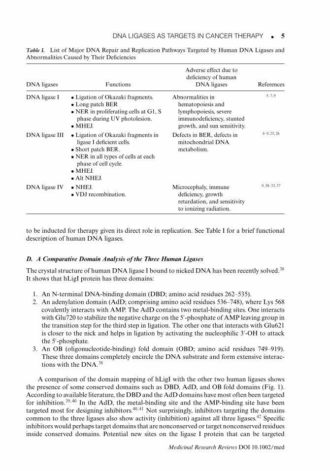
Table I. List of Major DNA Repair and Replication Pathways Targeted by Human DNA Ligases andAbnormalities Caused by Their Deficiencies
Adverse effect due todeficiency of human
DNA ligases Functions DNA ligases References
DNA ligase I • Ligation of Okazaki fragments.• Long patch BER• NER in proliferating cells at G1, S
phase during UV photolesion.• MHEJ.
Abnormalities inhematopoiesis andlymphopoiesis, severeimmunodeficiency, stuntedgrowth, and sun sensitivity.
5–7, 9
DNA ligase III • Ligation of Okazaki fragments inligase I deficient cells.
• Short patch BER.• NER in all types of cells at each
phase of cell cycle.• MHEJ.• Alt NHEJ.
Defects in BER, defects inmitochondrial DNAmetabolism.
6–9, 25, 26
DNA ligase IV • NHEJ.• VDJ recombination.
Microcephaly, immunedeficiency, growthretardation, and sensitivityto ionizing radiation.
9, 30–33, 37
to be inducted for therapy given its direct role in replication. See Table I for a brief functionaldescription of human DNA ligases.
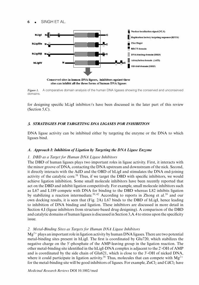
D. A Comparative Domain Analysis of the Three Human Ligases
The crystal structure of human DNA ligase I bound to nicked DNA has been recently solved.38
It shows that hLigI protein has three domains:
1. An N-terminal DNA-binding domain (DBD; amino acid residues 262–535).2. An adenylation domain (AdD; comprising amino acid residues 536–748), where Lys 568
covalently interacts with AMP. The AdD contains two metal-binding sites. One interactswith Glu720 to stabilize the negative charge on the 5′-phosphate of AMP leaving group inthe transition step for the third step in ligation. The other one that interacts with Glu621is closer to the nick and helps in ligation by activating the nucleophilic 3′-OH to attackthe 5′-phosphate.
3. An OB (oligonucleotide-binding) fold domain (OBD; amino acid residues 749–919).These three domains completely encircle the DNA substrate and form extensive interac-tions with the DNA.38
A comparison of the domain mapping of hLigI with the other two human ligases showsthe presence of some conserved domains such as DBD, AdD, and OB fold domains (Fig. 1).According to available literature, the DBD and the AdD domains have most often been targetedfor inhibition.39, 40 In the AdD, the metal-binding site and the AMP-binding site have beentargeted most for designing inhibitors.40, 41 Not surprisingly, inhibitors targeting the domainscommon to the three ligases also show activity (inhibition) against all three ligases.42 Specificinhibitors would perhaps target domains that are nonconserved or target nonconserved residuesinside conserved domains. Potential new sites on the ligase I protein that can be targeted
Medicinal Research Reviews DOI 10.1002/med
6 � SINGH ET AL.
Figure 1. A comparative domain analysis of the human DNA ligases showing the conserved and unconserveddomains.
for designing specific hLigI inhibitor/s have been discussed in the later part of this review(Section 5,C).
3. STRATEGIES FOR TARGETING DNA LIGASES FOR INHIBITION
DNA ligase activity can be inhibited either by targeting the enzyme or the DNA to whichligases bind.
A. Approach I: Inhibition of Ligation by Targeting the DNA Ligase Enzyme
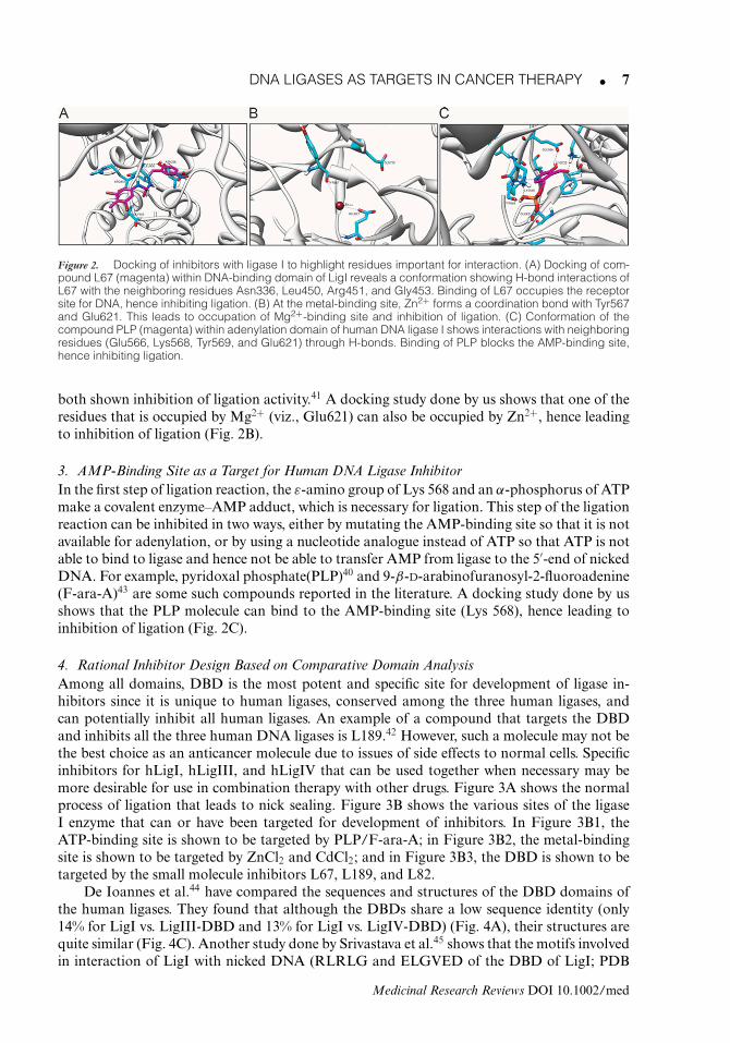
1. DBD as a Target for Human DNA Ligase InhibitorsThe DBD of human ligases plays two important roles in ligase activity. First, it interacts withthe minor groove of DNA, contacting the DNA upstream and downstream of the nick. Second,it directly interacts with the AdD and the OBD of hLigI and stimulates the DNA end-joiningactivity of the catalytic core.38 Thus, if we target the DBD with specific inhibitors, we wouldachieve ligation inhibition. Some small molecule inhibitors have been recently reported thatact on the DBD and inhibit ligation competitively. For example, small molecule inhibitors suchas L67 and L189 compete with DNA for binding to the DBD whereas L82 inhibits ligationby stabilizing a reaction intermediate.39, 42 According to reports in Zhong et al.39 and ourown docking results, it is seen that (Fig. 2A) L67 binds to the DBD of hLigI, hence leadingto inhibition of DNA binding and ligation. These inhibitors are discussed in more detail inSection 4,I (ligase inhibitors from structure-based drug designing). A comparison of the DBDand catalytic domains of human ligases is discussed in Section 3,A.4 to stress upon the specificityissue.
2. Metal-Binding Sites as Targets for Human DNA Ligase InhibitorsMg2+ plays an important role in ligation activity by human DNA ligases. There are two potentialmetal-binding sites present in hLigI. The first is coordinated by Glu720, which stabilizes thenegative charge on the 5′-phosphate of the AMP-leaving group in the ligation reaction. Theother metal-binding site identified in the hLigI-DNA complex is adjacent to the 2′-OH of AMPand is coordinated by the side chain of Glu621, which is close to the 3′-OH of nicked DNA,where it could participate in ligation activity.38 Thus, molecules that can compete with Mg2+
for the metal-binding site will be good inhibitors of ligases. For example, ZnCl2 and CdCl2 have
Medicinal Research Reviews DOI 10.1002/med
DNA LIGASES AS TARGETS IN CANCER THERAPY � 7
Figure 2. Docking of inhibitors with ligase I to highlight residues important for interaction. (A) Docking of com-pound L67 (magenta) within DNA-binding domain of LigI reveals a conformation showing H-bond interactions ofL67 with the neighboring residues Asn336, Leu450, Arg451, and Gly453. Binding of L67 occupies the receptorsite for DNA, hence inhibiting ligation. (B) At the metal-binding site, Zn2+ forms a coordination bond with Tyr567and Glu621. This leads to occupation of Mg2+-binding site and inhibition of ligation. (C) Conformation of thecompound PLP (magenta) within adenylation domain of human DNA ligase I shows interactions with neighboringresidues (Glu566, Lys568, Tyr569, and Glu621) through H-bonds. Binding of PLP blocks the AMP-binding site,hence inhibiting ligation.
both shown inhibition of ligation activity.41 A docking study done by us shows that one of theresidues that is occupied by Mg2+ (viz., Glu621) can also be occupied by Zn2+, hence leadingto inhibition of ligation (Fig. 2B).
3. AMP-Binding Site as a Target for Human DNA Ligase InhibitorIn the first step of ligation reaction, the ε-amino group of Lys 568 and an α-phosphorus of ATPmake a covalent enzyme–AMP adduct, which is necessary for ligation. This step of the ligationreaction can be inhibited in two ways, either by mutating the AMP-binding site so that it is notavailable for adenylation, or by using a nucleotide analogue instead of ATP so that ATP is notable to bind to ligase and hence not be able to transfer AMP from ligase to the 5′-end of nickedDNA. For example, pyridoxal phosphate(PLP)40 and 9-β-D-arabinofuranosyl-2-fluoroadenine(F-ara-A)43 are some such compounds reported in the literature. A docking study done by usshows that the PLP molecule can bind to the AMP-binding site (Lys 568), hence leading toinhibition of ligation (Fig. 2C).
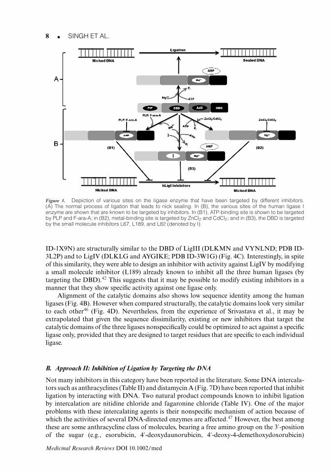
4. Rational Inhibitor Design Based on Comparative Domain AnalysisAmong all domains, DBD is the most potent and specific site for development of ligase in-hibitors since it is unique to human ligases, conserved among the three human ligases, andcan potentially inhibit all human ligases. An example of a compound that targets the DBDand inhibits all the three human DNA ligases is L189.42 However, such a molecule may not bethe best choice as an anticancer molecule due to issues of side effects to normal cells. Specificinhibitors for hLigI, hLigIII, and hLigIV that can be used together when necessary may bemore desirable for use in combination therapy with other drugs. Figure 3A shows the normalprocess of ligation that leads to nick sealing. Figure 3B shows the various sites of the ligaseI enzyme that can or have been targeted for development of inhibitors. In Figure 3B1, theATP-binding site is shown to be targeted by PLP/F-ara-A; in Figure 3B2, the metal-bindingsite is shown to be targeted by ZnCl2 and CdCl2; and in Figure 3B3, the DBD is shown to betargeted by the small molecule inhibitors L67, L189, and L82.
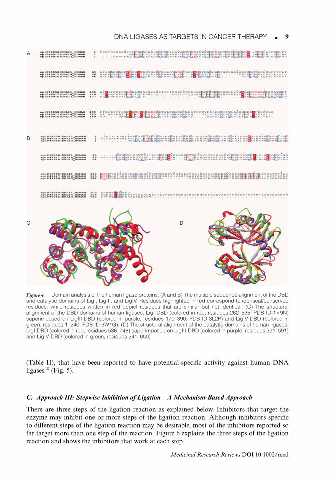
De Ioannes et al.44 have compared the sequences and structures of the DBD domains ofthe human ligases. They found that although the DBDs share a low sequence identity (only14% for LigI vs. LigIII-DBD and 13% for LigI vs. LigIV-DBD) (Fig. 4A), their structures arequite similar (Fig. 4C). Another study done by Srivastava et al.45 shows that the motifs involvedin interaction of LigI with nicked DNA (RLRLG and ELGVED of the DBD of LigI; PDB
Medicinal Research Reviews DOI 10.1002/med
8 � SINGH ET AL.
Figure 3. Depiction of various sites on the ligase enzyme that have been targeted by different inhibitors.(A) The normal process of ligation that leads to nick sealing. In (B), the various sites of the human ligase Ienzyme are shown that are known to be targeted by inhibitors. In (B1), ATP-binding site is shown to be targetedby PLP and F-ara-A; in (B2), metal-binding site is targeted by ZnCl2 and CdCl2; and in (B3), the DBD is targetedby the small molecule inhibitors L67, L189, and L82 (denoted by I).
ID-1X9N) are structurally similar to the DBD of LigIII (DLKMN and VYNLND; PDB ID-3L2P) and to LigIV (DLKLG and AYGIKE; PDB ID-3W1G) (Fig. 4C). Interestingly, in spiteof this similarity, they were able to design an inhibitor with activity against LigIV by modifyinga small molecule inhibitor (L189) already known to inhibit all the three human ligases (bytargeting the DBD).42 This suggests that it may be possible to modify existing inhibitors in amanner that they show specific activity against one ligase only.
Alignment of the catalytic domains also shows low sequence identity among the humanligases (Fig. 4B). However when compared structurally, the catalytic domains look very similarto each other46 (Fig. 4D). Nevertheless, from the experience of Srivastava et al., it may beextrapolated that given the sequence dissimilarity, existing or new inhibitors that target thecatalytic domains of the three ligases nonspecifically could be optimized to act against a specificligase only, provided that they are designed to target residues that are specific to each individualligase.
B. Approach II: Inhibition of Ligation by Targeting the DNA
Not many inhibitors in this category have been reported in the literature. Some DNA intercala-tors such as anthracyclines (Table II) and distamycin A (Fig. 7D) have been reported that inhibitligation by interacting with DNA. Two natural product compounds known to inhibit ligationby intercalation are nitidine chloride and fagaronine chloride (Table IV). One of the majorproblems with these intercalating agents is their nonspecific mechanism of action because ofwhich the activities of several DNA-directed enzymes are affected.47 However, the best amongthese are some anthracycline class of molecules, bearing a free amino group on the 3′-positionof the sugar (e.g., esorubicin, 4′-deoxydaunorubicin, 4′-deoxy-4-demethoxydoxorubicin)
Medicinal Research Reviews DOI 10.1002/med
DNA LIGASES AS TARGETS IN CANCER THERAPY � 9
Figure 4. Domain analysis of the human ligase proteins. (A and B) The multiple sequence alignment of the DBDand catalytic domains of LigI, LigIII, and LigIV. Residues highlighted in red correspond to identical/conservedresidues, while residues written in red depict residues that are similar but not identical. (C) The structuralalignment of the DBD domains of human ligases. LigI-DBD (colored in red, residues 262–535; PDB ID-1×9N)superimposed on LigIII-DBD (colored in purple, residues 170–390; PDB ID-3L2P) and LigIV-DBD (colored ingreen, residues 1–240; PDB ID-3W1G). (D) The structural alignment of the catalytic domains of human ligases.LigI-DBD (colored in red, residues 536–748) superimposed on LigIII-DBD (colored in purple, residues 391–591)and LigIV-DBD (colored in green, residues 241–650).
(Table II), that have been reported to have potential-specific activity against human DNAligases48 (Fig. 5).
C. Approach III: Stepwise Inhibition of Ligation—A Mechanism-Based Approach
There are three steps of the ligation reaction as explained below. Inhibitors that target theenzyme may inhibit one or more steps of the ligation reaction. Although inhibitors specificto different steps of the ligation reaction may be desirable, most of the inhibitors reported sofar target more than one step of the reaction. Figure 6 explains the three steps of the ligationreaction and shows the inhibitors that work at each step.
Medicinal Research Reviews DOI 10.1002/med
10 � SINGH ET AL.
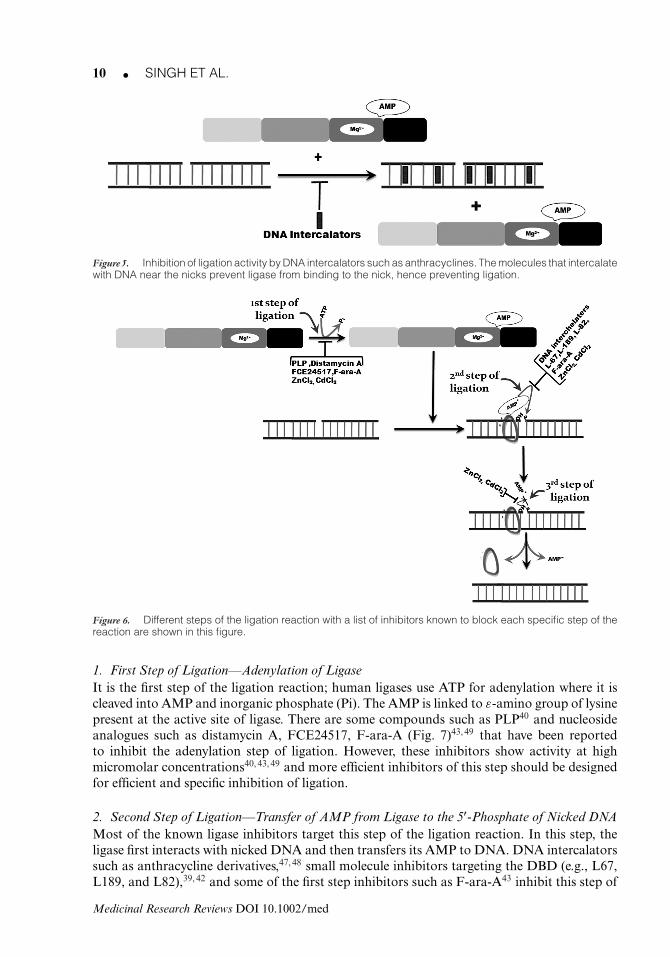
Figure 5. Inhibition of ligation activity by DNA intercalators such as anthracyclines. The molecules that intercalatewith DNA near the nicks prevent ligase from binding to the nick, hence preventing ligation.
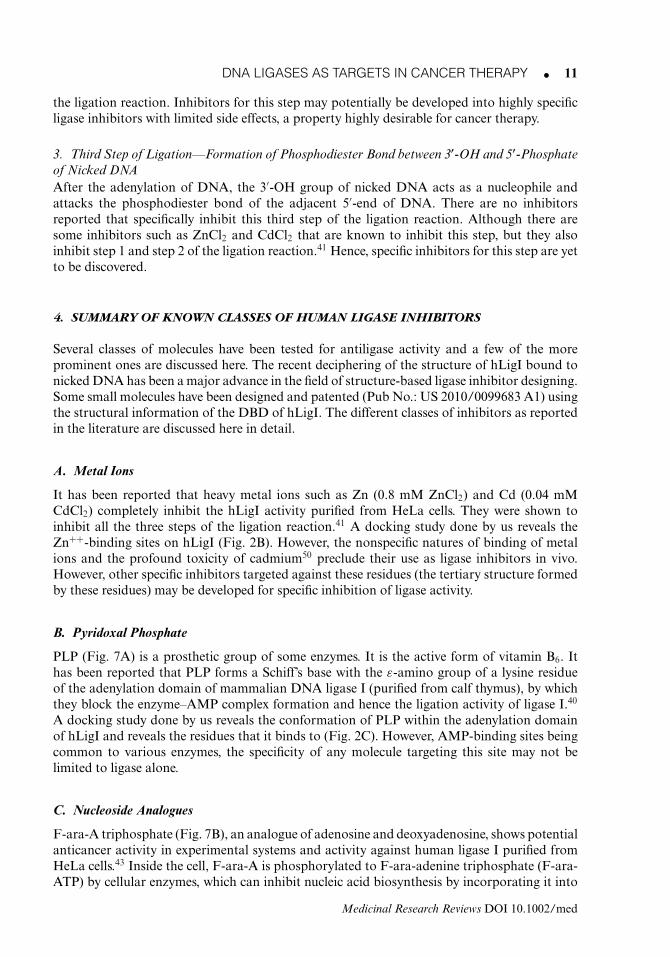
Figure 6. Different steps of the ligation reaction with a list of inhibitors known to block each specific step of thereaction are shown in this figure.
1. First Step of Ligation—Adenylation of LigaseIt is the first step of the ligation reaction; human ligases use ATP for adenylation where it iscleaved into AMP and inorganic phosphate (Pi). The AMP is linked to ε-amino group of lysinepresent at the active site of ligase. There are some compounds such as PLP40 and nucleosideanalogues such as distamycin A, FCE24517, F-ara-A (Fig. 7)43, 49 that have been reportedto inhibit the adenylation step of ligation. However, these inhibitors show activity at highmicromolar concentrations40, 43, 49 and more efficient inhibitors of this step should be designedfor efficient and specific inhibition of ligation.
2. Second Step of Ligation—Transfer of AMP from Ligase to the 5′-Phosphate of Nicked DNAMost of the known ligase inhibitors target this step of the ligation reaction. In this step, theligase first interacts with nicked DNA and then transfers its AMP to DNA. DNA intercalatorssuch as anthracycline derivatives,47, 48 small molecule inhibitors targeting the DBD (e.g., L67,L189, and L82),39, 42 and some of the first step inhibitors such as F-ara-A43 inhibit this step of
Medicinal Research Reviews DOI 10.1002/med
DNA LIGASES AS TARGETS IN CANCER THERAPY � 11
the ligation reaction. Inhibitors for this step may potentially be developed into highly specificligase inhibitors with limited side effects, a property highly desirable for cancer therapy.
3. Third Step of Ligation—Formation of Phosphodiester Bond between 3′-OH and 5′-Phosphateof Nicked DNAAfter the adenylation of DNA, the 3′-OH group of nicked DNA acts as a nucleophile andattacks the phosphodiester bond of the adjacent 5′-end of DNA. There are no inhibitorsreported that specifically inhibit this third step of the ligation reaction. Although there aresome inhibitors such as ZnCl2 and CdCl2 that are known to inhibit this step, but they alsoinhibit step 1 and step 2 of the ligation reaction.41 Hence, specific inhibitors for this step are yetto be discovered.
4. SUMMARY OF KNOWN CLASSES OF HUMAN LIGASE INHIBITORS
Several classes of molecules have been tested for antiligase activity and a few of the moreprominent ones are discussed here. The recent deciphering of the structure of hLigI bound tonicked DNA has been a major advance in the field of structure-based ligase inhibitor designing.Some small molecules have been designed and patented (Pub No.: US 2010/0099683 A1) usingthe structural information of the DBD of hLigI. The different classes of inhibitors as reportedin the literature are discussed here in detail.
A. Metal Ions
It has been reported that heavy metal ions such as Zn (0.8 mM ZnCl2) and Cd (0.04 mMCdCl2) completely inhibit the hLigI activity purified from HeLa cells. They were shown toinhibit all the three steps of the ligation reaction.41 A docking study done by us reveals theZn++-binding sites on hLigI (Fig. 2B). However, the nonspecific natures of binding of metalions and the profound toxicity of cadmium50 preclude their use as ligase inhibitors in vivo.However, other specific inhibitors targeted against these residues (the tertiary structure formedby these residues) may be developed for specific inhibition of ligase activity.
B. Pyridoxal Phosphate
PLP (Fig. 7A) is a prosthetic group of some enzymes. It is the active form of vitamin B6. Ithas been reported that PLP forms a Schiff’s base with the ε-amino group of a lysine residueof the adenylation domain of mammalian DNA ligase I (purified from calf thymus), by whichthey block the enzyme–AMP complex formation and hence the ligation activity of ligase I.40
A docking study done by us reveals the conformation of PLP within the adenylation domainof hLigI and reveals the residues that it binds to (Fig. 2C). However, AMP-binding sites beingcommon to various enzymes, the specificity of any molecule targeting this site may not belimited to ligase alone.
C. Nucleoside Analogues
F-ara-A triphosphate (Fig. 7B), an analogue of adenosine and deoxyadenosine, shows potentialanticancer activity in experimental systems and activity against human ligase I purified fromHeLa cells.43 Inside the cell, F-ara-A is phosphorylated to F-ara-adenine triphosphate (F-ara-ATP) by cellular enzymes, which can inhibit nucleic acid biosynthesis by incorporating it into
Medicinal Research Reviews DOI 10.1002/med
12 � SINGH ET AL.
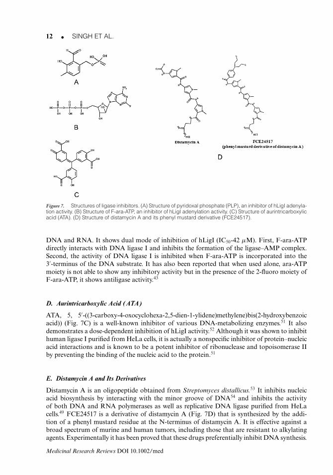
Figure 7. Structures of ligase inhibitors. (A) Structure of pyridoxal phosphate (PLP), an inhibitor of hLigI adenyla-tion activity. (B) Structure of F-ara-ATP, an inhibitor of hLigI adenylation activity. (C) Structure of aurintricarboxylicacid (ATA). (D) Structure of distamycin A and its phenyl mustard derivative (FCE24517).
DNA and RNA. It shows dual mode of inhibition of hLigI (IC50-42 μM). First, F-ara-ATPdirectly interacts with DNA ligase I and inhibits the formation of the ligase–AMP complex.Second, the activity of DNA ligase I is inhibited when F-ara-ATP is incorporated into the3′-terminus of the DNA substrate. It has also been reported that when used alone, ara-ATPmoiety is not able to show any inhibitory activity but in the presence of the 2-fluoro moiety ofF-ara-ATP, it shows antiligase activity.43
D. Aurintricarboxylic Acid (ATA)
ATA, 5, 5′-((3-carboxy-4-oxocyclohexa-2,5-dien-1-ylidene)methylene)bis(2-hydroxybenzoicacid)) (Fig. 7C) is a well-known inhibitor of various DNA-metabolizing enzymes.51 It alsodemonstrates a dose-dependent inhibition of hLigI activity.52 Although it was shown to inhibithuman ligase I purified from HeLa cells, it is actually a nonspecific inhibitor of protein–nucleicacid interactions and is known to be a potent inhibitor of ribonuclease and topoisomerase IIby preventing the binding of the nucleic acid to the protein.51
E. Distamycin A and Its Derivatives
Distamycin A is an oligopeptide obtained from Streptomyces distallicus.53 It inhibits nucleicacid biosynthesis by interacting with the minor groove of DNA54 and inhibits the activityof both DNA and RNA polymerases as well as replicative DNA ligase purified from HeLacells.49 FCE24517 is a derivative of distamycin A (Fig. 7D) that is synthesized by the addi-tion of a phenyl mustard residue at the N-terminus of distamycin A. It is effective against abroad spectrum of murine and human tumors, including those that are resistant to alkylatingagents. Experimentally it has been proved that these drugs preferentially inhibit DNA synthesis.
Medicinal Research Reviews DOI 10.1002/med
DNA LIGASES AS TARGETS IN CANCER THERAPY � 13
Although these two drugs interfere with the action of all the proteins involved in DNA replica-tion, but the adenylation step of DNA ligation is most sensitive to these two distamycins.49
It has also been reported that the DNA intercalating or DNA-binding drugs interferingwith the minor groove of DNA can inhibit DNA ligases,49, 55 suggesting the possibility ofdetermining the mechanism of inhibition of DNA ligases for distamycin A and FCE24517by assaying their effects on different steps of the ligation reaction. These molecules have thepotential to be developed into antiligase molecules with the proper kind of tweaking andmolecular dynamics studies. Interestingly, in our own screening for ligase inhibitors, we havefound a lipidated natural product derivative belonging to the glutarimide class that targets boththe DBD domain of hLigI as well as the minor groove of DNA and is a potent inhibitor ofhLigI. The molecule has shown selective anticancer activity in certain human cancer cell linesand seems to have the potential for drug development.
F. Anthracycline and Its Derivatives
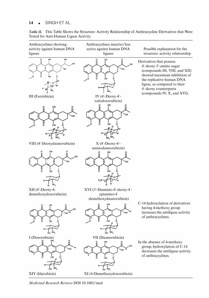
Anthracyclines are an important class of antibiotics derived from Streptomyces. They are alsoa class of drugs used in cancer chemotherapy [e.g., doxorubicin (adriamycin) and daunomycin(daunorubicin)]. Nearly a hundred derivatives have been synthesized to try and reduce theirtoxic effect on cells and to improve their anticancer properties. The biological properties ofthese compounds are generally attributed to the intercalation and reversible binding to DNA,although stabilization of the DNA topoisomerase II-DNA complex may be an importantfactor in the cytotoxicity.48 It is still unclear how anthracyclines attain their varied biologicalactivities and this characteristic may be attributed to activity against multiple targets. GiovanniCiarrocchi et al.48 found that DNA ligases are resistant to several intercalating anthracyclines,but very sensitive to ones bearing a free amino group on the 3′-position of the sugar. Thestructure–activity relationship (SAR) studies of various anthracycline derivatives done by themrevealed that maximal inhibition of DNA-joining activity require a 4′-deoxy-3′-amino sugar(compounds III, VIII, and XII; Table II).
Initially, 4′-deoxy derivatives (III, IV, VIII, X, XII, and XVI) were tested for their antiligaseactivity on replicative DNA ligase purified from HeLa cells. It was found that the derivativesthat possess 4′-deoxy-3′-amino sugar (compound III, VIII, and XII) showed maximum inhibi-tion of human DNA ligase as compared to their 4′-hydroxy counterparts (Table II).48, 56 Otherderivatives having 4′-deoxy-4′-amino sugar (compound XVI with amino group present on4′-epi position) show very low activity. If the 4′-deoxy-3′-amino sugar is replaced by3′,4′-diamino sugar (compound X), this shows lesser activity than 4′-deoxy-3′-amino sugarderivatives, but similar activity as compared with 4′-hydroxy-3′-amino sugar counterparts(daunomycin, VIII) (Table II).
However, it is clear from the studies of Giovanni Ciarrocchi et al.48 that anthracyclines arepoor inhibitors of the first step of the DNA-joining reaction and that inhibition of DNA-joiningactivity is obtained via interaction with the substrate (DNA), as proposed for some other DNAbinders such as ethidium bromide. However, although DNA intercalation is necessary, it isnot in itself sufficient for the activity against human replicative DNA ligase. Crystallographicstudies revealed that during DNA intercalation, while the anthracycline aglycone-chromophoremoiety lies between base pairs perpendicular to the axis of the double helix, the sugar residuehangs in the minor groove with the 3′-amino group exposed.48 Although it is still unclear whatkind of interactions occur between the hanging amino group and the ligase, studies suggestthat the interaction must be very specific.48 It was also found that closely related anthracyclineswith different sensitivity to ligases did not show the same variations to the stability of the DNAtopoisomerase II-DNA complex. If this hypothesis is correct then it should be possible to findsome anthracyclines that have specific activity for ligases.48
Medicinal Research Reviews DOI 10.1002/med
14 � SINGH ET AL.
Table II. This Table Shows the Structure–Activity Relationship of Anthracycline Derivatives that WereTested for Anti-Human Ligase Activity
Anthracyclines showingactivity against human DNAligases
Anthracyclines inactive/lessactive against human DNA
ligasesPossible explanation for thestructure–activity relationship
Derivatives that possess4′-deoxy-3′-amino sugar(compounds III, VIII, and XII)showed maximum inhibition ofthe replicative human DNAligase, as compared to their4′-deoxy counterparts(compounds IV, X, and XVI).
III (Esorubicin) IV (4′-Deoxy-4′-iododoxorubicin)
VIII (4′-Deoxydaunorubicin) X (4′-Deoxy-4′-aminodaunorubicin)
XII (4′-Deoxy-4-demethoxydoxorubicin)
XVI (3′-Deamino-4′-deoxy-4′-epiamino-4
-demethoxydaunorubicin)C-14 hydroxylation of derivatives
having 4-methoxy groupincreases the antiligase activityof anthracyclines.
I (Doxorubicin) VII (Daunorubicin)In the absence of 4-methoxy
group, hydroxylation of C-14decreases the antiligase activityof anthracyclines.
XIV (Idarubicin) XI (4-Demethoxydoxorubicin)
Medicinal Research Reviews DOI 10.1002/med
DNA LIGASES AS TARGETS IN CANCER THERAPY � 15
Table II also lists other derivatives of anthracyclines that were created by modifications inthe C-14 position of the carbon chain, by the addition of hydroxyl group or methyl group at thatposition. The addition of hydroxyl group at C-14 position of daunorubicin (VII) creates a newmolecule doxorubicin (I) that is more active than daunorubicin. This suggests that hydroxylationpromotes the inhibitory activity but 4-demethoxy molecule carrying same modifications (XI,XIV) shows negative effect of hydroxylation, that is, decreases the activity of the inhibitors.When the new analogues were created only by 4′-demethoxy modification like the 4′-demethoxy-doxorubicin (XI), differences were seen in their activity. Doxorubicin (I) was more active than its4′-demethoxy counterpart whereas daunorubicin (VII) was less effective than its 4′-demethoxyderivative (XIV).
G. Arylamino Compounds
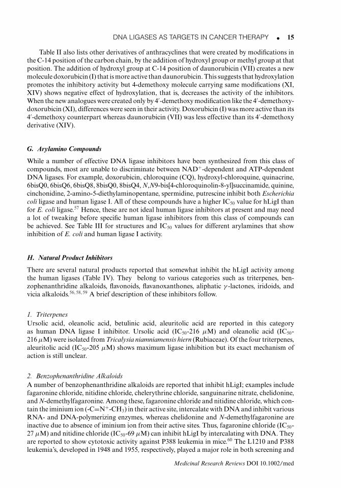
While a number of effective DNA ligase inhibitors have been synthesized from this class ofcompounds, most are unable to discriminate between NAD+-dependent and ATP-dependentDNA ligases. For example, doxorubicin, chloroquine (CQ), hydroxyl-chloroquine, quinacrine,6bisQ0, 6bisQ6, 6bisQ8, 8bisQ0, 8bisQ4, N,N9-bis[4-chloroquinolin-8-yl]succinamide, quinine,cinchonidine, 2-amino-5-diethylaminopentane, spermidine, putrescine inhibit both Escherichiacoli ligase and human ligase I. All of these compounds have a higher IC50 value for hLigI thanfor E. coli ligase.57 Hence, these are not ideal human ligase inhibitors at present and may needa lot of tweaking before specific human ligase inhibitors from this class of compounds canbe achieved. See Table III for structures and IC50 values for different arylamines that showinhibition of E. coli and human ligase I activity.
H. Natural Product Inhibitors
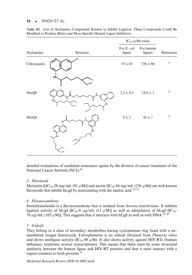
There are several natural products reported that somewhat inhibit the hLigI activity amongthe human ligases (Table IV). They belong to various categories such as triterpenes, ben-zophenanthridine alkaloids, flavonoids, flavanoxanthones, aliphatic γ -lactones, iridoids, andvicia alkaloids.56, 58, 59 A brief description of these inhibitors follow.
1. TriterpenesUrsolic acid, oleanolic acid, betulinic acid, aleuritolic acid are reported in this categoryas human DNA ligase I inhibitor. Ursolic acid (IC50-216 μM) and oleanolic acid (IC50-216 μM) were isolated from Tricalysia niamniamensis hiern (Rubiaceae). Of the four triterpenes,aleuritolic acid (IC50-205 μM) shows maximum ligase inhibition but its exact mechanism ofaction is still unclear.
2. Benzophenanthridine AlkaloidsA number of benzophenanthridine alkaloids are reported that inhibit hLigI; examples includefagaronine chloride, nitidine chloride, chelerythrine chloride, sanguinarine nitrate, chelidonine,and N-demethylfagaronine. Among these, fagaronine chloride and nitidine chloride, which con-tain the iminium ion (-C=N+-CH3) in their active site, intercalate with DNA and inhibit variousRNA- and DNA-polymerizing enzymes, whereas chelidonine and N-demethylfagaronine areinactive due to absence of iminium ion from their active sites. Thus, fagaronine chloride (IC50-27 μM) and nitidine chloride (IC50-69 μM) can inhibit hLigI by intercalating with DNA. Theyare reported to show cytotoxic activity against P388 leukemia in mice.60 The L1210 and P388leukemia’s, developed in 1948 and 1955, respectively, played a major role in both screening and
Medicinal Research Reviews DOI 10.1002/med
16 � SINGH ET AL.
Table III. List of Arylamino Compounds Known to Inhibit Ligation. These Compounds Could BeModified to Produce Better and More Specific Human Ligase Inhibitors
IC50 (μM) value
For E. coli For humanArylamines Structure ligase ligases References
Chloroquine 53 ± 63 720 ± 80 57
6bisQ6 2.2 ± 0.3 14.8 ± 3 57
8bisQ4 9 ± 2 43 ± 7 57
detailed evaluations of candidate anticancer agents by the division of cancer treatment of theNational Cancer Institute (NCI).60
3. FlavonoidsMyricetin [(IC50-28 mg/mL (91 μM)] and morin [IC50-68 mg/mL (236 μM)] are well-knownflavonoids that inhibit hLigI by intercalating with the nucleic acid.56, 59
4. FlavanoxanthoneSwertifrancheside is a flavonoxanthone that is isolated from Swertia franchetiana. It inhibitsligation activity of hLigI [IC50-8 μg/mL (11 μM)] as well as adenylation of hLigI [IC50-76 μg/mL (105 μM)]. This suggests that it interacts with hLigI as well as with DNA.56, 59
5. IridoidsThey belong to a class of secondary metabolites having cyclopentane ring fused with a six-membered oxygen heterocycle. Fulvoplumerin is an iridoid obtained from Plumeria rubraand shows antiligase activity (IC50-88 μM). It also shows activity against HIV-RTs (humandeficiency syndrome reverse transcriptases). This means that there must be some structuralsimilarity between the human ligase and HIV-RT proteins and that it must interact with aregion common to both proteins.59
Medicinal Research Reviews DOI 10.1002/med
DNA LIGASES AS TARGETS IN CANCER THERAPY � 17
Table IV. List of Different Categories of Known Natural Product Human Ligase Inhibitors
Natural product Examples with IC50 (μM) Possible explanationinhibitor classes chemical structures values for their activity References
Triterpenes 216 Triterpenes directly interactwith ligases, but the exactmechanism of inhibition(possible interaction andstructural features) is notknown.
56, 59
Oleanolic acid
216
Ursolic acid
205
Aleuritolic acid
Benzo-phenanthridinealkaloids
Notavailable
In the presence of iminium ion,nitidine chloride andfagaronine chlorideintercalate with DNA andinhibit DNA ligases.However, N-demethyl-fagaronine and chelidonineare inactive up to 200 μMconcentration in the absenceof iminium ion.
56, 59, 60
N-Demethylfagaronine
Notavailable
Chelidonine
27
Fagaronine chloride
69
Nitidine chloride
Medicinal Research Reviews DOI 10.1002/med
18 � SINGH ET AL.
Table IV. Continued
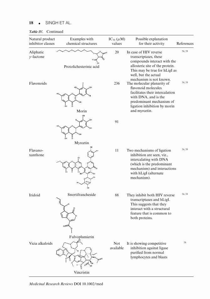
Natural product Examples with IC50 (μM) Possible explanationinhibitor classes chemical structures values for their activity References
Aliphaticγ -lactone
20 In case of HIV reversetranscriptases, thesecompounds interact with theallosteric site of the protein.This may be true for hLigI aswell, but the actualmechanism is not known.
56, 59
Protolichesterinic acid
Flavonoids 236 The molecular planarity offlavonoid moleculesfacilitates their intercalationwith DNA, and is thepredominant mechanism ofligation inhibition by morinand myrcetin.
56, 59
Morin
91
Myrcetin
Flavano-xanthone
11 Two mechanisms of ligationinhibition are seen, viz.,intercalating with DNA(which is the predominantmechanism) and interactionswith hLigI (alternatemechanism).
56, 59
SwertifranchesideIridoid 88 They inhibit both HIV reversetranscriptases and hLigI.This suggests that theyinteract with a structuralfeature that is common toboth proteins.
56, 59
Fulvoplumierin
Vicia alkaloids Notavailable
It is showing competitiveinhibition against ligasepurified from normallymphocytes and blasts
58
Vincristin
Medicinal Research Reviews DOI 10.1002/med
DNA LIGASES AS TARGETS IN CANCER THERAPY � 19
6. Aliphatic γ -LactoneProtolichesterinic acid is an aliphatic α-methylase-γ -lactone obtained from the lichen Cetrariaislandica. It inhibits hLigI activity at an IC50 value of 20 μM and blocks its adenylation at amuch higher concentration (387 μM).59 Although the molecule binds to ligase, it also bindsto HIV-1 RT, highlighting its nonspecific nature of binding. The molecule does not interactor intercalate with DNA and should therefore be potentially nonmutagenic in nature. Themolecule holds promise if it can be tweaked to specifically target hLigI or the HIV-1 RT foruse in anticancer or antiretroviral therapy, respectively.
7. Vicia AlkaloidsThey are obtained from Catharanthus roseus. Various vicia alkaloids such as vincristine, vin-blastin, and vindesine have been tested for antiligase activity. Only vincristine (at 100 μMconcentration) showed activity against ligase purified from thymocytes and normal lympho-cytes, while having little or no effect on the enzyme purified from lymphoblast cells.58
Although several categories of natural products exhibit ligase inhibition, compounds show-ing specific inhibition at low micro- or nanomolar concentrations still remain elusive. We suggestthat natural products with modified synthetic adages may be developed to achieve the requiredparameters of specificity, activity, stability (along with high bioavailability and low toxicity),and other features that are essential for drug-like molecules. This is a relatively unexplored areawith a lot of potential, although unfortunately, an area where not much research is known tobe happening.
I. Ligase Inhibitors from Structure-Based Drug Designing
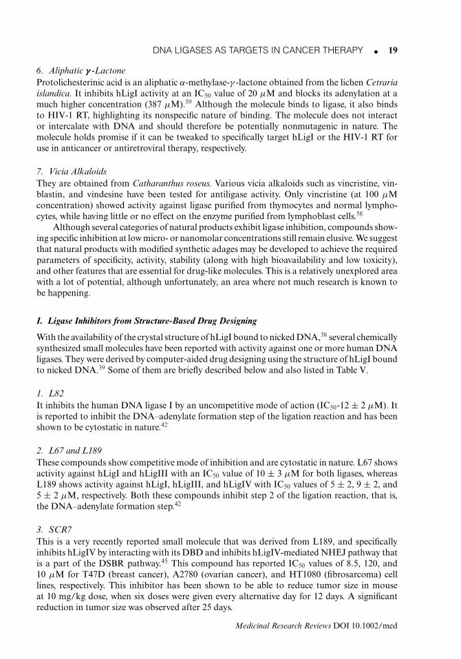
With the availability of the crystal structure of hLigI bound to nicked DNA,38 several chemicallysynthesized small molecules have been reported with activity against one or more human DNAligases. They were derived by computer-aided drug designing using the structure of hLigI boundto nicked DNA.39 Some of them are briefly described below and also listed in Table V.
1. L82It inhibits the human DNA ligase I by an uncompetitive mode of action (IC50-12 ± 2 μM). Itis reported to inhibit the DNA–adenylate formation step of the ligation reaction and has beenshown to be cytostatic in nature.42
2. L67 and L189These compounds show competitive mode of inhibition and are cytostatic in nature. L67 showsactivity against hLigI and hLigIII with an IC50 value of 10 ± 3 μM for both ligases, whereasL189 shows activity against hLigI, hLigIII, and hLigIV with IC50 values of 5 ± 2, 9 ± 2, and5 ± 2 μM, respectively. Both these compounds inhibit step 2 of the ligation reaction, that is,the DNA–adenylate formation step.42
3. SCR7This is a very recently reported small molecule that was derived from L189, and specificallyinhibits hLigIV by interacting with its DBD and inhibits hLigIV-mediated NHEJ pathway thatis a part of the DSBR pathway.45 This compound has reported IC50 values of 8.5, 120, and10 μM for T47D (breast cancer), A2780 (ovarian cancer), and HT1080 (fibrosarcoma) celllines, respectively. This inhibitor has been shown to be able to reduce tumor size in mouseat 10 mg/kg dose, when six doses were given every alternative day for 12 days. A significantreduction in tumor size was observed after 25 days.
Medicinal Research Reviews DOI 10.1002/med
20 � SINGH ET AL.
Table V. List of Small Molecule Inhibitors Derived from Rational Structure-Based Designing for HumanLigase Inhibitors after the Elucidation of hLigI-Nicked DNA Crystal Structure.39, 42 The CompoundsLI-01 and LI-02 were However Found from Random Screening of a Chemical Library from Micro-Source Discovery Systems by Sun et al.52
IC50 (μM)value (in
human cancerCompound Structure cell lines) Mechanism of action Reference
L67 10 ± 3 for hLigIand hLigIII
It inhibits the secondstep of ligation byhLigI and hLigIII in acompetitive manner.
42
L82 12 ± 2 for hLigI It inhibits second step ofligation by hLigI in anuncompetitivemanner.
42
L189 • 5 ± 2 for hLigIand hLigIV.
• 9 ± 2 forhLigIII.
It inhibits the secondstep of ligation byhLigI, hLigIII, andhLigIV in acompetitive manner.
42
SCR7 8.5 It interacts with theDBD of hLigIV andinhibits its activitycompetitively.
45
LI-01 Not available It inhibits activity ofhLigI by an unknownmechanism.
52
LI-02 Not available It inhibits activity ofhLigI by an unknownmechanism.
52
Medicinal Research Reviews DOI 10.1002/med
DNA LIGASES AS TARGETS IN CANCER THERAPY � 21
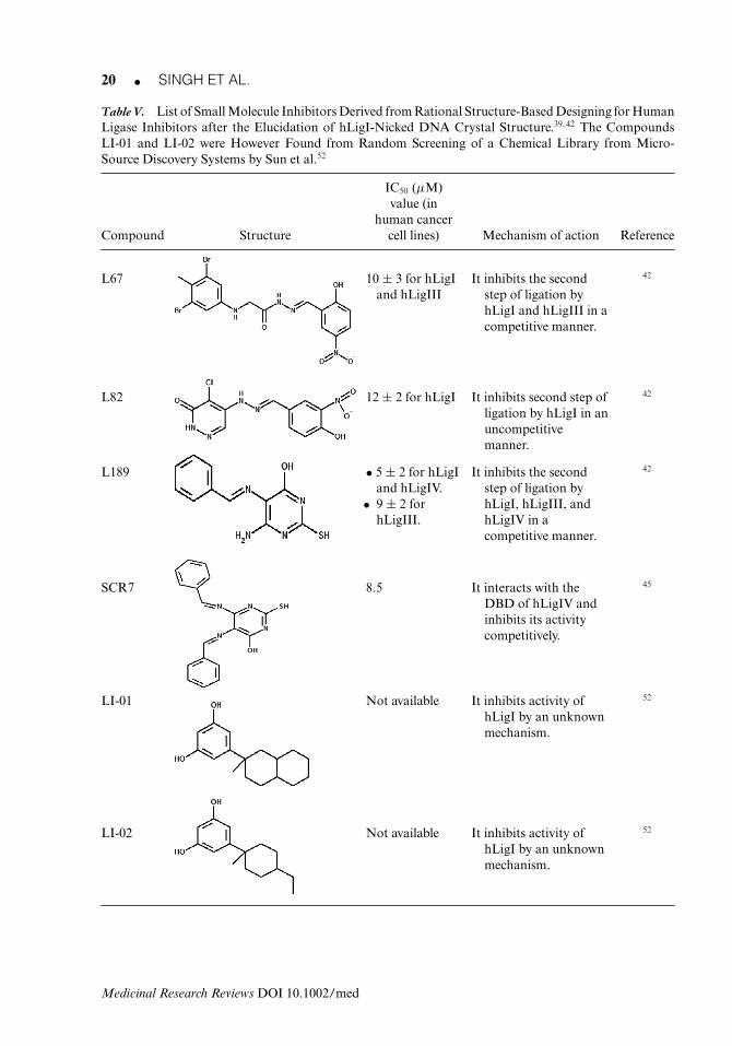
Figure 8. Schematic representation of the potential use of ligase inhibitors for successful combination therapyin treating cancers (SSBs, single-strand breaks; DSBs, double-strand breaks; SSBR, single-strand break repair;DSBR, double-strand break repair).
J. Ligase Inhibitors Discovered from Random Screening
Apart from the above, some molecules such as LI-01 (2-(3,5-dihydroxyphenyl)-2 methyldecalin)and LI-02 (2-(3,5-dihydroxyphenyl)-4-ethyl-1-methylcyclohexane) show hLigI inhibition. Thesecompounds were found from a random screening of chemical libraries from Micro-SourceDiscovery Systems.52 LI-01 was found to be more active than LI-02 but the exact IC50 valuesare not known.
5. STRATEGIES IN CANCER THERAPY AND THE FUTURE OF LIGASE INHIBITORS
The current state of knowledge clearly shows that human DNA ligases are crucial enzymesfor DNA replication and repair pathways. It has been reported that hLigI is highly expressedin cancer cells.14, 61 Hence they are potentially good targets for development of anticancerdrugs. However, there may be problems of specificity and drug resistance, problems that can beovercome by selective targeting and combination therapy of drugs, respectively (Fig. 8). Herewe discuss how ligase inhibitors may be useful in combination cancer therapy and how hLigIcan be targeted for development of more specific inhibitors for future therapy.
A. Ligase Inhibitors in Combination Therapy
Ligase inhibitors should work well in combination with DNA damaging and intercalationagents that can first cause breaks in the DNA and then help sustain the damage throughinhibition of ligation. Hence, radiation therapy to induce ss and ds breaks, followed by antiligasetherapy should be more effective than radiation therapy alone where such damages may berepaired by the DNA repair processes (involving ligases I, III, and IV). In fact, a combinationof molecules, or a single molecule that can inhibit all three ligases (and hence inhibit all DNArepair processes), may be used for a limited time to ensure cancer cell killing and then withdrawnfrom therapy to avoid too much damage to normal cells over the long term. Again, DNA
Medicinal Research Reviews DOI 10.1002/med
22 � SINGH ET AL.
intercalators such as doxorubicin and daunorubicin that are already in therapy and can inhibitligation by physical occlusion of ligases (from binding the DNA), should become more effectivewhen used in combination with other ligase inhibitors that can take care of any residual ligationactivity.
Among the recent lot of promising anticancer drugs, several poly-ADP ribose polymerase(PARP) inhibitors were recently in the second and third phase of clinical trials being conductedby Astra-Zeneca (olaparib), Sanofi-Aventis (iniparib), Pfizer (PF-01367338), and others.62 It isunderstood that inhibiting PARP-1 leads to the accumulation of ss DNA lesions, which duringDNA replication can degenerate to form ds breaks. When PARP-1 is blocked, backup DNArecombination mechanisms involving BRCA1/2 (that are critical players in the homologous-recombination repair pathway), kick in. PARP inhibition in BRCA-defective cancer cells wouldtherefore lead to massive genetic damage and cell death (a synthetic lethal effect).62 However,recent reports suggest that PARP-1 inhibitors have failed to considerably prolong lifespan inthe third phase of clinical trials.62 Given the fact that the strand breaks left behind by PARPinhibitors need to be sealed by ligases for the cells to survive, we believe that human ligaseinhibitors (specially hLigI inhibitors) would be very effective when used in combination withPARP-1 inhibitors in keeping DNA damage levels (strand breaks) high enough to inducecell death. PARP inhibitors, when used alone, work best in BRCA-negative cells (or in cellswith altered DNA damage response),63 whereas normal tissue with at least one functionalBRCA1 or BRCA2 allele will be able to repair its DNA by alternate repair mechanisms andsurvive. At the same time, if ligase inhibitors are used simultaneously, then the alternate mech-anisms will be halted for want of ligation, hence blocking the complete repair of damagedDNA. This will perhaps lead to a faster accumulation of DNA damage and induce apoptosis(Fig. 8).63, 64
B. Specific Targeting of Ligase Inhibitors
As with all chemotherapeutic agents, targeting of ligase inhibitors to sites of tumors or tocancer cells dissipated in the body will be a major challenge and will depend on the discoveryof specific identifiers of cancer cells that can be attached to the delivery modules. We suggestthat chemotherapeutic agents, including ligase inhibitors can be packaged inside lipid micellesthat carry target identifiers if possible. Lipid-based drug delivery is a very well establishedfield and pharmacists/chemists in our group are currently working on ways to molecularlytarget lipids for drug delivery. The idea is that once these micelles reach the targeted cells, theycan fuse with the cell membrane and release their load inside the cells, hence delivering thedrugs.65
However, even in the absence of specific targeting, issues of side effects for ligase inhibitorsshould be minimal since several fast replicating cancer cells have high levels of hLigI comparedto normal cells.14 Also, most normal adult cells in the body do not replicate at all or replicateonly sporadically (e.g., hematopoietic stem cells). Thus, the high proliferative index of cancercells compared to the sporadic nature of normal stem cell multiplication should make anantiligase I strategy clinically viable. This combination of specificity and selectivity (perhapsin combination with targeted radiation therapy to induce DNA strand breaks in cancer cells)could become successful in anticancer therapy.
For issues of specific targeting of inhibitors to individual ligases, structure-based ap-proaches highlighted by recent publications39, 45 indicate that it may be possible to designinhibitors with activity against an individual ligase (e.g., L82 and SCR7 against hLigI andhLigIV, respectively), or against a combination of ligases (e.g., L67 against hLigI and hLigIIIand L189 against all three ligases). Our group is presently engaged in design and synthesis ofspecific ligase inhibitors.
Medicinal Research Reviews DOI 10.1002/med
DNA LIGASES AS TARGETS IN CANCER THERAPY � 23
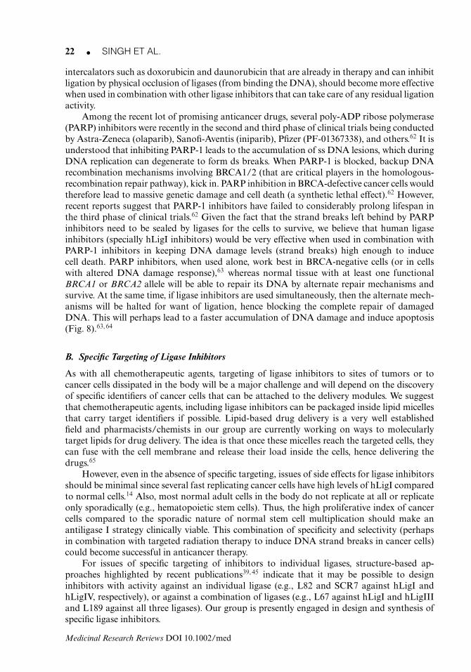
Figure 9. Schematic representation of hLigI domain structure showing PIP box, NLS, and Cy-motifs as potentialnew targets for development of specific hLigI inhibitors.
C. New Targets for Ligase Inhibitors
1. The PIP BoxIn human DNA ligase I, the first 19 amino acids from the N-terminal region acts as a pro-liferating cell nuclear antigen (PCNA)-binding motif, also known as the PIP box (Fig. 9).It is unique among ligases in being found only in the ligase I sequence. Mutational studiesshow that if phenylalanine residues at the eighth and ninth position are replaced by alanine,PCNA binding with hLigI is abolished.66 It is known that interaction of hLigI with PCNAis necessary for efficient ligation in vivo.9 Hence, it is expected that if we can block the PIPbox of ligase I by designing an inhibitor against it, it will block hLigI–PCNA interaction,and lead to a considerable reduction in ligation in vivo. Currently, no synthetic or natu-ral product inhibitors have been reported against this region and it could potentially be anexcellent target for the development of specific inhibitors against hLigI. As a proof of con-cept, it has recently been reported67 that T2-amino alcohol (T2AA; IC50-1 μM), which isa derivative of 3,3′,5-triiodothyronine (T3, thyroid hormone), is a synthetic compound thatinhibits PCNA binding with PIP-box sequence peptide by competing for the same bindingsite. The inhibitor also inhibits PCNA binding with interaction partners such as p21 andPol δ, leading to de novo DNA synthesis inhibition and cell cycle arrest in the S phase. Thisresults in inhibition of growth and replication of cancer cells.67
2. Cyclin-Binding Motif (Cy Motif) and Nuclear Localization Signal (NLS Motif)At the C-terminal region of hLigI, cyclin-binding site (RRQL) is found at amino acid position678-681. This region plays two important roles. First, it acts as a cyclin A binding site thathelps phosphorylation of Ser51 (at G1/S phase), Ser76 (at G2/M phase), and Ser91 (at G1/Sphase). These phosphorylation events play a very important role in interaction of hLigI withother proteins involved in DNA replication such as PCNA and RFC.68, 69 Second, deletionstudies of the nuclear localization sequences (NLS) and Cy-motif revealed that binding ofcyclin A at Cy-motif is enough for nuclear translocation of hLigI from the cytosol (in theG1/S phase), even in the absence of the NLS.69 Hence, the Cy-motif could be an excellenttarget for specific inhibition of hLigI by designing peptides mimicking the Cy-motif bindingregion of cyclin A and thus restricting the binding of cyclin A to hLigI. This will restrict thephosphorylation of hLigI and its interaction with partner proteins, thus limiting its activityin vivo.69 The NLS is another obvious target for peptide inhibitors as binding of peptides tothis region will preclude the translocation of hLigI from the cytoplasm into the nucleus, thuslimiting the interaction of hLigI with either its interaction partners (PCNA, RFC, etc.) or itssubstrate (DNA), both of which are present in the nucleus (see Fig. 9).
Medicinal Research Reviews DOI 10.1002/med

24 � SINGH ET AL.
6. CONCLUDING REMARKS
Given the fact that combination therapies are more effective for cancer treatment than singleagent therapy,70 we believe that adding ligase inhibitors to the existing or new arsenal ofanticancer (or antibiotic) compounds might prove to be the missing link in broad-spectrumanticancer (or antibiotic) therapy. Having said that, a lot of research still needs to go intodiscovering or designing ligase inhibitors that are specific and effective enough to be used intherapy.
ACKNOWLEDGMENTS
The authors thank the Department of Science and Technology (DST), India (grantno. SB/FT/LS-163/2012), Department of Biotechnology (DBT), India (grant no.BT/PR6421/GBD/27/436/2012), and the Council for Scientific and Industrial Research(CSIR), India (funds from BSC0106) for funding and support in writing this review. D.K.S.,S.C., S.K., M.S., and A.D. thank CSIR for their JRF’s (Junior Research Fellowships). We ac-knowledge Dr. M.I. Siddiqi, Scientist, Molecular and Structural Biology Division, CSIR-CDRIfor his comments and suggestions in improving the manuscript. The CSIR-CDRI institutionalnumber allocated for this manuscript is 8488.
REFERENCES
1. Lehman IR. DNA ligase: Structure, mechanism, and function. Science 1974;186(4166):790–797.
2. Pascal JM. DNA and RNA ligases: Structural variations and shared mechanisms. Curr Opin StructBiol 2008;18(1):96–105.
3. Wilkinson A, Day J, Bowater R. Bacterial DNA ligases. Mol Microbiol 2001;40(6):1241–1248.
4. Moser J, Kool H, Giakzidis I, Caldecott K, Mullenders LH, Fousteri MI. Sealing of chromosomalDNA nicks during nucleotide excision repair requires XRCC1 and DNA ligase III alpha in a cell-cycle-specific manner. Mol Cell 2007;27(2):311–323.
5. Robertson AB, Klungland A, Rognes T, Leiros I. DNA repair in mammalian cells: Base excisionrepair: The long and short of it. Cell Mol Life Sci 2009;66(6):981–993.
6. Dianov GL. Base excision repair targets for cancer therapy. Am J Cancer Res 2011;1(7):845–851.
7. Wilson TE, Grawunder U, Lieber MR. Yeast DNA ligase IV mediates non-homologous DNA endjoining. Nature 1997;388(6641):495–498.
8. Liang L, Deng L, Nguyen SC, Zhao X, Maulion CD, Shao C, Tischfield JA. Human DNA ligasesI and III, but not ligase IV, are required for microhomology-mediated end joining of DNA double-strand breaks. Nucleic Acids Res 2008;36(10):3297–3310.
9. Ellenberger T, Tomkinson AE. Eukaryotic DNA ligases: Structural and functional insights. AnnuRev Biochem 2008;77:313–338.
10. Chrzanowska KH, Gregorek H, Dembowska-Baginska B, Kalina MA, Digweed M. Nijmegen break-age syndrome (NBS). Orphanet J Rare Dis 2012;7:13.
11. Noguiez P, Barnes DE, Mohrenweiser HW, Lindahl T. Structure of the human DNA ligase I gene.Nucleic Acids Res 1992;20(15):3845–3850.
12. Teraoka H, Sawai M, Yamamoto K, Tsukada K. DNA ligase I mRNA and enzyme levels in humanhematopoietic cells under dimethyl sulfoxide-induced growth-arrest and differentiation. Biochem Int1992;26(5):963–971.
Medicinal Research Reviews DOI 10.1002/med
DNA LIGASES AS TARGETS IN CANCER THERAPY � 25
13. Jessberger R, Schar P, Robins P, Ferrari E, Riwar B, Hubscher U. Regulation of DNA metabolicenzymes upon induction of preB cell development and V(D)J recombination: Up-regulation of DNApolymerase delta. Nucleic Acids Res 1997;25(2):289–296.
14. Sun D, Urrabaz R, Nguyen M, Marty J, Stringer S, Cruz E, Medina-Gundrum L, Weitman S.Elevated expression of DNA ligase I in human cancers. Clin Cancer Res 2001;7(12):4143–4148.
15. Barnes DE, Tomkinson AE, Lehmann AR, Webster AD, Lindahl T. Mutations in the DNA ligaseI gene of an individual with immunodeficiencies and cellular hypersensitivity to DNA-damagingagents. Cell 1992;69(3):495–503.
16. Webster AD, Barnes DE, Arlett CF, Lehmann AR, Lindahl T. Growth retardation and immunode-ficiency in a patient with mutations in the DNA ligase I gene. Lancet 1992;339(8808):1508–1509.
17. Prigent C, Satoh MS, Daly G, Barnes DE, Lindahl T. Aberrant DNA repair and DNA replicationdue to an inherited enzymatic defect in human DNA ligase I. Mol Cell Biol 1994;14(1):310–317.
18. Hsieh CL, Arlett CF, Lieber MR. V(D)J recombination in ataxia telangiectasia, Bloom’s syndrome,and a DNA ligase I-associated immunodeficiency disorder. J Biol Chem 1993;268(27):20105–20109.
19. Aboussekhra A, Biggerstaff M, Shivji MK, Vilpo JA, Moncollin V, Podust VN, Protic M, HubscherU, Egly JM, Wood RD. Mammalian DNA nucleotide excision repair reconstituted with purifiedprotein components. Cell 1995;80(6):859–868.
20. Lakshmipathy U, Campbell C. The human DNA ligase III gene encodes nuclear and mitochondrialproteins. Mol Cell Biol 1999;19(5):3869–3876.
21. Cappelli E, Taylor R, Cevasco M, Abbondandolo A, Caldecott K, Frosina G. Involvement of XRCC1and DNA ligase III gene products in DNA base excision repair. J Biol Chem 1997;272(38):23970–23975.
22. Grawunder U, Zimmer D, Fugmann S, Schwarz K, Lieber MR. DNA ligase IV is essential for V(D)Jrecombination and DNA double-strand break repair in human precursor lymphocytes. Mol Cell1998;2(4):477–484.
23. Smith J, Riballo E, Kysela B, Baldeyron C, Manolis K, Masson C, Lieber MR, Papadopoulo D,Jeggo P. Impact of DNA ligase IV on the fidelity of end joining in human cells. Nucleic Acids Res2003;31(8):2157–2167.
24. Caldecott KW, Tucker JD, Stanker LH, Thompson LH. Characterization of the XRCC1-DNA ligaseIII complex in vitro and its absence from mutant hamster cells. Nucleic Acids Res 1995;23(23):4836–4843.
25. Della-Maria J, Zhou Y, Tsai MS, Kuhnlein J, Carney JP, Paull TT, Tomkinson AE. HumanMre11/human Rad50/Nbs1 and DNA ligase IIIalpha/XRCC1 protein complexes act together in analternative nonhomologous end joining pathway. J Biol Chem 2011;286(39):33845–33853.
26. Arakawa H, Bednar T, Wang M, Paul K, Mladenov E, Bencsik-Theilen AA, Iliakis G. Functionalredundancy between DNA ligases I and III in DNA replication in vertebrate cells. Nucleic Acids Res2012;40(6):2599–2610.
27. Le Chalony C, Hoffschir F, Gauthier LR, Gross J, Biard DS, Boussin FD, Pennaneach V. Partialcomplementation of a DNA ligase I deficiency by DNA ligase III and its impact on cell survival andtelomere stability in mammalian cells. Cell Mol Life Sci 2012;69(17):2933–2949.
28. Tomkinson AE, Tappe NJ, Friedberg EC. DNA ligase I from Saccharomyces cerevisiae: Physical andbiochemical characterization of the CDC9 gene product. Biochemistry 1992;31(47):11762–11771.
29. McVey M, Lee SE. MMEJ repair of double-strand breaks (director’s cut): Deleted sequences andalternative endings. Trends Genet 2008;24(11):529–538.
30. Timson DJ, Singleton MR, Wigley DB. DNA ligases in the repair and replication of DNA. MutatRes 2000;460(3–4):301–318.
31. Featherstone C, Jackson SP. DNA double-strand break repair. Curr Biol 1999;9(20):R759–761.
32. Wei YF, Robins P, Carter K, Caldecott K, Pappin DJ, Yu GL, Wang RP, Shell BK, Nash RA, Schar P,Barnes DE, Haseltine WA, Lindahl T. Molecular cloning and expression of human cDNAs encodinga novel DNA ligase IV and DNA ligase III, an enzyme active in DNA repair and recombination.Mol Cell Biol 1995;15(6):3206–3216.
Medicinal Research Reviews DOI 10.1002/med
26 � SINGH ET AL.
33. Robins P, Lindahl T. DNA ligase IV from HeLa cell nuclei. J Biol Chem 1996;271(39):24257–24261.
34. Frank KM, Sekiguchi JM, Seidl KJ, Swat W, Rathbun GA, Cheng HL, Davidson L, Kangaloo L,Alt FW. Late embryonic lethality and impaired V(D)J recombination in mice lacking DNA ligaseIV. Nature 1998;396(6707):173–177.
35. Grawunder U, Wilm M, Wu X, Kulesza P, Wilson TE, Mann M, Lieber MR. Activity of DNAligase IV stimulated by complex formation with XRCC4 protein in mammalian cells. Nature1997;388(6641):492–495.
36. Barnes DE, Stamp G, Rosewell I, Denzel A, Lindahl T. Targeted disruption of the gene encodingDNA ligase IV leads to lethality in embryonic mice. Curr Biol 1998;8(25):1395–1398.
37. Tobin LA, Robert C, Nagaria P, Chumsri S, Twaddell W, Ioffe OB, Greco GE, Brodie AH, TomkinsonAE, Rassool FV. Targeting abnormal DNA repair in therapy-resistant breast cancers. Mol CancerRes 2012;10(1):96–107.
38. Pascal JM, O’Brien PJ, Tomkinson AE, Ellenberger T. Human DNA ligase I completely encirclesand partially unwinds nicked DNA. Nature 2004;432(7016):473–478.
39. Zhong S, Chen X, Zhu X, Dziegielewska B, Bachman KE, Ellenberger T, Ballin JD, Wilson GM,Tomkinson AE, MacKerell AD, Jr. Identification and validation of human DNA ligase inhibitorsusing computer-aided drug design. J Med Chem 2008;51(15):4553–4562.
40. Tomkinson AE, Totty NF, Ginsburg M, Lindahl T. Location of the active site for enzyme-adenylateformation in DNA ligases. Proc Natl Acad Sci USA 1991;88(2):400–404.
41. Yang SW, Becker FF, Chan JY. Inhibition of human DNA ligase I activity by zinc and cadmiumand the fidelity of ligation. Environ Mol Mutagen 1996;28(1):19–25.
42. Chen X, Zhong S, Zhu X, Dziegielewska B, Ellenberger T, Wilson GM, MacKerell AD, Jr., Tomkin-son AE. Rational design of human DNA ligase inhibitors that target cellular DNA replication andrepair. Cancer Res 2008;68(9):3169–3177.
43. Yang SW, Huang P, Plunkett W, Becker FF, Chan JY. Dual mode of inhibition of purified DNAligase I from human cells by 9-beta-D-arabinofuranosyl-2-fluoroadenine triphosphate. J Biol Chem1992;267(4):2345–2349.
44. De Ioannes P, Malu S, Cortes P, Aggarwal AK. Structural basis of DNA ligase IV-Artemis interactionin nonhomologous end-joining. Cell Rep 2012;2(6):1505–1512.
45. Srivastava M, Nambiar M, Sharma S, Karki SS, Goldsmith G, Hegde M, Kumar S, Pandey M,Singh RK, Ray P, Natarajan R, Kelkar M, De A, Choudhary B, Raghavan SC. An inhibitor ofnonhomologous end-joining abrogates double-strand break repair and impedes cancer progression.Cell 2012;151(7):1474–1487.
46. Ochi T, Wu Q, Chirgadze DY, Grossmann JG, Bolanos-Garcia VM, Blundell TL. Structural insightsinto the role of domain flexibility in human DNA ligase IV. Structure 2012;20(7):1212–1222.
47. Aubel-Sadron G, Londos-Gagliardi D. Daunorubicin and doxorubicin, anthracycline antibiotics, aphysicochemical and biological review. Biochimie 1984;66(5):333–352.
48. Ciarrocchi G, Lestingi M, Fontana M, Spadari S, Montecucco A. Correlation between anthracyclinestructure and human DNA ligase inhibition. Biochem J 1991;279(Pt 1):141–146.
49. Montecucco A, Fontana M, Focher F, Lestingi M, Spadari S, Ciarrocchi G. Specific inhibition ofhuman DNA ligase adenylation by a distamycin derivative possessing antitumor activity. NucleicAcids Res 1991;19(5):1067–1072.
50. Nordberg GF. Historical perspectives on cadmium toxicology. Toxicol Appl Pharmacol2009;238(3):192–200.
51. Givens JF, Manly KF. Inhibition of RNA-directed DNA polymerase by aurintricarboxylic acid.Nucleic Acids Res 1976;3(2):405–418.
52. Sun D, Urrabaz R. Development of non-electrophoretic assay method for DNA ligases and itsapplication to screening of chemical inhibitors of DNA ligase I. J Biochem Biophys Methods2004;59(1):49–59.
Medicinal Research Reviews DOI 10.1002/med
DNA LIGASES AS TARGETS IN CANCER THERAPY � 27
53. Arcamone F, Penco S, Orezzi P, Nicolella V, Pirelli A. Structure and synthesis of distamycin A.Nature 1964;203:1064–1065.
54. Zimmer C, Wahnert U. Nonintercalating DNA-binding ligands: Specificity of the interaction andtheir use as tools in biophysical, biochemical and biological investigations of the genetic material.Prog Biophys Mol Biol 1986;47(1):31–112.
55. Barbieri B GF, Pozzoni G, Lazzari E, Arcamone F, Mongelli N. In vivo antitumor activity ofFCE24517, a novel distamycin A derivative with specificity for ATP dependent DNA ligase. ProcAm Assoc Cancer Res 1988;29:330.
56. Dwivedi N, Dube D, Pandey J, Singh B, Kukshal V, Ramachandran R, Tripathi RP. NAD(+)-dependent DNA ligase: A novel target waiting for the right inhibitor. Med Res Rev 2008;28(4):545–568.
57. Ciarrocchi G, MacPhee DG, Deady LW, Tilley L. Specific inhibition of the eubacterial DNA ligaseby arylamino compounds. Antimicrob Agents Chemother 1999;43(11):2766–2772.
58. David JC, Bassez T, Bonhommet M, Rusquet R. Inhibition of DNA ligase from human thymocytesand normal or leukemic lymphocytes by antileukemic drugs. Cancer Res 1985;45(5):2177–2183.
59. Tan GT, Lee S, Lee IS, Chen J, Leitner P, Besterman JM, Kinghorn AD, Pezzuto JM. Natural-product inhibitors of human DNA ligase I. Biochem J 1996;314(Pt 3):993–1000.
60. Messmer WM, Tin-Wa M, Fong HH, Bevelle C, Farnsworth NR, Abraham DJ, Trojanek J. Faga-ronine, a new tumor inhibitor isolated from Fagara zanthoxyloides Lam. (Rutaceae). J Pharm Sci1972;61(11):1858–1859.
61. Montecucco A, Biamonti G, Savini E, Focher F, Spadari S, Ciarrocchi G. DNA ligase I geneexpression during differentiation and cell proliferation. Nucleic Acids Res 1992;20(23):6209–6214.
62. Guha M. PARP inhibitors stumble in breast cancer. Nat Biotechnol 2011;29(5):373–374.
63. Curtin NJ, Szabo C. Therapeutic applications of PARP inhibitors: Anticancer therapy and beyond.Mol Aspects Med 2013. In press. doi: 10.1016/j.mam.2013.01.006.
64. Tobin LA, Robert C, Rapoport AP, Gojo I, Baer MR, Tomkinson AE, Rassool FV. Targetingabnormal DNA double-strand break repair in tyrosine kinase inhibitor-resistant chronic myeloidleukemias. Oncogene 2013;32(14):1784–1793.
65. Porter CJ, Trevaskis NL, Charman WN. Lipids and lipid-based formulations: Optimizing the oraldelivery of lipophilic drugs. Nat Rev Drug Discov 2007;6(3):231–248.
66. Montecucco A, Rossi R, Levin DS, Gary R, Park MS, Motycka TA, Ciarrocchi G, Villa A, BiamontiG, Tomkinson AE. DNA ligase I is recruited to sites of DNA replication by an interaction withproliferating cell nuclear antigen: Identification of a common targeting mechanism for the assemblyof replication factories. EMBO J 1998;17(13):3786–3795.
67. Punchihewa C, Inoue A, Hishiki A, Fujikawa Y, Connelly M, Evison B, Shao Y, Heath R, KuraokaI, Rodrigues P, Hashimoto H, Kawanishi M, Sato M, Yagi T, Fujii N. Identification of small moleculeproliferating cell nuclear antigen (PCNA) inhibitor that disrupts interactions with PIP-box proteinsand inhibits DNA replication. J Biol Chem 2012;287(17):14289–14300.
68. Vijayakumar S, Dziegielewska B, Levin DS, Song W, Yin J, Yang A, Matsumoto Y, BermudezVP, Hurwitz J, Tomkinson AE. Phosphorylation of human DNA ligase I regulates its interactionwith replication factor C and its participation in DNA replication and DNA repair. Mol Cell Biol2009;29(8):2042–2052.
69. Ferrari G, Rossi R, Arosio D, Vindigni A, Biamonti G, Montecucco A. Cell cycle-dependent phospho-rylation of human DNA ligase I at the cyclin-dependent kinase sites. J Biol Chem 2003;278(39):37761–37767.
70. LoRusso PM, Canetta R, Wagner JA, Balogh EP, Nass SJ, Boerner SA, Hohneker J. Acceleratingcancer therapy development: The importance of combination strategies and collaboration. Summaryof an institute of medicine workshop. Clin Cancer Res 2012;18(22):6101–6109.
Medicinal Research Reviews DOI 10.1002/med
28 � SINGH ET AL.
Deependra Kumar Singh was born in Allahabad, India in 1987. He obtained his B.Sc. degree fromAllahabad University, Allahabad in 2008. In 2010, he obtained his M.Sc. degree in Biotechnologyfrom Babasaheb Bhimrao Ambedkar University, Lucknow. Currently, he is working as a JuniorResearch Fellow at the Molecular and Structural Biology Division, Central Drug Research In-stitute, Lucknow, under the guidance of Dr. Dibyendu Banerjee. He is following a rational drugdesign approach to find and screening for human DNA ligase inhibitors and then wants to checktheir potential as novel anticancer drugs.
Shagun Krishna was born in Varanasi, India in 1986. She obtained her B.Sc. degree in Chemistryhonors from Banaras Hindu University, Varanasi in 2008. She got her Master’s degree in Bioinfor-matics from BHU in 2011. Currently, she is working as a Project Assistant in the Molecular andStructural Biology Division, CSIR-Central Drug Research Institute, Lucknow, under the guidanceof Dr. M.I. Siddiqi. Her areas of interest include pharmacophore modeling, virtual screening, andcomputer-aided drug design.
Sharat Chandra was born in Lucknow, India in 1987. He obtained his B.Sc. degree from BipinBihari College, Bundelkhand University, Jhansi in 2007 in Zoology and Chemistry. In 2010, heobtained his Master’s degree from Devi Ahilya Vishwavidyalaya, Indore in biotechnology. Atpresent he is a Junior Research Fellow in the Molecular and Structural Biology Division, CSIR-Central Drug Research Institute, Lucknow, under the guidance of Dr. M.I. Siddiqi. His area ofinterest is computational biology and chemistry and development of novel approaches to discovernovel therapeutically relevant molecules using virtual screening, molecular docking, and othermolecular modeling tools.
Mohammad Shameem was born in 1984 in Jaloun, Uttar Pradesh, India. In 2008, he obtainedhis M.Sc. degree in Biotechnology from University of Jammu, Jammu (J&K), India. Presently,he is pursuing his Ph.D. under the mentorship of Dr. Dibyendu Banerjee in the Molecular andStructural Biology Division, CSIR-Central Drug Research Institute, Lucknow, India. His area ofresearch includes characterization of replicating proteins.
Amit Laxmikant Deshmukh was born in Parbhani Maharshtra, India in 1986. He did his B.Sc.and M.Sc. Biotechnology from Swami Ramanand Tirth (SRT) Marathwada University Nanded,Maharashtra in 2007 and 2009, respectively. Presently, he is working as Junior Research Fellow atthe Molecular and Structural Biology Division, Central Drug Research Institute, Lucknow, underthe Supervision of Dr. Dibyendu Banerjee. His area of research is Molecular Human Genetics.
Dibyendu Banerjee was born in 1976, in the city of Kolkata, West Bengal, India. In 1998, he ob-tained his Bachelor’s Degree in Science from Calcutta University, Kolkata, India. In the year 2000,he obtained his Master’s Degree in Human Genetics from Banaras Hindu University, Varanasi,India. He obtained his Ph.D. degree in 2007 studying the phenomenon of multidrug resistance(MDR) in the human pathogenic fungus, Candida albicans using genomics and proteomics ap-proaches under the guidance of Prof. Rajendra Prasad and Prof. Gauranga Mukhopadhyay, fromthe Special Centre for Molecular Medicine, Jawaharlal Nehru University, New Delhi. After that hejoined the lab of Dr. Tapas Hazra in the group of Prof. Sankar Mitra at the University of Galveston,Texas, USA. There he studied the role of a human glycosylase NEIL2 in transcription-coupledbase excision repair (TC-BER) pathway. Subsequently, he joined the lab of Prof. Alan Tomkinson
Medicinal Research Reviews DOI 10.1002/med

DNA LIGASES AS TARGETS IN CANCER THERAPY � 29
at the University of Maryland School of Medicine, where he worked on human ligases and theirroles in DNA replication and repair processes. In the middle of 2011, he joined the Molecularand Structural Biology Division of the CSIR-Central Drug Research Institute, Lucknow, India, tolead a Molecular Genetics group concentrating on finding new targets for treating human diseasesincluding cancer. His research background includes protein biochemistry, molecular biology, andgenetics, and in collaboration with bioinformaticians, structural biologists, and medicinal chemistsat the Institute, he is applying his skills in finding molecules of potential therapeutic importance.
Medicinal Research Reviews DOI 10.1002/med


