Embed Size (px)
Citation preview

Human Hippocampal CA1 Involvement during AllocentricEncoding of Spatial Information
Nanthia A. Suthana1, Arne D. Ekstrom1, Saba Moshirvaziri1, Barbara Knowlton2, and SusanY. Bookheimer1,21 Center for Cognitive Neurosciences, Semel Institute, Department of Psychiatry and BiobehavioralSciences, University of California, Los Angeles, Los Angeles, California 90095-17592 Department of Psychology, University of California, Los Angeles, Los Angeles, California90095-1563
AbstractA central component of our ability to navigate an environment is the formation of a memoryrepresentation that is allocentric and thus independent of our starting point within that environment.Computational models and rodent electrophysiological recordings suggest a critical role for the CA1subregion of the hippocampus in this type of coding; however, the hippocampal neural basis of spatiallearning in humans remains unclear. We studied subjects learning virtual environments using high-resolution functional magnetic resonance imaging (1.6 mm × 1.6 mm in-plane) and computationalunfolding to better visualize substructural changes in neural activity in the hippocampus. We showthat the right posterior CA1 subregion is active and positively correlated with performance whensubjects learn a spatial environment independent of starting point and direction. Altogether, ourresults demonstrate that the CA1 subregion is involved in our ability to learn a map-like representationof an environment.
IntroductionForming an internal representation of an environment underlies our ability to form novel routesand therefore find our way within that environment. The process of linking landmarks andlocations within their spatial context is thought to involve the formation of a cognitive map ofspace (O’Keefe and Nadel, 1978; Nadel and MacDonald, 1980; Kumaran and Maguire,2005; Tolman, 1948). Electrophysiological recordings during navigation identified place cellsin the rodent and human hippocampus that fire preferentially in response to specific locationswithin an environment (O’Keefe and Dostrovsky, 1971; Wilson and McNaughton, 1993;Ekstrom et al., 2003), supporting the idea that the hippocampus plays an important role informing spatial representations. The hippocampus comprises CA fields 1, 2, and 3, dentategyrus, and subiculum. Medial temporal lobe cortices adjacent to the hippocampus include theparahippocampal, entorhinal, and perirhinal cortices. These cortical regions, in addition to thehippocampus, support the general formation of declarative memories (Squire et al., 2004).
Due to the small and convoluted nature of the hippocampus, visualization and localization ofactivity within human hippocampal subregions is challenging. Previous high-resolutionimaging studies have shown CA2, CA3, and dentate gyrus activity increases during encodingof novel face–name and object–object associations, while subiculum activity increases during
Correspondence should be addressed to: Dr. Susan Bookheimer, Center for Cognitive Neuroscience, Semel Institute, Department ofPsychiatry and Biobehavioral Sciences, University of California, Los Angeles, 760 Westwood Plaza, Suite C8-881, Los Angeles, CA90095-1759. [email protected].
NIH Public AccessAuthor ManuscriptJ Neurosci. Author manuscript; available in PMC 2010 May 20.
Published in final edited form as:J Neurosci. 2009 August 26; 29(34): 10512–10519. doi:10.1523/JNEUROSCI.0621-09.2009.
NIH
-PA Author Manuscript
NIH
-PA Author Manuscript
NIH
-PA Author Manuscript
retrieval of these learned associations (Zeineh et al., 2003; Eldridge et al., 2005). No studiesto date, however, in humans have demonstrated activity specific to CA1, a region stronglyimplicated in memory formation, although bilateral loss of this region does result in memorydeficits (Zola-Morgan et al., 1986). Furthermore, these high-resolution functional magneticresonance imaging (fMRI) methods have yet to be applied to a spatial learning paradigminvolving virtual reality.
Computational theories of hippocampal involvement in learning and memory suggest thatdifferent cortical regions and hippocampal CA fields subserve distinct roles. Specifically, CA3may be involved in pattern separation processes necessary for encoding and CA1 in updatingor integrating these memories with novel information entering via the entorhinal cortex (Levy,1989; Vinogradova, 2001; Lee et al., 2004; Bakker et al., 2008; Goodrich-Hunsaker et al.,2008). The CA1 region of the hippocampus receives input from both the CA3 pyramidal cellsand the entorhinal cortex (Witter et al., 2000). Encoding spatial information into a cognitivemap would involve the integration of information about novel routes with that of previouslylearned routes to a particular learned location in space. We hypothesized a central role for CA1in this type of spatial learning in humans. In this study, we use high-resolution (1.6 × 1.6 mmin-plane) fMRI to measure activation in the medial temporal lobe while subjects learned tolocate landmarks in a virtual environment from multiple start points. In a second task, subjectslearned to locate landmarks using a repeated start point. We hypothesized that learning tonavigate from novel starting points would engage CA1 in that it requires the integration ofnovel and previously learned information. In contrast, we hypothesized that learning locationsrelative to a repeated start point would activate medial temporal lobe structures but would resultin less activation in CA1 than learning locations from multiple starting points.
Materials and MethodsSubjects
Eighteen right-handed, healthy subjects (nine male, nine female) between the ages of 20 and31 (24.89 ± 0.72 years) provided informed consent and participated in the experiment. Werecruited 19 subjects but dropped one who was unable to complete the study. Studies wereperformed under University of California, Los Angeles Institutional Review Board testingprotocols.
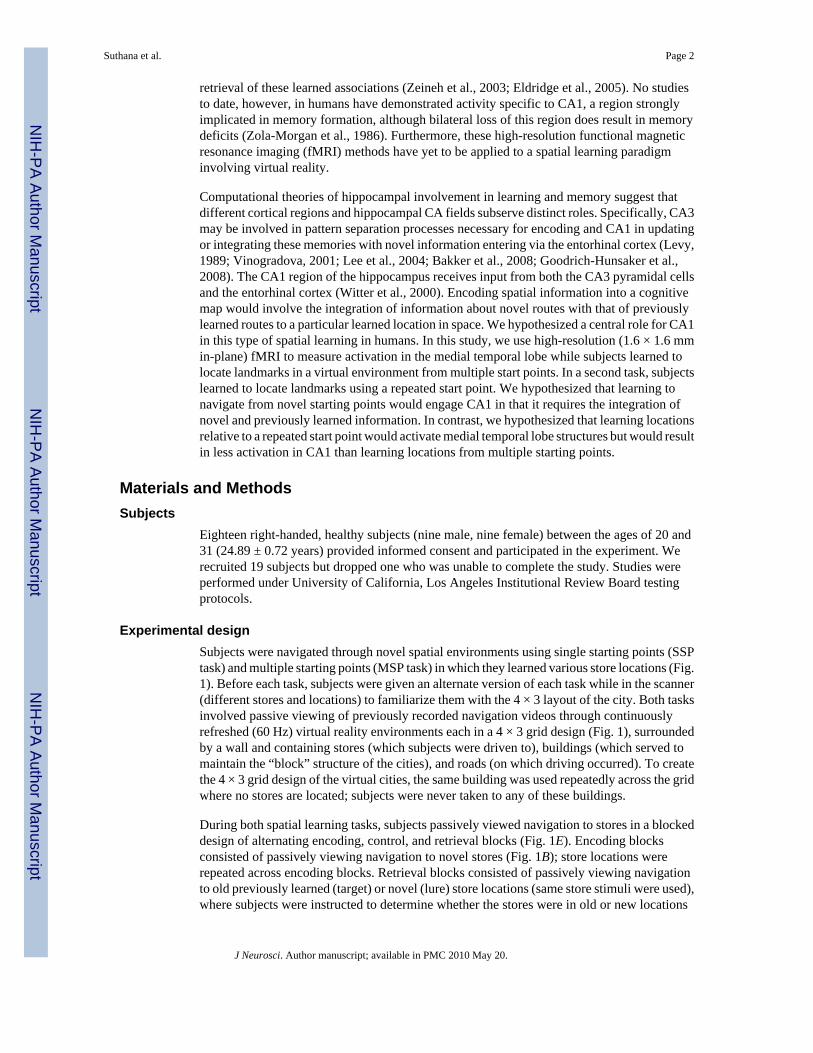
Experimental designSubjects were navigated through novel spatial environments using single starting points (SSPtask) and multiple starting points (MSP task) in which they learned various store locations (Fig.1). Before each task, subjects were given an alternate version of each task while in the scanner(different stores and locations) to familiarize them with the 4 × 3 layout of the city. Both tasksinvolved passive viewing of previously recorded navigation videos through continuouslyrefreshed (60 Hz) virtual reality environments each in a 4 × 3 grid design (Fig. 1), surroundedby a wall and containing stores (which subjects were driven to), buildings (which served tomaintain the “block” structure of the cities), and roads (on which driving occurred). To createthe 4 × 3 grid design of the virtual cities, the same building was used repeatedly across the gridwhere no stores are located; subjects were never taken to any of these buildings.
During both spatial learning tasks, subjects passively viewed navigation to stores in a blockeddesign of alternating encoding, control, and retrieval blocks (Fig. 1E). Encoding blocksconsisted of passively viewing navigation to novel stores (Fig. 1B); store locations wererepeated across encoding blocks. Retrieval blocks consisted of passively viewing navigationto old previously learned (target) or novel (lure) store locations (same store stimuli were used),where subjects were instructed to determine whether the stores were in old or new locations
Suthana et al. Page 2
J Neurosci. Author manuscript; available in PMC 2010 May 20.
NIH
-PA Author Manuscript
NIH
-PA Author Manuscript
NIH
-PA Author Manuscript

within the city and respond by pressing one of two assigned buttons. Subjects learned andrecalled 12 store locations (12 trials) in each task during a scan time of 477 s for each task. Inthe direction-pressing control condition for both tasks (Fig. 1D), subjects passively viewednavigation through identical cities but without stores (just buildings and roads), and wereinstructed to press the corresponding button on the keypad every time the direction was changed(left and right). This baseline task was chosen over typical rest tasks for its higher demand onnon-mnemonic cognitive processes. It has previously been shown that using a simple rest taskactivates the hippocampus and thus provides a nonoptimal baseline level of activity (Stark andSquire, 2001). Because we were solely interested in the mnemonic processes associated withour tasks, we chose a baseline task that differed in only this respect. SSP and MSP spatiallearning tasks were matched in aspects including experimental design (Fig. 1E), baselinecondition (Fig. 1D), virtual city layout (Fig. 1C), length and timing of navigation videos, andnumber of stores driven to. Alternate stores were used in each task and task order wascounterbalanced across subjects.
What differed between spatial learning tasks were the instructions and starting points usedwithin and between encoding and retrieval. For encoding (study) during the SSP task, subjectswere instructed to learn store locations from an initial starting point within the city; the startingpoint was repeated across learning trials (Fig. 1C, red arrows). Instructions during retrieval(test) were the same for both tasks (“Is the store in the same location? Press ‘1’ for yes and ‘2’for no.”). During SSP retrieval, subjects’ starting point and route were also repeated acrossretrieval trials. The same stores were used in encoding and retrieval, although locations werechanged for lure trials. In the SSP version, study and test both relied on the same startinglocation and route used in the city. For the MSP task, subjects were instructed to learn storelocations relative to other stores; the starting point within the city varied across trials duringencoding blocks as well as between encoding and retrieval blocks (Fig. 1C, blue arrows).Therefore, subjects could not learn locations relative to the starting point. Study and test bothrely on learning locations from multiple starting points; starting points and routes presentedduring retrieval differed with respect to encoding, and therefore subjects could not depend onpreviously presented routes for learning. All subjects completed both the SSP and MSPexperimental tasks; order was counterbalanced across subjects.
Navigation videos were displayed and recorded using pyepl (http://pyepl.sourceforge.net/),Snapz Pro X (Ambrosia software), and an adapted version of yellowcab2 (with buildings, citylayout changed, and passengers removed; original downloadhttp://memory.psych.upenn.edu/Software). Stimuli were edited using iMovie (Apple) andpresented using MacStim 3.2.1 software (WhiteAnt Occasional Publishing).
Imaging procedureSubjects were scanned with a Siemens Allegra head-only 3 tesla scanner at the Ahmanson-Lovelace Brain Mapping Center at the University of California, Los Angeles. High in-planeresolution structural images with a matrix size of 512 × 512 [spin echo, repetition time (TR)= 5200 ms, echo time (TE) = 105 ms, 19 slices, contiguous; voxel size: 0.391 × 0.391 × 3 mm]and echo-planar images (TR = 3000 ms, TE = 39 ms, 128 × 128, 19 slices, contiguous; voxelsize = 1.6 × 1.6 × 3 mm) were acquired in the same oblique coronal plane and registered usinga matched-bandwidth sequence (TR = 5000 ms, TE = 66 ms, 19 slices, contiguous; voxel size= 1.6 × 1.6 × 3 mm). Two volumes were introduced at the beginning of each functional scanto allow equilibration to steady state and were subsequently excluded from the analysis. Thecoronal plane was chosen because the structures are relatively homogeneous along the longaxis but differ in-plane; thus, we maximized in-plane resolution. For all scans, 19 slices wereacquired perpendicular to the long axis of the hippocampus during acquisition. See Zeineh etal. (2000) for further details on method. Visual stimuli were presented to the subject using 512
Suthana et al. Page 3
J Neurosci. Author manuscript; available in PMC 2010 May 20.
NIH
-PA Author Manuscript
NIH
-PA Author Manuscript
NIH
-PA Author Manuscript

× 512 resolution magnet-compatible three-dimensional (3-D) goggles and headphones undercomputer control (Resonance Technologies). The stimuli were presented using a MacintoshG4 Powerbook computer, and key presses were recorded for behavioral analysis.
Imaging and statistical analysisfMRI analysis was conducted using FEAT (fMRI Expert Analysis Tool), part of FMRIBSoftware Analysis (FSL version 3.3, www.fmrib.ox.ac.uk/fsl), to investigate differences inoverall activity in contrasts of interest. Functional volumes were motion corrected to the medianvolume using MCFLIRT (Jenkinson et al., 2002) (FMRIB’s motion correction linear imageregistration tool) using a normalized correlation ratio cost function and linear interpolation.Brains were skull stripped using BET (brain extraction tool) (Smith, 2002). Images werespatially smoothed using a Gaussian kernel of full-width at half-maximum 3 mm and intensitynormalized, and a high-pass temporal filter (Gaussian-weighted least-squares straight linefitting, with σ = 100.0 s) was applied. Time-series statistical analysis was performed usingFILM (FMRIB’s improved linear model) with local autocorrelation correction (Woolrich etal., 2001). Regressors of interest were created by convolving a delta function representing trialonset times with a canonical (gamma) hemodynamic response function, along with theirtemporal derivatives. Contrasts were cluster corrected at Z > 2.3 and p < 0.05. Functionalimages were aligned using FMRIB’s Linear Image Registration Tool to high-resolutioncoplanar images via an affine transformation with 6 degrees of freedom. The high-resolutioncoplanar images were then aligned to the subject’s high-resolution structural images using anaffine transformation with 6 degrees of freedom.
For those slices selected for functional scanning, the 3-D gray matter of the MTL subregionswere created (Fig. 2A) by manually segmenting the high-resolution structural images into whitematter and CSF using mrGray segmentation software (Teo et al., 1997). The gray matter wasthen computationally unfolded using an iterative algorithm based on metric multidimensionalscaling using mrUnfold software and interpolated by a factor of 7 to improve segmentationalong the long axis of the hippocampus (Engel et al., 1997) (mrGray and mrUnfold download:http://white.stanford.edu/~brian/mri/segmentUnfold.htm), yielding a final voxel size of 0.391× 0.391 × 0.429 mm (Fig. 2D). The position of the various CA fields, subiculum, entorhinalcortex (ERC), perirhinal cortex (PRC), parahippocampal cortex (PHC), and fusiform gyrus onthe unfolded map were found by mapping pixels from points demarcated (Fig. 2B) in thestructural images based on the atlases by Amaral and Insausti (1990) and Duvernoy (1998).The raw functional images were aligned with the structural images using matched-bandwidthimages and then onto the unfolded hippocampus using the same transformation parameters(Fig. 2D). The activation maps for the functional images were superimposed onto the structuralimages for precise localization of effects. In this way, we were able to clearly differentiateactivation in the substructures of the hippocampus and the parahippocampal gyrus. Anatomicalregions of interest (ROIs) were created a priori by defining voxels in 2-D space and thenprojected into 3-D space (Fig. 2C). These ROIs included anterior and posterior CA2,3 anddentate gyrus (CA23DG), anterior and posterior CA1, anterior and posterior subiculum, ERC,PRC, PHC, and fusiform gyrus. With our current human imaging methods, the dentate gyrusis not distinguishable from adjacent CA fields and therefore grouped in an encompassing regionlabeled CA23DG (but see Ekstrom et al., 2009). The average percentage signal change wascomputed in FSL for each ROI from average parameter estimates using the height of an isolatedevent as the scaling factor, and is relative to the voxel mean (seehttp://mumford.bol.ucla.edu/perchange_guide.pdf). Correlationanalysis was done bycalculating the Spearman rank coefficient (p < 0.05, corrected) between percentage signalchange and subjects’ average behavioral performance (percentage correct) on the task. Wechose the Spearman rank correlation coefficient, rather than the Pearson ρ correlation, becausewe did not assume a linear correlation between metrics but rather a monotonically increasing
Suthana et al. Page 4
J Neurosci. Author manuscript; available in PMC 2010 May 20.
NIH
-PA Author Manuscript
NIH
-PA Author Manuscript
NIH
-PA Author Manuscript

relationship between the variables. Conducting the analysis using the Pearson ρ correlation,however, did not change our overall results.
A group 2-D hippocampal template shown in Figure 3 was created based on the 18 individualsubject anatomical images and boundaries. Each subject’s individual anatomical and timefunctional activations were warped into the template. Group activation maps were producedby averaging individual subregional boundaries and then transforming each subject’s flat mapto this template. The degree of fit between each individual subject’s behavior and fMRI signal(e.g., β values), based on model contrasts discussed earlier, were then compared across subjectsfor each voxel using a mixed-effects t test (t ≥ 2.4; p < 0.05, corrected). See Thompson et al.(2000) and Zeineh et al. (2001) for details on these methods. Correction for multiplecomparisons for single-subject contrasts were cluster corrected at Z > 2.3 and p < 0.05. Groupactivation maps were corrected using a Bonferroni correction for 10 ROIs (p < 0.005).Spearman rank correlation analysis was corrected for multiple comparisons using a Bonferronicorrection (p < 0.025).
ResultsSubjects’ average performance and learning rates on the SSP and MSP spatial tasks did notdiffer significantly (supplemental Fig. S1, available at www.jneurosci.org as supplementalmaterial) (n = 18; percentage correct: mean ± SEM, SSP, 73.33 ±3.89; MSP, 68.33 ± 5.18;t(17) = 0.85; p = 0.41).
The focus of this paper was to determine hippocampal subregion activity during the processingof spatial information, specifically during encoding. To investigate changes during encodingof store locations, activity during encoding stimuli from both tasks were analyzed separatelyduring periods of navigation without stores (where subjects were navigated through a citylayout that lacked store landmarks) (Fig. 1A) and periods of navigation with stores (wherestores were present in the city and subjects could make store–place associations) (Fig. 1B). Asexpected, no significant increases were found during navigation without stores during encodingblocks compared with navigation during the direction-pressing control blocks (baseline).However, in both spatial tasks activity during navigation with stores showed significantincreases compared with the control condition across various MTL subregions bilaterally,including the parahippocampal gyrus and adjacent fusiform gyrus (Fig. 3A). To determine anysignificant differences between the MSP and SSP conditions, we directly contrasted encodingblocks from these two conditions. Learning locations from varying compared with a singlestart point yielded a cluster of significant increase within the right CA1 subregion of thehippocampus (Fig. 3B). No significant clusters were found in any extrahippocampal regionsor hippocampal subregions CA23DG and subiculum. Furthermore, the reverse contrast yieldedno areas of significant difference in activity within the MTL (for example, activation datasuperimposed on an oblique-coronal high-resolution anatomical image) (see supplemental Fig.3, available at www.jneurosci.org as supplemental material).
In addition to the voxelwise analysis performed above, we completed an independent analysisbased solely on anatomical ROIs. ROIs were determined independent of functional activity,and average percentage signal change was then calculated for each region. These ROIs werebased on the anatomical definitions of CA23DG, anterior and posterior CA1, anterior andposterior subiculum, ERC, PRC, PHC, and fusiform gyrus. Average percentage signal changecompared with baseline within hippocampal regions is shown in Figure 4. For the CA1 andsubiculum, the posterior anatomical ROIs are shown; anterior regions yielded no significantdifferences between conditions or hemispheres. Based on the a priori hypothesis that CA1activity would differ between learning conditions, we performed a 2 × 2 hemisphere (right vsleft) × condition (MSP vs SSP) ANOVA in posterior CA1, which yielded a significant main
Suthana et al. Page 5
J Neurosci. Author manuscript; available in PMC 2010 May 20.
NIH
-PA Author Manuscript
NIH
-PA Author Manuscript
NIH
-PA Author Manuscript

effect of hemisphere (F(2,34) = 7.93; p = 0.006) and significant interaction between hemisphereand condition (F(2,34) = 4.90; p = 0.029). Post hoc comparisons using paired t tests foranatomical regions of interest found that activity within the right posterior CA1 subregion wassignificantly greater during the MSP compared with SSP encoding (t(17) = 2.36; p = 0.026).Furthermore, activity in the right posterior CA1 during MSP encoding correlated significantlywith behavioral performance across subjects (Fig. 5A) (Spearman’s ρ = 0.53; p = 0.02; n = 18).Activity in this region during SSP encoding was not significantly correlated with performance(Fig. 5B) (ρ = −0.488; n.s.; n = 18). No significant differences between SSP and MSP encodingconditions were found in the left CA1 subregion (t(17) = −0.93; n.s.), although overall activitywas significantly greater in the right hemisphere compared with the left (right > left, t(17) =3.29; p = 0.004). In contrast to this pattern, activity within the right and left CA23DG regiondid not differ between learning conditions (SSP > MSP, right, t(17) = 0.81; n.s.; left, t(17) =0.17; n.s.), nor was there a significant right versus left difference (right > left, t(17) = 1.75; n.s.).Similar to CA23DG, the anterior and posterior subiculum showed no significant differencesin condition or hemisphere (right, t(17) = −0.41; n.s.; left, t(17) = −0.33; p = 0.74; right > left,t(17) = 0.98; n.s.).
For the adjacent PHC, activation increased bilaterally during both encoding conditionscompared with baseline (MSP right, t(17) = 4.83; MSP left, t(17) = 5.27; SSP right, t(17) = 4.00;SSP left, t(17) = 4.80; p < 0.0001) (supplemental Fig. 2C, available at www.jneurosci.org assupplemental material). Similarly we found increases in activity within the fusiform gyrus(MSP right, t(17) = 6.39; MSP left, t(17) = 5.39; SSP left, t(17) = 6.19; SSP right, t(17) = 5.79;p < 0.0001) (supplemental Fig. 2D, available at www.jneurosci.org as supplemental material).However, no significant differences were found in these regions with respect to condition(supplemental Fig. 2C,D, available at www.jneurosci.org as supplemental material). Activitywithin the entorhinal cortex showed no significant increase from baseline (Fig. 3A;supplemental Fig. 2A, available at www.jneurosci.org as supplemental material). Within theright PRC, activity increased during both MSP and SSP conditions (MSP, t(17) = 2.28; p =0.03; SSP, t(17) = 3.37; p = 0.003); however no significant differences were found with respectto condition (supplemental Fig. 2B, available at www.jneurosci.org as supplemental material).
DiscussionThe present findings demonstrate that while MTL regions such as the PHC are engaged duringlearning of spatial information, processing of locations from multiple starting points furtherrecruits the hippocampus. We isolated the increase in hippocampal activation to CA1,supporting computational models suggesting a role for CA1 in updating memories with novelinformation. Furthermore, CA1 activity positively correlated with behavioral performancewhen navigating from multiple starting points, suggesting that this region supportsperformance during this type of learning.
Our results are consistent with the idea that the hippocampus plays a critical role in forming aviewpoint-independent map of an environment. Spatial information can be processed using aview-dependent (egocentric) or view-independent (allocentric) frame of reference (Tolman,1948; Howard, 1982). A key difference between allocentric and egocentric spatialrepresentations is that allocentric representations can support navigation from novel startingpoints. Studies in rodents have suggested that an allocentric representation of space is formedin the CA1 subfield of hippocampus (Leutgeb et al., 2005; Brun et al., 2008) and place cellsin CA1 are able to abruptly shift map representations during an incremental shift in theenvironment, suggesting these cells code for distinct, orthogonal map representations fordifferent environments (Wilson and McNaughton, 1993; Wills et al., 2005). Lesions of CA1impair memory for relative placement of landmarks to each other (Goodrich-Hunsaker et al.,2008). Hippocampal activity increases during a spatial memory task using novel starting points
Suthana et al. Page 6
J Neurosci. Author manuscript; available in PMC 2010 May 20.
NIH
-PA Author Manuscript
NIH
-PA Author Manuscript
NIH
-PA Author Manuscript
(Parslow et al., 2004), and hippocampal damage impairs recall of object–place associationswhen tested from shifted viewpoints (King et al., 2002). While several studies have suggestedthat the hippocampus plays a major role in forming an allocentric representation of anenvironment (Morris et al., 1982; Spiers et al., 2001; Kumaran and Maguire, 2005), thesestudies were not able to measure the contribution of subregions. Thus, our results provide novelevidence suggesting that CA1 is involved in allocentric spatial computations necessary forspatial navigation.
Various studies of spatial learning have shown that the PHC is preferentially involved duringencoding and retrieval of object–place associations (Köhler et al., 2002; Malkova and Mishkin,2003; Sommer et al., 2005). In these studies the objects are presented in front of the observeron a screen and are likely to be encoded egocentrically. Therefore, we would not expect thiscondition to necessitate formation of a viewpoint-independent spatial memory representation.In contrast, navigation in a virtual environment from different starting points involvesmovement of the observer relative to objects and thus requires allocentric encoding. Consistentwith prior studies, we show increases in activity within the PHC during SSP and MSP learningconditions. However, using virtual reality to manipulate starting point may further recruitallocentric processing and thus hippocampal involvement. Interestingly, learning a map froman aerial perspective does not result in hippocampal activation, while learning a map throughground-level exploration results in significant hippocampal activation (Shelton and Gabrieli,2002). It may be that the hippocampus plays an important role in building a flexible map-likememory representation of space from route-level information rather than learning spatial mapsper se.
Although we did not find increases in activity during SSP compared with MSP learning, thereare likely to be regions outside the MTL, which show increased activity. Previous studies haveshown caudate nucleus involvement during route learning from single starting points (Hartleyet al., 2003; Iaria et al., 2003). Thus, it is possible the caudate nucleus would show increasedactivity in SSP compared with MSP learning. In the present study, we restricted our field ofview to MTL structures to increase resolution necessary to differentiate hippocampalsubregions. Using whole-brain fMRI methods could describe the role of regions outside theMTL in spatial learning from single and multiple start points.
A possible difference between allocentric and egocentric spatial learning is that forming anallocentric map requires a higher memory load resulting from multiple viewpoints (Shrager etal., 2007). Increased CA1 activation may therefore be due to a higher memory load in our MSPencoding condition. However, although subjects learned locations from only one starting pointin the SSP task, we used routes that were sufficiently complex to ensure that performance wasat the same level as in the MSP condition. Thus, the amount of information in memory neededto support performance was approximately equivalent in the two tasks, at least as assessed bylearning rate. During MSP encoding and retrieval subjects approached stores from differentdirections and locations were learned and recalled relative to other stores rather than relativeto a repeated starting location. Integration of previously learned routes in memory with novelroutes necessitates the formation of a viewpoint-independent memory representation of theenvironment. We therefore suggest increase in CA1 activity in the MSP condition reflects thenature of spatial representations learned rather than memory load.
CA1 regions active during the MSP learning condition were in the posterior hippocampus.These findings are consistent with other studies indicating that the posterior hippocampus isactivated during spatial learning (Gabrieli et al., 1997; Colombo et al., 1998; Fernández et al.,1998; Maguire et al., 2006). In a recent study, patterns of activity in the posterior hippocampuspredicted the location of an individual in a virtual environment (Hassabis et al., 2009). These
Suthana et al. Page 7
J Neurosci. Author manuscript; available in PMC 2010 May 20.
NIH
-PA Author Manuscript
NIH
-PA Author Manuscript
NIH
-PA Author Manuscript

data suggest that the posterior hippocampus may be particularly involved in mnemonicfunctions involving spatial representations.
Previous neuroimaging studies using virtual reality navigation found PHC activity in absenceof hippocampal activation (Aguirre et al., 1996; Mellet et al., 2000; Rosenbaum et al., 2004).One explanation for hippocampal activity detected in our study may be that subregions withinthe hippocampus are likely to serve functionally distinct roles and thus averaging across theseareas would result in reduced signal. In this study, we separately evaluate individual subregionswithout losing information during group averaging. Individual hippocampi are segmented andwarped to an averaged group template constrained by individual subregional boundaries. Inaddition to unfolding we used an ROI-based method to calculate percentage signal change inanatomical regions of individual subjects. This analysis independently confirmed our unfoldingresults and allowed for an additional method of high-resolution hippocampal imaging analysis.However, current in vivo imaging methods are unable to reach the resolution necessary tovisualize cell histology to determine exact subregional boundaries. Furthermore, thehippocampus is small in structure and variable across subjects. We attempted to deal with thischallenge by demarcating each individual subject’s boundaries using atlases by Amaral andInsausti (1990) and Duvernoy (1998), which identify landmarks on the MR images usingpostmortem histological images.
Surprisingly, we did not see CA3 activity differences between MSP and SSP conditions,although this region has been shown to be involved in encoding associative information (Zeinehet al., 2003; Eldridge et al., 2005). While there was activity within voxels of the regionencompassing CA23DG in both learning conditions, there was no differential activity whenlearning from multiple start points. Region CA3 has been associated with pattern separationto form distinct memory traces during learning (Leutgeb et al., 2007; Bakker et al., 2008). Itmay be that learning a route using a single start point and learning a cognitive map from multiplestart points depends to the same degree on pattern separation processes.
In the MSP condition, subjects have to integrate different paths stored in memory to infer wherethey are located in relation to the goal. Thus, this encoding condition may rely more heavilyon retrieval. Navigation depending on an allocentric spatial representation is thought to dependon memory retrieval (Knowlton and Fanselow, 1998). Stronger demands on retrieval tointegrate previously learned information with novel information might cause an increase inactivation in CA1. One possibility is that the CA1 activation reflects retrieval of other routesduring the task. Previous work has shown an increase in CA1 activity during memory retrievalin a nonspatial memory task, although this activation was not extremely robust (Eldridge etal., 2005).
Last, our findings are consistent with computational models proposing that CA1 integratespreviously learned information (encoded in CA3) with incoming novel information from theneocortex (via ERC) (Levy, 1989; Vinogradova, 2001). Navigating through a novel spatialenvironment requires integrating multiple spatial relationships into a flexible memoryrepresentation. Forming an allocentric cognitive map requires integration of previously learnedviewpoints with newly encountered viewpoints to update the memory representation into onethat is independent of starting point. This process would thus presumably recruit more cellswithin CA1, leading to a detectable fMRI response. In the rodent hippocampus, place cells arecontext specific (Muller and Ranck, 1987; Wilson and McNaughton, 1993) and remap whena rat is placed in a different spatial environment. CA1 neurons have been shown to be sensitiveto the intended destination and trajectory of the animal (Frank et al., 2000; Ainge et al.,2007). These lines of evidence support a role for this structure in acquiring a flexible memoryrepresentation of a spatial environment. Overall, the present results suggest that the CA1 regionmay also play a critical role in allocentric spatial memory formation in humans.
Suthana et al. Page 8
J Neurosci. Author manuscript; available in PMC 2010 May 20.
NIH
-PA Author Manuscript
NIH
-PA Author Manuscript
NIH
-PA Author Manuscript

Supplementary MaterialRefer to Web version on PubMed Central for supplementary material.
AcknowledgmentsThis work was supported by National Institute of Mental Health Grant 5T32 MH015795, National Institute ofNeurological Disorders and Stroke Grant F32 NS50067-03, National Institutes of Health Grant T90 431587-BH-29793, National Science Foundation Grant GK-12 0742410, and National Institute on Aging Grants 2R01AG013308 and 5P01 AG025831. For generous support, we thank the Brain Mapping Medical Research Organization,Brain Mapping Support Foundation, Pierson-Lovelace Foundation, The Ahmanson Foundation, William M. and LindaR. Dietel Philanthropic Fund at the Northern Piedmont Community Foundation, Tamkin Foundation, Jennifer Jones-Simon Foundation, Capital Group Companies Charitable Foundation, Robson Family, and Northstar Fund. We thankMichael J. Kahana for sharing and Aaron Geller and Josh Jacobs for support of the virtual navigation task “yellowcab”with financial support from Dr. Kahana’s National Institutes of Health Grant MH61975. We also thank Michael Zeinehand Paul Thompson for assistance with group unfolding scripts, and Michael Jones for technical assistance. Finally,we also thank all of the subjects for their participation in this study.
ReferencesAguirre GK, Detre JA, Alsop DC, D’Esposito M. The parahippocampus subserves topographical learning
in man. Cereb Cortex 1996;6:823–829. [PubMed: 8922339]Ainge JA, Tamosiunaite M, Woergoetter F, Dudchenko PA. Hippocampal CA1 place cells encode
intended destination on a maze with multiple choice points. J Neurosci 2007;27:9769–9779. [PubMed:17804637]
Amaral, DG.; Insausti, R. The hippocampal formation. In: Paxinos, G., editor. The human nervous system.San Diego: Academic; 1990. p. 711-755.
Bakker A, Kirwan CB, Miller M, Stark CE. Pattern separation in the human hippocampal CA3 and dentategyrus. Science 2008;319:1640–1642. [PubMed: 18356518]
Brun VH, Leutgeb S, Wu HQ, Schwarcz R, Witter MP, Moser EI, Moser MB. Impaired spatialrepresentation in CA1 after lesion of direct input form entorhinal cortex. Neuron 2008;57:290–302.[PubMed: 18215625]
Colombo M, Fernandez T, Nakamura K, Gross CG. Functional differentiation along the anterior-posterioraxis of the hippocampus in monkeys. J Neurophysiol 1998;80:1002–1005. [PubMed: 9705488]
Duvernoy, HM. The human hippocampus: functional anatomy, vascularization, and serial sections withMRI. Berlin: Springer; 1998.
Ekstrom AD, Kahana MJ, Caplan JB, Fields TA, Isham EA, Newman EL, Fried I. Cellular networksunderlying human spatial navigation. Nature 2003;425:184–188. [PubMed: 12968182]
Ekstrom AD, Bazih AJ, Suthana NA, Al-Hakim R, Ogura K, Zeineh M, Burggren AC, Bookheimer SY.Advances in high-resolution imaging and computational unfolding of the human hippocampus.Neuroimage 2009;47:42–49. [PubMed: 19303448]
Eldridge LL, Engel SA, Zeineh MM, Bookheimer SY, Knowlton BJ. A dissociation of encoding andretrieval processes in the human hippocampus. J Neurosci 2005;25:3280–3286. [PubMed: 15800182]
Engel SA, Glover GH, Wandell BA. Retinotopic organization in human visual cortex and the spatialprecision of functional MRI. Cereb Cortex 1997;7:181–192. [PubMed: 9087826]
Fernández G, Weyerts H, Schrader-Bölsche M, Tendolkar I, Smid HG, Tempelmann C, Hinrichs H,Scheich H, Elger CE, Mangun GR, Heinze HJ. Successful verbal encoding into episodic memoryengages the posterior hippocampus: a parametrically analyzed functional magnetic resonanceimaging study. J Neurosci 1998;18:1841–1847. [PubMed: 9465008]
Frank LM, Brown EN, Wilson M. Trajectory encoding in the hippocampus and entorhinal cortex. Neuron2000;27:169–178. [PubMed: 10939340]
Gabrieli JD, Brewer JB, Desmond JE, Glover GH. Separate neural bases of two fundamental memoryprocesses in the human medial temporal lobe. Science 1997;276:264–266. [PubMed: 9092477]
Goodrich-Hunsaker NJ, Hunsaker MR, Kesner RP. The interactions and dissociations of the dorsalhippocampus subregions: how the dentate gyrus, CA3, and CA1 process spatial information. BehavNeurosci 2008;122:16–26. [PubMed: 18298245]
Suthana et al. Page 9
J Neurosci. Author manuscript; available in PMC 2010 May 20.
NIH
-PA Author Manuscript
NIH
-PA Author Manuscript
NIH
-PA Author Manuscript

Hartley T, Maguire EA, Spiers HJ, Burgess N. The well-worn route and the path less traveled: distinctneural bases of route following and wayfinding in humans. Neuron 2003;37:877–888. [PubMed:12628177]
Hassabis D, Chu C, Rees G, Weiskopf N, Molyneux PD, Maguire EA. Decoding neuronal ensembles inthe human hippocampus. Curr Biol 2009;19:546–554. [PubMed: 19285400]
Howard, IP. Human visual orientation. Chichester, UK: Wiley; 1982.Iaria G, Petrides M, Dagher A, Pike B, Bohbot VD. Cognitive strategies dependent on the hippocampus
and caudate nucleus in human navigation: variability and change with practice. J Neurosci2003;23:5945–5952. [PubMed: 12843299]
Jenkinson M, Bannister P, Brady M, Smith S. Improved optimization for the robust and accurate linearregistration and motion correction of brain images. Neuroimage 2002;17:825–841. [PubMed:12377157]
King JA, Burgess N, Hartley T, Vargha-Khadem F, O’Keefe J. The human hippocampus and viewpointdependence in spatial memory. Hippocampus 2002;12:811–820. [PubMed: 12542232]
Knowlton BJ, Fanselow MS. Hippocampus, consolidation, and online memory. Curr Opin Neurobiol1998;8:293–296. [PubMed: 9635216]
Köhler S, Crane J, Milner B. Networks of domain-specific and general regions involved in episodicmemory for spatial location and object identity. Hippocampus 2002;12:718–723. [PubMed:12542224]
Kumaran D, Maguire EA. The human hippocampus: cognitive maps or relational memory? J Neurosci2005;25:7254–7259. [PubMed: 16079407]
Lee I, Yoganarasimha D, Rao G, Knierim JJ. Comparison of population coherence of place cells inhippocampal subfields CA1 and CA3. Nature 2004;430:456–459. [PubMed: 15229614]
Leutgeb JK, Leutgeb S, Treves A, Meyer R, Barnes CA, McNaughton BL, Moser MB, Moser EI.Pregressive transformation of hippocampal neuronal representations in “morphed” environments.Neuron 2005;48:345–358. [PubMed: 16242413]
Leutgeb JK, Leutgeb S, Moser MB, Moser EI. Pattern separation in the dentate gyrus and CA3 of thehippocampus. Science 2007;315:961–966. [PubMed: 17303747]
Levy, WB. A computational approach to the hippocampal function. In: Hawkins, RD.; Gower, GH.,editors. Computational models of learning in simple neural systems. Orlando, FL: Academic; 1989.p. 243-305.
Maguire EA, Woollett K, Spiers HJ. London taxi drivers and bus drivers: a structural MRI andneuropsychological analysis. Hippocampus 2006;12:1091–1101. [PubMed: 17024677]
Malkova L, Mishkin M. One-trial memory for object-place associations after separate lesions ofhippocampus and posterior parahippocampal region in the monkey. J Neurosci 2003;23:1956–1965.[PubMed: 12629201]
McNamara TP, Shelton AL. Cognitive maps and the hippocampus. Trends Cogn Sci 2003;7:333–335.[PubMed: 12907223]
Mellet E, Briscogne S, Tzourio-Mazoyer N, Ghaëm O, Petit L, Zago L, Etard O, Berthoz A, Mazoyer B,Denis M. Neural correlates of topographic mental exploration: the impact of route versus surveyperspective learning. Neuroimage 2000;12:588–600. [PubMed: 11034866]
Morris RG, Garrud P, Rawlins JN, O’Keefe J. Place navigation impaired in rats with hippocampal lesions.Nature 1982;297:681–683. [PubMed: 7088155]
Muller RU, Kubie JL, Ranck JB Jr. Spatial firing patterns of hippocampal complex-spike cells in a fixedenvironment. J Neurosci 1987;7:1935–1950. [PubMed: 3612225]
Nadel L, MacDonald L. Hippocampus: cognitive map or working memory? Behav Neural Biol1980;29:405–409. [PubMed: 7417203]
O’Keefe J, Dostrovsky J. The hippocampus as a spatial map. Preliminary evidence from unit activity inthe freely-moving rat. Brain Res 1971;34:171–175. [PubMed: 5124915]
O’Keefe, J.; Nadel, L. The hippocampus as a cognitive map. Oxford: Clarendon; 1978. p. 190-230.Parslow DM, Rose D, Brooks B, Fleminger S, Gray JA, Giampietro V, Brammer MJ, Williams S, Gasston
D, Andrew C, Vythelingum GN, Loannou G, Simmons A, Morris RG. Allocentric spatial memory
Suthana et al. Page 10
J Neurosci. Author manuscript; available in PMC 2010 May 20.
NIH
-PA Author Manuscript
NIH
-PA Author Manuscript
NIH
-PA Author Manuscript

activation of the hippocampal formation measured with fMRI. Neuropsychology 2004;18:450–461.[PubMed: 15291723]
Rosenbaum RS, Ziegler M, Winocur G, Grady CL, Moscovitch M. I have often walked down this streetbefore: fMRI studies of hippocampus and other structures during mental navigation of an oldenvironment. Hippocampus 2004;14:826–835. [PubMed: 15382253]
Shelton AL, Gabrieli JD. Neural correlates of encoding space from route and survey perspectives. JNeurosci 2002;22:2711–2717. [PubMed: 11923436]
Shrager Y, Bayley PJ, Bontempi B, Hopkins RO, Squire LR. Spatial memory and the humanhippocampus. Proc Natl Acad Sci U S A 2007;104:2961–2966. [PubMed: 17296931]
Smith SM. Fast robust automated brain extraction. Hum Brain Mapp 2002;17:143–155. [PubMed:12391568]
Sommer T, Rose M, Gläscher J, Wolbers T, Büchel C. Dissociable contributions within the medialtemporal lobe to encoding of object-location associations. Learn Mem 2005;12:343–351. [PubMed:15897257]
Spiers HJ, Burgess N, Maguire EA, Baxendale SA, Hartley T, Thompson PJ, O’Keefe J. Unilateraltemporal lobectomy patients show lateralized topographical and episodic memory deficits in a virtualtown. Brain 2001;124:2476–2489. [PubMed: 11701601]
Squire LR, Stark CEL, Clark RE. The medial temporal lobe. Annu Rev Neurosci 2004;27:279–306.[PubMed: 15217334]
Stark CE, Squire LR. When zero is not zero: the problem of ambiguous baseline conditions in fMRI.Proc Natl Acad Sci U S A 2001;98:12760–12766. [PubMed: 11592989]
Teo PC, Sapiro G, Wandell BA. Creating connected representations of cortical gray matter for functionalMRI visualization. IEEE Trans Med Imaging 1997;16:852–863. [PubMed: 9533585]
Thompson PM, Woods RP, Mega MS, Toga AW. Mathematical/computational challenges in creatingdeformable and probabilistic atlases of the human brain. Hum Brain Mapp 2000;9:81–92. [PubMed:10680765]
Tolman EC. Cognitive maps in rats and men. Psychol Rev 1948;55:189–208. [PubMed: 18870876]Vinogradova OS. Hippocampus as comparator: role of the two input and two output systems of the
hippocampus in selection and registration of information. Hippocampus 2001;11:578–598.[PubMed: 11732710]
Wills TJ, Lever C, Cacucci F, Burgess N, O’Keefe J. Attractor dynamics in the hippocampalrepresentation of the local environment. Science 2005;308:873–876. [PubMed: 15879220]
Wilson MA, McNaughton BL. Dynamics of the hippocampal ensemble code for space. Science1993;261:1055–1058. [PubMed: 8351520]
Witter MP, Wouterlood FG, Naber PA, Van Haeften T. Anatomical organization of the parahippocampal-hippocampal network. Ann N Y Acad Sci 2000;911:1–24. [PubMed: 10911864]
Woolrich MW, Ripley BD, Brady M, Smith SM. Temporal autocorrelation in univariate linear modelingof FMRI data. Neuroimage 2001;14:1370–1386. [PubMed: 11707093]
Zeineh MM, Engel SA, Bookheimer SY. Application of cortical unfolding techniques to functional MRIof the human hippocampal region. Neuroimage 2000;11:668–683. [PubMed: 10860795]
Zeineh MM, Engel SA, Thompson PM, Bookheimer SY. Unfolding the human hippocampus with highresolution structural and functional MRI. Anat Rec 2001;265:111–120. [PubMed: 11323773]
Zeineh MM, Engel SA, Thompson PM, Bookheimer SY. Dynamics of the hippocampus during encodingand retrieval of face-name pairs. Science 2003;299:577–580. [PubMed: 12543980]
Zola-Morgan S, Squire LR, Amaral DG. Human amnesia and the medial temporal lobe region: enduringmemory impairment following a bilateral lesion limited to field CA1 of the hippocampus. J Neurosci1986;6:2950–2967. [PubMed: 3760943]
Suthana et al. Page 11
J Neurosci. Author manuscript; available in PMC 2010 May 20.
NIH
-PA Author Manuscript
NIH
-PA Author Manuscript
NIH
-PA Author Manuscript

Figure 1.Virtual city snapshots. A, Snapshot of virtual city from a sample starting point. B, Sample storestimulus used. C, Subjects’ learned store locations within 4 × 3 grid cities from a varying initialstarting point within a city for the MSP encoding condition (blue arrows) and from a singleinitial starting point within the city for the SSP encoding condition (red arrows). D, Layout ofcity without stores used in the direction-pressing control baseline condition. Both tasks usedthis control condition. E, Both the SSP and MSP tasks consisted of alternating blocks ofencoding (Learn) and retrieval (Recall) interspersed with blocks of control (Ctl).
Suthana et al. Page 12
J Neurosci. Author manuscript; available in PMC 2010 May 20.
NIH
-PA Author Manuscript
NIH
-PA Author Manuscript
NIH
-PA Author Manuscript

Figure 2.Unfolding method. A, B, Each subject’s gray matter (green) is created by segmenting whitematter and CSF. The gray matter is then computationally unfolded (A) and boundaries betweenregions are projected onto the unfolded flat map (B). C, Voxels in 2-D space are projected into3-D space to create anatomical regions of interest showing posterior regions (left): CA23DG(red), CA1 (orange), subiculum (yellow), PHC (green), and fusiform gyrus (blue). D, Anaveraged group flat map (shown is the left) is created showing regions CA2, 3, and dentategyrus, CA1, subiculum (Sub), ERC, PRC, PHC, and fusiform gyrus.
Suthana et al. Page 13
J Neurosci. Author manuscript; available in PMC 2010 May 20.
NIH
-PA Author Manuscript
NIH
-PA Author Manuscript
NIH
-PA Author Manuscript

Figure 3.A, Group voxel-based mixed-effects unfolded t test maps (n = 18, statistical maps ofsignificantly activated and deactivated regions; − 2.4 ≥ t ≥ 2.4; p < 0.05 corrected) for the leftand right MTL regions during the MSP and SSP tasks separately compared with baseline. B,Group voxel-based mixed-effects unfolded t test maps (n = 18, statistical maps of significantlydifference in activity between MSP and SSP conditions; −2.4 ≥ t ≥ 2.4; p < 0.05 corrected) forthe left and right MTL regions. Regions shown include CA2, 3, and dentate gyrus, CA1,subiculum (Sub), ERC, PRC, PHC, and fusiform gyrus.
Suthana et al. Page 14
J Neurosci. Author manuscript; available in PMC 2010 May 20.
NIH
-PA Author Manuscript
NIH
-PA Author Manuscript
NIH
-PA Author Manuscript

Figure 4.Results from hippocampal anatomical regions of interest. Average percentage signal change(n = 18, error bars correspond to the SE across subjects) for the SSP and MSP tasks separatelycompared with baseline within left and right hippocampal subregions CA23DG (A) andsubiculum (C) show no significant differences between MSP and SSP conditions or betweenright and left hemispheric regions. B, Average percentage signal change from baseline withinleft and right posterior CA1 for MSP and SSP encoding indicate a significant differencebetween hemisphere (right > left, t(17) = 3.29; p < 0.05) and conditions (MSP > SSP, t(17) =2.26; p < 0.05).
Suthana et al. Page 15
J Neurosci. Author manuscript; available in PMC 2010 May 20.
NIH
-PA Author Manuscript
NIH
-PA Author Manuscript
NIH
-PA Author Manuscript

Figure 5.Behavioral performance (percentage correct) versus percentage signal change. A, Subjects’performance on the MSP task significantly correlated with percentage signal change in theright CA1 subregion during encoding (n = 18, Spearman’s ρ = 0.53; p < 0.05). B, Subjects’performance on the SSP task did not significantly correlate with percentage signal change inthe right CA1 subregion during encoding (n = 18, Spearman’s ρ = −0.488; n.s.).
Suthana et al. Page 16
J Neurosci. Author manuscript; available in PMC 2010 May 20.
NIH
-PA Author Manuscript
NIH
-PA Author Manuscript
NIH
-PA Author Manuscript


