Embed Size (px)
Citation preview
Hungnes, I. N., Al-Salemee, F., Gawne, P. J., Eykyn, T., Atkinson, R.A., Terry, S. Y. A., Clarke, F., Blower, P. J., Pringle, P. G., & Ma, M. T.(2021). One-step, kit-based radiopharmaceuticals for molecularSPECT imaging: a versatile diphosphine chelator for 99mTcradiolabelling of peptides. Dalton Transactions, 50(44), 16156-16165.https://doi.org/10.1039/d1dt03177e
Publisher's PDF, also known as Version of recordLicense (if available):CC BYLink to published version (if available):10.1039/d1dt03177e
Link to publication record in Explore Bristol ResearchPDF-document
This is the final published version of the article (version of record). It first appeared online via Royal Society ofChemistry at https://doi.org/10.1039/D1DT03177E .Please refer to any applicable terms of use of the publisher.
University of Bristol - Explore Bristol ResearchGeneral rights
This document is made available in accordance with publisher policies. Please cite only thepublished version using the reference above. Full terms of use are available:http://www.bristol.ac.uk/red/research-policy/pure/user-guides/ebr-terms/
DaltonTransactions
PAPER
Cite this: Dalton Trans., 2021, 50,16156
Received 17th September 2021,Accepted 15th October 2021
DOI: 10.1039/d1dt03177e
rsc.li/dalton
One-step, kit-based radiopharmaceuticals formolecular SPECT imaging: a versatile diphosphinechelator for 99mTc radiolabelling of peptides†
Ingebjørg N. Hungnes,a Fahad Al-Salemee, a Peter J. Gawne, a Thomas Eykyn,a
R. Andrew Atkinson,b,c Samantha Y. A. Terry,a Fiona Clarke,d Philip J. Blower, a
Paul G. Pringle e and Michelle T. Ma *a
Radiotracers labelled with technetium-99m (99mTc) enable accessible diagnostic imaging of disease, pro-
vided that radiotracer preparation is simple. Whilst 99mTc radiopharmaceuticals for imaging perfusion are
routinely prepared from kits, and regularly used in healthcare, there are no 99mTc-labelled receptor-tar-
geted radiopharmaceuticals in widespread clinical use. This is in part due to the multistep radiosyntheses
required for the latter. We demonstrate that the diphosphine, 2,3-bis(diphenylphosphino)maleic anhydride
(BMA), is an excellent platform for preparation of kit-based, receptor-targeted 99mTc-labelled radiotra-
cers: its conjugates are simple to prepare and can be easily labelled with 99mTc using one-step, kit-based
protocols. Here, reaction of BMA with the αvβ3-integrin receptor targeted cyclic peptide, Arg-Gly-Asp-
DPhe-Lys (RGD), provided the first diphosphine-peptide conjugate, DP-RGD. DP-RGD was incorporated
into a “kit”, and addition of a saline solution containing 99mTcO4− to this kit, followed by heating, furnished
the radiotracer [99mTcO2(DP-RGD)2]+ in consistently high radiochemical yields (>90%). The analogous
[ReO2(DP-RGD)2]+ compound was prepared and characterised, revealing that both [99mTcO2(DP-RGD)2]
+
and [ReO2(DP-RGD)2]+ consist of a mixture of cis and trans geometric isomers. Finally,
[99mTcO2(DP-RGD)2]+ exhibited high metabolic stability, and selectively targeted αvβ3-integrin receptors,
enabling in vivo SPECT imaging of αvβ3-integrin receptor expression in mice.
Introduction
The γ-emitting radionuclide, technetium-99 m (99mTc, t1/2 =6 h, 90% γ, 140 keV), is used in over 30 million routine nuclearmedicine SPECT/γ-scintigraphy procedures every year, for diag-nostic imaging of perfusion and anatomical processes.1,299mTc is produced by bench-top generators, enabling this wide-spread access. Despite the availability of 99mTc and the highprevalence of SPECT and γ-scintigraphy infrastructure, few
receptor-targeted 99mTc molecular imaging agents haveentered late stage clinic trials, and none are used routinely. Incontrast, modern PET imaging with peptide-based, receptor-targeted radiotracers has had significant clinical impact. 68Ga-labelled peptides that target receptors over-expressed in pros-tate and neuroendocrine cancers have resulted in betterdisease management for patients and are now used in routineclinical practice.3,4
99mTc radiopharmaceuticals for imaging heart, kidney andbrain perfusion are based on one-step, kit-based radiosynth-eses, in which generator-produced 99mTcO4
− is simply addedto commercially available “kit” vials that contain a reducingagent, chelator and other reagents.2,5 These simple radiosyn-thetic procedures allow staff in hospital radiopharmacies toroutinely prepare patient doses of 99mTc radiopharmaceuticalson a daily basis.
Several 99mTc-labelled chelator–peptide conjugates haverecently demonstrated clinical utility in SPECT imaging ofreceptor expression. These include 99mTc-MIP-1404 and deriva-tives, and 99mTc-PSMA-I&S, which target PSMA (prostatespecific membrane antigen) receptors that are overexpressedin prostate cancer.6,7 In 99mTc-MIP-1404, the tridentate N3 che-
†Electronic supplementary information (ESI) available: ESI-MS data, NMR, dataHPLC chromatograms, SPECT/CT and biodistribution data. See DOI: 10.1039/d1dt03177e
aKing’s College London, School of Biomedical Engineering and Imaging Sciences, 4th
Floor Lambeth Wing, St Thomas’ Hospital, London, UK.
E-mail: [email protected]’s College London, Randall Centre for Cell and Molecular Biophysics, and
Centre for Biomolecular Spectroscopy, London, UKcInstitut de Pharmacologie et de Biologie Structurale, IPBS, Université de Toulouse,
CNRS, Université Paul Sabatier, 31077 Toulouse, FrancedKing’s College London, Centre for Inflammation Biology and Cancer Immunology,
Faculty of Life Sciences and Medicine, London, UKeUniversity of Bristol, School of Chemistry, Cantock’s Close, Bristol, UK
16156 | Dalton Trans., 2021, 50, 16156–16165 This journal is © The Royal Society of Chemistry 2021
Ope
n A
cces
s A
rtic
le. P
ublis
hed
on 1
5 O
ctob
er 2
021.
Dow
nloa
ded
on 1
/6/2
022
9:43
:41
AM
. T
his
artic
le is
lice
nsed
und
er a
Cre
ativ
e C
omm
ons
Attr
ibut
ion
3.0
Unp
orte
d L
icen
ce.
View Article OnlineView Journal | View Issue
lator (Chart 1) coordinates to a fac-[99mTc(CO)3]+ moiety.6 In
99mTc-PSMA-I&S (Chart 1), a modified tripeptide, mercaptoace-tyl-D-Ser-D-Ser-D-Ser, coordinates to the [99mTcO]3+ motif, via athiol and three deprotonated amide groups.7 These PSMA-tar-geted radiopharmaceuticals are prepared from kits. However,whilst 99mTc-PSMA-I&S is prepared in a single step at highradiochemical yields, preparation of 99mTc-MIP-1404 involvestwo “kits” – the first to generate the labile fac-[99mTc(CO)3(H2O)3]
+ precursor and the second to form the 99mTccomplex of the targeting chelator–peptide bioconjugateMIP-1404. Other molecular 99mTc radiopharmaceuticals arebased on 6-hydrazinopyridine-3-carboxylic acid (HYNIC,Chart 1), which coordinates to 99mTc and acts as an attach-ment point for targeting peptides.8 Co-ligands such as ethyle-nediamine or tricine occupy remaining coordination sites onthe Tc. Some of these HYNIC-based radiopharmaceuticals canbe prepared from a single kit,9,10 but their structures remainill-defined: it is unknown whether HYNIC coordinates to Tcvia the hydrazino group only, or as a bidentate ligand, via thehydrazino and pyridyl groups.11,12
The radiopharmaceutical “Myoview” is used to imagecardiac perfusion. In Myoview, two bidentate diphosphinescoordinate to a trans-[TcO2]
+ motif (Chart 1).13 Myoview is alsoprepared using a single step: 99mTcO4
− is added to a kit con-taining sodium gluconate, tin chloride, sodium bicarbonateand diphosphine chelator, followed by incubation at roomtemperature for 15 min to produce Myoview in >90% radio-chemical yield and purity. It is then administered to patientswithout further processing.14 Other chelators containing phos-phines, notably a P2S2 chelator (Chart 1), have also exhibitedefficient radiolabelling properties when reacted with [TcO2]
+
derivatives.15–17
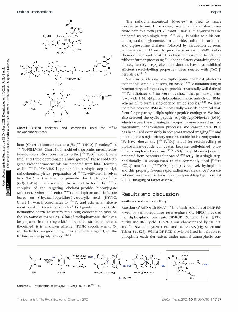
We aim to identify new diphosphine chemical platformsthat enable simple, one-step, kit-based 99mTc-radiolabelling ofreceptor-targeted peptides, to provide structurally well-defined99mTc radiotracers. Prior work has shown that primary aminesreact with 2,3-bis(diphenylphosphino)maleic anhydride (BMA,Scheme 1) to form a ring-opened amide species.18,19 We havetherefore selected BMA as a potentially versatile chemical plat-form for preparing a diphosphine-peptide conjugate. We havealso selected the cyclic peptide, Arg-Gly-Asp-DPhe-Lys (RGD),which targets the αvβ3-integrin receptor over-expressed in neo-vasculature, inflammation processes and cancer cells. RGDhas been used extensively in receptor-targeted imaging,9,20 andit contains a single primary amine suitable for bioconjugation.We have chosen the [99mTcVO2]
+ motif for radiolabelling ofdiphosphine-peptide conjugates because well-defined phos-phine complexes based on [99mTcVO2]
+ (e.g. Myoview) can beprepared from aqueous solutions of 99mTcO4
− in a single step.Additionally, in comparison to the commonly used [99mTc(CO)3]
+ motif, the [99mTcVO2]+ group is relatively hydrophilic,
and this property favours rapid radiotracer clearance from cir-culation via a renal pathway, potentially enabling high contrastSPECT imaging of target disease.
Results and discussionSynthesis and radiolabelling
Reaction of RGD with BMA21,22 in a basic solution of DMF fol-lowed by semi-preparative reverse-phase C18 HPLC providedthe diphosphine conjugate DP-RGD (Scheme 1) in ≥95%purity and 86% yield. DP-RGD was characterised by 1H, 13Cand 31P NMR, analytical HPLC and HR-ESI-MS (Fig. S1–S6 andTables S1, S2†). Whilst DP-RGD slowly oxidised in solution tophosphine oxide derivatives under normal atmospheric con-
Chart 1 Existing chelators and complexes used for 99mTcradiopharmaceuticals.
Scheme 1 Preparation of [MO2(DP-RGD)2]+ (M = Re, 99mTc).
Dalton Transactions Paper
This journal is © The Royal Society of Chemistry 2021 Dalton Trans., 2021, 50, 16156–16165 | 16157
Ope
n A
cces
s A
rtic
le. P
ublis
hed
on 1
5 O
ctob
er 2
021.
Dow
nloa
ded
on 1
/6/2
022
9:43
:41
AM
. T
his
artic
le is
lice
nsed
und
er a
Cre
ativ
e C
omm
ons
Attr
ibut
ion
3.0
Unp
orte
d L
icen
ce.
View Article Online
ditions, in the solid state, DP-RGD was stable to oxidation: itcan be handled either as a dry powder, in basic organic solu-tions, or in aqueous solutions at near-neutral pH. However, inacidic solutions, the reverse reaction was observed, and theDP-RGD conjugate decomposed to re-form RGD and BMA.
The chemistry of Re and Tc are closely similar. As Tc has nostable isotopes, it was convenient to prepare [ReO2(DP-RGD)2]
+
in order to obtain full characterisation. Reaction of [ReO2I(PPh3)2] with an excess of DP-RGD furnished geometricisomers of [ReO2(DP-RGD)2]
+ (Scheme 1), which are labelledcis and trans to denote the relative positions of the RGD moi-eties, and which were formed in the ratio of 54% and 46%respectively. The isomers were separated by reverse phase C18
HPLC. For both species, the most intense signals in theESI-MS at m/z = 785.92 and 1178.38, corresponded to the ions[M + 2H]3+ and [M + H]2+ where M = [ReO2(DP-RGD)2]
+ (seeFig. S1†).
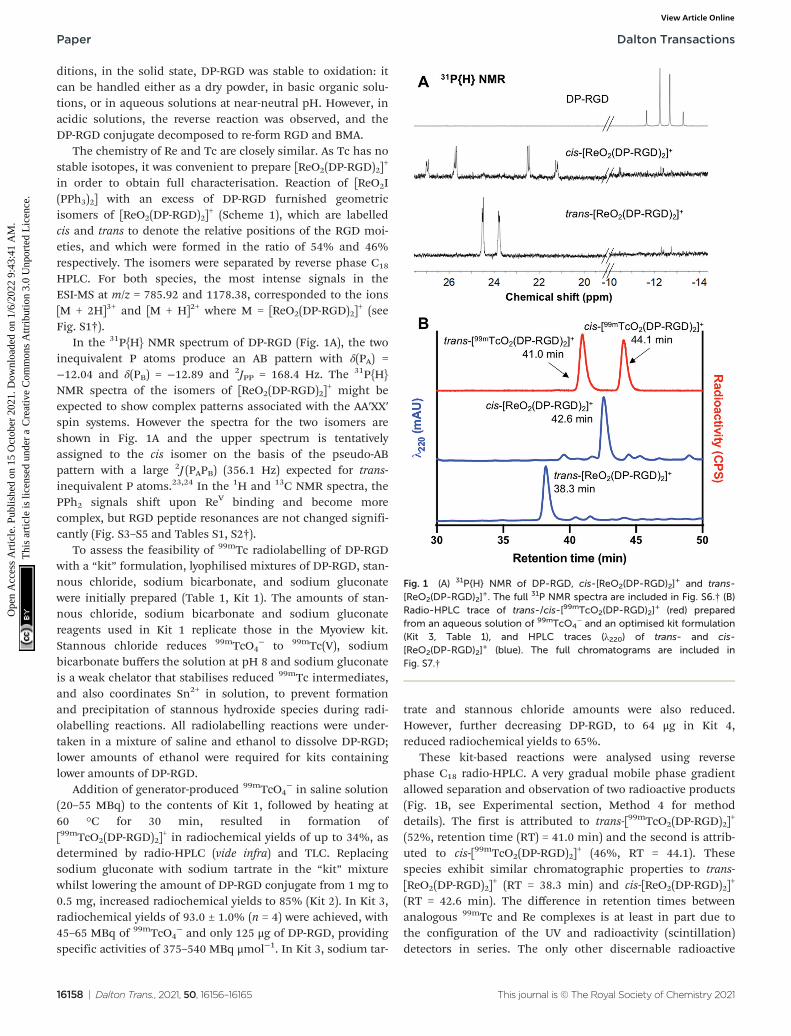
In the 31P{H} NMR spectrum of DP-RGD (Fig. 1A), the twoinequivalent P atoms produce an AB pattern with δ(PA) =−12.04 and δ(PB) = −12.89 and 2JPP = 168.4 Hz. The 31P{H}NMR spectra of the isomers of [ReO2(DP-RGD)2]
+ might beexpected to show complex patterns associated with the AA′XX′spin systems. However the spectra for the two isomers areshown in Fig. 1A and the upper spectrum is tentativelyassigned to the cis isomer on the basis of the pseudo-ABpattern with a large 2J (PAPB) (356.1 Hz) expected for trans-inequivalent P atoms.23,24 In the 1H and 13C NMR spectra, thePPh2 signals shift upon ReV binding and become morecomplex, but RGD peptide resonances are not changed signifi-cantly (Fig. S3–S5 and Tables S1, S2†).
To assess the feasibility of 99mTc radiolabelling of DP-RGDwith a “kit” formulation, lyophilised mixtures of DP-RGD, stan-nous chloride, sodium bicarbonate, and sodium gluconatewere initially prepared (Table 1, Kit 1). The amounts of stan-nous chloride, sodium bicarbonate and sodium gluconatereagents used in Kit 1 replicate those in the Myoview kit.Stannous chloride reduces 99mTcO4
− to 99mTc(V), sodiumbicarbonate buffers the solution at pH 8 and sodium gluconateis a weak chelator that stabilises reduced 99mTc intermediates,and also coordinates Sn2+ in solution, to prevent formationand precipitation of stannous hydroxide species during radi-olabelling reactions. All radiolabelling reactions were under-taken in a mixture of saline and ethanol to dissolve DP-RGD;lower amounts of ethanol were required for kits containinglower amounts of DP-RGD.
Addition of generator-produced 99mTcO4− in saline solution
(20–55 MBq) to the contents of Kit 1, followed by heating at60 °C for 30 min, resulted in formation of[99mTcO2(DP-RGD)2]
+ in radiochemical yields of up to 34%, asdetermined by radio-HPLC (vide infra) and TLC. Replacingsodium gluconate with sodium tartrate in the “kit” mixturewhilst lowering the amount of DP-RGD conjugate from 1 mg to0.5 mg, increased radiochemical yields to 85% (Kit 2). In Kit 3,radiochemical yields of 93.0 ± 1.0% (n = 4) were achieved, with45–65 MBq of 99mTcO4
− and only 125 μg of DP-RGD, providingspecific activities of 375–540 MBq μmol−1. In Kit 3, sodium tar-
trate and stannous chloride amounts were also reduced.However, further decreasing DP-RGD, to 64 μg in Kit 4,reduced radiochemical yields to 65%.
These kit-based reactions were analysed using reversephase C18 radio-HPLC. A very gradual mobile phase gradientallowed separation and observation of two radioactive products(Fig. 1B, see Experimental section, Method 4 for methoddetails). The first is attributed to trans-[99mTcO2(DP-RGD)2]
+
(52%, retention time (RT) = 41.0 min) and the second is attrib-uted to cis-[99mTcO2(DP-RGD)2]
+ (46%, RT = 44.1). Thesespecies exhibit similar chromatographic properties to trans-[ReO2(DP-RGD)2]
+ (RT = 38.3 min) and cis-[ReO2(DP-RGD)2]+
(RT = 42.6 min). The difference in retention times betweenanalogous 99mTc and Re complexes is at least in part due tothe configuration of the UV and radioactivity (scintillation)detectors in series. The only other discernable radioactive
Fig. 1 (A) 31P{H} NMR of DP-RGD, cis-[ReO2(DP-RGD)2]+ and trans-
[ReO2(DP-RGD)2]+. The full 31P NMR spectra are included in Fig. S6.† (B)
Radio-HPLC trace of trans-/cis-[99mTcO2(DP-RGD)2]+ (red) prepared
from an aqueous solution of 99mTcO4− and an optimised kit formulation
(Kit 3, Table 1), and HPLC traces (λ220) of trans- and cis-[ReO2(DP-RGD)2]
+ (blue). The full chromatograms are included inFig. S7.†
Paper Dalton Transactions
16158 | Dalton Trans., 2021, 50, 16156–16165 This journal is © The Royal Society of Chemistry 2021
Ope
n A
cces
s A
rtic
le. P
ublis
hed
on 1
5 O
ctob
er 2
021.
Dow
nloa
ded
on 1
/6/2
022
9:43
:41
AM
. T
his
artic
le is
lice
nsed
und
er a
Cre
ativ
e C
omm
ons
Attr
ibut
ion
3.0
Unp
orte
d L
icen
ce.
View Article Online
species (<1%) eluted with the solvent front, and correspondsto either unreacted 99mTcO4
− or 99mTc intermediates bound toother kit-based components (e.g. tartrate ligand).
Lastly, to unambiguously assign the stoichiometry of the[99mTcO2(DP-RGD)2]
+ compounds, experiments with long-livedtechnetium-99g (99gTc, t1/2 = 211 000 years) were undertaken. A
Table 1 Materials used in kit-based reactions for preparation of [99mTcO2(DP-RGD)2]+ and Myoview
Kit Kit components Radiochemical yielda
1 DP-RGD: 1.0 mg (0.93 μmol) ≤34%Sodium gluconate (NaC6H11O7): 1.0 mg (4.58 μmol)SnCl2·2H2O: 50 μg (0.22 μmol)NaHCO3: 1.8 mg (21.43 μmol)99mTcO4
− in 150 μL saline/150 μL EtOH2 DP-RGD: 500 μg (0.47 μmol) 85%
Sodium tartrate (Na2C4H4O6): 1.05 mg (4.58 μmol)SnCl2·2H2O: 50 μg (0.22 μmol)NaHCO3: 1.8 mg (21.4 μmol)99mTcO4
− in 150 μL saline/150 μL EtOH3 DP-RGD: 125 μg (0.12 μmol) ≥90%
Sodium tartrate: 0.26 mg (1.15 μmol)SnCl2·2H2O: 25 μg (0.11 μmol)NaHCO3: 0.9 mg (10.71 μmol)99mTcO4
− in 250 μL saline/50 μL EtOH4 DP-RGD: 64 μg (0.06 μmol) 65%
Sodium tartrate: 0.26 mg (1.15 μmol)SnCl2·2H2O: 25 μg (0.11 μmol)NaHCO3: 0.9 mg (10.71 μmol)99mTcO4
− in 260 μL saline/40 μL EtOHMyoview (single dose kits) Diphosphine: 250 μg (0.65 μmol) Routinely > 90%
Sodium gluconate: 1.0 mg (4.6 μmol)SnCl2·2H2O: 50 μg (0.22 μmol)NaHCO3: 1.8 mg (21.4 μmol)99mTcO4
− in saline
a Reactions were undertaken in duplicate to ensure reproducibility of radiochemical yields, except for radiolabelling reactions with Kit 3, wherethe reaction was replicated four times to give an average radiochemical yield of 93.0 ± 1.0%.
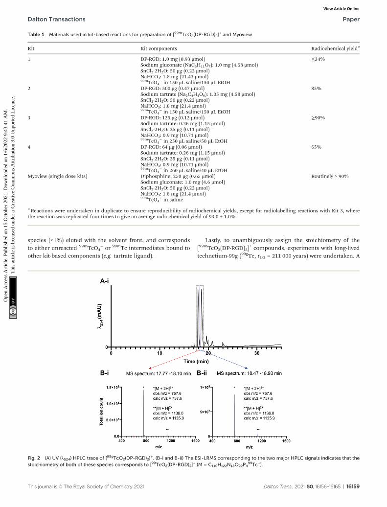
Fig. 2 (A) UV (λ524) HPLC trace of [99gTcO2(DP-RGD)2]+. (B-i and B-ii) The ESI-LRMS corresponding to the two major HPLC signals indicates that the
stoichiometry of both of these species corresponds to [99TcO2(DP-RGD)2]+ (M = C110H122N18O22P4
99Tc+).
Dalton Transactions Paper
This journal is © The Royal Society of Chemistry 2021 Dalton Trans., 2021, 50, 16156–16165 | 16159
Ope
n A
cces
s A
rtic
le. P
ublis
hed
on 1
5 O
ctob
er 2
021.
Dow
nloa
ded
on 1
/6/2
022
9:43
:41
AM
. T
his
artic
le is
lice
nsed
und
er a
Cre
ativ
e C
omm
ons
Attr
ibut
ion
3.0
Unp
orte
d L
icen
ce.
View Article Online
sample of [N(C4H9)4][99gTcOCl4] was reacted with 3 equivalents
of DP-RGD in methanol, and analysed by reverse phase C18
HPLC methods, which revealed the formation of two major Tcproduct complexes, with closely similar retention times.LC-ESI-LRMS analysis (Fig. 2) of [99gTcO2(DP-RGD)2]
+ showedthat these two major species possessed LRMS signals consist-ent with the stoichiometry of [99TcO2(DP-RGD)2]
+ (m/z = 757.6and 1136.0, corresponding to the ions [M + 2H]3+ and [M +H]2+ where M = [99TcO2(DP-RGD)2]
+). Additionally, these twomajor products, tentatively assigned as trans-[99gTcO2(DP-RGD)2]
+ and cis-[99gTcO2(DP-RGD)2]+ co-eluted
with radioactive signals of [99mTcO2(DP-RGD)2]+ (Fig. S8†).
Biological characterisation of [99mTcO2(DP-RGD)2]+
For all subsequent experiments, including in vivo experiments,[99mTcO2(DP-RGD)2]
+ was prepared from generator-produced99mTcO4
− and “Kit 3”, using our newly established one-stepradiolabelling protocol. Solutions containing both the desiredcis-[99mTcO2(DP-RGD)2]
+ and trans-[99mTcO2(DP-RGD)2]+ pro-
ducts, as well as unreacted DP-RGD ligand, were used withoutfurther purification.
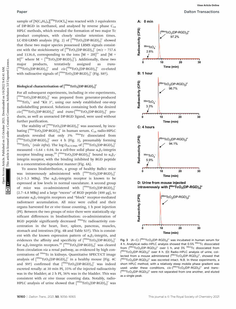
The stability of [99mTcO2(DP-RGD)2]+ was assessed, by incu-
bating [99mTcO2(DP-RGD)2]+ in human serum. C18 radio-HPLC
analysis revealed that only 3% 99mTc dissociated from[99mTcO2(DP-RGD)2]
+ over 4 h (Fig. 3), presumably forming99mTcO4
− (vide infra). The logDOCT/PBS of [99mTcO2(DP-RGD)2]
+
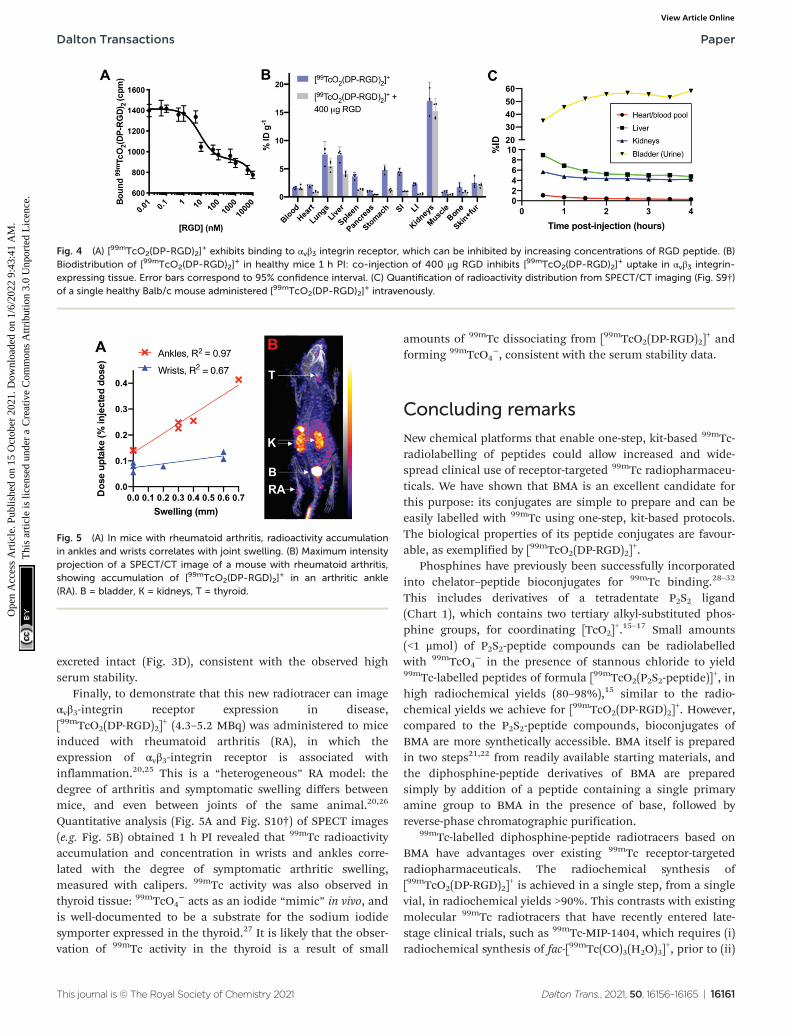
measured −1.64 ± 0.04. In a cell-free solid phase αvβ3-integrinreceptor binding assay,20 [99mTcO2(DP-RGD)2]
+ bound to αvβ3-integrin receptor, with the binding inhibited by RGD peptidein a concentration-dependent manner (Fig. 4A).
To assess biodistribution, a group of healthy Balb/c micewas intravenously administered with [99mTcO2(DP-RGD)2]
+
(4.3–5.3 MBq). The αvβ3-integrin receptor is known to beexpressed at low levels in normal vasculature. A second groupof mice was co-administered with [99mTcO2(DP-RGD)2]
+
(2.7–4.8 MBq) and a large “excess” of RGD peptide (400 μg), tosaturate αvβ3-integrin receptors and “block” receptor-mediatedradiotracer accumulation. All mice were culled and theirorgans harvested for ex vivo tissue counting, 1 h post injection(PI). Between the two groups of mice there were statistically sig-nificant differences in biodistribution: co-administration ofRGD peptide significantly decreased 99mTc radioactivity con-centration in the heart, liver, spleen, pancreas, muscles,stomach and intestines (Fig. 4B and Table S3†). This is consist-ent with the known expression pattern of αvβ3-integrin, andevidences the affinity and specificity of [99mTcO2(DP-RGD)2]
+
for αvβ3-integrin receptors.20 [99mTcO2(DP-RGD)2]+ was cleared
from circulation via a renal pathway, as evidenced by high con-centrations of 99mTc in kidneys. Quantitative SPECT/CT imageanalysis of [99mTcO2(DP-RGD)2]
+ in a healthy mouse (Fig. 4Cand S9†) confirmed that [99mTcO2(DP-RGD)2]
+ was indeedexcreted renally: at 30 min PI, 35% of the injected radioactivitywas in the bladder; at 2 h PI, 56% was in the bladder. This wasconsistent with ex vivo tissue counting data. Notably, radio-HPLC analysis of urine showed that [99mTcO2(DP-RGD)2]
+ was
Fig. 3 (A–C) [99mTcO2(DP-RGD)2]+ was incubated in human serum for
4 h. Analytical radio-HPLC analysis showed that 0.5% 99mTc dissociatedfrom [99mTcO2(DP-RGD)2]
+ over 1 h, and 3% 99mTc dissociated from[99mTcO2(DP-RGD)2]
+ over 4 h. (D) Radio-HPLC analysis of urine, col-lected from a mouse administered [99mTcO2(DP-RGD)2]
+, showed that[99mTcO2(DP-RGD)2]
+ was excreted intact. N.B. In these experiments, ashort HPLC method, with a relatively steep mobile phase gradient wasused: under these conditions, cis-[99mTcO2(DP-RGD)2]
+ and trans-[99mTcO2(DP-RGD)2]
+ were not separated from one another, and elutedas a single peak.
Paper Dalton Transactions
16160 | Dalton Trans., 2021, 50, 16156–16165 This journal is © The Royal Society of Chemistry 2021
Ope
n A
cces
s A
rtic
le. P
ublis
hed
on 1
5 O
ctob
er 2
021.
Dow
nloa
ded
on 1
/6/2
022
9:43
:41
AM
. T
his
artic
le is
lice
nsed
und
er a
Cre
ativ
e C
omm
ons
Attr
ibut
ion
3.0
Unp
orte
d L
icen
ce.
View Article Online
excreted intact (Fig. 3D), consistent with the observed highserum stability.
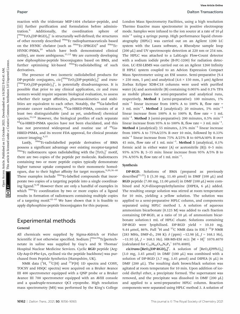
Finally, to demonstrate that this new radiotracer can imageαvβ3-integrin receptor expression in disease,[99mTcO2(DP-RGD)2]
+ (4.3–5.2 MBq) was administered to miceinduced with rheumatoid arthritis (RA), in which theexpression of αvβ3-integrin receptor is associated withinflammation.20,25 This is a “heterogeneous” RA model: thedegree of arthritis and symptomatic swelling differs betweenmice, and even between joints of the same animal.20,26
Quantitative analysis (Fig. 5A and Fig. S10†) of SPECT images(e.g. Fig. 5B) obtained 1 h PI revealed that 99mTc radioactivityaccumulation and concentration in wrists and ankles corre-lated with the degree of symptomatic arthritic swelling,measured with calipers. 99mTc activity was also observed inthyroid tissue: 99mTcO4
− acts as an iodide “mimic” in vivo, andis well-documented to be a substrate for the sodium iodidesymporter expressed in the thyroid.27 It is likely that the obser-vation of 99mTc activity in the thyroid is a result of small
amounts of 99mTc dissociating from [99mTcO2(DP-RGD)2]+ and
forming 99mTcO4−, consistent with the serum stability data.
Concluding remarks
New chemical platforms that enable one-step, kit-based 99mTc-radiolabelling of peptides could allow increased and wide-spread clinical use of receptor-targeted 99mTc radiopharmaceu-ticals. We have shown that BMA is an excellent candidate forthis purpose: its conjugates are simple to prepare and can beeasily labelled with 99mTc using one-step, kit-based protocols.The biological properties of its peptide conjugates are favour-able, as exemplified by [99mTcO2(DP-RGD)2]
+.Phosphines have previously been successfully incorporated
into chelator–peptide bioconjugates for 99mTc binding.28–32
This includes derivatives of a tetradentate P2S2 ligand(Chart 1), which contains two tertiary alkyl-substituted phos-phine groups, for coordinating [TcO2]
+.15–17 Small amounts(<1 μmol) of P2S2-peptide compounds can be radiolabelledwith 99mTcO4
− in the presence of stannous chloride to yield99mTc-labelled peptides of formula [99mTcO2(P2S2-peptide)]
+, inhigh radiochemical yields (80–98%),15 similar to the radio-chemical yields we achieve for [99mTcO2(DP-RGD)2]
+. However,compared to the P2S2-peptide compounds, bioconjugates ofBMA are more synthetically accessible. BMA itself is preparedin two steps21,22 from readily available starting materials, andthe diphosphine-peptide derivatives of BMA are preparedsimply by addition of a peptide containing a single primaryamine group to BMA in the presence of base, followed byreverse-phase chromatographic purification.
99mTc-labelled diphosphine-peptide radiotracers based onBMA have advantages over existing 99mTc receptor-targetedradiopharmaceuticals. The radiochemical synthesis of[99mTcO2(DP-RGD)2]
+ is achieved in a single step, from a singlevial, in radiochemical yields >90%. This contrasts with existingmolecular 99mTc radiotracers that have recently entered late-stage clinical trials, such as 99mTc-MIP-1404, which requires (i)radiochemical synthesis of fac-[99mTc(CO)3(H2O)3]
+, prior to (ii)
Fig. 4 (A) [99mTcO2(DP-RGD)2]+ exhibits binding to αvβ3 integrin receptor, which can be inhibited by increasing concentrations of RGD peptide. (B)
Biodistribution of [99mTcO2(DP-RGD)2]+ in healthy mice 1 h PI: co-injection of 400 μg RGD inhibits [99mTcO2(DP-RGD)2]
+ uptake in αvβ3 integrin-expressing tissue. Error bars correspond to 95% confidence interval. (C) Quantification of radioactivity distribution from SPECT/CT imaging (Fig. S9†)of a single healthy Balb/c mouse administered [99mTcO2(DP-RGD)2]
+ intravenously.
Fig. 5 (A) In mice with rheumatoid arthritis, radioactivity accumulationin ankles and wrists correlates with joint swelling. (B) Maximum intensityprojection of a SPECT/CT image of a mouse with rheumatoid arthritis,showing accumulation of [99mTcO2(DP-RGD)2]
+ in an arthritic ankle(RA). B = bladder, K = kidneys, T = thyroid.
Dalton Transactions Paper
This journal is © The Royal Society of Chemistry 2021 Dalton Trans., 2021, 50, 16156–16165 | 16161
Ope
n A
cces
s A
rtic
le. P
ublis
hed
on 1
5 O
ctob
er 2
021.
Dow
nloa
ded
on 1
/6/2
022
9:43
:41
AM
. T
his
artic
le is
lice
nsed
und
er a
Cre
ativ
e C
omm
ons
Attr
ibut
ion
3.0
Unp
orte
d L
icen
ce.
View Article Online
reaction with the tridentate MIP-1404 chelator–peptide, and(iii) further purification and formulation before adminis-tration.6 Additionally, the coordination sphere of[99mTcO2(DP-RGD)2]
+ is structurally well-defined; the structuresof other recently described 99mTc radiopharmaceuticals basedon the HYNIC chelator (such as 99mTc-3PRGD29 and 99mTc-HYNIC-PSMA,10 which have both demonstrated clinicalutility), are more ambiguous.11,12 We are currently developingnew diphosphine-peptide bioconjugates based on BMA, andfurther optimising kit-based 99mTc-radiolabelling of suchderivatives.
The presence of two isomeric radiolabelled products forDP-peptide conjugates, cis-[99mTcO2(DP-peptide)2]
+ and trans-[99mTcO2(DP-peptide)2]
+, is potentially disadvantageous. It ispossible that prior to any clinical application, cis and transisomers would require separate biological evaluation, to assesswhether their target affinities, pharmacokinetics and stabi-lities are equivalent to each other. Notably, the 68Ga-labelledprostate cancer radiotracer, 68Ga-HBED-PSMA, consists of atleast two distinguishable (and as yet, undefined) chemicalspecies.33,34 However, the biological profiles of each separate68Ga-HBED-PSMA species have not been elucidated, and thishas not prevented widespread and routine use of 68Ga-HBED-PSMA, and its recent FDA approval, for clinical prostatecancer imaging.4,35
Lastly, 99mTc-radiolabelled peptide derivatives of BMApossess a significant advantage over existing receptor-targeted99mTc radiotracers: upon radiolabelling with the [TcO2]
+ motif,there are two copies of the peptide per molecule. Radiotracerscontaining two or more peptide copies typically demonstratehigher tumour uptake compared to their monomeric homol-ogues, due to their higher affinity for target receptors.9,20,36–40
These examples include 99mTc-labelled compounds that incor-porate two copies of a targeting peptide into a single coordinat-ing ligand.9,38 However there are only a handful of examples inwhich 99mTc coordination by two or more copies of a ligandresults in formation of radiotracers containing multiple copiesof a targeting motif.41–43 We have shown that it is feasible toapply diphosphine-peptide bioconjugates for this purpose.
Experimental methodsGeneral
All chemicals were supplied by Sigma-Aldrich or FisherScientific if not otherwise specified. Sodium [99m/99Tc]pertech-netate in saline was supplied by Guy’s and St Thomas’Hospital Nuclear Medicine Services. Cyclic RGD peptide (Arg-Gly-Asp-D-Phe-Lys, cyclised via the peptide backbone) was pur-chased from Peptide Synthetics (Hampshire, UK).
NMR data (1H, 13C{H} and 31P{H} 1D spectra and COSY,TOCSY and HSQC spectra) were acquired on a Bruker AvanceIII 400 spectrometer equipped with a QNP probe or a BrukerAvance III 700 spectrometer equipped with an AVIII consoleand a quadruple-resonance QCI cryoprobe. High resolutionmass spectrometry (MS) was performed by the King’s College
London Mass Spectrometry Facilities, using a high resolutionThermo Exactive mass spectrometer in positive electrospraymode. Samples were infused to the ion source at a rate of 10 μlmin−1 using a syringe pump. High performance liquid chrom-atography (HPLC) was carried out on an Agilent 1200 LCsystem with the Laura software, a Rheodyne sample loop(200 μL) and UV spectroscopic detection at 220 nm or 254 nm.The HPLC was attached to a LabLogic Flow-Count detectorwith a sodium iodide probe (B-FC-3200) for radiation detec-tion. LC-ESI-LRMS was carried out on an Agilent 1260 InfinityII HPLC system coupled to an Advion Expression CompactMass Spectrometer using an ESI source. Semi-preparative (9.4× 250 mm, 5 μm) and analytical (4.6 × 150 mm, 5 μm) AgilentZorbax Eclipse XDB-C18 columns were used with purifiedwater (A) and acetonitrile (B) containing 0.005% and 0.1% TFAas mobile phases for semi-preparative and analytical runs,respectively. Method 1 (semi-preparative): 100 minutes, 1%min−1 linear increase from 100% A to 100% B, flow rate =3 mL min−1. Method 2 (analytical): 20 minutes, 5% min−1
linear increase from 100% A to 100% B, flow rate = 1 mLmin−1. Method 3 (semi-preparative): 200 minutes, 0.5% min−1
linear increase from 95% A to 100% B, flow rate = 3 mL min−1.Method 4 (analytical): 55 minutes, 2.5% min−1 linear increasefrom 100% A to 75%A/25% B over 10 min, followed by 0.33%min−1 linear increase from 75% A/25% B to 60%A/40% B over45 min, flow rate of 1 mL min−1. Method 5 (analytical, 0.1%formic acid in either water (A) or acetonitrile (B)): 0–5 min:95% A/5% B; 5–35 min: linear increase from 95% A/5% B to5% A/95% B; flow rate of 1 mL min−1.
Synthesis
DP-RGD. Solutions of BMA (prepared as previouslydescribed21,22) 1 (5.30 mg, 11.40 μmol) in DMF (100 μL) andRGD peptide (7.00 mg, 11.00 μmol) in DMF (100 μL) were com-bined and N,N-diisopropylethylamine (DIPEA, 6 μL) added.The resulting orange solution was stirred at room temperaturefor 10 min, yielding a yellow solution. The solution wasapplied to a semi-preparative HPLC column, and componentsseparated using HPLC method 1. A solution of aqueousammonium bicarbonate (0.125 M) was added to each fractioncontaining DP-RGD, at a ratio of 10 μL of ammonium bicar-bonate solution:1 mL of HPLC eluate. Solutions containingDP-RGD were lyophilised. DP-RGD yield = 10.10 mg,9.44 μmol, 86%. Full 1H and 13C NMR data in ESI.† 31P NMR(283 MHz, DMF-d7, 298 K): δ (ppm) −12.98 (d, J = 168.1 Hz),−11.95 (d, J = 168.1 Hz). HR-MS-ESI m/z: [M + H]+ 1070.4070(calculated for C55H62O10N9P2
+ 1070.4089).cis/trans-[ReO2(DP-RGD)2]
+. A solution of [ReO2I(PPh3)2](3.0 mg, 3.45 μmol) in DMF (100 μL) was combined with asolution of DP-RGD (3.7 mg, 3.45 μmol) and DIPEA (6 μL) inDMF (200 μL). The resulting dark brown/black solution wasagitated at room temperature for 10 min. Upon addition of ice-cold diethyl ether, a precipitate formed. The supernatant wasremoved, and the precipitate was dissolved in DMF (200 μL)and applied to a semi-preparative HPLC column. Reactioncomponents were separated using HPLC method 3. A solution of
Paper Dalton Transactions
16162 | Dalton Trans., 2021, 50, 16156–16165 This journal is © The Royal Society of Chemistry 2021
Ope
n A
cces
s A
rtic
le. P
ublis
hed
on 1
5 O
ctob
er 2
021.
Dow
nloa
ded
on 1
/6/2
022
9:43
:41
AM
. T
his
artic
le is
lice
nsed
und
er a
Cre
ativ
e C
omm
ons
Attr
ibut
ion
3.0
Unp
orte
d L
icen
ce.
View Article Online
aqueous ammonium bicarbonate (0.125 M) was added to eachfraction containing cis/trans-[ReO2(DP-RGD)2]
+ at a ratio of 10 μLof ammonium bicarbonate solution: 1 mL of HPLC eluate.Solutions containing cis/trans-[ReO2(DP-RGD)2]
+ were lyophilised.The lyophilised fractions that eluted at 65–67 min and68–70 min were identified as trans-[ReO2(DPP-N-RGD)2]
+ 0.8 mg,0.34 μmol, 9.9% and cis-[ReO2(DPP-N-RGD)2]
+ (0.9 mg,0.38 μmol, 11.0%), respectively. Full 1H and 13C NMR data inESI.† trans-[ReO2(DP-RGD)2]
+: 31P NMR (283 MHz, DMF-d7,298 K): δ (ppm) 23.781 (m), 24.506 (m). HR-MS-ESI m/z: [M +H]2+ 1179.3826 (calculated for C110H123N18O22P4Re
2+ 1179.3773),[M + 2H]3+ 786.5921 (calculated for C110H124N18O22P4Re
3+
786.5906). cis-[ReO2(DP-RGD)2]+: 31P NMR (283 MHz, DMF-d7,
298 K): δ (ppm) 21.848 (dm, J1 = 356.1 Hz), 26.335 (dm, J1 =356.1 Hz). HRMS-ESI m/z: [M + H]2+ 1179.3826 (calculated forC110H123N18O22P4Re
2+ 1179.3773), [M + 2H]3+ 786.5921 (calcu-lated for C110H124N18O22P4Re
3+ 786.5906).
Preparation and characterisation of [99m/99gTcO2(DP-RGD)2]+
Radiolabelling kits. An aqueous stock solution was preparedcontaining the required amounts of sodium bicarbonate, tinchloride dihydrate and either sodium gluconate or sodium tar-trate dibasic dihydrate. The pH of this solution was adjustedto 8.5 by dropwise addition of an aqueous solution of sodiumhydroxide (0.1 M). Aliquots of the stock solution were mixedwith the required amount of DP-RGD (in ethanol), and theresulting solutions (Table 1) were frozen and lyophilised. Thelyophilised kits were stored at −18 °C prior to use.
99mTc radiolabelling. DP-RGD was radiolabelled with genera-tor-produced 99mTcO4
− in saline solution (0.9% NaCl in water,w/v). Saline solution containing 99mTcO4
− and ethanol wereadded to the contents of a “kit” (amounts listed in Table 1).The radiolabelling kit mixture was heated at 60 °C for 30 min,and then analysed by analytical HPLC (method 2) and instantthin layer chromatography (iTLC) using iTLC SGI0001 strips (9or 10 cm length; Varian Medical Systems, Crawley, UK). TheiTLC plates were scanned with a PerkinElmer StoragePhosphor System (Cyclone) or a LabLogic miniScan TLCreader equipped with Laura software.
Two separate iTLC analyses were undertaken, to enablequantification of 99mTc-colloids, unreacted 99mTcO4
− and[99mTcO2(DP-RGD)2]
+. To quantify amounts of unreacted99mTcO4
−, acetone was used as a mobile phase: Rf values:99mTcO4
− >0.9, 99mTc colloids <0.1, [99mTcO2(DP-RGD)2]+ <0.1.
To quantify 99mTc-colloid formation, a 1 : 1 mixture of metha-nol and 2 M aqueous ammonium acetate solution was used asa mobile phase: 99mTcO4
− >0.9, 99mTc colloids <0.1,[99mTcO2(DP-RGD)2]
+ >0.9.99gTc radiolabelling. [N(C4H9)4][
99gTcOCl4]44 (0.4 mg) dis-
solved in methanol (50 μL) was added to DP-RGD (2.4 mg, 3equiv.) dissolved in methanol (200 μL), resulting in a yellow-orange solution. The sample was analysed by reverse-phaseanalytical HPLC (method 4, vide infra), and LC-ESI-LRMS(method 5). ESI-LRMS m/z: [M + H]2+ 1136.0 (calculated forC110H123N18O22P4
99Tc2+ 1135.9), [M + 2H]3+ 757.6 (calculatedfor C110H124N18O22P4
99Tc3+ 757.6).
Co-elution of [99mTcO2(DP-RGD)2]+ with cis/trans-
[ReO2(DP-RGD)2]+ and cis/trans-[99gTcO2(DP-RGD)2]
+.[99mTcO2(DP-RGD)2]
+ was prepared in >90% RCY as describedabove, and co-injected with cis-[ReO2(DP-RGD)2]
+ and separately,trans-[ReO2(DP-RGD)2]
+, onto a reverse-phase analytical HPLCcolumn (method 4). A sample of cis/trans-[99gTcO2(DP-RGD)2]
+
was also analysed, using the same HPLC method. Retentiontimes: trans/cis-[99mTcO2(DP-RGD)2]
+ 41.0 min and 44.1 min(NaI scintillator detection); trans/cis-[99gTcO2(DP-RGD)2]
+
40.70 min and 43.1 min; trans-[ReO2(DP-RGD)2]+ 38.3 min and
cis-[ReO2(DP-RGD)2]+ 42.6 min.
LogD (pH 7.4). The following procedure was carried out intriplicate. A solution containing [99mTcO2(DP-RGD)2]
+ (1 MBqin 7.5 μL) was combined with phosphate buffered saline (pH7.4, 500 μL) and octanol (500 μL), and the mixture was agitatedfor 30 min. The mixture was then centrifuged (10 000 rpm,10 minutes), and aliquots of octanol and aqueous PBS wereanalysed for radioactive using a gamma counter. Log D = −1.64± 0.04.
Serum stability. A solution containing [99mTcO2(DP-RGD)2]+
(100 μL, 79 MBq) was added to filtered human serum (Sigma-Aldrich, 900 μL) and incubated at 37 °C for 4 h. At 1 and 4 h,aliquots were taken. Each aliquot (300 μL) was treated with ice-cold acetonitrile (300 μL) to precipitate and remove serum pro-teins. Acetonitrile in the supernatant was then removed byevaporation under a stream of N2 gas (40 °C, 30 min). Thefinal solution was then analysed by reverse-phase analyticalHPLC (method 2).
αvβ3-integrin solid-phase competitive binding assay. Theaffinity of [99mTcO2(DP-RGD)2]
+ for αvβ3 integrin was deter-mined in a solid-phase competitive binding assay.20 In brief,wells of a 96 well plate were coated with 150 ng mL−1 integrinαvβ3 in 100 μL coating buffer (25 mM Tris HCl pH 7.4, 150 mMNaCl, 1 mM CaCl2, 0.5 mM MgCl2, and 1 mM MnCl2) over-night at 4 °C. Wells were then washed twice in binding buffer(coating buffer plus 0.1% bovine serum albumin (BSA)) beforebeing blocked for 2 hours at room temperature with blockingbuffer (coating buffer plus 1% BSA). After a further two washesin binding buffer, both [99mTcO2(DP-RGD)2]
+ (RCY > 96%, 1–2kBq in 50 μL binding buffer, containing 1.2 pmol DP-RGDpeptide) and RGD peptide (10.0 pM to 10 000 nM, 50 μL inbinding buffer) were added simultaneously to wells, and left toincubate for 1 h at room temperature, before being washedtwice as before. Finally, the amount of activity bound to thewells was counted. Binding of [99mTcO2(DP-RGD)2]
+ to αvβ3integrin was displaced by RGD peptide in a concentration-dependent manner. The pseudo-IC50 value of 8.54 ± 3.45 nM(95% CI: 1.67–15.41 nM) was calculated using a non-linearregression model (Binding/Saturation, one site – total) inGraphPad Prism (n = 6 from one experiment).
Pre-clinical imaging and in vivo biodistribution studies of[99mTcO2(DP-RGD)2]
+
Animal imaging studies were ethically reviewed and carriedout in accordance with the Animals (Scientific Procedures) Act1986 (ASPA) UK Home Office regulations governing animal
Dalton Transactions Paper
This journal is © The Royal Society of Chemistry 2021 Dalton Trans., 2021, 50, 16156–16165 | 16163
Ope
n A
cces
s A
rtic
le. P
ublis
hed
on 1
5 O
ctob
er 2
021.
Dow
nloa
ded
on 1
/6/2
022
9:43
:41
AM
. T
his
artic
le is
lice
nsed
und
er a
Cre
ativ
e C
omm
ons
Attr
ibut
ion
3.0
Unp
orte
d L
icen
ce.
View Article Online
experimentation. SPECT/CT imaging was accomplished usinga pre-clinical nanoScan SPECT/CT Silver Upgrade instrument(Mediso) calibrated for technetium-99m. All scans wereacquired by helical SPECT (4-head scanner with 4 × 9 [1.4 mm]pinhole collimators), and helical CT with 1.4 mm aperture col-limators. All acquired images were reconstructed using a full3D Monte Carlo-based iterative algorithm (Tera-Tomo; Mediso)and further processed and analysed using VivoQuant software(inviCRO, USA).
SPECT/CT imaging and biodistribution in healthy mice. Afemale, balb/c mouse (2 months old) was anaesthetised (2–3%v/v isofluorane in oxygen), scanned by CT and injected intra-venously (tail vein) with [99mTcO2(DP-RGD)2]
+ (21 MBq contain-ing 22 μg of DP-RGD peptide). SPECT images (8 × 30 minimages) were acquired over 4 h. At the end of the imaging pro-cedure, the mouse was culled by cervical dislocation and asample of the urine analysed by reverse-phase HPLC (analyti-cal, method 2).
Female balb/c mice (2 months old) were anaesthetised(2–3% v/v isofluorane in oxygen) and injected intravenously(tail vein) with [99mTcO2(DP-RGD)2]
+ (2.7–5.3 MBq containing5 μg of DP-RGD). For blocking studies, animals were co-injected with RGD peptide (400 μg). Mice remained underanaesthetic for 1 h, after which they were culled (pentabarbi-tone by i.v. injection). Tissues and organs were harvested andweighed, and radioactivity counted using a Gamma Counter(Wallac 1282 CompuGamma Universal Gamma Counter).
SPECT/CT imaging and biodistribution in mice inducedwith rheumatoid arthritis. We used a K/BxN serum transferarthritis (STA) model of rheumatoid arthritis.20,26 On day 0and 2, female C57Bl/6J mice (2 months old) were injectedintraperitoneally with arthritogenic serum in sterile filteredPBS (150 μL, 50% v/v, serum obtained from arthritic K/B × Ntransgenic mice). Disease severity was evaluated in micethroughout the induction period, by measuring weight, thick-ness of swollen paws using microcallipers, and visual scoringon a scale of 0–3 per paw. SPECT/CT imaging and biodistribu-tion was undertaken on day 7.
Mice were anesthetised (2.5–3% v/v isofluorane) and theirpaws were measured using microcallipers. Mice were theninjected intravenously with [99mTcO2(DP-RGD)2]
+ (approx. 5MBq containing 5 μg of DP-RGD) and allowed to recover fromanaesthetic administration. At 1 h post-injection of radiotracer,mice were culled (sodium pentabarbitone), and underwentSPECT/CT scanning post-mortem for 60–180 min. Finally,tissues and organs were harvested and weighed, and radioac-tivity counted using a Gamma Counter (Wallac 1282CompuGamma Universal Gamma Counter). The acquiredimages were processed to units of %ID and the regions ofinterest (ROIs) delineated by CT using VivoQuant software(inviCRO, USA). Radioactivity in ankle and wrist ROIs wereobtained in units of %ID and %ID/cm−3. Each “ankle” ROIwas defined as the area between the tibiofibula joint and thebase of phalanx V. Each “wrist” ROI was defined as the areabetween the narrowest point of the wrist (ulna and radius) andthe end of the forepaw.
Conflicts of interest
There are no conflicts to declare.
Acknowledgements
This research was supported by a Cancer Research UK CareerEstablishment Award (C63178/A24959), King’s College London& Imperial College London EPSRC Centre for DoctoralTraining in Medical Imaging (EP/L015226/1), the EPSRC pro-gramme for Next Generation Molecular Imaging and Therapywith Radionuclides (EP/S032789/1, “MITHRAS”), RosetreesTrust (M685, M606), the Wellcome Multiuser EquipmentRadioanalytical Facility funded by Wellcome Trust (212885/Z/18/Z), and the Centre for Medical Engineering funded by theWellcome Trust and the Engineering and Physical SciencesResearch Council (WT088641/Z/09/Z), and the King’s CollegeLondon Centre for Biomolecular Spectroscopy funded byWellcome Trust (202762/Z/16/Z) and British Heart Foundation(IG/16/2/32273).
References
1 J. A. Jackson, I. N. Hungnes, M. T. Ma and C. Rivas,Bioconjugate Chem., 2020, 31, 483–491.
2 C. Rivas, J. A. Jackson, I. N. Hungnes and M. T. Ma,Radioactive Metals in Imaging and Therapy, in ComprehensiveCoordination Chemistry III, ed. E. C. Constable, G. Parkin,L. Que Jr. and N. J. Long, 2021, ch. 17, vol. 9, pp. 706–740,DOI: 10.1016/B978-0-08-102688-5.00010-6.
3 J. Strosberg, G. El-Haddad, E. Wolin, A. Hendifar, J. Yao,B. Chasen, E. Mittra, P. L. Kunz, M. H. Kulke, H. Jacene,D. Bushnell, T. M. O’Dorisio, R. P. Baum, H. R. Kulkarni,M. Caplin, R. Lebtahi, T. Hobday, E. Delpassand, E. VanCutsem, A. Benson, R. Srirajaskanthan, M. Pavel, J. Mora,J. Berlin, E. Grande, N. Reed, E. Seregni, K. Öberg,M. L. Sierra, P. Santoro, T. Thevenet, J. L. Erion,P. Ruszniewski, D. Kwekkeboom and E. Krenning, N.Engl. J. Med., 2017, 376, 125–135.
4 M. S. Hofman, N. Lawrentschuk, R. J. Francis, C. Tang,I. Vela, P. Thomas, N. Rutherford, J. M. Martin,M. Frydenberg, R. Shakher, L. M. Wong, K. Taubman,S. Ting Lee, E. Hsiao, P. Roach, M. Nottage, I. Kirkwood,D. Hayne, E. Link, P. Marusic, A. Matera, A. Herschtal,A. Iravani, R. J. Hicks, S. Williams and D. G. Murphy,Lancet, 2020, 395, 1208–1216.
5 S. Liu and S. Chakraborty, Dalton Trans., 2011, 40, 6077–6086.6 S. M. Hillier, K. P. Maresca, G. Lu, R. D. Merkin,
J. C. Marquis, C. N. Zimmerman, W. C. Eckelman, J. L. Joyaland J. W. Babich, J. Nucl. Med., 2013, 54, 1369–1376.
7 S. Robu, M. Schottelius, M. Eiber, T. Maurer, J. Gschwend,M. Schwaiger and H.-J. Wester, J. Nucl. Med., 2017, 58, 235–242.
8 M. J. Abrams, M. Juweid, C. I. TenKate, D. A. Schwartz,M. M. Hauser, F. E. Gaul, A. J. Fuccello, R. H. Rubin,
Paper Dalton Transactions
16164 | Dalton Trans., 2021, 50, 16156–16165 This journal is © The Royal Society of Chemistry 2021
Ope
n A
cces
s A
rtic
le. P
ublis
hed
on 1
5 O
ctob
er 2
021.
Dow
nloa
ded
on 1
/6/2
022
9:43
:41
AM
. T
his
artic
le is
lice
nsed
und
er a
Cre
ativ
e C
omm
ons
Attr
ibut
ion
3.0
Unp
orte
d L
icen
ce.
View Article Online
H. W. Strauss and A. J. Fischman, J. Nucl. Med., 1990, 31,2022–2028.
9 Z. Zhu, W. Miao, Q. Li, H. Dai, Q. Ma, F. Wang, A. Yang,B. Jia, X. Jing, S. Liu, J. Shi, Z. Liu, Z. Zhao, F. Wang andF. Li, J. Nucl. Med., 2012, 53, 716–722.
10 J. Zhang, J. Zhang, X. Xu, L. Lu, S. Hu, C. Liu, J. Cheng,S. Song, Y. Zhang and L. Q. Shi, Sci. Rep., 2020, 10, 1–9.
11 L. K. Meszaros, A. Dose, S. C. G. Biagini and P. J. Blower,Inorg. Chim. Acta, 2010, 363, 1059–1069.
12 R. C. King, M. B.-U. Surfraz, S. C. G. Biagini, P. J. Blowerand S. J. Mather, Dalton Trans., 2007, 4998–5007.
13 J. D. Kelly, A. M. Forster, B. Higley, C. M. Archer, F. S. Booker,L. R. Canning, K. W. Chiu, B. Edwards, H. K. Gill,M. McPartlin, K. R. Nagle, I. A. Latham, R. D. Pickett,A. E. Storey and P. M. Webbon, J. Nucl. Med., 1993, 34, 222–227.
14 J. Veggeland, G. K. Madsen and S. Hemsted,Radiopharmaceutical Composition, US9549999B, 2017.
15 H. Gali, T. J. Hoffman, G. L. Sieckman, N. K. Owen,K. V. Katti and W. A. Volkert, Bioconjugate Chem., 2001, 12,354–363.
16 S. R. Karra, R. Schibli, H. Gali, K. V. Katti, T. J. Hoffman,C. Higginbotham, G. L. Sieckman and W. A. Volkert,Bioconjugate Chem., 1999, 10, 254–260.
17 H. Gali, S. R. Karra, V. S. Reddy and K. V. Katti, Angew.Chem., Int. Ed., 1999, 38, 2020–2023.
18 J. S. Lewis, S. L. Heath, A. K. Powell, J. Zweit andP. J. Blower, J. Chem. Soc., Dalton Trans., 1997, 855–861.
19 J. S. Lewis, J. Zweit, J. L. J. Dearling, B. C. Rooney andP. J. Blower, Chem. Commun., 1996, 1093–1094.
20 C. Imberti, S. Y. A. Terry, C. Cullinane, F. Clarke,G. H. Cornish, N. K. Ramakrishnan, P. Roselt, A. P. Cope,R. J. Hicks, P. J. Blower and M. T. Ma, Bioconjugate Chem.,2017, 28, 481–495.
21 D. Fenske and H. J. Becher, Chem. Ber., 1974, 107, 117–122.22 M. Fei, S. K. Sur and D. R. Tyler, Organometallics, 1991, 10,
419–423.23 R. G. Cavell, R. W. Hilts, H. Luo and R. McDonald, Inorg.
Chem., 1999, 38, 897–905.24 H. Luo, I. Setyawati, S. J. Rettig and C. Orvig, Inorg. Chem.,
1995, 34, 2287–2299.25 J. D. Young, V. Abbate, C. Imberti, L. K. Meszaros,
M. T. Ma, S. Y. A. Terry, R. C. Hider, G. E. Mullen andP. J. Blower, J. Nucl. Med., 2017, 58, 1270–1277.
26 P. A. Monach, D. Mathis and C. Benoist, Curr. Protoc.Immunol., 2008, 81, 15.22.1–15.22.12.
27 A. Khoshnevisan, K. Chuamsaamarkkee, M. Boudjemeline,A. Jackson, G. E. Smith, A. D. Gee, G. O. Fruhwirth andP. J. Blower, J. Nucl. Med., 2017, 58, 156–161.
28 K. K. Kothari, H. Gali, K. R. Prabhu, N. Pillarsetty,N. K. Owen, K. V. Katti, T. J. Hoffman and W. A. Volkert,Nucl. Med. Biol., 2002, 29, 83–89.
29 C. Bolzati, A. Caporale, S. Agostini, D. Carta, M. Cavazza-Ceccato, F. Refosco, F. Tisato, E. Schievano and G. Bandoli,Nucl. Med. Biol., 2007, 34, 511–522.
30 C. Bolzati, E. Malagò, A. Boschi, A. Cagnolini,M. Porchia and G. Bandoli, New J. Chem., 1999, 23, 807–809.
31 R. Kannan, N. Pillarsetty, H. Gali, T. J. Hoffman,C. L. Barnes, S. S. Jurisson, C. J. Smith and W. A. Volkert,Inorg. Chem., 2011, 50, 6210–6219.
32 K. K. Kothari, K. Raghuraman, N. K. Pillarsetty,T. J. Hoffman, N. K. Owen, K. V. Katti and W. A. Volkert,Appl. Radiat. Isot., 2003, 58, 543–549.
33 M. I. Tsionou, C. E. Knapp, C. A. Foley, C. R. Munteanu,A. Cakebread, C. Imberti, T. R. Eykyn, J. D. Young,B. M. Paterson, P. J. Blower and M. T. Ma, RSC Adv., 2017,7, 49586–49599.
34 M. Eder, O. Neels, M. Müller, U. Bauder-Wüst, Y. Remde,M. Schäfer, U. Hennrich, M. Eisenhut, A. Afshar-Oromieh,U. Haberkorn and K. Kopka, Pharmaceuticals, 2014, 7, 779–796.
35 U. Hennrich and M. Eder, Pharmaceuticals, 2021, 14, 713.36 N. A. Zia, C. Cullinane, J. K. Van Zuylekom, K. Waldeck,
L. E. McInnes, G. Buncic, M. B. Haskali, P. D. Roselt,R. J. Hicks and P. S. Donnelly, Angew. Chem., Int. Ed., 2019,58, 14991–14994.
37 J. Notni, K. Pohle and H.-J. Wester, Nucl. Med. Biol., 2013,40, 33–41.
38 A. Frei, E. Fischer, B. C. Childs, J. P. Holland andR. Alberto, Dalton Trans., 2019, 48, 14600–14605.
39 Z. Li, W. Cai, Q. Cao, K. Chen, Z. Wu, L. He and X. Chen,J. Nucl. Med., 2007, 48, 1162–1171.
40 X. Zhang, Z. Xiong, Y. Wu, W. Cai, J. R. Tseng,S. S. Gambhir and X. Chen, J. Nucl. Med., 2006, 47, 113–121.
41 J. K. Bordoloi, D. Berry, I. U. Khan, K. Sunassee,R. T. M. De Rosales, C. Shanahan and P. J. Blower, DaltonTrans., 2015, 44, 4963–4975.
42 A. J. North, J. A. Karas, M. T. Ma, P. J. Blower,U. Ackermann, J. M. White and P. S. Donnelly, Inorg.Chem., 2017, 56, 9725–9741.
43 Y. Mizuno, T. Uehara, H. Hanaoka, Y. Endo, C. W. Jen andY. Arano, J. Med. Chem., 2016, 59, 3331–3339.
44 A. Davison, C. Orvig, H. S. Trop, M. Sohn,B. V. DePamphilis and A. G. Jones, Inorg. Chem., 2002, 19,1988–1992.
Dalton Transactions Paper
This journal is © The Royal Society of Chemistry 2021 Dalton Trans., 2021, 50, 16156–16165 | 16165
Ope
n A
cces
s A
rtic
le. P
ublis
hed
on 1
5 O
ctob
er 2
021.
Dow
nloa
ded
on 1
/6/2
022
9:43
:41
AM
. T
his
artic
le is
lice
nsed
und
er a
Cre
ativ
e C
omm
ons
Attr
ibut
ion
3.0
Unp
orte
d L
icen
ce.
View Article Online


![iGalerie.cz: [Rejstriky] Psychologie (Rita L. Atkinson a kol](https://img.pdfslide.net/doc/110x75/63367c9f4e9c1ac02e081a27/igaleriecz-rejstriky-psychologie-rita-l-atkinson-a-kol.jpg)


