Embed Size (px)
Citation preview
Hyaluronate Fragments Reverse Skin Atrophyby a CD44-Dependent MechanismGurkan Kaya
1*, Christian Tran
1, Olivier Sorg
1, Raymonde Hotz
1, Denise Grand
1, Pierre Carraux
1, Liliane Didierjean
1,
Ivan Stamenkovic2
, Jean-Hilaire Saurat1
1 Department of Dermatology, University of Geneva, Geneva, Switzerland, 2 Institute of Pathology, University of Lausanne, Lausanne, Switzerland
Funding: This work was supportedby the University of GenevaDermatology Fund and GendermResearch Program to GK and JHS,and Swiss National ScienceFoundation grant 3100-65090-01and the Molecular OncologyNational Centre for Competence inResearch (NCCR) to IS. The fundershad no role in study design, datacollection and analysis, decision topublish, or preparation of themanuscript.
Competing Interests: The authorshave declared that no competinginterests exist.
Academic Editor: Kim Yancey,Medical College of Wisconsin, UnitedStates of America
Citation: Kaya G, Tran CT, Sorg O,Hotz R, Grand D. et al. (2006)Hyaluronate fragments reverse skinatrophy by a CD44-dependentmechanism. PLoS Med 3(12): e493.doi:10.1371/journal.pmed.0030493
Received: June 12, 2006Accepted: October 9, 2006Published: December 19, 2006
Copyright: � 2006 Kaya et al. This isan open-access article distributedunder the terms of the CreativeCommons Attribution License, whichpermits unrestricted use,distribution, and reproduction in anymedium, provided the originalauthor and source are credited.
Abbreviations: ADAM; a disintegrinand metalloproteinase; AR,amphiregulin; EGF, epidermal-likegrowth factor; HA, hyaluronate;HABP, HA-binding protein; HAF, HAfragment(s); HAFi, intermediate-sizeHAF; HAFl, large-size HAF; HAFs,small-size HAF; HAS, HA synthase;HB-EGF, heparin-binding EGF;HUVEC, human umbilical veinendothelial cell; MMP-7, matrixmetalloproteinase 7; pro-HB-EGF,HB-EGF precursor; SEM, standarderror of the mean; TIMP-3, tissueinhibitor of metalloproteinase-3; wt,wild-type
* To whom correspondence shouldbe addressed. E-mail: [email protected]
A B S T R A C T
Background
Skin atrophy is a common manifestation of aging and is frequently accompanied byulceration and delayed wound healing. With an increasingly aging patient population,management of skin atrophy is becoming a major challenge in the clinic, particularly in light ofthe fact that there are no effective therapeutic options at present.
Methods and Findings
Atrophic skin displays a decreased hyaluronate (HA) content and expression of the major cell-surface hyaluronate receptor, CD44. In an effort to develop a therapeutic strategy for skinatrophy, we addressed the effect of topical administration of defined-size HA fragments (HAF)on skin trophicity. Treatment of primary keratinocyte cultures with intermediate-size HAF (HAFi;50,000–400,000 Da) but not with small-size HAF (HAFs; ,50,000 Da) or large-size HAF (HAFl;.400,000 Da) induced wild-type (wt) but not CD44-deficient (CD44�/�) keratinocyteproliferation. Topical application of HAFi caused marked epidermal hyperplasia in wt but notin CD44�/� mice, and significant skin thickening in patients with age- or corticosteroid-relatedskin atrophy. The effect of HAFi on keratinocyte proliferation was abrogated by antibodiesagainst heparin-binding epidermal growth factor (HB-EGF) and its receptor, erbB1, which forma complex with a particular isoform of CD44 (CD44v3), and by tissue inhibitor ofmetalloproteinase-3 (TIMP-3).
Conclusions
Our observations provide a novel CD44-dependent mechanism for HA oligosaccharide-induced keratinocyte proliferation and suggest that topical HAFi application may provide anattractive therapeutic option in human skin atrophy.
The Editors’ Summary of this article follows the references.
PLoS Medicine | www.plosmedicine.org December 2006 | Volume 3 | Issue 12 | e4932291
PLoSMEDICINE
Introduction
Skin atrophy is a frequent and clinically relevant manifes-tation of aging, often complicated by ulceration and impairedwound healing. Experimental evidence suggests that defectivefunction of the principal cell-surface hyaluronate (HA)receptor CD44 [1,2], which is associated with impaired HAmetabolism, may underlie the pathogenesis of this disorder[3]. HA is the most abundant glycosaminoglycan in cutaneoustissues [4] and is deposited in the extracellular matrix as ahigh-molecular-weight polymer. Cleavage of the HA polymerduring tissue remodeling gives rise to lower-molecular-weightfragments that elicit a variety of CD44-mediated cellularresponses, including proliferation, migration, and cytokinesynthesis. Increasing evidence suggests that cellular responseselicited by HA depend on the size of the HA oligosaccharidesas well as on the responding cell type [5,6]. Thus, HAfragments (HAF), but not native HA polymer or HAdisaccharides, could activate Ras and PKCf in tumor cells ina CD44-dependent fashion [7]; 35,000- to 280,000-Da HApreparations were shown to stimulate CD44-mediated che-mokine gene expression in alveolar macrophages [8]; HAoligosaccharides have been reported to induce a toll-likereceptor–mediated response in endothelial cells [9]; and onlytetra- and hexasaccharide HA fragments have been observedto induce immunophenotypic maturation of human den-dritic cells [10].
Cellular responses to HA are primarily mediated by CD44,the most broadly expressed cell-surface HA receptor whosefunction is regulated at transcriptional, translational, andpost-translational levels. Expression levels, variant isoformselection, and glycosylation all play an important role in theability of CD44 to bind HA [11,12]. Because of its polymericstructure, HA can crosslink several CD44 molecules on thesurface of a cell, an effect that is believed to be essential intriggering proliferative, invasive, and promigratory signals[11]. HA-mediated CD44 clustering can have a variety ofeffects, depending, at least in part, on the CD44 isoform thatis implicated in HA binding. Thus, in mouse mammary anduterine epithelia, HA can induce heparan sulfate–bearingvariants of CD44 (CD44v3) to recruit a cell-surface complexthat includes matrix metalloproteinase 7 (MMP-7), heparin-binding epidermal-like growth factor (HB-EGF) precursorHB-EGF (pro-HB-EGF), and erbB4 [13]. MMP-mediated HB-EGF activation within this complex promotes epithelial cellsurvival in the lactating mammary gland and postpartumuterus [13]. CD44v3 isoforms are expressed in skin keratino-cytes, where HA has been shown to regulate keratinocyteproliferation [3]. CD44 in turn appears to play an importantrole in maintaining HA homeostasis in skin [3], and its levelsappear to correlate with skin trophicity. Thus, decreasedepidermal CD44 expression is associated with the patho-genesis of an atrophic skin disorder known as lichensclerosus et atrophicus [14]. Conversely, epidermal hyper-plasia induced by topical administration of retinoids inmouse skin is accompanied by increased CD44 and HAsynthase (HAS) expression with a concomitant increase inHA deposition [15]. To elucidate the mechanism whereby HAmight induce keratinocyte proliferation, and to determinethe response of the epidermis to its application in vivo, weexamined the effect of HAF of varying size on in vitro–cultured mouse keratinocyte growth, and skin trophicity of
DBA/1 wild-type (wt) and CD44�/� mice SKH1 hairless mice,as well as normal and atrophic human skin. Our observationsindicate that HAF of 50,000–400,000 Da, defined as inter-mediate-size HAF (HAFi), induce keratinocyte proliferationand skin hyperplasia by a CD44-dependent mechanism thatrequires proteolytic activation of HB-EGF and engagementof erbB1.
Methods
Preparation of HAFiHA from rooster comb (IAL) for clinical application was
provided by Transbussan (http://www.transbussan.com).Three types of HA from IAL were used: (1) HAFl: high-molecular-weight HA (IAL); (2) HAFi: fragmentation prod-ucts of HA of medium size (50,000–400,000 Da) was preparedusing a Braun sonifier (http://www.bbraunusa.com) for 30 minat 400 W on ice. The fragments were then separated on aSephacryl S-400 HR 4 3 80 cm size exclusion gel filtrationcolumn (Pharmacia Diagnostics, http://www.phadia.com). Thecolumn was calibrated using Pullulan standards (Showa-Denko, http://www.showadenko.com) from 20,000 to 800,000Da. Fractions (100) of 10 ml each were collected from thecolumn with a Frac 100 fraction collector (Pharmacia, http://www.pharmacia.com) at a flow rate of 1 ml/min. The HAconcentration of each sample was determined by a colori-metric dosage according to a BCA-reducing sugar assay, andan elution profile for the fragments was obtained. Thefractions corresponding to the 50,000–400,000 Da werepooled, dialyzed against distilled water, and lyophilized. (3)HAFs: small fragmentation products of HA (1,000–50,000 Da)were generated by enzymatic digestion of high-molecular-weight HA with bovine testis hyaluronidase (Sigma, http://www.sigmaaldrich.com) for 1 h at 37 8C in 0.1 M sodiumacetate buffer, 0.15 M NaCl, and 1mM EDTA (pH 5.0), andlyophilized. The fragments were separated by 15% polyacry-lamide gel (TBS) electrophoresis and visualized with an alcianblue and silver staining. The HAF of different sizes wereprepared in the form of cream by the same vehicle.
Treatment of MiceGroups of five adult (.3 mo-old) SKH1 hairless, DBA/1
(The Jackson Laboratory, http://www.jax.org), or CD44-defi-cient (CD44�/�) mice [13] were used. HAF (0.2%) or vehiclecream samples of 0.5 g were applied twice daily for 3 d to thedorsal skin of SKH1 hairless, DBA/1, or CD44�/� mice.Biotinylated 0.2% HAF (Carbomer, http://www.carbomer.com) in Liposol (Sigene, http://www.sigene.com) was appliedtwice daily for 3 d on the back skin of SKH1 hairless mice.TPA (Sigma; 0.005% solution in acetone) was applied twicedaily for 3 d to the dorsal skin of DBA/1 or CD44�/� mice.Animals were killed 2 h after the last application. Allexperiments were approved by the Ethical Commission onAnimal Experimentation of the University of Geneva and theCantonal Veterinary Office of Geneva.
Treatment of Healthy Participants and PatientsSeven healthy young adults (seven males) between 19 and
32 y (mean age, 25.5 y), ten healthy women in menopausewithout hormone replacement therapy between 55 and 65 y(mean age, 60 y), three patients with advanced age-relatedskin atrophy (two females, one male) between 60 and 88 y
PLoS Medicine | www.plosmedicine.org December 2006 | Volume 3 | Issue 12 | e4932292
Hyaluronate Reverses Skin Atrophy
(mean age, 76 y) and three patients with skin atrophy due toprolonged use of oral corticosteroids for rheumatoid arthritis(three females) between 74 and 86 y (mean age, 81 y) wereincluded in this study after obtaining informed consent.Clinical studies were conducted with the authorization andaccording to the guidelines of Ethical Commission on HumanResearch of the University Hospital of Geneva. HAFi (1%) orvehicle cream samples of 0.5 g were applied twice daily for 1mo on the posterior side of the right or left arm, respectively.
HistologyDorsal skin samples were fixed in 10% phosphate-buffered
formaldehyde, embedded in paraffin, and processed forhistological analysis. Sections were cut at 5 lm, mountedonto slides, and stained with hematoxylin-eosin, Sirius red,and van Gieson elastin according to standard procedures.
Staining of Skin Sections for CD44, CD44v3, HA-BindingProtein, Ki-67, Biotinylated HAF, Vimentin, Filaggrin,Loricrin, K14, and CD31
Paraffin-embedded sections (5 lm) were mounted ontoslides, dewaxed in xylene, rehydrated in a graded ethanolseries, and prepared for immunoperoxidase staining accord-ing to standard procedures. Primary antibodies includedanti-CD44v3 (1:100; Bender MedSystems, http://www.bendermedsystems.com), anti–Ki-67 (1:20; Dako, http://www.dako.com), anti-filaggrin (1:750; Abcam, http://www.abcam.com), anti-loricrin (1:500; Abcam); anti-K14 (1:100;Novocastra, http://www.novocastra.co.uk), and anti-CD31(1:100; Dako). After staining with the primary reagent for 1h at room temperature, sections were washed, incubated withbiotinylated affinity-purified secondary antibody or withbiotinylated anti-CD44 (2.5 lg/ml; PharMingen, http://www.bdbiosciences.com) or HA-binding protein (HABP, 25 ng/ml;Seikagaku Kogyo, Japan, http://www.seikagaku.co.jp) for 30min at room temperature, washed, and treated with avidin-biotin-peroxidase for 30 min at room temperature. Thesections were then washed with buffer and incubated in0.05% DAB (3,39-diaminobenzidine; Sigma) and 0.03% H2O2
in phosphate buffer at room temperature. Cryostat sections(5 lm) of skin samples of mice treated with biotinylated HAFwere frozen in liquid nitrogen, mounted onto slides, air-dried, and incubated with anti-vimentin antibody (1:20; SantaCruz Biotechnology, http://www.scbt.com) for 1 h at roomtemperature. The sections were then washed and incubatedwith biotinylated immunoglobulins for anti-vimentin anti-body and then streptavidin-FITC (1:500; Invitrogen, http://www.invitrogen.com) and streptavidin rhodamine (1:500;Pierce Biotechnology, http://www.piercenet.com). All sectionswere examined under a Zeiss axiophot microscope (http://www.zeiss.com) using appropriate filters.
Hyaluronidase DigestionHyaluronidase treatment of tissues was performed by
incubating the tissue sections with 1.5 lg/ml bovine testicularhyaluronidase (Sigma) in phosphate-buffered saline (PBS) for5 h at 37 8C. The sections were then stained either withcolloidal iron or with HABP as described above. Thehyaluronidase digestion experiments also included negativecontrols incubated under otherwise similar conditions butlacking the enzyme. An HA-secreting mesothelioma sectionwas used as a positive control.
Epidermal and Cutaneous Thickness MeasurementsEpidermal thickness of mice was measured by a graded
ocular and multiplied by ten to correct the scale. Cutaneousthickness measurements of the healthy participants andpatients were performed using a skin ultrasound system(Episcan; Longport, http://www.longportinc.com).
Cell ProliferationEpidermal and dermal Ki-67þ cells were counted in ten
fields per section at 403magnification; the average value wascalculated.
Western Blot AnalysisFrozen mouse epidermis or dermis and human biopsy
samples were incubated in extraction buffer containing 20mM Tris-HCl (pH 7.5), 100 mM NaCl, 10 mM EDTA, 1% SDS,10% glycerol, and protease inhibitor cocktail (complete;Boehringer, http://www.boehringer-ingelheim.com), minced,polytron-homogenized, and sonicated on ice. Cell cultureextracts were treated with the same buffer. Homogenateswere spun in a microcentrifuge for 20 min, and the solublefraction was extracted and subjected to western blot analysiswith appropriate antibodies.Samples were loaded in nonreducing SDS sample buffer,
subjected to electrophoresis, and transblotted onto 0.45 lmpore–size nitrocellulose membrane. Antibodies used forWestern blot analysis were anti-CD44 standard (BenderMedSystems); anti-CD44v3 (Bender MedSystems); anti–MMP-7 (G-20; Santa Cruz Biotechnology); anti–pro-HB-EGF(M-18; Santa Cruz Biotechnology); anti–HB-EGF neutralizingantibody (R&D Systems, http://www.rndsystems.com); andanti-erbB1 and anti-erbB4 (Santa Cruz Biotechnology).
ImmunoprecipitationLysates were incubated overnight at 4 8C with anti-CD44
antibodies and protein AþG agarose beads (Pierce Biotech-nology) in the presence of BSA and rat IgG. Elution ofantigen–antibody complex was performed in nonreducingSDS sample buffer by heating 5 min at 95 8C. Aftercentrifugation for 3 min at 15,000g, supernatant was loadedon a 10% polyacrylamide gel.
Quantification of CD44, HA, and erbB1Quantification of CD44, HA, and erbB1 in the skin samples
by an enzyme-linked binding protein assay was performedusing the sCD44 ELISA Kit (Bender MedSystems), theCorgenix Hyaluronic Acid Quantitative Test Kit (Endotell,http://www.endotell.ch), and the erbB1 ELISA Kit (Sigma),respectively, according to the manufacturers’ instructions.
In Vitro Keratinocyte Proliferation AssayEpidermal keratinocytes were isolated and cultured in 96-
well plates (Becton Dickinson, http://www.bd.com) as de-scribed previously [16]. On day 2 of culture, HAFs, HAFi, orHAFl (100 lg/ml), monoclonal anti-human amphiregulin (AR)neutralizing antibody (100 ng/ml), monoclonal anti-humanerbB1 neutralizing antibody (isotype IgG1; 100 ng/ml), mouserecombinant tissue inhibitor of metalloproteinase 3 (TIMP-3;100 ng/ml), or human HB-EGF (5 ng/ml; R&D Systems), TPA (1ng/ml; Sigma), and human EGF (50 ng/ml; Sigma), in thepresence or absence of anti–HB-EGF neutralizing antibody(10 ng/ml; R&D Systems), was added to the cultures. MouseIgG1 was used as a control of anti-erbB1. Human fibroblasts
PLoS Medicine | www.plosmedicine.org December 2006 | Volume 3 | Issue 12 | e4932293
Hyaluronate Reverses Skin Atrophy
PLoS Medicine | www.plosmedicine.org December 2006 | Volume 3 | Issue 12 | e4932294
Hyaluronate Reverses Skin Atrophy
and human umbilical vein endothelial cells (HUVECs) werecultured in 96-well plates. Culture medium contained DMEM,10% FCS, 1% antibiotics for the fibroblasts; and M199, 10%FCS, 1% antibiotics, glutamine, 1% heparin, 1% ECGS(endothelial cell growth supplement; Upstate, http://www.upstate.com), 1% hydrocortisone, and 1% vitamin C for theHUVECs. On day 2 of culture, bFGF (1 ng/ml), VEGF (20 ng/ml), or HAFi (100 lg/ml) was added to the cultures. On day 5of culture, supernatants alone or with HAFi (100 lg/ml) wereadded to the keratinocyte cultures (1:4 ratio). Later (48 h),0.037 MBq of [3H]thymidine (Amersham, http://www.amersham.com) was added to each well. Isotope incorpora-tion was evaluated 24 h later in a Beckman LS 1801 (http://www.beckmancoulter.com). b counter (Beckman Coulter,Fullerton, California). All experiments were done in triplicateand repeated 5 times, and the results were expressed as themean of incorporated counts per minute for each conditiontested.
Results
HAFi Induce CD44-Dependent Keratinocyte Proliferationand Skin Hyperplasia
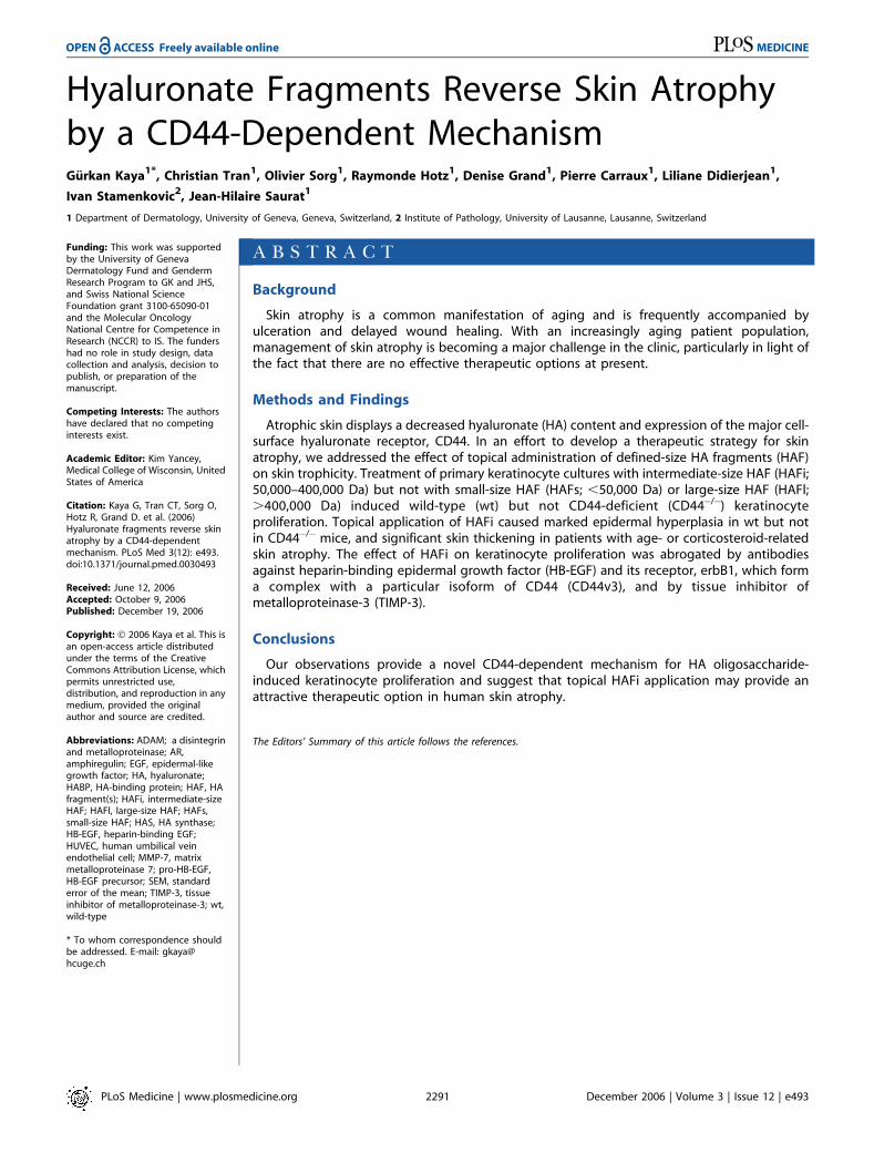
Cultured keratinocytes from the dorsal skin of wt andCD44�/� DBA/1 mice were incubated with HAF generatedfrom high-molecular-weight HA by sonication, enzymaticdigestion, and size exclusion gel filtration (Figure 1A).Exposure to HAFi but not to HAFl or HAFs resulted inincreased keratinocyte proliferation (Figure 1B). Consistentwith the notion that the proliferative response to HA is CD44mediated, HAFi failed to induce proliferation in CD44�/�
keratinocytes (Figure 1C).To verify that the inability of HAFi to induce proliferation
of CD44�/� keratinocytes was indeed due to the lack of CD44and not to a more general proliferation defect of these cells,
we assessed the response of CD44�/� keratinocytes to phorbolester and EGF stimulation. CD44�/� keratinocytes wereobserved to mount a proliferative response to both mitogens,albeit to a lesser degree than wt cells (Figure 1D). It wouldtherefore seem reasonable to attribute the complete lack ofkeratinocyte response to HAFi to the absence of the HAreceptor CD44.The stimulatory effect of HAFi was not limited to cultured
keratinocytes. Incubation of primary human fibroblasts froma healthy donor and HUVECs with HAFi at concentrationsthat induced keratinocyte proliferation resulted in increasedproliferation of both cell types (Figure 1E and 1F).To determine whether HA fragments may induce kerati-
nocyte proliferation in vivo, the effect of repeated localadministration of HAFi to the dorsal skin of wt and CD44�/�
DBA/l mice was assessed. To control for possible strainspecific effects, SKH1 hairless mice were also subjected tolocal HAFi treatment. Daily topical application of a solutionof 0.2% HAFi for 3 d resulted in significant epidermalhyperplasia (Figure 2A and 2B) that reflected increased cellproliferation, as demonstrated by increased numbers ofepidermal and dermal Ki-67þ cells, in all six wt but in noneof the six CD44�/�mice tested (Figure 2C and 2D). Consistentwith the behavior of CD44�/� keratinocytes in vitro, the skinof CD44�/� mice displayed moderate hyperplasia in responseto local phorbol ester application (Figure S1A and S1B),supporting the notion that the complete lack of hyperplasiain response to HAFi could be attributed to the absence ofCD44 itself.The potential effect of HAFi on keratinocyte differ-
entiation in vivo was assessed by staining of HAFi- andvehicle-treated skin sections with antibodies against differ-entiation markers, including keratin-14, filaggrin, and lor-icrin. All three differentiation markers were found to displayincreased expression in hyperplastic HAFi-treated skin that
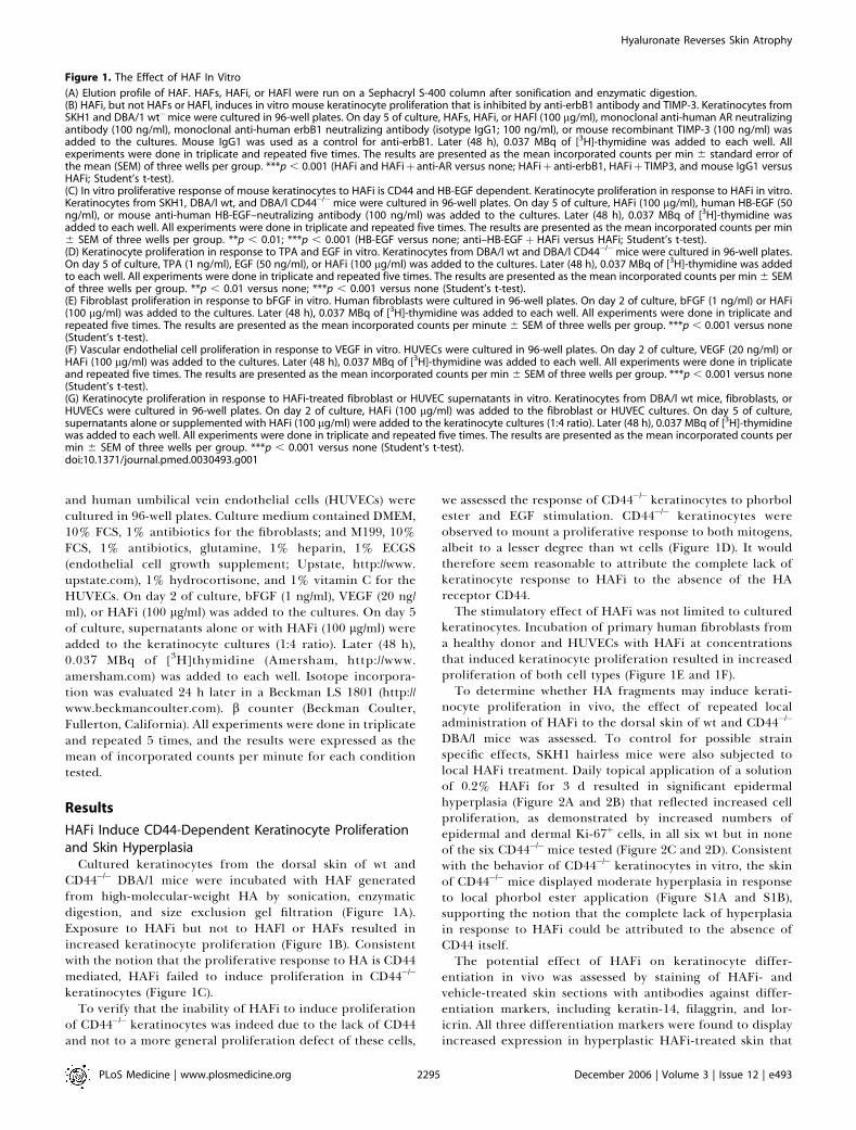
Figure 1. The Effect of HAF In Vitro
(A) Elution profile of HAF. HAFs, HAFi, or HAFl were run on a Sephacryl S-400 column after sonification and enzymatic digestion.(B) HAFi, but not HAFs or HAFl, induces in vitro mouse keratinocyte proliferation that is inhibited by anti-erbB1 antibody and TIMP-3. Keratinocytes fromSKH1 and DBA/1 wt�mice were cultured in 96-well plates. On day 5 of culture, HAFs, HAFi, or HAFl (100 lg/ml), monoclonal anti-human AR neutralizingantibody (100 ng/ml), monoclonal anti-human erbB1 neutralizing antibody (isotype IgG1; 100 ng/ml), or mouse recombinant TIMP-3 (100 ng/ml) wasadded to the cultures. Mouse IgG1 was used as a control for anti-erbB1. Later (48 h), 0.037 MBq of [3H]-thymidine was added to each well. Allexperiments were done in triplicate and repeated five times. The results are presented as the mean incorporated counts per min 6 standard error ofthe mean (SEM) of three wells per group. ***p , 0.001 (HAFi and HAFiþ anti-AR versus none; HAFiþ anti-erbB1, HAFiþ TIMP3, and mouse IgG1 versusHAFi; Student’s t-test).(C) In vitro proliferative response of mouse keratinocytes to HAFi is CD44 and HB-EGF dependent. Keratinocyte proliferation in response to HAFi in vitro.Keratinocytes from SKH1, DBA/l wt, and DBA/l CD44�/�mice were cultured in 96-well plates. On day 5 of culture, HAFi (100 lg/ml), human HB-EGF (50ng/ml), or mouse anti-human HB-EGF–neutralizing antibody (100 ng/ml) was added to the cultures. Later (48 h), 0.037 MBq of [3H]-thymidine wasadded to each well. All experiments were done in triplicate and repeated five times. The results are presented as the mean incorporated counts per min6 SEM of three wells per group. **p , 0.01; ***p , 0.001 (HB-EGF versus none; anti–HB-EGFþ HAFi versus HAFi; Student’s t-test).(D) Keratinocyte proliferation in response to TPA and EGF in vitro. Keratinocytes from DBA/l wt and DBA/l CD44�/�mice were cultured in 96-well plates.On day 5 of culture, TPA (1 ng/ml), EGF (50 ng/ml), or HAFi (100 lg/ml) was added to the cultures. Later (48 h), 0.037 MBq of [3H]-thymidine was addedto each well. All experiments were done in triplicate and repeated five times. The results are presented as the mean incorporated counts per min 6 SEMof three wells per group. **p , 0.01 versus none; ***p , 0.001 versus none (Student’s t-test).(E) Fibroblast proliferation in response to bFGF in vitro. Human fibroblasts were cultured in 96-well plates. On day 2 of culture, bFGF (1 ng/ml) or HAFi(100 lg/ml) was added to the cultures. Later (48 h), 0.037 MBq of [3H]-thymidine was added to each well. All experiments were done in triplicate andrepeated five times. The results are presented as the mean incorporated counts per minute 6 SEM of three wells per group. ***p , 0.001 versus none(Student’s t-test).(F) Vascular endothelial cell proliferation in response to VEGF in vitro. HUVECs were cultured in 96-well plates. On day 2 of culture, VEGF (20 ng/ml) orHAFi (100 lg/ml) was added to the cultures. Later (48 h), 0.037 MBq of [3H]-thymidine was added to each well. All experiments were done in triplicateand repeated five times. The results are presented as the mean incorporated counts per min 6 SEM of three wells per group. ***p , 0.001 versus none(Student’s t-test).(G) Keratinocyte proliferation in response to HAFi-treated fibroblast or HUVEC supernatants in vitro. Keratinocytes from DBA/l wt mice, fibroblasts, orHUVECs were cultured in 96-well plates. On day 2 of culture, HAFi (100 lg/ml) was added to the fibroblast or HUVEC cultures. On day 5 of culture,supernatants alone or supplemented with HAFi (100 lg/ml) were added to the keratinocyte cultures (1:4 ratio). Later (48 h), 0.037 MBq of [3H]-thymidinewas added to each well. All experiments were done in triplicate and repeated five times. The results are presented as the mean incorporated counts permin 6 SEM of three wells per group. ***p , 0.001 versus none (Student’s t-test).doi:10.1371/journal.pmed.0030493.g001
PLoS Medicine | www.plosmedicine.org December 2006 | Volume 3 | Issue 12 | e4932295
Hyaluronate Reverses Skin Atrophy
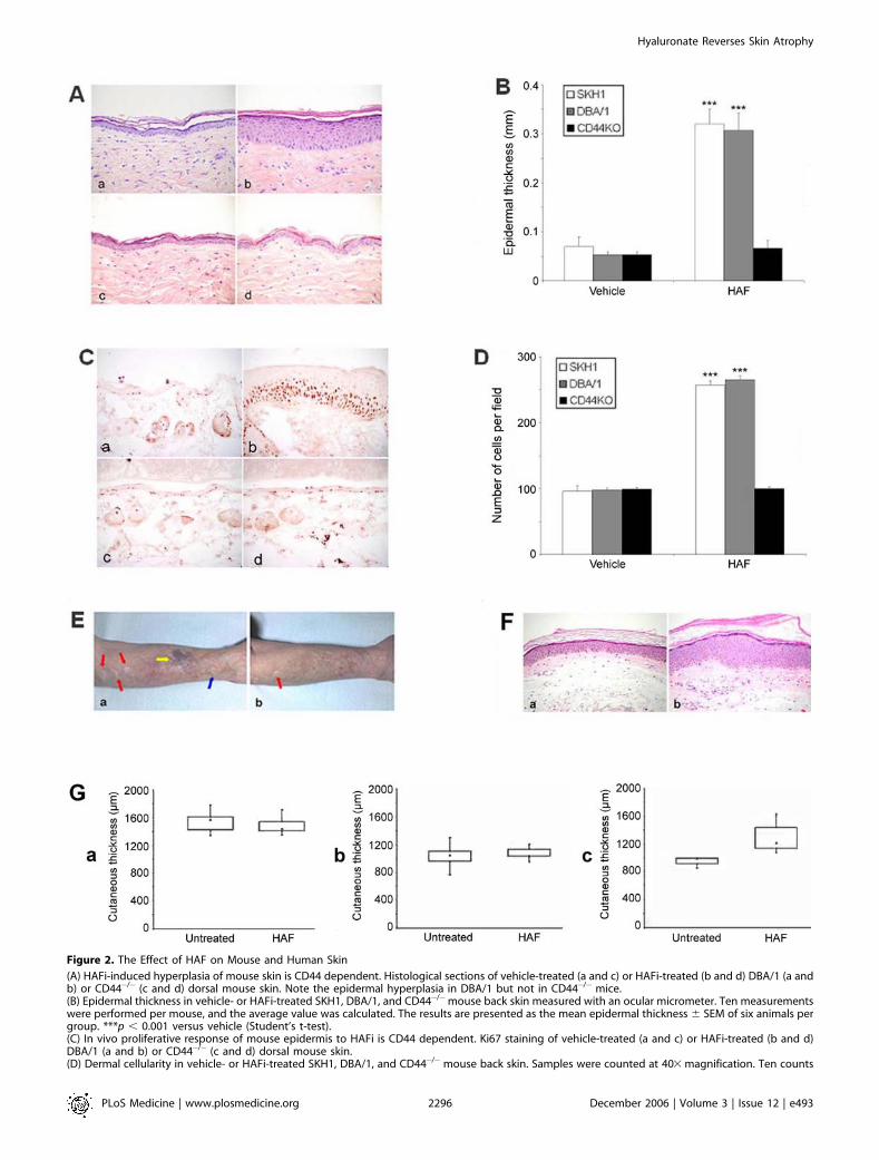
Figure 2. The Effect of HAF on Mouse and Human Skin
(A) HAFi-induced hyperplasia of mouse skin is CD44 dependent. Histological sections of vehicle-treated (a and c) or HAFi-treated (b and d) DBA/1 (a andb) or CD44�/� (c and d) dorsal mouse skin. Note the epidermal hyperplasia in DBA/1 but not in CD44�/� mice.(B) Epidermal thickness in vehicle- or HAFi-treated SKH1, DBA/1, and CD44�/�mouse back skin measured with an ocular micrometer. Ten measurementswere performed per mouse, and the average value was calculated. The results are presented as the mean epidermal thickness 6 SEM of six animals pergroup. ***p , 0.001 versus vehicle (Student’s t-test).(C) In vivo proliferative response of mouse epidermis to HAFi is CD44 dependent. Ki67 staining of vehicle-treated (a and c) or HAFi-treated (b and d)DBA/1 (a and b) or CD44�/� (c and d) dorsal mouse skin.(D) Dermal cellularity in vehicle- or HAFi-treated SKH1, DBA/1, and CD44�/�mouse back skin. Samples were counted at 403 magnification. Ten counts
PLoS Medicine | www.plosmedicine.org December 2006 | Volume 3 | Issue 12 | e4932296
Hyaluronate Reverses Skin Atrophy
was proportional to the degree of hyperplasia (Figure S2A).Thus, keratin-14 expression was observed throughout theepidermis, whereas filaggrin and loricrin expression wasconfined to the granular layer, suggesting that HAFiapplication had no obvious effect on keratinocyte differ-entiation in vivo.
The effect of HAFi on the composition of the dermis wasassessed by addressing changes in collagen expression andvascularization. Staining with Sirius red revealed an increasein collagen content of the superficial dermis (Figure S2B[parts a and b]). Elastic fiber increase in the dermis wasrevealed by van Gieson elastin staining (Figure S2B [parts cand d]), and an increase in blood vessel content was observedwith anti-CD31 antibody (Figure S2B [parts e and f]),reflecting the effect of HAFi on fibroblasts and HUVECsobserved in vitro.
Finally, to determine whether HAFi-mediated stimulationof fibroblasts and endothelial cells might indirectly partic-ipate in keratinocyte proliferation, keratinocytes derivedfrom wt mice were incubated with 1:4 diluted conditionedculture media from 72-h HAFi- and vehicle-treated fibro-blasts and HUVECs. Neither HAFi-treated fibroblasts norHUVEC culture supernatants displayed any significant effecton keratinocyte proliferation in vitro (Figure 1G). However,supplementation of the 72-h HAFi-treated endothelial celland fibroblast supernatants with 100 lg/ml of fresh HAFiinduced keratinocyte proliferation to a degree that wassimilar to that when HAFi were directly added to keratino-cyte cultures (Figure 1G), rendering unlikely the possibilitythat nutrient depletion of the supernatants inhibitedproliferation. It would seem conceivable that the HAFi usedto stimulate endothelial cells and fibroblasts had beeninternalized and/or degraded by 72 h of culture, providing aplausible explanation for the lack of keratinocyte stimulationby the 72-h conditioned culture media. Taken together, theseobservations suggest that HAFi-induced epidermal hyper-plasia in vivo reflected a direct effect of HAFi on keratinocyteproliferation, independent of fibroblast and endothelial cellstimuli.
Topical Application of HAFi Restores Atrophic Skin Lesionsin Humans
To assess the effect of HAFi administration to human skin,six patients with atrophic skin lesions and 17 controlparticipants, including seven healthy men (age, 29–32 y;mean age, 25.5 y) and ten healthy postmenopausal womenwho had not received hormone replacement therapy (age, 55–65 y; mean age, 60 y) were subjected to daily topicalapplication to the forearm of a 1% preparation of HAFifor 1 mo. Following termination of the treatment, none of thecontrol participants revealed a measurable increase in skin
thickness, signs of inflammation, or scaling (Figure 2G [parta]). By contrast, all six patients with skin atrophy that waseither age-related (three patients, aged 60–88 y; mean age, 76y) or associated with corticosteroid therapy for rheumatoidarthritis (three patients, aged 74–86 y; mean age, 81 y)responded to topical HAFi application by developing markedskin thickening (Figure 2E and 2F, and Figure 2G [part b]) atthe end of the treatment period. Most notably, pseudoscars(representing the healing stage of hyperextended skin; Figure2E, red arrows), hemorrhage (Figure 2E, yellow arrow), andvisible superficial vessels (Figure 2E, blue arrow), all of whichare associated with skin atrophy, disappeared in response toHAFi-mediated restoration of skin trophicity (Figure 2E and2F). Contrary to HAFi, the same concentration of HAFl andHAFs applied for the same duration to the six patients withskin atrophy had no effect on skin thickness (unpublisheddata).
HAFi Applied to the Skin Induce Expression of CD44 andEnzymes Implicated in HA MetabolismTo begin to address the putative mechanism of HAFi-
induced skin hyperplasia, we assessed changes in CD44expression and HA synthesis in response to local HAapplication. Topical HAFi application, but not HAFl orHAFs (unpublished data) application, resulted in increasedCD44 expression at the RNA and protein levels throughoutthe epidermis (Figure 3A [parts a and b] and unpublisheddata). HAFi also selectively increased CD44v3 isoformexpression in basal and suprabasal keratinocytes, with theexception of the granular layer (Figure 3A [parts c and d]).The superficial dermis of HAFi-treated DBA/1 mouse
dorsal skin sections displayed strong reactivity with biotiny-lated HABP, an established probe of tissue HA (Figure 3B),which was abrogated by hyaluronidase pretreatment of thetissue sections (unpublished data). An enzyme-linked bindingprotein assay confirmed the presence of increased amountsof HA in the epidermis and dermis (Figure 3B [parts c and d]and unpublished data) of HAFi-treated DBA/l and SKH1hairless mice.To address the fate of locally administered HAF to the skin,
size-fractionated HA was biotinylated, and its localization wastraced with fluorescein-labeled streptavidin. BiotinylatedHAFs-treated SKHI hairless mice and, to a lesser extent,HAFi- but not HAFl-treated SKH1 hairless mice displayedbiotin deposits in the superficial dermis after 3 d of topicalapplication (Figure 3C). The observed biotin deposits wereremoved by hyaluronidase treatment of the skin sections(unpublished data), consistent with the notion that bothHAFs and HAFi can penetrate the epidermis. To furtherassess HAFi localization in tissues, double staining usingbiotinylated HAFi and fluorescein-labeled streptavidin as well
were made per mouse, and the average value was calculated. The results are presented as the mean number of cells 6 SEM of six animals per group.***p , 0.001 versus vehicle (Student’s t-test).(E) HAFi corrects age- and corticosteroid-related atrophy in human skin. Atrophic human forearm skin 1 mo after topical treatment with vehicle (a) or1% HAFi (b). Note the decrease of wrinkles, hemorrhage (yellow arrow), and pseudoscars (red arrows), the visibility of superficial vessels (blue arrow),and the smoothening of the skin with after HAFi treatment.(F) HAFi corrects age- and corticosteroid-related atrophy in human skin. Histology of atrophic human forearm skin 1 mo after topical treatment withvehicle (a) or 1% HAFi (b). Note the significant epidermal hyperplasia after HAFi treatment.(G) HAFi results in skin hyperplasia in atrophic but not normal human skin. Skin thickness in HAFi-treated young (a), nonlesional aged (b), or atrophicaged (c) human skin measured by echography. The results are presented as boxplots with median values (triangles). Young untreated versusnonlesional aged untreated, p , 0.001; young untreated versus atrophic aged untreated, p ¼ 0.001; atrophic aged untreated versus atrophic agedtreated, p , 0.01 (nonparametric Mann-Whitney U test).doi:10.1371/journal.pmed.0030493.g002
PLoS Medicine | www.plosmedicine.org December 2006 | Volume 3 | Issue 12 | e4932297
Hyaluronate Reverses Skin Atrophy
PLoS Medicine | www.plosmedicine.org December 2006 | Volume 3 | Issue 12 | e4932298
Hyaluronate Reverses Skin Atrophy
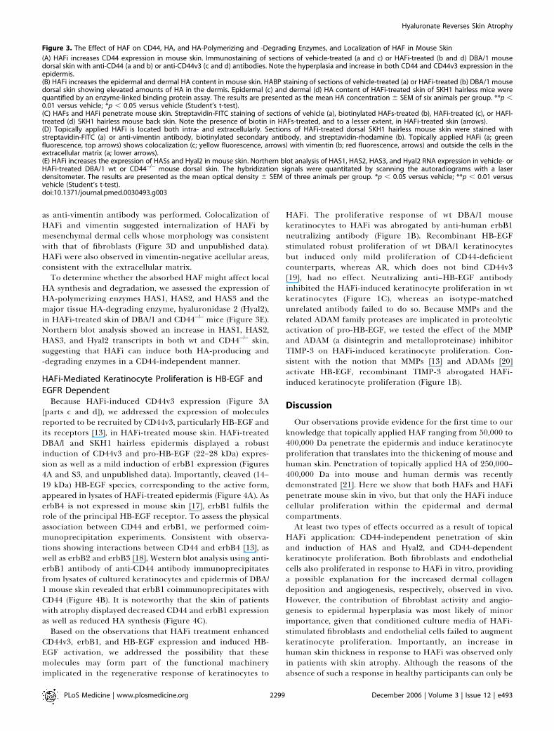
as anti-vimentin antibody was performed. Colocalization ofHAFi and vimentin suggested internalization of HAFi bymesenchymal dermal cells whose morphology was consistentwith that of fibroblasts (Figure 3D and unpublished data).HAFi were also observed in vimentin-negative acellular areas,consistent with the extracellular matrix.
To determine whether the absorbed HAF might affect localHA synthesis and degradation, we assessed the expression ofHA-polymerizing enzymes HAS1, HAS2, and HAS3 and themajor tissue HA-degrading enzyme, hyaluronidase 2 (Hyal2),in HAFi-treated skin of DBA/1 and CD44�/�mice (Figure 3E).Northern blot analysis showed an increase in HAS1, HAS2,HAS3, and Hyal2 transcripts in both wt and CD44�/� skin,suggesting that HAFi can induce both HA-producing and-degrading enzymes in a CD44-independent manner.
HAFi-Mediated Keratinocyte Proliferation is HB-EGF andEGFR Dependent
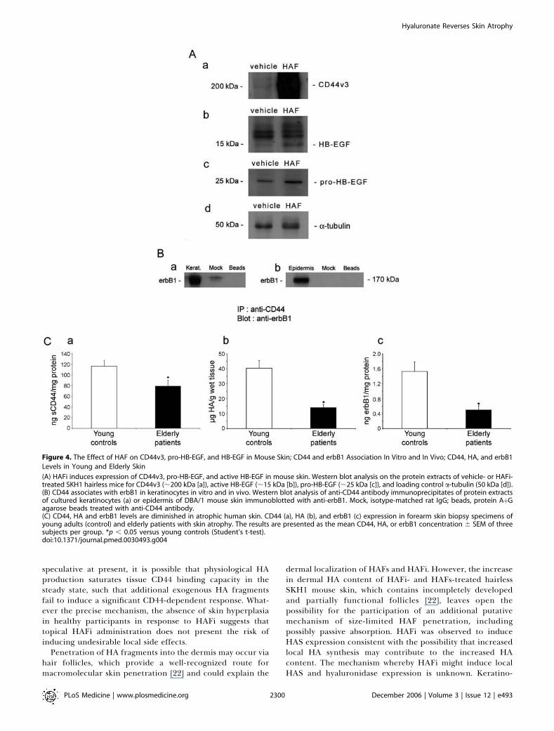
Because HAFi-induced CD44v3 expression (Figure 3A[parts c and d]), we addressed the expression of moleculesreported to be recruited by CD44v3, particularly HB-EGF andits receptors [13], in HAFi-treated mouse skin. HAFi-treatedDBA/l and SKH1 hairless epidermis displayed a robustinduction of CD44v3 and pro-HB-EGF (22–28 kDa) expres-sion as well as a mild induction of erbB1 expression (Figures4A and S3, and unpublished data). Importantly, cleaved (14–19 kDa) HB-EGF species, corresponding to the active form,appeared in lysates of HAFi-treated epidermis (Figure 4A). AserbB4 is not expressed in mouse skin [17], erbB1 fulfils therole of the principal HB-EGF receptor. To assess the physicalassociation between CD44 and erbB1, we performed coim-munoprecipitation experiments. Consistent with observa-tions showing interactions between CD44 and erbB4 [13], aswell as erbB2 and erbB3 [18], Western blot analysis using anti-erbB1 antibody of anti-CD44 antibody immunoprecipitatesfrom lysates of cultured keratinocytes and epidermis of DBA/1 mouse skin revealed that erbB1 coimmunoprecipitates withCD44 (Figure 4B). It is noteworthy that the skin of patientswith atrophy displayed decreased CD44 and erbB1 expressionas well as reduced HA synthesis (Figure 4C).
Based on the observations that HAFi treatment enhancedCD44v3, erbB1, and HB-EGF expression and induced HB-EGF activation, we addressed the possibility that thesemolecules may form part of the functional machineryimplicated in the regenerative response of keratinocytes to
HAFi. The proliferative response of wt DBA/1 mousekeratinocytes to HAFi was abrogated by anti-human erbB1neutralizing antibody (Figure 1B). Recombinant HB-EGFstimulated robust proliferation of wt DBA/1 keratinocytesbut induced only mild proliferation of CD44-deficientcounterparts, whereas AR, which does not bind CD44v3[19], had no effect. Neutralizing anti–HB-EGF antibodyinhibited the HAFi-induced keratinocyte proliferation in wtkeratinocytes (Figure 1C), whereas an isotype-matchedunrelated antibody failed to do so. Because MMPs and therelated ADAM family proteases are implicated in proteolyticactivation of pro-HB-EGF, we tested the effect of the MMPand ADAM (a disintegrin and metalloproteinase) inhibitorTIMP-3 on HAFi-induced keratinocyte proliferation. Con-sistent with the notion that MMPs [13] and ADAMs [20]activate HB-EGF, recombinant TIMP-3 abrogated HAFi-induced keratinocyte proliferation (Figure 1B).
Discussion
Our observations provide evidence for the first time to ourknowledge that topically applied HAF ranging from 50,000 to400,000 Da penetrate the epidermis and induce keratinocyteproliferation that translates into the thickening of mouse andhuman skin. Penetration of topically applied HA of 250,000–400,000 Da into mouse and human dermis was recentlydemonstrated [21]. Here we show that both HAFs and HAFipenetrate mouse skin in vivo, but that only the HAFi inducecellular proliferation within the epidermal and dermalcompartments.At least two types of effects occurred as a result of topical
HAFi application: CD44-independent penetration of skinand induction of HAS and Hyal2, and CD44-dependentkeratinocyte proliferation. Both fibroblasts and endothelialcells also proliferated in response to HAFi in vitro, providinga possible explanation for the increased dermal collagendeposition and angiogenesis, respectively, observed in vivo.However, the contribution of fibroblast activity and angio-genesis to epidermal hyperplasia was most likely of minorimportance, given that conditioned culture media of HAFi-stimulated fibroblasts and endothelial cells failed to augmentkeratinocyte proliferation. Importantly, an increase inhuman skin thickness in response to HAFi was observed onlyin patients with skin atrophy. Although the reasons of theabsence of such a response in healthy participants can only be
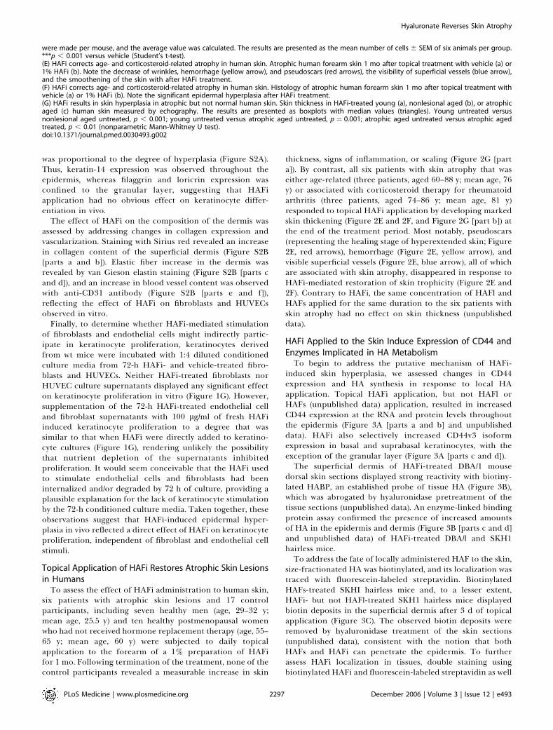
Figure 3. The Effect of HAF on CD44, HA, and HA-Polymerizing and -Degrading Enzymes, and Localization of HAF in Mouse Skin
(A) HAFi increases CD44 expression in mouse skin. Immunostaining of sections of vehicle-treated (a and c) or HAFi-treated (b and d) DBA/1 mousedorsal skin with anti-CD44 (a and b) or anti-CD44v3 (c and d) antibodies. Note the hyperplasia and increase in both CD44 and CD44v3 expression in theepidermis.(B) HAFi increases the epidermal and dermal HA content in mouse skin. HABP staining of sections of vehicle-treated (a) or HAFi-treated (b) DBA/1 mousedorsal skin showing elevated amounts of HA in the dermis. Epidermal (c) and dermal (d) HA content of HAFi-treated skin of SKH1 hairless mice werequantified by an enzyme-linked binding protein assay. The results are presented as the mean HA concentration 6 SEM of six animals per group. **p ,0.01 versus vehicle; *p , 0.05 versus vehicle (Student’s t-test).(C) HAFs and HAFi penetrate mouse skin. Streptavidin-FITC staining of sections of vehicle (a), biotinylated HAFs-treated (b), HAFi-treated (c), or HAFl-treated (d) SKH1 hairless mouse back skin. Note the presence of biotin in HAFs-treated, and to a lesser extent, in HAFi-treated skin (arrows).(D) Topically applied HAFi is located both intra- and extracellularly. Sections of HAFi-treated dorsal SKH1 hairless mouse skin were stained withstreptavidin-FITC (a) or anti-vimentin antibody, biotinylated secondary antibody, and streptavidin-rhodamine (b). Topically applied HAFi (a; greenfluorescence, top arrows) shows colocalization (c; yellow fluorescence, arrows) with vimentin (b; red fluorescence, arrows) and outside the cells in theextracellular matrix (a; lower arrows).(E) HAFi increases the expression of HASs and Hyal2 in mouse skin. Northern blot analysis of HAS1, HAS2, HAS3, and Hyal2 RNA expression in vehicle- orHAFi-treated DBA/1 wt or CD44�/� mouse dorsal skin. The hybridization signals were quantitated by scanning the autoradiograms with a laserdensitometer. The results are presented as the mean optical density 6 SEM of three animals per group. *p , 0.05 versus vehicle; **p , 0.01 versusvehicle (Student’s t-test).doi:10.1371/journal.pmed.0030493.g003
PLoS Medicine | www.plosmedicine.org December 2006 | Volume 3 | Issue 12 | e4932299
Hyaluronate Reverses Skin Atrophy
speculative at present, it is possible that physiological HAproduction saturates tissue CD44 binding capacity in thesteady state, such that additional exogenous HA fragmentsfail to induce a significant CD44-dependent response. What-ever the precise mechanism, the absence of skin hyperplasiain healthy participants in response to HAFi suggests thattopical HAFi administration does not present the risk ofinducing undesirable local side effects.
Penetration of HA fragments into the dermis may occur viahair follicles, which provide a well-recognized route formacromolecular skin penetration [22] and could explain the
dermal localization of HAFs and HAFi. However, the increasein dermal HA content of HAFi- and HAFs-treated hairlessSKH1 mouse skin, which contains incompletely developedand partially functional follicles [22], leaves open thepossibility for the participation of an additional putativemechanism of size-limited HAF penetration, includingpossibly passive absorption. HAFi was observed to induceHAS expression consistent with the possibility that increasedlocal HA synthesis may contribute to the increased HAcontent. The mechanism whereby HAFi might induce localHAS and hyaluronidase expression is unknown. Keratino-
Figure 4. The Effect of HAF on CD44v3, pro-HB-EGF, and HB-EGF in Mouse Skin; CD44 and erbB1 Association In Vitro and In Vivo; CD44, HA, and erbB1
Levels in Young and Elderly Skin
(A) HAFi induces expression of CD44v3, pro-HB-EGF, and active HB-EGF in mouse skin. Western blot analysis on the protein extracts of vehicle- or HAFi-treated SKH1 hairless mice for CD44v3 (;200 kDa [a]), active HB-EGF (;15 kDa [b]), pro-HB-EGF (;25 kDa [c]), and loading control a-tubulin (50 kDa [d]).(B) CD44 associates with erbB1 in keratinocytes in vitro and in vivo. Western blot analysis of anti-CD44 antibody immunoprecipitates of protein extractsof cultured keratinocytes (a) or epidermis of DBA/1 mouse skin immunoblotted with anti-erbB1. Mock, isotype-matched rat IgG; beads, protein AþGagarose beads treated with anti-CD44 antibody.(C) CD44, HA and erbB1 levels are diminished in atrophic human skin. CD44 (a), HA (b), and erbB1 (c) expression in forearm skin biopsy specimens ofyoung adults (control) and elderly patients with skin atrophy. The results are presented as the mean CD44, HA, or erbB1 concentration 6 SEM of threesubjects per group. *p , 0.05 versus young controls (Student’s t-test).doi:10.1371/journal.pmed.0030493.g004
PLoS Medicine | www.plosmedicine.org December 2006 | Volume 3 | Issue 12 | e4932300
Hyaluronate Reverses Skin Atrophy
cytes and fibroblasts provide the principal source of HA inthe epidermis and dermis, respectively [23], and control localHA metabolism. CD44 is believed to play a major role in theuptake of HA by receptor-mediated endocytosis [24] in bothcell types. Following internalization, HA undergoes intra-cellular degradation that is possibly mediated by endosomal/lysosomal hyaluronidases [25]. HAF released by keratinocytestraverse the basement membrane to the dermis, where theyare cleared via lymphatic vessels [26]. It would appear thatmechanisms independent of CD44 might sense changes inlocal HA concentration, or that HAFi stimulate receptorsother than CD44 to induce HAS and hyaluronidase expres-sion. Increased tissue HA concentration typically occurs inthe context of development, injury, and tumor growth, and isbelieved to actively participate in the process of tissueremodeling. It is conceivable that an increase in tissue HA,whether of endogenous or exogenous origin, is interpreted bylocal fibroblasts to reflect a remodeling process, triggeringadditional HA synthesis and degradation.
Proliferation in response to HAFi is a CD44-dependentevent. Our present observations provide evidence that inaddition to CD44, HB-EGF, erbB1, and MMPs/ADAMs arerequired for HA-dependent in vitro keratinocyte prolifer-ation. The absence of a proliferative response of CD44�/�
keratinocytes to HB-EGF is consistent with the notion thatHB-EGF interaction with its receptors requires presentationby heparan sulfate side chains of CD44v3-containing isoforms[19]. Similar to other members of the EGF family, pro-HB-EGF is expressed as an integral membrane protein of 22–28kDa [27], which, following stimulation of cells with mitogens,undergoes MMP-mediated release of mature soluble 14–19kDa HB-EGF [28]. Accordingly, topical application of HAFioligosaccharide resulted in a significant increase in pro-HB-EGF and HB-EGF, while inhibition of MMP activity by TIMP-3 abrogated the corresponding proliferative response.The observation that anti-erbB1– and anti–HB-EGF–block-
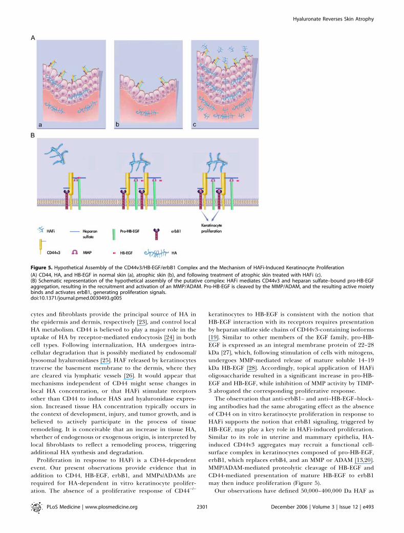
ing antibodies had the same abrogating effect as the absenceof CD44 on in vitro keratinocyte proliferation in response toHAFi supports the notion that erbB1 signaling, triggered byHB-EGF, may play a key role in HAFi-induced proliferation.Similar to its role in uterine and mammary epithelia, HA-induced CD44v3 aggregates may recruit a functional cell-surface complex in keratinocytes composed of pro-HB-EGF,erbB1, which replaces erbB4, and an MMP or ADAM [13,20].MMP/ADAM-mediated proteolytic cleavage of HB-EGF andCD44-mediated presentation of mature HB-EGF to erbB1may then induce proliferation (Figure 5).Our observations have defined 50,000–400,000 Da HAF as
Figure 5. Hypothetical Assembly of the CD44v3/HB-EGF/erbB1 Complex and the Mechanism of HAFi-Induced Keratinocyte Proliferation
(A) CD44, HA, and HB-EGF in normal skin (a), atrophic skin (b), and following treatment of atrophic skin treated with HAFi (c).(B) Schematic representation of the hypothetical assembly of the putative complex: HAFi mediates CD44v3 and heparan sulfate–bound pro-HB-EGFaggregation, resulting in the recruitment and activation of an MMP/ADAM. Pro-HB-EGF is cleaved by the MMP/ADAM, and the resulting active moietybinds and activates erbB1, generating proliferation signals.doi:10.1371/journal.pmed.0030493.g005
PLoS Medicine | www.plosmedicine.org December 2006 | Volume 3 | Issue 12 | e4932301
Hyaluronate Reverses Skin Atrophy
reagents capable of inducing a proliferative response inmouse and human skin. Clinically, topical HAFi applicationresulted in epidermal hyperplasia with restoration to normalthickness of atrophic human skin as early as 1 mo afterinitiation of treatment. This effect was accompanied bysignificant clinical improvement, suggesting that HAFi mayprovide the basis for the development of novel therapeuticstrategies for skin diseases characterized by atrophy.
Supporting Information
Figure S1. The Effect of TPA on CD44�/� Mouse Skin
(A) TPA induces a reduced skin hyperplasia in CD44�/� mice.Histological sections of acetone-treated (parts a and c) or TPA-treated (parts b and d) DBA/1 (parts a and b) or CD44�/� (parts c andd) dorsal mouse skin. Note the decreased epidermal hyperplasia inCD44�/� mice.(B) Epidermal thickness in acetone- or TPA-treated DBA/1 andCD44�/� mouse back skin measured with an ocular micrometer. Tenmeasurements were performed per mouse, and the average value wascalculated. The results are presented as the mean epidermal thickness6 SEM of six animals per group. ***p , 0.001 versus acetone(Student’s t-test).
Found at doi:10.1371/journal.pmed.0030493.sg001 (293 KB JPG).
Figure S2. The Effect of HAFi on Mouse Epidermal Differentiationand Atrophic Human Dermis
(A) HAFi has no effect on epidermal differentiation in mouse skin.Immunostaining of sections of vehicle-treated (parts a, c, and e) orHAFi-treated (parts b, d, and f) DBA/1 mouse dorsal skin with anti-K14 (parts a and b), anti-filaggrin (parts c and d), or anti-loricrin(parts e and f) antibody. Note the lack of increase in staining intensitydespite the increased number of stained cells in HAFi-treated skin.(B) HAFi increases the collagen, elastin and vascular content inatrophic human skin. Histology of atrophic human forearm skin 1month after topical treatment with vehicle (part a) or 1% HAFi (partb), stained with Sirius red (parts a and b), van Gieson elastin (parts cand d), or anti-CD31 antibody (parts e and f). Note the increase indermal collagen, elastic fibers, and vessels after HAFi treatment.
Found at doi:10.1371/journal.pmed.0030493.sg002 (882 KB JPG).
Figure S3. HAFi Induces Expression of CD44v3 in Mouse Skin
Western blot analysis on the protein extracts of vehicle- or HAFi-treated SKH1 hairless mice for CD44v3.Found at doi:10.1371/journal.pmed.0030493.sg003 (454 KB JPG).
Acknowledgments
IS and JHS share senior authorship of this article. We thank EricAugsburger, Marie-Jo Cartier, and Evelyne Leemans at the Derma-topathology Laboratory of the University Hospital of Geneva fortheir excellent technical help.
Author contributions. GK, IS, and JHS designed the study. GK, CT,OS, LD, IS, and JHS analyzed the data. GK and JHS enrolled patients.GK, CT, OS, LD, IS, and JHS contributed to writing the paper. GK,RH, DG, and PC collected data or did experiments for the study.
References1. Aruffo A, Stamenkovic I, Melnick M, Underhill CB, Seed B (1990) CD44 is
the principal cell surface receptor for hyaluronate. Cell 61: 1303–1313.2. Miyake K, Underhill CB, Lesley J, Kincade PW (1990) Hyaluronate can
function as a cell adhesion molecule and CD44 participates in hyaluronaterecognition. J Exp Med 172: 69–75.
3. Kaya G, Rodriguez I, Jorcano JL, Vassalli P, Stamenkovic I (1997) Selectivesuppression of CD44 in keratinocytes of mice bearing an antisense CD44
transgene driven by a tissue-specific promoter disrupts hyaluronatemetabolism in the skin and impairs keratinocyte proliferation. GenesDev 11: 996–1007.
4. Laurent TC, Fraser JR (1992) Hyaluronan. FASEB J 6: 2397–2404.5. Forrester JV, Balazs EA (1980) Inhibition of phagocytosis by high molecular
weight hyaluronate. Immunology 40: 435–446.6. West DC, Hampson IN, Arnold F, Kumar S (1985) Angiogenesis induced by
degradation products of hyaluronic acid. Science 228: 1324–1326.7. Fitzgerald KA, Bowie AG, Skeffington BS, O’Neill LA (2000) Ras, protein
kinase C zeta, and I kappa B kinases 1 and 2 are downstream effectors ofCD44 during the activation of NF-kappa B by hyaluronic acid fragments inT-24 carcinoma cells. J Immunol 164: 2053–2063.
8. McKee CM, Penno MB, Cowman M, Burdick MD, Strieter RM, et al. (1996)Hyaluronan (HA) fragments induce chemokine gene expression in alveolarmacrophages. The role of HA size and CD44. J Clin Invest 98: 2403–2413.
9. Taylor KR, Trowbridge JM, Rudisill JA, Termeer CC, Simon JC, et al. (2004)Hyaluronan fragments stimulate endothelial recognition of injury throughTLR4. J Biol Chem 279: 17079–17084.
10. Termeer CC, Hennies J, Voith U, Ahrens T, Weiss JM, et al. (2000)Oligosaccharides of hyaluronan are potent activators of dendritic cells. JImmunol 165: 1863–1870.
11. Ponta H, Sherman L, Herrlich PA (2003) CD44: From adhesion moleculesto signalling regulators. Nature Rev Mol Biol 4: 33–45.
12. Skelton TP, Zeng C, Nocks A, Stamenkovic I (1998) Glycosylations providesboth stimulatory and inhibitory effects on cell surface and soluble CD44binding to hyaluronan. J Cell Biol 140: 431–446.
13. Yu WH, Woessner JF Jr, McNeish JD, Stamenkovic I (2002) CD44 anchorsthe assembly of matrilysin/MMP-7 with heparin-binding epidermal growthfactor precursor and erbB4 and regulates female reproductive organremodeling. Genes Dev 16 : 307–323.
14. Kaya G, Augsburger E, Stamenkovic I, Saurat JH (2000) Decrease inepidermal CD44 expression as a potential mechanism for abnormalhyaluronate accumulation in superficial dermis in lichen sclerosus etatrophicus. J Invest Dermatol 115: 1054–1058.
15. Kaya G, Grand D, Hotz R, Augsburger E, Carraux P, et al. (2005)Upregulation of CD44 and hyaluronate synthases by topical retinoids inmouse skin. J Invest Dermatol 124: 284–287.
16. Limat A, Hunziker T (1996) Cultivation of keratinocytes from the outerroot sheath of human hair follicles. In: Jones GE, editor. Methods inmolecular medicine. Human cell culture protocols. Totowa (New Jersey):Humana Press Inc. pp. 21–31.
17. Xian W, Rosenberg MP, DiGiovanni J (1997) Activation of erbB2 and c-srcin phorbol ester-treated mouse epidermis: Possible role in mouse skintumor promotion. Oncogene 14: 1435–1444.
18. Sherman LS, Rizvi TA, Karyala S, Ratner N (2000) CD44 enhancesneuregulin signalling by Schwann cells. J Cell Biol 150: 1071–1083.
19. Bennett KL, Jackson DG, Simon JC, Tanczos E, Tanczos R, et al. (1995)CD44 isoforms containing exon V3 are responsible for the presentation ofheparin-binding growth factor. J Cell Biol 128: 687–698.
20. Higashiyama S, Nanba D (2005) ADAM-mediated ectodomain shedding ofHB-EGF in receptor cross-talk. Biochim Biophys Acta 1751: 110–117.
21. Brown TJ, Alcorn D, Fraser JR (1999) Absorption of hyaluronan applied tothe surface of intact skin. J Invest Dermatol 113: 740–746.
22. Dokka S, Cooper SR, Kelly S, Hardee GE, Karras JG (2005) Dermal deliveryof topically applied oligonucleotides via follicular transport in mouse skin.J Invest Dermatol 124: 971–975.
23. Wang C, Tammi M, Tammi R (1992) Distribution of hyaluronan and itsCD44 receptor in the epithelia of human skin appendages. Histochemistry98: 105–112.
24. Tammi R, Saamanen AM, Maibach HI, Tammi M (1991) Degradation ofnewly synthesized high molecular mass hyaluronan in the epidermal anddermal compartments of human skin in organ culture. J Invest Dermatol97: 126–130.
25. Tammi R, Rilla K, Pienimaki JP, MacCallum DK, Hogg M, et al. (2001)Hyaluronan enters keratinocytes by a novel endocytic route for catabolism.J Biol Chem 276: 35111–35122.
26. Tammi R, Agren UM, Tuhkanen AL, Tammi M (1994) Hyaluronanmetabolism in skin. Prog Histochem Cytochem 29: 1–81.
27. Higashiyama S, Lau K, Besner GE, Abraham JA, Klagsbrun M (1992)Structure of heparin-binding EGF-like growth factor. J Biol Chem 276:6205–6212.
28. Prenzel N, Zwick E, Daub H, Leserer M, Abraham R, et al. (1999) EGFreceptor transactivation by G-protein-coupled receptors requires metal-loproteinase cleavage of proHB-EGF. Nature 402: 884–888.
PLoS Medicine | www.plosmedicine.org December 2006 | Volume 3 | Issue 12 | e4932302
Hyaluronate Reverses Skin Atrophy
Editors’ Summary
Background. Time wreaks many changes in the human body but theskin is where one of the first visible signs of aging—wrinkles—occurs.The skin consists of three main layers. The outermost layer is theepidermis. It is the thickness of a sheet of paper and forms a barrier thatprevents the body losing water or infectious agents entering it. The cellsin the epidermis are mainly keratinocytes. These specialized skin cells arecontinually produced at the base of the epidermis. From there, theymove toward the skin’s surface where they are shed. The middle layer isthe dermis. It is about ten times thicker than the epidermis and containsthe blood vessels that feed the skin, nerves, sebaceous glands, and hairfollicles. The final, subcutaneous layer contains sweat glands, some hairfollicles, blood vessels and fat. The dermis contains collagen fibers thatsupport the skin and elastin fibers that provide flexibility. Human skinbegins to age in early adulthood. By the time a person is 80 years old,their epidermis may be half its original thickness because of decreasedkeratinocyte proliferation. The dermis also thins, and loss of collagen andelastin fibers means that the skin becomes less elastic. The gradual lossof epidermis and dermis—skin atrophy—is clinically important becauseaging skin is more fragile and heals slower than young skin and is alsoprone to ulceration.
Why Was This Study Done? No one knows why skin atrophy occurs, butit is becoming more common as people live longer, and there is noeffective treatment for it. One characteristic of atrophic skin is that,compared to normal skin, it contains less hyaluronate (also calledhyaluronan and hyaluronic acid)—a large carbohydrate component ofthe extracellular matrix, the material that surrounds cells. It also containsless CD44, a cell-surface protein that interacts with hyaluronate. Thisinteraction can stimulate cell proliferation and migration. Given theseobservations, in this study the researchers have investigated whethertreating atrophic skin with fragments of hyaluronate might counteractatrophy.
What Did the Researchers Do and Find? The researchers isolatedkeratinocytes from normal mice and from CD44-deficient mice (CD44�/�
mice) and treated them with different sized fragments of hyaluronate.Intermediate sized hyaluronate fragments (so-called HAFi) but not large
or small fragments increased the proliferation of normal keratinocytesbut not CD44�/� keratinocytes. This suggests that proliferation inresponse to HAFi is CD44-dependent. Similarly, a cream of HAFi appliedto the backs of normal mice caused thickening of the epidermal layer buthad no effect on CD44�/� mice. Finally, topical application of HAFi forone month caused skin thickening and clinical improvement in sixpeople with skin atrophy but had no effect on normal human skin. Thecollagen, elastic fiber, and blood vessel content of the dermis alsoincreased in treated patients. By using antibodies to block the functionof various proteins, the researchers also discovered that heparin-bindingepidermal growth factor (HB-EGF, a protein that stimulates keratinocyteproliferation), erbB1 (a cell-surface protein that binds HB-EGF), andmatrix metalloproteinases (proteins that activate HB-EGF) are all requiredfor the stimulation of keratinocyte proliferation by HAFi.
What Do These Findings Mean? Taken together, these results providethe first indication that application of HAFi to atrophic skin might beuseful therapeutically. The absence of any effect on normal human skin isreassuring but puzzling given the thickening seen in normal mouse skin,so this finding needs confirmation before hyaluronate fragments areused clinically. Longer trials in more people are also needed tocharacterize the clinical effects fully. Finally, the mechanism by whichhyaluronate fragments have their effect needs to be studied in moredepth. Such studies might reveal other potential therapeutic options forthe treatment of skin atrophy.
Additional Information. Please access these Web sites via the onlineversion of this summary at http://dx.doi.org/10.1371/journal.pmed.0030493.� MedlinePlus encyclopedia entry on aging changes in skin� US National Institute on Aging, patient information on skin care and
aging� American Academy of Dermatology, patient information on aging skin� Information on CD44, the hyaluronan receptor, provided by
Glycoforum a source of information on glycobiology� Wikpedia pages on skin and on hyaluronan (note that Wikipedia is a
free online encyclopedia that anyone can edit)
PLoS Medicine | www.plosmedicine.org December 2006 | Volume 3 | Issue 12 | e4932303
Hyaluronate Reverses Skin Atrophy


