Embed Size (px)
Citation preview
INTERNATIONAL JOURNAL OF SYSTEMATIC BACTERIOLOGY, July 1989, p. 319-333
Copyright 0 1989, International Union of Microbiological Societies 0020-771 3/89/0303 19-15$02.00/0
Vol. 39, No. 3
Hydrogenophaga, a New Genus of Hydrogen-Oxidizing Bacteria That Includes Hydrogenophaga flava comb. nov.
(Formerly Pseudomonas f lava) , Hydrogenophaga palleronii (Formerly Pseudomonas palleronii), Hydrogenophaga pseudoflava
(Formerly Pseudomonas pseudoflava and “Pseudomonas carboxydoflava ”), and Hydrogenophaga taeniospiralis
(Formerly Pseudomonas taeniospiralis) A. WILLEMS,’ J. BUSSE,2 M. GOOR,’ B. POT,l E. FALSEN,3 E. JANTZEN,4 B. HOSTE,’ M. GILLIS,’
K. KERSTERS,l G. AULING,2 AND J. DE LEY1*
La horatorium voor Microbiologie en Microbiele Genetica, Rijksuniversiteit, B-9000 Ghent, Belgium‘; Institut fur Mikrobiologie, Universitiit Hannover, 0-3000 Hannover, Federal Republic of Germany2; Culture Collection,
Department of Clinical Bacteriology, University of Goteborg, S-413 46 Goteborg, Sweden3; and Department of Methodology, National Institute of Public Health, N-0462 Oslo, Norway4
The relationships of the yellow-pigmented hydrogen-oxidizing species Pseudomonas flava, Pseudomonas pseudoflava, Pseudomonas palleronii, Pseudomonas taeniospiralis, and “Pseudomonas carboxydoflava,” which are all members of the acidovorans ribosomal ribonucleic acid (rRNA) complex in rRNA superfamily 111, were studied by using deoxyribonucleic acid (DNA):rRNA hybridization, immunotyping, numerical analysis of biochemical and auxanographic features, polyacrylamide gel electrophoresis of cellular proteins, numerical analysis of fatty acid patterns, and DNA:DNA hybridization. Our results show that these five yellow-pigmented hydrogen-oxidizing Pseudomonas species are more closely related to each other than to other taxa belonging to the acidovorans rRNA complex. We propose the transfer of these species to a new genus, Hydrogenophaga, with the following four species: Hydrogenophaga flava (formerly Pseudomonas flava), Hydrogenophaga pseudoflava (to accommodate both Pseudomonas pseudoflava and “Pseudomonas carboxydoflava”), Hydrogenophaga taeniospiralis (formerly Pseudomonas taeniospiralis), and Hydrogenophaga palleronii (formerly Pseudomonas palleronii). The type species is H.flava, with monotype strain DSM 619 (= LMG 2185 = CCUG 1658). Because H. Java grows slowly and unreliably, but is genotypically and protein electrophoretically very similar to H. pseudoflava, the latter species can be used as an alternative reference taxon for the new genus. The type strains of H. pseudoflava, H. taeniospiralis, and H . palleronii are strains GA3 (= LMG 5945 = CCUG 13799), DSM 2082 (= LMG 7170 = CCUG 15921), and Stanier 362t1 (= LMG 2366t1 = CCUG 20334), respectively.
The genus Hydrogenomonas was originally created to shelter gram-negative, facultatively autotrophic hydrogen bacteria (34). In 1969 Davis et al. (7) proposed that this genus should be abandoned and transferred the various species to other genera. Because of their deoxyribonucleic acid (DNA) base compositions, their polar flagellation, and their overall phenotypic properties, the yellow-pigmented organism Hy- drogenomonas flava and the nonpigmented organisms Hy- drogenomonas facilis and Hydrogenomonas ruhlandii were assigned to the genus Pseudomonas. Later, Pseudomonas palleronii was described as an additional yellow-pigmented hydrogen-oxidizing species, differing from Pseudomonas flava in the nature of its carotenoid pigments and its inability to grow on several carbohydrates (8).
Within the phenotypically defined genus Pseudomonas, five ribosomal ribonucleic acid (rRNA) homology groups were delineated by means of DNA:rRNA hybridization (37). Later, De Vos and De Ley (14) extended these five rRNA groups considerably and showed that they are only very remotely interrelated and cannot be maintained in a single genus. This conclusion was confirmed by results from sev- eral other techniques (6,46). Most Pseudomonas species are
* Corresponding author
located on three rRNA branches; only the Pseudomonas f luorexens rRNA branch in rRNA superfamily I1 represents the true pseudomonads (14). We consider all of the other Pseudomonas species to be generically misnamed (14). Below all misnamed taxa are indicated by brackets. The yellow-pigmented hydrogen-oxidizing species [Pseudomo- nus] flava, [Pseudomonas] palleronii, [Pseudomonas] pseudoflava, “[Pseudomonas] carboxydoflava,” and [Pseu- domonas] taeniospiralis belong to the acidovorans rRNA complex, i.e., rRNA homology group I11 sensu Palleroni (36). This rRNA complex belongs to rRNA superfamily I11 (sensu De Ley [ lo]) , which corresponds to the beta subclass of the Proteobacteria (41). In addition, this rRNA complex contains the following species: [Pseudomonas] acidovorans, [Pseudomonas] avenue, [Pseudomonas] cattleyae, [Pseudo- monas] delajieldii, [Pseudomonas] facilis, [Pseudomonas] pseudoalcaligenes subsp. citrulli, [Pseudomonas] pseudoal- caligenes subsp. konjaci, [Pseudomonas] rubrilineans, [Pseudomonas] saccharophila, “ [Pseudomonas] seturiae,” [Pseudomonas] testosteroni, Comamonas terrigena, [Alcali- genes] paradoxus, [Alcaligenes] latus, [Rhodocyclus] gela- tinosus, and Xylophilus ampelinus (15, 16, 44). [Pseudomo- nus] acidovorans and [Pseudomonas] testosteroni were recently transferred to the genus Comamonas (42).
So far, within the acidovorans rRNA complex, labeled
319

320 WILLEMS ET AL. INT. J . SYST. BACTERIOL.
rRNAs have been available from the type strains of only Comamonas acidovorans, [Alcaligenes] paradoxus, and X y - lophilus ampelinus. Hybridization of these rRNAs with DNAs from strains belonging to the acidovorans rRNA complex resulted in delineation of three rRNA subbranches linked at a Tm(e) level [T,,(c, is the temperature, in degrees Celsius, at which one-half of the DNA:rRNA duplex is denatured] of 76.2 & 0.7”C and containing one species each. All of the other species of the acidovorans rRNA complex are presently located on the T,,,(,) dendrogram at the branch- ing level of these three rRNA subbranches (16, 44). Within this phenotypically very diverse group, some taxa may be more closely related to each other. We are in the process of revealing these possible subgroups by using a repeated process of rRNA preparation from new strains and DNA- rRNA hybridization. The combination of DNA:rRNA hy- bridization data with the results of other techniques should provide a clearer view of the relationships within the aci- dovorans rRNA complex. In this study, we used a broad spectrum of methods to investigate the taxonomy of the yellow-pigmented hydrogen-oxidizing [Pseudomonas] spe- cies belonging to the acidovorans rRNA complex. We pro- pose the creation of a new genus, Hydrogenophaga, for these organisms.
MATERIALS AND METHODS
Bacterial strains. The strains which we used are listed in Table 1. “[Pseudomonas] gazotropha” was grown on PSS medium, which contained 1% (wthol) neutralized bacterio- logical peptone (type L34; Oxoid Ltd., London, England), 0.1% (wthol) succinic acid, 0.1% (wt/vol) (NH,),SO,, 0.1% (wthol) MgSO, . 7H,O, 0.0002% (wthol) FeC1, . 6H,O, and 0.0002% (wthol) MnSO, . H,O; the pH was adjusted to 7.0 with 1 M KOH. The media used for growth of [Rhoducyclus] gelatinosus and Xylophilus ampelinus have been described previously (44); all other strains were grown on nutrient agar (0.1% [wthol] beef extract, 0.2% [wt/vol] yeast extract, 0.5% [wthol] NaCI, 0.5% [wt/vol] peptone, 2% [wt/vol] agar, pH 7.4). For fatty acid analysis, cells were grown for 40 h on Columbia agar (Oxoid) containing 5% (vol/vol) defibri- nated horse blood. All strains except Xylophilus ampelinus NCPPB 2217T (T = type strain) were grown at 28°C; Xylophilus ampelinus NCPPB 2217T was grown at 24°C.
Preparation of high-molecular-weight DNA. Cells were grown in Roux flasks for 2 to 3 days. DNA was isolated by the method of Marmur (29).
DNA base composition. The average guanine-plus-cytosine (G +C) contents were determined by the thermal denatur- ation method (13) and were calculated by using the equation of Marmur and Doty (30), as modified by De Ley (9).
DNA:rRNA hybridization. DNA was further purified by CsCl gradient centrifugation (12). Fixation of thermally denatured DNA on cellulose nitrate filters and chemical estimation of the amount of filter-fixed DNA were performed as described previously (12, 43). [‘HIrRNAs from [Pseudo- monas] palleronii Stanier 362tlT and Comamonas terrigena NCIB 8193T and [ 14C]rRNA from [Pseudomonas] pseudo- Java GA3T were isolated and purified as described by De Ley and De Smedt (12). Purified 23s [‘HJrRNA from Coma- monas acidovorans Stanier 14T and 16s [14C]rRNA from [Alcaligenes] paradoxus ATCC 17713tlT were available in our research group (15, 16). Hybridizations between labeled 16s or 23s rRNA and filter-fixed DNA were carried out as described previously (12). Each hybrid was characterized by its TrpZce, and by the percentage of rRNA binding (the amount
of rRNA [in micrograms] bound to 100 pg of filter-fixed DNA after ribonuclease treatment).
DNA:DNA hybridization. The degree of binding, ex- pressed as a percentage, was determined spectrophotomet- rically by using the initial renaturation rate method (11). We used a model 2600 spectrophotometer (Gilford Instrument Laboratories, Inc., Oberlin, Ohio) equipped with a thermo- statically controlled cuvette chamber and a model 7225A plotter (Hewlett-Packard Co., Palo Alto, Calif.). Renatur- ations were performed in 2X SSC (12 SSC is 0.15 M NaCl plus 0.015 M sodium citrate, pH 7.0) at the optimal renatur- ation temperature of 80.9”C and with a total DNA concen- tration of 0.101 mM base pairs. Degrees of binding of 25% or less indicate no significant DNA homology.
Taxonomic immunotyping. The preparation of antisera by immunizing rabbits with bacterial extracts in Freund incom- plete adjuvant and the immunodiffusion technique have been described previously (16, 17).
Morphological and biochemical features. To determine morphological and biochemical features, we used the meth- ods described by De Vos et al. (16). Nitrite reduction was tested as described by Rossau et al. (39).
Carbon substrate assimilation tests. API SOCH, API SOAO, and API 50AA galleries (API System S.A., Montalieu- Vercieu, France) were used to test the assimilation of 147 organic compounds as sole carbon sources. The experimen- tal procedure which we used has been described previously (23). For “[Pseudomonas] gazotropha” 0.1 M potassium phosphate buffer (pH 7) was used to prepare the medium for the auxanographic tests instead of sodium phosphate buffer, and the API galleries were incubated at 28°C instead of 30°C.
Numerical analysis of phenotypic features. The scoring of the auxanographic and biochemical tests has been described previously (39). Of the 251 features tested, 192 were used in the numerical analysis, The API 20NE tests (21 features) were not included in the computer analysis as they dupli- cated other tests, and 38 other features were omitted be- cause all 38 strains tested were either negative or displayed the same positive value. The levels of interstrain similarity were calculated by using the Gower similarity coefficient (S,) (40). Cluster analysis by the unweighted average pair group method (40) was performed with the CLUSTAN 2.1 program of Wishart (45) on the Siemens model 7551 com- puter of the Centraal Digitaal Rekencentrum, Rijksuniver- siteit Ghent, Ghent, Belgium. The reproducibility of the clustering was estimated by including duplicate tests for seven strains.
Polyacrylamide gel electrophoresis of proteins. All strains were grown on nutrient agar at 28°C for 40 h in Roux flasks. Whole-cell protein extracts were prepared, and sodium dodecyl sulfate-polyacrylamide gel electrophoresis was per- formed by using small modifications of the procedure of Laemmli (27), as described previously (24). The normalized densitometric traces of the protein electrophoretic patterns were grouped by numerical analysis, using the Pearson product moment correlation coefficient ( r ) and the tech- niques described by Pot et al. (38).
Gas chromatography of cellular fatty acids. The following strains were examined for cellular fatty acid composition: [Pseudomonas] Java DSM 619T; [P,~eudomonas] palleronii Stanier 362tlT, Stanier 362t2T, Stanier 366, RH2, 214, 217, and 232; [Pseudomonas] pseudoflaw GA3T, GA2, and GA4; “[Pseudomonas] carboxydojava” DSM 1084T; [Pseudomo- nas] taeniospiralis DSM 2082T; [Pseudomonas] delajieldii ATCC 17505T; [Pseudomonas] facilis ATCC 11228T; [Pseu- domonas] avenae NCPPB lo l lT ; “[Pseudomonas] gazotro-

VOL. 39, 1989 HYDROGENOPHAGA GEN. NOV. 321 TABLE 1. Strains used
Species Strain“ Other strain designation(s) Source and place and year of isolation
Organisms assigned to the genus
“[Pseudomonas] carboxydo-
“[Pseudomonas] carboxydo-
[Pseudomonas] Java [Pseudomonas] palleronii
Hydrogenophaga
fava”
fava”
DSM 1084’ LMG 7584T, CCUG 20741T, Z-1107T (= LMG 835ST = CCUG 22764T)
LMG 8356. CCUG22765
Mud and soil, River Moskwa, USSR
2-1607 Soil near River Nischenka, Moscow
Mud from ditch, 1942 Water enriched for hydrogen bacteria
in an atmosphere containing 6% 0, Water enriched for hydrogen bacteria
in an atmosphere containing 6% 0, Water enriched for hydrogen bacteria
in an atmosphere containing 6% 0, Gottingen, Federal Republic of
Germany Russia Russia Russia Water, River Weende, Federal
area, USSR
Republic of Germany
DSM 619T Stanier 362tlTb
LMG 2 M T , CCUG 1658T, ATCC 33667T LMG 2366tlTh (= CCUG 20334T), ATCC
LMG 2366t2Tb (= CCUG 20335T), ATCC
LMG 6346, CCUG 17387, CCUG 20336,
LMG 6348, CCUG 20338
17724T (= CCUG 1780T)
17724T (= CCUG 1780T)
ATCC 17728
[Pseudomonas] palleronii Stanier 362t2Th
[Pseudomonas] palleronii Stanier 366
[Pseudomonas] palleronii RH2
[Pseudomonas] palleronii [Pseudomonas] palleronii [Pseudomonas] palleronii [Pseudomonas] pseudoJava
214 217 232 GA3T
LMG 6349, CCUG 21796 LMG 6350, CCUG 21797 LMG 6347, CCUG 17388, CCUG 20337 LMG 5945T, CCUG 13799T, ATCC 33668T
[Pseudomonasl pseudoJava [Pseudomonasl pseudofava [Pseudomonas] taeniospiralis
[Pseudomonas] avenue [Pseudomonasl cattleyae [Pseudomonas] delajeldii
Reference organisms
GA2 GA4 DSM 2082T
LMG 6351, CCUG 17389, CCUG 20339 LMG 6352, CCUG 17390, CCUG 20340 LMG 7170T, CCUG 15921T Soil, Spain
NCPPB lol lT NCPPB 961T ATCC 1750ST
LMG 2117T, CCUG 15838T, ATCC 19860T LMG S286T, CCUG 21975T, ATCC 33619T LMG 5943T, CCUG 1779T
Zea mays, United States, 1958
Soil with PHB as the sole carbon
Soil with PHB as the sole carbon
Soil with PHB as the sole carbon
Lawn soil, United States
source‘
source
source
[Pseudomonas] delajeldii Stanier 134tlh LMG 1792tlb, CCUG 14277, ATCC 17506
[Pseudomonasl delajieldii ATCC 17508 LMG 5944, CCUG 14478
ATCC 11228T ATCC 15376 DSM 550 DSM 1085T NCPPB 920T
LMG 2193T, CCUG 2113T LMG 2194, CCUG 14278 LMG 6598, CCUG 15919, ATCC 17695 LMG 7583T, CCUG 20742T, CCUG 21978T LMG 2281T, CCUG 1S837T, ATCC 19307T
[Pseudomonas] facilis [Pseudomonasl facilis [Pseudomonas] facilis “[Pseudomonas] gazotropha” [Pseudomonas] rubrilineans
Lawn soil Mud, River Moskwa, USSR Saccharum oficinarum cv. R445,
Mud from a stagnant pool Oryza sativa, Japan, 1955 Soil enriched with acetamide, Delft,
The Netherlands, 1926 Soil enriched with p-hydroxybenzoate Great Britain Urine, male, Goteborg, Sweden, 1968 Hay infusion filtrate, United States
RCunion, 1960 [Pseudomonas] saccharophila “[Pseudomonas] setariae” Cornamonas acidovorans
ATCC 15946T NCPPB 1392T Stanier 14T
LMG 2256T, LMG 7831T LMG 1806T, CCUG 15836T, ATCC 19882= LMG 1226T, ATCC 15668T, CCUG 14481T
Comumonas acidovorans Com am onas acidovo runs Comamonas acidovorans Coma mo nus t e rr ig e nu
ATCC 17406 ATCC 17476 CCUG 274B NCIB 8193T
LMG 1790, CCUG 15340 LMG 1791, CCUG 15337 LMG 7098 LMG 1253T, CCUG 21MT (= LMG
LMG 1249, CCUG 2474 LMG 5520 LMG 1251, CCUG 2475, ATCC 14636 LMG 1786T, CCUG 1426*, ATCC 11996T LMG 1787, CCUG 15341 LMG 1788. CCUG 15339
5929T), CCUG 15327T, ATCC 8461T Comamonas terrigena Comamonas terrigena Comamonas terrigena Comamonas testosteroni Comumonas testosteroni Comamonas testosteroni
NCIB 2581 CCUG 12940 NCIB 2582 NCTC 10698T ATCC 17407 ATCC 17409
Soil Blood, 1982 Soil Soil, Berkeley, Calif. Soil enriched with anthranilate Soil enriched with kynurenate,
Soil, California Soil in mineral medium under an
Berkeley, Calif., 1963
atmosphere containing 91% H,, 4% O,, and 5% CO,
Soil in atmosphere containing H,, 6% 0, and 5% C 0 2
Soil enriched with panthotenate Acetate enrichment, pH 6.6
Palleroni H-4T ATCC 17713tlTh
LMG 3321T, CCUG 10983T, ATCC 29712T LMG 1797tlTb, CCUG 1777T
[Alcaligenes] latus [Alcaligenes] paradoxus
[Alcaligenes] paradoxus ATCC 17712 LMG 3572. CCUG 15916
[ A lcalig e nes] paradoxus [Rhodocyclus] gelatinosus
ATCC 17549tlb NCIB 8290T
LMG 1796tlh, CCUG 1778 LMG 4311T, ATCC 17011T, CCUG
15841T, CCUG 21977T LMG 5S5ST, ATCC 33914T, CCUG 21976T Xylophilus ampelinus NCPPB 2217T Vitis vinifera var. sultana, Crete, 1966
Strain number as received. ATCC, American Type Culture Collection, Rockville, Md.; CCUG, Culture Collection of the University of Goteborg, Department of Clinical Bacteriology, University of Goteborg, Goteborg, Sweden; DSM, Deutsche Sammlung von Mikroorganismen, Braunschweig, Federal Republic of Germany; LMG, Culture Collection, Laboratorium voor Microbiologie, State University of Ghent, Ghent, Belgium; NCIB, National Collection of Industrial Bacteria, Aberdeen, Scotland; NCPPB, National Collection of Plant Pathogenic Bacteria, Hatching Green, England; NCTC, National Collection of Type Cultures, Central Public Health Laboratory, London, England. ’ We isolated two stable colony types from the original culture and labeled them t l and t2. Since both types have almost identical protein electrophoretic
patterns, we only used colony type t l in some tests. PHB, Poi y-p-hydrox ybut yrate.

322 WILLEMS ET AL. INT. J . SYST. BACTERIOL.
pha” DSM 10UT; Comamonas acidovorans Stanier 14T, ATCC 17406, and ATCC 17476; Comamonas terrigena NCIB 8193T, NCIB 2581, NCIB 2582, and CCUG 12940: Comamonas testosteroni NCTC 1069ST, ATCC 17407, and ATCC 17409; [Alcaligenes] paradoxus ATCC 17713tlT. ATCC 17712, and ATCC 17549t1; [Alcaligenes] latus Palle- roni H-4T; and [Rhodocyclus] gelatinosus NCIB 8290T. The strains were grown on Columbia blood agar as described above, washed, harvested, freeze-dried, and methanolyzed (both alkaline and acidic) as described previously (18, 19). The resulting fatty acid methyl esters were analyzed on a gas chromatograph equipped with a flame ionization detector as described previously (39). The hydroxylated fatty acids were separated from the nonhydroxylated fatty acids on a silica BOND ELUT extraction column (Analytichem Interna- tional, Harbor City, Calif.) and analyzed and identified separately by gas chromatography-mass spectrometry (Ion- trap 700 instrument; Finnigan, San Jose, Calif.) after triflu- oroacetylation and trimethylsilylation (31).
Numerical analysis of gas chromatographic data. Principal component analysis was carried out by using the soft inde- pendent modeling of class analogy method (47), as previ- ously described (20). The software program (in a PC-DOS version) was obtained from Pattern Recognition Systems, Bergen, Norway.
Determination of quinone and polyamine contents. Qui- nones were analyzed by thin-layer chromatography as de- scribed by Auling et al. (3), and polyamines were analyzed by high-performance liquid chromatography as described by Busse and Auling (5 ) .
RESULTS
DNA base composition. The average G+C values of the strains studied are shown in Table 2.
DNA:rRNA hybridizations. The specific activities of the 23s [14C]rRNA fraction from [Pseudomonas] pseudoflava GA3T, the 23s [‘HIrRNA fraction from [Pseudomonas] palleronii Stanier 362tlT, and the 16s and 23s E3H]rRNA fractions from Comamonas terrigena NCIB 8193T were 1,757, 82,684, 14,236, and 21,426 cprn/pg of rRNA, respec- tively. The results of the DNA:rRNA hybridizations are shown in Table 2 and are presented as a dendrogram based on 7‘m(el values in Fig. 1. Our results allow the delineation of three new rRNA groups within the acidovorans rRNA complex. The first group, formed by the four Comamonas terrigena strains, is linked to the other taxa in the aci- dovorans rRNA complex at the branching level of the three existing rRNA subbranches [T,(,,, 76.1 k 1.loC] and thus constitutes a new rRNA subbranch. The second rRNA group exclusively comprises [Pseudomonas] palleronii strains, and the third one is formed by [Pseudomonas] pseudoflava, “[Pseudomonas] carboxydoflava,” and [Pseic- domonas] flava strains. Reciprocal hybridizations between the [Pseudomonas] palleronii and [Pseudomonas] pseudo- f l a w rRNA groups yielded a mean clustering level of 78.0 ? 0.7”C. Hybridizations between rRNAs from both [Pseudo- monas] palleronii Stanier 362tlT and [Pseudomonas] pseudoflava GA3T and DNAs from reference strains belong- ing to the acidovorans rRNA complex showed that these rRNAs are linked to the other four rRNA subbranches at the branching level of 76.1 +- 1.1”C. Therefore we consider the [Pseudomonas] palleronii and [Pseudomonas] pseudoflava rRNA groups to be a single bifurcated rRNA subbranch. [Pseudomonas] taeniospiralis DSM 2082T is located on this rRNA subbranch at the bifurcation level (Table 2).
DNA:DNA hybridizations. The results of our DNA:DNA hybridizations are summarized in Fig. 2. We distinguished two DNA homology groups, one formed by the [Pseudomo- nus] palleronii strains and the other formed by the [Pseudo- monas] pseudoflava, “[Pseudomonas] carboxydoflava,” and [Pseudomonas] flava strains. Within the latter DNA homology group, [Pseudomonas] flava DSM 619T was the most aberrant strain (degree of binding, 48 to 62%). The members of the [Pseudomonas] palleronii DNA homology group showed no significant DNA binding with the type strains of [Pseudomonas] pseudoflava, “[Pseudomonas] carboxydoflava,” [Pseudomonas] jlavcz, and [Pseudomonas] taeniospiralis. [Pseudomonas] taeniospiralis showed no sig- nificant DNA binding with representative strains of any of the other yellow-pigmented hydrogen-oxidizing [Pseudomo- nus] species.
Immunotyping properties. Our immunotyping results are summarized in Table 3. [Pseudomonczs] f lava, [Pseudomo- nus] pseudojava, “[Pseudomonas] cmboxydojlava ,” and [Pseudomonas] taeniospiralis were very similar to each other and could easily be separated from [Pseudomonas] palleronii. [Pseudomonas] pseudoflaw and [Pseudomonas] Java gave different precipitation patterns of similar magni- tude. The antiserum of [Pseudomonas] pseudoflava re- vealed a fairly high level of relatedness between this species and [Pseudomonas] palleronii. By extending the time of diffusion, the antiserum of [Psecidomonas] palleronii confirmed this observation. [Pseudomonas] delafeldii ap- peared to be the closest immunological neighbor of the yellow-pigmented hydrogen oxidizers.
Numerical analysis of phenotypic features. A total of 13 representative strains of the yellow-pigmented hydrogen- oxidizing [Pseudomonas] species and a number of reference strains belonging to the acidovorans rRNA complex were compared by performing a numerical analysis of auxano- graphic and biochemical features. Under our test conditions [Alcaligenes] latus Palleroni H-4T did not grow on any of the substrates of the auxanographic galleries and was therefore excluded from the numerical analysis. Xylophilus ampelinus was not included either because this organism requires special incubation conditions which would have produced imcompatible results. Strains tested in duplicate clustered, on the average, at an S, value of 95% (10 sets of compari- sons, with one exceptional minimum having an S, value of 90%). The following phena separated at S, values of more than 79% (Fig. 3): [Pseudomonas] pseudoflava and “[Pseu- domonas] carboxydoflava”; [Pseudomonas] palleronii; Co- mamonas spp. ; [Pseudomonas] facilis and [Pseudomonas] delafeldii; a phenon formed by the type strains of [Pseudo- monas] avenue, [Pseudomonas] rubrilineans, [Pseudomo- nus] cattleyae, and “[Pseudomonas] setariae” ; and [Alcali- genes] paradoxus. [Pseudomonas] jlava and [Pseudomonas] taeniospiralis were linked to the [Pseudomonas] pseudo- flava-“[Pseudomonas] carboxydoflava” phenon at an S, value of 74%. “[Pseudomonas] gazotropha” and [Rhodocy- clus] gelatinosus occupy separate positions on the dendro- gram.
Protein electrophoresis. Above an r value of 0.8 the follow- ing two electrophoretic groups of yellow-pigmented hydro- gen-oxidizing species could be delineated: one formed by the [Pseudomonas] palleronii strains and the other formed by the [Pseudomonas]pseudoflava and “Pseudomonas carboxy- doflava” strains (Fig. 4). [Pseudomonas] flava DSM 619* and [Pseudomonas] taeniospiralis DSM 2082T occupy sepa- rate positions on the dendrogram, although the pattern of [Pseudomonas] flava DSM 619T has some overall visual

VOL. 39, 1989 HYDROGENOPHAGA GEN. NOV. 323
TABLE 2. DNA base compositions of strains and parameters of hybrids of DNAs with labeled rRNAs from [Pseudomonas] pseudojlava GA3T, [Pseudomonas] palleronii Stanier 362tlT, Comamonas terrigena NCIB 8193T, Comamonas acidovorans Stanier 14T,
and [Alcaligenes] paradoxus ATCC 17713tlT
Hybridized with:
DNA from strain:
[l4C1rRNA [3H]rRNA L3H]rRNA [3H]rRNA [14C]rRNA from from from from from
G+C [Pseudomonusl [Pseudomon~sl Cornamonus Comamonas [Alcaligenes] content pseudqfluvci palleronii terrigena acidovorans paradoxus (mol%) GA3T Stanier 362tlT NCIB 8193T Stanier 14T ATCC 17713tlT
rRNA rRNA rRNA rRNA binding TwzrcJ binding TmceJ binding binding Tm(e) binding
rRNA
(OC) (%) (OC) (%) rC) (%) (%) (OC) (%)
[Pseudomonas] flava DSM 619T [Pseudomonas] pseudojlava GA3T [Pseudomonas] pseudoflava GA2 [Pseudomonas] pseudojaw GA4 L'[Pseudomonas] carboxydoflava" DSM 1084T "[Pseudomonas] carboxydoflava" 2-1607 [Pseudomonas] palleronii Stanier 362tlT [Pseudomonas] palleronii Stanier 362t2T [Pseudomonas] palleronii Stanier 366 [Pseudomonas] palleronii RH2 [Pseudomonas] palleronii 214 [Pseudomonas] palleronii 217 [Pseudomonas] palleronii 232 [Pseudomonas] taeniospiralis DSM 2082T [Pseudornonas] facilis ATCC 1122gT [Pseudomonas] facilis ATCC 15376 [Pseudornonas] delafeldii ATCC 1750jT [Pseudomonas] delafeldii Stanier 134tl [Pseudomonas] avenae NCPPB lo l lT [Pseudomonas] saccharophila ATCC 15946T b'[Pserndomonas] gazotropha" DSM 108ST Comamonas terrigena NCIB 8193T Cornurnonas terrigena NCIB 2581 Comamonas terrigena CCUG 12940 Comamonas terrigena NCIB 2582 Cornamonas acidovorans Stanier 14T Cornamonus acidovorans ATCC 17406 Comamonas acidovorans ATCC 17476 Comamonas testosteroni NCTC 1069gT Comamonas testosteroni ATCC 17407 Comamonas testosteroni ATCC 17409 [Alcaligenes] paradoxus ATCC 17713tlT [Alcaligenes] latus Palleroni H-4T [Rhodocyclus] gelatinosus NCIB 8290T Xylophilus ampelinus NCPPB 2217T
66.7" 79.6 66.4 80.8 68.6 81.1 66.2 81.1 66.9 80.8 66.8 79.2 67.3 78.1 67.3 78.4 67.5 77.4 68.3 79.0 68.5 77.8 68.3 77.7 68.0 79.1 64.8 78.5 64.7" 76.8 63.7" 76.1 65.6' 75.7 65.3 77.0 69.8 76.8 69.1 72.5 58.0 67.3 64.0' 74.2 65.8" 66.1 66.1" 66.6" 73.6 68.4" 68.4" 62.5" 74.5 64.5" 63.0" 67.0" 76.0 70.0" 70.2 70.7 70.8 68.4' 75.5
0.08 78.2 0.04 77.4 0.06 77.7 0.07 78.0 0.07 77.3 0.04 76.7 0.06 80.4 0.04 79.1 0.06 79.1 0.05 79.5 0.06 79.1 0.05 78.6 0.04 79.0 0.09 79.0 0.06 76.3 0.06 76.0 0.04 75.6 0.07 75.3 0.04 75.6 0.07 73.4
68.0 0.41 73.9
0.04 73.6
0.36 75.6
0.11 76.0 0.05 73.4 0.02 71.9 0.06 75.8
0.07 0.09 75.2 0.08 0.09 0.04 0.06 0.07 73.4 0.07 0.06 0.06 0.09 0.05 0.06 0.05 0.06 75.0 0.06 0.06 75.1 0.09 0.07 75.7 0.05 71.8
0.14 81.1 79.4 80.5 81.2
0.07 75.0 76.2 75.8
0.17 76.7 75.9 77.7
0.03 74.8 0.10 71.6 0.01 73.5 0.06 75.6
75.5" 0.02" 76.0' 0.04' 0.09 76.0 0.08
75.3 0.05
0.06 76.0" 0.05" 77.5' 0.06'
0.08
0.05
0.09 0.05
0.32 0.27 0.23 0.30 0.15 0.16 0.16 0.13 0.14 0.24 0.05 0.15 0.06 0.10
75.0 77.0" 76.5 77.2' 78.0 76.6' 72.6'
75.9" 75.5" 76.9 76.0' 80.6" 79.5" 81.0" 76.5" 77.5" 77.5" 76.5" 74.5' 745b 76.0'
0.04 0.09" 0.09 0.11' 0.10 0.12' O.OSb
0.19' 0.16" 0.14 0.19" 0.12" 0.12" 0.10" 0.17" 0.17" 0.15" 0.03" 0.12' 0.05' 0.09'
77.5' 78.0 76.6b 77.5 76.5' 73.4
75.5" 76.0'
76.0" 77.0' 76.5' 77.0' 76.5"
77 .o' 81.0" 70.0' 71.0b 76.2
0.07' 0.08 0.02' 0.09 0.09' 0.04
0.16' 0.20"
0.19" 0.10' 0.10" 0.10' 0.18"
0.17' 0.06' 0.11' 0.02' 0.06
Data from reference 14. Data from reference 44. Data from reference 16. Data from reference 2 2 .
similarities to the patterns of [Pseudomonas] pseudoJava strains. The protein electropherogram of [Pseudomonas] taeniospiralis DSM 2082T is also visually unique. To com- pare representative reference strains from the acidovorans rRNA complex, we included the following three strains from which labeled rRNAs are available: Cornurnonas aci- dovorans Stanier 14T, Comamonas terrigena NCIB 8193T, and [Alcaligenes] paradoxus ATCC 17713tlT. Also included were the type strains of the following representative species that are equidistantly related to each of the three above- mentioned rRNA subbranches: Cornamonas testosteroni, [Pseudomonas] avenae, [Pseudomonas] facilis, and [Pseu- domonas] delafieldii. Xylophilus ampelinus NCPPB 2217T cannot be grown on nutrient agar and therefore was not included. There was no resemblance between the protein patterns of the yellow-pigmented hydrogen-oxidizing [Pseu-
domonas] species and the pattern of any of the reference strains from the acidovorans rRNA complex investigated (Fig. 4).
Numerical analysis of gas chromatographic cellular fatty acid patterns. Table 4 shows the cellular fatty acid compo- sitions of the species examined. The common pattern was characterized by unbranched fatty acids with chain lengths ranging from 12 to 18 carbon atoms, with palmitoleic acid (16: l), palmitic acid (16:0), and cis-vaccenic acid (All-18:l) as the most abundant constituents. Generally present, but less abundant, were also lauric acid (12:0), myristic acid (14:0), n-pentadecanoic acid (15:0), n-heptadecanoic acid (17:0), stearic acid (18:0), and cyclopropane-substituted methylene-hexadecanoic acid (17:cyc). The two hydroxy- lated acids, 3-hydroxy-octanoic acid (3-OH-8:O) and 3-hy- droxy-decanoic acid (3-OH-10:0), were more unevenly dis-

324 WILLEMS ET AL.
ACIDOVORANS rRNA COMPLEX r 1
I i HYDROGENOPHAGA
INT. J. SYST. BACTERIOL.
r i d I -
2 \[PJ TENlOSPlRALlS
ti [P.] FACILIS, [P.] DELAFlELDll
(R.] GELATINOSUS, [A.] UTLJS. [P.] SACCHAROPHIIA
m
I
H
FIG. 1. rRNA cistron similarity dendrogram of the acidovorans rRNA complex. The T,,(,, values are from Table 2 and reference 14. Solid bars represent T,,,cc) ranges within the individual rRNA branches. The roman numerals indicate the roots of the rRNA superfamilies sensu De Ley (10); I+II, 111, and IV correspond to the gamma, beta, and alpha subclasses, respectively, of the Proteobacreria (41). [A.], misnamed Alcaligenes; C., Comamonas; [P.], misnamed Pseudomonas; [R.], misnamed Rhodocyclus; X., Xylophilus.
tributed among the species and represent the main distinguishing fatty acids within the strain material. The species [Pseudomonas] palleronii was most easily distin- guished by its relatively high content of 17:cyc, its relatively low content of E l , and the presence of 3-OH-8:O as the only hydroxylated fatty acid. The latter feature was shared with [Pseudomonas] taeniospiralis, but this species contained less 17:cyc. The three other yellow-pigmented hydrogen- oxidizing species, [Pseudomonas] f lava, [Pseudomonas] pseudoflaw, and “[Pseudomonas] carboxydoflava.” showed nearly indistinguishable patterns, with low levels of 17:cyc and the presence of the two hydroxylated fatty acids 3-OH-8:0 and 3-OH-1O:O as the main features.
The results of a soft independent modeling of class anal-
ogy principal component analysis of the data in Table 4 (except the data for “[Pseudomonas] gazotropha”) are shown in Fig. 5. In this figure similarities and dissimilarities are expressed as distances in a two-dimensional plot. The first and second principal components cover 43 and 23% of the variation, respectively, whereas the third principal com- ponent (data not shown) covers 10%. Evidently, the individ- ual fatty acid compositions were too similar for the forma- tion of very distinct clusters, although some separation can be seen. Thus, the strains of [Pseudomonas] palleronii and [Pseudomonas] taeniospiralis group far to the right (Fig. 5, species 4 and 5) and are well separated from the three other yellow-pigmented hydrogen-oxidizing species (Fig. 5 , spe- cies 1 through 3). Most species form relatively homogeneous
[P.] PALLERONll STANIER 362tlT [P.] PALLERONll STANIER 362t2T
[p.] PALLERONII STANIER 366 [I?] PALLERONll 232
[P.] PALLERONll RH2 [P.] PALLERONll 214 [P.] PALLERONII 217
[i?] PSEUDOFLAVA GA3’
[P.] PSEUDOFLAVA GA2 [i?] PSEUDOFLAVA GA4
‘It?] CARBOXYDOFLAVA” DSM 1084’
[P.] FLAVA DSM 619’
[P.] TAENlOSPlRALlS DSM 2082’
r4[I?] CA RBOXYDOFLAVA” Z-1607
100 100 la!,
FIG. 2. Average degrees of binding (expressed as percentages) obtained from DNA:DNA hybridizations between representative strains of the yellow-pigmented hydrogen-oxidizing [Pseudomonas] species now assigned to the genus Hydrogenophaga . For abbreviations see the legend to Fig. 1.

VOL. 39. 1989 HYDROGENOPHAGA GEN. NOV. 325
TABLE 3. Immunodiffusion ana lyses
Antiserum against:
Antigen from: [Pseudomonas] [Pseudomoncisj Comamonas Comamonas [Alcaligenes] palleronii pseudojl~r vci acidovorcins terrigenir paradoxus
CCUG 1780’ CCUG 13799.r CCUG 274B CCUG 218gT CCUG 1778
[Pseudomonas] j?ava DSM 619T [Pseudornonas] pseudofava GA3T [Pseudomonas] pseudoflava GA2 [Pseudomonas] pseudofava GA4 “[Pseudomonas] carboxydoflava” DSM 1084T “ [Pseudomonas] carboxydofla va ’ ’ 2- 1607 [Pseudomonas] palleronii CCUG 1780T [Pseudomonas] palleronii Stanier 362tlT [Pseudomonas] palleronii Stan ie r 362t2T [Pseudomonas] palleronii Stan ie r 366 [Pseudomonas] palleronii RH2 [Pseudomonas] palleronii 214 [Pseudomonas] palleronii 217 [Pseudomonas] palleronii 232 [Pseudomonas] taeniospiralis DSM 2082T [Pseudomonas] facilis ATCC 1 122gT [Pseudomonas] facilis ATCC 15376 [Pseudomonas] delujieldii ATCC 17505T [Pseudomonas] delafieldii Stan ie r 134tl [Pseudomonas] defafieldii ATCC 17508 [Pseudomonas] avenue NCPPB lol lT [Pseudomonas] cuttleyae NCPPB 961T [Pseudomonas] rubrilineans NCPPB 920T “[Pseudomonas] setariae” NCPPB 1392T “[Pseudomonas] gazotropha” DMS 1085T Comamonas testosteroni NCTC 1069gT Comamonas testosteroni ATCC 17407 Comumonas testosteroni ATCC 17409 Comamonas terrigena NCIB 8193T Comamonus terrigena NCIB 2581 Comumonus terrigena CCUG 12940 Comamonas terrigena NCIB 2582 Comamonas acidivorans Stan ie r 14T Cornamonas acidovorans ATCC 17406 Comamonus acidovorans ATCC 17476 Comamonas acidovorans CCUG 274B [Alcaligenes] paradoxus ATCC 17713tlT [Alcaligenes] paradoxus ATCC 17712 [Alculigenes] paradoxus CCUG 1778 [Alculigenes] latus Palleroni H-4T
0“ 2 2 1 2 2 5 6 6 6 6 6 6 6 1 1 1 0 0 0 0 0 0 0 0 0 0 0 0 0 0 0 1 0 0 1 0 1 0 0
5 7 6 6 6 6 3 2 3 2 3 3 4 4 5 3 3 3 4 2 2 1 2 1 0 2 2 1 0 1 2 1 2 1 2 2 1 1 1 1
1 0 0 0 0 0 0 0 0 0 0 0 1 0 0 1 2 0 1 1 1 0 0 1 0 2 1 1 3 3 3 2 7 5 6 8 0 1 0 2
1 0 0 0 0 0 0 0 0 0 0 1 1 0 0 0 1 0 1 1 1 0 1 0 0 3 3 2 6 5 5 5 1 3 2 4 0 1 0 0
0 0 0 0 0 0 0 0 0 0 0 1 1 1 0 1 1 0 1 0 1 1 1 1 0 0 0 0 0 0 1 0 0 0 0 1 4 5 6 0
0, No precipitate; 1 or 2 , weak reaction with uncertain interpretation; 3, weak reaction usually revealing relatedness when serum specificity was high; 4 or 5 , moderate reaction revealing relatedness or identity with unsatisfactory serum; 6, 7, or 8, strong reaction observed only with closely related strains. Each value is the average of at least two successful immunodiffusion analyses performed under similar conditions.
clusters; however, both the interspecies and intergeneric distances appear to be uncorrelated with the genetic affilia- tions observed.
Quinone and polyamine contents. A ubiquinone with eight isoprenoid units in the side chain was found in [Pseudomo- nus] j a v a DSM 619T, [Pseudomonas] pseudojava GA3T, “[Pseudomonas] carhoxydojlava” DSM 1084T, [Pseudomo- nus] t a eniospiralis DSM 2082T, [ Pseudomonas] palleronii Stanier 362tlT, and LLIPseudomonus] gazotropha” DSM 1085T. A quantitative analysis of the polyamines of these strains showed nearly identical patterns. The following data for [Pseudomonas]flava DSM 619T are representative: 1 g (dry weight) of cells contained 36.3 pmol of hydroxypu- trescine and 35.6 pmol of putrescine. [Pseudomonas] pseudoflava GA3T and “[Pseudomonas] carboxydojavu” DSM 1084T contained low amounts of spermidine and sper- mine in addition. This polyamine pattern is typical for members of superfamily I11 (5).
DISCUSSION
Our results show that [Pseudomonas] j a v a , [Pseudomo- nus] palleronii, [Pseudomonas] pseudoflava, “[Pseudo- monas] carboxydojava,” and [Pseudomonas] taeniospiralis form a separate group within the acidovorans rRNA com- plex. Our arguments are given below. (i) The hybrids be- tween rRNAs from [Pseudomonas] pseudojava and [Pseu- domonas] palleronii and DNAs from strains of [Pseudomonas] j a v a , [Pseudomonas] pseudojava, ‘[Pseu- domonas] carboxydoflava, ” [Pseudomonas] palleronii, and [Pseudomonas] taeniospiralis have Tm(r) values ranging from 77.3 to 81.1”C. Hybrids between the same rRNAs and DNAs from representative strains of other taxa belonging to the acidovorans rRNA complex have distinctly lower Tm(,, values (76.1 rlr 1.1”C). Therefore, [Pseudomonas] f lava, [Pseudomonas] palleronii, [Pseudomonas] pseudojlava,

326 WILLEMS ET AL.
DSM 2082’ [P.] TAENlOSPlRA L/S
I N T . J. SYST. BACTERIOL.
”IP. I GAZOTRO P HA”
232 STANIER 36211’ DSM 10857
ATCC 17406 C. AClDOVORANS STANIER 14l
ATCC 17476 I ” ATCC 17407 ATCC 17409 NCTC i o 6 9 a ~ NCIB 2581 NCIB 2582 NClR 8193l CCUG 12940 ] - ATCC 112287 i
C. TES TOS TERON I
C. TERRlGENA
J“
~ ~ ~ ~ 1 7 5 0 8 J NCPPB 961T [P.]CATTLEYAE NCPPB 101 1’ [P.]AVENAE NCPPB 920’ [P.]RUBRIL/N€ANS NCPPB 1392’ ”[P.]SETARIAE”
ATCC 177 1311 [A.]PA RADOXUS ATCC 17712
ATCC 1754911 @ NCIB 8290’ [R.]G€LA TlNOSUS
FIG. 3 . Dendrogram obtained by unweighted average linkage cluster analysis of SG values. Each of the 38 strains was tested for 251 phenotypic features. For abbreviations see the legend to Fig. 1.
I
“ [Pseudomonas] carboxydoflava ,” and [Pseudomonas] tae- niospiralis are more closely related to each other than to the other taxa belonging to the acidovorans rRNA complex. (ii) High levels of cross-reaction for the group of five species described above versus antisera from [ Pseudomonas] palle- ronii and [Pseudomonas] pseudoflatla and low levels of cross-reaction versus the other reference antisera from or- ganisms belonging to the acidovorans rRNA complex were obtained. (iii) [Pseudomonas] Java , [Pseudomonas] pseudoJava, “[Pseudomonas] carboxydoflava,” and [Pseu- domonas] taeniospiralis formed a separate phenon in our numerical analysis of phenotypic features (Fig. 3 ) . The [Pseudomonas] palleronii phenon is linked with the phenon formed by the four yellow-pigmented hydrogen-oxidizing [Pseudomonas] species at an S, value of only 66.8% (Fig. 3 ) . This is due to the limited number of carbohydrates used for growth by [Pseudomonasj palleronii strains and even more to the great substrate versatility of the other species.
Information from the literature is not opposed to our data. No significant DNA homology was found between [Alcali- genes] yaradoxus, [Pseudomonas] jkcilis, or [Pseudomo- nus] saccharophila and either [Psezrdomonas] palleronii, [Pseudomonas] Java , or [Pseudomonas] pseudoJava (2). The 3-hydroxy fatty acid and quinone patterns were similar in [Pseudomonas] f lava, [Pseudomonas] pseudoflava, [Pseudomonas] acidovorans, [Pseudomonas] testosteroni, and Comamonas terrigena; [Pseudomonas] palleronii was placed in a separate group (35). However, these data do not allow conclusions at the generic level.
The acidovorans rRNA complex consists of at least 10 rRNA subbranches, which are very heterogeneous pheno- typically and genotypically (Willems, Gillis, and De Ley, manuscript in preparation). Several genera are needed to accommodate the component groups; two of these genera,
FIG. 4. Normalized sodium dodecyl sulfate-polyacrylamide gel electrophoretic patterns of representative strains of Hydrogenophaga species ([Pseudomonas] f lava, [Pseudomonas] pseudoJava, “[Pseudornonas] carboxydojlava,” [Pseudomonas] palleronii, [Pseudomonasl taeniospirulis) and the type strains of some reference taxa. The data are presented in a correlation coefficient r dendrogram as calculated by the unweighted average pair group method. Each branch of the dendrogram faces the corresponding electrophoretic trace in the photograph. For abbreviations see the legend to Fig. 1.

TABLE 4. Cellular fatty acid compositions of [Pseudomonas] jlava, [Pseudomonas] palleronii, [Pseudomonas] pseudoflava, c‘[Pseudomonas] carboxydoflava, ” [Pseudomonas] taeniospiralis, [Pseudomonas] facilis, [Pseudomonas] delafieldii, [Pseudomonas] avenae, Comamonas terrigena , Comamonus acidovorans, Comamonas
testosteroni, [Alcaligenes] paradoxus, [Alcaligenes] latus, Rhodocyclus gelatinosus, and “[Pseudomonas] guzotropha”
% of total in:
[Pseudo- “[Pseudo- [Pseudo- Fatty acid [:::$- iPseudo- monasl monasl monasl [Pseudo- [Pseudo- [Pseudo- Cornamo- Comamo- Comamo- [Alcali- [Alcali- F::$- ‘‘ [Pseudo-
monas] carboxy- taenio- monas] monas] monas] nus tern’- nus aci- nus test- genes] genes] gelatino- monas] sus gazotro-
(1) (1) (1) (4) (3) (3) (3) (1) (1) pha” (1) Pava palleronii pseudo- ,, spiralis facilis delafieldii avenae gena dovorans osteroni paradoxus latus flava
(3) (1) (1) (1)” (7)
12:P 14:O 15:l 15:o 16:l 16:O 17:l 17:O 69-18: Id
18:O
19:cyc
A 11- 18: 1“
17:cyc
3-OH-8:O 3-OH-lO:O 3-OH-18:O 2-OH-14:O 2-OH-1610 2-OH-18:O
0.2 2.8 tr 0.4
51.7 19.4 tr 1.3 0.4
13.3 1.5 tr
1.2 2.9
0.0-tf 0.2-0.4 0.1-3.1 0.2-5.1
25.3-34.4 16.3-27.2 0.0-0.7 0.2-7.1 0.1-0.2
13.5-23.3 0.2-0.5
12.9-21.1 tr
1.9-5.6
tr 4.5-4.7 0.1-0.4 0.6-0 -7
42.4-49.8 23.1-25.9
tr 0.6-0.8 0.2-0.7 7.8-9.4 0.6-0.7 1.9-2.5
1.0-1.5 1.1-4.1
tr
tr 3.3 tr 0.2
45.0 21.3
tr 0.3 0.2 7.2 0.7 2.4
1.2 6.3
tr
1.4 2.7 3.1 2.7 0.2 2.7 0.3
43.1 42.4 27.1 24.8 0.7 0.7 0.2 0.7 0.7 9.6 18.0 0.3 0.2 5.3 0.2
2.7 1.6 4.4
2.7 4.1 tr 0.4
39.5 24.9
tr 0.2 0.5
20.3 0.3 0.4
1.2 4.5
2.3 2.4
0.3 41.6 29.0
0.2 0.5
15.9 0.3 0.2
5.8
2.7-6.1 3 -0-3.9
0.8-1.0 37.5-41.5 20 -4-24.9
tr 0.2-0.4 0.4-0.5
11.6-14.4 tr
1.24.8
7.4-9.0
2.6-3.4 0.7-1.1
0.3-1.1 30.2-35.4
tr 0.1-0.6 0.4-4.6
14.7-16.6 0.2-0.5 9.0-14.5
tr 2.0-2.3
23.9-28.3
4.9-6.7
3.6-6.5
tr 0.2-0.6
22.2-33.5
0.5-0.6
14.4-24.3
tr-6.8 0.0-0.8
12.4-16.1 0.2-0.8 1.5-15.2
tr
7.8-17.3
0.3-0.8 3.9-6.9 0.2-1.7
2.7-3.0 0.6-1.0 0.1-0.7 0.1-1.9
32.2-33.4 20.7-26.5
tr 0.2-0.9 0.6-0.8
12.3-20.1 0.3-0.5 3.7-6.0
1.9-2.3 4.8-5.6
3.0
1.1 2.3
tr 35.3 19.8
0.9 15.5 2.7 tr
7.2
3.7 4.3 0.2 0.4
38.7 20.3 0.2 0.2 0.6
15.5 0.2 0.2
7.4
1.8 3.2 tr tr
50.3 32.2
tr tr
2.0 0.2 1.7
5.6
a The numbers in parentheses are the numbers of strains investigated; the type strain was included for each taxon. ” Number of carbon atoms: number of double bonds.
Range among strains. tr, Less than 0.1%. Oleic acid. cis-Vaccenic acid.

328
hl 0
z 0
Ln W
Iy: 0 0 m
a
WILLEMS ET AL.
13 +
14 6 + +
/ _ - - - - - - _ _
;’+ 1
7 3 - - - - _ - -
\ l o
I
H YDROGENOPH AGA
- - - -
SCORES ON PC1 FIG. 5. Score plot of species on the basis of cellular fatty acid
data. PC, principal component. Species: 1, [Pseudomonas] jlava; 2, [Pseudomonas] pseudojlava; species 3, “[Pseudomonas] carboxy- dojlava ”; 4, [Pseudomonas] palleronii; 5 , [Pseudomonas] taenio- spiralis; 6 , [Pseudomonas] avenue; 7 , [Pseudomonas] delafieldii; 8, [Pseudomonas] facilis; 9, Comamonas temgena ; 10, Comamonas acidovorans; 11, Comamonas testosteroni; 12, [Alcaligenes] para- doxus; 13, [Alcaligenes] latus; and 14, [Rhodocyclus] gelatinosus.
Xylophilus (44) and Comamonas (16, 42), exist already. Inclusion of our five species in the genus Xylophilus is ruled out because of too many phenotypic differences (44). Trans- fer to the genus Comamonas is not acceptable either. Tamaoka et al. (42) proposed emendation of the genus Comamonas by including [Pseudomonas] acidovorans and [Pseudomonas] testosteroni in this genus. This proposal was based on a combination of data from several techniques, although no other taxa belonging to the acidovorans rRNA complex were included. Our DNA:rRNA hybridization data show that Comamonas acidovorans and Comamonas testos- teroni do not belong to the Comamonas temgena rRNA subbranch, but are located at the branching level, together with all other taxa in the acidovorans rRNA complex. This shows that a genus can consist of one or more rRNA subbranches, but the acidovorans rRNA complex is too heterogeneous to be included in one genus. For taxonomic conclusions DNA:rRNA hybridization data should be eval- uated in combination with the results from other techniques (44). There are important genotypic and immunotypic differ- ences between the members of the genus Comamonas and the five yellow-pigmented hydrogen-oxidizing species (Fig. 1 and Table 3). Moreover, phenotypic differentiation of the species belonging to the genus Comamonas is clearly possi- ble (Table 5), although in some features [Pseudomonas] palleronii is aberrant from its four neighbors. The separate position of the five yellow-pigmented hydrogen-oxidizing species should be reflected by creation of a new genus. We propose to name the new genus Hydrogenophaga. Features differentiating the genus Hydrogenophaga from the other hydrogen-oxidizing taxa of the acidovorans rRNA complex and from the genus Comamonas are given in Table 5.
The internal taxonomy of the genus Hydrogenophaga was
I +
4-a
I +
I I
u + +
Y
INT. J. SYST. BACTERIOL.
-? A
+ + + + + l + l I I + ’ z b
A
+ a I + I I
h
v A
I
1 d , - -
+I I l l l l l a a + a ~ a
a b

VOL. 39. 1989 HYDROGENOPHAGA G E N . NOV. 329
studied by using several suitable techniques. “[Pseudo- monas] carboxydoflava” and [Pseudomonas] pseudoflava strains produce very similar protein electropherograms (Fig. 4) and have very similar fatty acid compositions (Table 4 and Fig. 5 ) and phenotypic features (S, value, 82.3%) (Fig. 3). A DNA-binding level of at least 79% (Fig. 2) and T,?(=) values for hybridizations between “[Pseudomonas] carboxydo- JEava” DNA and rRNA of [Pseudomonas] pseudoflava GA3T of 79.2 to 80.8”C (Table 2) justify the fusion of these two taxa into one species, Hydrogenophaga pseudoflava. The ability of “[Pseudomonas] carboxydoflava” to grow at the expense of carbon monoxide (33) should not be an argument for maintaining it as a separate species. Plasmids have been found in “[Pseudomonas] carboxydojlava” DSM 1084T, and carbon monoxide oxidation has been determined to be a plasmid-encoded, and thus not stable, trait in other gram-negative bacteria (26).
Our DNA:DNA hybridization results indicate a close relationship among [Pseudomonas] f lava, [Pseudomonas] pseudojlava, and “[Pseudomonas] carboxydofiava” (Fig. 2) (degree of binding, 48 to 62%). The protein electrophoretic pattern of [Pseudomonas]jlava resembles the patterns of the [Pseudomonas] pseudoflava strains, but it is clearly different from them in the central part of the gel (Fig. 4). On the other hand, the fatty acid compositions did not reveal any signif- icant differences between [Pseudomonas] jlava and [Pseu- domonas] pseudojlava. As Fig. 5 shows, these species form a uniform cluster without any visible subgrouping, but distinctly separated from the cluster formed by [Pseudomo- nas] palleronii and [Pseudomonas] taeniospiralis. However, because of the considerable phenotypic and other differ- ences with [Pseudomonas] flava, Auling et al. (4) created [Pseudomonas] pseudoflava as a separate species. This is also reflected in the results of our phenotypic analysis (Fig. 3), and we therefore prefer to maintain the two taxa as separate species, Hydrogenophaga Java and Hydrogeno- phaga pseudoflava.
All [Pseudomonas] palleronii strains form a homogeneous and separate group as determined by sodium dodecyl sul- fate-polyacrylamide gel electrophoresis (Fig. 4), as well as by phenotypic analyses (Fig, 3), numerical analysis of fatty acid patterns (Fig. 5), and DNA:DNA hybridization (Fig. 2). These strains are transferred to the genus Hydrogenophaga as a separate species, Hydrogenophaga palleronii. Auling et al. (2) also reported very low DNA:DNA hybridization values between [Pseudomonas] palleronii and both [Pseu- domonas] pseudoflava and [Pseudomonas] flava (degrees of binding, 25 and 34%, respectively).
[Pseudomonas] taeniospiralis, containing only one strain, occupies gel electrophoretically (Fig. 4), genotypically (Fig. 2), and phenotypically (Fig. 3) a separate position among the yellow-pigmented hydrogen-oxidizing [Pseudomonas] spe- cies. From the numerical analysis of fatty acid patterns (Fig. 5) a closer relationship with [Pseudomonas] pulleronii can be presumed, but this could not be confirmed by DNA:DNA hybridizations (Fig. 2). [Pseudomonas] taeniospiralis is transferred to the genus Hydrogenophaga as Hydrogeno- phaga taeniospiralis. Differentiating features for the four Hydrogenophaga species are shown in Table 6.
“[Pseudomonas] gazotropha” was included in this study because its facultatively lithoautotrophic growth at the ex- pense of hydrogen or carbon monoxide (48) could indicate a closer relationship to [Pseudomonas] pseudoflava. Our re- sults (Tables 2 and 3) prove that “[Pseudomonas] gazotro- pha” is a member of rRNA superfamily 111, but its exact position within this group is still unknown. The presence of
hydroxyputrescine also indicates that it belongs in rRNA superfamily I11 (5) . It is not related to [Pseudomonas] pseudoJava and does not belong to the acidovorans rRNA complex. Furthermore, the fatty acid pattern of “[Pseudo- monas] gazotropha” DSM 1085T does not contain oleic acid (A9-18:1), which is present in all of the strains belonging to the acidovorans rRNA complex which we tested.
Description of Hydrogenophagu gen. nov. Hydrogenophaga (Hy.dro.ge.no’pha.ga. Gr. n. hydoor, water; Gr. n. gennao, to create; M.L. hydrogenum, hydrogen, that which produces water; Gr. v. phagein, to eat; M.L. fem. n. Hydrogeno- phaga, eater of hydrogen). Cells are straight to slightly curved rods (0.3 to 0.6 by 0.6 to 5.5 pm); they occur singly or in pairs. Motile by means of one, rarely two, polar to subpolar flagella. Gram negative. Oxidase positive. Catalase variable. A nondiffusible yellow pigment is produced. Aer- obic. Chemoorganotrophic or chemolithoautotrophic, using the oxidation of hydrogen as an energy source. Oxidative carbohydrate metabolism with oxygen as a terminal electron acceptor or heterotrophic denitrification of nitrate (by Hy- drogenophaga pseudofiava and Hydrogenophaga taeniospi- ralis). Cells grow well on media containing organic acids, amino acids, or peptone, but are less versatile in the use of carbohydrates. A cyclopropane-substituted fatty acid (17: cyc) is present; 3-hydroxy-octanoic acid (3-OH-8:O) is present alone or together with 3-hydroxy-decanoic acid (3-OH-10:O). 2-Hydroxy-substituted fatty acids are absent. Ubiquinone Q-8 is present as the main quinone. 2-Hydroxy- putrescine and putrescine are present in roughly equimolar concentrations either exclusively or as the dominant poly- amine compounds. The mean G+C content of the DNA ranges from 65 to 69 mol%. In DNA:rRNA hybridizations T,(,, values of 77.3 to 81.1”C are obtained versus rRNA from Hydrogenophaga pseudoflava GA3T or Hydrogenophaga palleronii Stanier 362tlT. The genus Hydrogenophaga be- longs to the acidovorans rRNA complex. It is equidistantly related to the following other taxa in this complex: the genera Comamonas and Xylophilus and the generically misnamed species [Pseudomonas] facilis, [Pseudomonas] delajeldii, [Pseudomonas] avenue, and [Alcaligenes] para- doxus. The type species is Hydrogenophaga f lava (Ni- klewski 1910) comb. nov. This species grows slowly and easily loses its chemolithoautotrophic ability (4). Because of its high genotypic and protein electrophoretic similarity to the type species, Hydrogenophaga pseudoflava can be used as an alternative reference species for the genus.
Description of Hydrogenophaga fluva (Niklewski 1910) comb. nov. The description of Hydrogenophaga flava (fla’va. L . fem. adj.fiava, yellow) is the same as that for the genus. The ability to grow lithoautotrophically at the ex- pense of hydrogen is easily lost (4). Aragno et al. (1) reported the presence of pili spread over the total cell surface. Morphological, biochemical, and nutritional features have been described by Davis et al. (8) and by Palleroni (36). Our findings, comprising more than 250 features, corroborate these data fairly well except for the following features (our results are indicated in parentheses): amylase (negative) and the use of the following substrates as sole carbon sources- D-arabinose (positive), salicin (positive), levdinate (POSi- tive), L-isoleucine (positive), L-histidine (positive), butyrate (late [7 to 15 days]), amylamine (late), L-tartrate (negative), 2-ketoglutarate (negative), and L-a-alanine (negative). The hydrolysis of esculin and the urease reaction are positive. Hydrogenophaga fiava was isolated from mud. The mean G+C content of the DNA is 67 mol%. The monotype strain is strain DSM 619 (= LMG 2185 = CCUG 1658 = ATCC

330 WILLEMS ET AL. INT. J. SYST. BACTERIOL.
TABLE 6. Differentiation among the four Hydrogenophaga species
Hydro- Hydro- Hydro- Hydro- geno- geno- geno- phuga phaga phagu geno-
$at:: pallero- pseudo- tnenio- nii flava spirulis
Feature
Growth on: Glycolate + + L-Arabinose, sucrose, D-galac- + -
tose, D-fructose, D-mannose, mannitol, D-arabitol, sorbitol, D-cellobiose, butylamine, eth- an o 1 am i n e
Maltose, D-turanose, L-histidine + -
L-Fucose + Azelate - D-Xylose, DL-Saminovalerate - -
Lactose DL-3-Aminobutyrate - -
Denitrification - -
Reduction of nitrate + Reduction of nitrite - -
Presence of 3-hydroxy-decanoic + -
-
+ - -
-
acid
+ +
+ + + + + + + +
-
-
-
+
-
+ + + + + + +
-
-
33667). This strain is the neotype strain proposed by Klyuver and Manten (25) to replace the original [Pseudomonas] Java type strain of Niklewski (32), which has been lost.
Description of Hydrogenophaga palleronii. (Davis, Stanier, Doudoroff, and Mandel 1970) comb. nov. The description of Hydrogenophaga palleronii (pal.1e.ro'ni.i. M.L. gen. n. pal- leronii, of Palleroni, named after N. J. Palleroni, who first isolated the type strain) is the same as that for the genus. Two strains, including the type strain, have been reported to bear pili at the polar caps of the cells (1). Biochemical and nutritional features have been described by Davis et al. (8) and by Palleroni (36). The findings of these authors are in good agreement with our own extensive findings, except for the auxanographic features listed below. All of the strains tested use isovalerate and sebacate as sole carbon sources. Only some strains (listed in parentheses) use the following substrates as sole carbon sources: L-a-alanine, L-leucine, and L-phenylalanine (Stanier 362tlT, RH2, 214, 217, and 232); pimelate (Stanier 362tlT, Stanier 366, RH2, 232, and 217); DL-norleucine (Stanier 362tlT, 214, 217, and 232); L-norleucine and D-a-alanine (Stanier 362tlT, RH2, and 232); L-tyrosine and L-trypthophan (RH2, 214, and 217); n-valerate (Stanier 362tlT, Stanier 366, and RH2); ter- ephthalate (Stanier 362tlT and Stanier 366); L-isoleucine (Stanier 362tlT and 232); propionate (Stanier 366 and RH2); and itaconate (Stanier 362tlT). None of the strains tested use the following organic acids as sole carbon sources: acetate, L-tartrate, 2-ketoglutarate, aconitate, and citrate. For four of the seven strains tested, weak production of acid in oxida- tive-fermentative medium containing D-glucose occurs; the type strain does not produce acid, in agreement with the report by Auling et al. (4). Hydrogenophaga palleronii strains were isolated from water enriched for hydrogen bacteria. The mean G+C content of the DNA ranges from 67 to 68 mol%. The type strain is strain Stanier 362t1 (= LMG 2366t1 = CCUG 20334 = ATCC 17724). Its characteristics are the same as those listed above for the species, and the mean G+C content of its DNA is 67 mol%.
Description of Hydrogenophaga pseudojlava (Auling, Reh, Lee, and Schlegel 1978) comb. nov. The description of
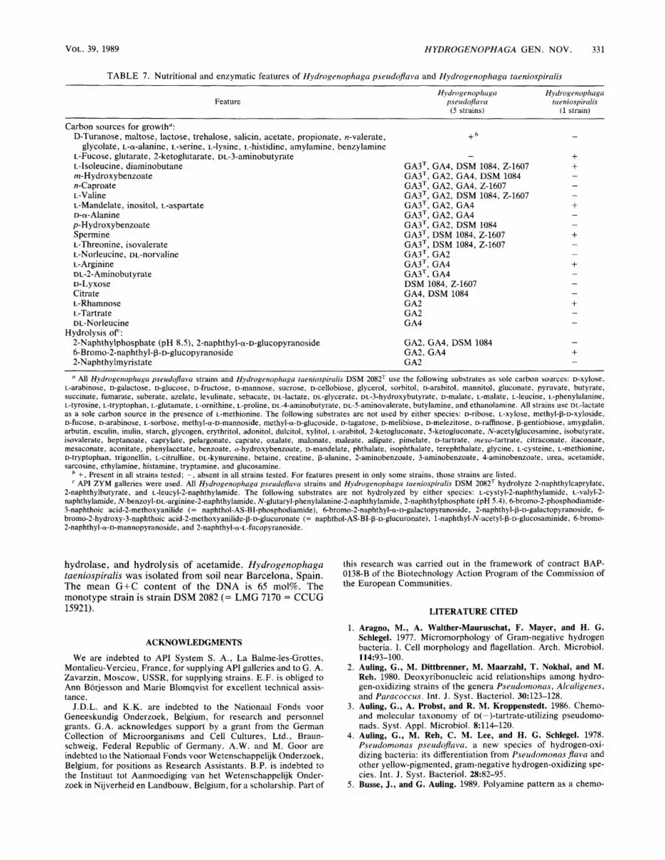
Hydrogenophaga pseudoflava (pseu.do.fla'va. Gr. adj. pseudes, false; L. fem. adj. f lava, yellow; M. L. fem. adj. pseudoJava, not the true [Pseudornonas] Java) is the same as that for the genus. Some strains of this species (originally described as "[Pseudornonas] carboxydoJava") are faculta- tive autotrophs, growing at the expense of carbon monoxide oxidation (33). Aragno et al. (1) reported the presence of pili over the total cell surface for two strains, including the type strain. Since this species includes strains of both the former species [Pseudomonas] pseudoflava and the former species LLIPseudornonas] carboxydojava," we provide a new de- scription for Hydrogenophaga pseudoflava based on our own results, which are generally in good agreement with the original species descriptions (4, 48). Nutritional and enzy- matic features are listed in Table 7. The following biochem- ical and physiological characteristics are present in all Hy- drogenophaga pseudoflava strains: growth at 30 and 37"C, growth in the presence of 0.5% NaCI, hydrolysis of Tween 80 and esculin, reduction of nitrate and nitrite, denitrifica- tion, and P-galactosidase. The following features are lacking in all Hydrogenophaga pseudoflava strains: catalase, hemo- lysis, resistance to penicillin (10 pg per disk), growth on Drigalski-Conradi agar at 30°C, growth at 42"C, growth in the presence of 1.5, 3, 4.5, or 6.5% NaCl, production of acid in 10% lactose, triple sugar iron medium, and oxidative-fer- mentative medium containing D-glucose , D-fructose, D-XY- lose, maltose, or adonitol, fluorescence, growth on cetrim- ide, production of H,S in triple sugar iron medium, amylase, lysine and ornithine decarboxylase, arginine djhydrolase, urease, and hydrolysis of gelatin. Hydrolysis of DNA and acetamide and production of acid from indole are absent in all strains except the type strain, which was not tested. Recently, Jenni et al. (21) reported nitrogen fixation for a strain (strain NEU 2226) which they assigned to [Pseudorno- nus] pseudoJava, although this feature could not be detected in strain GA3T. Hydrogenophaga pseudoflava was isolated from soil, mud, and water by enrichment for hydrogen bacteria (4, 33). The mean G+C content of the DNA is 66 to 69 mol%. The type strain is strain GA3 (= LMG 5945 = CCUG 13799 = ATCC 33668). Its characteristics are the same as those listed above for the species, and the mean G+C content of its DNA is 66 mo%.
Description of Hydrogenophaga taeniospiralis (Lalucat, Pares, and Schlegel 1982) comb. nov. The description of Hydrogenophaga taeniospiralis (tae.ni.o.spi.ra'1is. Gr. n. taenia, ribbon; L. adj. spiralis, coiled; M.L. adj. taeniospi- ralis, ribbon coiled, named after Caedibacter taeniospiralis, an organism also containing R bodies) is the same as that for the genus. The original description by Lalucat et al. (28) was verified. This description was found to be in good agreement with our own results, except for the following features (our results are indicated in parentheses): reduction of nitrite (positive), hydrolysis of esculin (positive), urease (weak), catalase (negative), growth at 37°C (negative), and use of the following substrates as sole carbon sources-butyrate (pos- itive), trehalose (negative), and L-lysine (negative). Addi- tions to the original description are presented below. Nutri- tional and enzymological features are listed in Table 7. Hydrogenophaga taeniospiralis grows at 30°C, hydrolyzes Tween 80, and possesses P-galactosidase. The following features are lacking: growth at 42"C, growth on Drigalski- Conradi agar at 30°C, growth in the presence of 0.5, 1.5, 3, 4.5, or 6.5% NaC1, resistance to penicillin (10 pg per disk), hemolysis, production of acid in oxidative-fermentative me- dium containing D-glucose, maltose, or adonitol, growth on cetrimide, lysine and ornithine decarboxylase, arginine di-
VOL. 39, 1989 HYDROGENOPHAGA GEN. NOV. 331
TABLE 7. Nutritional and enzymatic features of Hydrogenophaga pseudojlava and Hydrogenophaga taeniospiralis
Feature Hydrogenophaga Hydrogenophaga
pseudojla v u taeniospirulis (1 strain) (5 strains)
Carbon sources for growth": D-Turanose, maltose, lactose, trehalose, salicin, acetate, propionate, n-valerate,
glycolate, L-a-alanine, L-serine, L-lysine, L-histidine, amylamine, benzylamine L-Fucose, glutarate, 2-ketoglutarate, DL-3-aminobutyrate L-Isoleucine, diaminobutane m-H ydrox ybenzoate n-Caproate L-Valine L-Mandelate, inositol, L-aspartate D-a- Alanine p-H ydrox ybenzoate Spermine L-Threonine, isovalerate L-Norleucine, DL-norvaline L- Arginine DL-2-Aminobutyrate D-Lyxose Citrate L-Rhamnose L-Tartrate DL-Norleucine
Hydrolysis OF: 2-Naphthylphosphate (pH S S ) , 2-naphthyl-ol-~-glucopyranoside 6-Bromo-2-naphthyl-~-~-glucopyranoside 2-Naphthylmyristate
+ b
-
GA3T, GA4, DSM 1084, 2-1607 GA3T, GA2, GA4, DSM 1084 GA3T, GA2, GA4, 2-1607 GA3T, GA2, DSM 1084, 2-1607 GA3T, GA2, GA4 GA3T, GA2, GA4 GA3T, GA2, DSM 1084 GA3T, DSM 1084, 2-1607 GA3=, DSM 1084, 2-1607 GA3T, GA2 GA3T, GA4 GA3T, GA4
GA4, DSM 1084 GA2 GA2 GA4
DSM 1084, 2-1607
GA2, GA4, DSM 1084 GA2, GA4 GA2
~ ~ ~~
a All Hydrogenophaga pseudofiava strains and Hydrogenophuga taeniospiralis DSM 2082T use the following substrates as sole carbon sources: D-xylose, L-arabinose, D-galactose, D-glucose, D-fructose, D-mannose, sucrose, D-cellobiose, glycerol, sorbitol, D-arabitol, mannitol, gluconate, pyruvate, butyrate, succinate, fumarate, suberate, azelate, levulinate, sebacate, DL-lactate, DL-glycerate, ~~-3-hydroxybutyrate, D-malate, L-malate, L-leucine, L-phenylalanine, L-tyrosine, L-tryptophan, L-glutamate, L-ornithine, L-proline, ~~-4-aminobutyrate, ~~-5-aminovalerate, butylamine, and ethanolamine. All strains use oL-lactate as a sole carbon source in the presence of L-methionine. The following substrates are not used by either species: D-ribose, L-xylose, methyl-p-D-xyloside, D-fucose, D-arabinose, L-sorbose, methyl-a-D-mannoside, methyl-a-D-glucoside, D-tagatose, D-melibiose, D-melezitose, D-raffinose, p-gentiobiose, amygdalin, arbutin, esculin, inulin, starch, glycogen, erythritol, adonitol, dulcitol, xylitol, L-arabitol, 2-ketogluconate, 5-ketogluconate, N-acetylglucosamine, isobutyrate, isovalerate, heptanoate, caprylate, pelargonate, caprate, oxalate, malonate, maleate, adipate, pimelate, D-tartrate, rneso-tartrate, citraconate, itaconate, mesaconate, aconitate, phenylacetate, benzoate, o-hydroxybenzoate, ~-mandelate, phthalate, isophthalate, terephthalate, glycine, L-cysteine, L-methionine, D-tryptophan, trigonellin, ~-citrulline, DL-kynurenine, betaine, creatine, p-alanine, 2-aminobenzoate, 3-aminobenzoate, 4-aminobenzoate, urea, acetamide, sarcosine, ethylamine, histamine, tryptamine, and glucosamine.
+, Present in all strains tested; -, absent in all strains tested. For features present in only some strains, those strains are listed. API ZYM galleries were used. All Hydrogenophaga pseudoJava strains and Hydrogenophaga taeniospiralis DSM 2082T hydrolyze 2-naphthylcaprylate,
2-naphthylbutyrate, and L-leucyl-2-naphthylamide. The following substrates are not hydrolyzed by either species: ~-cystyl-2-naphthylamide, ~-valyl-2- naphthylamide, N-benzoyl-~~-arginine-2-naphthylamide, N-glutaryl-phenylalanine-2-naphthylamide, 2-naphthylphosphate (pH 5.4), 6-bromo-2-phosphodiamide- 3-naphthoic acid-2-methoxyanilide (= naphthol-AS-BI-phosphodiamide), 6-bromo-2-naphthyl-a-~-galactopyranoside, 2-naphthyl-p-~-galactopyranoside, 6- bromo-2-hydroxy-3-naphthoic acid-2-methoxyanilide-~-~-glucuronate (= naphthol-AS-BI-p-D-glucuronate), 1-naphthyl-N-acetyl-P-o-glucosaminide, 6-bromo- 2-naphthyl-a-~-mannopyranoside, and 2-naphthyl-a-~-fucopyranoside.
hydrolase, and hydrolysis of acetamide. Hydrogenophaga taeniospiralis was isolated from soil near Barcelona, Spain. The mean G+C content of the DNA is 65 mol%. The monotype strain is strain DSM 2082 (= LMG 7170 = CCUG 15921).
ACKNOWLEDGMENTS
We are indebted to API System S. A. , La Balme-les-Grottes, Montalieu-Vercieu, France, for supplying API galleries and to G. A. Zavarzin, Moscow, USSR, for supplying strains. E.F. is obliged to Ann Borjesson and Marie Blomqvist for excellent technical assis- tance.
J.D.L. and K.K. are indebted to the Nationaal Fonds voor Geneeskundig Onderzoek, Belgium, for research and personnel grants. G.A. acknowledges support by a grant from the German Collection of Microorganisms and Cell Cultures, Ltd., Braun- schweig, Federal Republic of Germany. A.W. and M. Goor are indebted to the Nationaal Fonds voor Wetenschappelijk Onderzoek, Belgium, for positions as Research Assistants. B.P. is indebted to the Instituut tot Aanmoediging van het Wetenschappelijk Onder- zoek in Nijverheid en Landbouw, Belgium, for a scholarship. Part of
this research was carried out in the framework of contract BAP- 0138-B of the Biotechnology Action Program of the Commission of the European Communities.
LITERATURE CITED
1. Aragno, M., A. Walther-Mauruschat, F. Mayer, and H. G. Schlegel. 1977. Micromorphology of Gram-negative hydrogen bacteria. I. Cell morphology and flagellation. Arch. Microbiol.
2. Auling, G., M. Dittbrenner, M. Maarzahl, T. Nokhal, and M. Reh. 1980. Deoxyribonucleic acid relationships among hydro- gen-oxidizing strains of the genera Pseudomonas, Alcaligenes, and Paracoccus. Int. J . Syst. Bacteriol. 30:123-128.
3. Auling, G., A. Probst, and R. M. Kroppenstedt. 1986. Chemo- and molecular taxonomy of D( -)-tartrate-utilizing pseudomo- nads. Syst. Appl. Microbiol. 8:114-120.
4. Auling, G., M. Reh, C. M. Lee, and H. G. Schlegel. 1978. Pseudomonas pseudoflava, a new species of hydrogen-oxi- dizing bacteria: its differentiation from Pseudomonas flava and other yellow-pigmented, gram-negative hydrogen-oxidizing spe- cies. Int. J. Syst. Bacteriol. 28:82-95.
5. Busse, J., and G. Auling. 1989. Polyamine pattern as a chemo-
114:93-100.

332 WILLEMS ET AL. INT. J. SYST. BACTERIOL.
taxonomic marker within the Proteobacteria. Syst. Appl. Mi- crobiol. 11:l-8.
6. Byng, G. S., J. L. Johnson, R. J. Whitaker, R. L. Gherna, and R. A. Jensen. 1983. The evolutionary pattern of aromatic amino acid biosynthesis and the emerging phylogeny of pseudomonad bacteria. J. Mol. Evol. 19:272-282.
7. Davis, D. H., M. Doudoroff, R. Y. Stanier, and M. Mandel. 1969. Proposal to reject the genus Hydrogenomonas: taxonomic im- plications. Int. J. Syst. Bacteriol. 19:375-390.
8. Davis, D. H., R. Y. Stanier, M. Doudoroff, and M. Mandel. 1970. Taxonomic studies on some Gram negative polarly flagellated “hydrogen bacteria” and related species. Arch. Mikrobiol.
9. De Ley, J. 1970. Reexamination of the association between melting point, buoyant density, and chemical base composition of deoxyribonucleic acid. J. Bacteriol. 101:738-754.
10. De Ley, J. 1978. Modern molecular methods in bacterial taxon- omy: evaluation, application, prospects, p. 347-357. I n Pro- ceedings of the 4th International Conference of Plant Pathogenic Bacteria, vol. 1. Gibert-Clarey, Tours, France.
11. De Ley, J., H. Cattoir, and A. Reynaerts. 1970. The quantitative measurement of DNA hybridization from renaturation rates. Eur. J. Biochem. 12:133-142.
12. De Ley, J., and J. De Smedt. 1975. Improvements of the membrane filter method for DNA:rRNA hybridization. Antonie van Leeuwenhoek J. Microbiol. Serol. 41:287-307.
13. De Ley, J., and J. Van Muylem. 1963. Some applications of deoxyribonucleic acid base composition in bacterial taxonomy. Antonie van Leeuwenhoek J. Microbiol. Serol. 29:34&358.
14. De Vos, P., and J. De Ley. 1983. Intra- and intergeneric similarities of Pseudomonas and Xanthomonus ribosomal ribo- nucleic acid cistrons. Int. J. Syst. Bacteriol. 33:487-509.
15. De Vos, P., M. Goor, M. Gillis, and J. De Ley. 1985. Ribosomal ribonucleic acid cistron similarities of phytopathogenic Pseudo- monas species. Int. J. Syst. Bacteriol. 35169-184.
16. De Vos, P., K. Kersters, E. Falsen, B. Pot, M. Gillis, P. Segers, and J. De Ley. 1985. Cornamonas Davis and Park 1962 gen. nov. nom. rev. emend., and Cornamonas terrigena Hugh 1962 sp. nov., nom. rev. Int. J. Syst. Bacteriol. 35443453.
17. Falsen, E. 1983. Immunodiffusion as an aid in routine identifi- cation of uncommon aerobic gram negative bacteria, p. 477-483. In H. Leclerc (ed.), Gram negative bacteria of medical and public health importance: taxonomy-identification-applications . Les Cditions de 1’Institut National de la SantC et de la Recherche MCdicale, Paris.
18. Jantzen, E., and K. Bryn. 1985. Whole-cell and lipopolysaccha- ride fatty acids and sugars of gram-negative bacteria, p. 145- 171. In M. Goodfellow and D. Minnikin (ed.), Chemical meth- ods in bacterial systematics. Academic Press, Inc., New York.
19. Jantzen, E., K. Bryn, N. Hagen, T. Bergan, and K. Bdvre. 1978. Fatty acids and monosaccharides of Neisseriaceae in relation to established taxonomy. Natl. Inst. Public Health Ann. (Norway)
20. Jantzen, E., 0. M. Kvalheim, T. A. Hauge, N. Hagen, and K. Bbvre. 1987. Grouping of bacteria by SIMCA pattern recogni- tion on gas chromatographic lipid data: patterns among Morax- ella and rod-shaped Neisseria. Syst. Appl. Microbiol. 9:142- 150.
21. Jenni, B., C. Isch, and M. Aragno. 1989. Nitrogen fixation by new strains of Pseudomonas pseudojava and related bacteria. J. Gen. Microbiol. 135461467.
22. Kersters, K., and J. De Ley. 1984. Genus Alcaligenes Castellani and Chalmers 1919, p. 361-373. I n N. R. Krieg and J. G. Holt (ed.), Bergey’s manual of systematic bacteriology, vol. 1. The Williams & Wilkins Co., Baltimore.
23. Kersters, K., K.-H. Hinz, A. Hertle, P. Segers, A. Lievens, 0. Siegmann, and J. De Ley. 1984. Bordetella avium sp. nov., isolated from the respiratory tracts of turkeys and other birds. Int. J. Syst. Bacteriol. 3456-70.
24. Kiredjian, M., B. Holmes, K. Kersters, I. Guilvout, and J. De Ley. 1986. Alcaligenes piechaudii, a new species from human
7O:l-13.
1 : 59-7 1.
clinical specimens and the environment. Int. J. Syst. Bacteriol.
25. Kluyver, A. J., and A. Manten. 1942. Some observations on the metabolism of bacteria oxidizing molecular hydrogen. Antonie van Leeuwenhoek. J . Microbiol. Serol. 8:71-85.
26. Kraut, M., and 0. Meyer. 1988. Plasmids in carboxydotrophic bacteria: physical and restriction analysis. Arch. Microbiol. 149540-546.
27. Laemmli, U. K. 1970. Cleavage of structural proteins during the assembly of the head of bacteriophage T4. Nature (London) 227:680-685.
28. Lalucat, J., R. Pares, and H. G. Schlegel. 1982. Pseudomonas taeniospiralis sp. nov., an R-body-containing hydrogen bacte- rium. Int. J . Syst. Bacteriol. 32:332-338.
29. Marmur, J. A. 1961. A procedure for the isolation of deoxyri- bonucleic acid from micro-organisms. J. Mol. Biol. 3:208- 218.
30. Marmur, J., and P. Doty. 1962. Determination of the base composition of deoxyribonucleic acid from its thermal denatur- ation temperature. J. Mol. Biol. 5109--118.
31. Mayberry, W. R. 1981. Dihydroxy and monohydroxy fatty acids in Legionella pneumcphila. J. Bacteriol. 147:373-381.
32. Niklewski, B. 1910. Uber die Wassersloffoxydation durch Mik- roorganismen. Jahrb. Wiss. Bot. 48:113-142.
33. Nozhevnikova, A. N., and G. A. Zavarzin. 1974. On the taxon- omy of CO-oxidizing Gram-negative bacteria. Izv. Akad. Nauk SSSR Ser. Biol. 3:436-440.
34. Orla-Jensen, S. 1909. Die Hauptlinien des natiirlichen Bakte- riensystems. Centralbl. Bakteriol. Parasitenkd. Infektionskr.
35. Oyaizu, H., and K. Komagata. 1983. Grouping of Pseudomonas species on the basis of cellular fatty acid composition and the quinone system with special reference to the existence of 3-hydroxy fatty acids. J. Gen. Appl. Microbiol. 29:1740.
36. Palleroni, N. J. 1984. Genus I . Pseudomonas Migula 1894, p. 141-199. In N. R. Krieg and J. G. Holt (ed.), Bergey’s manual of systematic bacteriology, vol. 1. The Williams & Wilkins Co., Baltimore.
37. Palleroni, N. J., R. Kunisawa, R. Contopoulou, and M. Doudor- off. 1973. Nucleic acid homologies in the genus Pseudomonas. Int. J. Syst. Bacteriol. 23:333-339.
38. Pot, B., M. Gillis, B. Hoste, A. Van De Velde, F. Bekaert, K. Kersters, and J. De Ley. 1989. Intra- and intergeneric relation- ships of the genus Oceanospirillum, Int. J. Syst. Bacteriol.
39. Rossau, R., K. Kersters, E. Falsen, E. Jantzen, P. Segers, A. Union, L. Nehls, and J. De Ley. 1987. Oligella, a new genus including Oligella urethralis comb. nov. (formerly Moraxella urethralis) and Oligella ureolytica sp. nov. (formerly CDC group We): relationship to Taylorelfa equigenitalis and related taxa. Int. J. Syst. Bacteriol. 37:198-2 LO.
40. Sneath, P. H. A., and R. R. Sokal. 1973. Numerical taxonomy. The principles and practice of numerical classification. W. H. Freeman and Co., San Francisco.
41. Stackebrandt, E., R. G. E. Murray, and H. G. Truper. 1988. Proteobacteriu classis nov., a name for the phylogenetic taxon that includes the “purple bacteria and their relatives.” Int. J . Syst. Bacteriol. 38:321-325.
42. Tamaoka, J., D.-M. Ha, and K. Komagata. 1987. Reclassifica- tion of Pseudomonas acidovorans den Dooren de Jong 1926 and Pseudomonas testosteroni Marcus and Talalay 1956 as Coma- monas acidovorans comb. nov. and Cornamonas testosteroni comb. nov., with an emended description of the genus Coma- monas. Int. J. Syst. Bacteriol. 3752-59.
43. Van Landschoot, A., and J. De Ley. 1983. Intra- and intergeneric similarities of the rRNA cistrons of Afteromonas, Marinomonas (gen. nov.) and some other Gram-negative bacteria. J. Gen. Microbiol. 129:3057-3074.
44. Willems, A,, M. Gillis, K. Kersters, L. Van den Broecke, and J. De Ley. 1987. Transfer of Xanthomonus ampelina Panagopoulos 1969 to a new genus, Xylophilus gen. nov., as Xylophilus ampelinus (Panagopoulos 1969) comb. nov. Int. J. Syst. Bacte- rial. 37:422430.
36:282-287.
Abt. 2 22:305-346.
39:23-34.
VOL. 39, 1989 HYDROGENOPHAGA GEN. NOV. 333
45. Wishart, D. 1978. Clustan user manual, 3rd ed. Program Library Unit, Edinburgh University, Edinburgh, Scotland.
46. Woese, C. R. , P. Blanz, and C. Hahn. 1984. What isn’t a pseudomonad: the importance of nomenclature in bacterial classification. Syst. Appl. Microbiol. 5:179-195.
47. Wold, S., C. Albano, W. J. DunnIII, U. Edlund, K. Esbensen, P.
Geladi, S. Hellberg, S. Johanson, W. Lindberg, and M. Sjostrom. 1984. Multivariate data analyses in chemistry, p. 17-95. I n B. R. Kowalsky (ed.), Chemometrics, mathematics and statistics in chemistry. V, Dordrecht, The Netherlands.
48. Zavarzin, G. A., and A. N. Nozhevnikova. 1977. Aerobic car- boxydobacteria. Microb. Ecol. 3:305-326.