Embed Size (px)
Citation preview
Am. J. Hum. Genet. 71:143–153, 2002
143
Hyperhomocysteinemia Due to Methionine Synthase Deficiency, cblG:Structure of the MTR Gene, Genotype Diversity, and Recognition of aCommon Mutation, P1173LDavid Watkins,1 Ming Ru,1 Hye-Yeon Hwang,7 Caroline D. Kim,1,3 Angus Murray,1,3
Noah S. Philip,1,3 William Kim,1,3 Helen Legakis,1,3 Timothy Wai,1,3 John F. Hilton,1,3
Bing Ge,2 Carole Dore,2 Angela Hosack,1 Aaron Wilson,8 Roy A. Gravel,8 Barry Shane,7Thomas J. Hudson,1,2,4 and David S. Rosenblatt1,3,4,5,6
1Division of Medical Genetics, Department of Medicine, and 2Montreal Genome Centre, McGill University Health Centre, and Departmentsof 3Biology, 4Human Genetics, 5Medicine, and 6Pediatrics, McGill University, Montreal; 7Department of Nutritional Sciences and Toxicology,University of California at Berkeley, Berkeley; and 8Department of Biochemistry and Molecular Biology, University of Calgary, Calgary
Mutations in the MTR gene, which encodes methionine synthase on human chromosome 1p43, result in themethylcobalamin deficiency G (cblG) disorder, which is characterized by homocystinuria, hyperhomocysteinemia,and hypomethioninemia. To investigate the molecular basis of the disorder, we have characterized the structure ofthe MTR gene, thereby identifying exon-intron boundaries. This enabled amplification of each of the 33 exons ofthe gene, from genomic DNA from a panel of 21 patients with cblG. Thirteen novel mutations were identified.These included five deletions (c.12-13delGC, c.381delA, c.2101delT, c.2669-2670delTG, and c.2796-2800del-AAGTC) and two nonsense mutations (R585X and E1204X) that would result in synthesis of truncated proteinsthat lack portions critical for enzyme function. One mutation was identified that resulted in conversion of A to Cof the invariant A of the 3′ splice site of intron 9. Five missense mutations (A410P, S437Y, S450H, H595P, andI804T) were identified. The latter mutations, as well as the splice-site mutation, were not detected in a panel of50 anonymous DNA samples, suggesting that these sequence changes are not polymorphisms present in the generalpopulation. In addition, a previously described missense mutation, P1173L, was detected in 16 patients in anexpanded panel of 24 patients with cblG. Analysis of haplotypes constructed using sequence polymorphisms iden-tified within the MTR gene demonstrated that this mutation, a CrT transition in a CpG island, has occurred onat least two separate genetic backgrounds.
Introduction
The methylcobalamin deficiency G (cblG) disorder (MIM250940) is the result of mutations affecting the MTRgene, which encodes the cytoplasmic enzyme methio-nine synthase (EC 2.1.1.13). This enzyme catalyzes themethylation of homocysteine to generate methionine,using 5-methyltetrahydrofolate as a methyl-group do-nor, and requires the presence of an enzyme-boundcobalamin prosthetic group for activity. The reactionproceeds by transfer of the methyl group from 5-meth-yltetrahydrofolate to the enzyme-bound cobalamin toform methylcobalamin and by subsequent transfer of themethyl group from methylcobalamin to homocysteine
Received February 21, 2002; accepted for publication April 22,2002; electronically published May 30, 2002.
Address for correspondence and reprints: Dr. David Watkins, Di-vision of Medical Genetics, Room H5-63, McGill University HealthCentre/Royal Victoria Hospital Site, 687 Pine Avenue West, Montreal,Quebec, H3A 1A1, Canada. E-mail: [email protected]
� 2002 by The American Society of Human Genetics. All rights reserved.0002-9297/2002/7101-0015$15.00
to form methionine (Matthews 1999; Matthews andLudwig 2001). Activity of methionine synthase dependson the presence of a second protein, methionine syn-thase reductase, which maintains the methionine syn-thase–bound cobalamin in its fully reduced active state.When the cobalamin becomes oxidized, methionine syn-thase reductase catalyzes its reductive methylation, usingS-adenosylmethionine as a methyl donor to regeneratemethylcobalamin (Olteanu and Banerjee 2001).
Methionine synthase activity is important for main-taining adequate levels of methionine and for preventingaccumulation of homocysteine. Increased blood levelsof homocysteine have been associated with increasedlikelihood of developing cardiovascular disease, birthdefects, and Down syndrome, and they may affect thedevelopment of some types of cancer (Carmel and Ja-cobsen 2001). Methionine synthase is also critical in themethylation cycle, which maintains the cellular level ofthe methionine derivative S-adenosylmethionine. Thismolecule acts as methyl group donor in a wide varietyof cellular processes, including DNA and RNA meth-ylation and neurotransmitter synthesis. Because methio-
144 Am. J. Hum. Genet. 71:143–153, 2002
nine synthase is the only enzyme in mammalian cellsthat uses 5-methyltetrahydrofolate, deficient activityalso results in the trapping of cellular folate as 5-methyl-tetrahydrofolate, which becomes unavailable for otherfolate-dependent reactions involved in purine and py-rimidine biosynthesis and other single-carbon–transferreactions.
Inborn errors of metabolism resulting in functional de-ficiency of methionine synthase are characterized by ho-mocystinuria, hyperhomocysteinemia, and hypomethio-ninemia. Patients have typically presented during the first2 years of life with megaloblastic anemia and develop-mental delay, but patients who presented as adults havebeen reported (Rosenblatt and Fenton 2001). Comple-mentation analysis demonstrated the existence of twoforms of the disorder—cblG and a second form thatis designated “methylcobalamin deficiency cblE type”(cblE)—that presumably result from mutations at sep-arate loci (Watkins and Rosenblatt 1989). Cloning ofthe MTR gene (GDB accession number 119440) onchromosome 1q43 (Leclerc et al. 1996; Li et al. 1996;Chen et al. 1997) led to the demonstration that mu-tations in this gene are the cause of the cblG disorder(Gulati et al. 1996; Leclerc et al. 1996). It was subse-quently shown that the cblE disorder is the result ofmutations affecting the MTRR gene, on chromosome5p15.2-15.3, that encodes methionine synthase reduc-tase (Leclerc et al. 1998).
The cDNA for the human gene encoding methioninesynthase was cloned on the basis of homology with theEscherichia coli gene encoding cobalamin-dependentmethionine synthase, which had been previously iden-tified. The human gene encodes a protein that is 1,265amino acids in length. Several mutations in the genehave been described in patients with cblG (Gulati et al.1996; Leclerc et al. 1996; Wilson et al. 1998). We reportfurther characterization of this gene, defining its intronicand exonic structure. This has permitted sequencing ofthe entire coding sequence of methionine synthase fromgenomic DNA of a panel of patients with the cblG dis-order. A panel of 18 patients with cblG was investigatedby sequence analysis, and an expanded panel of 24 pa-tients was investigated using restriction-endonucleaseanalysis. To date, 28 patients with the cblG disorderhave been identified (Suormala et al. 2001; D.S.R., un-published data). Thus, this panel of patients with cblGrepresents a majority of all recognized patients with thisdisorder.
Patients and Methods
Patients
DNA was extracted and sequenced from a panel of18 fibroblast lines derived from patients with cblG.DNA from an additional six patients with cblG was
tested for identified mutations, using restriction endo-nuclease–based assays, as described below. All cell lineswere characterized by decreased functional activity ofmethionine synthase and decreased synthesis of meth-ylcobalamin in intact cells, in the presence of normalfunctional activity of a second cobalamin-dependent en-zyme, methylmalonyl-CoA mutase, and normal synthe-sis of its adenosylcobalamin coenzyme. The diagnosis ofcblG was in all cases confirmed by somatic cell comple-mentation analysis. These studies were approved by theresearch ethics board of the Royal Victoria Hospital.
Genomic Organization of the Coding Region of theHuman Methionine Synthase Gene
Some of the cDNA clones used for the original se-quencing of the human methionine synthase cDNA(Chen et al. 1997) contained intronic sequences. Addi-tional exon-intron junctions were determined by PCR,using primers based on the cDNA sequence. PCR prod-ucts that were larger than the expected size of the cDNAsequence were assumed to contain intronic sequencesand were sequenced across the junctions. Additional in-tronic junctions were identified by genomic sequencingof putative exons. Intron sizes were determined by se-quencing through the region or by PCR using flankingprimers.
Sequencing of the MTR Gene
Sequencing was performed using genomic DNAfrom 18 cblG cell lines. Six additional cell lines(WG1655, WG1670, WG1671, WG2867, WG2918,and WG2989) were tested for presence of mutationsdetected by sequencing of the other cell lines, usingthe restriction endonuclease–based tests described be-low, but were not sequenced themselves.
Each of the 33 exons of the MTR gene was amplifiedseparately, using PCR primers within the flanking in-tronic sequences, such that the entire exon (as well asexon-intron boundaries) could be sequenced. Primerswere designed using the Primer 3.0 software availableon the Whitehead Institute/MIT Center for Genome Re-search server. The sequences of the PCR primers usedare shown in table 1. The annealing temperature for allprimer pairs was 60�C. PCR was performed in a totalvolume of 50 ml containing 1.25 U AmpliTaq Gold poly-merase (Perkin-Elmer Cetus) in the buffer provided bythe manufacturer, 2.5 mM MgCl2, 0.2 mM dNTPs,primers at a final concentration of 0.5 mM, and 100 ngof template genomic DNA. PCR products were purifiedwith BioMag DNA Sep magnetic beads (PerSeptive Bio-systems). Sequencing reactions were performed using theBigDye Primer Cycle Sequencing Ready Reactions-21M13 kit (PE Applied Biosystems), and products wereanalyzed on ABI 377 automated DNA sequencers (PEApplied Biosystems). Gel files were processed using Se-
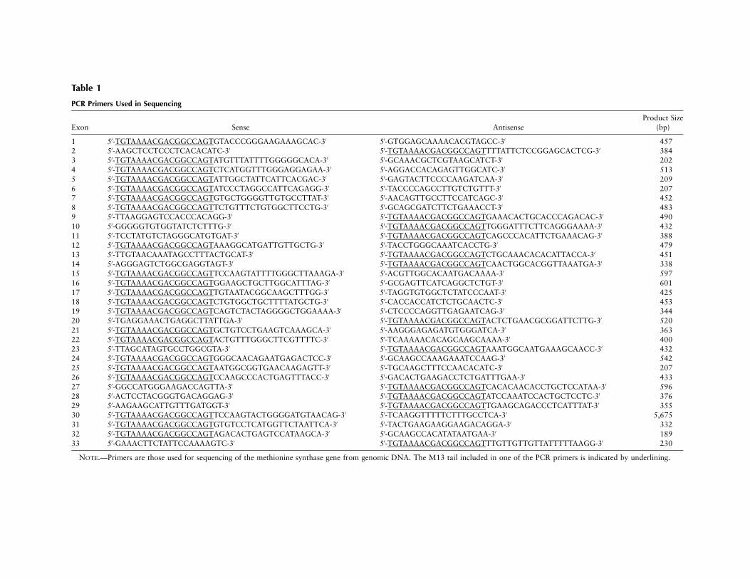
Table 1
PCR Primers Used in Sequencing
Exon Sense AntisenseProduct Size
(bp)
1 5′-TGTAAAACGACGGCCAGTGTACCCGGGAAGAAAGCAC-3′ 5′-GTGGAGCAAAACACGTAGCC-3′ 4572 5′-AAGCTCCTCCCTCACACATC-3′ 5′-TGTAAAACGACGGCCAGTTTTATTCTCCGGAGCACTCG-3′ 3843 5′-TGTAAAACGACGGCCAGTATGTTTATTTTGGGGGCACA-3′ 5′-GCAAACGCTCGTAAGCATCT-3′ 2024 5′-TGTAAAACGACGGCCAGTCTCATGGTTTGGGAGGAGAA-3′ 5′-AGGACCACAGAGTTGGCATC-3′ 5135 5′-TGTAAAACGACGGCCAGTATTGGCTATTCATTCACGAC-3′ 5′-GAGTACTTCCCCAAGATCAA-3′ 2096 5′-TGTAAAACGACGGCCAGTATCCCTAGGCCATTCAGAGG-3′ 5′-TACCCCAGCCTTGTCTGTTT-3′ 2077 5′-TGTAAAACGACGGCCAGTGTGCTGGGGTTGTGCCTTAT-3′ 5′-AACAGTTGCCTTCCATCAGC-3′ 4528 5′-TGTAAAACGACGGCCAGTTCTGTTTCTGTGGCTTCCTG-3′ 5′-GCAGCGATCTTCTGAAACCT-3′ 4839 5′-TTAAGGAGTCCACCCACAGG-3′ 5′-TGTAAAACGACGGCCAGTGAAACACTGCACCCAGACAC-3′ 49010 5′-GGGGGTGTGGTATCTCTTTG-3′ 5′-TGTAAAACGACGGCCAGTTGGGATTTCTTCAGGGAAAA-3′ 43211 5′-TCCTATGTCTAGGGCATGTGAT-3′ 5′-TGTAAAACGACGGCCAGTCAGCCCACATTCTGAAACAG-3′ 38812 5′-TGTAAAACGACGGCCAGTAAAGGCATGATTGTTGCTG-3′ 5′-TACCTGGGCAAATCACCTG-3′ 47913 5′-TTGTAACAAATAGCCTTTACTGCAT-3′ 5′-TGTAAAACGACGGCCAGTCTGCAAACACACATTACCA-3′ 45114 5′-AGGGAGTCTGGCGAGGTAGT-3′ 5′-TGTAAAACGACGGCCAGTCAACTGGCACGGTTAAATGA-3′ 33815 5′-TGTAAAACGACGGCCAGTTCCAAGTATTTTGGGCTTAAAGA-3′ 5′-ACGTTGGCACAATGACAAAA-3′ 59716 5′-TGTAAAACGACGGCCAGTGGAAGCTGCTTGGCATTTAG-3′ 5′-GCGAGTTCATCAGGCTCTGT-3′ 60117 5′-TGTAAAACGACGGCCAGTTGTAATACGGCAAGCTTTGG-3′ 5′-TAGGTGTGGCTCTATCCCAAT-3′ 42518 5′-TGTAAAACGACGGCCAGTCTGTGGCTGCTTTTATGCTG-3′ 5′-CACCACCATCTCTGCAACTC-3′ 45319 5′-TGTAAAACGACGGCCAGTCAGTCTACTAGGGGCTGGAAAA-3′ 5′-CTCCCCAGGTTGAGAATCAG-3′ 34420 5′-TGAGGAAACTGAGGCTTATTGA-3′ 5′-TGTAAAACGACGGCCAGTACTCTGAACGCGGATTCTTG-3′ 52021 5′-TGTAAAACGACGGCCAGTGCTGTCCTGAAGTCAAAGCA-3′ 5′-AAGGGAGAGATGTGGGATCA-3′ 36322 5′-TGTAAAACGACGGCCAGTACTGTTTGGGCTTCGTTTTC-3′ 5′-TCAAAAACACAGCAAGCAAAA-3′ 40023 5′-TTAGCATAGTGCCTGGCGTA-3′ 5′-TGTAAAACGACGGCCAGTAAATGGCAATGAAAGCAACC-3′ 43224 5′-TGTAAAACGACGGCCAGTGGGCAACAGAATGAGACTCC-3′ 5′-GCAAGCCAAAGAAATCCAAG-3′ 54225 5′-TGTAAAACGACGGCCAGTAATGGCGGTGAACAAGAGTT-3′ 5′-TGCAAGCTTTCCAACACATC-3′ 20726 5′-TGTAAAACGACGGCCAGTCCAAGCCCACTGAGTTTACC-3′ 5′-GACACTGAAGACCTCTGATTTGAA-3′ 43327 5′-GGCCATGGGAAGACCAGTTA-3′ 5′-TGTAAAACGACGGCCAGTCACACAACACCTGCTCCATAA-3′ 59628 5′-ACTCCTACGGGTGACAGGAG-3′ 5′-TGTAAAACGACGGCCAGTATCCAAATCCACTGCTCCTC-3′ 37629 5′-AAGAAGCATTGTTTGATGGT-3′ 5′-TGTAAAACGACGGCCAGTTGAAGCAGACCCTCATTTAT-3′ 35530 5′-TGTAAAACGACGGCCAGTTCCAAGTACTGGGGATGTAACAG-3′ 5′-TCAAGGTTTTTCTTTGCCTCA-3′ 5,67531 5′-TGTAAAACGACGGCCAGTGTGTCCTCATGGTTCTAATTCA-3′ 5′-TACTGAAGAAGGAAGACAGGA-3′ 33232 5′-TGTAAAACGACGGCCAGTAGACACTGAGTCCATAAGCA-3′ 5′-GCAAGCCACATATAATGAA-3′ 18933 5′-GAAACTTCTATTCCAAAAGTC-3′ 5′-TGTAAAACGACGGCCAGTTTGTTGTTGTTATTTTTAAGG-3′ 230
NOTE.—Primers are those used for sequencing of the methionine synthase gene from genomic DNA. The M13 tail included in one of the PCR primers is indicated by underlining.
146 Am. J. Hum. Genet. 71:143–153, 2002
quence Analysis software (PE Applied Biosystems) andthen were assembled and analyzed using Autoassembler2.0 (PE Applied Biosystems).
Restriction Endonuclease Analysis
MaeIII was purchased from Boehringer Mannheim;the remaining restriction endonucleases (Bsp1286I, BsrI,BstNI, BstUI, DdeI, HinfI, MspI, and NspI) were pur-chased from New England Biolabs. When a sequencechange detected on sequence analysis resulted in the cre-ation or destruction of a restriction site, DNA was am-plified by the same primers used for sequencing, and thePCR product was digested with the appropriate restric-tion endonuclease, using the buffer and reaction con-ditions specified by the manufacturer. Digestion productswere separated by electrophoresis on 2% agarose gelsor on 10% polyacrylamide (29:1 acrylamide:bis-ac-rylamide) gels, depending on the sizes of the productsto be separated. Two sequence changes that did not re-sult in the creation or destruction of a restriction sitewere confirmed by use of primers that resulted in thecreation of artificial restriction sites in PCR products.For the c.1310CrA sequence change in exon 14, alter-native PCR primers were designed (sense: 5′-CCAGATT-TTGCAACTTAATTGATT-3′; antisense: 5′-CCCTGTT-TTCGAT GTTGAGA-3′) that resulted in the creation ofa HinfI restriction site in wild-type DNA that was absentin DNA containing the sequence change. For the c.2669-2670delTG sequence change in exon 25, alternative PCRprimers were designed (sense: 5′-CTGGACGCGTCCA-AGACT-3′; antisense: 5′-GACATGGTCACCTGGAC-CTC-3′) that resulted in the creation of a BsrI restrictionsite when the mutant sequence was present, a sequencethat was absent when wild-type DNA was amplified.For a third deletion that did not result in the creationor destruction of a restriction site (c.2796-2800del-AAGTC), the amplicon was incubated with NspI togenerate smaller DNA fragments, and mutations wererecognized on the basis of heteroduplex formation onpolyacrylamide gel electrophoresis.
Results
Organization of the Human MTR Gene
The coding region of the human methionine synthasegene is composed of 33 exons and 32 introns. The nu-cleotide sequences at the splice junctions and the ap-proximate size of each intron are summarized in table2. All the introns, except for intron 21, follow the GT-AG rule. The start ATG (�1) is in exon 1.
All 33 exons of the methionine synthase gene weresequenced in a panel of 18 cblG fibroblast lines. GenomicDNA from six additional patients with cblG was analyzedfor the presence of mutations detected in the original panelusing restriction endonuclease–based techniques de-
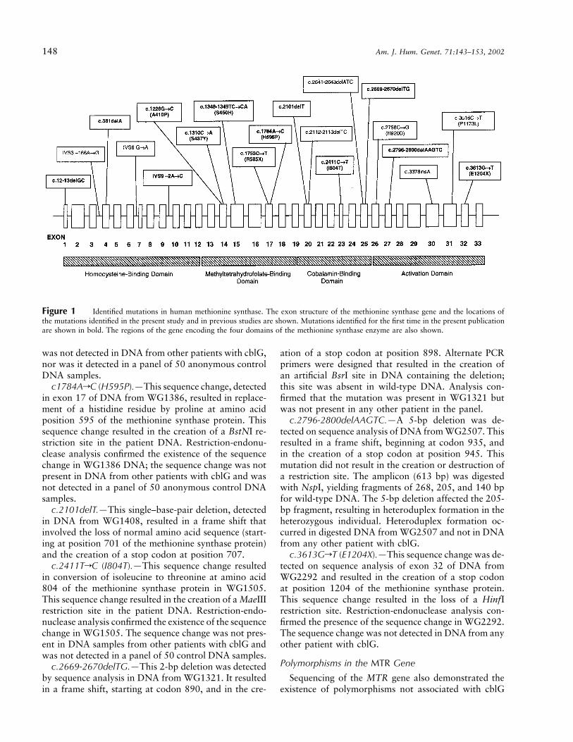
scribed below. This panel included cell lines that had beeninvestigated previously (WG1655, WG1670, WG1671,WG1765, WG1892, and WG2290) (Gulati et al. 1996;Leclerc et al. 1996; Wilson et al. 1998). Thirteen novelmutations were identified. Furthermore, one previouslyidentified mutation (c.3518CrT [P1173L]) was identifiedin several additional cell lines. All mutations in methioninesynthase that have been identified to date are shown infigure 1. All detected mutations were present in the het-erozygous state (table 3).
The Recurrent c.3518CrT Mutation (P1173L)
The c3518CrT mutation, which results in replacementof proline by leucine at position 1173 of the amino acidsequence, had been previously identified by Gulati et al.(1996). It was identified by sequence analysis in 14 of the18 cblG lines in the panel of the present study (table 3),including WG1892, the cell line in which it was originallyidentified. The c3518CrT mutation results in loss of anMspI site (Gulati et al. 1996). Restriction-endonucleaseanalysis confirmed the presence of the sequence changein DNA from the 14 cell lines; in addition, the sequencechange was detected in DNA from two additional cblGcell lines that had not undergone sequencing. Thec3518CrT mutation was thus detected in 16 of 24 patientcell lines in total. The sequence change was not detectedin a panel of 50 anonymous control DNA samples testedby restriction-endonuclease digestion.
Novel Methionine Synthase Mutations
c.12-13delGC.—Sequence analysis of DNA fromWG1765 demonstrated the presence of a 2-bp deletionat position 12 in exon 1, resulting in a frame shift, withthe loss of normal amino acid sequence starting at codon5 and creation of a stop codon at position 20. Thissequence change resulted in loss of a BstUI site. Restric-tion analysis with BstUI confirmed the presence of c.12-13delGC in WG1765; this change was not detected inDNA from any other patient with cblG.
c.381delA.—Sequence analysis indicated a single–base-pair deletion in exon 4 of DNA from WG2829.This resulted in a frame shift, with altered amino acidsequence, beginning at codon 128, and the creation ofa stop signal at codon 132.
IVS9�2ArC.—Sequence analysis of exon 10 fromWG1308 showed a sequence change resulting in alter-ation of the invariant A at the splice site at the 3′ endof intron 9. This mutation is expected to result in de-letion of exon 10 from the methionine synthase tran-script. This sequence change resulted in the creation ofan MspI restriction site. Restriction-endonuclease anal-ysis confirmed the sequence change in WG1308. Thesequence change was not detected in DNA from anyother patient with cblG, nor was it detected in a panelof 50 anonymous control DNA samples.
Watkins et al.: P1173L, a Common Mutation in the MTR Gene 147
Table 2
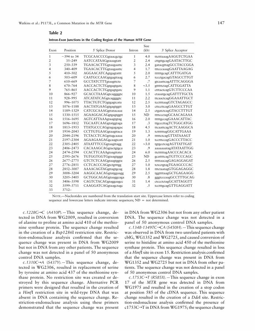
Intron-Exon Junctions in the Coding Region of the Human MTR Gene
Exon Position 5′ Splice Donor IntronSize(kb) 3′ Splice Acceptor
1 �394 to 34 TCGCAACCCGgtaacgctgc 1 4.0 ttctttaaagAAGGTCTGAA2 35–249 AATCCATAAGgtaaagtatt 2 2.4 cttgttgcagGAATACTTGC3 250–339 TGAACACTTGgtaagaattc 3 2.4 gatacgttagGCCTACCGGA4 340–409 TGAACACTTGgtaagaattc 4 1.7 tttcccaaagGAATTAAGAG5 410–502 AGGAACATCAgtgagtattt 5 2.0 ttttttgcagCATTTGATGA6 503–609 CAATGCCAAGgtgagttaag 6 2.7 tccctgacagGTAGCCTTGT7 610–669 GCCTATCTTTgtaagttcta 7 .7 gtcaattcagATTTCAGGGA8 670–764 AACCACTCTGgtgagtgatc 8 ∼3.5 gtttttctagCATTGGATTA9 765–865 AACCACTCTGgtgagtgatc 9 1.1 ctttactcagGTCTTCCCAA10 866–927 GCACCTAAAGgtcaggggtc 10 1.5 ctaaatgcagGATTTTGCTA11 928–995 ATCATATCAGgtcaggggtc 11 2.2 ttcaactcagGGAAATTGCT12 996–1075 TTACTGTCTGgtgagtcata 12 2.5 tccttttaagGTCTAGAGCC13 1076–1188 AACTATGAAGgtgagtggtt 13 3.0 cttccttcagGAAGCCTTGT14 1189–1329 CATCGCAAAGgttatacaaa 14 2.5 ctgatctcagGTACCTTTGT15 1330–1515 AGAAGGACAGgtgagtggtt 15 ND ttttccctagGCAACAGAAA16 1516–1695 AGTCATTAAAgtaagtgtag 16 2.0 tttttgccagGAAACATTAC17 1696–1812 TGCAATCAAGgtatggtaga 17 .3 ttgcccttagTCTGGCATGG18 1813–1953 TTATGCCCAGgtagagagac 18 4.5 ttcaattcagACTCAAGGCA19 1954–2043 CCTTGTGAAGgtaagttaca 19 1.3 tcttttttagGGCATTGAAA20 2044–2196 TCTACCTCAGgttagcaaaa 20 .9 tttttctcagGTTATAAAGT21 2197–2304 AGAAGAAGAGgcaagtcatt 21 1.0 tcctttccagGACCCTTACC22 2305–2405 ATAATTTCCGgtaagttagg 22 ∼3.0 tgtgcctcagAGTTATTGAT23 2406–2473 CACAAAGCAGgtactgtgca 23 .9 aaaaaaatagATATAATTGG24 2474–2594 CCACTTCAAAgtaagttata 24 6.0 ttctttttagAACCCACACA25 2595–2676 TGTGGTGGTGgtaagtgggt 25 ND gcattttcagTGTTCCCAGC26 2677–2775 GTCTCTCAAGgtaagtggta 26 2.5 tttttaacagGAGAGGAGAT27 2776–2851 CCTCACCCAGgtctgtttgg 27 1.0 tctcctgtagTGAAGCCCAC28 2852–3007 AAAACAGTAGgttagtgcag 28 1.8 tttctaatagGTGGAGAGGC29 3008–3204 AAGGCAACAGgtatggaagg 29 2.5 tggttttaagGCTGAGAAGG30 3205–3405 GCTGGCAGAGgtaaggcaga 30 .8 gggtcccaagGCCTTTGCAG31 3406–3598 CAGTCTACAGgtaggaagcc 31 1.4 ctccctctagGCATTAGGTT32 3599–3711 CAAGGATCAGgtaagctagc 32 .5 tcctttgcagGTTGAGGATT33 3712–
NOTE.—Nucleotides are numbered from the translation start site. Uppercase letters refer to codingsequence and lowercase letters indicate intronic sequences; ND p not determined.
c.1228GrC (A410P).—This sequence change, de-tected in DNA from WG2009, resulted in conversionof alanine to proline at amino acid 410 of the methio-nine synthase protein. The sequence change resultedin the creation of a Bsp1286I restriction site. Restric-tion-endonuclease analysis confirmed that the se-quence change was present in DNA from WG2009but not in DNA from any other patients. The sequencechange was not detected in a panel of 50 anonymouscontrol DNA samples.
c.1310CrA (S437Y).—This sequence change, de-tected in WG2306, resulted in replacement of serineby tyrosine at amino acid 437 of the methionine syn-thase protein. No restriction site was created or de-stroyed by this sequence change. Alternative PCRprimers were designed that resulted in the creation ofa HinfI restriction site in wild-type DNA that wasabsent in DNA containing the sequence change. Re-striction-endonuclease analysis using these primersdemonstrated that the sequence change was present
in DNA from WG2306 but not from any other patientDNA. The sequence change was not detected in apanel of 50 anonymous control DNA samples.
c.1348-1349TCrCA (S450H).—This sequence changewas observed in DNA from two unrelated patients withcblG, WG1352 and WG2725, and caused conversion ofserine to histidine at amino acid 450 of the methioninesynthase protein. This sequence change resulted in lossof a HinfI site in exon 15. Restriction analysis confirmedthat the sequence change was present in DNA fromWG1352 and WG2725 but not in DNA from other pa-tients. The sequence change was not detected in a panelof 50 anonymous control DNA samples.
c.1753CrT (R585X).—This sequence change in exon17 of the MTR gene was detected in DNA fromWG1975 and resulted in the creation of a stop codonat position 585 of the cDNA sequence. This sequencechange resulted in the creation of a DdeI site. Restric-tion-endonuclease analysis confirmed the presence ofc1753CrT in DNA from WG1975; the sequence change
148 Am. J. Hum. Genet. 71:143–153, 2002
Figure 1 Identified mutations in human methionine synthase. The exon structure of the methionine synthase gene and the locations ofthe mutations identified in the present study and in previous studies are shown. Mutations identified for the first time in the present publicationare shown in bold. The regions of the gene encoding the four domains of the methionine synthase enzyme are also shown.
was not detected in DNA from other patients with cblG,nor was it detected in a panel of 50 anonymous controlDNA samples.
c1784ArC (H595P).—This sequence change, detectedin exon 17 of DNA from WG1386, resulted in replace-ment of a histidine residue by proline at amino acidposition 595 of the methionine synthase protein. Thissequence change resulted in the creation of a BstNI re-striction site in the patient DNA. Restriction-endonu-clease analysis confirmed the existence of the sequencechange in WG1386 DNA; the sequence change was notpresent in DNA from other patients with cblG and wasnot detected in a panel of 50 anonymous control DNAsamples.
c.2101delT.—This single–base-pair deletion, detectedin DNA from WG1408, resulted in a frame shift thatinvolved the loss of normal amino acid sequence (start-ing at position 701 of the methionine synthase protein)and the creation of a stop codon at position 707.
c.2411TrC (I804T).—This sequence change resultedin conversion of isoleucine to threonine at amino acid804 of the methionine synthase protein in WG1505.This sequence change resulted in the creation of a MaeIIIrestriction site in the patient DNA. Restriction-endo-nuclease analysis confirmed the existence of the sequencechange in WG1505. The sequence change was not pres-ent in DNA samples from other patients with cblG andwas not detected in a panel of 50 control DNA samples.
c.2669-2670delTG.—This 2-bp deletion was detectedby sequence analysis in DNA from WG1321. It resultedin a frame shift, starting at codon 890, and in the cre-
ation of a stop codon at position 898. Alternate PCRprimers were designed that resulted in the creation ofan artificial BsrI site in DNA containing the deletion;this site was absent in wild-type DNA. Analysis con-firmed that the mutation was present in WG1321 butwas not present in any other patient in the panel.
c.2796-2800delAAGTC.—A 5-bp deletion was de-tected on sequence analysis of DNA from WG2507. Thisresulted in a frame shift, beginning at codon 935, andin the creation of a stop codon at position 945. Thismutation did not result in the creation or destruction ofa restriction site. The amplicon (613 bp) was digestedwith NspI, yielding fragments of 268, 205, and 140 bpfor wild-type DNA. The 5-bp deletion affected the 205-bp fragment, resulting in heteroduplex formation in theheterozygous individual. Heteroduplex formation oc-curred in digested DNA from WG2507 and not in DNAfrom any other patient with cblG.
c.3613GrT (E1204X).—This sequence change was de-tected on sequence analysis of exon 32 of DNA fromWG2292 and resulted in the creation of a stop codonat position 1204 of the methionine synthase protein.This sequence change resulted in the loss of a HinfIrestriction site. Restriction-endonuclease analysis con-firmed the presence of the sequence change in WG2292.The sequence change was not detected in DNA from anyother patient with cblG.
Polymorphisms in the MTR Gene
Sequencing of the MTR gene also demonstrated theexistence of polymorphisms not associated with cblG
Watkins et al.: P1173L, a Common Mutation in the MTR Gene 149
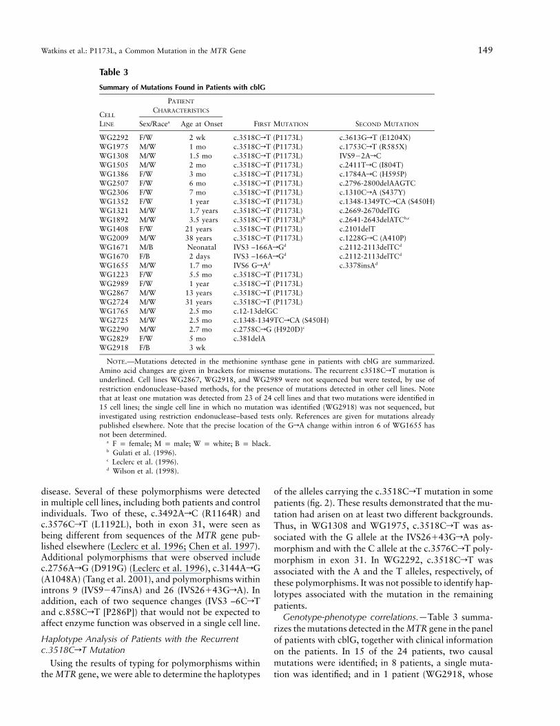
Table 3
Summary of Mutations Found in Patients with cblG
CELL
LINE
PATIENT
CHARACTERISTICS
FIRST MUTATION SECOND MUTATIONSex/Racea Age at Onset
WG2292 F/W 2 wk c.3518CrT (P1173L) c.3613GrT (E1204X)WG1975 M/W 1 mo c.3518CrT (P1173L) c.1753CrT (R585X)WG1308 M/W 1.5 mo c.3518CrT (P1173L) IVS9�2ArCWG1505 M/W 2 mo c.3518CrT (P1173L) c.2411TrC (I804T)WG1386 F/W 3 mo c.3518CrT (P1173L) c.1784ArC (H595P)WG2507 F/W 6 mo c.3518CrT (P1173L) c.2796-2800delAAGTCWG2306 F/W 7 mo c.3518CrT (P1173L) c.1310CrA (S437Y)WG1352 F/W 1 year c.3518CrT (P1173L) c.1348-1349TCrCA (S450H)WG1321 M/W 1.7 years c.3518CrT (P1173L) c.2669-2670delTGWG1892 M/W 3.5 years c.3518CrT (P1173L)b c.2641-2643delATCb,c
WG1408 F/W 21 years c.3518CrT (P1173L) c.2101delTWG2009 M/W 38 years c.3518CrT (P1173L) c.1228GrC (A410P)WG1671 M/B Neonatal IVS3 –166ArGd c.2112-2113delTCd
WG1670 F/B 2 days IVS3 –166ArGd c.2112-2113delTCd
WG1655 M/W 1.7 mo IVS6 GrAd c.3378insAd
WG1223 F/W 5.5 mo c.3518CrT (P1173L)WG2989 F/W 1 year c.3518CrT (P1173L)WG2867 M/W 13 years c.3518CrT (P1173L)WG2724 M/W 31 years c.3518CrT (P1173L)WG1765 M/W 2.5 mo c.12-13delGCWG2725 M/W 2.5 mo c.1348-1349TCrCA (S450H)WG2290 M/W 2.7 mo c.2758CrG (H920D)c
WG2829 F/W 5 mo c.381delAWG2918 F/B 3 wk
NOTE.—Mutations detected in the methionine synthase gene in patients with cblG are summarized.Amino acid changes are given in brackets for missense mutations. The recurrent c3518CrT mutation isunderlined. Cell lines WG2867, WG2918, and WG2989 were not sequenced but were tested, by use ofrestriction endonuclease–based methods, for the presence of mutations detected in other cell lines. Notethat at least one mutation was detected from 23 of 24 cell lines and that two mutations were identified in15 cell lines; the single cell line in which no mutation was identified (WG2918) was not sequenced, butinvestigated using restriction endonuclease–based tests only. References are given for mutations alreadypublished elsewhere. Note that the precise location of the GrA change within intron 6 of WG1655 hasnot been determined.
a F p female; M p male; W p white; B p black.b Gulati et al. (1996).c Leclerc et al. (1996).d Wilson et al. (1998).
disease. Several of these polymorphisms were detectedin multiple cell lines, including both patients and controlindividuals. Two of these, c.3492ArC (R1164R) andc.3576CrT (L1192L), both in exon 31, were seen asbeing different from sequences of the MTR gene pub-lished elsewhere (Leclerc et al. 1996; Chen et al. 1997).Additional polymorphisms that were observed includec.2756ArG (D919G) (Leclerc et al. 1996), c.3144ArG(A1048A) (Tang et al. 2001), and polymorphisms withinintrons 9 (IVS9�47insA) and 26 (IVS26�43GrA). Inaddition, each of two sequence changes (IVS3 –6CrTand c.858CrT [P286P]) that would not be expected toaffect enzyme function was observed in a single cell line.
Haplotype Analysis of Patients with the Recurrentc.3518CrT Mutation
Using the results of typing for polymorphisms withinthe MTR gene, we were able to determine the haplotypes
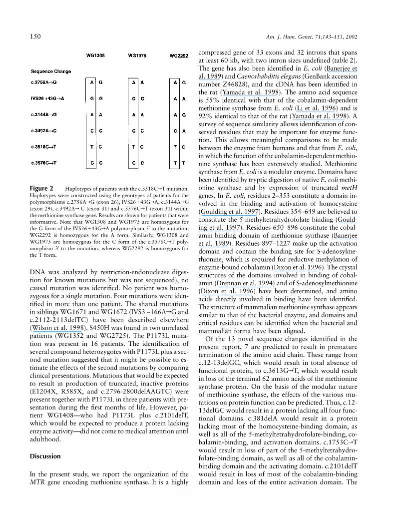
of the alleles carrying the c.3518CrT mutation in somepatients (fig. 2). These results demonstrated that the mu-tation had arisen on at least two different backgrounds.Thus, in WG1308 and WG1975, c.3518CrT was as-sociated with the G allele at the IVS26�43GrA poly-morphism and with the C allele at the c.3576CrT poly-morphism in exon 31. In WG2292, c.3518CrT wasassociated with the A and the T alleles, respectively, ofthese polymorphisms. It was not possible to identify hap-lotypes associated with the mutation in the remainingpatients.
Genotype-phenotype correlations.—Table 3 summa-rizes the mutations detected in the MTR gene in the panelof patients with cblG, together with clinical informationon the patients. In 15 of the 24 patients, two causalmutations were identified; in 8 patients, a single muta-tion was identified; and in 1 patient (WG2918, whose
150 Am. J. Hum. Genet. 71:143–153, 2002
Figure 2 Haplotypes of patients with the c.3518CrT mutation.Haplotypes were constructed using the genotypes of patients for thepolymorphisms c.2756ArG (exon 26), IVS26�43GrA, c.3144ArG(exon 29), c.3492Ar C (exon 31) and c.3576CrT (exon 31) withinthe methionine synthase gene. Results are shown for patients that wereinformative. Note that WG1308 and WG1975 are homozygous forthe G form of the IVS26�43GrA polymorphism 5′ to the mutation;WG2292 is homozygous for the A form. Similarly, WG1308 andWG1975 are homozygous for the C form of the c.3576CrT poly-morphism 3′ to the mutation, whereas WG2292 is homozygous forthe T form.
DNA was analyzed by restriction-endonuclease diges-tion for known mutations but was not sequenced), nocausal mutation was identified. No patient was homo-zygous for a single mutation. Four mutations were iden-tified in more than one patient. The shared mutationsin siblings WG1671 and WG1672 (IVS3 –166ArG andc.2112-2113delTC) have been described elsewhere(Wilson et al. 1998). S450H was found in two unrelatedpatients (WG1352 and WG2725). The P1173L muta-tion was present in 16 patients. The identification ofseveral compound heterozygotes with P1173L plus a sec-ond mutation suggested that it might be possible to es-timate the effects of the second mutations by comparingclinical presentations. Mutations that would be expectedto result in production of truncated, inactive proteins(E1204X, R585X, and c.2796-2800delAAGTC) werepresent together with P1173L in three patients with pre-sentation during the first months of life. However, pa-tient WG1408—who had P1173L plus c.2101delT,which would be expected to produce a protein lackingenzyme activity—did not come to medical attention untiladulthood.
Discussion
In the present study, we report the organization of theMTR gene encoding methionine synthase. It is a highly
compressed gene of 33 exons and 32 introns that spansat least 60 kb, with two intron sizes undefined (table 2).The gene has also been identified in E. coli (Banerjee etal. 1989) and Caenorhabditis elegans (GenBank accessionnumber Z46828), and the cDNA has been identified inthe rat (Yamada et al. 1998). The amino acid sequenceis 55% identical with that of the cobalamin-dependentmethionine synthase from E. coli (Li et al. 1996) and is92% identical to that of the rat (Yamada et al. 1998). Asurvey of sequence similarity allows identification of con-served residues that may be important for enzyme func-tion. This allows meaningful comparisons to be madebetween the enzyme from humans and that from E. coli,in which the function of the cobalamin-dependentmethio-nine synthase has been extensively studied. Methioninesynthase from E. coli is a modular enzyme. Domains havebeen identified by tryptic digestion of native E. coli methi-onine synthase and by expression of truncated metHgenes. In E. coli, residues 2–353 constitute a domain in-volved in the binding and activation of homocysteine(Goulding et al. 1997). Residues 354–649 are believed toconstitute the 5-methyltetrahydrofolate binding (Gould-ing et al. 1997). Residues 650–896 constitute the cobal-amin-binding domain of methionine synthase (Banerjeeet al. 1989). Residues 897–1227 make up the activationdomain and contain the binding site for S-adenosylme-thionine, which is required for reductive methylation ofenzyme-bound cobalamin (Dixon et al. 1996). The crystalstructures of the domains involved in binding of cobal-amin (Drennan et al. 1994) and of S-adenosylmethionine(Dixon et al. 1996) have been determined, and aminoacids directly involved in binding have been identified.The structure of mammalian methionine synthase appearssimilar to that of the bacterial enzyme, and domains andcritical residues can be identified when the bacterial andmammalian forma have been aligned.
Of the 13 novel sequence changes identified in thepresent report, 7 are predicted to result in prematuretermination of the amino acid chain. These range fromc.12-13delGC, which would result in total absence offunctional protein, to c.3613GrT, which would resultin loss of the terminal 62 amino acids of the methioninesynthase protein. On the basis of the modular natureof methionine synthase, the effects of the various mu-tations on protein function can be predicted. Thus, c.12-13delGC would result in a protein lacking all four func-tional domains. c.381delA would result in a proteinlacking most of the homocysteine-binding domain, aswell as all of the 5-methyltetrahydrofolate-binding, co-balamin-binding, and activation domains. c.1753CrTwould result in loss of part of the 5-methyltetrahydro-folate-binding domain, as well as all of the cobalamin-binding domain and the activating domain. c.2101delTwould result in loss of most of the cobalamin-bindingdomain and loss of the entire activation domain. The

Watkins et al.: P1173L, a Common Mutation in the MTR Gene 151
three remaining truncating mutations would result inloss of parts of the activating domain, including residuesknown to be critical for binding of S-adenosylmethio-nine. It has been shown that an intact activation domainis critical to the function of methionine synthase. Thus,a fragment of E. coli methionine synthase lacking theactivation domain is initially able to support methyla-tion of homocysteine, but activity decays with time andis ultimately lost (Drummond et al. 1993). Althoughthese are predictions of resulting structure, it is oftenthe case that mutations that introduce premature stopcodons are associated with instability of the mRNA.This was found to be the outcome for c.2112delTC andc.3378insA, which, coupled with mutations involvingintronic inserts, gave fibroblast mRNA nearly unde-tectable by northern blot analysis (Wilson et al. 1998).
Five missense mutations within the coding sequence ofthe MTR gene were identified in the present study. Al-though these mutations have not been expressed to con-firm that they disrupt methionine synthase activity, nonewere detected in a survey of DNA samples from the con-trol population. Three of the mutations (A410P, S450H,and H595P) affect residues that are conserved in methio-nine synthase from E. coli, C. elegans, and the rat. S437Yaffects a residue conserved in C. elegans and the rat; E.coli contains glycine at this position. I804T affects a res-idue conserved in methionine synthase from the rat; theequivalent residue in E. coli and C. elegans is valine.Thus, in four of the five missense mutations, the affectedresidue is conserved over considerable phylogenetic dis-tance, suggesting that these sequence changes have asignificant effect on methionine synthase function. Thefinal mutation, IVS9�2CrA, was not observed in thegeneral population and would be expected to result indeletion of part of the homocysteine-binding domain ofmethionine synthase.
One previously identified mutation, P1173L, washighly prevalent in the patient samples of the pres-ent study. It was detected in 16 of 24 mutant fibro-blast lines in the panel, including WG1892, in whichthe mutation had been previously identified (Gulati etal. 1996). This is clearly a disease-causing mutation,given its prevalence in patients and its absence in controlsubjects (consisting of a total of 210 control individualsanalyzed in the present study and the study by Gulatiet al. [1996]). In addition, it results in replacement ofa highly conserved proline residue at position 1173 byleucine; P1173 lies between two residues, R1172 andA1174, that interact directly with S-adenosylmethionine(Dixon et al. 1996).
The high frequency of the P1173L mutation in pa-tients with cblG raises the question of whether the mu-tation has arisen more than once during history. Themajority of the members of the patient panel were ofEuropean descent, including all of the patients who car-
ried the P1173L mutation. Analysis of haplotypes inpatients who carried the P1173L mutation (fig. 2) dem-onstrated that it has arisen on at least two differentbackgrounds. The mutation represents a CrT transitionat a CpG island; such sites are known to be prone tomutation (Templeton et al. 2000). These data suggestthat P1173L is truly a recurrent mutation that has arisenmore than once during human history.
Clinical findings in patients with the cblG disordervary widely. The identification—by the present studyand others—of causal mutations in the cblG disordermeans that it is now possible to attempt to correlatephenotype with genotype in this disorder, although sucha correlation is limited by the fact that both causal mu-tations have not been found in all patients. Variationin clinical presentation in patients carrying one copy ofP1173L in combination with a second mutation mayreflect the effect of the second mutation. Patients withP1173L in combination with a nonsense mutation thatwould be expected to result in production of a trun-cated, inactive protein showed a wide range of age atonset (table 3). Some patients (WG2292, WG1975, andWG2507) showed onset of symptoms within the firstmonths of life. However, WG1408, who had P1173Lplus a deletion resulting in loss of the cobalamin-bindingand activation domains of methionine synthase, cameto medical attention in adulthood. These results suggestthat genetic modifiers and environmental factors maysignificantly affect clinical presentation in patients withcblG.
Elevated blood levels of homocysteine have been as-sociated with increased risk of cardiovascular disease andadverse pregnancy outcomes (Ueland et al. 2001). Homo-cysteine has also been associated with increased likeli-hood of occurrence of birth defects, such as neural tubedefects and orofacial clefting (Christensen and Rosenblatt1995; Wong et al. 1999) and Down syndrome (James etal. 1999), and it may affect development of some typesof cancer (Kim 1999). Polymorphisms in genes encodingmethylenetetrahydrofolate reductase and methioninesynthase reductase (enzymes involved in homocysteineremethylation) have been shown to affect blood levels ofhomocysteine and to be associated with altered risk ofcardiovascular disease and birth defects (Rozen 2001).To date, studies of the c.2576ArG (D919G) MTR poly-morphism have been inconclusive. The G form appearsto be associated with lowered plasma total homocysteine(Wang et al. 1999; Chen et al. 2001; Dekau et al. 2001).However, no consistent effect on risk of developing car-diovascular disease (Morita et al. 1999; Hyndman et al.2000; Chen et al. 2001) or birth defects (Christensen etal. 1999; Shaw et al. 1999) has been documented. Furtherstudy of MTR mutations and polymorphisms such asthose described in the present publication may shed light
152 Am. J. Hum. Genet. 71:143–153, 2002
on the function of methionine synthase that will be im-portant beyond development of the cblG phenotype.
Acknowledgments
The authors offer thanks to the clinicians who providedpatient samples and clinical information, to Nora Matiaszukfor complementation analysis, and to Gail Dunbar for growthof patient fibroblasts. This work was supported by an oper-ating grant from the Canadian Institutes for Health Research(CIHR) and by the CIHR Group Grant in Medical Genetics.The work of B.S. and H.-Y.H. was supported by Public HealthService grants HL58991 and DK42033 from the United StatesDepartment of Health and Human Services. This is a publi-cation of the Hess B. and Diane Finestone Laboratory in theMemory of Jacob and Jenny Finestone.
Electronic-Database Information
Accession numbers and URLs for data presented herein areas follows:
GenBank, http://www.ncbi.nlm.nih.gov/Genbank (for C. ele-gans methionine synthase gene sequence [accession numberZ46828])
Genome Database, http://gdbwww.gdb.org (for MTR gene[GDB accession number 119440])
Online Mendelian Inheritance in Man (OMIM),http://www.ncbi.nlm.nih.gov/Omim/ (for cblG disorder [MIM 250940])
Whitehead Institute/MIT Center for Genome Research,http://www.genome.wi.mit.edu/cgi-bin/primer/primer3_www.cgi(for Primer 3.0 software)
References
Banerjee RV, Johnston NL, Sobeski JK, Datta P, Matthews RG(1989) Cloning and sequence analysis of the Escherichia colimetH gene encoding cobalamin-dependent methionine syn-thase and isolation of a tryptic fragment containing the co-balamin-binding domain. J Biol Chem 264:13888–13895
Carmel R, Jacobsen DW (eds) (2001) Homocysteine in healthand disease. Cambridge University Press, Cambridge
Chen J, Stampfer MJ, Ma J, Selhub J, Malinow MR, Hen-nekens CH, Hunter DJ (2001) Influence of a methioninesynthase (D919G) polymorphism on plasma homocysteineand folate levels and relation to risk of myocardial infarc-tion. Atherosclerosis 154:667–672
Chen LH, Liu M, Hwang H, Chen L, Korenberg J, Shane B(1997) Human methionine synthase: cDNA cloning, genelocalization and expression. J Biol Chem 272:3628–3634
Christensen B, Arbour L, Tran P, Leclerc D, Sabbaghian N,Platt R, Gilfix BM, Rosenblatt DS, Gravel RA, Forbes P,Rozen R (1999) Genetic polymorphisms in methylenetet-rahydrofolate reductase and methionine synthase, folate lev-els in red blood cells, and risk of neural tube defects. Am JMed Genet 84:151–157
Christensen B, Rosenblatt DS (1995) Effects of folate defi-ciency on embryonic development. In: Wickramasinghe SN(ed) Bailliere’s clinical haematology: megaloblastic anemia.Bailliere Tindall, London, pp 617–638
Dekau V, Gudnason V, Hawe E, Miller GJ, Stansbie D, Hum-phries SE (2001) Gene-environment and gene-gene inter-action in the determination of plasma homocysteine levelsin healthy middle-aged men. Thromb Haemost 85:67–74
Dixon MM, Huang S, Matthews RG, Ludwig M (1996) Thestructure of the C-terminal domain of methionine synthase:presenting S-adenosylmethionine for reductive methylationof B12. Structure 4:1263–1275
Drennan CL, Huang S, Drummond JT, Matthews RG, LudwigML (1994) How a protein binds B12: a 3.0 A X-ray structureof B12-binding domains of methionine synthase. Science 266:1669–1674
Drummond JT, Huang S, Blumenthal RM, Matthews RG(1993) Assignment of enzymatic function to specific proteinregions of cobalamin-dependent methionine synthase fromEscherichia coli. Biochemistry 32:9290–9295
Goulding CW, Postigo D, Matthews RG (1997) Cobalamin-dependent methionine synthase is a modular protein with dis-tinct regions for binding homocysteine, methyltetrahydrofo-late, and adenosylmethionine. Biochemistry 36:8082–8091
Gulati S, Baker P, Li YN, Fowler B, Kruger WD, Brody LC,Banerjee R (1996) Defects in human methionine synthase incblG patients. Hum Mol Genet Suppl 5:1859–1865
Hyndman ME, Bridge PJ, Warnica JW, Fick G, Parsons HG(2000) Effect of heterozygosity for the methionine synthase2756 ArG mutation on the risk for recurrent cardiovascularevents. Am J Cardiol 86:1144–1146
James SJ, Pogribna M, Pogribny II, Melnyk S, Hine RJ, Gib-son JB, Yi P, Tafoya DL, Swenson DH, Wilson VL, GaylorDW (1999) Abnormal folate metabolism and mutation inthe methylenetetrahydrofolate reductase gene may be ma-ternal risk factors for Down syndrome. Am J Clin Nutr 70:495–501
Kim Y-I (1999) Folate and cancer prevention: a new medicalapplication of folate beyond hyperhomocysteinemia and neu-ral tube defects. Nutr Rev 57:314–321
Leclerc D, Campeau E, Goyette P, Adjalla CE, Christensen B,Ross M, Eydoux P, Rosenblatt DS, Rozen R, Gravel RA(1996) Human methionine synthase: cDNA cloning andidentification of mutations in patients of the cblG comple-mentation group of folate/cobalamin disorders. Hum MolGenet 5:1867–1874
Leclerc D, Wilson A, Dumas R, Gafuik C, Song D, WatkinsD, Heng HHQ, Rommens JM, Scherer SW, Rosenblatt DS,Gravel RA (1998) Cloning and mapping of a cDNA formethionine synthase reductase, a flavoprotein defective inpatients with homocystinuria. Proc Natl Acad Sci USA 95:3059–3064
Li YN, Gulati S, Baker PJ, Brody LC, Banerjee R, Kruger WD(1996) Cloning, mapping and RNA analysis of the humanmethionine synthase gene. Hum Mol Genet 5:1851–1858
Matthews RG (1999) Cobalamin-dependent methionine syn-thase. In: Banerjee R (ed) Chemistry and biochemistry ofB12. John Wiley & Sons, New York, pp 681–706
Matthews RG, Ludwig ML (2001) Microbial modeling of hu-man disease: homocysteine metabolism. In: Carmel R, Ja-cobsen DW (eds) Homocysteine in health and disease. Cam-bridge University Press, Cambridge, pp 100–112
Morita H, Kurihara H, Sugiyama T, Hamada C, Kurihara Y,Shindo T, Oh-hashi Y, Yazaki Y (1999) Polymorphism ofthe methionine synthase gene: association with homocys-
Watkins et al.: P1173L, a Common Mutation in the MTR Gene 153
teine metabolism and late-onset vascular diseases in the Jap-anese population. Arterioscler Thromb Vasc Biol 19:298–302
Olteanu H, Banerjee R (2001) Human methionine synthasereductase, a soluble P-450 reductase-like dual flavoprotein,is sufficient for NADPH-dependent methionine synthase ac-tivation. J Biol Chem 276:35558–35563
Rosenblatt D, Fenton WA (2001) Inherited disorders of folateand cobalamin transport and metabolism. In: Scriver CR,Beaudet AL, Sly WS, Valle D, Childs B, Kinzler KW, VogelsteinB (eds) The metabolic and molecular bases of inherited dis-ease, 8th ed. McGraw-Hill, New York, pp 3897–3933
Rozen R (2001) Polymorphisms of folate and cobalamin me-tabolism. In: Carmel R, Jacobsen DW (eds) Homocysteinein health and disease. University of Cambridge Press, Cam-bridge, pp 259–269
Shaw GM, Todoroff K, Finnell RH, Lammer EJ, Leclerc D,Gravel RA, Rozen R (1999) Infant methionine synthase var-iants and risk for spina bifida. J Med Genet 36:86–87
Suormala ST, Baumgartner ER, Fowler B (2001) Characteri-zation of methylmalonic aciduria and/or hyperhomocystei-nemia: studies in cultured fibroblasts. J Inher Metab Dis 24:55
Tang B, Li YN, Kruger WD (2000) Defects in methylthioad-enosine phosphorylase are associated with but not responsiblefor methionine-dependent tumor cell growth. Cancer Res 60:5543–5547
Templeton AR, Clark AG, Weiss KM, Nickerson DA, Boer-winkle E, Sing CF (2000) Recombinational and mutation-al hotspots within the human lipoprotein lipase gene. Am JHum Genet 66:69–83
Ueland PM, Nygard O, Vollset SE, Refsum H (2001) The Hor-daland homocysteine studies. Lipids 36:S33–S39
Wang XL, Duarte N, Cai H, Adachi T, Sim AS, Cranney G,Wilcken DEL (1999) Relationship between total plasmahomocysteine, polymorphisms of homocysteine metabolismrelated enzymes, risk factors and coronary artery disease inthe Australian hospital-based population. Atherosclerosis146:133–140
Watkins D, Rosenblatt DS (1989) Functional methionine syn-thase deficiency cblE and cblG): clinical and biochemicalheterogeneity. Am J Med Genet 34:427–434
Wilson A, Leclerc D, Saberi F, Phillips JA III, Rosenblatt DS,Gravel RA (1998) Functionally null mutations in patientswith the cblG variant form of methionine synthase defi-ciency. Am J Hum Genet 63:409–414
Wong WY, Eskes TKAB, Kuijpers-Jagtman A-M, SprauwenPHM, Steegers EAP, Thomas CMG, Hamel BCJ, Blom HJ,Steegers-Theunissen RPM (1999) Non-syndromic orofa-cial clefts: association with maternal hyperhomocysteine-mia. Teratology 60:253–257
Yamada K, Tobimatsu T, Toraya T (1998) Cloning, sequenc-ing, and heterologous expression of rat methionine synthasecDNA. Biosci Biotechnol Biochem 62:2155–2160


