Embed Size (px)
Citation preview
This article appeared in a journal published by Elsevier. The attachedcopy is furnished to the author for internal non-commercial researchand education use, including for instruction at the authors institution
and sharing with colleagues.
Other uses, including reproduction and distribution, or selling orlicensing copies, or posting to personal, institutional or third party
websites are prohibited.
In most cases authors are permitted to post their version of thearticle (e.g. in Word or Tex form) to their personal website orinstitutional repository. Authors requiring further information
regarding Elsevier’s archiving and manuscript policies areencouraged to visit:
http://www.elsevier.com/authorsrights
Author's personal copy
Open-label, multi-center, non-randomized, single-arm study toevaluate the safety and efficacy of dendritic cell immunotherapy inpatients with refractory solid malignancies, on supportive care
POONAMALLE PARTHASARATHY BAPSY1, BANDANA SHARAN2,CHAITANYA KUMAR2, RAJEEV PATRICK DAS2, BHARATH RANGARAJAN3,MINISH JAIN4, VENKATA SATHYA SURESH ATTILI5, SUNDARAM SUBRAMANIAN6,SHYAM AGGARWAL7, MALA SRIVASTAVA8 & ASHOK VAID9
1Department of Oncology, Apollo Hospital, Bangalore, India, 2APAC Biotech Pvt Ltd, Gurgaon, India, 3NarayanHrudayalaya Hospital, Bangalore, India, 4Ruby Hall Clinic, Pune, India, 5Apollo Hospital, Hyderabad, India, 6V.S.Hospital, Chennai, India, 7Department of Medical Oncology, Sir Ganga Ram Hospital, New Delhi, India, 8NextvelConsulting LLP, Bangalore, India, and 9Department of Medical Oncology and Hematology, Medanta-the Medicity,Gurgaon, India
AbstractBackground aims. A phase II clinical trial of an autologous dendritic cell (DC) formulation for the management of refractorysolid malignant tumors was conducted across six sites in India with an objective to study safety and efficacy. Methods. A totalof 51 patients with refractory cancer (either sex) with life expectancy �3 months, Eastern Cooperative Oncology Groupscore �2, available tumor tissue and adequate organ and bone marrow function were recruited. Monocytes obtained byleukapheresis, differentiated into DCs by cytokines and primed with autologous tumor lysate (fresh tissue biopsy or paraffinblock). On the 8th day, mature DCs were analyzed for expression of CD40, CD80, CD83, CD86, DC205 and DC209. Thetreatment regime consisted of six doses (intravenous) over 14 weeks with 2 post-treatment follow-up visits, 6 weeks apart.Safety was assessed at all visits and responses were evaluated on days 58, 100 and 184 or at end of the study. Results. A total of38 patients were evaluated for safety and efficacy. One adverse event classified as possibly related was an episode of rigors orchills with mild pyrexia during one infusion. Objective response rate by Response Evaluation Criteria In Solid Tumors was28.9% (11/38) and immune-related response criteria was 42.1% (16/38); 90% confidence interval for objective response ratewas (17.2, 43.3) and (28.5, 56.7) by Response Evaluation Criteria In Solid Tumors and immune-related response criteria,respectively. The median time to treatment progression was >9 weeks. Median overall survival was 397 days. An increase inthe expression of interferon-g was not significant. Conclusions. Therapy was safe. The responses, time to treatment pro-gression and survival are encouraging for patients with aggressive refractory disease.
Key Words: cancer immunotherapy, dendritic cells, monocytes
Introduction
Metastases are the primary cause of death in patientswith solid cancers (1,2). Cancer accounts for ap-proximately 7% of deaths in India and 23% in theUnited States, with a prevalence of 2.5 million andapproximately 0.8 million new cases each year inIndia (3). Chemotherapy has been in the mainstay ofcancer treatment and has been found to be effective invarious types of cancer, but metastatic malignanciesoften develop resistance to standard chemotherapies,which are also responsible for considerable morbidityand death. This has shifted the focus to more specifictargeted therapies and immunotherapy. A viable
approach in cancer immunotherapy is the use ofdendritic cells (DCs) in orchestrating a repertoire ofboth innate (natural killer cells) and adaptive (T cells)immune responses against cancer. DC-based immu-notherapy has emerged as a rational new concept inthe treatment of malignant tumors, and there isincreasing evidence from animal studies and clinicaltrials showing that DC-based immunotherapy strat-egy may be a viable option in cancer treatment.
Discovered by Steinman et al. (4) in 1973, forwhich he received the 2011 Noble Prize in Medicine,DCs have evolved from subset curiosity to the most
Correspondence: Bandana Sharan, PhD, APAC Biotech Pvt Ltd, 69 JCM, DLF Phase II, Gurgaon-122002, India. E-mail: [email protected]
Cytotherapy, 2014; 16: 234e244
(Received 17 July 2013; accepted 30 November 2013)
ISSN 1465-3249 Copyright � 2014, International Society for Cellular Therapy. Published by Elsevier Inc. All rights reserved.http://dx.doi.org/10.1016/j.jcyt.2013.11.013
Author's personal copy
sought-after option in immunotherapy. DCs are themost potent antigen-presenting cells that play a keyrole in programming and regulating tumor-specificimmune responses by processing and presentingtumor antigens to naive or effector T lymphocytes(5). Numerous studies show that DCs loaded withtumor-associated antigens, exhibit protective anti-tumor responses that cause therapeutically regressionof preexisting tumors or increase time to progression(TTP) without any significant toxicity (6e9).
Various studies have shown that anomaly in DCnumber and function is linked to malignancies suchas breast cancer and multiple myeloma (10,11);further reduced DC counts in the peripheral blood ofpatients with cancer have also been associated withan accumulation of immunosuppressive immaturemyeloid cells (12,13), and these form the basis of theconcept of DC-based immunotherapy. The otherrationale for the success of DC-based immuno-therapy is that tumors can evade immune surveil-lance through tumor suppressor cells, release ofinhibitory cytokines (eg, interleukin-10, transforminggrowth factor -b) (14,15), loss of major histocom-patibility complex class I cell surface molecule andstructural abnormality of T-cell receptor -CD3complex and so forth (16); most of these defects maybe corrected by in vitro maturation of DCs, whichcan be used as immunotherapy.
Currently clinical trials on DC immunotherapymainly involve either the use of whole tumor lysate,recombinant protein or RNA transfection strategy tobe used as an antigen. This study involves the use ofwhole-tumor lysate attributable to several advantagesin DC-based immunotherapy preparation. First, allpatients are eligible for DC-whole tumor lysate ther-apy because patients are not selected on the basis oftheir human leukocyte antigeneA2 status. Second,whole-tumor lysate provides a rich array of tumor-associated antigens for both helper and cytotoxicT lymphocytes. This is important because the parallelpresentation of antigens to both T-lymphocyte sub-sets helps in evoking stronger immune responses andcould prevent the emergence of tumor escape. Thepresence of CD4þ T cells also promotes long-termCD8þ T-cell memory (17e19). In addition, DCspulsed with whole-tumor lysate have shown enhancedefficacy in patients with cancer over DCs loaded withdefined tumor-associated peptides or proteins, on thebasis of meta-analytical data (20).
Provenge (Sipuleucel-T) was approved by the USFood and Drug Administration on April 29, 2010,for asymptomatic or minimally symptomatic meta-static castrate resistant (hormone-refractory) prostatecancer (21). Various clinical trials on use of DCs incancer are currently ongoing in Europe, the UnitedStates and Asian countries. This multicentric phase II
clinical trial in India was undertaken to study thesafety, efficacy and tolerability of APCEDEN in re-fractory solid malignancies. The study was under-taken across six sites in India from September 2011 toDecember 2012.
Methods
This phase II study was an open-label, multi-centric,non-randomized, single-arm study in patients withrefractory solid malignancies who were only receivingsymptomatic care. Written informed consent of pa-tients was obtained according to the Helsinki Decla-ration; the study was approved by the respectiveinstitutional ethics committees, and the trial wasregistered with Clinical Trial Registry India (Regis-tration No. CTRI/2011/07/001917). ICH GCP (E6),Indian good clinical practices guidelines and ICMRethical guidelines for biomedical research on humansubjects were followed.
Generation of antigen-loaded mature DCs
APCEDEN is an autologous DC formulation inwhichDCs are derived fromCD14þ bloodmonocytesas previously described by Romani et al. (22) andloaded with whole-tumor lysate. In brief, the processbegins with separation of peripheral blood mono-nuclear cells by apheresis and further isolation ofmonocytes from apheresis harvest by plastic adher-ence; culturing in Roswell Park Memorial Institute1640 media (Lonza, Allendale, NJ, USA) supple-mented with cytokines interleukin-4 and granulocytemacrophage colony-stimulating factor (R&DSystems,Minneapolis, MN, USA) and autologous plasma invitro and exposure of the patient’s own tumor tissuelysate on the sixth day. For loading of DCs, fresh Tru-cut biopsy is preferred, but, in the case that an invasiveprocedure is not possible, paraffin block is used as thesource of antigen (23). Tumor lysate was prepared bythe freeze-thaw procedure as described by Nestle et al.(7), and protein concentration was determined ac-cording to Bradford’s protein assay (24). On the sixthday, 5 mg/mL of polyinosinic:polycytidylic acid (PolyIC) (InvivoGen, San Diego, CA, USA) was used asmaturation stimuli; after 3 h of adding poly IC, 1e20mg/mL protein was loaded on DCs.
Characterization of DCs
Mature DCs harvested on day 8 were analyzed bymeans of flow cytometry with the use of fluorochrome-labeled antibody against CD80, CD83, CD86 (BDBiosciences, San Jose, CA, USA), DC205, DC209and CD40 (Biolegend, San Diego, CA, USA). Via-bility of cells was assessed by 7AAD staining. Analysis
Phase II studies of dendritic cell therapy in solid tumors 235
Author's personal copy
was performed by means of flow cytometry (FACS-Calibur; Becton Dickinson) with the use of CellQuestsoftware. Adequate cell counts (>1 million DCs) perdose were used. All six doses were prepared at thesame time (>1 million DCs/dose) and cryopreserved(10% dimethyl sulfoxide and complete media). Ste-rility testing was performed according to the proceduredescribed in US Pharmacopoeia. Mycoplasma con-tamination was checked with the use of a MycoAlert-Mycoplasma detection kit (Lonza). Endotoxin wasassessed by means of the kinetic chromogenic limulusamoebocyte lysate test (Lonza).
Study objectives
The primary objectives were to determine the safety,tolerability and efficacy of the therapy. Response wasevaluated by (i) immune-related criteria (irRC)(25e27) and (ii) Response Evaluation Criteria InSolid Tumors (RECIST) criteria (28).
The secondary objectives were to measure qualityof life (QOL) by use of FACT-G (Functional Ass-essment of Cancer Therapy-General) (29); change inimmune response by measurement of pre- and post-therapy immune parameters such as CD4þ andCD8þ count and interferon (IFN)-g in peripheralblood by flow cytometry; and TTP defined as the timeperiod from the date of enrolment to the date whenprogression of disease was first documented.
Eligibility criteria
Both male and female adult patients with recurrentsolid malignancies, on symptomatic care with at least3 months of life expectancy, available tumor tissue,ECOG score of �2, having adequate organ and bonemarrow function were enrolled. Pregnant andlactating women were excluded from participation.
Dosing schedule
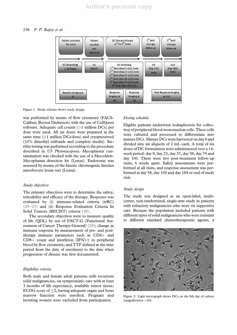
Eligible patients underwent leukapheresis for collec-tion of peripheral bloodmononuclear cells. These cellswere cultured and processed to differentiate intomatureDCs.MatureDCswere harvested onday 8 anddivided into six aliquots of 2 mL each. A total of sixdoses of DC formulation were administered over a 14-week period: day 9, day 23, day 37, day 58, day 79 andday 100. There were two post-treatment follow-upvisits, 6 weeks apart. Safety assessments were per-formed at all visits, and response assessment was per-formed at day 58, day 100 and day 184 or end of studyvisit.
Study design
The study was designed as an open-label, multi-center, non-randomized, single-arm study in patientswith refractory malignancies who were on supportivecare. Because the population included patients withdifferent types of solidmalignancies whowere resistantto different standard chemotherapeutic agents, a
Figure 1. Study schema shows study design.
Figure 2. Light micrograph shows DCs on the 8th day of culture(magnification �20).
236 P. P. Bapsy et al.
Author's personal copy
control arm could not be taken. The detailed studydesign of the trial is represented in Figure 1 (studyschema).
Route of administration
Each dose of APCEDEN (>1 � 106 cells along with100 mL of normal saline) was administered throughthe intravenous route as a slow infusion. Althoughvarious studies have preferred intradermal, subcu-taneous or intranodal routes of administration, in thepresent study, DCs were administered through theintravenous route because it has been associated witha significantly higher frequency and titer of Ag-spe-cific antibodies, which is desirable in some clinicalsituations in addition to the cellular immunity (30).
Safety and efficacy assessment
Primary end points were safety and tolerability asmeasured by the incidence and severity of treatment-emergent adverse events (TRAE) and incidence ofserious adverse events (SAE). Efficacy of therapy wasmeasured by means of tumor response and objectiveresponse assessment according to RECIST (version1.1) and irRC. The objective response rate (ORR)for this study included the percentage of patients whoshowed complete remission (CR), partial remission(PR) and stable disease.
Secondary end points were to assess QOL asmeasured by the FACT-G (31) and change in theimmune response by measuring immune parameters
before and after therapy by means of flow cytometryand TTP.
Safety events were graded by use of the revisedNational Cancer Institute Common TerminologyCriteria for Adverse Events (NCI CTCAE), version4.0 (published May 28, 2009) (25,26,32). AllSafety events were coded by use of MedDRA(version 13) (33).
Assessment of immune response by intracytoplasmicIFN-g release assay
Intracellular staining for IFN-g production of lym-phocytes was performed as described by Kern et al.
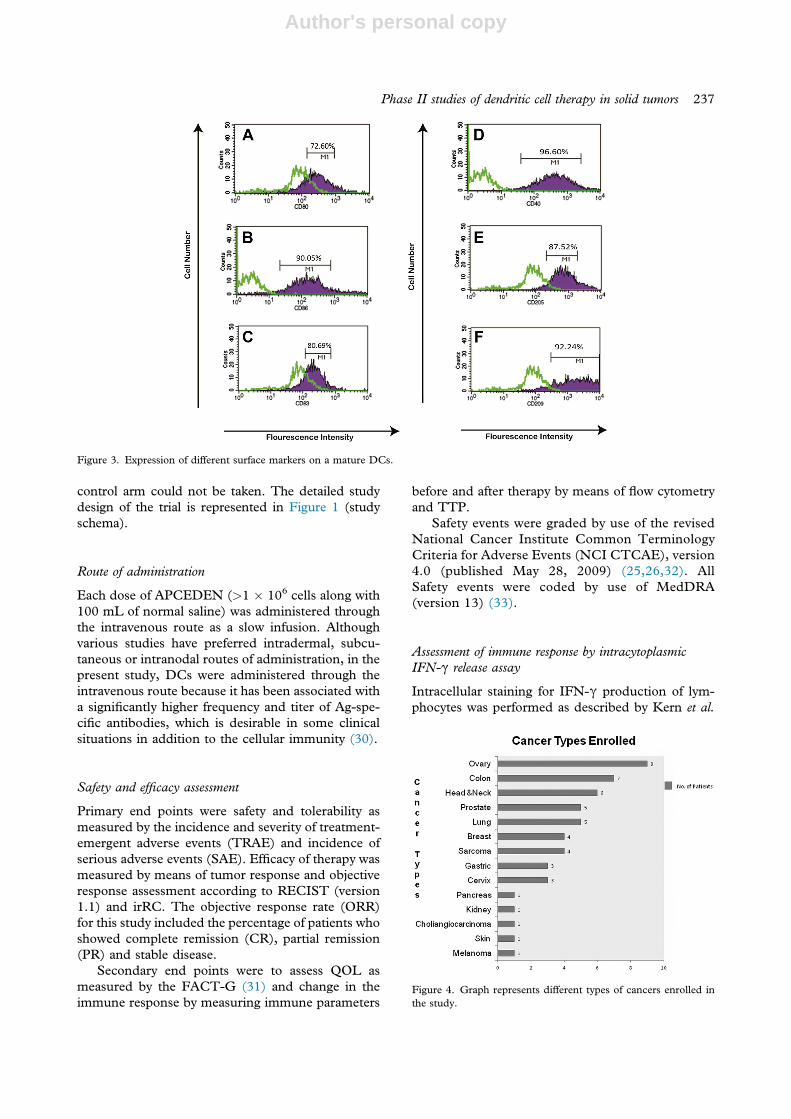
Figure 3. Expression of different surface markers on a mature DCs.
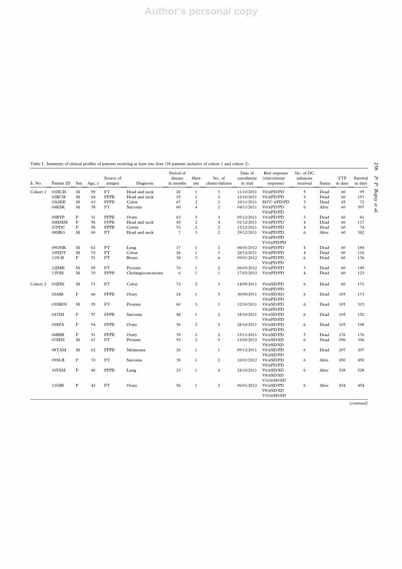
Figure 4. Graph represents different types of cancers enrolled inthe study.
Phase II studies of dendritic cell therapy in solid tumors 237
Author's personal copy
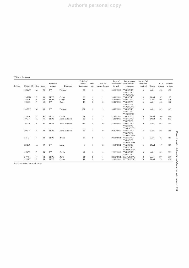
Table I. Summary of clinical profiles of patients receiving at least one dose (38 patients inclusive of cohort 1 and cohort 2).
S. No. Patient ID Sex Age, ySource ofantigen Diagnosis
Period ofdisease
in monthsMetssite
No. ofchemo-failures
Date ofenrollmentin trial
Best response(visit/criteria/
response)
No. of DCinfusionsreceived Status
TTPin days
Survivalin days
Cohort 1 01HCH M 59 FT Head and neck 20 1 3 11/10/2011 V6/irPD/PD 5 Dead 60 9502RCH M 64 FFPE Head and neck 15 2 2 12/10/2011 V6/irPD/PD 5 Dead 60 15703GKK M 63 FFPE Colon 67 2 2 10/11/2011 EOT/ irPD/PD 3 Dead 45 7204KSK M 38 FT Sarcoma 60 4 2 04/11/2011 V6/irPD/PD
V8/irPD/PD6 Alive 60 397
05RYP F 31 FFPE Ovary 63 3 3 05/12/2011 V6/irPD/PD 3 Dead 60 8106HMM F 56 FFPE Head and neck 45 2 4 01/12/2011 V6/irPD/PD 4 Dead 60 11707PDC F 50 FFPE Cervix 53 2 2 15/12/2011 V6/irPD/PD 4 Dead 60 7408SRG M 69 FT Head and neck 7 3 2 29/12/2011 V6/irPD/PD
V8/irPD/PDV10/irPD/PD
6 Alive 60 342
09GNK M 62 FT Lung 17 1 2 06/01/2012 V6/irPD/PD 4 Dead 60 18410NDT M 70 FT Colon 26 1 3 28/12/2011 V6/irPD/PD 4 Dead 60 11611N-B F 51 FT Breast 30 3 6 09/01/2012 V6/irPD/PD
V8/irPD/PD6 Dead 60 136
12JMK M 59 FT Prostate 70 1 2 06/03/2012 V6/irPD/PD 3 Dead 60 14913VSS M 70 FFPE Cholangiocarcinoma 6 1 1 17/03/2012 V6/irPD/PD 4 Dead 60 123
Cohort 2 01JHN M 71 FT Colon 72 2 3 14/09/2011 V6/irSD/PDV8/irPD/PD
6 Dead 60 171
02ARI F 66 FFPE Ovary 24 1 3 30/09/2011 V6/irSD/SDV8/irPD/PD
6 Dead 105 173
03MKN M 55 FT Prostate 60 3 3 12/10/2011 V6/irSD/PDV8/irPD/PD
6 Dead 105 315
04THI F 57 FFPE Sarcoma 48 1 2 18/10/2011 V6/irSD/PDV8/irPD/PD
6 Dead 105 152
05SPA F 54 FFPE Ovary 30 2 5 28/10/2011 V6/irSD/PDV8/irPD/PD
6 Dead 105 198
06BSR F 51 FFPE Ovary 35 2 2 15/11/2011 V6/irSD/PD 5 Dead 176 17607SEN M 67 FT Prostate 55 2 5 13/02/2012 V6/irSD/SD
V8/irSD/SD6 Dead 296 356
08TAM M 62 FFPE Melanoma 26 1 1 09/12/2011 V6/irSD/PDV8/irSD/PD
6 Dead 207 207
09M-B F 70 FT Sarcoma 39 1 2 10/01/2012 V6/irSD/PDV8/irPD/PD
6 Alive 450 450
10VSM F 40 FFPE Lung 25 1 4 24/10/2011 V6/irSD/SDV8/irSD/SDV10/irSD/SD
6 Alive 528 528
11GIB F 42 FT Ovary 56 1 2 06/01/2012 V6/irSD/PDV8/irSD/SDV10/irSD/SD
6 Alive 454 454
(continued)
238P.P.Bapsy
etal.
Author's personal copy
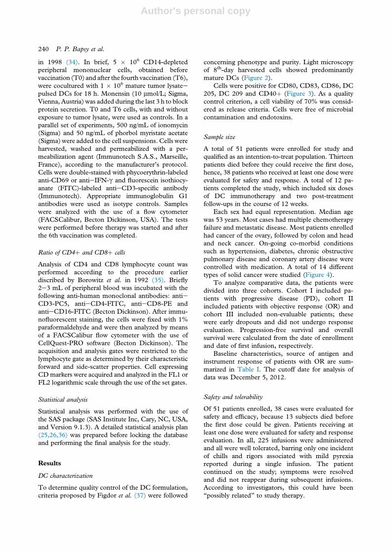
Table I. Continued
S. No. Patient ID Sex Age, ySource ofantigen Diagnosis
Period ofdisease
in monthsMetssite
No. ofchemo-failures
Date ofenrollmentin trial
Best response(visit/criteria/
response)
No. of DCinfusionsreceived Status
TTPin days
Survivalin days
12RVT M 73 FT Prostate 72 1 3 02/01/2012 V6/irSD/SDV8/irSD/SDV10/irSD/SD
6 Alive 458 458
13GBD F 36 FFPE Colon 60 1 1 29/11/2011 V6/irSD/SD 4 Dead 87 8714RVB F 60 FFPE Ovary 48 3 3 23/11/2011 V6/irSD/SD 5 Alive 498 49815SSK F 60 FT Ovary 40 2 2 29/12/2011 V6/irSD/PR
V8/irSD/PRV10/irSD/PR
6 Alive 462 462
16CSN M 68 FT Prostate 101 1 3 28/12/2011 V6/irSD/SDV8/irSD/SDV10/irSD/SD
6 Alive 463 463
17A-A F 42 FFPE Cervix 24 2 3 12/11/2011 V6/irSD/PD 5 Dead 246 24618C-N M 56 FFPE Head and neck 12 1 1 14/11/2011 V6/irSD/PD
V8/irPD/PD6 Dead 193 193
19K-B F 61 FFPE Head and neck 132 2 0 28/11/2011 V6/irSD/PDV8/irSD/PDV10/irSD/PD
6 Alive 493 493
20G-H F 33 FFPE Head and neck 17 1 0 06/12/2011 V6/irSD/PDV8/irPD/PDV10/irPD/PD
6 Alive 485 485
21I-V F 38 FFPE Breast 10 2 4 09/01/2012 V6/irSD/PDV8/irSD/PDV10 irPD/PD
6 Alive 451 451
22JKR M 57 FT Lung 8 1 2 13/01/2012 V6/irSD/SDV8/irSD/SDV10/irPD/PD
6 Dead 447 417
23BPS F 54 FT Cervix 17 2 2 17/03/2012 V6/irSD/SDV8/irSD/SD
6 Alive 383 383
24V-S F 54 FFPE RCC 14 2 1 22/02/2012 EOT/irSD/PD 2 Alive 397 39725SKT F 54 FFPE Colon 44 2 4 22/11/2011 EOT/irSD/SD 3 Dead 379 439
FFPE, formalin; FT, fresh tissue.
Phase
IIstudies
ofdendritic
celltherapyin
solidtum
ors239
Author's personal copy
in 1998 (34). In brief, 5 � 106 CD14-depletedperipheral mononuclear cells, obtained beforevaccination (T0) and after the fourth vaccination (T6),were cocultured with 1 � 106 mature tumor lysateepulsed DCs for 18 h. Monensin (10 mmol/L; Sigma,Vienna, Austria) was added during the last 3 h to blockprotein secretion. T0 and T6 cells, with and withoutexposure to tumor lysate, were used as controls. In aparallel set of experiments, 500 ng/mL of ionomycin(Sigma) and 50 ng/mL of phorbol myristate acetate(Sigma) were added to the cell suspensions. Cells wereharvested, washed and permeabilized with a per-meabilization agent (Immunotech S.A.S., Marseille,France), according to the manufacturer’s protocol.Cells were double-stained with phycoerythrin-labeledanti-CD69 or antieIFN-g and fluorescein isothiocy-anate (FITC)-labeled antieCD3-specific antibody(Immunotech). Appropriate immunoglobulin G1antibodies were used as isotype controls. Sampleswere analyzed with the use of a flow cytometer(FACSCalibur, Becton Dickinson, USA). The testswere performed before therapy was started and afterthe 6th vaccination was completed.
Ratio of CD4þ and CD8þ cells
Analysis of CD4 and CD8 lymphocyte count wasperformed according to the procedure earlierdiscribed by Borowitz et al. in 1992 (35). Briefly2e3 mL of peripheral blood was incubated with thefollowing anti-human monoclonal antibodies: antieCD3-PC5, antieCD4-FITC, antieCD8-PE andantieCD16-FITC (Becton Dickinson). After immu-nofluorescent staining, the cells were fixed with 1%paraformaldehyde and were then analyzed by meansof a FACSCalibur flow cytometer with the use ofCellQuest-PRO software (Becton Dickinson). Theacquisition and analysis gates were restricted to thelymphocyte gate as determined by their characteristicforward and side-scatter properties. Cell expressingCDmarkers were acquired and analyzed in the FL1 orFL2 logarithmic scale through the use of the set gates.
Statistical analysis
Statistical analysis was performed with the use ofthe SAS package (SAS Institute Inc, Cary, NC, USA,and Version 9.1.3). A detailed statistical analysis plan(25,26,36) was prepared before locking the databaseand performing the final analysis for the study.
Results
DC characterization
To determine quality control of the DC formulation,criteria proposed by Figdor et al. (37) were followed
concerning phenotype and purity. Light microscopyof 8th-day harvested cells showed predominantlymature DCs (Figure 2).
Cells were positive for CD80, CD83, CD86, DC205, DC 209 and CD40þ (Figure 3). As a qualitycontrol criterion, a cell viability of 70% was consid-ered as release criteria. Cells were free of microbialcontamination and endotoxins.
Sample size
A total of 51 patients were enrolled for study andqualified as an intention-to-treat population. Thirteenpatients died before they could receive the first dose,hence, 38 patients who received at least one dose wereevaluated for safety and response. A total of 12 pa-tients completed the study, which included six dosesof DC immunotherapy and two post-treatmentfollow-ups in the course of 12 weeks.
Each sex had equal representation. Median agewas 53 years. Most cases had multiple chemotherapyfailure and metastatic disease. Most patients enrolledhad cancer of the ovary, followed by colon and headand neck cancer. On-going co-morbid conditionssuch as hypertension, diabetes, chronic obstructivepulmonary disease and coronary artery disease werecontrolled with medication. A total of 14 differenttypes of solid cancer were studied (Figure 4).
To analyze comparative data, the patients weredivided into three cohorts. Cohort I included pa-tients with progressive disease (PD), cohort IIincluded patients with objective response (OR) andcohort III included non-evaluable patients; thesewere early dropouts and did not undergo responseevaluation. Progression-free survival and overallsurvival were calculated from the date of enrollmentand date of first infusion, respectively.
Baseline characteristics, source of antigen andinstrument response of patients with OR are sum-marized in Table I. The cutoff date for analysis ofdata was December 5, 2012.
Safety and tolerability
Of 51 patients enrolled, 38 cases were evaluated forsafety and efficacy, because 13 subjects died beforethe first dose could be given. Patients receiving atleast one dose were evaluated for safety and responseevaluation. In all, 225 infusions were administeredand all were well tolerated, barring only one incidentof chills and rigors associated with mild pyrexiareported during a single infusion. The patientcontinued on the study; symptoms were resolvedand did not reappear during subsequent infusions.According to investigators, this could have been“possibly related” to study therapy.
240 P. P. Bapsy et al.
Author's personal copy
Adverse events were reported in 29 patients(56.9%) irrespective of causal relationship to studytherapy. Twelve patients (23.5%) had TRAEs re-ported as unlikely to be caused by therapy. Only oneTRAE, chills and rigors, was adjudged as “possiblyrelated” to study therapy by the investigator. SAEswere reported in 21.6% of the patients but notrelated to the study therapy. All SAEs were attributedto the primary cancer condition and associated me-tastases and not adjudged related to study therapy. Atotal of 13 (25.4%) patients died while on study. Allthe deaths were caused by disease progression andwere unlikely to be related to study therapy.
Efficacy
Response rates were summarized as follows: ORR byRECIST was 28.9% (11/38) and irRC was 42.1%(16/38); 90% confidence interval for ORR was (17.2,43.3) and (28.5, 56.7) by RECIST and irRC,respectively. Intervals above 0 indicated that ORRwas estimated with sufficient precision. Nine of 12(75%) patients who completed the study continuedto show OR by RECIST (version 1.1) as well asirRC. One patient from this group showed PRthroughout the study. Best overall response was
recorded for 43 cases. Eleven of 43 (25.6%) casesshowed objective response.
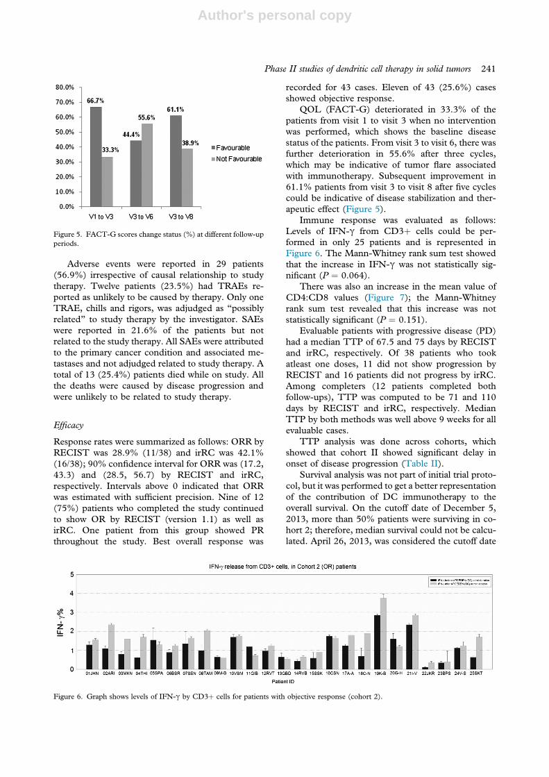
QOL (FACT-G) deteriorated in 33.3% of thepatients from visit 1 to visit 3 when no interventionwas performed, which shows the baseline diseasestatus of the patients. From visit 3 to visit 6, there wasfurther deterioration in 55.6% after three cycles,which may be indicative of tumor flare associatedwith immunotherapy. Subsequent improvement in61.1% patients from visit 3 to visit 8 after five cyclescould be indicative of disease stabilization and ther-apeutic effect (Figure 5).
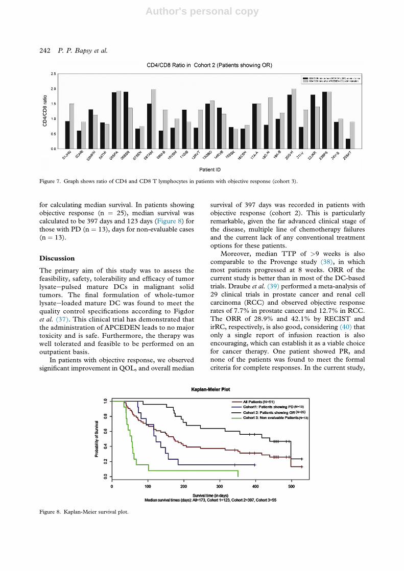
Immune response was evaluated as follows:Levels of IFN-g from CD3þ cells could be per-formed in only 25 patients and is represented inFigure 6. The Mann-Whitney rank sum test showedthat the increase in IFN-g was not statistically sig-nificant (P ¼ 0.064).
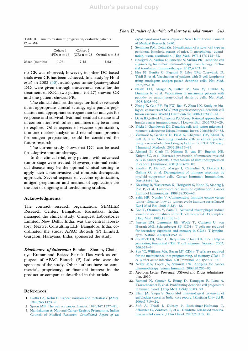
There was also an increase in the mean value ofCD4:CD8 values (Figure 7); the Mann-Whitneyrank sum test revealed that this increase was notstatistically significant (P ¼ 0.151).
Evaluable patients with progressive disease (PD)had a median TTP of 67.5 and 75 days by RECISTand irRC, respectively. Of 38 patients who tookatleast one doses, 11 did not show progression byRECIST and 16 patients did not progress by irRC.Among completers (12 patients completed bothfollow-ups), TTP was computed to be 71 and 110days by RECIST and irRC, respectively. MedianTTP by both methods was well above 9 weeks for allevaluable cases.
TTP analysis was done across cohorts, whichshowed that cohort II showed significant delay inonset of disease progression (Table II).
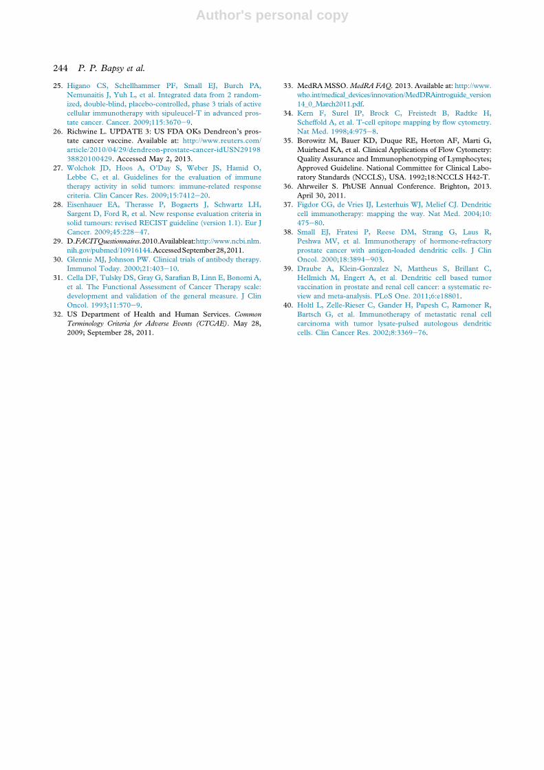
Survival analysis was not part of initial trial proto-col, but it was performed to get a better representationof the contribution of DC immunotherapy to theoverall survival. On the cutoff date of December 5,2013, more than 50% patients were surviving in co-hort 2; therefore, median survival could not be calcu-lated. April 26, 2013, was considered the cutoff date
Figure 5. FACT-G scores change status (%) at different follow-upperiods.
Figure 6. Graph shows levels of IFN-g by CD3þ cells for patients with objective response (cohort 2).
Phase II studies of dendritic cell therapy in solid tumors 241
Author's personal copy
for calculating median survival. In patients showingobjective response (n ¼ 25), median survival wascalculated to be 397 days and 123 days (Figure 8) forthose with PD (n ¼ 13), days for non-evaluable cases(n ¼ 13).
Discussion
The primary aim of this study was to assess thefeasibility, safety, tolerability and efficacy of tumorlysateepulsed mature DCs in malignant solidtumors. The final formulation of whole-tumorlysateeloaded mature DC was found to meet thequality control specifications according to Figdoret al. (37). This clinical trial has demonstrated thatthe administration of APCEDEN leads to no majortoxicity and is safe. Furthermore, the therapy waswell tolerated and feasible to be performed on anoutpatient basis.
In patients with objective response, we observedsignificant improvement in QOL, and overall median
survival of 397 days was recorded in patients withobjective response (cohort 2). This is particularlyremarkable, given the far advanced clinical stage ofthe disease, multiple line of chemotherapy failuresand the current lack of any conventional treatmentoptions for these patients.
Moreover, median TTP of >9 weeks is alsocomparable to the Provenge study (38), in whichmost patients progressed at 8 weeks. ORR of thecurrent study is better than in most of the DC-basedtrials. Draube et al. (39) performed a meta-analysis of29 clinical trials in prostate cancer and renal cellcarcinoma (RCC) and observed objective responserates of 7.7% in prostate cancer and 12.7% in RCC.The ORR of 28.9% and 42.1% by RECIST andirRC, respectively, is also good, considering (40) thatonly a single report of infusion reaction is alsoencouraging, which can establish it as a viable choicefor cancer therapy. One patient showed PR, andnone of the patients was found to meet the formalcriteria for complete responses. In the current study,
Figure 7. Graph shows ratio of CD4 and CD8 T lymphocytes in patients with objective response (cohort 3).
Figure 8. Kaplan-Meier survival plot.
242 P. P. Bapsy et al.
Author's personal copy
no CR was observed; however, in other DC-basedtrials even CR has been achieved. In a study by Holtlet al. in 2002 (40), autologous tumor lysateepulsedDCs were given through intravenous route for thetreatment of RCC; two patients (of 27) showed CRand one patient showed PR.
The clinical data set the stage for further researchin an appropriate clinical setting, right patient pop-ulation and appropriate surrogate marker analysis forresponse and survival. Minimal residual disease andin combination with other modalities may be an areato explore. Other aspects of vaccine optimization,immune marker analysis and recombinant proteinsfor antigen preparation, could be considered forfuture research.
The current study shows that DCs can be usedfor adoptive immunotherapy.
In this clinical trial, only patients with advancedtumor stage were treated. However, minimal resid-ual disease may be the optimal clinical setting toapply such a noninvasive and nontoxic therapeuticapproach. Several aspects of vaccine optimization,antigen preparation and method of application arethe foci of ongoing and forthcoming studies.
Acknowledgments
The contract research organization, SEMLERResearch Center, Bangalore, Karnataka, India,managed the clinical study; Oncquest LaboratoriesLimited, New Delhi, India, was the central labora-tory; Nextvel Consulting LLP, Bangalore, India, co-ordinated the study; APAC Biotech (P) Limited,Gurgaon, Harayana, India, sponsored the study.
Disclosure of interests: Bandana Sharan, Chaita-nya Kumar and Rajeev Patrick Das work as em-ployees of APAC Biotech (P) Ltd who were thesponsors of the study. Other authors have no com-mercial, proprietary, or financial interest in theproduct or companies described in this article.
References
1. Liotta LA, Kohn E. Cancer invasion and metastases. JAMA.1990;263:1123e6.
2. Sporn MB. The war on cancer. Lancet. 1996;347:1377e81.3. Nandakumar A. National Cancer Registry Programme, Indian
Council of Medical Research: Consolidated Report of the
Population-Based Cancer Registries. New Delhi: Indian Councilof Medical Research. 1990.
4. Steinman RM, Cohn ZA. Identification of a novel cell type inperipheral lymphoid organs of mice, I: morphology, quanti-tation, tissue distribution. J Exp Med. 1973;137:1142e62.
5. Bhargava A, Mishra D, Banerjee S, Mishra PK. Dendritic cellengineering for tumor immunotherapy: from biology to clin-ical translation. Immunotherapy. 2012;4:703e18.
6. Hsu FJ, Benike C, Fagnoni F, Liles TM, Czerwinski D,Taidi B, et al. Vaccination of patients with B-cell lymphomausing autologous antigen-pulsed dendritic cells. Nat Med.1996;2:52e8.
7. Nestle FO, Alijagic S, Gilliet M, Sun Y, Grabbe S,Dummer R, et al. Vaccination of melanoma patients withpeptide- or tumor lysate-pulsed dendritic cells. Nat Med.1998;4:328e32.
8. Zhang K, Gao PF, Yu PW, Rao Y, Zhou LX. Study on bio-logical characters of SGC7901 gastric cancer cell-dendritic cellfusion vaccines. World J Gastroenterol. 2006;12:3438e41.
9. Davis ID, JeffordM,ParenteP,Cebon J.Rational approaches tohuman cancer immunotherapy. J Leukoc Biol. 2003;73:3e29.
10. Fricke I, Gabrilovich DI. Dendritic cells and tumor microenvi-ronment: a dangerous liaison. Immunol Invest. 2006;35:459e83.
11. Vuckovic S, Gardiner D, Field K, Chapman GV, Khalil D,Gill D, et al. Monitoring dendritic cells in clinical practiceusing a new whole blood single-platform TruCOUNT assay.J Immunol Methods. 2004;284:73e87.
12. Almand B, Clark JI, Nikitina E, van BJ, English NR,Knight SC, et al. Increased production of immature myeloidcells in cancer patients: a mechanism of immunosuppressionin cancer. J Immunol. 2001;166:678e89.
13. Serafini P, De SC, Marigo I, Cingarlini S, Dolcetti L,Gallina G, et al. Derangement of immune responses bymyeloid suppressor cells. Cancer Immunol Immunother.2004;53:64e72.
14. Kiessling R, Wasserman K, Horiguchi S, Kono K, Sjoberg J,Pisa P, et al. Tumor-induced immune dysfunction. CancerImmunol Immunother. 1999;48:353e62.
15. Salih HR, Nussler V. Commentary: Immune escape versustumor tolerance: how do tumors evade immune surveillance?Eur J Med Res. 2001;6:323e32.
16. Aoe T, Okamoto Y, Saito T. Activated macrophages inducestructural abnormalities of the T cell receptor-CD3 complex.J Exp Med. 1995;181:1881e6.
17. Janssen EM, Lemmens EE, Wolfe T, Christen U, vonHerrath MG, Schoenberger SP. CD4þ T cells are requiredfor secondary expansion and memory in CD8þ T lympho-cytes. Nature. 2003;421:852e6.
18. Shedlock DJ, Shen H. Requirement for CD4 T cell help ingenerating functional CD8 T cell memory. Science. 2003;300:337e9.
19. Sun JC, Williams MA, Bevan MJ. CD4þ T cells are requiredfor the maintenance, not programming, of memory CD8þ Tcells after acute infection. Nat Immunol. 2004;5:927e33.
20. Neller MA, Lopez JA, Schmidt CW. Antigens for cancerimmunotherapy. Semin Immunol. 2008;20:286e95.
21. Approval Letter. Provenge, USFood and Drugs Administra-tion. 2010.
22. Romani N, Gruner S, Brang D, Kampgen E, Lenz A,Trockenbacher B, et al. Proliferating dendritic cell progenitorsin human blood. J Exp Med. 1994;180:83e93.
23. Khan JA, Yaqin S. Successful immunological treatment ofgallbladder cancer in India: case report. J Zhejiang Univ Sci B.2006;7:719e24.
24. Stift A, Friedl J, Dubsky P, Bachleitner-Hofmann T,Schueller G, Zontsich T, et al. Dendritic cell-based vaccina-tion in solid cancer. J Clin Oncol. 2003;21:135e42.
Table II. Time to treatment progression, evaluable patients(n ¼ 38).
Cohort 1(PD) n ¼ 13
Cohort 2(OR) n ¼ 25 Overall n ¼ 3 8
Mean (months) 1.96 7.52 5.62
Phase II studies of dendritic cell therapy in solid tumors 243
Author's personal copy
25. Higano CS, Schellhammer PF, Small EJ, Burch PA,Nemunaitis J, Yuh L, et al. Integrated data from 2 random-ized, double-blind, placebo-controlled, phase 3 trials of activecellular immunotherapy with sipuleucel-T in advanced pros-tate cancer. Cancer. 2009;115:3670e9.
26. Richwine L. UPDATE 3: US FDA OKs Dendreon’s pros-tate cancer vaccine. Available at: http://www.reuters.com/article/2010/04/29/dendreon-prostate-cancer-idUSN2919838820100429. Accessed May 2, 2013.
27. Wolchok JD, Hoos A, O’Day S, Weber JS, Hamid O,Lebbe C, et al. Guidelines for the evaluation of immunetherapy activity in solid tumors: immune-related responsecriteria. Clin Cancer Res. 2009;15:7412e20.
28. Eisenhauer EA, Therasse P, Bogaerts J, Schwartz LH,Sargent D, Ford R, et al. New response evaluation criteria insolid tumours: revised RECIST guideline (version 1.1). Eur JCancer. 2009;45:228e47.
29. D.FACITQuestionnaires.2010.Availableat:http://www.ncbi.nlm.nih.gov/pubmed/10916144.AccessedSeptember28,2011.
30. Glennie MJ, Johnson PW. Clinical trials of antibody therapy.Immunol Today. 2000;21:403e10.
31. Cella DF, Tulsky DS, Gray G, Sarafian B, Linn E, Bonomi A,et al. The Functional Assessment of Cancer Therapy scale:development and validation of the general measure. J ClinOncol. 1993;11:570e9.
32. US Department of Health and Human Services. CommonTerminology Criteria for Adverse Events (CTCAE). May 28,2009; September 28, 2011.
33. MedRAMSSO.MedRA FAQ. 2013. Available at: http://www.who.int/medical_devices/innovation/MedDRAintroguide_version14_0_March2011.pdf.
34. Kern F, Surel IP, Brock C, Freistedt B, Radtke H,Scheffold A, et al. T-cell epitope mapping by flow cytometry.Nat Med. 1998;4:975e8.
35. Borowitz M, Bauer KD, Duque RE, Horton AF, Marti G,Muirhead KA, et al. Clinical Applications of Flow Cytometry:Quality Assurance and Immunophenotyping of Lymphocytes;Approved Guideline. National Committee for Clinical Labo-ratory Standards (NCCLS), USA. 1992;18:NCCLS H42-T.
36. Ahrweiler S. PhUSE Annual Conference. Brighton, 2013.April 30, 2011.
37. Figdor CG, de Vries IJ, Lesterhuis WJ, Melief CJ. Dendriticcell immunotherapy: mapping the way. Nat Med. 2004;10:475e80.
38. Small EJ, Fratesi P, Reese DM, Strang G, Laus R,Peshwa MV, et al. Immunotherapy of hormone-refractoryprostate cancer with antigen-loaded dendritic cells. J ClinOncol. 2000;18:3894e903.
39. Draube A, Klein-Gonzalez N, Mattheus S, Brillant C,Hellmich M, Engert A, et al. Dendritic cell based tumorvaccination in prostate and renal cell cancer: a systematic re-view and meta-analysis. PLoS One. 2011;6:e18801.
40. Holtl L, Zelle-Rieser C, Gander H, Papesh C, Ramoner R,Bartsch G, et al. Immunotherapy of metastatic renal cellcarcinoma with tumor lysate-pulsed autologous dendriticcells. Clin Cancer Res. 2002;8:3369e76.
244 P. P. Bapsy et al.


