Embed Size (px)
Citation preview
lable at ScienceDirect
Insect Biochemistry and Molecular Biology 41 (2011) 178e190
Contents lists avai
Insect Biochemistry and Molecular Biology
journal homepage: www.elsevier .com/locate/ ibmb
Identification and characterization of functional aquaporin water channelprotein from alimentary tract of whitefly, Bemisia tabaci
Lolita G. Mathewa, Ewan M. Campbell b, Andrea J. Yool b, Jeffrey A. Fabrick a,*
aUSDA-ARS, U.S. Arid Land Agricultural Research Center, 21881 North Cardon Lane, Maricopa, AZ 85138, USAbUniversity of Adelaide, School of Medical Sciences, Frome Rd., Medical School South, Adelaide, SA 5005, Australia
a r t i c l e i n f o
Article history:Received 15 September 2010Received in revised form3 November 2010Accepted 2 December 2010
Keywords:AquaporinBemisia tabaciFilter chamberWhiteflyHemipteraPhloem sap
Abbreviations: AQP, aquaporin; MIP, major intrintabaci aquaporin-1; TRITC, tetramethyl rhodamine ismethylsulfonyl fluoride; eGFP, enhanced greencomplementary RNA; TEA, tetraethylammonium; NPalanine motif; DRIP, Drosophila intrinsic protein; BPyrocoelia rufa integral protein; TM, transmembrane.* Corresponding author. Tel.: þ1 520 316 6335; fax
E-mail address: [email protected] (J.A. Fabr
0965-1748/$ e see front matter Published by Elsevierdoi:10.1016/j.ibmb.2010.12.002
a b s t r a c t
Some hemipteran xylem and phloem-feeding insects have evolved specialized alimentary structures orfilter chambers that rapidly transport water for excretion or osmoregulation. In the whitefly, Bemisiatabaci, mass movement of water through opposing alimentary tract tissues within the filter chamber islikely facilitated by an aquaporin protein. B. tabaci aquaporin-1 (BtAQP1) possesses characteristicaquaporin topology and conserved pore-forming residues found in water-specific aquaporins. As pre-dicted for an integral transmembrane protein, recombinant BtAQP1 expressed in cultured insect cellslocalized within the plasma membrane. BtAQP1 is primarily expressed in early instar nymphs and adults,where in adults it is localized in the filter chamber and hindgut. Xenopus oocytes expressing BtAQP1were water permeable and mercury-sensitive, both characteristics of classical water-specific aquaporins.These data support the hypothesis that BtAQP1 is a water transport protein within the specialized filterchamber of the alimentary tract and functions to translocate water across tissues for maintenance ofosmotic pressure and/or excretion of excess dietary fluid.
Published by Elsevier Ltd.
1. Introduction
Hemipterans include both xylem and phloem feeders that ingestplant sap to meet their dietary requirements. Because plant xylemand phloem are nutritionally deficient, insects dependent on thesesolutions must typically ingest large quantities to achieve sufficientlevels of nitrogen and other nutrients (Douglas, 2006). As a result ofconsuming large volumes of phloem or xylem, unique problemssuch as excess dietary fluid and regulating osmotic pressure mustbe overcome. Some hemipterans have circular midguts, withanterior and posterior extremities joiningwithin specialized organscalled filter chambers (Gullan and Cranston, 2005; Lehane andBillingsley, 1996). These structures are thought to help alleviatethese physiological challenges.
Filter chambers are found in xylem feeders from families Cica-didae [Quesada: (Fonseca et al., 2010)], Cercopidae [Philaenus and
sic protein; BtAQP1, Bemisiaothiocyanate; PMSF, phenyl-fluorescent protein; cRNA,A motif, asparagine-proline-IB, Big Brain protein; PRIP,
: þ1 520 316 6330.ick).
Ltd.
Aphrophora: (LeCaherec et al., 1997)], and Cicadellidae [Cicadella:(Hubert et al., 1989; LeCaherec et al., 1997); Euscelidius and Sca-phoideus: (LeCaherec et al., 1997)], as well as in phloem feedersfrom Aleyrodidae [whiteflies: (Cicero et al., 1995; Ghanim et al.,2001; Rosell et al., 2003)] and Eurymelidae [Eurymela: (Lindsayand Marshall, 1981; Marshall and Cheung, 1974)]. Only limitedstudies have been published to determine the directional flow offluids and solutes through the alimentary tracts of phloem andxylem feeders. In Cicadella and Eurymela, injection of dye in variousparts of the gut and hemolymph suggest that water moves directlyfrom the anterior midgut, into the filter chamber, and into thehindgut (Cheung and Marshall, 1973; Lindsay and Marshall, 1981).Psyllids are phloem feeders and thought to excrete excess sapcomponents through a filter chamber directly into the hindgut(Cicero et al., 2009). Although not definitively proven, it has beenproposed that the whitefly filter chamber functions to shunt excesswater and some solutes from the anterior digestive system directlyto the hindgut (Cicero et al., 1995; Ghanim et al., 2001; Harris et al.,1996; Rosell et al., 2003; Salvucci et al., 1998). The rapid movementthrough adjacent tissues within the filter chamber suggests thathighly water permeable cell membranes are present. Many bio-logical membranes with high permeability facilitate water move-ment through water channel proteins or aquaporins [reviewed in(Benga, 2009; Campbell et al., 2008; Ishibashi et al., 2009; Maurelet al., 2008)].
L.G. Mathew et al. / Insect Biochemistry and Molecular Biology 41 (2011) 178e190 179
Aquaporins (AQPs) are members of the major intrinsic protein(MIP) family of integral membrane channel proteins found in mostliving organisms and facilitate mass transfer of water (andsometimes other substrates) across cell membranes for numerousessential physiological processes (Benga, 2009; Campbell et al.,2008; Gomes et al., 2009; Heymann and Engel, 1999; Yool, 2007).In invertebrates, these proteins have been involved in regulatingmovement of high volume liquid diets, cryoprotection andanhydrobiosis [a reviewon invertebrate aquaporins (Campbell et al.,2008)]. More recent publications report arthropod aquaporins inRhodnius prolixus (Echevarria et al., 2001), Acrythosiphon pisum(Shakesby et al., 2009), Rhipicephalus sanguineus (Ball et al., 2009),and Ixodes ricinus (Campbell et al., 2010), all requiring high volumeliquid diets. In the xylem feeder Cicadella viridis, AQPcic is localizedin the filter chamber and functions as a water-specific channel torapidly remove excess dietary fluid (LeCaherec et al., 1997). Thephloem-feeding aphid, A. pisum, uses ApAQP1 to transfer dietarywater across alimentary tract epithelium and is proposed to aid inosmoregulation of the midgut (Shakesby et al., 2009). AQPs aretherefore important facilitators of water in the alimentary tracts ofsap-feeding insects,where osmoregulation, concentration of dietarynutrients, and excretion of excess fluids are essential.
In addition to managing large quantities of water, sap-feedinginsects also face other unique dietary challenges. For example,phloem feeders ingest sap with high concentration of sucrose,which can exceed 1 M (Douglas, 2006). The challenge for theseinsects is to prevent the osmolarity of the phloem sap fromexceeding that of the hemolymph, which if not regulated can resultin mass transfer of water into the gut, resulting in osmotic collapseand desiccation. Phloem-feeding insects have not only evolvedspecialized alimentarymorphology, but also biochemical processingof carbohydrates to mediate adverse conditions. In both aphids andwhiteflies, biochemical conversions of sucrose to oligosaccharidesand/or other carbohydrates with reduced osmotic potential mayalter osmoregulatory properties of the dietary ingesta (Byrne et al.,2003; Douglas, 2006; Hendrix and Salvucci, 1998, 2001; Salvucci,2003; Salvucci and Crafts-Brandner, 2000; Salvucci et al., 1999,1997; Wolfe et al., 1998).
In this paper, we describe the characterization of an aquaporin,BtAQP1, from Bemisia tabaci (Gennadius) (Hemiptera: Aleyrodidae),a major polyphagous phloem-feeding insect pest of food, fiber, andornamental crops. Based on sequence conservation, bioinformaticpredictions, cellular and tissue localization, temporal expression,and functional analysis, BtAQP1 is a water-specific aquaporin thatplays an important role in water regulation within the alimentarysystem. Finally, we contrast those differences proposed for move-ment of dietary water in several xylem and phloem feeders withknown functional aquaporins.
2. Materials and methods
2.1. Insects
A colony of B. tabaci Biotype B (Gennadius) were mass-reared onBrassica oleracea in 3500 � 3500 � 8500 cages within a glasshousemaintained at 29 �C.
2.2. cDNA isolation and 50-RACE of BtAQP1
Total RNA was extracted from 100 mg of B. tabaci adults usingTRIzol� reagent (Invitrogen, Carlsbad, CAUSA). cDNAwas producedusing random hexamer primers and SuperScript III First-strandSynthesis System (Invitrogen, Carlsbad, CA USA) according tomanufacturer’s recommendations. Several sets of degenerate PCRprimers were designed from conserved regions identified in Vector
NTI AlignXmultiple sequence alignment of AQPs from Aedes aegypti(AF219314.1), Drosophila melanogaster (NM_165833.2), Haematobiairritans (U51638.1), Apis mellifera (XM_624528.1), Bombyx mori(NM_001043454.1), C. viridis (X97159.1), and Tribolium castaneum(XM_963249.2). A w400 bp fragment was amplified using primers2BtAQP5 (50-CACATCAAYCCMGCBGTCAC-30) and 8BtAQP3 (50-GGNCCCRCCCARTAMACCCA-30), each corresponding to asparagine-proline-alanine (NPA) motifs highly conserved in many AQPs andligated into pCR2.1-TOPO using TOPO TA Cloning Kit (Invitrogen,Carlsbad, CA USA). Plasmid DNAwas propagated in OneShot TOP10Electrocompetent Escherichia coli and purified using QIAprep SpinMiniPrep Kit (Qiagen, Valencia, CA USA). Inserts were sequencedwith T7 and M13 Reverse vector primers using the GenomeLab�DTCS Quick Start Kit on a CEQ8000 DNA Sequencer (Beckman-Coulter, Brea, CA USA).
The 50 and 30 ends of BtAQP1 were identified through rapidamplification of cDNA ends (RACE) using the GeneRacer� kit (Invi-trogen, Carlsbad, CA USA). For 50-RACE, two antisense primers(13BtAQP5: 50-AACATGTCCCGACACAGCTAGTCCA-30 and 14BtAQP3:50-ACCAGCAGCCGTGTAACCTGTTTTT-30) were designed from the438 bp partial BtAQP1 clone and used with GeneRacer 50-primer.Fully-nested PCR was used for 30-RACE, with the first reactionusing 10BtAQP5 (50-TAACTTGTGGACTAGCTGTGTCGGGACA-30) andGeneRacer 30-primer and the second round of PCR using 9BtAQP5(50-CCTTGGAGCCATCTGTGGAGCAATCA-30) andGeneRacer 30-nestedprimer. PCR products were sub-cloned into pCR2.1-TOPO andsequenced as indicated above.
The full-length BtAQP1 open reading frame (ORF)was confirmedby PCR amplification from B. tabaci cDNAusing 16AQUA5 (50-ATGGAGGACATATCATCTTCCGGCGAAG-30) and 15AQUA3 (50-GAAATCATAAGAGCTCTCATCCGATCT-30) and sub-cloned into pCR2.1-TOPO.
2.3. Bioinformatics and phylogeny
Sequences were analyzed in Vector NTI (Invitrogen, Carlsbad, CAUSA)andcomparisonofBtAQP1sequenceagainst thenon-redundantpublic sequence database was made using BLAST search programs(Altschul et al., 1990). Sequence analysis tools of the ExPASyMolecular Biology Server of Swiss Institute of Bioinformatics,including Translate, Compute pI/MW, and TMHMM, were used toanalyze the deduced BtAQP1 protein sequence. Multiple sequencealignments were performed using CLUSTALW (Larkin et al., 2007),AlignX from Vector NTI or Network Protein Sequence Analysis (NPS)(Combet et al., 2000). Phylogenetic analysis using predicted AQPprotein sequences was performed with the unweighted pair groupmethod with arithmetic mean (UPGMA, Sneath and Sokal, 1973),neighbor-joining (Saitou and Nei, 1987), minimum evolution andmaximumparsimony (Saitou and Nei,1986)methods and treeswereconstructed with 10,000 bootstrap replicates using MEGA 4 (Tamuraet al., 2007). Only the UPGMA tree is represented as all trees weresimilar in phylogeny. Glycosylation predictions were made usingpost-translational modification servers found at the Center forBiological Sequence Analysis (http://www.cbs.dtu.dk/services/).
2.4. Computational molecular modeling
To build model structures for BtAQP1, automated proteinhomology-based molecular modeling software was used [SWISS-MODEL, (Arnold et al., 2006; Kiefer et al., 2009; Peitsch, 1995);I-TASSER (Royet al., 2010; Zhang, 2008, 2009) andGeno3D, (Combetet al., 2002)]. Rat aquaporin 4 (PDB ID: 2D57) and humanAQP5 (PDBID: 3D9S) were the top threading templates used for automatedmodeling analyses using SWISS-MODEL and I-TASSER, respectively.BtAQP1 was also modeled in I-TASSER using 2D57 as template.Structures used as templates in Geno 3D included 2D57, Bos taurus
L.G. Mathew et al. / Insect Biochemistry and Molecular Biology 41 (2011) 178e190180
AQP1 (PDB ID: 1J4N) and AQP0 (PDB ID: 2B6P), and human AQP1(PDB ID: 1FQYand PDB ID: 1H6I). The overall stereochemical qualityof the Geno 3Dmodelswere assessed by Ramchandran plot analysisusing PROCHECK (Combet et al., 2002; Laskowski et al., 1993). Ras-Mol V2.7.4.2 Molecular Graphics Visualization Tool was used tovisualize the three-dimensional coordinates for the atoms of themodel (http://rasmol.org/).
2.5. RNA extraction and semi-quantitative reversetranscription PCR
A few hundred B. tabaci adults were introduced into clip cagesand allowed to lay eggs on broccoli leaves over a 48 h period tosynchronize hatching. Whiteflies representing each stage (egg, 1stinstar, 2nde3rd instar, 4th instar-pupa and adult) were collectedand frozen at �80 �C. Tissue samples were homogenized usingKontes pestles (Fisher Scientific, Pittsburg, PA USA) and total RNAwas extracted using TRIzol� reagent (Invitrogen, Carlsbad, CA USA).Total RNA was treated with DNA-free� DNase (Ambion, Austin, TXUSA) to remove genomic DNA contamination, if any, carried overduring total RNA extraction. Integrity of the total RNA wasconfirmed on the Agilent 2100 Bioanalyzer using the RNA Nano6000 LabChip kit (Agilent Technologies, Santa Clara, CA USA)according to manufacturer’s instructions.
RNA concentration was determined using NanoDrop ND1000spectrophotometer (NanoDrop Technologies/Thermo Scientific,Wilmington, DE USA). Samples were diluted and 1.5 mg of total RNAwas used to prepare cDNA using random decamer primersaccording to the protocol described in RETROscript Kit (Ambion,Austin, TX USA). For RT-PCR reactions, BtAQP1-specific primers16AQUA5 and 15AQUA3 were used to amplify the 786 bp full-length ORF. The appropriate number of PCR cycles was determinedempirically and included 20, 22, 25 and 30 cycles. PCR productswere analyzed using the Agilent 2100 Bioanalyzer with DNA 7500LabChip kit (Agilent Technologies, Santa Clara, CAUSA) according tothe manufacturer’s protocol.
2.6. Antibody production
A synthetic peptide (IMP-1) corresponding to the final 16 resi-dues of BtAQP1 (C-ITFKAKKRSDESSYDF) was identified by PacificImmunology (Ramona, CA USA) to be potentially immunogenic.IMP-1 was synthesized, conjugated to Keyhole Limpet Hemocyanin(KLH) carrier protein, and used to immunize two rabbits (PacificImmunology, Ramona, CA USA). Monospecific antibody waspurified from pooled serum from the two immunized rabbits(2965/2966), hereafter referred to as anti-BtAQP1, using covalently-coupled IMP-1 peptide affinity column.
2.7. Immunoblot and immunofluorescence analysisof B. tabaci tissues
B. tabaci adults (n¼ 40) were collected and either used as wholebody or dissected to separate gut, head, leg and whole bodywithout gut in dissection buffer [10 mM TriseHCl, pH 7.4, 0.15 MNaCl, 0.01% Triton X-100, 0.4 mM phenylmethylsulfonyl fluoride(PMSF)] under stereomicroscope. Tissues were homogenized usingKontes pestles (Fisher Scientific, Pittsburg, PA USA) and equalvolumes of homogenates were separated by 10% sodium dodecylsulfate-polyacrylamide gel electrophoresis (SDS-PAGE) usingNuPAGE Pre-Cast Gel System (Invitrogen, Carlsbad, CA USA).Proteins were transferred to nitrocellulose and probed with 1:500diluted affinity-purified rabbit anti-BtAQP1 antibody. Thesecondary antibody consisted of 1:3000 diluted goat anti-rabbit IgGwith alkaline phosphatase (AP) conjugate (Bio-Rad, Hercules, CA
USA) and bands were developed using Bio-Rad AP ConjugateSubstrate Kit.
For immunofluorescence, B. tabaci gut tracts were dissectedfrom adults (n ¼ 20) in dissection buffer under stereomicroscope.Tissues were washed twice with phosphate buffered saline (PBS)and fixed in 10% neutral buffered formalin at 4 �C overnight. Tissueswerewashed gently twice with PBS and permeabilized and blockedwith 0.1% Triton X-100 in PBS containing 10% Fetal Bovine Serum(FBS) for 2 h at 23 �C. Gut tissues were incubated with affinity-purified rabbit anti-BtAQP1 primary antibody (1:50 dilution) for 1 hat 23 �C and then washed three times with PBS. Final incubationwas followed for 1 h with secondary antibody, Rhodamine (TRITC)-conjugated goat anti-rabbit IgG (Southern Biotech, Birmingham, ALUSA) (1:400 dilution). The tissues were washed three times withPBS and analyzed using Olympus FSX 100with G excitation (Ex530-550 Em475IF) equivalent to U-MWIG3 filter set for TRITC to localizethe BtAQP1 antibody. Negative control gut tracts were similarlyhandled, but lacked incubation with anti-BtAQP1 antibody.
2.8. Expression of BtAQP1 in Sf9 cell cultureand immunofluorescence microscopy
Overlap extension PCR was used to construct the chimericexpression plasmid, eGFP-BtAQP1/pIB, by fusing the enhancedgreen fluorescent protein (eGFP) coding sequence (CDS) to theregion encoding the amino terminus of BtAQP1. The first PCRamplified the eGFP CDS from p3X3P3-eGFP using the gene-specificprimer, LM1eGFP5 (50-GTTATGGTGAGCAAGGGCGA-30) and thechimeric antisense primer, LM6BtAQP1-eGFP3 (50-GAAGATGA-TATGTCCTCCATcttgtacagctcgtccatgc-30). The second PCR amplifiedthe complete coding region of the BtAQP1 and the stop codon fromthe plasmid, BtAQP1/pCR2.1-TOPO, by using the chimeric senseprimer, LM5eGFP-BtAQP5 (50-gcatggacgagctgtacaagATGGAGGACA-TATCATCTTC-30) and the BtAQP1 antisense primer LM3BtAQP3. Thefinal step involved PCR amplification using the two previous reac-tions as templates with the eGFP gene-specific sense primerLM1eGFP5 and the BtAQP1-specific antisense primer LM3BtAQP3.The resulting PCR product was sub-cloned into the expressionvector pIB/V5-His-TOPO (Invitrogen, Carlsbad, CA USA) andsequenced to confirm the presence and orientation of insert. TakaraEx Taq (Takara Bio USA, Madison, WI USA) was used for all PCRamplifications.
The construct eGFP-BtAQP1-TR#1/pIB lacking the final 16carboxyl-terminal residues (ITFKAKKRSDESSYDF) was prepared byPCR amplification from eGFP-BtAQP1/pIB template using LM1eGFP5 and LM4BtAQP3 (50-ttaGGCGTAAAGCAGACTGGCAGT-30),which introduces a premature stop codon at position 247. A controlexpression plasmid (pIB/eGFP) lacking BtAQP1 was used.
Transfections were performed according to the Insect GeneJuicetransfection reagent protocol (Novagen/EMD Biosciences, Madison,WI USA) using adherent monolayer Sf9 cells and w2.0 mg ofplasmid per transfection. One day post transfection, fresh mediacontaining 10 mg ml�1 gentamycin was added and cells weremaintained for 24 h at 27 �C.
Immunofluorescence was performed using Olympus FSX 100fluorescencemicroscope and anti-BtAQP1 antibody. Transfected Sf9cells grown on 35mm glass bottom petri dishes werewashed twicewith PBS and fixed in 10% neutral buffered formalin for 20min. Cellswere permeabilized and blocked with 0.1% Triton X-100 in PBScontaining 10% FBS for 1 h at 23 �C. Cells were incubated with 1:50diluted anti-BtAQP1 for 1 h at 23 �C. Sf9 cells were washed threetimes with PBS and incubated for 1 h with 1 mg of the secondaryantibody, Rhodamine (TRITC)-conjugated goat anti-rabbit IgG(SouthernBiotech, Birmingham, ALUSA) per 1�106 cells. Cellswerewashed three times with PBS and analyzed for TRITC and eGFPwith
L.G. Mathew et al. / Insect Biochemistry and Molecular Biology 41 (2011) 178e190 181
B excitation (Ex460-495 Em510-550 DM505) and G excitation(Ex530-550 E475IF DM570) filters, respectively.
2.9. cRNA synthesis, Xenopus oocyte expressionand permeability assays
BtAQP1 ORF was sub-cloned into a modified Xenopus laevisb-globin plasmid expression vector (Anthony et al., 2000), linearizedwith SacII, and transcribed in vitro (T3 mMessage mMachine;Ambion, Austin, TX USA). In vitro-transcribed complementary RNA(cRNA) corresponded to the full-length ORF (residues 1e262) forBtAQP1. cRNA (at 20 ngmL�1)was resuspended in sterilewater. cDNAfor human AQP1 was provided by P. Agre (Preston et al., 1992;accession number NM_198098), linearized with BamHI andtranscribed with T3 polymerase. cDNA for Plasmodium PfAQP wasprovided by E. Beitz (Hansen et al., 2002; accession numberAJ413249), linearizedwithNotI and transcribedwith T7 polymerase.
UnfertilizedX. laevis oocyteswere defolliculatedwith collagenase(type 1A, 1.5 mg mL�1; Sigma, St. Louis, MO) and trypsin inhibitor(15mgmL�1) in OR-2 saline [82 mMNaCl, 2.5 mMKCl,1 mMMgCl2,and 5 mM 4-(2-Hydroxyethyl)piperazine-1-ethanesulfonic acid(HEPES)] at 18 �C for 1.5 h, washed in OR-2 saline solution, andmaintained in ND96 saline (96 mM NaCl, 2 mM KCl, 1 mM MgCl2,1.8 mM CaCl2, 5 mM HEPES, pH 7.55) supplemented with penicillinand streptomycin, and 10% (V/V) heat-inactivated horse serum.Oocyteswere injectedwith 50 nL ofwater containing 1 ng of BtAQP1cRNA andwere incubated for 2 ormore days at 18 �C to allowproteinexpression.
For quantitative swelling assays, oocytes were incubated inisotonic ND96 saline (without serum or antibiotics) for 1e2 h atroom temperature and were tested for water permeability byswelling in 50% hypotonic saline (isotonic diluted with an equalvolume of water). Glycerol permeability was assayed by measuringswelling rates of oocytes in saline in which 65 mM NaCl had beeniso-osmotically replaced with glycerol. Inhibitory effects ofmercury (Hg) or tetraethylammonium (TEA) chloride were testedby preincubation of injected oocytes in isotonic saline containingeither 1 mM HgCl2 or 100 mM TEA for 15 min prior to swellingassays. For TEA-preincubated oocytes, TEA also was included at100 mM in the hypotonic swelling solutions. Swelling rates werequantified by relative increases in oocyte cross-sectional areaimaged by videomicroscopy (charge-coupled device camera; Cohu,San Diego, CA) at 0.5 frames per s for 45 s using NIH ImageJ soft-ware. Rates were measured as described previously (Anthony et al.,2000; Boassa and Yool, 2003) using Prism (GraphPad Software Inc.,San Diego, CA). Data for cross-sectional oocyte area (A) standard-ized to the initial area (Ao) as a function of time were fit by linearregression to determine the swelling rate from the slope value(%A/Ao s�1 � 103). All surgical procedures were approved by theUniversity of Adelaide Animal Ethics committee and the proceduresfollowed protocols approved under the Australian Code of Practicefor the Care and Use of Animals for Scientific Purposes.
3. Results
3.1. Cloning, topology and homology of BtAQP1
Degenerate primers based on conserved regions from severalinsect aquaporins were used to PCR amplify a 438 bp partial frag-ment from B. tabaci cDNA. The cloned fragment was sequenced andBLAST analysis revealed high similarity with the aquaporin super-family (data not shown). Using the sequence from the partial clone,we identified the 50 and 30 end of putative AQP by RACE andfull-length BtAQP1 ORF (789 bp) was amplified from cDNA andsub-cloned. The coding sequence of BtAQP1 (Accession no.
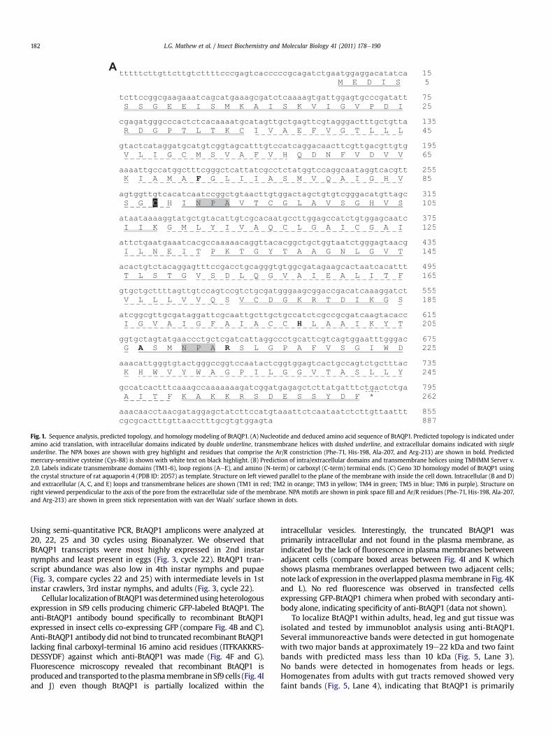
EU127479.1) consists of 262 amino acids with a predicted molec-ular weight of 27 kDa (Fig. 1A) and with isoelectric point of 6.5. Noputative N- or O-glycosylation sites were identified for BtAQP1.
Topology and hydrophobicity predictions using TMHMMprogram revealed that BtAQP1 consists of six transmembrane-spanning regions and intracellular amino and carboxyl termini(Fig. 1A and B), fitting the pattern known for other members of theaquaporin superfamily. Comparison of predicted topology andamino acid sequence of BtAQP1 with other insect and mammalianaquaporins reveal several key features that are common tomembersof the aquaporin superfamily, including six transmembranedomains, five alternating extracellular/intracellular loop regions,intracellular amino and carboxyl termini and two NPA motifs.
Three-dimensional structures are not yet available for arthropodaquaporins. Homology model predictions from SWISS-MODEL, I-TASSER, and Geno 3D using crystallography coordinates of Rattusnorvegicus aquaporin 4 [PDB ID: 2D57; (Hiroaki et al., 2006)] asa structural template depicted high similarity with BtAQP1 (49.5%identity) and reliable predicted model structures (SWISS-MODELRamachandran plot ¼ 81.7% residues in favorable region, 15.7% inallowable region, 2.6% in generously allowed region, and 0.0% indisallowed region; I-TASSER: c-score ¼ �0.09, TM-score ¼ 0.70 �0.12, RMSD ¼ 6.1 � 3.8; Geno 3D: Model energy ¼ �8356kcal mol�1, Ramachandran plot ¼ 79.1% residues in favorableregion, 16.6% in allowable region, 4.8% in generously allowedregion, and 0.0% in disallowed region). Fig. 1C shows predictedBtAQP1 model from Geno 3D using 2D57 as template. All modelsshow two tandem structural repeats, each consisting of threetransmembrane helices (TM1-3 and TM4-5) and a short a-helix inloops B and E each containing an NPA motif predicted to line oneside of the pore. This conserved structure is called the “aquaporinfold” (Murata et al., 2000). Residues that comprise the Ar/Rconstriction (Phe-71, His-198, Ala-207, and Arg-213) are found inBtAQP1 and are predicted to project into the putative cytoplasmicpore opening (Fig. 1A and C) and establish water selectivity.
We used CLUSTALW and MEGA 4.0 phylogenetic analysis tocompare BtAQP1with other insect aquaporins (Fig. 2). Phylogeneticanalysis of full-length AQP cDNA clones retrieved from GenBankreveals the existence three distinct families of insect AQPs,including Drosophila intrinsic proteins (DRIPs), Big Brain proteins(BIBs), and Pyrocoelia rufa integral proteins (PRIPs) as well as twounique sequences yet to be placed in a defined subfamily (Fig. 2;Campbell et al., 2008). Multiple insect AQP subfamilies other thanDRIPs, PRIPs, and BIBs likely exist based on the phylogeneticanalysis using cDNA and putative genomic AQP sequences (data notshown). BtAQP1 clusteredwith the known or predicted DRIP familyof proteins (Campbell et al., 2008) and overall shares 31% similaritywith shown members of that family. BtAQP1 was most closelyrelated to AQPs from the hemipteran leafhopper, C. viridis(54% identity), the human body louse, Pediculus humanus corporis(52% identity), and a putative AQP from the termite Coptotermesformosanus (53% identity). Interestingly, two splice variant AQPsfrom the pea aphid, A. pisum (ApAQP1 and 2) cluster together ona separate branch, indicating divergence from other insect DRIPs(41 and 43% identity to BtAQP1, respectively) (Fig. 2 and Supple-mental Fig. S1). Among the human AQPs, BtAQP1 is most similar toAQP4 (P55087.2), sharing 40% identity and key conserved sequencemotifs that are critical for water permeability (SupplementalFig. S2A and B).
3.2. Temporal expression and localization of BtAQP1
We investigated the expression pattern of BtAQP1 transcriptsover the life cycle of B. tabaci including egg, 4 nymphal stages(1st instar or crawler, 2nd, 3rd, 4th instar), pupal stage and adult.
A
Fig. 1. Sequence analysis, predicted topology, and homology modeling of BtAQP1. (A) Nucleotide and deduced amino acid sequence of BtAQP1. Predicted topology is indicated underamino acid translation, with intracellular domains indicated by double underline, transmembrane helices with dashed underline, and extracellular domains indicated with singleunderline. The NPA boxes are shown with grey highlight and residues that comprise the Ar/R constriction (Phe-71, His-198, Ala-207, and Arg-213) are shown in bold. Predictedmercury-sensitive cysteine (Cys-88) is shown with white text on black highlight. (B) Prediction of intra/extracellular domains and transmembrane helices using TMHMM Server v.2.0. Labels indicate transmembrane domains (TM1-6), loop regions (AeE), and amino (N-term) or carboxyl (C-term) terminal ends. (C) Geno 3D homology model of BtAQP1 usingthe crystal structure of rat aquaporin 4 (PDB ID: 2D57) as template. Structure on left viewed parallel to the plane of the membrane with inside the cell down. Intracellular (B and D)and extracellular (A, C, and E) loops and transmembrane helices are shown (TM1 in red; TM2 in orange; TM3 in yellow; TM4 in green; TM5 in blue; TM6 in purple). Structure onright viewed perpendicular to the axis of the pore from the extracellular side of the membrane. NPA motifs are shown in pink space fill and Ar/R residues (Phe-71, His-198, Ala-207,and Arg-213) are shown in green stick representation with van der Waals’ surface shown in dots.
L.G. Mathew et al. / Insect Biochemistry and Molecular Biology 41 (2011) 178e190182
Using semi-quantitative PCR, BtAQP1 amplicons were analyzed at20, 22, 25 and 30 cycles using Bioanalyzer. We observed thatBtAQP1 transcripts were most highly expressed in 2nd instarnymphs and least present in eggs (Fig. 3, cycle 22). BtAQP1 tran-script abundance was also low in 4th instar nymphs and pupae(Fig. 3, compare cycles 22 and 25) with intermediate levels in 1stinstar crawlers, 3rd instar nymphs, and adults (Fig. 3, cycle 22).
Cellular localizationofBtAQP1wasdeterminedusingheterologousexpression in Sf9 cells producing chimeric GFP-labeled BtAQP1. Theanti-BtAQP1 antibody bound specifically to recombinant BtAQP1expressed in insect cells co-expressing GFP (compare Fig. 4B and C).Anti-BtAQP1 antibody did not bind to truncated recombinant BtAQP1lacking final carboxyl-terminal 16 amino acid residues (ITFKAKKRS-DESSYDF) against which anti-BtAQP1 was made (Fig. 4F and G).Fluorescence microscopy revealed that recombinant BtAQP1 isproduced and transported to the plasmamembrane in Sf9 cells (Fig. 4Iand J) even though BtAQP1 is partially localized within the
intracellular vesicles. Interestingly, the truncated BtAQP1 wasprimarily intracellular and not found in the plasma membrane, asindicated by the lack of fluorescence in plasma membranes betweenadjacent cells (compare boxed areas between Fig. 4I and K whichshows plasma membranes overlapped between two adjacent cells;note lack of expression in the overlappedplasmamembrane in Fig. 4Kand L). No red fluorescence was observed in transfected cellsexpressing GFP-BtAQP1 chimera when probed with secondary anti-body alone, indicating specificity of anti-BtAQP1 (data not shown).
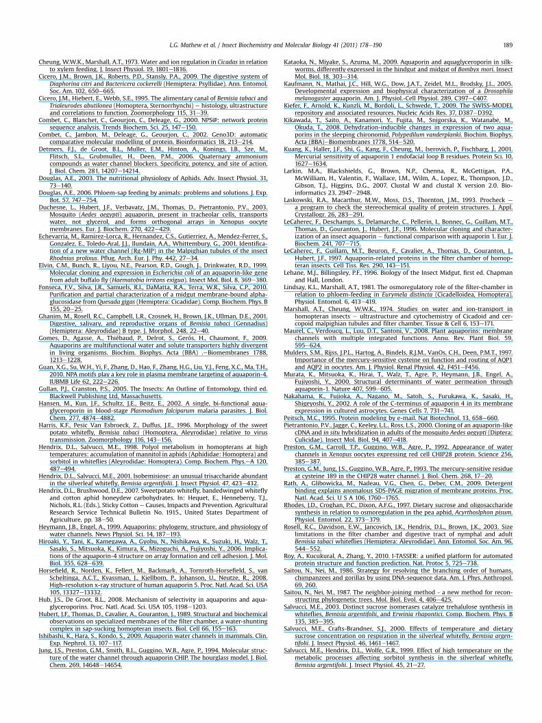
To localize BtAQP1 within adults, head, leg and gut tissue wasisolated and tested by immunoblot analysis using anti-BtAQP1.Several immunoreactive bands were detected in gut homogenatewith two major bands at approximately 19e22 kDa and two faintbands with predicted mass less than 10 kDa (Fig. 5, Lane 3).No bands were detected in homogenates from heads or legs.Homogenates from adults with gut tracts removed showed veryfaint bands (Fig. 5, Lane 4), indicating that BtAQP1 is primarily
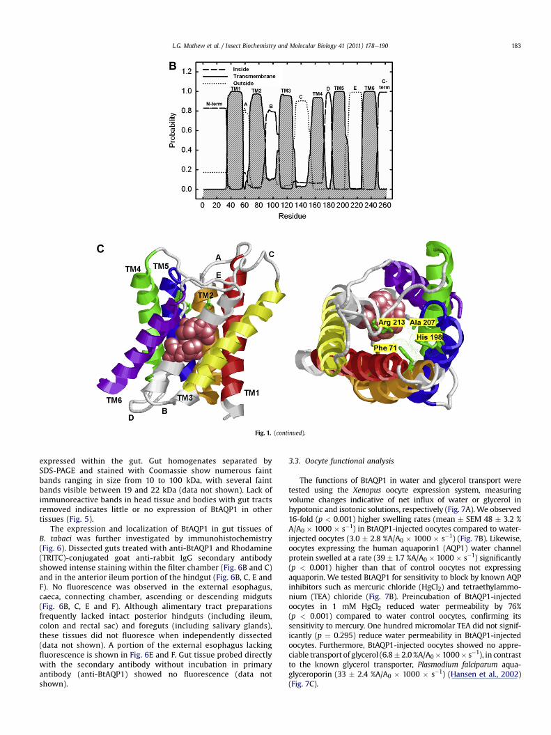
Fig. 1. (continued).
L.G. Mathew et al. / Insect Biochemistry and Molecular Biology 41 (2011) 178e190 183
expressed within the gut. Gut homogenates separated bySDS-PAGE and stained with Coomassie show numerous faintbands ranging in size from 10 to 100 kDa, with several faintbands visible between 19 and 22 kDa (data not shown). Lack ofimmunoreactive bands in head tissue and bodies with gut tractsremoved indicates little or no expression of BtAQP1 in othertissues (Fig. 5).
The expression and localization of BtAQP1 in gut tissues ofB. tabaci was further investigated by immunohistochemistry(Fig. 6). Dissected guts treated with anti-BtAQP1 and Rhodamine(TRITC)-conjugated goat anti-rabbit IgG secondary antibodyshowed intense staining within the filter chamber (Fig. 6B and C)and in the anterior ileum portion of the hindgut (Fig. 6B, C, E andF). No fluorescence was observed in the external esophagus,caeca, connecting chamber, ascending or descending midguts(Fig. 6B, C, E and F). Although alimentary tract preparationsfrequently lacked intact posterior hindguts (including ileum,colon and rectal sac) and foreguts (including salivary glands),these tissues did not fluoresce when independently dissected(data not shown). A portion of the external esophagus lackingfluorescence is shown in Fig. 6E and F. Gut tissue probed directlywith the secondary antibody without incubation in primaryantibody (anti-BtAQP1) showed no fluorescence (data notshown).
3.3. Oocyte functional analysis
The functions of BtAQP1 in water and glycerol transport weretested using the Xenopus oocyte expression system, measuringvolume changes indicative of net influx of water or glycerol inhypotonic and isotonic solutions, respectively (Fig. 7A). We observed16-fold (p < 0.001) higher swelling rates (mean � SEM 48 � 3.2 %A/A0 � 1000 � s�1) in BtAQP1-injected oocytes compared to water-injected oocytes (3.0 � 2.8 %A/A0 � 1000 � s�1) (Fig. 7B). Likewise,oocytes expressing the human aquaporin1 (AQP1) water channelprotein swelled at a rate (39 � 1.7 %A/A0 � 1000 � s�1) significantly(p < 0.001) higher than that of control oocytes not expressingaquaporin. We tested BtAQP1 for sensitivity to block by known AQPinhibitors such as mercuric chloride (HgCl2) and tetraethylammo-nium (TEA) chloride (Fig. 7B). Preincubation of BtAQP1-injectedoocytes in 1 mM HgCl2 reduced water permeability by 76%(p < 0.001) compared to water control oocytes, confirming itssensitivity to mercury. One hundred micromolar TEA did not signif-icantly (p ¼ 0.295) reduce water permeability in BtAQP1-injectedoocytes. Furthermore, BtAQP1-injected oocytes showed no appre-ciable transport of glycerol (6.8� 2.0 %A/A0�1000� s�1), in contrastto the known glycerol transporter, Plasmodium falciparum aqua-glyceroporin (33 � 2.4 %A/A0 � 1000 � s�1) (Hansen et al., 2002)(Fig. 7C).
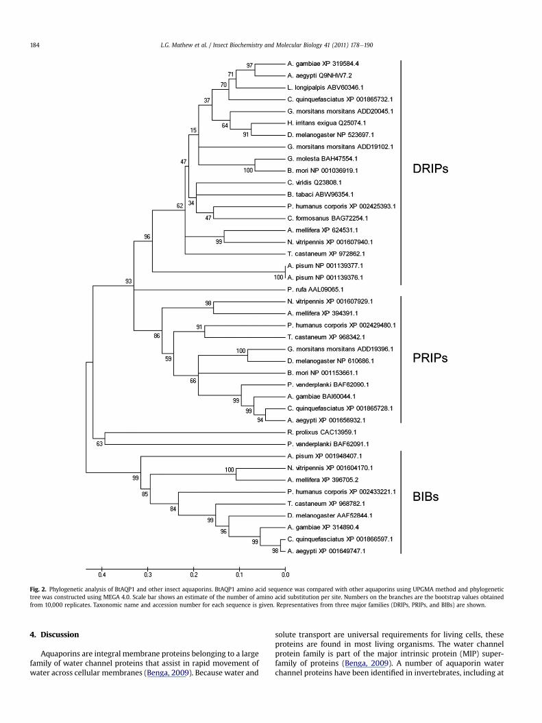
Fig. 2. Phylogenetic analysis of BtAQP1 and other insect aquaporins. BtAQP1 amino acid sequence was compared with other aquaporins using UPGMA method and phylogenetictree was constructed using MEGA 4.0. Scale bar shows an estimate of the number of amino acid substitution per site. Numbers on the branches are the bootstrap values obtainedfrom 10,000 replicates. Taxonomic name and accession number for each sequence is given. Representatives from three major families (DRIPs, PRIPs, and BIBs) are shown.
L.G. Mathew et al. / Insect Biochemistry and Molecular Biology 41 (2011) 178e190184
4. Discussion
Aquaporins are integral membrane proteins belonging to a largefamily of water channel proteins that assist in rapid movement ofwater across cellular membranes (Benga, 2009). Because water and
solute transport are universal requirements for living cells, theseproteins are found in most living organisms. The water channelprotein family is part of the major intrinsic protein (MIP) super-family of proteins (Benga, 2009). A number of aquaporin waterchannel proteins have been identified in invertebrates, including at
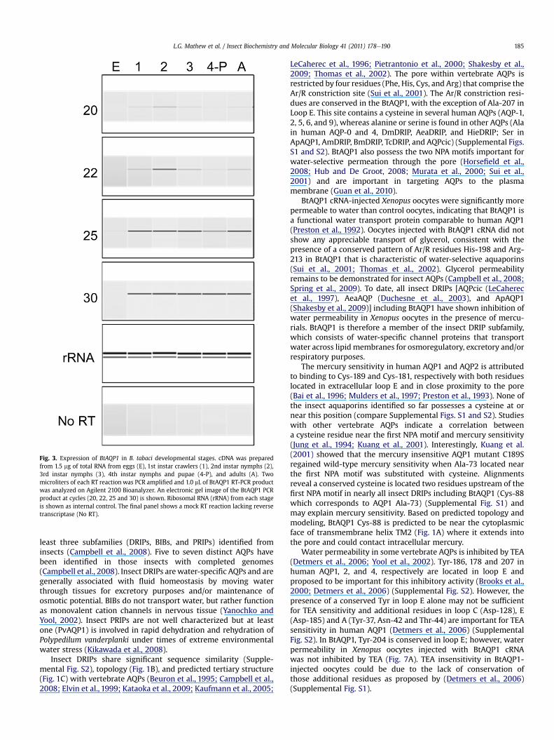
Fig. 3. Expression of BtAQP1 in B. tabaci developmental stages. cDNA was preparedfrom 1.5 mg of total RNA from eggs (E), 1st instar crawlers (1), 2nd instar nymphs (2),3rd instar nymphs (3), 4th instar nymphs and pupae (4-P), and adults (A). Twomicroliters of each RT reaction was PCR amplified and 1.0 mL of BtAQP1 RT-PCR productwas analyzed on Agilent 2100 Bioanalyzer. An electronic gel image of the BtAQP1 PCRproduct at cycles (20, 22, 25 and 30) is shown. Ribosomal RNA (rRNA) from each stageis shown as internal control. The final panel shows a mock RT reaction lacking reversetranscriptase (No RT).
L.G. Mathew et al. / Insect Biochemistry and Molecular Biology 41 (2011) 178e190 185
least three subfamilies (DRIPs, BIBs, and PRIPs) identified frominsects (Campbell et al., 2008). Five to seven distinct AQPs havebeen identified in those insects with completed genomes(Campbell et al., 2008). Insect DRIPs arewater-specific AQPs and aregenerally associated with fluid homeostasis by moving waterthrough tissues for excretory purposes and/or maintenance ofosmotic potential. BIBs do not transport water, but rather functionas monovalent cation channels in nervous tissue (Yanochko andYool, 2002). Insect PRIPs are not well characterized but at leastone (PvAQP1) is involved in rapid dehydration and rehydration ofPolypedilum vanderplanki under times of extreme environmentalwater stress (Kikawada et al., 2008).
Insect DRIPs share significant sequence similarity (Supple-mental Fig. S2), topology (Fig. 1B), and predicted tertiary structure(Fig. 1C) with vertebrate AQPs (Beuron et al., 1995; Campbell et al.,2008; Elvin et al., 1999; Kataoka et al., 2009; Kaufmann et al., 2005;
LeCaherec et al., 1996; Pietrantonio et al., 2000; Shakesby et al.,2009; Thomas et al., 2002). The pore within vertebrate AQPs isrestricted by four residues (Phe, His, Cys, and Arg) that comprise theAr/R constriction site (Sui et al., 2001). The Ar/R constriction resi-dues are conserved in the BtAQP1, with the exception of Ala-207 inLoop E. This site contains a cysteine in several human AQPs (AQP-1,2, 5, 6, and 9), whereas alanine or serine is found in other AQPs (Alain human AQP-0 and 4, DmDRIP, AeaDRIP, and HieDRIP; Ser inApAQP1, AmDRIP, BmDRIP, TcDRIP, and AQPcic) (Supplemental Figs.S1 and S2). BtAQP1 also possess the two NPA motifs important forwater-selective permeation through the pore (Horsefield et al.,2008; Hub and De Groot, 2008; Murata et al., 2000; Sui et al.,2001) and are important in targeting AQPs to the plasmamembrane (Guan et al., 2010).
BtAQP1 cRNA-injected Xenopus oocytes were significantly morepermeable to water than control oocytes, indicating that BtAQP1 isa functional water transport protein comparable to human AQP1(Preston et al., 1992). Oocytes injected with BtAQP1 cRNA did notshow any appreciable transport of glycerol, consistent with thepresence of a conserved pattern of Ar/R residues His-198 and Arg-213 in BtAQP1 that is characteristic of water-selective aquaporins(Sui et al., 2001; Thomas et al., 2002). Glycerol permeabilityremains to be demonstrated for insect AQPs (Campbell et al., 2008;Spring et al., 2009). To date, all insect DRIPs [AQPcic (LeCaherecet al., 1997), AeaAQP (Duchesne et al., 2003), and ApAQP1(Shakesby et al., 2009)] including BtAQP1 have shown inhibition ofwater permeability in Xenopus oocytes in the presence of mercu-rials. BtAQP1 is therefore a member of the insect DRIP subfamily,which consists of water-specific channel proteins that transportwater across lipid membranes for osmoregulatory, excretory and/orrespiratory purposes.
The mercury sensitivity in human AQP1 and AQP2 is attributedto binding to Cys-189 and Cys-181, respectively with both residueslocated in extracellular loop E and in close proximity to the pore(Bai et al., 1996; Mulders et al., 1997; Preston et al., 1993). None ofthe insect aquaporins identified so far possesses a cysteine at ornear this position (compare Supplemental Figs. S1 and S2). Studieswith other vertebrate AQPs indicate a correlation betweena cysteine residue near the first NPA motif and mercury sensitivity(Jung et al., 1994; Kuang et al., 2001). Interestingly, Kuang et al.(2001) showed that the mercury insensitive AQP1 mutant C189Sregained wild-type mercury sensitivity when Ala-73 located nearthe first NPA motif was substituted with cysteine. Alignmentsreveal a conserved cysteine is located two residues upstream of thefirst NPA motif in nearly all insect DRIPs including BtAQP1 (Cys-88which corresponds to AQP1 Ala-73) (Supplemental Fig. S1) andmay explain mercury sensitivity. Based on predicted topology andmodeling, BtAQP1 Cys-88 is predicted to be near the cytoplasmicface of transmembrane helix TM2 (Fig. 1A) where it extends intothe pore and could contact intracellular mercury.
Water permeability in some vertebrate AQPs is inhibited by TEA(Detmers et al., 2006; Yool et al., 2002). Tyr-186, 178 and 207 inhuman AQP1, 2, and 4, respectively are located in loop E andproposed to be important for this inhibitory activity (Brooks et al.,2000; Detmers et al., 2006) (Supplemental Fig. S2). However, thepresence of a conserved Tyr in loop E alone may not be sufficientfor TEA sensitivity and additional residues in loop C (Asp-128), E(Asp-185) and A (Tyr-37, Asn-42 and Thr-44) are important for TEAsensitivity in human AQP1 (Detmers et al., 2006) (SupplementalFig. S2). In BtAQP1, Tyr-204 is conserved in loop E; however, waterpermeability in Xenopus oocytes injected with BtAQP1 cRNAwas not inhibited by TEA (Fig. 7A). TEA insensitivity in BtAQP1-injected oocytes could be due to the lack of conservation ofthose additional residues as proposed by (Detmers et al., 2006)(Supplemental Fig. S1).
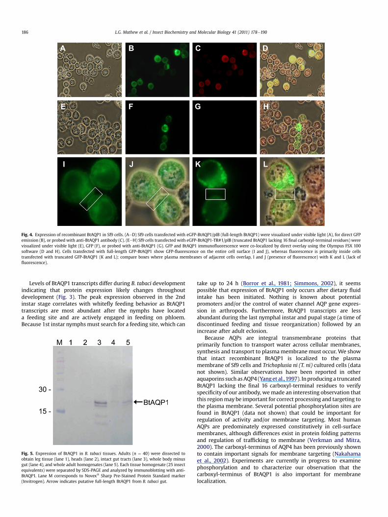
Fig. 4. Expression of recombinant BtAQP1 in Sf9 cells. (AeD) Sf9 cells transfected with eGFP-BtAQP1/pIB (full-length BtAQP1) were visualized under visible light (A), for direct GFPemission (B), or probed with anti-BtAQP1 antibody (C). (EeH) Sf9 cells transfected with eGFP-BtAQP1-TR#1/pIB (truncated BtAQP1 lacking 16 final carboxyl-terminal residues) werevisualized under visible light (E), GFP (F), or probed with anti-BtAQP1 (G). GFP and BtAQP1 immunofluorescence were co-localized by direct overlay using the Olympus FSX 100software (D and H). Cells transfected with full-length GFP-BtAQP1 show GFP-fluorescence on the entire cell surface (I and J), whereas fluorescence is primarily inside cellstransfected with truncated GFP-BtAQP1 (K and L); compare boxes where plasma membranes of adjacent cells overlap, I and J (presence of fluorescence) with K and L (lack offluorescence).
L.G. Mathew et al. / Insect Biochemistry and Molecular Biology 41 (2011) 178e190186
Levels of BtAQP1 transcripts differ during B. tabaci developmentindicating that protein expression likely changes throughoutdevelopment (Fig. 3). The peak expression observed in the 2ndinstar stage correlates with whitefly feeding behavior as BtAQP1transcripts are most abundant after the nymphs have locateda feeding site and are actively engaged in feeding on phloem.Because 1st instar nymphsmust search for a feeding site, which can
Fig. 5. Expression of BtAQP1 in B. tabaci tissues. Adults (n ¼ 40) were dissected toobtain leg tissue (lane 1), heads (lane 2), intact gut tracts (lane 3), whole body minusgut (lane 4), and whole adult homogenates (lane 5). Each tissue homogenate (25 insectequivalents) were separated by SDS-PAGE and analyzed by immunoblotting with anti-BtAQP1. Lane M corresponds to Novex� Sharp Pre-Stained Protein Standard marker(Invitrogen). Arrow indicates putative full-length BtAQP1 from B. tabaci gut.
take up to 24 h (Borror et al., 1981; Simmons, 2002), it seemspossible that expression of BtAQP1 only occurs after dietary fluidintake has been initiated. Nothing is known about potentialpromoters and/or the control of water channel AQP gene expres-sion in arthropods. Furthermore, BtAQP1 transcripts are lessabundant during the last nymphal instar and pupal stage (a time ofdiscontinued feeding and tissue reorganization) followed by anincrease after adult eclosion.
Because AQPs are integral transmembrane proteins thatprimarily function to transport water across cellular membranes,synthesis and transport to plasmamembrane must occur. We showthat intact recombinant BtAQP1 is localized to the plasmamembrane of Sf9 cells and Trichoplusia ni (T. ni) cultured cells (datanot shown). Similar observations have been reported in otheraquaporins such asAQP4 (Yanget al.,1997). Inproducing a truncatedBtAQP1 lacking the final 16 carboxyl-terminal residues to verifyspecificity of our antibody, wemade an interesting observation thatthis regionmay be important for correct processing and targeting tothe plasma membrane. Several potential phosphorylation sites arefound in BtAQP1 (data not shown) that could be important forregulation of activity and/or membrane targeting. Most humanAQPs are predominately expressed constitutively in cell-surfacemembranes, although differences exist in protein folding patternsand regulation of trafficking to membrane (Verkman and Mitra,2000). The carboxyl-terminus of AQP4 has been previously shownto contain important signals for membrane targeting (Nakahamaet al., 2002). Experiments are currently in progress to examinephosphorylation and to characterize our observation that thecarboxyl-terminus of BtAQP1 is also important for membranelocalization.
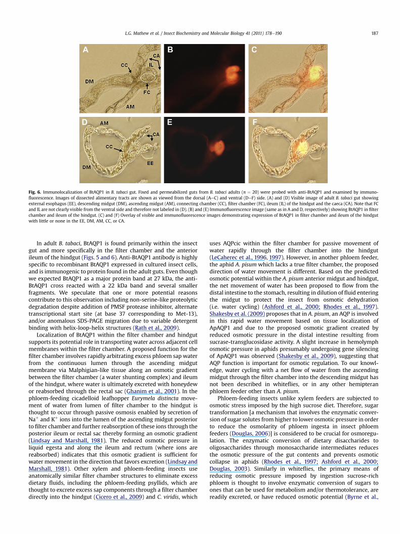
Fig. 6. Immunolocalization of BtAQP1 in B. tabaci gut. Fixed and permeabilized guts from B. tabaci adults (n ¼ 20) were probed with anti-BtAQP1 and examined by immuno-fluorescence. Images of dissected alimentary tracts are shown as viewed from the dorsal (AeC) and ventral (DeF) side. (A) and (D) Visible image of adult B. tabaci gut showingexternal esophagus (EE), descending midgut (DM), ascending midgut (AM), connecting chamber (CC), filter chamber (FC), ileum (IL) of the hindgut and the caeca (CA). Note that FCand IL are not clearly visible from the ventral side and therefore not labeled in (D). (B) and (E) Immunofluorescence image (same as in A and D, respectively) showing BtAQP1 in filterchamber and ileum of the hindgut. (C) and (F) Overlay of visible and immunofluorescence images demonstrating expression of BtAQP1 in filter chamber and ileum of the hindgutwith little or none in the EE, DM, AM, CC, or CA.
L.G. Mathew et al. / Insect Biochemistry and Molecular Biology 41 (2011) 178e190 187
In adult B. tabaci, BtAQP1 is found primarily within the insectgut and more specifically in the filter chamber and the anteriorileum of the hindgut (Figs. 5 and 6). Anti-BtAQP1 antibody is highlyspecific to recombinant BtAQP1 expressed in cultured insect cells,and is immunogenic to protein found in the adult guts. Even thoughwe expected BtAQP1 as a major protein band at 27 kDa, the anti-BtAQP1 cross reacted with a 22 kDa band and several smallerfragments. We speculate that one or more potential reasonscontribute to this observation including non-serine-like proteolyticdegradation despite addition of PMSF protease inhibitor, alternatetranscriptional start site (at base 37 corresponding to Met-13),and/or anomalous SDS-PAGE migration due to variable detergentbinding with helix-loop-helix structures (Rath et al., 2009).
Localization of BtAQP1 within the filter chamber and hindgutsupports its potential role in transporting water across adjacent cellmembranes within the filter chamber. A proposed function for thefilter chamber involves rapidly arbitrating excess phloem sap waterfrom the continuous lumen through the ascending midgutmembrane via Malphigian-like tissue along an osmotic gradientbetween the filter chamber (a water shunting complex) and ileumof the hindgut, where water is ultimately excreted with honeydewor reabsorbed through the rectal sac (Ghanim et al., 2001). In thephloem-feeding cicadelloid leafhopper Eurymela distincta move-ment of water from lumen of filter chamber to the hindgut isthought to occur through passive osmosis enabled by secretion ofNaþ and Kþ ions into the lumen of the ascending midgut posteriorto filter chamber and further reabsorption of these ions through theposterior ileum or rectal sac thereby forming an osmotic gradient(Lindsay and Marshall, 1981). The reduced osmotic pressure inliquid egesta and along the ileum and rectum (where ions arereabsorbed) indicates that this osmotic gradient is sufficient forwater movement in the direction that favors excretion (Lindsay andMarshall, 1981). Other xylem and phloem-feeding insects useanatomically similar filter chamber structures to eliminate excessdietary fluids, including the phloem-feeding psyllids, which arethought to excrete excess sap components through a filter chamberdirectly into the hindgut (Cicero et al., 2009) and C. viridis, which
uses AQPcic within the filter chamber for passive movement ofwater rapidly through the filter chamber into the hindgut(LeCaherec et al., 1996, 1997). However, in another phloem feeder,the aphid A. pisum which lacks a true filter chamber, the proposeddirection of water movement is different. Based on the predictedosmotic potential within the A. pisum anterior midgut and hindgut,the net movement of water has been proposed to flow from thedistal intestine to the stomach, resulting in dilution of fluid enteringthe midgut to protect the insect from osmotic dehydration(i.e. water cycling) (Ashford et al., 2000; Rhodes et al., 1997).Shakesby et al. (2009) proposes that in A. pisum, an AQP is involvedin this rapid water movement based on tissue localization ofApAQP1 and due to the proposed osmotic gradient created byreduced osmotic pressure in the distal intestine resulting fromsucrase-transglucosidase activity. A slight increase in hemolymphosmotic pressure in aphids presumably undergoing gene silencingof ApAQP1 was observed (Shakesby et al., 2009), suggesting thatAQP function is important for osmotic regulation. To our knowl-edge, water cycling with a net flow of water from the ascendingmidgut through the filter chamber into the descending midgut hasnot been described in whiteflies, or in any other hemipteranphloem feeder other than A. pisum.
Phloem-feeding insects unlike xylem feeders are subjected toosmotic stress imposed by the high sucrose diet. Therefore, sugartransformation [a mechanism that involves the enzymatic conver-sion of sugar solutes fromhigher to lower osmotic pressure in orderto reduce the osmolarity of phloem ingesta in insect phloemfeeders (Douglas, 2006)] is considered to be crucial for osmoregu-lation. The enzymatic conversion of dietary disaccharides tooligosaccharides through monosaccharide intermediates reducesthe osmotic pressure of the gut contents and prevents osmoticcollapse in aphids (Rhodes et al., 1997; Ashford et al., 2000;Douglas, 2003). Similarly in whiteflies, the primary means ofreducing osmotic pressure imposed by ingestion sucrose-richphloem is thought to involve enzymatic conversion of sugars toones that can be used for metabolism and/or thermotolerance, arereadily excreted, or have reduced osmotic potential (Byrne et al.,
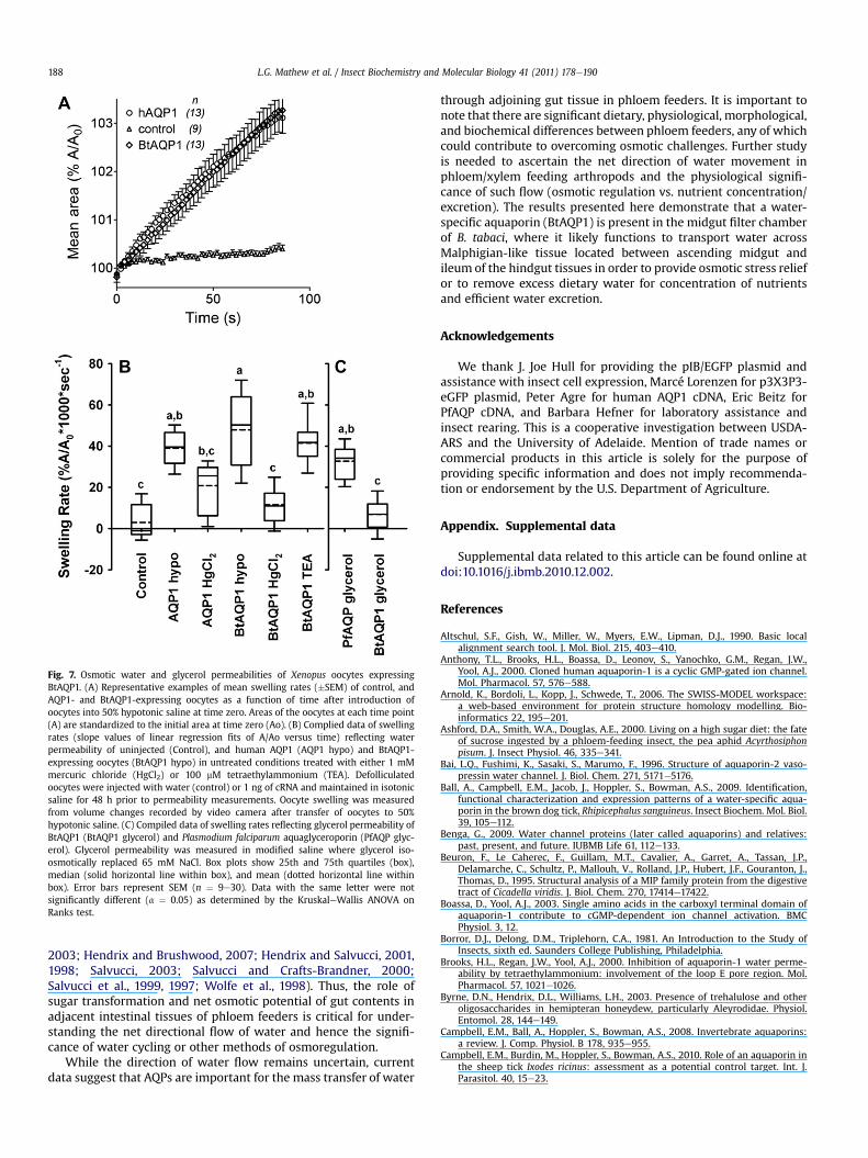
Fig. 7. Osmotic water and glycerol permeabilities of Xenopus oocytes expressingBtAQP1. (A) Representative examples of mean swelling rates (�SEM) of control, andAQP1- and BtAQP1-expressing oocytes as a function of time after introduction ofoocytes into 50% hypotonic saline at time zero. Areas of the oocytes at each time point(A) are standardized to the initial area at time zero (Ao). (B) Complied data of swellingrates (slope values of linear regression fits of A/Ao versus time) reflecting waterpermeability of uninjected (Control), and human AQP1 (AQP1 hypo) and BtAQP1-expressing oocytes (BtAQP1 hypo) in untreated conditions treated with either 1 mMmercuric chloride (HgCl2) or 100 mM tetraethylammonium (TEA). Defolliculatedoocytes were injected with water (control) or 1 ng of cRNA and maintained in isotonicsaline for 48 h prior to permeability measurements. Oocyte swelling was measuredfrom volume changes recorded by video camera after transfer of oocytes to 50%hypotonic saline. (C) Compiled data of swelling rates reflecting glycerol permeability ofBtAQP1 (BtAQP1 glycerol) and Plasmodium falciparum aquaglyceroporin (PfAQP glyc-erol). Glycerol permeability was measured in modified saline where glycerol iso-osmotically replaced 65 mM NaCl. Box plots show 25th and 75th quartiles (box),median (solid horizontal line within box), and mean (dotted horizontal line withinbox). Error bars represent SEM (n ¼ 9e30). Data with the same letter were notsignificantly different (a ¼ 0.05) as determined by the KruskaleWallis ANOVA onRanks test.
L.G. Mathew et al. / Insect Biochemistry and Molecular Biology 41 (2011) 178e190188
2003; Hendrix and Brushwood, 2007; Hendrix and Salvucci, 2001,1998; Salvucci, 2003; Salvucci and Crafts-Brandner, 2000;Salvucci et al., 1999, 1997; Wolfe et al., 1998). Thus, the role ofsugar transformation and net osmotic potential of gut contents inadjacent intestinal tissues of phloem feeders is critical for under-standing the net directional flow of water and hence the signifi-cance of water cycling or other methods of osmoregulation.
While the direction of water flow remains uncertain, currentdata suggest that AQPs are important for the mass transfer of water
through adjoining gut tissue in phloem feeders. It is important tonote that there are significant dietary, physiological, morphological,and biochemical differences between phloem feeders, any of whichcould contribute to overcoming osmotic challenges. Further studyis needed to ascertain the net direction of water movement inphloem/xylem feeding arthropods and the physiological signifi-cance of such flow (osmotic regulation vs. nutrient concentration/excretion). The results presented here demonstrate that a water-specific aquaporin (BtAQP1) is present in the midgut filter chamberof B. tabaci, where it likely functions to transport water acrossMalphigian-like tissue located between ascending midgut andileum of the hindgut tissues in order to provide osmotic stress reliefor to remove excess dietary water for concentration of nutrientsand efficient water excretion.
Acknowledgements
We thank J. Joe Hull for providing the pIB/EGFP plasmid andassistance with insect cell expression, Marcé Lorenzen for p3X3P3-eGFP plasmid, Peter Agre for human AQP1 cDNA, Eric Beitz forPfAQP cDNA, and Barbara Hefner for laboratory assistance andinsect rearing. This is a cooperative investigation between USDA-ARS and the University of Adelaide. Mention of trade names orcommercial products in this article is solely for the purpose ofproviding specific information and does not imply recommenda-tion or endorsement by the U.S. Department of Agriculture.
Appendix. Supplemental data
Supplemental data related to this article can be found online atdoi:10.1016/j.ibmb.2010.12.002.
References
Altschul, S.F., Gish, W., Miller, W., Myers, E.W., Lipman, D.J., 1990. Basic localalignment search tool. J. Mol. Biol. 215, 403e410.
Anthony, T.L., Brooks, H.L., Boassa, D., Leonov, S., Yanochko, G.M., Regan, J.W.,Yool, A.J., 2000. Cloned human aquaporin-1 is a cyclic GMP-gated ion channel.Mol. Pharmacol. 57, 576e588.
Arnold, K., Bordoli, L., Kopp, J., Schwede, T., 2006. The SWISS-MODEL workspace:a web-based environment for protein structure homology modelling. Bio-informatics 22, 195e201.
Ashford, D.A., Smith, W.A., Douglas, A.E., 2000. Living on a high sugar diet: the fateof sucrose ingested by a phloem-feeding insect, the pea aphid Acyrthosiphonpisum. J. Insect Physiol. 46, 335e341.
Bai, L.Q., Fushimi, K., Sasaki, S., Marumo, F., 1996. Structure of aquaporin-2 vaso-pressin water channel. J. Biol. Chem. 271, 5171e5176.
Ball, A., Campbell, E.M., Jacob, J., Hoppler, S., Bowman, A.S., 2009. Identification,functional characterization and expression patterns of a water-specific aqua-porin in the brown dog tick, Rhipicephalus sanguineus. Insect Biochem. Mol. Biol.39, 105e112.
Benga, G., 2009. Water channel proteins (later called aquaporins) and relatives:past, present, and future. IUBMB Life 61, 112e133.
Beuron, F., Le Caherec, F., Guillam, M.T., Cavalier, A., Garret, A., Tassan, J.P.,Delamarche, C., Schultz, P., Mallouh, V., Rolland, J.P., Hubert, J.F., Gouranton, J.,Thomas, D., 1995. Structural analysis of a MIP family protein from the digestivetract of Cicadella viridis. J. Biol. Chem. 270, 17414e17422.
Boassa, D., Yool, A.J., 2003. Single amino acids in the carboxyl terminal domain ofaquaporin-1 contribute to cGMP-dependent ion channel activation. BMCPhysiol. 3, 12.
Borror, D.J., Delong, D.M., Triplehorn, C.A., 1981. An Introduction to the Study ofInsects, sixth ed. Saunders College Publishing, Philadelphia.
Brooks, H.L., Regan, J.W., Yool, A.J., 2000. Inhibition of aquaporin-1 water perme-ability by tetraethylammonium: involvement of the loop E pore region. Mol.Pharmacol. 57, 1021e1026.
Byrne, D.N., Hendrix, D.L., Williams, L.H., 2003. Presence of trehalulose and otheroligosaccharides in hemipteran honeydew, particularly Aleyrodidae. Physiol.Entomol. 28, 144e149.
Campbell, E.M., Ball, A., Hoppler, S., Bowman, A.S., 2008. Invertebrate aquaporins:a review. J. Comp. Physiol. B 178, 935e955.
Campbell, E.M., Burdin, M., Hoppler, S., Bowman, A.S., 2010. Role of an aquaporin inthe sheep tick Ixodes ricinus: assessment as a potential control target. Int. J.Parasitol. 40, 15e23.
L.G. Mathew et al. / Insect Biochemistry and Molecular Biology 41 (2011) 178e190 189
Cheung, W.W.K., Marshall, A.T., 1973. Water and ion regulation in Cicadas in relationto xylem feeding. J. Insect Physiol. 19, 1801e1816.
Cicero, J.M., Brown, J.K., Roberts, P.D., Stansly, P.A., 2009. The digestive system ofDiaphorina citri and Bactericera cockerelli (Hemiptera: Psyllidae). Ann. Entomol.Soc. Am. 102, 650e665.
Cicero, J.M., Hiebert, E., Webb, S.E., 1995. The alimentary canal of Bemisia tabaci andTrialeurodes abutilonea (Homoptera, Sternorrhynchi) e histology, ultrastructureand correlations to function. Zoomorphology 115, 31e39.
Combet, C., Blanchet, C., Geourjon, C., Deleage, G., 2000. NPS@: network proteinsequence analysis. Trends Biochem. Sci. 25, 147e150.
Combet, C., Jambon, M., Deleage, G., Geourjon, C., 2002. Geno3D: automaticcomparative molecular modelling of protein. Bioinformatics 18, 213e214.
Detmers, F.J., de Groot, B.L., Muller, E.M., Hinton, A., Konings, I.B., Sze, M.,Flitsch, S.L., Grubmuller, H., Deen, P.M., 2006. Quaternary ammoniumcompounds as water channel blockers. Specificity, potency, and site of action.J. Biol. Chem. 281, 14207e14214.
Douglas, A.E., 2003. The nutritional physiology of Aphids. Adv. Insect Physiol. 31,73e140.
Douglas, A.E., 2006. Phloem-sap feeding by animals: problems and solutions. J. Exp.Bot. 57, 747e754.
Duchesne, L., Hubert, J.F., Verbavatz, J.M., Thomas, D., Pietrantonio, P.V., 2003.Mosquito (Aedes aegypti) aquaporin, present in tracheolar cells, transportswater, not glycerol, and forms orthogonal arrays in Xenopus oocytemembranes. Eur. J. Biochem. 270, 422e429.
Echevarria, M., Ramirez-Lorca, R., Hernandez, C.S., Gutierriez, A., Mendez-Ferrer, S.,Gonzalez, E., Toledo-Aral, J.J., Ilundain, A.A., Whittembury, G., 2001. Identifica-tion of a new water channel (Rg-MIP) in the Malpighian tubules of the insectRhodnius prolixus. Pflug. Arch. Eur. J. Phy. 442, 27e34.
Elvin, C.M., Bunch, R., Liyou, N.E., Pearson, R.D., Gough, J., Drinkwater, R.D., 1999.Molecular cloning and expression in Escherichia coli of an aquaporin-like genefrom adult buffalo fly (Haematobia irritans exigua). Insect Mol. Biol. 8, 369e380.
Fonseca, F.V., Silva, J.R., Samuels, R.I., DaMatta, R.A., Terra, W.R., Silva, C.P., 2010.Purification and partial characterization of a midgut membrane-bound alpha-glucosidase from Quesada gigas (Hemiptera: Cicadidae). Comp. Biochem. Phys. B155, 20e25.
Ghanim, M., Rosell, R.C., Campbell, L.R., Czosnek, H., Brown, J.K., Ullman, D.E., 2001.Digestive, salivary, and reproductive organs of Bemisia tabaci (Gennadius)(Hemiptera: Aleyrodidae) B type. J. Morphol. 248, 22e40.
Gomes, D., Agasse, A., Thiébaud, P., Delrot, S., Gerós, H., Chaumont, F., 2009.Aquaporins are multifunctional water and solute transporters highly divergentin living organisms. Biochim. Biophys. Acta (BBA) .eBiomembranes 1788,1213e1228.
Guan, X.G., Su, W.H., Yi, F., Zhang, D., Hao, F., Zhang, H.G., Liu, Y.J., Feng, X.C., Ma, T.H.,2010. NPA motifs play a key role in plasma membrane targeting of aquaporin-4.IUBMB Life 62, 222e226.
Gullan, P.J., Cranston, P.S., 2005. The Insects: An Outline of Entomology, third ed.Blackwell Publishing Ltd, Massachusetts.
Hansen, M., Kun, J.F., Schultz, J.E., Beitz, E., 2002. A single, bi-functional aqua-glyceroporin in blood-stage Plasmodium falciparum malaria parasites. J. Biol.Chem. 277, 4874e4882.
Harris, K.F., Pesic Van Esbroeck, Z., Duffus, J.E., 1996. Morphology of the sweetpotato whitefly, Bemisia tabaci (Homoptera, Aleyrodidae) relative to virustransmission. Zoomorphology 116, 143e156.
Hendrix, D.L., Salvucci, M.E., 1998. Polyol metabolism in homopterans at hightemperatures: accumulation of mannitol in aphids (Aphididae: Homoptera) andsorbitol in whiteflies (Aleyrodidae: Homoptera). Comp. Biochem. Phys.eA 120,487e494.
Hendrix, D.L., Salvucci, M.E., 2001. Isobemisiose: an unusual trisaccharide abundantin the silverleaf whitefly, Bemisia argentifolii. J. Insect Physiol. 47, 423e432.
Hendrix, D.L., Brushwood, D.E., 2007. Sweetpotato whitefly, bandedwinged whiteflyand cotton aphid honeydew carbohydrates. In: Hequet, E., Henneberry, T.J.,Nichols, R.L. (Eds.), Sticky Cotton e Causes, Impacts and Prevention. AgriculturalResearch Service Technical Bulletin No. 1915., United States Department ofAgriculture, pp. 38e50.
Heymann, J.B., Engel, A., 1999. Aquaporins: phylogeny, structure, and physiology ofwater channels. News Physiol. Sci. 14, 187e193.
Hiroaki, Y., Tani, K., Kamegawa, A., Gyobu, N., Nishikawa, K., Suzuki, H., Walz, T.,Sasaki, S., Mitsuoka, K., Kimura, K., Mizoguchi, A., Fujiyoshi, Y., 2006. Implica-tions of the aquaporin-4 structure on array formation and cell adhesion. J. Mol.Biol. 355, 628e639.
Horsefield, R., Norden, K., Fellert, M., Backmark, A., Tornroth-Horsefield, S., vanScheltinga, A.C.T., Kvassman, J., Kjellbom, P., Johanson, U., Neutze, R., 2008.High-resolution x-ray structure of human aquaporin 5. Proc. Natl. Acad. Sci. USA105, 13327e13332.
Hub, J.S., De Groot, B.L., 2008. Mechanism of selectivity in aquaporins and aqua-glyceroporins. Proc. Natl. Acad. Sci. USA 105, 1198e1203.
Hubert, J.F., Thomas, D., Cavalier, A., Gouranton, J., 1989. Structural and biochemicalobservations on specialized membranes of the filter chamber, a water-shuntingcomplex in sap-sucking homopteran insects. Biol. Cell 66, 155e163.
Ishibashi, K., Hara, S., Kondo, S., 2009. Aquaporin water channels in mammals. Clin.Exp. Nephrol. 13, 107e117.
Jung, J.S., Preston, G.M., Smith, B.L., Guggino, W.B., Agre, P., 1994. Molecular struc-ture of the water channel through aquaporin CHIP. The hourglass model. J. Biol.Chem. 269, 14648e14654.
Kataoka, N., Miyake, S., Azuma, M., 2009. Aquaporin and aquaglyceroporin in silk-worms, differently expressed in the hindgut and midgut of Bombyx mori. InsectMol. Biol. 18, 303e314.
Kaufmann, N., Mathai, J.C., Hill, W.G., Dow, J.A.T., Zeidel, M.L., Brodsky, J.L., 2005.Developmental expression and biophysical characterization of a Drosophilamelanogaster aquaporin. Am. J. Physiol.-Cell Physiol. 289, C397eC407.
Kiefer, F., Arnold, K., Kunzli, M., Bordoli, L., Schwede, T., 2009. The SWISS-MODELrepository and associated resources. Nucleic Acids Res. 37, D387eD392.
Kikawada, T., Saito, A., Kanamori, Y., Fujita, M., Snigorska, K., Watanabe, M.,Okuda, T., 2008. Dehydration-inducible changes in expression of two aqua-porins in the sleeping chironomid, Polypedilum vanderplanki. Biochim. Biophys.Acta (BBA)eBiomembranes 1778, 514e520.
Kuang, K., Haller, J.F., Shi, G., Kang, F., Cheung, M., Iserovich, P., Fischbarg, J., 2001.Mercurial sensitivity of aquaporin 1 endofacial loop B residues. Protein Sci. 10,1627e1634.
Larkin, M.A., Blackshields, G., Brown, N.P., Chenna, R., McGettigan, P.A.,McWilliam, H., Valentin, F., Wallace, I.M., Wilm, A., Lopez, R., Thompson, J.D.,Gibson, T.J., Higgins, D.G., 2007. Clustal W and clustal X version 2.0. Bio-informatics 23, 2947e2948.
Laskowski, R.A., Macarthur, M.W., Moss, D.S., Thornton, J.M., 1993. Procheck e
a program to check the stereochemical quality of protein structures. J. Appl.Crystallogr. 26, 283e291.
LeCaherec, F., Deschamps, S., Delamarche, C., Pellerin, I., Bonnec, G., Guillam, M.T.,Thomas, D., Gouranton, J., Hubert, J.F., 1996. Molecular cloning and character-ization of an insect aquaporin e functional comparison with aquaporin 1. Eur. J.Biochem. 241, 707e715.
LeCaherec, F., Guillam, M.T., Beuron, F., Cavalier, A., Thomas, D., Gouranton, J.,Hubert, J.F., 1997. Aquaporin-related proteins in the filter chamber of homop-teran insects. Cell Tiss. Res. 290, 143e151.
Lehane, M.J., Billingsley, P.F., 1996. Biology of the Insect Midgut, first ed. Chapmanand Hall, London.
Lindsay, K.L., Marshall, A.T., 1981. The osmoregulatory role of the filter-chamber inrelation to phloem-feeding in Eurymela distincta (Cicadelloidea, Homoptera).Physiol. Entomol. 6, 413e419.
Marshall, A.T., Cheung, W.W.K., 1974. Studies on water and ion-transport inhomopteran insects e ultrastructure and cytochemistry of Cicadoid and cer-copoid malpighian tubules and filter chamber. Tissue & Cell 6, 153e171.
Maurel, C., Verdoucq, L., Luu, D.T., Santoni, V., 2008. Plant aquaporins: membranechannels with multiple integrated functions. Annu. Rev. Plant Biol. 59,595e624.
Mulders, S.M., Rijss, J.P.L., Hartog, A., Bindels, R.J.M., VanOs, C.H., Deen, P.M.T., 1997.Importance of the mercury-sensitive cysteine on function and routing of AQP1and AQP2 in oocytes. Am. J. Physiol. Renal Physiol. 42, F451eF456.
Murata, K., Mitsuoka, K., Hirai, T., Walz, T., Agre, P., Heymann, J.B., Engel, A.,Fujiyoshi, Y., 2000. Structural determinants of water permeation throughaquaporin-1. Nature 407, 599e605.
Nakahama, K., Fujioka, A., Nagano, M., Satoh, S., Furukawa, K., Sasaki, H.,Shigeyoshi, Y., 2002. A role of the C-terminus of aquaporin 4 in its membraneexpression in cultured astrocytes. Genes Cells 7, 731e741.
Peitsch, M.C., 1995. Protein modeling by e-mail. Nat Biotechnol. 13, 658e660.Pietrantonio, P.V., Jagge, C., Keeley, L.L., Ross, L.S., 2000. Cloning of an aquaporin-like
cDNA and in situ hybridization in adults of the mosquito Aedes aegypti (Diptera:Culicidae). Insect Mol. Biol. 94, 407e418.
Preston, G.M., Carroll, T.P., Guggino, W.B., Agre, P., 1992. Appearance of waterchannels in Xenopus oocytes expressing red cell CHIP28 protein. Science 256,385e387.
Preston, G.M., Jung, J.S., Guggino, W.B., Agre, P., 1993. The mercury-sensitive residueat cysteine 189 in the CHIP28 water channel. J. Biol. Chem. 268, 17e20.
Rath, A., Glibowicka, M., Nadeau, V.G., Chen, G., Deber, C.M., 2009. Detergentbinding explains anomalous SDS-PAGE migration of membrane proteins. Proc.Natl. Acad. Sci. U S A 106, 1760e1765.
Rhodes, J.D., Croghan, P.C., Dixon, A.F.G., 1997. Dietary sucrose and oligosaccharidesynthesis in relation to osmoregulation in the pea aphid, Acyrthoslphon pisum.Physiol. Entomol. 22, 373e379.
Rosell, R.C., Davidson, E.W., Jancovich, J.K., Hendrix, D.L., Brown, J.K., 2003. Sizelimitations in the filter chamber and digestive tract of nymphal and adultBemisia tabaci whiteflies (Hemiptera: Aleyrodidae). Ann. Entomol. Soc. Am. 96,544e552.
Roy, A., Kucukural, A., Zhang, Y., 2010. I-TASSER: a unified platform for automatedprotein structure and function prediction. Nat. Protoc 5, 725e738.
Saitou, N., Nei, M., 1986. Strategy for resolving the branching order of humans,chimpanzees and gorillas by using DNA-sequence data. Am. J. Phys. Anthropol.69, 260.
Saitou, N., Nei, M., 1987. The neighbor-joining method - a new method for recon-structing phylogenetic trees. Mol. Biol. Evol. 4, 406e425.
Salvucci, M.E., 2003. Distinct sucrose isomerases catalyze trehalulose synthesis inwhiteflies, Bemisia argentifolii, and Erwinia rhapontici. Comp. Biochem. Phys. B135, 385e395.
Salvucci, M.E., Crafts-Brandner, S.J., 2000. Effects of temperature and dietarysucrose concentration on respiration in the silverleaf whitefly, Bemisia argen-tifolii. J. Insect Physiol. 46, 1461e1467.
Salvucci, M.E., Hendrix, D.L., Wolfe, G.R., 1999. Effect of high temperature on themetabolic processes affecting sorbitol synthesis in the silverleaf whitefly,Bemisia argentifolii. J. Insect Physiol. 45, 21e27.
L.G. Mathew et al. / Insect Biochemistry and Molecular Biology 41 (2011) 178e190190
Salvucci, M.E., Wolfe, G.R., Hendrix, D.L., 1997. Effect of sucrose concentration oncarbohydrate metabolism in Bemisia argentifolii: biochemical mechanism andphysiological role for trehalulose synthesis in the silverleaf whitefly. J. InsectPhysiol. 43, 457e464.
Salvucci, M.E., Wolfe, G.R., Hendrix, D.L., 1998. Purification and properties of anunusual NADPH-dependent ketose reductase from the silverleaf whitefly. InsectBiochem. Mol. Biol. 28, 357e363.
Shakesby, A.J., Wallace, I.S., Isaacs, H.V., Pritchard, J., Roberts, D.M., Douglas, A.E.,2009. A water-specific aquaporin involved in aphid osmoregulation. InsectBiochem. Mol. Biol. 39, 1e10.
Simmons, A.M., 2002. Settling of crawlers of Bemisia tabaci (Homoptera: Alcyr-odidae) on five vegetable hosts. Ann. Entomol. Soc. Am. 95, 464e468.
Sneath, Sokal, 1973. Numerical Taxonomy. W.H. Freeman and Company, San Fran-cisco, pp. 230e234.
Spring, J.H., Robichaux, S.R., Hamlin, J.A., 2009. The role of aquaporins in excretionin insects. J. Exp. Biol. 212, 358e362.
Sui, H., Han, B.G., Lee, J.K., Walian, P., Jap, B.K., 2001. Structural basis of water-specific transport through the AQP1 water channel. Nature 414, 872e878.
Tamura, K., Dudley, J., Nei, M., Kumar, S., 2007. MEGA4: molecular evolutionarygenetics analysis (MEGA) software version 4.0. Mol. Biol. Evol. 24,1596e1599.
Thomas, D., Bron, P., Ranchy, G., Duchesne, L., Cavalier, A., Rolland, J.P., Raguenes-Nicol, C., Hubert, J.F., Haase, W., Delamarche, C., 2002. Aquaglyceroporins, onechannel for two molecules. Biochim. Biophys. Acta 1555, 181e186.
Verkman, A.S., Mitra, A.K., 2000. Structure and function of aquaporin water chan-nels. Am. J. Physiol. Renal Physiol. 278, F13eF28.
Wolfe, G.R., Hendrix, D.L., Salvucci, M.E., 1998. A thermoprotective role for sorbitolin the silverleaf whitefly, Bemisia argentifolii. J. Insect Physiol. 44, 597e603.
Yang, B., van Hoek, A.N., Verkman, A.S., 1997. Very high single channel waterpermeability of aquaporin-4 in baculovirus-infected insect cells and liposomesreconstituted with purified aquaporin-4. Biochemistry 36, 7625e7632.
Yanochko, G.M., Yool, A.J., 2002. Regulated cationic channel function in Xenopusoocytes expressing Drosophila big brain. J. Neurosci. 22, 2530e2540.
Yool, A.J., 2007. Functional domains of aquaporin-1: keys to physiology, and targetsfor drug discovery. Curr. Pharm. Des. 13, 3212e3221.
Yool, A.J., Brokl, O.H., Pannabecker, T.L., Dantzler, W.H., Stamer, W.D., 2002. Tetrae-thylammonium block of water flux in aquaporin-1 channels expressed in kidneythin limbs of Henle’s loop and a kidney-derived cell line. BMC Physiol. 2, 4.
Zhang, Y., 2008. I-TASSER server for protein 3D structure prediction. BMC Bio-informatics 9, 40.
Zhang, Y., 2009. I-TASSER: fully automated protein structure prediction in CASP8.Proteins 77 (Suppl. 9), 100e113.


