Embed Size (px)
Citation preview

Available online at www.sciencedirect.com
gy, Part B 149 (2008) 419–427www.elsevier.com/locate/cbpb
Comparative Biochemistry and Physiolo
Identification and characterization of the receptor for the Bacillus sphaericusbinary toxin in the malaria vector mosquito, Anopheles gambiae
Onya Opota a, Jean-François Charles b, Sylvie Warot a, David Pauron a, Isabelle Darboux a,⁎
a UMR1112 « Réponses des Organismes aux Stress Environnementaux », INRA-UNSA, 400 Route des Chappes, BP 167, F-06903 Sophia-Antipolis, Franceb Génétique des Génomes Bactériens URA CNRS 2171, Institut Pasteur, 28 rue du Dr. Roux, F-75724 Paris, France
Received 6 October 2007; received in revised form 12 November 2007; accepted 12 November 2007Available online 17 November 2007
Abstract
The binary toxin (Bin) from Bacillus sphaericus exhibits a highly insecticidal activity against Culex and Anopheles mosquitoes. Thecytotoxicity of Bin requires an interaction with a specific receptor present on the membrane of midgut epithelial cells in larvae. A direct correlationexists between binding affinity and toxicity. The toxin binds with high affinity to its receptor in its primary target, Culex pipiens, and displays alower affinity to the receptor in Anopheles gambiae, which is less sensitive to Bin. Although the Bin receptor has previously been identified andnamed Cpm1 in C. pipiens, its structure in Anopheles remains unknown. In this study, we hypothesize that the Anopheles Bin receptor is anortholog of Cpm1. By screening the Anopheles genomic database, we identified a candidate gene (Agm3) which is expressed primarily on thesurface of midgut cells in larvae and which functions as a receptor for Bin. A Cpm1-like gene is also present in the Bin-refractory species Aedesaegypti. Overall, our results indicate that the three mosquito genes examined share a very similar organization and are strongly conserved at theamino acid level, in particular in the NH2-terminus, a region believed to contain the ligand binding site, suggesting that relatively few amino acidsresidues are critical for high affinity binding of the toxin.© 2007 Elsevier Inc. All rights reserved.
Keywords: Anopheles gambiae; Receptor; Binary toxin; Bacillus sphaericus; Biopesticide
1. Introduction
Mosquitoes of the genera Aedes, Anopheles and Culextransmit serious human diseases such as malaria, yellow fever,West Nile fever, and chikungunya. In the past, the control forthese vectors had been primarily undertaken using chemicalinsecticides. However, resistance of insects to all major classesof pesticides (Gubler, 1998; Zaim and Guillet, 2002; Nauen,2007), together with increasing limitation or discontinued use ofmost of the harmful synthetic pesticides have contributed to areduction in both the panel of insecticidal products and thenumber of methods available to control mosquito populations.Strategies based on genetic manipulations and the generation ofmosquitoes refractory to the development of pathogens havebeen proposed (James, 2002) but have not yet been developedto date.
⁎ Corresponding author. Tel.: +33 4 67 14 32 20; fax: +33 4 67 14 42 99.E-mail address: [email protected] (I. Darboux).
1096-4959/$ - see front matter © 2007 Elsevier Inc. All rights reserved.doi:10.1016/j.cbpb.2007.11.002
Other strategies have been developed based on environmen-tally safe biopesticides derived from the bacteria Bacillussphaericus and B. thuringiensis subsp. israelensis. These highlyselective microbial agents have been successfully and exten-sively used for many years in different parts of the world tocontrol mosquito and/or black fly larvae (Mulla, 1994; Becker,2000; Charles et al., 2005). Because the target site and mode ofaction of these microbial agents differ from that of chemicalpesticides, they have been used to overcome the resistance ofCulex and Anopheles to synthetic compounds, and have thuscontributed to a significant reduction in the incidence of humandiseases such as filariasis and malaria (Mulla et al., 2001; Silva-Filha et al., 2001; Fillinger and Lindsay, 2006). The insecticidalactivities of these bacteria are due to the production of tox-ins during the sporulation stage. B. thuringiensis subsp. israe-lensis produces δ-endotoxins, called Cry and Cyt, whereasB. sphaericus produces a single binary toxin, called Bin, whichamino acid sequence and mode of action both differ from thatof Cry and Cyt toxins. Bin is composed of two proteins,

420 O. Opota et al. / Comparative Biochemistry and Physiology, Part B 149 (2008) 419–427
the binding (BinB) and toxic (BinA) components, which areboth required for its mosquitocidal properties. B. thuringiensissubsp. israelensis has a broader mosquitocidal activity than B.sphaericus, but cannot be used efficiently to control Culexspecies as it loses its larvicidal activity in organically pollutedbreeding sites.
The susceptibility to Bin varies markedly among and withinmosquito species. Culex pipiens and C. quinquesfasciatus arehighly sensitive to this toxin, whereas Aedes aegypti is re-latively resistant to it (Berry et al., 1993). Anopheles specieswhich are believed to have diverged from C. pipiens andAe. aegypti, approximately 180 and 150 million years ago,respectively, display an intermediate susceptibility to Bin:Anopheles gambiae and Anopheles stephensi are 4-fold lesssensitive whereas Anopheles albimanus and Anopheles quad-rimaculatus are 10- and 50-fold less sensitive to Bin thanC. pipiens, respectively (Davidson, 1989). Previous studieshave shown that the ingestion, solubilization and activationof Bin are effective in both refractory and susceptible mosquitospecies (Broadwell and Baumann, 1987; Aly et al., 1989).Thus, it has been suggested that the restricted host range ofB. sphaericus might be defined by the binding step. In bothCulex and Anopheles-sensitive larvae, the toxin binds specifi-cally to a single class of receptor present on the brush-bordermembrane fractions (BBMF) of the midgut epithelial cells(Nielsen-LeRoux and Charles, 1992; Silva-Filha et al., 1997).Binding affinities and association kinetics are distinct for eachspecies, i.e. 8 nM for C. pipiens and 30 nM for A. gambiae, andare directly correlated to toxicity (Charles et al., 1997). Bindingof Bin is not detected for Ae. aegypti BBMF, suggesting that thefailure to control Ae. aegypti might be due to the low affinity ofBin to its receptor.
We have previously demonstrated that Cpm1, a glycosylpho-sphatidylinositol (GPI)-anchored alpha-glucosidase, is thereceptor for Bin in C. pipiens (Darboux et al., 2001, 2002).The Bin receptor has not yet been identified in A. gambiae. Inthis study, we hypothesized that the Cpm1 ortholog might be theBin receptor in A. gambiae. To test this possibility, we cloned agene that encodes an Anopheles Cpm1-like protein, that wecalled Agm3, and examined its interactions with Bin.
2. Materials and methods
2.1. Mosquito strains
A. gambiaeG3 strain was obtained from the Pasteur Institute,Plate-forme CEPIA, Centre de Production et d'Infection desAnophèles. Larvae were kept at 25 °C and fed with cat grundle.
2.2. Sequence analysis
TBLASTN searches were performed to identify candidateCpm1 gene in the A. gambiae genome, using the Cpm1 aminoacid sequence (accession number pls tag genbank AF222024).DNA database searches were performed using the Ensemblmosquito databases (http://www.ensembl.org/Anopheles_gambiae/) and (http://www.ensembl.org/Aedes_aegypti). The
Ensembl database was also used to predict the number ofalpha-glucosidase genes in the Anopheles gambiae genome withAgm3 as the search sequence. Multiple sequence alignment wasperformed using the CLUSTALW program. GPI-anchoring wasdetermined using the GPI Prediction Server (big-PI Predictor)(Eisenhaber et al., 1998) and GPI-SOM, available on theExPASy Proteomics Tools Website. The signalP (v. 3.0) server(Bendtsen et al., 2004) was used to predict the putative signalpeptide in Agm3 sequence. Potential glycosylation sites wereidentified using the NetNGlyc 1.0 prediction server.
2.3. Cloning of the Agm3 cDNA
Total RNA extraction from midgut of larvae was performedwith TRIzol Reagent following the instructions of the man-ufacturer (Invitrogen Life Technologies). The Agm3 cDNAwasobtained by using the One Step RNA PCR kit (Takara) andprimers that encompassed the entire open reading frame (ORF):AgC1-R1 (forward): (5′-CGGGAATTCGCAAATGAAGTTTTATCCGACC-3′) and AgC1-Anc (reverse): (5′-GCTGTTGAGCATTTGTTTCTAGACTAG-3′). Primers sequences weredesigned based on the sequence data obtained from the BLASTsearch, and contained EcoRI and XbaI sites (underlined) tofacilitate insertion of the PCR fragment into the expressionvector pIZ (Invitrogen). The PCR program consisted of 30 sat 65 °C, 30 min at 50 °C and then 2 min at 94 °C followedby 30 cycles of 30 s at 94 °C, 30 s at 53 °C and 3 min at 68 °C.PCR products were purified with QIAquick PCR purificationkit (Qiagen), digested with EcoRI and XbaI and ligated inpIZ (similarly digested). The resulting plasmid, pIZ-Ag1 wascharacterized by sequencing.
2.4. Reverse-transcription
Ten micrograms of total RNA were treated with 1.5 U ofDNase RQ1 RNase free (Promega) at 37 °C for 30 min and thenused to generate first-strand cDNA by using SuperScript IIIRNase H-Reverse Transcriptase (Life Technologies) and anoligo(dT) modified as described previously (Darboux et al.,2001). cDNA products were used as templates for PCR ampli-fication with nested primers: OR4 (5′-ATGCAGTGGGACAACTCCATTTCC-3′) and AGSL (5′-AACAACGGCCCGCAGCACGACGGAATC-3′). The actin cDNA was amplified byPCR using primers Act1 (5′-CTGAGGCCCCCCTCAACCCG-3′) and Act2 (5′-GGCCTCCGGGCAACGGAAACG-3′)specific to the Culex actin cDNA. The PCR cycling pa-rameters were 94 °C for 2 min followed by 25 cycles of 94 °Cfor 30 s, 53 °C for 30 s and 72 °C for 90 s. The PCR productswere resolved by electrophoresis on 1% TBE agarose gels.
2.5. Antibody production
A peptide comprising residues 243–258 (AQLRDEPPGWGAPGTY) of Agm3 was obtained from Eurogentec (Bel-gium), and coupled to keyhole limpet hemocyanin through thecysteine residue. The conjugate was then used as an antigenfor the immunization of rats to obtain polyclonal antibodies.

421O. Opota et al. / Comparative Biochemistry and Physiology, Part B 149 (2008) 419–427
Two rats were immunized with 100 µg peptide, and boostedevery third week with 50-70 µg antigen until final bleeding.Serum from each rat collected prior to immunization was usedas the control. The polyclonal anti-Cpm1 antibody was gen-erated as previously described (Darboux et al., 2002).
2.6. Immunohistochemistry
Preparation of tissue sections and immunodetection wereconducted as described in Darboux et al. (2002). Anti-Cpm1and anti-Agm3 antibodies were used as primary antibody with adilution of 1:500. The secondary antibody was anti-rat IgGlabelled with Alexa-488 (Molecular probes) at a dilution of1:1000. Confocal images were obtained with an Axiovert 200M microscope equiped with a confocal laser module LSM 510META, using a 20× lens (Carl Zeiss MicroImaging, Inc.).
2.7. Plasmid constructs
Expression vector for Agm3 was obtained by amplifying thecorresponding cDNA with the primers AgC1-R1 and AgC1.2-Anc (5′-GCGGTCTAGATTAAAACAAATGCTCACCAG-3′;XbaI site underlined), and subcloning the cDNA into the EcoRI-XbaI site of the pcDNA3.1 vector (Invitrogen). For GST pull-down assays using a GST fusion protein with BinB (see below)and alpha-glucosidase activity, Agm3 was expressed as asecreted recombinant protein. For that purpose, Agm3 cDNAwas amplified with the primers AgC1-R1 and AgC1-Sec (5′-CTAGTCTAGAGAAACAACGGCCCGCAG-3′; reverse,nucleotides 1684–1700, XbaI site underlined). AgC1-Sec wasdesigned so that the amplified fragment could be inserted inframe into the upstream region of the C-terminal V5 epitope ofpIZ (Invitrogen) for detection.
2.8. Cell culture and transfection
Sf9 cells (Invitrogen) were cultured in Grace's Insect me-dium (Invitrogen) and transfected with the jetPEI reagent(Qbiogen), as described in Darboux et al. (2002). MDCKcells were maintained at 37 °C in a humidified atmosphere(containing 5% CO2) and grown in Eagle's minimal essentialmedium (MEM) supplemented with 5% fetal calf serum.MDCK cells were transfected with Lipofectamine (Invitrogen)and a stable cell line was selected by resistance to G418. Clonalcell lines were then established by limiting dilution of stabletransfectant and maintained in the above medium supplementedwith G418.
2.9. Membrane preparations
Membrane-enriched fractionswere prepared by homogenizingtransfected cell pellets in PBS with 5 mM EDTA and Completeprotease inhibitor mixture (Roche Diagnostics). Lysates werecleared by centrifugation at 14,000× g for 10 min at 4 °C. Cellmembranes were then pelleted by centrifugation at 100,000× g for30 min and resuspended in buffer. Protein concentration wasdetermined using the method of Bradford.
2.10. SDS–PAGE and immunoblotting
For collection of midguts, larvae were homogenized in ahomogenization buffer (100 mM Tris-HCl, 150 mM NaCl, 1XComplete, pH 7.5). The homogenate was centrifuged at13,000× g for 15 min at 4 °C and the pellet was resuspendedin the homogenization buffer. Protein extracts were denatured at100 °C for 5 min and subjected to SDS–PAGE using 9%acrylamide gels. For immunoblotting, separated polypeptideswere transferred to PVDF Immobilon-P membranes (Milli-pore), and blocked with 5% dry milk TBT (TBS+0.05% Tween20. Anti-Cpm1 or Anti-Agm3 antibodies were used as pri-mary antibodies at 1:5000 dilution and detected by secondaryhorseradish peroxidase-conjugated rat antibodies (anti-rat IgG1:10,000, Jackson ImmunoResearch). The anti-V5-HRP anti-bodies (Invitrogen) were used at 1:5000 dilution.
2.11. Binding assays
Radioiodinated 125I-Bin was prepared as previously de-scribed (Darboux et al., 2002). Membranes preparations wereprepared from MDCK producing Agm3. Homologous competi-tion binding assays were performed using increasing concen-trations of the unlabelled toxin in the presence of a constant lowconcentration of the labelled toxin (30–50 pM) as described inDarboux et al. (2001). Binding data were analyzed using theGraphPad prism 3 software for Macintosh (http://www.graphpad.com/prism/Prism.htm).
2.12. GST pull down
GST-BinB was purified using a GST-glutathione affinitychromatography system from Amersham Pharmacia Biotechfollowing the manufacturer's protocol. One milliliter of themedium culture was incubated with 100 µl of a 50% slurry ofBinB-GST/glutahione-sepharose 4B, with gentle agitation for2 h 30 min at 5 °C. The suspension was then centrifuged at500× g for 5 min. The agarose matrix precipitate was collectedand washed ten times with 1 ml of NTEN buffer (20 mM Tris–HCl, 300 mM NaCl, 0.1 mM EDTA, 0.5% NP-40, pH 7.4).After the final wash, the matrix was directly suspended in theprotein loading buffer for SDS–PAGE analysis. In competitionexperiments, 5 µM Bin was pre-incubated with 1 ml of cellculture medium for 4 h 30 min at 24 °C prior to incubation withbeads linked to GST-BinB or GST.
2.13. Enzymatic activity assay for Agm3
Measurement ofAgm3 enzymatic activitywas conducted usingculture medium of Sf9 cells expressing a secreted form of Agm3.Culture medium (80 µl) was used in a total volume of 200 µl of areaction mixture. The substrate p-nitrophenyl α-D-glucopyrano-side (Sigma) at 2 mMwas added to the reaction, and the change inabsorbance was measured at 405 nm every hour for 12 h. Cellculturemedium fromSf9 cells expressing a secreted formof Cpm1was used as a positive control, and cell culture medium fromuntransfected Sf9 cells was used as a negative control.

Fig. 1. Alignment of the deduced amino acid sequences of Cpm1 genes in Culex, Anopheles and Aedes mosquitoes. Identical residues in the alignment are colored inblack boxes. Grey boxes indicate conservative replacements. Dots indicate alignment gaps. GenBank accession no: Anopheles, EU166335; Culex, AF222024; Aedes,AAEL010537-PA.
Fig. 2. Cpm1 sequences are strongly conserved among mosquito species. (A) Sequence homology of Cpm1 proteins in mosquitoes. The percentage of amino acididentities among mosquito species is shown. Scores in parenthesis show the percentage of amino acid similarity. ORF: open reading frame; D1: domain encoded byexon 1; D2: domain encoded by exon 2; D3: domain encoded by exon 3. (B) Shematic diagrams showing the genomic structure of the Cpm1 genes in mosquitospecies. Open boxes represent exons with intervening introns represented by connecting lines. Numerals in boxes refer to exon length, numerals below line refer tointron length.
422 O. Opota et al. / Comparative Biochemistry and Physiology, Part B 149 (2008) 419–427
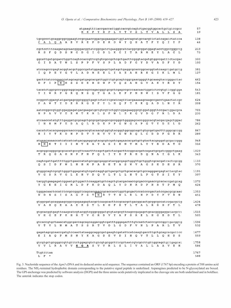
Fig. 3. Nucleotide sequence of the Agm3 cDNA and its deduced amino acid sequence. The sequence contained an ORF (1767 bp) encoding a protein of 588 amino acidresidues. The NH2-terminal hydrophobic domain corresponding to the putative signal peptide is underlined. Asparagines predicted to be N-glycosylated are boxed.The GPI-anchorage was predicted by software analysis (DGPI) and the three amino acids putatively implicated in the cleavage site are both underlined and in boldface.The asterisk indicates the stop codon.
423O. Opota et al. / Comparative Biochemistry and Physiology, Part B 149 (2008) 419–427

Fig. 4. Expression of Agm3 in A. gambiae. Total RNA was extracted fromdevelopmental stages of A. gambiae and analyzed by RT–PCR using sequence-specific primers for Agm3. An internal control corresponding to the actin genewas used for normalization (lower panel). L1, early larval stage; M-L4, midgutof fourth-instar larvae; C-L4, carcasses of fourth-instar larvae; P, pupa; A, adult.Experiments were done with (+) or without (−) reverse-transcriptase.
424 O. Opota et al. / Comparative Biochemistry and Physiology, Part B 149 (2008) 419–427
3. Results
3.1. Identification of putative orthologous Cpm1 genes inmosquitoes
BLAST search with the TBLASTN algorithm (Altschulet al., 1997) revealed the presence of a sequence with highamino acid homology to Cpm1 in the A. gambiae genome. Thepredicted protein (accession number XP_319712.3) is theproduct of the gene ENSANGG00000016933 (Ensembl Ano-pheles genome database). Ensembl also predicted a candidateortholog of this gene in the Bin-refractory Ae. aegypti species.The corresponding protein (AAEL010537-PA) is the product ofthe AAEL010537 gene. Fig. 1 shows a sequence alignment ofCpm1 with the putative orthologous proteins in Anopheles andAedes. The deduced amino acid sequences of each gene aresimilar to each other. Overall, the three sequences share N66%identity. The identity is higher in the region encoded by exons 1and 2, ranging from 62 to 78%, whereas the percentage identityfor the COOH-terminal domain, encoded by exon 3, rangesfrom 42 to 57% (Fig. 2A). The genomic overall organization ofthe Anopheles and Aedes genes is strikingly conserved incomparison with the organization of Culex Cpm1 sequence(Fig. 2B). The three genes are composed of three exons in-terrupted by two introns of variable lengths, which were in-serted in the same positions within the coding region.
3.2. Cloning of the Anopheles Cpm1
We designed specific primers from the Anopheles genomicsequence identified in silico to amplify the target sequencefrom midgut of fourth instar larvae. A 1767-bp cDNA fragmentwas obtained and cloned. The inferred amino acid sequencematchs 100% the predicted sequence from the gene productXP_319712, and corresponds to a 588-amino acid-long poly-peptide with a calculated molecular mass of 67,092 Da and apredicted isoelectric point of 5.4. The protein sequence con-tained a hydrophobic sequence of 24 residues at the NH2-terminus part that could act as a signal peptide (Fig. 3). Thesignature of GPI-anchored protein is also present in the se-quence with a predicted cleavage site at Gly567. Three putativeN-glycosylation sites are detected at positions 132, 291, 436. Incontrast to Cpm1, two cysteines (Cys20 and Cys283) were alsopresent. All structural and enzymatic domains characteristic ofmaltases are present within the predicted protein. In particular,the invariant residues in the active site of α-amylase family ofenzymes or that have structural role are conserved (Janecek,1997). As two other genes that encode maltase-like proteins,Agm1 and Agm2, were previously characterized in A. gambiae(Zheng et al., 1995), the Anopheles Cpm1-like gene was re-ferred as Agm3.
3.3. Temporal and spatial expression profiles
We analysed the expression pattern of Agm3 transcripts byreverse transcriptase-polymerase (RT-PCR) using total RNAextracted from developmental stages of A. gambiae (Fig. 4).
The partial cDNA of actin gene was used as an internal control.Amplified bands at the expected size were primarily detected inthe midgut of fourth instar larvae and at a lower level in the firstinstar extracts and pupa. A weak signal is also detected incarcasses of 4th instars, but may be due to contamination duringdissection.
To determine the localization of the protein in larvae, ratpolyclonal antibodies were raised against Agm3. Since Agm3belongs to a multigene family, a peptide located in a region oflow similarity between all ten maltases that we identified inA. gambiae genome was chosen to generate Agm3-specificantibodies. In immunohistochemistry assays on frozen sectionsof fourth-instar larvae, the polyclonal serum specificallylabelled the brush border membrane of posterior midgut cells(Fig. 5). A weak signal is also observed in cardia cells andgastric caecae (data not shown). A similar pattern ofimmunostaining was obtained by using anti-Cpm1 antibody(Fig. 5, C-D). Immunostaining with non-immune serum as acontrol did not show any positive stain (data not shown).
3.4. Bin toxin interaction with Agm3
To further investigate Bin interaction with Agm3, we usedmammalian MDCK cell lines, which have been previously usedsuccessfully to express Cpm1 (Pauchet et al., 2005). In Westernblot analysis, the Agm3 specific antibody failed to revealany signal in the membrane fractions from MDCK geneticallyengineered for stable expression of Agm3 (data not shown). Inthe other hand, the anti-Cpm1 antibody detected a single 67-kDa protein (Fig. 6A). Homologous competition binding assaysdemonstrated that the 125I-Bin toxin bound specifically to themembrane fractions from transfected cells (Fig. 6B).
As Agm3 exhibits all structural and enzymatic features ofalpha-glucosidases, we tested whether the Bin receptor is in-volved in the degradation of sugars in A. gambiae. In a previousreport, we showed that a secreted form of Cpm1 expressedin the cell culture medium of insect cells, exhibits enzy-matic activity and is able to interact with BinB, the binding

Fig. 5. Immunolocalization of Agm3 protein in larval tissue. Parasagittal sections of fourth-instar larvae were incubated with the anti-Agm3 (A–B) or anti-Cpm1 (C–D)antibodies. A strong signal was observed in the brush border membranes of the posterior stomach cells (arrows in B and D), while Agm3 is not detected in the anteriorstomach cells (A and C). L, lumen. (Bar, 50 µm).
425O. Opota et al. / Comparative Biochemistry and Physiology, Part B 149 (2008) 419–427
component of Bin (Darboux et al., 2007). Thus we constructed aplasmid encoding Agm3 lacking the GPI-anchoring site.Western blotting confirmed that Agm3 was translated andefficiently processed by the cellular export apparatus, resultingin its secretion into the cell culture medium (data not shown).GST pull-down assays performed using the medium containingAgm3 further demonstrated the capacity of the recombinantprotein to bind BinB (data not shown). However, we didnot detect significant alpha-glucosidase activity in our assays.
Fig. 6. Interaction of Agm3 with the Bin toxin. (A) Immunoblot analysis of Agm3 inuntransfected cells (Lane 2). Proteins were subjected to SDS/PAGE, transferred to nexperiments between membrane-enriched fractions from MDCK stably transfected wdeviation) of two independent experiments performed in duplicate.
Experiments with the secreted form of Cpm1 were used as apositive control.
4. Discussion
In this study, we attempted to identify the receptor for the Bintoxin from B. sphaericus in A. gambiae. We hypothesized thatthis receptor is orthologous to Cpm1, the Bin receptor inC. pipiens. A BLAST search with Cpm1 as a query revealed an
membrane-enriched fractions from Agm3-expressing MDCK cells (Lane 1) oritrocellulose and immunoblotted with anti-Cpm1. (B) Homologous competitionith Agm3 and the 125I-Bin toxin. Data points represent the mean (+/−standard

426 O. Opota et al. / Comparative Biochemistry and Physiology, Part B 149 (2008) 419–427
Anopheles sequence designated Agm3 that was 66.4% identicalto Cpm1. The high degree of conservation of the overallgenomic organization and the high amino acid identity bothsuggest that Agm3 is probably orthologous to Cpm1.
Our spatio-temporal distribution studies show that the mid-gut of fourth instar larvae is the major site of expression forAgm3 mRNA. In our assays, transcripts were nearly undetect-able in the adult sample containing male and non-bloodfedfemale mosquitoes. However, microarray analyses available onA. gambiae Gene Expression Profile database at UC Irvine(www.angaged.bio.uci.edu) indicate that Agm3 is weakly ex-pressed in non-bloodfed female, and displays a 5-fold increasein the midgut tissue three hours after the blood meal. Agm3immunolocalization in larvae was examined using a polyclonalantiserum raised against a 16-residue domain. This peptide isunique to Agm3 and not found in the primary amino acidsequence of the nine others maltase sequences identified in thewhole genome. Immunohistochemical staining of cryosectionsof Anopheles larvae showed that Agm3 was primarily detectedin the brush border membranes of posterior midgut cells. Thispattern is thus very similar to that of Cpm1 in Culex (Darbouxet al., 2002), and correlated well with the binding pattern of Binfluorescent derivatives in A. gambiae larvae (Davidson, 1989).Indeed, we demonstrated that the recombinant Agm3 proteinfunctions as a receptor for Bin in vitro.
Several observations suggest that the mode of action of Binmight be different in Culex and Anopheles. The ultrastructuralchanges observed in larvae after ingestion of Bs spores differbetween these species (Davidson, 1989). The localization of thefluorescent-labelled toxin is also less sharply delimited in Ano-pheles than in Culex and the Bin internalization in vesicles afterbinding to midgut cells is not observed in Anopheles. Moreover,in C. pipiens, BinB is the primary binding component, whereasin A. gambiae, both BinA and BinB seem to play an equivalentrole in the Bin binding (Charles et al., 1997). Altogether, theseobservations suggest that Bin uses the one-to-one orthologousprotein as a receptor, but its mode of action may be different indifferent mosquito species. The identification of Agm3 and itsexpression in a heterologous expression system will be a usefultool for further studies on the characterization of the mode ofaction of Bin in this mosquito species.
Alpha-glucosidase digestive enzymes are members of thealpha-amylase family that differ from each other with respect tosubstrate specificities, tissue distribution and mode of mem-brane anchorage. These enzymes play a major role in a numberof insect species. Primary sequence analysis of Agm3 suggeststhat it has an alpha-glucosidase activity. However, we couldnot detect any significant enzymatic activity for Agm3 in ourassays, using either membrane fractions from MDCK orconditioned cell culture medium from Sf9 cells. This apparentlack of enzymatic activity is not due to insufficient levels ofAgm3 being expressed in the cell culture system, as demon-strated by the intensity of the signal revealed by Western blotanalysis (data not shown). It is possible that using cell lines forthe production of Agm3 might have resulted in an inabilityto express the product in a correct form. This hypothesis issupported by the fact that anti-Agm3 antibodies recognized both
the native and the SDS-denatured protein (not shown) present inthe larval tissues, but did not react with the recombinant proteinproduced in MDCK cells. It is noteworthy that this product isrecognized by anti-Cpm1 antibodies, which are directed againsta region different to that of anti-Agm3 antibodies (Darbouxet al., 2002). The incorrect expression of the recombinant pro-tein, if any, did not affect the interactions between the toxin andits receptor, but might have influenced its enzymatic properties.
Another possibility is that enzymatic assays were unsuccessfulbecause conditions for enzymatic reactions were inappropriate orcorrect substrates were not available, although alpha-glucosidasesgenerally have a weak specificity. Further studies are required todetermine the physiological function of Agm3, but the highexpression levels of the protein in the principal site of nutrientdigestion and absorption, and its up-regulation after a blood meal,suggest that Agm3 acts as a digestive enzyme in A. gambiae.
A putative ortholog of Agm3was also detected in the genomeof the Bin-refractory mosquito A. aegypti. The predicted aminoacid sequence shows a high homology to Agm3 and Cpm1,particularly in the NH2-terminus, believed to contain the Binbinding site (Romao et al., 2006). However, specific binding isnot detected for Aedes BBMFs. As the ingestion, solubilizationand activation of Bin are effective in Ae. aegypti and otherrefractory insect species, it has been suggested that Bin bindingis the critical step leading to the narrow host range of B.sphaericus. These findings suggest that, in mosquitoes, the Binreceptor is a Cpm1-like protein, which amino acid sequencevaries slightly from one species to another, leading to differentBin affinity and consequently different susceptibility.
The recent availability of the complete genome of themalaria mosquito A. gambiae (Holt et al., 2002) has enabled theidentification of several maltase-like proteins, including thepreviously described Agm1 and Agm2 maltase-like polypep-tides (Zheng et al., 1995). Both proteins are specificallyexpressed in the adult midgut and are not detected in larvalstages. Furthermore, preliminary data from our group indicatethat two other maltase-like proteins identified in the wholegenome are co-expressed with Agm3 in the target tissue of Bin.Both sequences show a relatively high identity to Agm3,particularly in the polypeptide supposed to contain the bindingsite of Bin (54 to 56% identity) and, in contrast to all othermaltase-like proteins detected in the Anopheles genome, containsignatures for the GPI-anchorage to the epithelial cell mem-brane. A comparison between Agm3 and other Cpm1-likesequences from susceptible and refractory mosquitoes couldreveal amino acids residues important for the binding of thetoxin. Identification of such structural determinants could behighly informative to better understand the Bin-receptor in-teraction and should provide a basis for a rational design ofmore potent and wider-spectrum Bin toxins, particularly againstthe dengue vector, Ae. aegypti.
Acknowledgements
This work was supported by grants from the MENRT(Impact des OGM) and the INRA/CNRS program (Impact desbiotechnologies dans les agroécosystèmes).

427O. Opota et al. / Comparative Biochemistry and Physiology, Part B 149 (2008) 419–427
References
Altschul, S.F., Madden, T.L., Schaffer, A.A., Zhang, J., Zhang, Z., Miller, W.,Lipman, D.J., 1997. Gapped BLAST and PSI-BLAST: a new generation ofprotein database search programs. Nucleic Acids Res. 25, 3389–3402.
Aly, C., Mulla, M.S., Federici, B.A., 1989. Ingestion, dissolution, andproteolysis of the Bacillus sphaericus toxin by mosquito larvae. J. Invertebr.Pathol. 53, 12–20.
Becker, N., 2000. In: Charles, J.-F., Delécluse, A., Nielsen-LeRoux, C. (Eds.),Bacterial control of vector-mosquitoes and black flies. In Entomopathogenicbacteria: from laboratory to field application. Kluwer Academic Publishers,Dordrecht/Boston/London, pp. 383–398.
Bendtsen, J.D., Nielsen, H., von Heijne, G., Brunak, S., 2004. Improvedprediction of signal peptides: SignalP 3.0. J. Mol. Biol. 340, 783–795.
Berry, C., Hindley, J., Ehrhardt, A.F., Grounds, T., de Souza, I., Davidson, E.W.,1993. Genetic determinants of host ranges of Bacillus sphaericus mosquitolarvicidal toxins. J. Bacteriol. 175, 510–518.
Broadwell, A.H., Baumann, P., 1987. Proteolysis in the gut of mosquito larvaeresults in further activation of the Bacillus sphaericus toxin. Appl. Environ.Microbiol. 53, 1333–1337.
Charles, J.F., Silva-Filha, M.H., Nielsen-Le Roux, C., Humphreys, M.J., Berry,C., 1997. Binding of the 51- and 42-kDa individual components from theBacillus sphaericus crystal toxin to mosquito larval midgut membranesfrom Culex and Anopheles sp. (Diptera: Culicidae). FEMS Microbiol. Lett.156, 153–159.
Charles, J.F., Darboux, I., Pauron, D., Nielsen-LeRoux, C., 2005. MosquitocidalBacillus sphaericus: toxins, genetics, mode of action, use, and resistancemechanisms. In: Gilbert, L.I., Iatrou, K., Gill, S.S. (Eds.), ComprehensiveMolecular Insect Science. Elsevier Pergamon, pp. 207–231.
Darboux, I., Nielsen-LeRoux, C., Charles, J.F., Pauron, D., 2001. The receptorof Bacillus sphaericus binary toxin in Culex pipiens (Diptera: Culicidae)midgut: molecular cloning and expression. Insect Biochem. Mol. Biol. 31,981–990.
Darboux, I., Pauchet, Y., Castella, C., Silva-Filha, M.H., Nielsen-LeRoux, C.,Charles, J.F., Pauron, D., 2002. Loss of the membrane anchor of the targetreceptor is a mechanism of bioinsecticide resistance. Proc. Natl. Acad. Sci.U. S. A. 99, 5830–5835.
Darboux, I., Charles, J.F., Pauchet, Y., Warot, S., Pauron, D., 2007. Transposon-mediated resistance to Bacillus sphaericus in a field-evolved population ofCulex pipiens (Diptera: Culicidae). Cell. Microbiol. 9, 2022–2029.
Davidson, E.W., 1989. Variation in binding of Bacillus sphaericus toxin andwheat germ agglutinin to larval midgut cells of six species of mosquitoes.J. Invertebr. Pathol. 53, 251–259.
Eisenhaber, B., Bork, P., Eisenhaber, F., 1998. Sequence properties of GPI-anchored proteins near the omega-site: constraints for the polypeptidebinding site of the putative transamidase. Protein Eng. 11, 1155–1161.
Fillinger, U., Lindsay, S.W., 2006. Suppression of exposure to malaria vectorsby an order of magnitude using microbial larvicides in rural Kenya. Trop.Med. Int. Health 11, 1629–1642.
Gubler, D.J., 1998. Resurgent vector-borne diseases as a global health problem.Emerg. Infect. Dis. 4, 442–450.
Holt, R.A., Subramanian, G.M., Halpern, A., Sutton, G.G., Charlab, R.,Nusskern, D.R., Wincker, P., Clark, A.G., Ribeiro, J.M., Wides, R., et al.,2002. The genome sequence of the malaria mosquito Anopheles gambiae.Science 298, 129–149.
James, A.A., 2002. Engineering mosquito resistance to malaria parasites: theavian malaria model. Insect Biochem. Mol. Biol. 32, 1317–1323.
Janecek, S., 1997. Alpha-amylase family: molecular biology and evolution.Prog. Biophys. Mol. Biol. 67, 67–97.
Mulla, M.S., 1994. Mosquito control then, now, and in the future. J. Am. Mosq.Control Assoc. 10, 574–584.
Mulla, M.S., Thavara, U., Tawatsin, A., Kong-ngamsuk, W., Chompoosri, J.,Su, T., 2001. Mosquito larval control with Bacillus sphaericus: reduction inadult populations in low-income communities in Nonthaburi Province,Thailand. J. Vector Ecol. 26, 221–231.
Nauen, R., 2007. Insecticide resistance in disease vectors of public healthimportance. Pest Manag. Sci. 63, 628–633.
Nielsen-LeRoux, C., Charles, J.F., 1992. Binding of Bacillus sphaericus binarytoxin to a specific receptor on midgut brush-border membranes frommosquito larvae. Eur. J. Biochem. 210, 585–590.
Pauchet, Y., Luton, F., Castella, C., Charles, J.F., Romey, G., Pauron, D., 2005.Effects of a mosquitocidal toxin on a mammalian epithelial cell lineexpressing its target receptor. Cell. Microbiol. 7, 1335–1344.
Romao, T.P., de Melo Chalegre, K.D., Key, S., Ayres, C.F., Fontes de Oliveira,C.M., de-Melo-Neto, O.P., Silva-Filha, M.H., 2006. A second independentresistance mechanism to Bacillus sphaericus binary toxin targets its alpha-glucosidase receptor in Culex quinquefasciatus. FEBS J. 273, 1556–1568.
Silva-Filha, M.H., Nielsen-LeRoux, C., Charles, J.-F., 1997. Binding kinetics ofBacillus sphaericus binary toxin to midgut brush-border membranes ofAnopheles and Culex sp. mosquito larvae. Eur. J. Biochem. 247, 754–761.
Silva-Filha, M.H., Regis, L., Oliveira, C.M., Furtado, A.E., 2001. Impact of a26-month Bacillus sphaericus trial on the preimaginal density of Culexquinquefasciatus in an urban area of Recife, Brazil. J. Am. Mosq. ControlAssoc. 17, 45–50.
Zaim, M., Guillet, P., 2002. Alternative insecticides: an urgent need. TrendsParasitol. 18, 161–163.
Zheng, L., Whang, L.H., Kumar, V., Kafatos, F.C., 1995. Two genes encodingmidgut-specific maltase-like polypeptides from Anopheles gambiae. Exp.Parasitol. 81, 272–283.


