Embed Size (px)
Citation preview

This article appeared in a journal published by Elsevier. The attachedcopy is furnished to the author for internal non-commercial researchand education use, including for instruction at the authors institution
and sharing with colleagues.
Other uses, including reproduction and distribution, or selling orlicensing copies, or posting to personal, institutional or third party
websites are prohibited.
In most cases authors are permitted to post their version of thearticle (e.g. in Word or Tex form) to their personal website orinstitutional repository. Authors requiring further information
regarding Elsevier’s archiving and manuscript policies areencouraged to visit:
http://www.elsevier.com/copyright

Author's personal copy
Hyaluronic acid based self-assembling nanosystems for CD44 targetmediated siRNA delivery to solid tumors
Shanthi Ganesh a,b,1, Arun K. Iyer a,1, David V. Morrissey b, Mansoor M. Amiji a,*aDepartment of Pharmaceutical Sciences, School of Pharmacy, Bouvé College of Health Sciences, Northeastern University, Boston, MA 02115, USAbNovartis Institutes for Biomedical Research Inc., Cambridge, MA 02139, USA
a r t i c l e i n f o
Article history:Received 3 January 2013Accepted 22 January 2013Available online 11 February 2013
Keywords:Self-assembling nanosystemssiRNA deliveryMultidrug resistanceTumor targetingCD44Hyaluronic acid
a b s t r a c t
Anticancer therapeutics employing RNA interference mechanism holds promising potentials forsequence-specific silencing of target genes. However targeted delivery of siRNAs to tumor tissues andcells and more importantly, their intracellular release at sites of interest still remains a major challengethat needs to be addressed before this technique could become a clinically viable option. In the currentstudy, we have engineered and screened a series of CD44 targeting hyaluronic acid (HA) based self-assembling nanosystems for targeted siRNA delivery. The HA polymer was functionalized with lipidsof varying carbon chain lengths/nitrogen content, as well as polyamines for assessing siRNA encapsu-lation. From the screens, several HA-derivatives were identified that could stably encapsulate/complexsiRNAs and form self-assembled nanosystems, as determined by gel retardation assays and dynamic lightscattering. Many HA derivatives could transfect siRNAs into cancer cells overexpressing CD44 receptors.Interestingly, blocking the CD44 receptors on the cells using free excess soluble HA prior to incubation ofcy3-labeled-siRNA loaded HA nano-assemblies resulted in >90% inhibition of the receptor mediateduptake, confirming target specificity. In addition, SSB/PLK1 siRNA encapsulated in HA-PEI/PEG nano-systems demonstrated dose dependent and target specific gene knockdown in both sensitive andresistant A549 lung cancer cells overexpressing CD44 receptors. More importantly, these siRNA encap-sulated nanosystems demonstrated tumor selective uptake and target specific gene knock down in vivoin solid tumors as well as in metastatic tumors. The HA based nanosystems thus portend to be promisingsiRNA delivery vectors for systemic targeting of CD44 overexpressing cancers including tumor initiating(stem-) cells and metastatic lesions.
� 2013 Elsevier Ltd. All rights reserved.
1. Introduction
Multi drug resistance (MDR) poses as a great challenge for thetreatment of several recurrent cancers including lung cancers in theclinic [1]. MDR develops due to multiple factors that include poorsystemic drug delivery efficiency, inefficient drug residence at thetumor site, poor intracellular availability and micro-environmentalselection pressures that allow certain cells to survive despiteaggressive chemotherapy [2]. RNA interference (RNAi) therapy hasemerged as a powerful strategy for combating drug resistance byselective down-regulation of key genes involved in the develop-ment of MDR phenotype [3]. However, delivery of small interferingRNAs (siRNAs) to specific tumor tissues and cells followed by theirintracellular release from the endosomal/lysosomal compartments
into the cytoplasm still remains a major hurdle that needs to beaddressed before this experimental technique could becomea clinically viable therapeutic option [4,5].
Naked siRNAs are unfavorable for systemic delivery because oftheir inherent limitations such as negative charge, large molecularweight, size, and instability. They are easily degraded by serumendonucleases and are quickly eliminated by renal excretion [6]. Inthis regard, engineered nanocarriers that can stably encapsulate/complex, protect and selectively deliver siRNAs to target tumortissues and cells are highly promising as next generation gene de-livery vehicles [7]. The ideal system should preferably be non-toxic,biodegradable, non-immunogenic and able to efficiently deliversiRNAs to target tumors. More importantly, after tumor tissuespecific delivery, the nanosystem should facilitate intracellularuptake and promote endosome release/escape of the payload siR-NAs into cytoplasm allowing their interactionwith the endogenousRISC [8]. Although viral vectors can serve as efficient gene deliveryvehicles, the apparent toxicity and safety concerns limit their utilityfor human applications, necessitating the development of safer
* Corresponding author.E-mail address: [email protected] (M.M. Amiji).
1 These authors contributed equally to this work.
Contents lists available at SciVerse ScienceDirect
Biomaterials
journal homepage: www.elsevier .com/locate/biomater ia ls
0142-9612/$ e see front matter � 2013 Elsevier Ltd. All rights reserved.http://dx.doi.org/10.1016/j.biomaterials.2013.01.077
Biomaterials 34 (2013) 3489e3502

Author's personal copy
delivery technologies. In this regard, non-viral/polymeric nano-particle based delivery platforms are becoming increasingly viablealternatives for siRNA delivery [9].
For the current study, we selected hyaluronic acid (HA) polymerwith a relatively low molecular weight of 20 kDa as our startingbuilding block. HA is a naturally occurring mucopolysaccharidecomposed of alternating disaccharide units of D-glucuronic acid andN-acetyl D glucosamine (NAG) with b (1e4) interglycosidic linkage[10]. HA is a highly anionic biopolymer present in the extracellularmatrix and synovial fluids. It is biodegradable, non-toxic, non-immunogenic and non-inflammatory, which makes it an idealcarrier polymer for systemic drug delivery applications [11]. Also,a relatively simple coupling chemistry allows for modification ofthe sugar residues on the polymer to arrive at variety of functionalmacrostructures that facilitates self-assembly and encapsulation ofdiverse drug and gene payloads for targeted delivery [12]. Of pri-mary importance to tumor selective delivery, the HA backbone initself is endowed with tumor targeting moieties that specificallyrecognizes CD44 e an integral membrane glycoprotein overex-pressed on several tumors cell surfaces, including tumor initiatingstem cells [13e16]. Interestingly, it has already been reported thatthe CD44 receptors are highly expressed in majority of non-smallcell lung cancers (NSCLC) and is a known biomarker for NSCLCtumors [17], however their expression levels are diminished in H69and H69AR sensitive and resistant small cell lung cancers respec-tively [17].
Although HA is an attractive carrier polymer, its anionic naturecan pose difficulty in encapsulating negatively charged siRNAmolecules [18]. However, we propose to overcome this limitationby hydrophobic modification of HA backbone, with fatty aminesand cationic polyamines that would not only help reduce the netnegative charge density on the backbone polymer but also facilitatesiRNA encapsulation via self-assembly [11], and intracellular de-livery by endocytosis [19]. High counter-ion density has also beenreported to promote efficient endosome release/escape after cel-lular entry [20,21].
In the current study, we have developed a series of CD44 tar-geting hyaluronic acid (HA) based versatile self-assembling nano-systems for targeted delivery of siRNA with the aim to treat CD44overexpressing cancer cells. Furthermore, we evaluated the sys-temic delivery efficiency and gene silencing activity of the siRNAloaded in select HA nanosystems using drug resistant and sensitivetumors models overexpressing CD44 receptors including meta-static cancer models.
2. Materials and methods
2.1. Materials
Sodium hyaluronate (HA) with an average molecular weight of 20 kDa wasobtained from Lifecore Biomedical Co. (Chaska, MN). Polyethyleneimine (bPEI,MWw10,000 Da) was obtained from Polysciences Inc. (Warrington, PA). Sulfo-NHSwas purchased from Thermo Scientific Corp (Billerica, MA), mono-functional poly-ethyleneglycol amine (PEG2k-NH2, MW ¼ 2000 Da) was purchased from CreativePEG Works (Winston Salem, NC). All the fatty amines and other reagents for syn-thesis were obtained at high purity (>99%) from SigmaeAldrich Chemical Co(Milwaukee,WI) or Acros Organics (Thermo Fisher, Pittsburgh, PA) and usedwithoutfurther purification.
2.2. Cell lines
The human cell lines used in this study included non-small cell lung cancer(NSCLC) A549 and A549DDP, small cell lung cancer (SCLC) H69 and H69AR, breastcancer MDA-MB468, liver cancer Hep3B, murine melanoma B16F10. Except forA549DDP, all the other cell lines were obtained from ATCC. The cisplatin resistantA549DDP cells were obtained from Massachusetts General Hospital, Boston, MA.Except Hep3B and MDA-MB468 cells, which were cultured using Dulbecco’sModified Eagle Medium (DMEM) supplemented with 10% FBS, all the other cell lineswere cultured using RPMI-1640 medium supplemented with 10% FBS. All the cells
were grown as adherent cultures except H69whichwas grown as suspension cells at37 �C and 5% CO2.
2.3. Synthesis of functional hyaluronic acid based polymers
2.3.1. Modification of hyaluronic acid with mono-functional fatty aminesMono-functional fatty amines of the general formula CH3(CH2)nNH2 (where
n ¼ 3,4,5,7,9,11,13 and 17) such as n-butyl amine, n-pentylamine, n-hexylamine, n-octylamine, n-decylamine, n-dodecylamine, tetradecylamine and stearylaminewerechemically conjugated to the backbone of hyaluronic acid by coupling the end groupprimary amine of the fatty amine with the carboxylic acid or hydroxyl groupof hyaluronic acid in the presence of a coupling agent, 1-Ethyl-3-[3-dimethylaminopropyl]carbodiimide hydrochloride (EDC) and n-hydrox-ysuccinimide (NHS). In brief, multiple glass scintillation vials were used to dissolvesodium hyaluronate (MW 20 kDa, 100 mg, 5 mmole) in 5 ml of dry formamide bywarming up the reaction mixture up to 50 �C under stirring. After obtaining a clearsolution the reaction mixture was allowed to cool to room temperature and thenvarying feed ratio of fatty amines (w2e25 mg,w40e100 mmole) was added to eachvial containing the hyaluronic acid solution under magnetic stirring. Subsequently,EDC (10e20mg, 50e100 mmole) and NHS (5e15mg, 50e150 mmole) mixture in DMF(2ml)was added dropwise into each of the reaction vial undermagnetic stirring. Thereaction vials were allowed to stir for 12 h using multi-position magnetic stirrer(IKAMAG, IKA Labs, Germany). The resulting solution was added to a large excess ofethanol (EtOH, 250 ml) to precipitate the derivatized polymer and dissolve theunreacted fatty amines. The polymer precipitate was centrifuged and the washingswere discarded. The above step of EtOH precipitation and washing was repeatedthrice to purify the polymer. Finally the polymerwas re-dissolved in deionizedwaterand subjected to ultrafiltration using Millipore Labscale Tangential Flow Filtration(TFF) systemwith Biomax 10 kDa MWCOmembrane (EMD-Millipore, Billerica, MA).The concentrated and purified polymer was finally lyophilized using a freeze dryer(Freezone-6, Labconco Inc., Kansas, MO) and stored at�20 �C until further use (yield:85e90 mg, w81e85%, off-white fibrous product). A 3 mg portion of the lyophilizedproduct was dissolved in 600 ml of D2O and characterized by 400 MHz 1H NMRspectroscopy (Varian Inc., CA) for determining the % lipid modification.
2.3.2. Modification of hyaluronic acid with bifunctional fatty aminesAn aqueous solution of sodium hyaluronate (HA, MW 20 kDa, 100 mg, 5 mmole)
prepared in deionized water and mixed with bifunctional fatty amines (w2e25 mg,w40e100 mmole) having a general formula of NH2(CH2)nNH2 (where n ¼ 4,5,6...)such as 1,4 diaminobutane, 1,6 diaminohexane or 1,8 diaminoocatne. The pH of thereaction mixture was adjusted to 7.5 with 0.1 M NaOH/0.1 M HCl and the pH adjustedsolution was added slowly into a mixture of EDC (10e50 mge50e250 mmole) andNHS (5e25 mg, 50e250 mmole) in deionized water. After mixing the reactants usinga magnetic stirrer, the pH of the reaction was maintained at 7.5 by the addition of0.1 M NaOH for about 2 h and the reaction was allowed to proceed for 12 h. The HA-lipid modified derivatives were purified by extensively dialysis using cellulosemembrane (MWCOw12e14 kDa) against deionized water for 48 h with frequentreplacement of deionized water. The purified product was then lyophilized andstored at �20 �C until further use (yield: 90 mg,w86.5%, off-white fibrous product).A 3 mg portion of the lyophilized product was dissolved in 600 ml of D2O andcharacterized by 400 MHz 1H-NMR spectroscopy (Varian Inc., CA) for determiningthe % lipid modification.
2.3.3. Chemical modification of hyaluronic acid with polyaminesHA was chemically modified with polyethyleneimine (PEI, 10 kDa) or Poly (L-
Lysine) (10e14 kDa) by using a coupling agent, 1-Ethyl-3-[3-dimethylaminopropyl]carbodiimide hydrochloride (EDC). In brief, sodium hyaluronate (MW 20 kDa,100 mg, 5 mmole) was dissolved in 5 ml of dry formamide in a glass scintillation vialby warming up the reaction vial up to 50 �C. After obtaining a clear solution thereaction mixture was allowed to cool to room temperature and thenw3.3 mg of thePEI or PLL (10 kDae0.33 mmole) was added to the solution. Then EDC (10 mg,50 mmole) was added into the reaction mixture. The solution turned hazy andthen became clear after 1 h. The reaction was stirred overnight or for 12 h usinga magnetic stirrer. The resulting solution was added to a large excess of EtOH(250 ml) to precipitate the polymer. The precipitate was centrifuged and collectedand the washings were discarded. The above step of EtOH precipitation andwashing was repeated thrice to purify the polymer. Finally the precipitatepolymer was re-dissolved in deionized water and subjected to ultrafiltration usingLabscale Millipore Tangential Flow Filtration (TFF) system with Biomax 10 kDaMWCO membrane (EMD-Millipore, Billerica, MA). The concentrated and purifiedpolymer was then lyophilized using a freeze dryer (Freezone-6, Labconco Inc.,Kansas, MO) and stored at�20 �C (yield: 75 mg,w75%, off-white fibrous product). A3 mg portion of the lyophilized product was dissolved in 600 ml of D2O and char-acterized by 400 MHz 1H-NMR spectroscopy (Varian Inc., CA) for determining the %lipid modification.
2.3.4. Synthesis of thiolated and PEGylated hyaluronic acidThe synthesis of thiolated-HA was based on a reported procedure [22]. Briefly,
the HA polymer (5 g, MW 20 kDa, 0.25 mmol) was dissolved in 250 ml of PBS
S. Ganesh et al. / Biomaterials 34 (2013) 3489e35023490

Author's personal copy
solution (pH ¼ 7.0) and stirred well using a magnetic stirrer until the solutionbecame clear. Three molar excess amounts of EDC (1 g, 5 mmol) and N-Hydrox-ybenzotrizole (HoBt) (750 mg, 5 mmol) were added and stirred for 2 h, and thencystamine dihydrochloride (565 mg, 5 mmol) was added drop wise and mixedovernight. The reaction mixture was dialyzed thoroughly using cellulose dialysismembranes (MW cut offw 12e14 kDa) (Spectrapore, Spectrum Labs, CA) for 48 h toremove unreacted reactants. Subsequently, the thiol-cross-linked HA polymer wastreated with five molar excess of dithiothreitol (DTT) and stirred for 24 h to cleavethe disulfide bridges to yield the water soluble conjugate. The product was dialyzedagainst deionized water at pH 3.0 for 96 h (MWCO 12e14 kDa) (Spectrapore,Spectrum Labs, CA). The % thiolation was determined using Ellman reagent asdescribed by Habeeb [23]. The final product was lyophilized and stored at �20 �Cuntil use.
For the preparation of the PEGlyate hyaluronic acid, HA was chemically conju-gated with polyethyleneglycol amine (PEG-NH2) (PEG, 2 kDa or 5 kDa) using 1-Ethyl-3-[3imethylaminopropyl]carbodiimide hydrochloride (EDC) and n-hydroxysulfo succinimide (sulfo-NHS). In brief, EDC (19 mg, 100 mmole) and sulfo-NHS(21 mg, 100 mmole) were mixed in 5 ml deionized water and kept for 1 h to formthe amine reactive intermediate. Sodium hyaluronate (MW 20 kDa, 200 mg,10 mmole) was dissolved in 10 ml of deionized water in a glass scintillation vial. Afterstirring and obtaining a clear solution 50 mg or 125 mg of the PEG (50 mmole, 2 k or5 k PEG for 5% modification) was added to the solution and stirred. Then EDC/NHSsolution prepared earlier was added into the HA/PEG solution dropwise overa period of 1 h. The reaction mixture was stirred for 24 h and then dialyzed usingcellulose dialysis membranes (MW cut off w12e14 kDa) against deionized water/methanol mixture (1v/3ve1v/1v) for 24 h and subsequently with deionized waterfor 48 h. The purified product was then lyophilized and stored at �20 �C (yield:80 mg, w79%, off-white fibrous product). A 3 mg portion of the lyophilized productwas dissolved in 600 ml of D2O and characterized by 400 MHz 1H-NMR spectroscopy(Varian Inc., CA) for determining the % modification.
2.4. Assessing self-assembling capability of HA based functional macrostructuresand nanosystems
In order to assess the self-assembling ability of the HA based macrostructures(such as HA-butylamine, HA-hexylamine, HA-octylamine, HA-stearylamine, HA-choline, HA-spermine, HA-PLL or HA-PEI), the respective derivative was sus-pended in deionized water at varying concentrations (1e10 mg/ml) at room tem-perature. After vortex mixing for 1 min followed by 15 min incubation, their averagehydrodynamic size and size distribution were measured by using a dynamic lightscattering (DLS) instrument (Malvern Zetasizer Nano-S, Malvern Inc., UK). Further,in order to determine the zeta potentials or the surface charge of the prepared HAderivatives they were transferred to specially designed electrophoretic cells and thezeta potentials were recorded using the same instrument.
2.5. Formation of siRNA loaded HA nanosystems, determination of siRNAencapsulation and release by polyanion competition by gel retardation assay
In order to assess the ability of the series of functionalized-HA derivatives(containing varying lipid content, nitrogen content and polyamine content) to formself-assembling nanosystems with siRNAs, the respective derivatives were incu-bated with known concentration of siRNA with varying concentrations of the HA-derivatives in solution. The PLK1 and SSB siRNA sequences that were used in thisstudy are:
PLK1: sense: UUGUCUuCAGGuCUuCAGuuantisense: AGAUcacCCUccuuAAAUauu
SSB: sense: AcAAcAGAcuuuAAuGuAAuuantisense:UUAcAuuAAAGucuGuuGuuu
The solution containing the polymer and siRNA was vortexed and mixed fora minute and incubated at room temperature for 15 min to form self-assembly. Thenano-assemblies were prepared at several polymer-to-siRNA weight ratios, rangingfrom 9:1 to 450:1 to study the effect of polymer concentration on siRNA encapsu-lation. The formation of siRNA loaded self-assembling nanosystems was also studiedusing the dynamic light scattering (DLS) instrument by measuring the size andcharge of the particles formed. Also, transmission electron microscopy (JEOL, JEM-1000, Tokyo, Japan) was performed to assess the formation of siRNA loaded nano-assemblies. A uranyl acetate ribonucleic acid stain was used to demarcate siRNAsfrom the polymer. The dark staining of siRNA by heavy metals such as uranyl acetateprovides a high contrast compared to hyaluronic acid polymer that can helpascertain loading of genes in polymeric nanosystems.
The ability of these complexes to release siRNAwas then determined by treatingthemwith a highly charged polyanionic polymer, polyacrylic acid (PAA) and runningon gel. The anionic polymer (PAA) would compete with the anionic polymer andrelease the siRNA which then appears as a free band in the gel. For this purpose,a 5 ml polymer-siRNA complex was diluted up to 10 ml with deionized water. Then10 ml of 2% PAA was added to this mixture and vortexed. The 20 ml sample was then
run on Agarose gel along with the PAA untreated samples (with same amount oftotal siRNA content) to compare their ability for de-complexation of siRNA bymonitoring the released/free siRNA bands.
2.6. Assessment of siRNA uptake by confocal microscopy and flow cytometry
To assess the ability of the polymeric nanosystems to release the siRNA into cells,a NIR dye cy3-labeled siRNA was formulated using functionally variant HA de-rivatives at optimal polymer:siRNA weight ratios according to the above mentionedprotocol. These particles were then incubated with 2e4 MDA-MB468 cells con-taining 100 nM siRNA at 37 �C. After incubation for 12 h, the cells were washed withPBS and trypsinized to detach the cells. The uptake of cy3-labeled siRNA wasdetermined by using a flow cytometer (BD FACSCalibur, BD Biosciences, San Jose,CA). The data was analyzed with BD Cell Quest software. In parallel, identicalpolymer-siRNA nanosystems were incubated with cancer cells overexpressing CD44receptors grown on the glass cover slips for 12 h to assess the intracellular traffickingof siRNA. At the end of the incubation period, the cells were washed thrice with PBS,fixed in paraformaldehyde in PBS for 10 min. The localization of siRNA within thecells was visualized by a confocal microscope (Carl Zeiss Microscopy GmbH, Jena,Germany). Flow cytometry was also used to determine the CD44 receptor levels inthe cells. For this purpose 200 ml of 1e6 A549, A549DDP, H69, H69AR, Hep3B, MDA-MB468 and B16F10 cells were incubated separatelywith antibody against CD44withPE tag for 1 h and washed thrice with PBS. Cells were then reconstituted in PBS andran through flow cytometer to assess the CD44 receptor levels. The anti-CD44monoclonal antibodies used in this study was obtained from R&D systems.
2.7. Competition assays to assess receptor mediated cellular entry
In order to determine the receptor mediated cellular entry of the engineeredHA-nanosystems, cy3-labeled siRNA encapsulated HA-nanosystems were incubatedin MDA-MB468 cells for 12 h. In parallel a competitive inhibition (or blockingcontrol) studywas also undertaken. For the blocking studies the cell culturemediumwas replaced with serum free media containing excess free soluble HA polymer ata concentration of 10 mg/ml for an hour to block the receptors prior to incubationwith the cy3-labeled-siRNA/HA-nanosystem for 12 h under identical conditions.Both sets of cells were then washed with 1� PBS and visualized under the confocalfluorescence microscope (Zeiss, Germany). In parallel, two other sets of cells weretreated similarly and harvested as described previously for flow cytometric analysisto quantitate the cy3 positive cells. For negative control studies, a cell line that doesnot express CD44 (or expresses very low levels of CD44) was treated with cy3-labeled-siRNA encapsulated HA-nanosystems.
2.8. Transfection of PLK1 targeted siRNA to assess silencing of target gene expression
PLK1 or CTL siRNA was encapsulated in a series of HA-derivatives at mass ratiosranging from 450:1 to 9:1 (polymer: siRNA) as previously described. These siRNAloaded nanosystems were then reverse transfected into MDA-MB468 cells andincubated for 48 h at siRNA concentrations of 300, 200 and 100 nM. After incubation,cells were harvested and the RNAwas extracted. The extracted RNAwas used to runquantitative PCR to assess the mRNA levels. mRNA knockdown was determined bynormalizing the PLK1 expression levels to the endogenous GAPDH levels.
2.9. Assessment of siRNA release using endosome-disrupting agents
To determine if chloroquine, an endosome-disrupting agent could release thesiRNA/nanosystem trapped in the endosome, the MDA-MB468 cells were incubatedwith HA-PEI/PLK1 complex prepared using a mass ratio of 27:1 in the presence orabsence of chloroquine in the medium and incubated for 48 h. After the incubation,the RNAwas extracted from the cells as previously described and the PCR was run todetermine siRNA release by measuring the PLK1 mRNA knockdown.
2.10. Establishment of subcutaneous, metastatic and syngeneic tumor models
Animal procedures were performed according to a protocol approved byNortheastern University, Institutional Animal Care and Use Committee (NU-IACUC).The tumor models for this study were developed in nude mice obtained fromHarlanLaboratories, South Easton, MA. 5e6 week old nude mice were injected subcuta-neously (s.c.) with tumor cells A549 (5 � 106 cells þ matrigel), A549DDP (1 � 107
cells), H69 (1 � 106 cells þ matrigel), H69AR (1 � 106 cells þ matrigel), Hep3B(7 � 106 cells þ matrigel), MDA-MB468 (5 � 106 cells þ matrigel) and B16F10(1 �106 cells) under the right shoulder. Tumor sizes were measured at least once ortwice a week to monitor the tumor growth. In addition, the B16F10 cells wereintravenously (i.v.) injected (5 � 105 cells/mouse) into nude mice to generate anexperimental metastatic lung or syngeneic model with the idea of checking out thedelivery and activity when the tumors are located on lungs as metastatic lesions.Since these cells do not express luciferase, several mice were opened up on day 7and 10 to make sure that they developed tumors in the lung. In a similar trend, theA549-luc cells (5e6) were also i.v. injected to generate lungmetastatic tumormodelsin mice. Tumor growth progression was monitored using an in vivo imaging system
S. Ganesh et al. / Biomaterials 34 (2013) 3489e3502 3491

Author's personal copy
(IVIS, Carestream Molecular Imaging Inc., Rochester, NY) and measuring the luci-ferase activity in mice at different time points.
2.11. Evaluating target gene knockdown in tumors with differential CD44 expressionand vascularity using ‘tool’ siRNA
In order to correlate the CD44 expression levels and the vascularity of the tu-mors, we examined several tumor types in mice as described above. Typically, whenA549 subcutaneous tumors were approximately w150e200 mm3 in size, the ani-mals were randomized into groups such that each group had similar tumor size.These groups of micewith A549 tumors then received either PLK1 or SSB formulatedHA-PEI/siRNA, HA-PEI/HA-PEG/siRNA or HA-PEI/HA-PEG/HA-SH/siRNA at a dose of0.5 mg/kg every day for three consecutive days. Subsequently, 24 h after the lastdose, the tumors were excised, RNAwas extracted and PCRwas run to determine thePLK1 or SSB knockdown. Likewise, similar doses were evaluated in other tumormodels as well (A549DDP (s.c.), H69 (s.c.), H69AR (s.c.), B16F10 (s.c. and met), Hep3B(s.c.), MDA-MB468 (s.c.) and A549 (s.c. and met) as described.
3. Results and discussion
Development of new and versatile non-toxic and biodegradablepolymers that are safe in systemic circulation, efficient in encap-sulation of therapeutic genes, and can selectively deliver the pay-load to the sites of interest (such as tumors tissues and cells), isurgently needed for successful translation of RNAi based mecha-nisms from bench to the bedside [24]. In the current study, we havesynthesized a series of functionally variant library of self-assembling CD44 targeting HA based macrostructures by varyingthe carbon chain length, nitrogen content and polyamine side chaingrafting onto the HA backbone. The modification of anionic HApolymer resulted in effective lowering/shielding of its negativecharge density that could enable siRNA encapsulation. The
hydrophobic modification of HA polymer could be efficiently ach-ieved by chemical conjugation/grafting of fatty amines with varyingalkyl chains lengths onto the hydrophilic HA backbone by usingEDC/NHS coupling chemistry (Fig. 1). It was observed that byvarying the molar feed ratio of the fatty amines and the polyaminecontent, the degree of HA modification could be controlled in therange of 5e20% as determined by 1H-NMR spectroscopy (Fig. S1).As described, several different approaches were undertaken toarrive at HA based functional macrostructures that varied in ni-trogen content, carbon chain lengths (or lipid content), polyaminecontent, and other variables to understand the structureeactivityrelationship. Some of the different classes of functional HA blocksthat were synthesized included HA derivatized with mono-functional fatty amines (namely, n-butylamine, C4; n-hexylamine,C6; n-octylamine, C8; dodecylamine, C12; and stearylamine, C18);bifunctional fatty amines (namely, 1e6 diaminohexane, C6N2; 1e8diaminohexane, C8N2); and multiple nitrogen containing de-rivatives and polyamines such as pipirazine, spermine, choline,poly (L-lysine) and polyethyleneimine (PEI).
In order to assess the ability of the modified HAmacrostructuresto form self-assembly, the free polymers (without the payloadsiRNA) were suspended in water at concentrations ranging from 1to 10mg/ml. It was observed that the critical micellar concentration(as determine by DLS) varied for different classes of HA-lipids andpolyamine containing HA derivatives. Notably, the HA conjugatedto fatty amines with one or two amine residues (such as HA-hexylamine, HA-octylamine, HA-dodecylamine, HA-stearlyamine,HA-diaminohexane and HA-diaminooctane), with a lipid mod-ification of w8e10% required at least a concentration of w5 mg/ml
Fig. 1. Synthesis of HA based functional macrostructures. A series of functional HA based derivatives were synthesized using a simple and versatile EDC/NHS conjugation chemistryas shown.1, Hyaluronic acid (HA) of 20 kDa molecular weight; 2, HA conjugated to monofunctional fatty amines with the general formula CH3(CH2)nNH2 (where n ¼ 3,4,5.); 3, HAconjugated to PEG of 2000 Da molecular weight; 4, HA conjugated to bifunctional fatty amines with the general formula NH2(CH2)nNH2 (where n ¼ 4,5.); 5, Thiolated-HA (HA-SH)formed by conjugating HA with cysteamine dihydrochloride followed by reduction; and 6, HA conjugated to polyamines such as polyethyleneimine (PEI) of 10 kDa molecularweight.
S. Ganesh et al. / Biomaterials 34 (2013) 3489e35023492
Author's personal copy
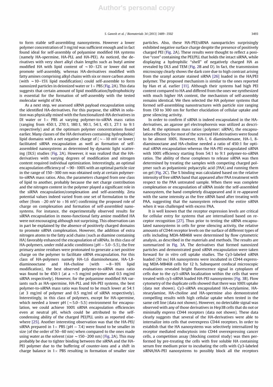
to form stable self-assembling nanosystems. However a lowerpolymer concentration of 3mg/mlwas sufficient enough and in factfound ideal for self-assembly of polyamine modified HA systems(namely HA-spermine, HA-PLL and HA-PEI). As noticed, the de-rivatives with very short alkyl chain lengths such as butyl aminemodified HA with lipid content of w10e12% or lower did notpromote self-assembly, whereas HA-derivatives modified withfatty amines comprising alkyl chains with six or more carbon atoms(with w10e15% lipid modification) could self-assemble to formnanosized particles in deionizedwater or 1� PBS (Fig. 2A). This datasuggests that certain amount of lipid modification/hydrophobicityis essential for the formation of self-assembly with the testedmolecular weight of HA.
As a next step, we assessed siRNA payload encapsulation usingthe identified HA-derivatives. For this purpose, the siRNA in solu-tionwas physically mixedwith the functionalized-HA derivatives inDI water or 1� PBS at varying polymer-to-siRNA mass ratios(ranging from 450:1, 270:1, 180:1, 90:1, 54:1, 45:1, 27:1 to 9:1respectively) and at the optimum polymer concentrations foundearlier. Many classes of the HA derivatives containing hydrophobic/lipid domains with a net negative charge of (w�10 mV or lower)facilitated siRNA encapsulation as well as formation of self-assembled nanosystems as determined by dynamic light scatter-ing (DLS) studies (Fig. 2A). However, it was realized that the HA-derivatives with varying degrees of modification and nitrogencontent required individual optimization. Interestingly, an optimalencapsulation efficiency ofw90e100%with an optimal particle sizein the range of 150e300 nmwas obtained only at certain polymer-to-siRNA mass ratios. Also, the parameters changed from one classof lipid to another, probably because the net charge contributionand the nitrogen content in the polymer played a significant role inthe siRNA encapsulation/complexation and self-assembly. Zetapotential values indeed varied from one set of formulation to theother (from �20 mV to þ16 mV) confirming the proposed role ofcharge on complexation and formation of self-assembled nano-systems. For instance, the experimentally observed results forsiRNA encapsulation in mono-functional fatty amine modified HAwere not encouraging although not surprising. This observation canin part be explained by the absence of positively charged domainsto promote siRNA complexation. However, the addition of extranitrogen atoms to the fatty acid chains (such as diamine containingHA) favorably enhanced the encapsulation of siRNAs. In this class ofHA-polymers, under mild acidic conditions (pH¼ 5.0e5.5), the freeprimary amine residues were protonated to generate a net positivecharge on the polymer to facilitate siRNA encapsulation. For thisclass of HA-polymers namely HA-1,6 diaminohexane, HA-1,8-diaminooctane and HA-choline (with w8e10% lipidmodification), the best observed polymer-to-siRNA mass ratiowas found to be 450:1 (at a w5 mg/ml polymer and 0.5 mg/mlsiRNA respectively). However, for the polyamine modified HA var-iants such as HA-spermine, HA-PLL and HA-PEI systems, the bestpolymer-to-siRNA mass ratio was found to be much lower at 54:1(at 3 mg/ml of polymer and 0.5 mg/ml of siRNA respectively).Interestingly, in this class of polymers, except for HA-spermine,which needed a lower pH (w5.0e5.5) environment for encapsu-lation, we could achieve 100% siRNA encapsulation efficiencieseven at neutral pH, which could be attributed to the self-condensing ability of the charged PEI/PLL units as reported else-where [25]. Another interesting observation was that the HA-PEI/siRNA prepared in 1� PBS (pH ¼ 7.4) were found to be smaller insize (of the order of 50e60 nm) when compared to the ones madeusing water as the solvent (size w150e200 nm) (Fig. 2A). This mayprobably be due to tighter binding between the siRNA and the HA-PEI polymer due to the buffering of counter-ions and a shift incharge balance in 1� PBS resulting in formation of smaller size
particles. Also, these HA-PEI/siRNA nanoparticles surprisinglyexhibited negative surface charge despite the presence of positivelycharged PEI (Fig. 2A). These results were thought to reflect a posi-tive “core” containing the PEI/PLL that holds the intact siRNA, whiledisplaying a hydrophilic “shell” of negatively charged HA asrevealed by DLS and TEM (Fig. 2B and D). In fact, the transmissionmicroscopy clearly shows the dark core due to high contrast arisingfrom the uranyl acetate stained siRNA [26] loaded in the HA/PEIpolymer. The proposed mechanism is similar to the ones reportedby Han et al. earlier [11]. Although their systems had high PEIcontent compared to HA and differed from the ones we synthesizedwith much higher HA content, the mechanism of self-assemblyremains identical. We then selected the HA polymer systems thatformed self-assembling nanostructures with particle size rangingfrom 150 to 300 nm for further elucidation of cellular uptake andgene silencing activity.
In order to confirm if siRNA is indeed encapsulated in the HA-derivatives, an agarose gel electrophoresis was utilized as descri-bed. At the optimum mass ratios (polymer: siRNA), the encapsu-lation efficiency formost of the screened HAderivatives were foundto be almost 100%. However, HA-1, 6 diaminohexane, HA-1, 8-diaminooctane and HA-choline needed a ratio of 450:1 for opti-mal siRNA encapsulation whereas the HA-PEI encapsulated siRNAwith 100% efficiency starting from 54:1 to 9:1 polymer-to-siRNAratios. The ability of these complexes to release siRNA was thendetermined by treating the samples with competing charged pol-ymer such as polyanionic polyacrylic acid (PAA) and running themon gel (Fig. 2C). The % binding was calculated based on the relativeintensity of free siRNA band that appeared after PAA treatmentwithrespect to the PAA untreated sample. When there was completecomplexation or encapsulation of siRNA inside the self-assemblednanosystem, the band completely disappeared and it re-appearedwith the same intensity as the free siRNA band after treating withPAA, suggesting that the nanosystem released the entire siRNAwhen it was challenged with excess PAA.
It is well known that the receptor expression levels are criticalfor cellular entry for systems that are internalized based on re-ceptor recognition [27]. Thus prior to testing the siRNA encapsu-lated nanosystems in cells for gene silencing activity, the relativeamounts of CD44 receptor levels on the surface of different types ofcells including MDA-MB468 were determined by flow cytometricanalysis, as described in the materials and methods. The results aresummarized in Fig. 3A. The derivatives that formed nanosizedparticles and demonstrated good siRNA encapsulation were takenforward for in vitro cell uptake studies. The Cy3-labeled siRNAloaded (50 nM) HA nanosystems were incubated in CD44 express-ing MDA-MB468 cells for 12 h. Subsequent confocal microscopicevaluations revealed bright fluorescence signal in cytoplasm ofcells due to the cy3-siRNA localization within the cells that weretreated with cy3-siRNA loaded HA-PEI nanosystems (Fig. 3B). Flowcytometry of the duplicate cells showed that therewas 100% uptake(data not shown). Cy3-siRNA encapsulated HA-octylamine, HA-stearylamine, HA-choline and HA-spermine also demonstratedcompelling results with high cellular uptake when tested in thesame cell line (data not shown). However, no detectable signal wasobserved with any of those derivatives in Hep3B cells that do not orminimally express CD44 receptors (data not shown). These dataclearly suggests that several of the HA-derivatives were able tointernalize into cells that overexpress CD44 receptors. In order toestablish that the HA nanosystems was selectively internalized byreceptor mediated endocytosis into CD44 overexpressing cancercells, a competition assay (blocking control study) was then per-formed by pre-treating the cells with free soluble HA containingserum free medium prior to incubating the cells with Cy3-labeledsiRNA/HA-PEI nanosystems to possibly block all the receptors
S. Ganesh et al. / Biomaterials 34 (2013) 3489e3502 3493

Author's personal copy
Functionalized HA Derivative Self
assembly
siRNA
encapsulation
Activity in
cancer cells
Size (nm)
+siRNA
Charge (mV)
+ siRNA
HA- butylamine in water (C4) - - - - -
HA- hexylamine in water (C6) + - - 1000±1.0 -20±0.8
HA-octylamine in water (C8) + - - 200±0.3 -20±0.4
HA-stearylamine in water (C18
) + - - 190±0.3 -15±0.6
HA-1,6 diaminohexane in water + + - 320±0.5 -8±0.9
HA-1,8 diaminooctane in water + + - 225±0.2 -10±0.1
HA-choline in water + + - 175±0.4 0±0.1
HA-spermine in water + + + 190±0.3 +16.5±0.9
HA-PEI in water + + + 180±0.1 -6.5±0.4
HA-PEI in PBS + + + 50±0.9 -6.5±0.5
HA-PEI/HA-PEG in PBS + + + 85±0.9 -5.5±0.3
HA-PEI/HA-PEG/ HA-SH in PBS + + + 90±1.2 -8.5±0.8
A
B
C
...............................................
..........
..........
..........
..........
..........
..........
...
100nm
HA-PEI/siRNA +PAA +PAA +PAA
54:1 1:91:72
Fig. 2. Characteristics of HA derivative/siRNA particles. Illustrative examples from each class of lipid chain that was used for derivatization (A). The HA-PEI/siRNA particles made at54:1 ratio in PBS were visualized by TEM (B). Electrophoretic retardation analysis of siRNA binding by HA-PEI derivatives at different mass ratios (54:1, 27:1 and 9:1). The release ofintact siRNA by polyacrylic acid was shown in each case (C). Schematic representation of HA-PEI/siRNA nanosystem suggesting a positive core containing the siRNA with a neg-atively charged HA shell structure (D).
S. Ganesh et al. / Biomaterials 34 (2013) 3489e35023494

Author's personal copy
expressed on the cell surface. Indeed, a highly diminished cellularlocalization/uptake (>90% inhibition) of the cy3-labeled siRNAwasobserved in the cells that were pre-treated with excess HA, sug-gesting that these particles are predominantly trafficked into thecells via receptor mediated endocytosis pathway (Fig. 3C). Flow
cytometry also suggests that the uptake was reduced by >85%when the cells were pre-treated with excess HA corroborating theconfocal microscopy results.
After confirming the cellular uptake and pathway of internal-ization, the ability of these nanosystems to deliver a functional
Cell line Indication CD44 expression levels
H69 SCLC ~60%
H69AR SCLC ~90%
A549 NSCLC >99%
A549DDP NSCLC >99%
MDA-MB468
Breast >99%
Hep3B Liver ~4%
B16F10 Murine melanoma
~65%
HA-PEI/cy3 siRNA HA-PEI/cy3 siRNA + free HA
HA-PEI/cy3 siRNAA B
C
Fig. 3. CD44 receptor levels in different type of cells and the corresponding cell uptake. NSCLC (non-small cell lung cancer), SCLC (small cell lung cancer), breast, liver and murinemelanoma cells were treated with monoclonal antibody against CD44 receptors to determine the receptor levels on the surface of the cells by flow cytometry (A). To demonstratethat the particles enter the cells via the receptors, MDA-MB468 cells were treated with HA-PEI/siRNA nanosystems at 50 nM for 12 h and observed under confocal microscope tocapture images. The internalized cy3-labeled-siRNA appears as red (B). For competitive inhibition study, the cells were incubated with HA-PEI/Cy3-loaded-siRNA in the presenceand absence of excess free soluble HA (C) and observed under confocal microscope as shown.
Fig. 2. (continued).
S. Ganesh et al. / Biomaterials 34 (2013) 3489e3502 3495

Author's personal copy
siRNAwas evaluated using PLK1 targeted siRNA to silence the PLK1gene expression in cells overexpressing CD44 receptors. For thispurpose, breast cancer cells, MDA-MB468 (with >99% CD44expression levels) were transfected with varying HA derivative/siRNA at concentrations ranging from 50 to 300 nM. Although, all
the fatty amine modified HAs derivatives demonstrated good cel-lular uptake, most of them were unsuccessful in down regulatingPLK1 gene expression in vitro. The spermine derivatized HA dem-onstrated about 40% target gene knock down at 100, 200 and300 nM while the control siRNA/HA-spermine in the same study did
00.20.40.60.8
11.21.4
PLK1
/hG
APD
H
200nM siRNA, 100uM chloroquine
HA-SP:siRNA 27:1
*
00.20.40.60.8
11.21.4
PLK1
/hG
APD
H
~38%*
~40%
100nM siRNA, 100uM chloroquine
HA-PEI:siRNA 27:1
00.20.40.60.8
11.21.4
PLK1
/hG
APD
H
00.20.40.60.8
11.21.4
PLK1
/hG
APD
H
HA-PEI:siRNA 54:1
***
HA-PEI:siRNA 45:1 or 27:1 or 9:1
PBS
PBSch
loro
HA-SP/PLK
1
HA-SP/PLK
1+ch
loro
HA-SP/C
TL
HA-SP/C
T+chlo
roPBS
chlor
o
HA-PEI/P
LK1
HA-PEI/P
LK1+
chlor
o
HA-PEI/C
TL
HA-PEI/C
TL+ch
loro
HA-PEI/P
LK-30
0
HA-PEI/P
LK-10
0
HA-PEI/P
LK-50
HA-PEI/P
LK-10
HA-PEI/C
TL-300
HA-PEI/C
TL-100
HA-PEI/C
TL-50
HA-PEI/C
TL-10
PBS
HA-PEI/P
LK-30
0
HA-PEI/P
LK-10
0
HA-PEI/P
LK-50
HA-PEI/P
LK-10
HA-PEI/C
TL-300
HA-PEI/C
TL-100
HA-PEI/C
TL-50
HA-PEI/C
TL-10
0
0.2
0.4
0.6
0.8
1
1.2
PLK1
/hG
APD
H
0
0.2
0.4
0.6
0.8
1
1.2
PLK1
/hG
APD
H
HA-SP:siRNA 54:1 HA-SP:siRNA 45:1 or 27:1 or 9:1
* *
PBS
HA-SP/PLK
-300
HA-SP/PLK
-200
HA-SP/PLK
1-100
HA-SP/PLK
-50
HA-SP/C
TL-300
HA-SP/C
TL-200
HA-SP/C
TL-100
HA-SP/C
TL-50
PBS
HA-SP/PLK
1-300
HA-SP/PLK
1-200
HA-SP/PLK
1-100
HA-SP/PLK
1-50
HA-SP/C
TL-300
HA-SP/C
TL-200
HA-SP/C
TL-100
HA-SP/C
TL-50
A
B
C
Fig. 4. HA-spermine and HA-PEI/siRNA mediated PLK1 gene silencing in MDA-MB 468 cells. Cells were treated with PLK1 or CTL siRNA formulated HA-spermine (A) or HA-PEI (B) atpolymer-to-siRNA mass ratios of (1) 54:1 or (2) 45:1 or 27:1 or 9:1 respectively for 48 h. The PLK1 gene expression was measured by qPCR. Data represented as a mean � SD (n ¼ 3).* P ¼ 0.01 compared to PBS and CTL treatment groups for A and B. ** P ¼ 0.02 compared to PBS and CTL treatment groups for B. In order to assess if the nanosystems formulated atpolymer-to-siRNA ratios other than 54:1 were trapped in the endosome without being released, the HA-spermine and HA-PEI/PLK1 siRNA nanosystems were formulated at 27:1ratio. The PLK1 gene silencing study was carried out in the presence and absence of chloroquine in MDA-MB 468 cells for 48 h (C). The PLK1 gene expressionwas measured by qPCR.Data represented as a mean � SD (n ¼ 3). * P ¼ 0.01 compared to PBS and all the other CTL groups.
S. Ganesh et al. / Biomaterials 34 (2013) 3489e35023496

Author's personal copy
not demonstrate any silencing (Fig. 4A). It is interesting to note thatthe HA-spermine demonstrated activity only at the mass ratio of54:1 (polymer: siRNA) and failed to demonstrate gene silencing atother ratios of 45:1, 27:1 or 9:1 (polymer: siRNA) or lower (Fig. 4A).It is also worth noting that the nanoassembly formed using 54:1polymer-to-siRNA ratio had zeta potentials of around þ16.5 mVwhereas the others with ratios of 27:1 or 9:1 revealed a lowersurface charge of þ5e6 mV or close to neutral. In addition to HA-spermine, the PEI modified HA also demonstrated comparablegene silencing activity in the CD44 expressing MDA-MB 468 cells(Fig. 4B). In this case too the 54:1 of polymer-to-siRNA demon-strated dose dependent response and good gene silencing activity.The most interesting observation was, unlike HA-spermine/siRNAnanosystem, the HA-PEI/siRNA revealed a completely negativesurface charge, and despite of this, it exhibited the best genesilencing activity in cells, indicative of a positive core that holds theintact siRNA, with a negatively charged HA shell/surface. IndeedDLS, zeta potentials measurements and transmission electron mi-croscopy all indicated the formation of a siRNA-PEI condensed“core” (which stains dark in the presence of uranyl acetate stain)with a hydrophilic HA polymer shell (Fig. 2D).
As such, it has been demonstrated clearly that the cell entry wasreceptor mediated and it is independent of the charge on the sur-face. All the HA derivatives demonstrated cellular uptake butshowed no gene down-regulation except the HA-spermine and HA-PEI derivatives at a specific ratio (Fig. 4A and B). The nanosystemsthat were unable to exhibit gene silencing activity in cells weremost likely due to lack of entry into cells or more critically due to
lack of release from endosomes or a combination of both. This issueof endosome escape was then addressed by proper selection offunctional derivatives to conjugate onto the HA backbone, namelyby introducing small % of polyamines (such as spermine, PLL andPEI), in which the polyamines contribute to more positive chargesthat can not only afford efficient encapsulation and self-assembly ofthe siRNA payload but also facilitated endosome release, resultingin pronounced gene silencing activity. Interestingly, the activity incells was noticed for both HA-spermine and HA-PEI only at a par-ticular polymer-to-siRNA ratio suggesting that a critical balance ofcharge is essential in order to design nanosystems that can promoteendosome escape and at the same time remain as a safe carrier forin vivo delivery.
In order to confirm that the nanosystems loadedwith siRNA thatdemonstrated cellular uptake but no gene silencing were indeedtrapped in the endosome, the transfectionwas done in the presenceof a weak base, chloroquine. As literature suggests [28,29], thissmall molecule helps to disrupt the endosome in addition toinhibiting endosomeelysosome fusion. Indeed, treatment of cellswith HA-PEI/siRNA or HA-spermine/siRNA at 27:1 ratio withchloroquine demonstrated activity in cells (Fig. 4C) whereas thesame complex without chloroquine failed to show gene silencingactivity in cells (Fig. 4C). Also, the nanosystems constructed withother class of functional HA macrostructures that failed to showactivity in cells in the absence of chloroquine showed activity in thepresence of chloroquine (data not shown) again suggesting thatthese self-assembled nanosystems efficiently enter the cells but gettrapped in the endosome without the ability to release the siRNA
0
0.2
0.4
0.6
0.8
1
1.2
PLK1
/hG
APD
H
0
0.2
0.4
0.6
0.8
1
1.2
PLK1
/hG
APD
H
0
0.2
0.4
0.6
0.8
1
1.2
PLK1
/hG
APD
H
0
0.2
0.4
0.6
0.8
1
1.2
PLK1
/hG
APD
H
~70%
~47%
HA-PEI HA-PLL
~50% ~50%
~30%
HA-PEI HA-PLL
HA-PEI HA-PLL HA-PEI HA-PLL
A549 A549DDP
H69 H69AR
PBS
HA-PEI/PLK1-100
HA-PEI/CTL-100
HA-PLL/PLK1-100
HA-PLL/CTL-100
PBS
HA-PEI/PLK1-300
HA-PEI/CTL-300
HA-PLL/PLK1-300
HA-PLL/CTL-300PBS
HA-PEI/PLK1-300
HA-PEI/CTL-300
HA-PLL/PLK1-300
HA-PLL/CTL-300
PBS
HA-PEI/PLK1-100
HA-PEI/CTL-100
HA-PLL/PLK1-100
HA-PLL/CTL-100
A
B
Fig. 5. Target (PLK1) knockdown in sensitive and resistant lung cancer cells. A549/A549DDP (A, NSCLC) and H69/H69AR (B, SCLC) cells were transfected with PLK1 or SSBencapsulated HA-PEI or HA-PLL nanosystems at 100 or 300 nM concentrations. Cells were harvested and RNA was extracted after 48 h. qPCR was run to determine the targetknockdown.
S. Ganesh et al. / Biomaterials 34 (2013) 3489e3502 3497

Author's personal copy
payload. As a result of our study, we were able to identify one classof HA derivatives, namely the polyamine modified functional HAmacrostructures such as HA-spermine, HA-PEI and HA-PLL thatshowed favorable cellular entry in cells as well as endosome escapeto demonstrate pronounced gene silencing activity. These HA basedpolymers were thus carried forward for more thorough in vitro andin vivo evaluation.
Since our ultimate goal of engineering such nanosystems was totest their utility in reversing drug resistance in lung cancers, weevaluated their activity in both resistant and sensitive NSCLC andSCLC lung cancer cells. When the PLK1/CTL siRNA encapsulated HA-PEI particles were tested (100 nM for 24 h) in A549/A549DDP cells(sensitive/resistance NSCLC pair) that express greater than 99%CD44 expression levels, the gene knockdown was close to 70% and
00.20.40.60.8
PBS
HA-PEI/SSB
HA-PEI/PEG/SSB
HA-PEI/PEG/SH/SSB
HA-PEI/PLK1
HA-PEI/PEG/PLK1
HA-PEI/PEG/SH/PLK1
11.2
SSB/
hGAP
DH
~55%
~40% ~40%
#1 #2 #3 A549 tumors
HA-PEI/siRNA
#1
HA-PEI/HAPEG/siRNA
#2
HA-PEI/HA-PEG/HA-SH/siRNA
#3
Size (nm)
50±1.2 85±2.4 90±2.2
Charge(mV)
-6.5±0.6 -5.5±0.4 -8.5±0.7
0
0.2
0.4
0.6
0.8
1
1.2
SSB/
hGAP
DH ~20%
A549DDP
0
0.2
0.4
0.6
0.8
1
1.2
SSB/
hGAP
DH
H69AR
0
0.2
0.4
0.6
0.8
1
1.2
PBS
HA-PEI/P
EG/SSB
HA-PEI/P
EG/PLK1
PBS
HA-PEI/P
EG/SSB
HA-PEI/P
EG/PLK1
PBS
HA-PEI/P
EG/SSB
HA-PEI/P
EG/PLK1
PBS
HA-PEI/P
EG/SSB
HA-PEI/P
EG/PLK1
SSB/
GAP
DH
~55%
A549
0
0.2
0.4
0.6
0.8
1
1.2
SSB/
hGAP
DH
H69
A
C
B
Fig. 6. In vivo gene knockdown activity mediated by siRNA loaded HA-nanosystems. Size and charge characterization (A) and gene silencing activity (B) of HA-nanosystemsengineered with various functional blocks in A549 tumors following 3, i.v doses of 0.5 mg/kg followed by further evaluation of HA-PEI/PEG/siRNA nanosystem in both sensitiveand resistant tumors. Sensitive/resistant tumor (A549/A549DDP and H69/H69AR) bearing mice were injected with PLK1 or SSB siRNA encapsulated HA-PEI/HA-PEG nanosystem at0.5 mg/kg for 3 days. 24 h after the last injection, tumors were harvested and RNA was extracted. qPCR was run to determine the target gene knockdown (C).
S. Ganesh et al. / Biomaterials 34 (2013) 3489e35023498

Author's personal copy
50% respectively (Fig. 5A). The silencing was down to 30% at 300 nM
siRNA concentration in H69AR cells that express w92% CD44 re-ceptor levels. However, we observed no down-regulation activity inH69 cells that express only 60% receptors even at a high doses of(w300 nM) siRNA (Fig. 5B) suggesting that the CD44 receptors needto be at a certain threshold level for these particles to actively enterand mediate gene silencing. Similar trend was noticed with PLL(poly-L-lysine) modified HA nanosystems as well.
Finally, to translate the observed in vitro activity in vivo, wetested their potentials in animal tumor models. Our choice of tu-mors mainly included the lung cancer models with and withoutresistance to chemotherapeutic drugs. A pair of subcutaneous tu-mors fromNSCLC indication (A549 and cisplatin resistant A549 alsocalled A549DDP) and a pair from SCLC indication (H69 and doxor-ubicin resistance H69 also called H69AR) were thus used. Thesesubcutaneous xenografts are human tumors and therefore theywould grow only in immune deficient mice, an animal model withno humoral immune system [30]. More importantly, these tumorsmimic the heterogeneity and complexity of the actual human tu-mors. However, in addition to these xenograft models, it is alsouseful to look at other types of relevant tumor models such asmetastatic tumor models, whichmimic the more realistic advancedtumors in clinical settings. As most of the patients with late stage
cancers succumb to metastasis and in majority of cases, and theseare the tumors being treated in the clinic, it is important to evaluatethese tumormodels as well in the pre-clinical settings [31]. Primarytumors are generally surgically removed at the time of treatment.Apart from these two types of models, understanding the deliveryto syngeneic mouse models is also important, as these tumors aredeveloped in normal mice with fully developed immune system tomimic what is present in human patients. As noted, these differenttumor types provide different benefit, so collective informationfrom these models will be more predictive of a future clinicaloutcome.
For the in vivo studies, we thought it to be relevant to introducetwo additional functional HA blocks, namely a PEGlylated-HAfunctional block and a thiolated-HA block that can self-assemblewith the siRNA condensing HA-PEI functional block, forming mul-tifunctional nanoparticles as described [32]. Our hypothesis wasthat the HA-PEI block would bind with siRNA, the sulfhydryl cross-linked HA-thiol blocks apart from stabilizing the nanosystem incirculation would facilitate endosome escape and the amphiphilicHA-PEG block will enhance in vivo circulation time and facilitatepassive tumor targeting based on the EPR effect as observed withseveral other nanosystems [32e34] (Fig. 6A). The multifunctionalHA-blocks in solution when mixed together would form self-
0
0.2
0.4
0.6
0.8
1
1.2
mSS
B/m
b-ac
tin
0
0.2
0.4
0.6
0.8
1
1.2
mSS
B/m
-bac
tin
~40%
0
0.2
0.4
0.6
0.8
1
1.2
SSB/
hGAP
DH
0
0.2
0.4
0.6
0.8
1
1.2
SSB/
GAP
DH
~55%
B16F10 lung mets
A549 lung mets
B16F10 metsB16F10 s.c
A549 metsA549 s.c
~25%
PBS
HA-PEI/P
EG/SSB
HA-PEI/P
EG/PLK1
PBS
HA-PEI/P
LK1
HA-PEI/S
SB
PBS
HA-PEI/P
EG/SSB
HA-PEI/P
EG/PLK1
PBS
HA-PEI/P
EG/SSB
HA-PEI/P
EG/PLK1
A
B
Fig. 7. Target gene knockdown in metastatic lung models vs. subcutaneous models. B16F10 cells (A) and A549 cells (B) were subcutaneously implanted in nude mice to get s.c.tumors. These cells were also injected intravenously to generate experimental metastatic lung tumors. Mice with both types of tumors were injected with SSB/PLK1 encapsulatedHA-PEI/PEG nanosystem at 0.5 mg/kg for 3 days. 24 h after the last dose, tumors were harvested and the target gene knockdown was measured.
S. Ganesh et al. / Biomaterials 34 (2013) 3489e3502 3499

Author's personal copy
assembled nanosystems [32,35]. The nanosystems with and with-out all the HA-blocks were first tested in subcutaneous A549 tu-mors to identify the best combination for developing the idealsiRNA delivery system. The outcome of our evaluation indicatedthat the HA-nanosystem using HA-PEI/HA-PEG/siRNAwas found tobe the ideal system that could deliver the siRNA most efficientlyand showed the highest target gene knockdown (55%) compared tothe other groups that had either HA-PEI alone with siRNA or withall the three HA components with siRNA proposed earlier (Fig. 6B).Based on these results, the HA-PEI/HA-PEG/siRNA system wasselected for further testing in other tumor models. Similarly, themice with A549DDP, H69 and H69AR tumors were given the samedoses and mRNA knockdownwas examined 24 h after the last dose(Fig. 6C). There was only marginal activity detected in A549DDP
tumors at the time point tested. However, no activity was seen inthe other two s.c. SCLC tumors at the doses administered to mice.Given that the CD44 levels were not that high in H69/H69AR paircompared to A549 pair, it is not very surprising to see this outcome.However, as the doses used in these studies were very low, it ispossible that the siRNA delivered to these tumors might also bevery low. In order to understand if other factors such as vascularityalso play a role in tumor delivery apart from the receptors, these
particles with PLKI/SSB siRNAwere also tested in other tumor typesthat either expresses CD44 receptors at higher or lower levels withvaried levels of vascularity, to understand the correlation betweengene silencing activity, CD44 expression levels and vascularity. Toaddress this, B16F10, a murine melanoma type tumor that ex-presses reasonable levels of CD44 receptors (w65%) were implan-ted subcutaneously into nude mice. These tumors were treated thesameway as described before. Reasonable gene knockdown activityof w40% was seen in these tumors confirming the role of CD44levels. It is also worthwhile to note that these tumors seem to behighly vascularized. In order to see if the HA nanosystems could stilldeliver siRNA to the same tumor type when it is present in lung asmetastatic lesions, theywere tested in themetastatic B16F10model(Fig. 7A). The study results suggested that there was no demon-strable activity in these metastatic tumors. In a similar trend toB16F10 tumors, the metastatic A549-luc lung lesions were alsodeveloped by intravenous injection of the cancer cells. As this cellline express luciferase, the mice were imaged to monitor the tumorgrowth. Both s.c. and metastatic tumors were treated with PLK1 orSSB encapsulated HA-PEI/PEG particles at the same doses (3 dosesof 0.5 mg/kg each). We observed a target knockdown ofw55% in s.ctumors whereas the metastatic lesions in lung exhibited only 25%
0
0.2
0.4
0.6
0.8
1
1.2
SSB/
GAP
DH
0
0.2
0.4
0.6
0.8
1
1.2
mSS
B/m
-bac
tin
~55%
~40%
~15%
0
0.2
0.4
0.6
0.8
1
1.2
SSB/
GAP
DH
~55%
0
0.2
0.4
0.6
0.8
1
1.2
1.4
SSB/
GAP
DH ~15%
A549 (>99% CD44, moderate vascularity) B16F10 (65% CD44, highly vascularized)
Hep3B (~4% CD44, highly vascularized) MDA-MB468 (>99% CD44, poor vascularity)
PBS
HA-PEI/P
EG/SSB
HA-PEI/P
EG/PLK1
PBS
HA-PEI/P
EG/SSB
HA-PEI/P
EG/PLK1
PBS
HA-PEI/S
SB
HA-PEI/P
LK1
PBS
HA-PEI/P
EG/PLK1
HA-PEI/P
EG/SSB
A
B
Fig. 8. Target gene knockdown in tumors with differential CD44 expression levels and vascularity. Different types of subcutaneous tumors (A549, B16F10, Hep3B and MDA-MB468)with varied levels of CD44 expression and varied levels of vascularity were used to test the delivery efficiency of the HA based nanosystems is presented.
S. Ganesh et al. / Biomaterials 34 (2013) 3489e35023500

Author's personal copy
target knockdown (Fig. 7B). The interesting observation from thesestudies was that the difference in activity that was noted in s.c.tumors versus the activity in metastatic tumors of the same tumortype. It was surprising to see no knockdown in the B16F10 lunglesions and lower knockdown in A549 lung lesions compared totheir corresponding s.c tumors at the same doses used. Oneexplanationwould be that these metastatic tumors that are formedin the lung may not have the same architecture or tumor micro-environment compared to the ones that are developed at thesubcutaneous space. Previous studies have also identified the het-erogeneity within the same tumor mass as well as the distinctmolecular differences between primary tumors and metastatictumors based on meta- analysis and profiling experiments [36,37],indicating that tumor micro-environment and location can alsoplay an important role in responding to treatment.
In order to further understand the correlation between theCD44 expression levels and vascularity of tumors for siRNA delivery(and gene silencing potential), we used a Hep3B tumor model inmice, a highly vascularized human hepatic tumor which has rela-tively very low expression of CD44 receptors (w4%). When thesetumors were treated with the similar doses of HA-PEI/PEG/siRNAnanosystems, the activity was very minimal (w15%), but notcompletely absent. These results suggest that both vascularity andCD44 expression play an important role in tumor delivery and ac-tivity. Judge et al. reported efficient delivery of siRNA to thesehighly vascularized Hep3B tumors using a liposomal non-targeteddelivery system named as SNLAP [38,39]. It was also reported byYan et al. that siRNA could be delivered to few other vascularizedhepatic tumors other than Hep3B using the same liposomal systemmainly by the EPR effect [40]. However, these non-targeted systemsfailed to deliver siRNA to tumors that are not vascularized enough(un-published data), indicating that targeting may be necessary topenetrate hypovascular or solid tumors. Yet in another study, weused a hypovascular MDA-MB468 tumor model in mice that ex-presses very high levels of CD44 (>99%) (Fig. 8). In this case as well,we observed only 15% gene knockdown activity in the tumors at thesame doses of HA-PEI/siRNA used for other tumor studies. Theseresults substantiate that the CD44 expression level may not serve asthe only factor for achieving gene silencing in tumors. In otherwords, receptors and favorable vascular architecture are crucial tofacilitate tumor selective delivery followed by intracellular uptakeand endosome escape/release to show necessary gene silencingactivity. In selection of the ideal candidate tumors obviously theones with higher levels of CD44 (that promotes receptor mediatedinternalization) and higher levels of vascularity that facilitate tu-mor accumulation based on the EPR effect will show pronouncedeffect [34,41].
4. Conclusions
In summary, in this study we have synthesized and evaluateda series of HA based functional macrostructures that can form self-assembled nanosystems encapsulating siRNA payload as discussed.Several HA based nanosystems were effective in entering tumorcells overexpressing CD44 receptors and selected candidate HAderivatives showed gene silencing activity in vitro and in vivo. Alinear correlation between CD44 expression levels and activity incells was observed, however, it was not exactly translated in vivo insolid tumor models in mice. Although the heterogeneity presentedby tumors of same and different origins, their sites of localization,region of metastasis and the impact of tumor vasculature presenton tumors poses serious challenge in the development of idealdelivery systems, the comprehensive screening of modular HAbased self-assembling nanosystems developed in this study por-tend to be promising candidates and the way forward for effective
treatment of sensitive and resistant tumors overexpressing CD44receptors, including tumor initiating stem cells and metastatic le-sions warranting further evaluations.
Acknowledgments
The authors wish to thank Dr. Amit Singh for assistance withTEM image acquisition. The authors also wish to gratefullyacknowledge the funding support from the National Cancer In-stitute’s (NCI’s) Alliance for Nanotechnology in Cancer, Center forCancer Nanotechnology Excellence (CCNE) grant U54-CA151881and the Cancer Nanotechnology Platform Partnership (CNPP) grantU01-CA151452.
Appendix A. Supplementary material
Supplementary material related to this article can be foundonline at http://dx.doi.org/10.1016/j.biomaterials.2013.01.077.
References
[1] Saad M, Garbuzenko OB, Minko T. Co-delivery of siRNA and an anticancer drugfor treatment of multidrug-resistant cancer. Nanomedicine (Lond) 2008;3:761e76.
[2] Tredan O, Galmarini CM, Patel K, Tannock IF. Drug resistance and the solidtumor microenvironment. J Natl Cancer Inst 2007;99:1441e54.
[3] Pai SI, Lin YY, Macaes B, Meneshian A, Hung CF, Wu TC. Prospects of RNAinterference therapy for cancer. Gene Ther 2006;13:464e77.
[4] Aagaard L, Rossi JJ. RNAi therapeutics: principles, prospects and challenges.Adv Drug Deliv Rev 2007;59:75e86.
[5] Aigner A. Applications of RNA interference: current state and prospects forsiRNA-based strategies in vivo. Appl Microbiol Biotechnol 2007;76:9e21.
[6] Bumcrot D, Manoharan M, Koteliansky V, Sah DW. RNAi therapeutics: a po-tential new class of pharmaceutical drugs. Nat Chem Biol 2006;2:711e9.
[7] Weinstein S, Peer D. RNAi nanomedicines: challenges and opportunitieswithin the immune system. Nanotechnology 2010;21:232001.
[8] Oh YK, Park TG. siRNA delivery systems for cancer treatment. Adv Drug DelivRev 2009;61:850e62.
[9] Ditto AJ, Shah PN, Yun YH. Non-viral gene delivery using nanoparticles. ExpertOpin Drug Deliv 2009;6:1149e60.
[10] Ossipov DA. Nanostructured hyaluronic acid-based materials for active de-livery to cancer. Expert Opin Drug Deliv 2010;7:681e703.
[11] Han SE, Kang H, Shim GY, Kim SJ, Choi HG, Kim J, et al. Cationic derivatives ofbiocompatible hyaluronic acids for delivery of siRNA and antisense oligonu-cleotides. J Drug Target 2009;17:123e32.
[12] Yadav AK, Mishra P, Agrawal GP. An insight on hyaluronic acid in drug tar-geting and drug delivery. J Drug Target 2008;16:91e107.
[13] Platt VM, Szoka Jr FC. Anticancer therapeutics: targeting macromolecules andnanocarriers to hyaluronan or CD44, a hyaluronan receptor. Mol Pharm 2008;5:474e86.
[14] Rivkin I, Cohen K, Koffler J, Melikhov D, Peer D, Margalit R. Paclitaxel-clusterscoated with hyaluronan as selective tumor-targeted nanovectors. Bio-materials 2010;31:7106e14.
[15] Prince ME, Sivanandan R, Kaczorowski A, Wolf GT, Kaplan MJ, Dalerba P, et al.Identification of a subpopulation of cells with cancer stem cell properties inhead and neck squamous cell carcinoma. Proc Natl Acad Sci USA 2007;104:973e8.
[16] Collins AT, Berry PA, Hyde C, Stower MJ, Maitland NJ. Prospective identifica-tion of tumorigenic prostate cancer stem cells. Cancer Res 2005;65:10946e51.
[17] Coradini D, Pellizzaro C, Abolafio G, Bosco M, Scarlata I, Cantoni S, et al. Hy-aluronic-acid butyric esters as promising antineoplastic agents in human lungcarcinoma: a preclinical study. Invest New Drugs 2004;22:207e17.
[18] Ruponen M, Honkakoski P, Ronkko S, Pelkonen J, Tammi M, Urtti A. Extrac-ellular and intracellular barriers in non-viral gene delivery. J Control Release2003;93:213e7.
[19] Cullis PM, Green RE, Merson-Davies L, Travis N. Probing the mechanism oftransport and compartmentalisation of polyamines in mammalian cells. ChemBiol 1999;6:717e29.
[20] Akinc A, Thomas M, Klibanov AM, Langer R. Exploring polyethylenimine-mediated DNA transfection and the proton sponge hypothesis. J Gene Med2005;7:657e63.
[21] Xiong XB, Uludag H, Lavasanifar A. Biodegradable amphiphilic poly(ethyleneoxide)-block-polyesters with grafted polyamines as supramolecular nano-carriers for efficient siRNA delivery. Biomaterials 2009;30:242e53.
[22] Lee H, Choi SH, Park TG. Direct visualization of hyaluronic acid polymer chainby self-assembled one-dimensional array of gold nanoparticles. Macromole-cules 2006;39:23e5.
S. Ganesh et al. / Biomaterials 34 (2013) 3489e3502 3501

Author's personal copy
[23] Habeeb AF. A sensitive method for localization of disulfide containing pep-tides in column effluents. Anal Biochem 1973;56:60e5.
[24] Shim MS, Kwon YJ. Efficient and targeted delivery of siRNA in vivo. FEBS J2010;277:4814e27.
[25] Jiang G, Park K, Kim J, Kim KS, Oh EJ, Kang H, et al. Hyaluronic acid-polyethyleneimine conjugate for target specific intracellular delivery ofsiRNA. Biopolymers 2008;89:635e42.
[26] Huxley HE, Zubay G. Preferential staining of nucleic acid-containing structuresfor electron microscopy. J Biophys Biochem Cytol 1961;11:273e96.
[27] Kim TH, Nah JW, Cho MH, Park TG, Cho CS. Receptor-mediated gene deliveryinto antigen presenting cells using mannosylated chitosan/DNA nanoparticles.J Nanosci Nanotechnol 2006;6:2796e803.
[28] Murphy EA, Waring AJ, Murphy JC, Willson RC, Longmuir KJ. Development ofan effective gene delivery system: a study of complexes composed of a pep-tide-based amphiphilic DNA compaction agent and phospholipid. NucleicAcids Res 2001;29:3694e704.
[29] Canine BF, Wang Y, Hatefi A. Evaluation of the effect of vector architecture onDNA condensation and gene transfer efficiency. J Control Release 2008;129:117e23.
[30] Richmond A, Su Y. Mouse xenograft models vs GEM models for human cancertherapeutics. Dis Model Mech 2008;1:78e82.
[31] Steeg PS, Theodorescu D. Metastasis: a therapeutic target for cancer. Nat ClinPract Oncol 2008;5:206e19.
[32] Abeylath SC, Ganta S, Iyer AK, Amiji M. Combinatorial-designed multifunc-tional polymeric nanosystems for tumor-targeted therapeutic delivery. AccChem Res 2011;44:1009e17.
[33] Susa M, Iyer AK, Ryu K, Choy E, Hornicek FJ, Mankin H, et al. Inhibition ofABCB1 (MDR1) expression by an siRNA nanoparticulate delivery system toovercome drug resistance in osteosarcoma. PLoS One 2010;5:e10764.
[34] Iyer AK, Khaled G, Fang J, Maeda H. Exploiting the enhanced permeability andretention effect for tumor targeting. Drug Discov Today 2006;11:812e8.
[35] Abeylath SC, Amiji MM. ‘Click’ synthesis of dextran macrostructures forcombinatorial-designed self-assembled nanoparticles encapsulating diverseanticancer therapeutics. Bioorg Med Chem 2011;19:6167e73.
[36] Dexter DL, Kowalski HM, Blazar BA, Fligiel Z, Vogel R, Heppner GH. Hetero-geneity of tumor cells from a single mouse mammary tumor. Cancer Res1978;38:3174e81.
[37] Poste G, Tzeng J, Doll J, Greig R, Rieman D, Zeidman I. Evolution of tumor cellheterogeneity during progressive growth of individual lung metastases. ProcNatl Acad Sci USA 1982;79:6574e8.
[38] Judge AD, Robbins M, Tavakoli I, Levi J, Hu L, Fronda A, et al. Confirming theRNAi-mediated mechanism of action of siRNA-based cancer therapeutics inmice. J Clin Invest 2009;119:661e73.
[39] Li L, Wang R, Wilcox D, Zhao X, Song J, Lin X, et al. Tumor vasculature is a keydeterminant for the efficiency of nanoparticle-mediated siRNA delivery. GeneTher 2012;19:775e80.
[40] Lee YH, Andersen JB, Song HT, Judge AD, Seo D, Ishikawa T, et al. Definition ofubiquitination modulator COP1 as a novel therapeutic target in human he-patocellular carcinoma. Cancer Res 2010;70:8264e9.
[41] Matsumura Y, Maeda H. A new concept for macromolecular therapeutics incancer chemotherapy: mechanism of tumoritropic accumulation of proteinsand the antitumor agent smancs. Cancer Res 1986;46:6387e92.
S. Ganesh et al. / Biomaterials 34 (2013) 3489e35023502


