Embed Size (px)
Citation preview
Ia
MTa
b
c
a
ARRAA
KFJCCRIA
1
naMiaMCt(Tafii1Pa
0d
Acta Tropica 112 (2009) 164–173
Contents lists available at ScienceDirect
Acta Tropica
journa l homepage: www.e lsev ier .com/ locate /ac ta t ropica
dentification, expression and immunolocalization of cathepsin B3,stage-specific antigen expressed by juvenile Fasciola gigantica
anussabhorn Sethadavit a, Krai Meemon a, Armando Jardim b,erry W. Spithill b,c, Prasert Sobhon a,∗
Department of Anatomy, Faculty of Science, Mahidol University, Rama VI Road, Bangkok 10400, ThailandInstitute of Parasitology and FQRNT Centre for Host-Parasite Interactions, McGill University, Ste Anne de Bellevue, QC, CanadaSchool of Animal and Veterinary Sciences, Charles Sturt University, Locked Bag 588, Wagga Wagga, NSW 2678, Australia
r t i c l e i n f o
rticle history:eceived 4 February 2009eceived in revised form 1 July 2009ccepted 18 July 2009vailable online 24 July 2009
eywords:asciola gigantica
a b s t r a c t
To identify antigens that could potentially be developed as vaccines against Fasciola gigantica, somaticantigens were analyzed by immunoprecipitation using pooled sera from rats infected with F. gigan-tica metacercariae. A prominent antigen of the newly excysted juveniles (NEJ), cathepsin B3 protease(FgCatB3), was identified by N-terminal sequencing and PCR screening of a cDNA library. RecombinantFgCatB3 (rFgCatB3) was expressed in Pichia pastoris, and shown to catalyse the digestion of gelatin, the flu-orometric substrate Z-Phe-Arg-AMC and native fibronectin, suggesting that this enzyme may be involvedin digesting host connective tissues during the fluke’s penetration and migration in the host. Rabbit poly-
uvenile flukeathepsin Bysteine proteaseecombinant protein
mmunolocalizationntigen
clonal sera against rFgCatB3 was produced and used to determine the distribution of the native cathepsinB3 protease in various developmental stages of F. gigantica. By Western blotting, cathepsin B3 was detectedin the whole body (WB) extract of metacercariae and NEJ but not in 4-week-old juveniles or adult parasiteswhich confirmed the stage-specific characteristics of cathepsin B3. Immunolocalization of cathepsin B3protease in each parasite stage showed that high levels of FgCatB3 were present in the caecal epitheliumof the metacercariae and NEJ. The differential distribution of FgCatB3 in the different life cycle stages
e is fu
suggests that this proteas. Introduction
Tropical fasciolosis caused by Fasciola gigantica poses a sig-ificant economic burden to the livestock industry of developingnd underdeveloped countries of the world (Spithill et al., 1999).oreover, fasciolosis is considered a true zoonotic disease with
ncreasing incidence of human infections worldwide (Kyroseppand Goldsmid, 1978; Srihakim and Pholpark, 1991; Anon., 1995;as-Coma et al., 1999; Torgerson and Claxton, 1999; Hillyer, 2005).
hemotherapy has been used to control the disease, but resis-ance to the most effective drug triclabendazole is now widespreadOverend and Bowen, 1995; Mitchell et al., 1998; Moll et al., 2000;homas et al., 2000; Fairweather, 2005). The most cost-effectivend more sustainable method of control is vaccination. Many puri-ed native and recombinant antigens have been shown to have
mmunoprophylactic potential against fasciolosis (Dalton et al.,996, 2003; Estuningsih et al., 1997; Spithill and Dalton, 1998;iacenza et al., 1999; Hillyer, 2005; Acosta et al., 2008; Jayaraj etl., 2009).
∗ Corresponding author. Tel.: +66 2 201 5406; fax: +66 2 354 7168.E-mail address: [email protected] (P. Sobhon).
001-706X/$ – see front matter © 2009 Elsevier B.V. All rights reserved.oi:10.1016/j.actatropica.2009.07.016
nctionally important for the juvenile stage of F. gigantica.© 2009 Elsevier B.V. All rights reserved.
Several observations suggest that proteins which are pre-dominantly synthesized and released during the early juvenile-immature stage have the highest potential as vaccine candidates.Firstly, the adult parasites, compared to the earlier parasitic stages,reside in the biliary tracts where they are not exposed to attackby immune effector cells. Secondly, data from different vaccina-tion studies in cattle and sheep showed that killing of parasitesoccurs within about 6 weeks of infection but only after some dam-age occurs to the liver parenchyma and before significant damageto the bile ducts (Dalton et al., 1996; Roberts et al., 1997; Piacenzaet al., 1999; Hoyle et al., 2003; Piedrafita et al., 2004). Thirdly, ratscan be protected by vaccination with extracts from juvenile F. hep-atica (van Milligen et al., 2000). Lastly, resistant sheep express anantibody-dependent cell cytotoxicity mechanism that kills juvenileF. gigantica in vitro, suggesting that a protective immune mechanismdoes exist that targets juvenile parasites (Piedrafita et al., 2004,2007). These observations suggest that the newly excysted juvenile(NEJ) or immature parasite, not the adult parasite, is the primary
target of the effective immune response in vaccinated hosts. There-fore, the chance of killing or stopping the migration of the parasiteswould be higher if the vaccine is targeted at the earlier stages ofthe parasite, especially the newly excysted juvenile which is theinvasive stage.
a Trop
teota2siie(waC2o
bFatiopRjocaaam2iesthbi2dipfifegwiaaa
2
2
iTlfw
M. Sethadavit et al. / Act
Cathepsins L and B are the most highly expressed cysteine pro-eases in various stages of Fasciola spp. (Wilson et al., 1998; Daltont al., 2003; Meemon et al., 2004; Cancela et al., 2008). Cathepsinsbtained from NEJ and immature juvenile flukes have been showno be different from the enzymes from adult parasites (Carmona etl., 1993; Tkalcevic et al., 1995; Wilson et al., 1998; Dalton et al.,003; Meemon et al., 2004; Cancela et al., 2008). Several cathep-in L genes constituting a cysteine protease gene family have beendentified in F. hepatica and F. gigantica, which play important rolesn the development of the fluke in its host environment (Yamasakit al., 2002; Dalton et al., 2003; Dixit et al., 2008). Two cathepsin LsCL1 and CL2) are predominant in the secretion from adult flukes,hile a different cathepsin L (CL3) (Tkalcevic et al., 1995; Dalton et
l., 2003; Harmsen et al., 2004; Cancela et al., 2008) and cathepsinB2 and CB3 (Wilson et al., 1998; Law et al., 2003; Cancela et al.,008) have been detected as major components of somatic extractf NEJs.
A family of cathepsin B-like proteases has been demonstrated toe one of the abundant proteases produced by NEJ and immatureasciola parasites (Tkalcevic et al., 1995; Wilson et al., 1998; Law etl., 2003; Meemon et al., 2004; Cancela et al., 2008). One isotype ofhe cathepsin B proteases, FhCatB1 (also known as CB2), was firstdentified in both the somatic and excretory–secretory (ES) extractsf F. hepatica newly excysted juveniles and 5-week-old immaturearasites (Tkalcevic et al., 1995; Wilson et al., 1998; Law et al., 2003).ecently, 3 cathepsin B isotypes (CB1, CB2, CB3) were identified in
uvenile F. hepatica (Cancela et al., 2008), and these are orthologuesf the 3 isotypes of cathepsin B proteases in F. gigantica which wereloned from stage-specific cDNA libraries, namely, FgCatB1, FgCatB2nd FgCatB3 (Meemon et al., 2004). It was demonstrated by RT-PCRnalysis that FgCatB2 and FgCatB3 were expressed in metacercariaend NEJ parasites while FgCatB1 was expressed in all stages butainly in the adult parasites (Meemon et al., 2004; Cancela et al.,
008). The function of cathepsin B-like proteases in liver flukess still not clearly defined. However, the observation that they arexpressed in the early stage of the parasites suggested that cathep-in Bs may play important roles in excystation and invasion of hostissues (Wilson et al., 1998; Law et al., 2003). Consistent with thisypothesis, a recent study on silencing of F. hepatica FhCatB1(CB2)y RNA interference (RNAi) in NEJ revealed a significant reduction
n gut penetration by this parasite stage in vitro (McGonigle et al.,008). The vaccine potential of juvenile cathepsin B was recentlyemonstrated in rats where vaccination with F. hepatica CatB1 (CB2)
nduced significant reduction in worm burden, liver damage andarasite mass (Jayaraj et al., 2009). In this study, we have identi-ed cathepsin B3 as a major antigen from F. gigantica NEJ using sera
rom infected rats. The corresponding recombinant FgCatB3 wasxpressed in yeast and functionally tested for its activity in digestingelatin, synthetic fluorometric and natural substrates. The proteinas used to raise antibodies which could detect FgCatB3 specifically
n the caecal epithelium and lumen of metacercariae and NEJ par-sites. The co-localization by immunostaining with antiCatB3 serand rat infection sera is consistent with the notion that FgCatB3 ismajor protease antigen in F. gigantica metacercariae and NEJ.
. Materials and methods
.1. Parasite specimens
F. gigantica eggs were obtained from gall bladders of naturally
nfected cattle killed at a local abattoir in Pathumthani province,hailand. F. gigantica metacercariae were obtained by infecting theaboratory-cultured snail, Lymnae ollula, with miracidia hatchedrom the fully mature eggs that were incubated in the dark for 2
eeks and then hatched by incubation under warm light. Each snail
ica 112 (2009) 164–173 165
was infected with one miracidia. After 8 weeks, the cercariae wereshed from the snails and attached to floating plastic sheets to formmetacercariae. To activate the excystment of NEJs, metacercariaecollected from the plastic sheets were incubated in a solution con-taining 1% (w/v) pepsin (pepsin A, from porcine gastric mucosa,P-7000, Sigma–Aldrich Co.) and 0.4% (v/v) HCl at 37 ◦C for 30 min,washed with water and then suspended in 0.02 M sodium dithionite(Fluka BioChemika), 1% (w/v) NaHCO3, 0.8% (w/v) NaCl; subse-quently 0.5% (v/v) HCl was added to the tube to generate CO2 beforethe metacercariae were incubated at 37 ◦C for further 30 min. Themetacercariae were excysted in RPMI-1640 containing 0.2% (w/v)sodium taurocholate (T-4009, Sigma–Aldrich Co.) containing gen-tamycin 10 �g/ml, fungizone 2 �g/ml at 37 ◦C for 2 h. Thereafter,the contents were transferred to an excystment tower fitted with100 �m meshes within a 24-well plate and incubated at 37 ◦C forfurther 30–60 min; the NEJ migrated across the mesh and werecollected (Wilson et al., 1998). The 4-week-old juvenile parasiteswere obtained from 5-week-old ICR female mice infected with 30F. gigantica metacercariae via a feeding tube. Adult parasites werecollected from the gall bladder and intrahepatic bile duct of nat-urally infected cattle culled at a local abattoir. All stages of theparasites were washed several times in 0.85% normal saline solutionand stored in liquid nitrogen.
2.2. Antigen preparations
Excretory–secretory antigens were collected by incubating 4-week-old or adult parasites in RPMI-1640 supplemented withgentamycin 10 �g/ml, fungizone 2 �g/ml at 37 ◦C in a 5% CO2 incu-bator for 2 h. Insoluble debris was removed from the medium bycentrifugation at 12,000 × g at 4 ◦C for 45 min. The supernatant wasconcentrated and was exchanged with phosphate buffered saline(PBS) using an Amicon® Ultra centrifugal device, 10,000 NMWL(Millipore Corporation, USA) and stored at −80 ◦C. Whole body(WB) antigens were obtained by extracting metacercariae, NEJs, 4-week-old juveniles and adult parasites in homogenization buffercontaining 10 mM Tris–HCl, 150 mM NaCl, 0.5% (v/v) Triton X-100,10 mM EDTA, pH 7.4 and 100 �M of PMSF (P-7626, Sigma–AldrichCo.). The parasite samples were homogenized with a hand homog-enizer on ice, then sonicated for 5 min with 15-s pulse and pausecycles in an ice bath. After rotation at 4 ◦C overnight, the suspen-sions were centrifuged at 12,000 × g at 4 ◦C for 45 min. Tegumentantigens (TA) were extracted from adult parasites by immersing inthe non-ionic detergent solution containing 1% (v/v) Triton X-100 in0.05 M Tris–HCl, 0.01 M EDTA, 0.15 M NaCl, pH 8.0 for 20 min at 25 ◦C(Chaithirayanon et al., 2002). Insoluble material was removed fromthe suspension by centrifugation at 12,000 × g at 4 ◦C for 45 min.The supernatant was collected and used as the enriched fraction ofTA antigens. The protein concentrations of all antigens were deter-mined using a DCTM protein assay kit (Bio-Rad).
2.3. Identification of major antigens in F. gigantica NEJ byimmunoprecipitation and N-terminal sequencing
The immunoprecipitation was performed using Seize® X ProteinG Immunoprecipitation Kit (PIERCE) by following the manufac-turer’s instruction. Briefly, IgG antibodies from pooled infectedrat sera collected at 21, 35 and 49 days post-infection (dpi) wereisolated using protein G immobilized on agarose beads, and theimmunoglobulins were crosslinked to protein G using disuccin-imidyl suberate (DSS). The pooled sera from uninfected control
rats were used as the negative control for immunoprecipitation.The immunoprecipitation was performed by adding WB antigensof F. gigantica NEJ to the protein G-IgG experimental and controlcolumns and incubated with gentle mixing at 4 ◦C overnight. Thecolumns were washed and the immunoprecipitated antigens were
1 a Trop
ew
tpCsd
firuts(a
2
vt(bar6(tTau2wt
2
2uatrwf1peca
2Ba
u2(oTp4(o
66 M. Sethadavit et al. / Act
luted using the ImmunoPure® elution buffer and the fractionsere neutralized with 1 M Tris, pH 8.0 prior to SDS-PAGE analysis.
For Edman degradation sequence analysis immunoprecipi-ated proteins were separated on 12.5% SDS-PAGE, transferred toolyvinylidene difluoride (PVDF) membrane, then stained withoomassie Blue R250, and the protein bands excised for N-terminalequence analysis. Protein sequencing was performed at the Shel-on Biotechnology Centre, McGill University, Quebec, Canada.
The open reading frames (ORF) for the NEJ proteins were ampli-ed using degenerate primers designed against the N-terminalegion of the protein sequences according to F. gigantica codonsage (Nakamura et al., 2000) and the Entelechon back translationool (http://www.entelechon.com/backtranslation), and used forcreening of a F. gigantica NEJ cDNA library by PCR analysis. BLASTblastp) was used to search for short, nearly exact matches of themino acid sequences in the NCBI database.
.4. Screening of F. gigantica cDNA library by PCR
The F. gigantica NEJ cDNA library, constructed in a �TriplEx2ector (Meemon et al., 2004) was used to infect an overnight cul-ure of E. coli XL1-Blue at the titer of 6 × 10⁴ plaques per plateMeemon, 2004). Screening of the cDNA library was performedy a PCR-based method (Israel, 1993). Phage DNA was used astemplate for standard PCR with Taq DNA polymerase (Invit-
ogen; 30 cycles at 94 ◦C for 45 s, gradient between 45 ◦C and5 ◦C for 45 s and 72 ◦C for 90 s) using S2F degenerate primer5′-(C/T)T(A/G)CC(C/G)GAAAA(T/C)TTC-3′) designed from the N-erminal sequence of the 29 kDa antigen, as a forward primer and7 promoter primer as a reverse primer. For secondary and tertiarymplification, phage from single positive wells were titered, andsed to infect E. coli XL1-Blue at approximately 30 pfu/100 �l andpfu/100 �l, respectively. The PCR products from tertiary screenere further subcloned into pGEM®-T Easy vector (Promega) and
he sequence confirmed by DNA sequence analysis.
.5. Generating proFgCatB3 cDNA and subcloning into pPICZ˛A
Using the full length sequence for proFgCatB3 (Meemon et al.,004), the proFgCatB3 ORF was subcloned into the pPICZ�A vectorsing the primer pairs 5′-GCTGAAGCT GAATTCAAGCCAAACTAC-3′
nd 5′-ATCTTGGTAGACGCGGCCGCAGGTAATCCGGC-3′ containinghe EcoRI and NotI restriction endonuclease sites (underlined),espectively, using Platinum® Pfx DNA polymerase (Invitrogen)ith the following conditions: 94 ◦C for 2 min, 30 cycles of 94 ◦C
or 15 s, 55 ◦C for 30 s and 68 ◦C for 90 s, and finally at 68 ◦C for0 min. The sequence was confirmed by DNA sequence analysis. ThePICZ�A containing proFgCatB3 gene was linearized with SacI thenlectroporated into Pichia pastoris GS115. Genomic integration wasonfirmed by screening genomic DNA by direct PCR using 5′AOX1nd 3′AOX1 primers (Linder et al., 1996).
.6. Expression, purification of recombinant F. gigantica cathepsin3 (rFgCatB3) in P. pastoris and production of rabbit polyclonalnti-rFgCatB3
A single colony of GS115-pPICZ�A-FgCatB3 was used to inoc-late 25 ml of YPD [1% (w/v) yeast extract, 2% (w/v) peptone,% (w/v) dextrose] and grown at 30 ◦C for 48 h without aerationBeckham et al., 2006). This culture was used to inoculate 200 mlf YPD and was incubated at 30 ◦C for 24 h on a shaking incubator.
he culture was used to inoculate 1 l of YPD containing 100 mMotassium phosphate, pH 6.0, 1.34% (w/v) yeast nitrogen base,× 10−5% (w/v) D-biotin and 0.5% (v/v) methanol with antifoamSigma–Aldrich Co.). The culture was incubated for 3 days at 30 ◦Cn a shaking incubator and methanol was added to a final con-
ica 112 (2009) 164–173
centration of 0.5% every 24 h to induce expression of proFgCatB3.The culture supernatant containing recombinant proFgCatB3 wasextensively dialyzed against 25 mM NaH2PO4, 250 mM NaCl, 10 mMimidazole, pH 7.6 (SSI buffer) and purified by Ni2+-NTA affinity-chromatography (QIAGEN) at 4 ◦C. Unbound material was removedand proFgCatB3 protein was eluted with SSI buffer containing250 mM imidazole. The protein concentration of the fractions wasdetermined by a DCTM protein assay kit (Bio-Rad) and analyzed bySDS-PAGE. The peak fractions were pooled and concentrated usingAmicon® Ultra Centrifugal Devices, 10,000 NMWL (Millipore Cor-poration, USA). Purified proFgCatB3 was stored in 50% glycerol at−80 ◦C.
Deglycosylation of rFgCatB3 was performed using endoglycosi-dase H (New England Biolab® Inc., USA) under denaturing andnative conditions. Under denaturing conditions, 20 �g of rFgCatB3was denatured in 10 �l of Glycoprotein Denaturing Buffer (0.5% SDS,0.04 M DTT) at 100 ◦C for 10 min. Then, 10 U endoglycosidase H wasintroduced in 0.05 M sodium citrate, pH 5.5. The enzymatic reac-tion was performed at 37 ◦C for 1 h. For the native conditions, thedenaturing step was omitted.
Antisera against proFgCatB3 were produced by immunizing twoNew Zealand White rabbits three times at 2-week intervals with200 �g of purified proFgCatB3 mixed with Freund’s adjuvant. Ani-mal experiments were approved by Mahidol University Animal Careand Use Committee (SCMU-ACUC), Faculty of Science, Mahidol Uni-versity, Thailand.
2.7. Determination of rFgCatB3 enzymatic activity
For analyzing the protease activity of rFgCatB3, the non-denaturing gelatin substrate gel, as modified from Carmona et al.(1994) and Wijffels et al. (1994), was employed. Briefly, the sam-ple was mixed with non-reducing SDS-PAGE loading buffer (0.5 MTris–HCl, pH 6.8, 20% glycerol, 0.01% bromophenol blue), and theproteins separated in the 12.5% SDS-PAGE with 1% gelatin (w/v) at4 ◦C. Then the gel was renatured by incubation in buffer (2.5% TritonX-100 (v/v) in ddH2O) for 20 min. After that, the gel was devel-oped with activation buffer (100 mM sodium acetate, 100 mM NaCl,0.5 mM EDTA, 0.005% Brij 35, 50 �g/�l dextran sulfate (DS 500 K)and 10 mM DTT, pH 4.5) at 37 ◦C overnight. The gel was stained withCoomassie Brilliant Blue R250.
For enzymatic activity assay, hydrolysis of benzyloxycarbonyl-phenylalanyl-arginine 4-methyl-7-coumarylamide (Z-Phe-Arg-AMC) was carried out in 96-well plate and monitored in a FluostarGalaxy Spectrophotometer. The reaction was performed in a totalvolume of 200 �l and a final substrate concentration of 10 �M.The 55 ng of proFgCatB3 was pre-incubated in activation buffer,pH 4.5 at 37 ◦C for 30 min (Law et al., 2003), before adding thesubstrate. The enzymatic activity was expressed as the increasedfluorescence units detected by the spectrophotometer.
For cleavage of the natural substrate Fibronectin, 5 �g of proFg-CatB3 was pre-activated in activation buffer, pH 4.5 in the presenceof DTT and dextran sulfate (500 K) for 1 h at 37 ◦C. The acti-vated sample was subsequently co-incubated with 5 �g fibronectin(F2006, Sigma–Aldrich Co.) at 37 ◦C for 0, 10, 30, and 60 min andthen rapidly frozen to −80 ◦C to prevent further activity. The sam-ples were thawed in the presence of SDS-PAGE sample buffer andheated to 100 ◦C for 5 min. The samples were immediately sepa-rated on a 7% SDS-PAGE gel and visualized with Coomassie BrilliantBlue R250 staining.
2.8. Immunoblotting analysis
Proteins from WB antigens of metacercariae, NEJs, 4-week-oldjuveniles and adult parasites, tegument antigens of adult para-sites, and ES antigens of 4-week-old juveniles and adult parasites

M. Sethadavit et al. / Acta Trop
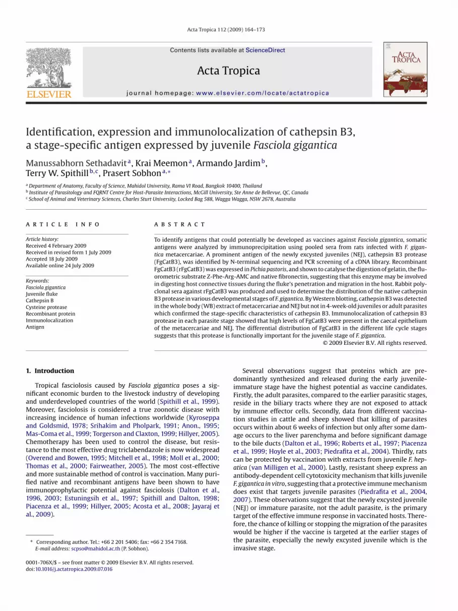
Fig. 1. Electrophoretic pattern of the major antigens from F. gigantica NEJ immuno-precipitated by antisera from rats infected with F. gigantica NEJ. Proteins wereseparated on a 12.5% SDS-PAGE and transferred onto PVDF membrane and stainedwith Coomassie Blue dye. (A) SDS-PAGE analysis of bands corresponding to themwT
wnmTa1Pcdr
2
w4efim(
fidctrcsfg
Immunoprecipitation of whole body antigen from 4000 NEJsshowed that the dominant antigens captured by the antisera frominfected rats were 38, 29, and 27 kDa proteins (Fig. 1). Edman degra-dation analysis of these proteins revealed the major N-terminal
Fig. 2. Characterization of recombinant proFgCatB3. Recombinant proFgCatB3was purified from the yeast culture supernatant using Ni2+-NTA affinity-chromatography. The purified protein was separated by 12.5% SDS-PAGE under
ost prominent immunoprecipitated proteins of 27, 29, and 38 kDa; the bandsere excised and the N-termini determined by Edman degradation analysis. (B)
he negative control immunoprecipitation using pooled naïve rat sera.
ere separated on a 12.5% SDS-PAGE, then transferred onto aitrocellulose membrane. The blot was blocked with 5% skimilk in TBS (40 mM Tris–HCl, 0.5 M NaCl, pH 7.4) containing 0.1%
ween®-20 (TBST) for 1 h at 25 ◦C, then incubated with the rabbitnti-proFgCatB3 antisera diluted to 1:5000 in 5% skim milk-TBST forh at 25 ◦C with gentle agitation. The blot was washed with TBST.rimary antibodies were visualized using horseradish peroxidase-onjugated goat anti-rabbit IgG (Zymed Laboratory Inc.) at ailution of 1:20,000 in TBST and the West Pico chemiluminescenceeagent (PIERCE).
.9. Immunolocalization
For localization of FgCatB3, F. gigantica metacercariae and NEJere fixed with 4% (w/v) paraformaldehyde in PBS, pH 7.4 at◦C overnight, then dehydrated with increasing concentrations ofthyl alcohol (70–100%), dioxane and finally embedded in paraf-n. The tissue blocks were sectioned at 5 �m thickness using aicrotome and placed onto 3-aminopropyl-triethoxy-silane (APES)
Sigma–Aldrich Co.) coated slides.For immunoperoxidase detection, the protocol was modi-
ed from Pankao et al. (2006). Briefly, the tissue sections wereeparaffinized and rehydrated through xylene and decreasing con-entrations of ethyl alcohol (100–70%). After washing with ddH2O,he slides were washed with PBS before treating with 10 mM cit-ate buffer, pH 6.0 in a microwave oven at 700 W using three 5-min
ycles (Shi et al., 1991). The endogenous peroxidase in the tis-ues was quenched by treatment with 3% (w/v) H2O2 in the darkor 30 min. Non-specific bindings were blocked with 3% normaloat serum in PBS for 20 min at 25 ◦C and then the sections wereica 112 (2009) 164–173 167
incubated with rabbit anti-rFgCatB3 antisera diluted to 1:500 inPBS containing 0.1% Tween®-20 (PBST) overnight at 25 ◦C. Sectionswere washed then incubated with HRP-conjugated goat anti-rabbitIgG (Zymed Laboratory Inc.) diluted to 1:500 in PBST for 1 h at25 ◦C. Sections were exhaustively washed, incubated with AEC(3-amino-9-ethylcarbazole) substrate solution (Zymed LaboratoryInc.), counterstained with Mayer’s hematoxylin, and mounted with90% glycerol in PBS. The slides were observed and photographed bya light microscope.
For immunofluorescence detection, the protocol was modi-fied from Levano-Garcia et al. (2007). Briefly, deparaffinized andrehydrated sections were washed with PBS, pH 7.4 containing0.1% Tween®-20 (PBST) for 5 min, and non-specific bindings wereblocked with 1% (w/v) bovine serum albumin (BSA) in PBST for 1 hat 25 ◦C. Sections were incubated with antisera (pooled infected ratsera from 21, 35 and 49 dpi or rabbit anti-rFgCatB3 sera) dilutedto 1:500 with PBST for 2 h at 25 ◦C. Immune complexes weredetected by fluorescein isothiocyanate (FITC)-conjugated goat anti-rat IgG (SouthernBiotech, Birmingham, AL, USA) and tetramethylrhodamine isothiocyanate (TRITC)-conjugated goat anti-rabbit IgG(Zymed Laboratory Inc.) diluted to 1:500 in PBST for 1 h at 25 ◦C.Sections were washed and mounted with Vectashield® (Vector Lab-oratory Inc., Burlingame, CA, USA) and visualized by a confocal laserscanning microscopy (CLSM) Olympus FV1000.
3. Results
3.1. Detection of FgCatB3 in NEJ by immunoprecipitation
reducing conditions and stained with Coomassie Blue dye. The purified recombi-nant glycosylated proFgCatB3 forms migrated at approximately 55–75 kDa (lane1), deglycosylation of rFgCatB3 with endoglycosidase H under denaturing (lane 2)and native conditions (lane 3) produced protein migrating at 38–39 kDa. Molecularweight markers are shown on the left side.

168 M. Sethadavit et al. / Acta Tropica 112 (2009) 164–173
Fig. 3. Enzymatic activity of purified rFgCatB3. (A) Lane 1: non-reducing gelatin substrate gel (12.5%) of rFgCatB3, subsequently activated with activation buffer, pH 4.5 in thepresence of DTT and dextran sulfate (500 K) overnight at 37 ◦C, showed gelatinolytic activity at 37 kDa and 50–75 kDa; lane 2: Coomassie stained proteins shown in lane 1. (B)Activity of FgCatB3 using synthetic fluorometric substrate Z-Phe-Arg-AMC incubated with rFgCatB3 for 30 min. Activity is shown in arbitrary fluorescence units (Beckhame s pre-a 7 ◦C fi FgCata
a2dpiSai2DshF1efbaXp
pNr
t al., 2006). (C) Digestion of fibronectin (FbN) by rFgCatB3. pro FgCatB3 (5 �g) wat 37 ◦C. The activated samples were subsequently co-incubated with 5 �g FbN at 3ncubated with activation buffer for 60 min, lane 2: FbN incubated with activated rnd lane 6: activated rFgCatB3 alone incubated for 60 min).
mino acid sequences, SNDELCG and DVPACG for the 38 and7 kDa bands, respectively. BLAST analysis of the NCBI genomeatabases using these N-terminal sequences identified severalroteins showing high sequence similarity including the hypothet-
cal proteins from S. japonicum SJCHGC02299 (38 kDa band) andJCHGC06544 (27 kDa band) (GenBank accession no. AAW26631nd AAW26825, respectively). Sequence AAW26631 shows 76%dentity to an omega-type GST from S. mansoni. Interestingly, the9 kDa antigen (Fig. 1) revealed two N-terminal sequences. TheLPESF sequence showed 100% identity to the family of cathep-
in B sequences from NEJ somatic and ES antigens of Fasciola (F.epatica: CB1, CB2 (also known as FhCatB1) and CB3; F. gigantica:gCatB1, FgCatB2, FgCatB3) (Tkalcevic et al., 1995; Wilson et al.,998; Meemon, 2004; Cancela et al., 2008); the DLPENF sequencexhibited 100% identity with cathepsin B-like cysteine proteasesrom the aphid Myzus persicae (accession number DAA06112), theeet army worm Spodoptera exigua (accession number ABK90823)nd sea urchin Strongylocentrotus purpuratus (accession numberP 001182630). These data suggest that the 29 kDa band is com-
rised of cathepsin B sequences.A screen of the F. gigantica NEJ cDNA library by PCR using T7romoter and S2F degenerate primers (designed from the DLPENF-terminal sequence) generated two identical clones with an open
eading frame showing 99% identity with F. gigantica cathepsin
activated in activation buffer, pH 4.5 in the presence of DTT and DS (500 K) for 1 hor 0, 10, 30 and 60 min and the products were analyzed by SDS-PAGE (lane 1: FbNB3 for 0 min, lanes 3–5: FbN incubated with activated rFgCatB3 for 10, 30, 60 min,
B3 (accession number AY227675) (Meemon et al., 2004). The fulllength ORF encodes a pre pro-enzyme of 337 aa (predicted size37.8 kDa) consisting of a 15 aa signal sequence, a 70 aa prosequenceand a 252 aa mature sequence; the predicted sizes of the proenzymeand mature enzyme are 36.2 kDa and 27.9 kDa, respectively.
3.2. Expression and purification of recombinant FgCatB3
The proFgCatB3 containing a propeptide region and taggedwith the c-myc epitope and hexahistidine at the C-terminus wasexpressed in the yeast P. pastoris GS115 using the pPICZ�A vectordesigned to secrete recombinant proteins into the extracellular cul-ture media (Beckham et al., 2006). Together with the c-myc andhistidine tags, the predicted size of the preproenzyme is 40.8 kDa,the proenzyme is 39.2 kDa and the mature enzyme is 31 kDa. SDS-PAGE analysis of the protein eluted from the Ni2+-NTA columnrevealed a minor band at about 38–39 kDa while the bulk of theprotein migrated as a smear ranging from 55–75 kDa (Fig. 2). SincePichia is known to glycosylate secreted proteins that contain an
N-linked sugar glycosylation motif, two of which are present inthe mature enzyme domain of proFgCatB3, it was rationalized thatthis higher molecular weight species was most likely due to gly-cosylation, as observed earlier with the F. hepatica cathepsin CB2sequence (Beckham et al., 2006). Treatment of the denatured or
a Tropica 112 (2009) 164–173 169
nast
3
tCt5rbaat(mtl
tawDp1wswawr
3r
dgajaAsttefis(
3
oNscsiasoi
Fig. 4. The reactivity of anti-rFgCatB3 sera to native cathepsin B3 of F. giganticain whole body extract (WB), tegument (TA), and excretory–secretory (ES) antigensby immunoblotting. Panel (A) The proteins from F. gigantica were separated by12.5% SDS-PAGE and visualized by Coomassie Blue staining. Panel (B) The blottedF. gigantica proteins were subsequently probed with rabbit anti-rFgCatB3 sera as theprimary antibody and goat anti-rabbit IgG conjugated to HRP as the secondary anti-body. Lane 1: WB of metacercariae; lane 2: WB of newly excysted juvenile (NEJ);lane 3: WB of 4-week-old juvenile (4 weeks) whole body extract; lane 4: WB ofadult; lane 5: adult tegument antigen (TA); lane 6: 4-week-old juvenile (4 weeks)
M. Sethadavit et al. / Act
ative recombinant proFgCatB3 with endoglycosidase H resulted inquantitative conversion of the recombinant protein to a 38–39 kDapecies which most likely represents the pre-pro cathepsin B3 pro-ein (Fig. 2).
.3. Enzymatic activity of rFgCatB3
Gelatin substrate gel analysis was performed to demonstratehe enzymatic activity of purified rFgCatB3 (proFgCatB3). proFg-atB3 was separated by non-denaturing SDS-PAGE which showedhe protein migrated at 37 kDa as well as the glycosylated form at0–75 kDa (Fig. 3A, lane 2). Activation of the protease in the gelevealed a clear thin band of protease activity at 37 kDa and a thickand at 50–75 kDa (Fig. 3A, lane 1). The activity of purified rFgCatB3gainst the synthetic fluorometric substrate, Z-Phe-Arg-AMC, waslso determined. The proFgCatB3 was pre-activated with activa-ion buffer at pH 4.5 in the presence of DTT and dextran sulfate500 K) for 30 min at 37 ◦C (Beckham et al., 2006) and the enzy-
atic activity was measured immediately for 30 min, during whichhe amount of fluorescent substrate being cleaved increased almostinearly (Fig. 3B).
Since FgCatB3 may be a leading enzyme that NEJ use in pene-rating the gut wall and for migration in the host’s tissues, its actiongainst a natural substrate was also tested. The purified proFgCatB3as pre-activated in activation buffer at pH 4.5 in the presence ofTT and dextran sulfate (500 K) for 1 h at 37 ◦C. The activated sam-les were subsequently co-incubated with fibronectin at 37 ◦C for 0,0, 30, and 60 min. After the incubation, the co-incubated samplesere separated by 7% SDS-PAGE and visualized by Coomassie Blue
taining. The co-incubation results showed that the fibronectin,hose molecular weight was approximately 250 kDa, was digested,
nd a group of proteins with lower molecular weights appearedhich increased in prominence with incubation time (Fig. 3C). The
esults show that rFgCatB3 is able to cleave fibronectin.
.4. Detection of native F. gigantica cathepsin B3 antigen withabbit anti-rFgCatB3
Immunoblotting with rabbit anti-rFgCatB3 antisera was used toetermine the expression of native F. gigantica cathepsin B3 anti-ens in the WB extracts of metacercariae, NEJ, 4-week-old juvenile,dult parasites; tegument antigens; and ES antigens of 4-week-olduvenile and adult parasites. The proteins from WB extracts werenalyzed by 12.5% SDS-PAGE under reducing condition (Fig. 4A).fter immunoblotting, the rabbit anti-rFgCatB3 antisera reactedpecifically with WB extracts of metacercariae at MW of 34 kDa andhose of NEJ at MW of 34 kDa and 29 kDa (Fig. 4B). Interestingly, inhree separate experiments, no positive bands were detected in WBxtracts of 4-week-old juvenile or of adult parasites. This result con-rmed the specific expression of cathepsin B3 RNA (detected by initu hybridization) in metacercariae and NEJ stages of F. giganticaMeemon et al., 2004).
.5. Immunolocalization of cathepsin B3 in F. gigantica
The distribution of cathepsin B3 was investigated in vari-us developmental stages of the F. gigantica (i.e., metacercariae,EJ, 4-week-old juvenile and adult stage) by immunoperoxidase
taining using rabbit anti-rFgCatB3 antisera. In metacercariae, spe-ific immunostaining was observed in the caecal epithelium andome parts of the tegument (Fig. 5B). In NEJ, the intense reddish
mmunostaining was observed in the caecal epithelium (Fig. 5Dnd E), while the parenchymal tissue which lies around the caecumhowed weak immunostaining (Fig. 5D–F). At higher magnificationf sections from NEJ, intense reddish immunostaining was observedn both caecal epithelium (Fig. 5E) and in the lumen of the caecum
excretory–secretory (ES) antigen; lane 7: Adult excretory–secretory (ES) antigen;lane 8: glycosylated rFgCatB3 used as a positive control. Dual Color Precision PlusProteinTM Standards were used as molecular weight (MW) markers as indicated onthe left side.
(Fig. 5F). The negative control using rabbit pre-immune sera showedno staining in any of the tissues from both parasitic stages (Fig. 5Aand C). Consistent with the immunoblotting experiment, there wasno immunostaining in 4-week-old juvenile and adult stages (Fig. 5Gand H).
In order to study the relationship between the major antigenssecreted from NEJ and cathepsin B3, co-localization was performedon F. gigantica NEJ and metacercariae sections using pooled anti-
sera from rats infected with F. gigantica metacercariae, and rabbitanti-rFgCatB3. Confocal laser scanning micrographs of the NEJ andmetacercariae sections were acquired (Fig. 6). In NEJ, the major anti-gens (marked with FITC) and cathepsin B3 proteases (marked withTRITC) were co-localized in both caecal epithelium (Fig. 6C and D)
170 M. Sethadavit et al. / Acta Tropica 112 (2009) 164–173
Fig. 5. Immunostaining of FgCatB3 in F. gigantica. Light micrographs of F. gigantica metacercariae (panels (A and B) ME) and newly excysted juvenile (panels (C–F) NEJ) stainedwith rabbit anti FgCatB3 sera using the immunoperoxidase method. The sections were counterstained with Mayer’s hematoxylin. In ME sections, the reddish immunostainingof cathepsin B3 was shown in the caecal epithelium (Ca) and some part of the tegument (Te), while there was no immunostaining in the parenchymal tissues (Pc) and outercyst wall (CW2) (panel (B)). In NEJ sections, the intense reddish immunostaining of cathepsin B3 was observed in both caecal epithelium (panels (D and E)) and its lumen(panel (F)), while the parenchymal tissues (Pc) around the caecum showed weak immunostaining (panels (D–F)). The immunostaining was not observed in the oral sucker(Os), pharynx (Ph) and tegument (Te). The negative control sections of ME (panel A) and NEJ (panel (C)) stained with rabbit pre-immune sera showed no immunostaining inthe parasite’s tissues. In addition, there was no immunostaining of cathepsin B3 in 4-week-old juvenile and adult parasite tissues (panels (G and H), respectively).

M. Sethadavit et al. / Acta Tropica 112 (2009) 164–173 171
Fig. 6. Fluorescence confocal microscopy and corresponding differential interference contrast (DIC) micrographs showing co-localization in sections on NEJ and metacercariaeprobed by rabbit anti-rFgCatB3 and pooled antisera from rats infected with F. gigantica (RIS). In newly excysted juvenile (NEJ) sections, the pooled antisera from rats at 21, 35,49 days post-infection (dpi) and anti-rFgCatB3 sera, and corresponding secondary antibodies conjugated to FITC (green) and TRITC (red) were used for fluorescence detectionof the major antigens (panels (A and E)) and cathepsin B3 protease (panels (B and F)), respectively. The fluorescence staining showed co-localization of these proteins in bothcaecal epithelium (panels (C and D)) and in the caecal lumen (panels (G and H)) of the NEJ. Pre-immune sera from rats and rabbit were used as negative controls and do nots oresce( onlyc uoresc
aacasrtNah
4
a
how any positive staining (panels (I–L)). In the metacercariae (ME) sections, the fluN)) was observed. The co-localizations of these immunogenic proteins were shownontrol sections using pre-immune sera from rats and rabbit showed only the autoflolor in this figure legend, the reader is referred to the web version of the article.)
nd the caecal lumen (Fig. 6G and H). In metacercariae, the majorntigens and cathepsin B3 proteases were co-localized only in theaecal epithelium (Fig. 6O and P). Pre-immune sera from both ratnd rabbit, used as negative controls, did not show any positivetaining in both NEJ and metacercariae (Fig. 6I–L and Q–T). Theseesults confirmed a significant degree of co-localization betweenhe major antigens of NEJ and cathepsin B3 in the caecum of bothEJ and metacercariae, and suggest that CatB3 is one of the majorntigens from the caeca of these stages that could elicit the host’sumoral immune response.
. Discussion
In this study, we have immunoprecipitated three prominentntigens of 27, 29 and 38 kDa in F. gigantica NEJ, using pooled
nce staining of the major antigens (panel (M)) and the cathepsin B3 protease (panelin the caecal epithelium of the metacercariae (panels (O and P)), while the negativecence of the outer cyst wall (panels (Q–T)). (For interpretation of the references to
antisera from rats infected with F. gigantica for 3–7 weeks, anddetermined that the N-terminal amino acid sequences of these anti-gens show sequence similarity to either two hypothetical proteinsfrom S. japonicum (38, 27 kDa antigens), or cathepsin B sequences(29 kDa antigen) previously described from juvenile F. gigantica(Meemon et al., 2004). The antigenicity of cathepsin B proteinsduring F. gigantica infection in rats is consistent with a previousstudy which showed that the juvenile F. hepatica CatB1 (CB2) pro-tein is antigenic during the first 5–10 weeks of F. hepatica infectionin rats and sheep, with peak antigenicity observed at 5 weeks
after infection (Law et al., 2003). Interestingly, kinetic analysisshowed that the F. hepatica CatB1 (CB2) antigen was antigenic ininfected sheep within 2 weeks of infection suggesting that thesejuvenile-specific enzymes are released early after invasion of hosttissues.
1 a Trop
wcs(bNsBn1cc
l(ppistta2
oactsttneftte
wtlBcIcteFfipea
atamflpa2ttgt
72 M. Sethadavit et al. / Act
The N-terminal amino acid sequences of the 29 kDa proteinere used to design degenerate primers for screening the NEJ
DNA library which generated two clones showing 99% DNAequence identity to F. gigantica CB3, previously cloned by our groupMeemon et al., 2004). In a previous study, it was shown indirectlyy PCR that cathepsin B2 and B3 could be detected in F. giganticaEJ and metacercariae whereas cathepsin B1 was detected in all
tages (Meemon et al., 2004). Two closely related 29 kDa cathepsins (termed FhCatB1 or CB2; CB3) have also been detected predomi-antly in F. hepatica NEJ excretory–secretory extracts (Wilson et al.,998; Cancela et al., 2008). Taken together, the data suggest thatathepsin B3 is one of the dominant antigens in NEJ and metacer-ariae stages of F. gigantica.
The recombinant F. gigantica cathepsin B3 containing two N-inked glycosylation sites at positions 180 and 223 in the sequenceMeemon et al., 2004) was expressed in P. pastoris. The entireropeptide region of FgCatB3 was included in the expressionlasmid to ensure proper folding (Müntener et al., 2005) and self-
nhibition to prevent toxicity in the heterologous yeast expressionystem (Mort and Buttle, 1997). The rFgCatB3 secreted from P. pas-oris culture medium was heterogeneous which can be attributedo its N-linked glycosylation as seen with FhCatB1 (CB2) which waslso expressed as a glycosylated enzyme in Pichia (Beckham et al.,006).
In whole body extracts of F. gigantica, the antisera to FgCatB3 rec-gnized a single band of ∼34 kDa in metacercariae and two bands atbout ∼34 and ∼29 kDa in NEJ WB extract. This 34 kDa band likelyorresponds to the procathepsin B3 based on its close approxima-ion to the theoretical MW of 36.2 kDa deduced from the amino acidequences of native FgCatB3 (Meemon et al., 2004). The 29 kDa pro-ein which was found only in the NEJ WB extract likely representshe native mature FgCatB3 which is similar in size to the matureative cathepsin B3 protease from F. hepatica ES antigens (Cancelat al., 2008). The anti-rFgCatB3 sera did not react with any proteinrom 4-week-old juvenile and adult WB extracts, further confirminghe stage-specific expression of the cathepsin B3 gene of F. gigan-ica (Meemon et al., 2004) and consistent with the stage-specificxpression of F. hepatica CB3 (Cancela et al., 2008).
By immunohistochemistry the F. gigantica cathepsin B3 proteaseas localized in the caecum of metacercariae, while in the NEJ,
his protease was found in both caecal epithelium and the caecalumen. This observation suggests that the mature native cathepsin3 may be processed and secreted by NEJ, whereas in metacer-ariae, only procathepsin B3 was present in the epithelial cells.n F. hepatica, cathepsin B could be detected by rabbit anti-bovineathepsin B antibodies in the electron-dense secretory granules ofhe caecal epithelium and within the caecal lumen of NEJs (Creaneyt al., 1996). The immunoblot analysis and immunolocalization of. gigantica cathepsin B3 protease in metacercariae and NEJ con-rms that the juvenile flukes express specific forms of processedroteases during the two developmental stages, as demonstratedarlier (Yoshihara and Goto, 1993; Wilson et al., 1998; Mohamed etl., 2005; Cancela et al., 2008).
The rFgCatB3 showed enzymatic activity when autoprocessedt pH 4.5 in the presence of DTT and dextran sulfate (DS 500 K),hus confirming that the enzyme was correctly folded in Pichias seen with FhCatB1 (CB2) (Beckham et al., 2006). Further-ore, rFgCatB3 was also able to digest gelatin, and cleave the
uorometric Z-Phe-Arg-AMC substrate, which was similar to theroperties of mammalian and F. hepatica cathepsin B (Rowan etl., 1992; Rozman et al., 1999; Law et al., 2003; Beckham et al.,
006; Cancela et al., 2008). Recombinant FgCatB3 was also ableo digest fibronectin which is one of the major components inhe host’s connective tissues suggesting that cathepsin B3 from F.igantica may be involved in the parasites migration in the hostissues.ica 112 (2009) 164–173
Even though the exact functions of cathepsin B proteases in liverflukes are still unclear, the findings that the mature cathepsin B3was present only in F. gigantica NEJ, and not in 4-week-old or adultparasites, together with its ability to digest fibronectin, a nativecomponent of host’s connective tissue, suggest that the putativefunction of secreted cathepsin B3 may be in digesting the host’s tis-sues to facilitate the migration of the excysted parasite to the liver(Wilson et al., 1998; Sajid and McKerrow, 2002; Law et al., 2003;Meemon et al., 2004; Cancela et al., 2008). This is consistent withdata from McGonigle et al. (2008) showing that RNAi-mediatedsuppression of F. hepatica FhCatB1 (CB2) expression in NEJ signif-icantly reduced gut penetration in an in vitro model system. It isalso possible that the synergistic activity of several cathepsin B pro-teases (such as CB2 and CB3), or cathepsin B and cathepsin L, arerequired for NEJ penetration of host tissues. This biological role mayexplain the need for the parasite to express high levels of these pro-teases. Thus, it is possible that a vaccine that stimulates an immuneresponse against cathepsin B3 would block the migration of youngflukes to the liver and would lead to death of the parasite. The poten-tial for a cathepsin B-based vaccine against Fasciola is supported bythe observation that FhCatB1 (CB2) is highly antigenic in vaccinatedsheep and rats and can induce high levels of protection in rats (Lawet al., 2003; Kennedy et al., 2006; Jayaraj et al., 2009). Our group iscurrently evaluating F. gigantica CB3 as a vaccine. One preliminarystudy in ICR mice showed that intraperitoneal immunization of rFg-CatB3 mixed with Freund’s adjuvant could stimulate IgG1 antibodyresponse and conferred a significant protection (41.8%, p ≤ 0.05)against F. gigantica when compared with non-immunized controlinfection (unpublished data). Future work will confirm and extendthis observation and determine whether the rFgCatB3 vaccine isable to induce significant protection in livestock.
Acknowledgements
This research was supported by Mahidol University, the ThailandResearch Fund (Senior Research Fellowship to Prasert Sobhon), theCommission on Higher Education (Ph.D. Scholarship to Manuss-abhorn Sethadavit), the Le Fonds québécois de la recherche sur lanature et les technologies (FQRNT), Centre for Host-Parasite Inter-actions and the Canada Research Chair program. T. Spithill holdsa Canada Research Chair in Immunoparasitology. A. Jardim and T.Spithill were supported by a Discovery grants from the NaturalSciences and Engineering Research Council of Canada.
References
Acosta, D., Cancela, M., Piacenza, L., Roche, L., Carmona, C., Tort, J.F., 2008. Fasciolahepatica leucine aminopeptidase, a promising candidate for vaccination againstruminant fasciolosis. Mol. Biochem. Parasitol. 158, 52–64.
Anon., 1995. Control of food borne, trematode infections. WHO Technical Series, vol.849. WHO, Geneva, p. 157.
Beckham, S.A., Law, R.H., Smooker, P.M., Quinsey, N.S., Caffrey, C.R., McKerrow, J.H.,Pike, R.N., Spithill, T.W., 2006. Production and processing of a recombinant Fas-ciola hepatica cathepsin B-like enzyme (FhcatB1) reveals potential processingmechanisms in the parasite. Biol. Chem. 387, 1053–1061.
Carmona, C., Dowd, A.J., Smith, A.M., Dalton, J.P., 1993. Cathepsin L proteinasesecreted by Fasciola hepatica in vitro prevents antibody-mediated eosinophilattachment to newly excysted juveniles. Mol. Biochem. Parasitol. 62, 9–17.
Carmona, C., McGonigle, S., Dowd, A.J., Smith, A.M., Coughlan, S., McGowran, E., Dal-ton, J.P., 1994. A dipeptidylpeptidase secreted by Fasciola hepatica. Parasitology109, 113–118.
Cancela, M., Acosta, D., Rinaldi, G., Silva, E., Durán, R., Roche, L., Zaha, A., Carmona,C., Tort, J.F., 2008. A distinctive repertoire of cathepsins is expressed by juvenileinvasive Fasciola hepatica. Biochimie 90, 1461–1475.
Chaithirayanon, K., Wanichanon, C., Vichasri-Grams, S., Ardseungneon, P., Grams, R.,Viyanant, V., Upatham, E.S., Sobhon, P., 2002. Production and characterization of
a monoclonal antibody against 28.5 kDa tegument antigen of Fasciola gigantica.Acta Trop. 84, 1–8.Creaney, J., Wilson, L., Dosen, M., Sandeman, M., Spithill, T.W., Parsons, J.C., 1996.Fasciola hepatica: irradiation induced alterations in carbohydrate and cathepsinB protease expression in newly excysted juvenile liver fluke. Exp. Parasitol. 83,202–215.

a Trop
D
D
D
E
F
F
H
H
H
I
J
K
K
L
L
L
M
M
M
M
M
M
M
M
M. Sethadavit et al. / Act
alton, J.P., McGonigle, S., Rolph, T.P., Andrews, S.J., 1996. Induction of protectiveimmunity in cattle against infection with Fasciola hepatica by vaccination withcathepsin L proteinases and with hemoglobin. Infect. Immun. 64, 5066–5074.
alton, J.P., Neill, S.O., Stack, C., Collins, P., Walshe, A., Sekiya, M., Doyle, S., Mulcahy,G., Hoyle, D., Khaznadji, E., Moiré, N., Brennan, G., Mousley, A., Kreshchenko, N.,Maule, A.G., Donnelly, S.M., 2003. Fasciola hepatica cathepsin L-like proteases:biology, function, and potential in the development of first generation liver flukevaccines. Int. J. Parasitol. 33, 1173–1181.
ixit, A.K., Dixit, P., Sharma, R.L., 2008. Immunodiagnostic/protective role of Cathep-sin L cysteine proteinase secreted by Fasciola species. Vet. Parasitol. 154, 177–184.
stuningsih, S.E., Smooker, P.M., Wiedosari, E., Widjajanti, S., Vaiano, S., Partoutomo,S., Spithill, T.W., 1997. Evaluation of antigens of Fasciola gigantica as vaccinesagainst tropical fasciolosis in cattle. Int. J. Parasitol. 27, 1419–1428.
airweather, I., 2005. Triclabendazole: new skills to unravel an old(ish) enigma. J.Helminthol. 79, 227–234.
ischer, M. Backtranslation Tool. Entelechon GmbH, Regensburg, Germany.http://www.entelechon.com/backtranslation.
armsen, M.M., Cornelissen, J.B., Buijs, H.E., Boersma, W.J., Jeurissen, S.H., vanMilligen, F.J., 2004. Identification of a novel Fasciola hepatica cathepsin L pro-tease containing protective epitopes within the propeptide. Int. J. Parasitol. 43,675–682.
illyer, G.V., 2005. Fasciola antigens as vaccines against fascioliasis and schistoso-miasis. J. Helminthol. 79, 241–247.
oyle, D.V., Dalton, J.P., Chase-Topping, M., Taylor, D.W., 2003. Pre-exposure of cat-tle to drug-abbreviated Fasciola hepatica infections: the effect upon subsequentchallenge infection and the early immune response. Vet. Parasitol. 111, 65–82.
srael, D.I., 1993. A PCR-based method for high stringency screening of DNA libraries.Nucleic Acids Res. 21, 2627–2631.
ayaraj, R., Piedrafita, D., Dynon, K., Grams, R., Spithill, T.W., Smooker, P.M., 2009. Vac-cination against fasciolosis by a multivalent vaccine of stage-specific antigens.Vet. Parasitol. 160, 230–236.
ennedy, N.J., Spithill, T.W., Tennent, J., Wood, P.R., Piedrafita, D., 2006. DNA vaccinesin sheep: CTLA-4 mediated targeting and CpG motifs enhance immunogenicityin a DNA prime/protein boost strategy. Vaccine 24, 970–979.
yroseppa, H.J., Goldsmid, J.M., 1978. Studies on the intestinal parasites in Africanpatients in Owamboland, South West Africa. Trans. R. Soc. Trop. Med. Hyg. 72,16–21.
aw, R.H., Smooker, P.M., Irving, J.A., Piedrafita, D., Ponting, R., Kennedy, N.J., Whis-stock, J.C., Pike, R.N., Spithill, T.W., 2003. Cloning and expression of the majorsecreted cathepsin B-like protein from juvenile Fasciola hepatica and analysis ofimmunogenicity following liver fluke infection. Infect. Immun. 71, 6921–6932.
evano-Garcia, J., Mortara, R.A., Verjovski-Almeida, S., DeMarco, R., 2007. Character-ization of Schistosoma mansoni ATPDase2 gene, a novel apyrase family member.Biochem. Biophys. Res. Commun. 352, 384–389.
inder, S., Schliwa, M., Kube-Granderath, E., 1996. Direct PCR screening of Pichiapastoris clones. Biotechniques 20, 980–982.
as-Coma, S., Esteban, J.G., Bargues, M.D., 1999. Epidemiology of human fascioliasis:a review and proposed new classification. Bull. World Health Organ. 77, 340–346.
cGonigle, L., Mousley, A., Marks, N.J., Brennab, G.P., Dalton, J.P., Spithill, T.W., Day,T.A., Maule, A.G., 2008. The silencing of cysteine proteases in Fasciola hepaticanewly excysted juveniles using RNA interference reduces gut penetration. Int. J.Parasitol. 38, 149–155.
eemon, K., 2004. Molecular cloning, characterization and vaccine potential ofcathepsin B-encoding genes from Fasciola gigantica. Ph.D. Thesis. Mahidol Uni-versity, Bangkok, Thailand.
eemon, K., Grams, R., Vichasri-Grams, S., Hofmann, A., Korge, G., Viyanant, V.,Upathum, E.S., Habe, S., Sobhon, P., 2004. Molecular cloning and analysis ofstage and tissue-specific expression of cathepsin B encoding genes from Fasciolagigantica. Mol. Biochem. Parasitol. 136, 1–10.
itchell, G.B., Maris, L., Bonniwell, M.A., 1998. Triclabendazole-resistant liver flukein Scottish sheep. Vet. Rec. 143, 399.
ohamed, S.A., Fahmy, A.S., Mohamed, T.M., Hamdy, S.M., 2005. Proteases inegg, miracidium and adult of Fasciola gigantica. Characterization of serine
and cysteine proteases from adult. Comp. Biochem. Physiol. Part B 142,192–200.oll, L., Gaasenbeek, C.P.H., Vellema, P., Borgsteede, F.H.M., 2000. Resistance of Fas-ciola hepatica against triclabendazole in cattle and sheep in the Netherlands. Vet.Parasitol. 91, 153–158.
ort, J.S., Buttle, D.J., 1997. Cathepsin B. Int. J. Biochem. Cell. Biol. 29, 715–720.
ica 112 (2009) 164–173 173
Müntener, K., Willimann, A., Zwicky, R., Svoboda, B., Mach, L., Baici, A., 2005. Foldingcompetence of N-terminally truncated forms of human procathepsin B. J. Biol.Chem. 280, 11973–11980.
Nakamura, Y., Gojobori, T., Ikemura, T., 2000. Codon usage tabulated from the inter-national DNA sequence databases: status for the year 2000. Nucleic Acids Res.28, 292.
Overend, D.J., Bowen, F.L., 1995. Resistance of Fasciola hepatica to triclabendazole.Aust. Vet. J. 72, 275.
Pankao, V., Sirisriro, A., Grams, R., Vichasri-Grams, S., Meepool, A., Kangwanrangsan,N., Wanichanon, C., Ardseungneon, P., Viyanant, V., Upatham, E.S., Sobhon, P.,2006. Classification of the parenchymal cells in Fasciola gigantica based onultrastructure and their expression of fatty acid binding proteins (FABPs). Vet.Parasitol. 142, 281–292.
Piacenza, L., Acosta, D., Basmadjian, I., Dalton, J.P., Carmona, C., 1999. Vaccina-tion with cathepsin L proteinases and with leucine aminopeptidase induceshigh levels of protection against fascioliasis in sheep. Infect. Immun. 67,1954–1961.
Piedrafita, D., Raadsma, H.W., Prowse, R., Spithill, T.W., 2004. Immunology of thehost parasite relationship in fasciolosis (Fasciola hepatica and Fasciola gigantica).Can. J. Zool. 82, 233–250.
Piedrafita, D., Estuningsih, E., Pleasance, J., Prowse, R., Raadsma, H.W., Meeusen, E.N.,Spithill, T.W., 2007. Peritoneal lavage cells of Indonesian thin-tail sheep medi-ate antibody-dependent superoxide radical cytotoxicity in vitro against newlyexcysted juvenile Fasciola gigantica but not juvenile Fasciola hepatica. Infect.Immun. 75, 1954–1963.
Roberts, J.A., Estuningsih, E., Wiedosari, E., Spithill, T.W., 1997. Acquisition of resis-tance against Fasciola gigantica by Indonesian Thin Tail sheep. Vet. Parasitol. 73,215–224.
Rowan, A., Mason, P., Mach, L., Mort, J.S., 1992. Rat pro-cathepsin B. J. Biol. Chem.267, 15993–15999.
Rozman, J., Stojan, J., Kuheji, R., Turk, V., Turk, B., 1999. Autocatalytic processing ofrecombinant human procathepsin B is a bimolecular process. FEBS 459, 358–362.
Sajid, M., McKerrow, J.H., 2002. Cysteine proteases of parasitic organism: review.Mol. Biochem. Parasitol. 120, 1–21.
Shi, S.R., Key, M.E., Kalra, K.L., 1991. Antigen retrieval in formalin-fixed, paraffin-embedded tissues: an enhancement method for immunohistochemical stainingbased on microwave oven heating of tissue sections. J. Histochem. Cytochem. 39,741–748.
Spithill, T.W., Dalton, J.P., 1998. Progress in development of liver fluke vaccines.Parasitol. Today 14, 224–228.
Spithill, T.W., Smooker, P.M., Copeman, D.B., 1999. Fasciola gigantica: epidemiology,control, immunology and molecular biology. In: Dalton, J.P. (Ed.), Fasciolosis.CABI Publishing, Oxon, pp. 465–525.
Srihakim, S., Pholpark, M., 1991. Problem in fasciolosis in animal husbandry in Thai-land. Southeast Asian J. Trop. Med. Pub. Health 22 (Suppl.), 352–355.
Thomas, I., Coles, G.C., Duffus, K., 2000. Triclabendazole-resistant Fasciola hepaticain southwest Wales. Vet. Rec. 146, 200.
Tkalcevic, J., Ashman, K., Meeusen, E., 1995. Fasciola hepatica: rapid identifica-tion of newly excysted juvenile proteins. Biochem. Biophys. Res. Commun. 213,169–174.
Torgerson, P., Claxton, J., 1999. Epidemiology and control. In: Dalton, J.P. (Ed.), Fasci-olosis. CABI Publishing, Oxon, pp. 113–139.
van Milligen, F.J., Cornelissen, J.B., Bokhout, B.A., 2000. Fasciola hepatica: an antigenfraction derived from newly excysted juveniles, containing an immunoreactive32-kDa protein, induces strong protective immunity in rats. Exp. Parasitol. 94,163–171.
Wijffels, G.L., Salvatore, L., Dosen, M., Waddington, J., Wilson, L., Thompson, C., Camp-bell, N., Sexton, J., Wicker, J., Bowen, F., Spithill, T.W., 1994. Vaccination of sheepwith purified cysteine proteinases of Fasciola hepatica decreases worm fecundity.Exp. Parasitol. 78, 132–148.
Wilson, L.R., Good, R.T., Panaccio, M., Wijffels, G.L., Sandeman, R.M., Spithill, T.W.,1998. Fasciola hepatica: characterization and cloning of the major cathepsinB protease secreted by newly excysted juvenile liver fluke. Exp. Parasitol. 88,
85–94.Yamasaki, H., Mineki, R., Murayama, K., Ito, A., Aoki, T., 2002. Characterizationand expression of the Fasciola gigantica cathepsin L gene. Int. J. Parasitol. 32,1031–1042.
Yoshihara, S., Goto, N., 1993. Distribution of antigens of Fasciola gigantica at variousstages of development. Res. Vet. Sci. 54, 252–255.


