Embed Size (px)
Citation preview
1
Original Paper
Identification of an unusual variant peroxisome biogenesis disorder caused by mutations
in the PEX16 gene
Merel S. Ebberink1, Barbara Csanyi2, Wui K. Chong3, Simone Denis1, Peter Sharp4,
Petra A.W. Mooijer1, Conny J.M. Dekker1, Claire Spooner5, Lock H. Ngu6, Carlos De Sousa7,
Ronald J.A. Wanders1, Michael J. Fietz4, Peter T. Clayton2, Hans R. Waterham1,
Sacha Ferdinandusse1
1Academic Medical Centre, University of Amsterdam, Laboratory Genetic Metabolic
Diseases, Department of Paediatrics/Emma Children's Hospital, Amsterdam, The
Netherlands; 2Biochemistry Research Group, UCL Institute of Child Health, Great Ormond
Street Hospital for Children NHS Trust, London, United Kingdom, 3Department of Radiology,
Great Ormond Street Hospital for Children NHS Trust, London, United Kingdom, 4National
Referral Laboratory, SA Pathology, North Adelaide, Australia, 5Starship Children's Hospital,
Auckland District Health Board, New Zealand, 6Genetics Department, Kuala Lumpur
Hospital, Kuala Lumpur, Malaysia, 7Department of Neurology, Great Ormond Street
Hospital for Children NHS Trust, London, United Kingdom
Corresponding author:
Sacha Ferdinandusse, PhD
Laboratory Genetic Metabolic Diseases (F0-220)
Academic Medical Centre, University of Amsterdam
Meibergdreef 9, 1105 AZ Amsterdam
The Netherlands
Tel: +31 20 5665958/5797
Fax: +31 20 696 2596
Email: [email protected]
Key words: Zellweger syndrome, peroxisome, leukodystrophy
Word count: 3971 (excluding abstract, table and figure legends)
peer
-005
5738
2, v
ersi
on 1
- 19
Jan
201
1Author manuscript, published in "Journal of Medical Genetics 47, 9 (2010) 608"
DOI : 10.1136/jmg.2009.074302
2
ABSTRACT
Objective Zellweger syndrome spectrum disorders are caused by mutations in any of at least
12 different PEX genes. This includes PEX16, which encodes an integral peroxisomal
membrane protein involved in peroxisomal membrane assembly. PEX16-defective patients
have been reported to have a severe clinical presentation. Fibroblasts of these patients
displayed a defect in import of peroxisomal matrix and membrane proteins, resulting in a total
absence of peroxisomal remnants. Here, we report 6 patients with an unexpected mild variant
peroxisome biogenesis disorder due to mutations in the PEX16 gene. Patients presented in the
preschool years with progressive spastic paraparesis and ataxia (with a characteristic pattern
of progressive leukodystrophy and brain atrophy on MRI scan) and later developed cataracts
and peripheral neuropathy. Surprisingly, their fibroblasts showed enlarged, import-competent
peroxisomes.
Results Plasma analysis revealed biochemical abnormalities suggesting a peroxisomal
disorder. Biochemical parameters in fibroblasts were only mildly abnormal or within the
normal range. Immunofluorescence microscopy analyses revealed the presence of import-
competent peroxisomes, which were increased in size but reduced in number. Subsequent
sequencing of all known PEX genes revealed five novel apparent homozygous mutations in
the PEX16 gene.
Conclusion We identified an unusual variant peroxisome biogenesis disorder caused by
mutations in the PEX16 gene, with a relatively mild clinical phenotype and an unexpected
phenotype in fibroblasts. Although PEX16 is involved in peroxisomal membrane assembly,
PEX16 defects can present with enlarged import-competent peroxisomes in fibroblasts. This
is important for future diagnostics of patients with a peroxisomal disorder.
peer
-005
5738
2, v
ersi
on 1
- 19
Jan
201
1
3
INTRODUCTION
The Zellweger Syndrome Spectrum (ZSS), including Zellweger Syndrome (ZS, MIM
214100), neonatal adrenoleukodystrophy (NALD, MIM 202370) and infantile Refsum disease
(IRD, MIM 266510), comprises a spectrum of severe, often lethal, inherited multisystemic
disorders. Variable neurodevelopmental delay, liver disease, retinopathy and perceptive
deafness are characteristic for the disorders within the ZSS. ZS is the most severe disorder
within this spectrum. ZS patients have profound neurological abnormalities and display
typical craniofacial dysmorphia.[1] ZS patients generally die within the first year of life. Like
ZS patients, patients with NALD suffer from neonatal hypotonia and seizures. They may
suffer from progressive white matter disease, and usually die in late infancy.[2] Patients with
IRD have no neuronal migration defect, but can develop a progressive white matter defect.
Their survival is variable, but most patients survive beyond infancy and some even reach
adulthood.[3]
The disorders of the ZSS are characterized by the absence of functional peroxisomes and a
generalized loss of peroxisomal functions. Defects in any of at least 12 different peroxins
(PEX), encoded by PEX genes, have been found to result in a peroxisome biogenesis disorder
(PBD, MIM 601539) of the ZSS type. Peroxins are involved in the import of proteins into the
peroxisome and/or the biogenesis of these organelles. Different mutations in the same PEX
gene can lead to different phenotypes within the spectrum. PEX1, PEX2, PEX5, PEX6,
PEX10, PEX12, PEX13, PEX14 and PEX26 are involved in the import of peroxisomal matrix
proteins. A defect in one of the genes encoding these peroxins results in impaired peroxisomal
matrix protein import, but peroxisomal membrane structures (peroxisomal ghosts) are still
present. In contrast, fibroblasts with a defect in the genes encoding PEX3, PEX16 or PEX19,
peer
-005
5738
2, v
ersi
on 1
- 19
Jan
201
1
4
which are involved in the peroxisomal membrane protein import, were shown to have no
peroxisomal remnants at all.[4, 5]
Human PEX16 is an integral peroxisomal membrane protein (PMP) with two membrane-
spanning domains. So far, only three patients have been reported with a defect in PEX16, all
displaying the severe ZS phenotype. Fibroblasts of these patients showed a complete lack of
peroxisomes, including membranes, and peroxisomal functions.[4, 6] In this paper, we report
6 patients, including one sib pair, who all have a defect in PEX16, but who showed enlarged,
protein import-competent peroxisomes in their fibroblasts.
peer
-005
5738
2, v
ersi
on 1
- 19
Jan
201
1
5
PATIENTS AND METHODS
Patients
Patient 1, a girl, was the first child of consanguineous Turkish parents. She was born at term
with a normal birth weight and Apgar scores. She had no dysmorphic features and developed
normally until 9 months of age. She started with toe walking at 13 months. Her walking
remained unsteady with frequent falls. On examination at the age of 3 years, she presented a
normal mental status, a bilateral horizontal nystagmus, increased tone in lower limbs, but
normal in upper limbs, normal sensation, ataxia, slight dysmetria, a head and bilateral hand
tremor, very brisk tendon reflexes at the knees, extensor plantar reflex and sustained bilateral
ankle clonus. She showed no cognitive impairment, no organomegaly and no cranial nerve
abnormalities. She stopped walking completely at the age of 5 years and ever since has been
wheelchair bound. At the age of 10 years she developed dysarthria and dysphagia, and from
that time she required full gastrostomy feeding. At the age of 13 years, ophthalmological
examination revealed optic atrophy and very mild lens opacities. At the age of 14 years she
was admitted to a hospital due to constipation and severe neuropathic pain. On examination
she had normal sensory responses to touch, temperature and pain, but decreased vibration
sense (absent in the ankles). Neurophysiology studies revealed evidence of a progressive
demyelinating motor and sensory neuropathy affecting upper and lower limbs without axonal
involvement on electroneuromyography (ENMG).
By the age of 16 years she was suffering from spasticity in all four limbs and had no
independent mobility. Her cataracts had become sufficiently dense as to require surgical
removal. Repeated electroretinograms (ERGs) revealed normal flash responses suggesting
normal function of the outer retinal receptor layer, whereas the visual evoked potentials
peer
-005
5738
2, v
ersi
on 1
- 19
Jan
201
1
6
(VEPs) showed small and very delayed potentials suggesting marked impairment of the visual
pathways bilaterally. There have never been any concerns about her hearing.
Magnetic resonance imaging (MRI) scans were performed at the age of 4, 6 (Figure 1A), 15
and 17 years. They showed extensive, diffuse and symmetrical signal abnormalities of
myelinated white matter in the form of increased signal on T2-weighted images and near
normal signal on T1-weighted images. These changes initially involved the dorsal brainstem,
internal capsules (more severely in the posterior limb) and deep peritrigonal and parietal white
matter, and spared the corpus callosum and subcortical U-fibres of white matter. There was no
evidence of a malformation of cortical development. Later scans showed extension of disease
into the corpus callosum and the subcortical white matter with some reduction in signal of the
involved regions of white matter on T1-weighted images, whilst remaining more severely
abnormal on T2-weighted images. There was generalized prominence of the ventricles and
sulci in line with reduction in cerebral volume, however at the same time, more selective
atrophy of the corpus callosum and cerebellar vermis was also observed.
Patient 2 is the six years younger brother of patient 1, who was born at term with normal birth
weight. He started to walk independently at the age of 17 months, despite lower limb
spasticity, with an unsteady gait and frequent falls. He ceased independent walking at 25
months of age and was wheelchair bound from the age of 3 years. On examination at the age
of 4 years, he had rigidity particularly in his lower limbs, brisk reflexes, progressive upper
limb tremor, dysmetria and dysarthria. A neuropathy became apparent from the age of 5 years
with increasing difficulty in emptying his bladder and worsening constipation. At the age of
10 years a demyelinating motor and sensory neuropathy was shown with ENMG and he
developed dysaesthesia in his legs. As for his sister, his flash ERG responses were normal,
peer
-005
5738
2, v
ersi
on 1
- 19
Jan
201
1
7
whereas the VEP responses were delayed and not very well formed. Cataract was
demonstrated. Currently, he is 11 years old and his cognition is relatively spared. His MRI
findings (Figure 1B and 1C) resembled those of his sister, showing a similar pattern of
progressive leukodystophy and brain atrophy from the age of 3 years.
The case report of patient 3 was published previously. [7]
No detailed clinical information is available for patient 4.
Patient 5, a girl, was born following an uneventful pregnancy and delivery. Her development
was normal in the first year of life and she started walking independently at the age of 14-15
months. At the age of two years she developed an ataxic gait. At the age of 6 years, she had
mild cognitive impairment, moderate dysarthria and abnormal eye saccades, but no problems
with speech or swallowing. MRI scans revealed widespread white matter changes on a
background pattern of global delay in myelin maturation, with symmetrical white matter
abnormality of the dorsal brainstem and capsular white matter, sparing of the corpus callosum
and appearing more extensive and conspicuous on T2-weighted images than on T1-weighted
images. There was reduced cerebellar volume, more severely affecting the vermis. Similar
MRI findings were observed in the sister of patient 5 who was two years younger.
Patient 6, a girl, is the second child of consanguineous parents of Indian ethnicity. She was
born at term with a normal birth weight. Her development was normal during the first year of
life. She walked independently at 13 months and spoke a few short phrases at 18 months.
After this she was noted to lose previously acquired skills and did not gain new skills. She lost
her ability to walk independently at 24 months due to a combination of spasticity and mild
ataxia. Her speech and other cognitive functions also deteriorated slowly over time. At the age
peer
-005
5738
2, v
ersi
on 1
- 19
Jan
201
1
8
of 5 years, nystagmus and cataracts in both eyes were observed. In her lower limbs, the
muscular tone was increased, reflexes were brisk, clonus was present and plantar reflexes
were up-going. She had mild cerebellar signs. She did not have organomegaly. At 6 years, her
cerebral white matter was noted to be diffusely hyperintense compared to grey matter on T2-
weighted images in nearly all areas, particularly the deep and capsular white matter. The
posterior fossa and callosal white matter was isointense with grey matter; that is, relatively
spared. At the same time, all except the subcortical white matter was hyperintense compared
to grey matter on T1-weighted images (that is, the comparatively normal appearance of
myelinated white matter on these images). In addition, there was evidence of more focal
atrophy of the cerebellum and corpus callosum. Peripheral nerve velocity studies of the lower
limbs were suggestive of demyelination. Currently, at the age of 9, she is able to stand with a
supportive frame, communicate in short sentences, and she is able to read and write simple
sentences.
Skin fibroblasts of the patients used in this study were sent to the Laboratory Genetic
Metabolic Diseases at the Academic Medical Center of the University of Amsterdam for
diagnostic purposes and informed consent was obtained for publication of the data.
Cell culturing
Primary skin fibroblasts were cultured in DMEM medium with 4.5 g/L glucose, L- glutamine
(BioWhittaker, Lonza, Verviers, Belgium) and 25 mM Hepes, or in HAM F-10 medium with
L-glutamine and Hepes 25 mM (Gibco, Invitrogen, Carlsbad, CA), each supplemented with
10% fetal bovine serum (FBS, BioWhittaker), 100 U/ml penicillin, 100 µg/ml streptomycin,
in a humidified atmosphere of 5% CO2, at 37°C. DMEM medium was used for the
transfection experiments and HAM F-10 medium for the biochemical experiments.
peer
-005
5738
2, v
ersi
on 1
- 19
Jan
201
1
9
Biochemical analysis
Levels of very long chain fatty acids (VLCFAs), phytanic and pristanic acid, and C27-bile acid
intermediates were measured in plasma as described before.[8] Plasmalogens were
determined in erythrocytes as previously described.[9] Dihydroxyacetonephosphate
acyltransferase (DHAPAT),[10] acyl-CoA oxidase I (AOXI)[11] and D-bifunctional protein
(DBP)[12] activity, concentrations of VLCFAs,[13] β-oxidation of C26:0, C16:0 and pristanic
acid,[14] and α-oxidation of phytanic acid[15] were measured in cultured fibroblasts as
previously described. Catalase immunofluorescence and immunoblot analysis using
antibodies against peroxisomal thiolase 1, AOXI, and DBP were performed as described.[12,
16]
Mutation analysis
Mutation analysis was performed by either sequencing all exons plus flanking intronic
sequences of the PEX gene amplified by PCR from genomic DNA or by sequencing cDNAs
prepared from total mRNA fractions. Genomic DNA was isolated from skin fibroblasts using
the NucleoSpin Tissue genomic DNA purification kit (Macherey-nagel, Germany, Düren).
Total RNA was isolated from skin fibroblasts using Trizol (Invitrogen, Carlsbad, CA)
extraction, after which cDNA was prepared using a first strand cDNA synthesis kit for RT-
PCR (Roche, Mannheim, Germany). All forward and reverse primers (PEX16 primer
sequences are listed in supplemental table 1) were tagged with a -21M13 (5-
’TGTAAAACGACGGCCAGT-3’) sequence or M13rev (5’-CAGGAAACAGCTATGACC-
3’) sequence, respectively. PCR fragments were sequenced in two directions using ‘-21M13’
and ‘M13rev’ primers by means of BigDye Terminator v1.1 Cycle Sequencing Kits (Applied
Biosystems, Foster City, CA, USA) and analyzed on an Applied Biosystems 377A automated
peer
-005
5738
2, v
ersi
on 1
- 19
Jan
201
1
10
DNA sequencer, following the manufacturer’s protocol (Applied Biosystems, Foster City,
CA, USA).
Mutation analysis of PEX1 and PEX3 was performed by sequencing of cDNA. Mutations in
the PEX2 gene were identified by sequencing exon 4 from gDNA. Mutation analysis of
PEX5L, PEX6 PEX10, PEX12, PEX13, PEX14, PEX19 and PEX26 were performed by
sequencing of all exons plus flanking intronic sequences.
Mutation analysis of the PEX16 gene was performed by sequencing the PEX16 cDNA.
Mutations were confirmed by sequencing the corresponding exons and flanking intronic
sequences of the PEX16 gene. Sequences were compared to the reference PEX16 sequence
(NM_004813) with nucleotide numbering starting at the first adenine of the translation
initiation codon ATG.
Functional complementation assay
Fibroblasts of the five unrelated patients (2-6) were co-transfected with a pcDNA3 expression
plasmid containing either the PEX16 cDNA (transcription variant 1 containing exon 11a,
NM_004813.2) or, as a control, the PEX12 cDNA (gift of Dr. S.J. Gould), and the pDsRed-
Express-DR vector (Clontech Laboratories; used to identify transfected cells) using the
AMAXA nucleofector technology (Amaxa, Cologne, Germany). The fibroblasts were
examined by catalase immunofluorescence 72 hours after transfection. [12] Two independent
transfections per construct were examined by counting the number of peroxisomes in at least
100 cells. Similar sized cells with the nucleus in focus were counted under the microscope
using a counter.
Expression of mutant PEX16 cDNA
peer
-005
5738
2, v
ersi
on 1
- 19
Jan
201
1
11
The mutant PEX16 cDNAs of patient 1-4 were amplified by PCR from cDNA prepared from
mRNA isolated from the patient fibroblasts and subcloned in the mammalian expression
vector pcDNA3 (Invitrogen). The wild type and mutant PEX16 cDNAs were separately
expressed in a previously reported PEX16-deficient fibroblast cell line (homozygous for an
R298fsX38 mutation) with complete lack of peroxisomal structures[6] and a control fibroblast
cell line. Four days after transfection the fibroblasts were examined by catalase
immunofluorescence to assess the appearance of peroxisomes. Two independent transfections
per construct were examined by counting the number of peroxisomes in at least 100 cells.
Similar sized cells with the nucleus in focus were counted under the microscope using a
counter. To control for transfection efficiency the constructs were also co-transfected with a
GFP-SKL expression vector in parallel experiments. The transfection efficiency was
comparable for the different constructs.
peer
-005
5738
2, v
ersi
on 1
- 19
Jan
201
1
12
RESULTS
Biochemical analysis
The major presenting symptom in all cases described for the first time in this paper was
difficulty with walking at around 2 years due to combination of spasticity and ataxia.
Presentation with a leukodystrophy at this age has been seen previously in a few children with
disorders of peroxisome biogenesis. For this reason, alongside other tests, peroxisomal
metabolites were measured in plasma and erythrocytes. The levels of VLCFAs, the branched-
chain fatty acids phytanic acid and pristanic acid and the C27-bile acid intermediates were all
elevated in plasma (Table 1), with the exception of the branched-chain fatty acid levels in
patients 1, 5 and 6, which were normal. The levels of plasmalogens in erythrocytes were
normal in all examined patients. Studies in cultured skin fibroblasts (Table 2) revealed
increased levels of VLCFAs with a marginally decreased β-oxidation rate of C26:0 in patient
3-6. All other parameters (phytanic acid α-oxidation, pristanic acid β-oxidation and the
activity of DHAPAT, the first enzyme of the etherphospholipid biosynthesis pathway) were
within the control range. In addition, immunoblot analysis showed that the peroxisomal
enzymes AOXI, DBP and thiolase I were normally processed (supplemental Figure S1).
Immunofluorescence microscopy analysis using antibodies raised against catalase, a
peroxisomal matrix enzyme, and antibodies against ALD protein, a peroxisomal membrane
protein, revealed the presence of peroxisomes. However, the peroxisomes were markedly
enlarged in size and reduced in number when compared to control fibroblasts (Figure 2). The
presence of enlarged peroxisomes and normal immunoblot profiles, in combination with
normal plasmalogens levels and DHAPAT activity usually points to a single peroxisomal
enzyme deficiency (i.e. AOXI or DBP deficiency). However, measurement of the activities of
AOXI and DBP in the patient cells revealed no abnormalities (Table 2). Based on these
results a novel variant of a PBD with enlarged peroxisomes was suspected.
peer
-005
5738
2, v
ersi
on 1
- 19
Jan
201
1
13
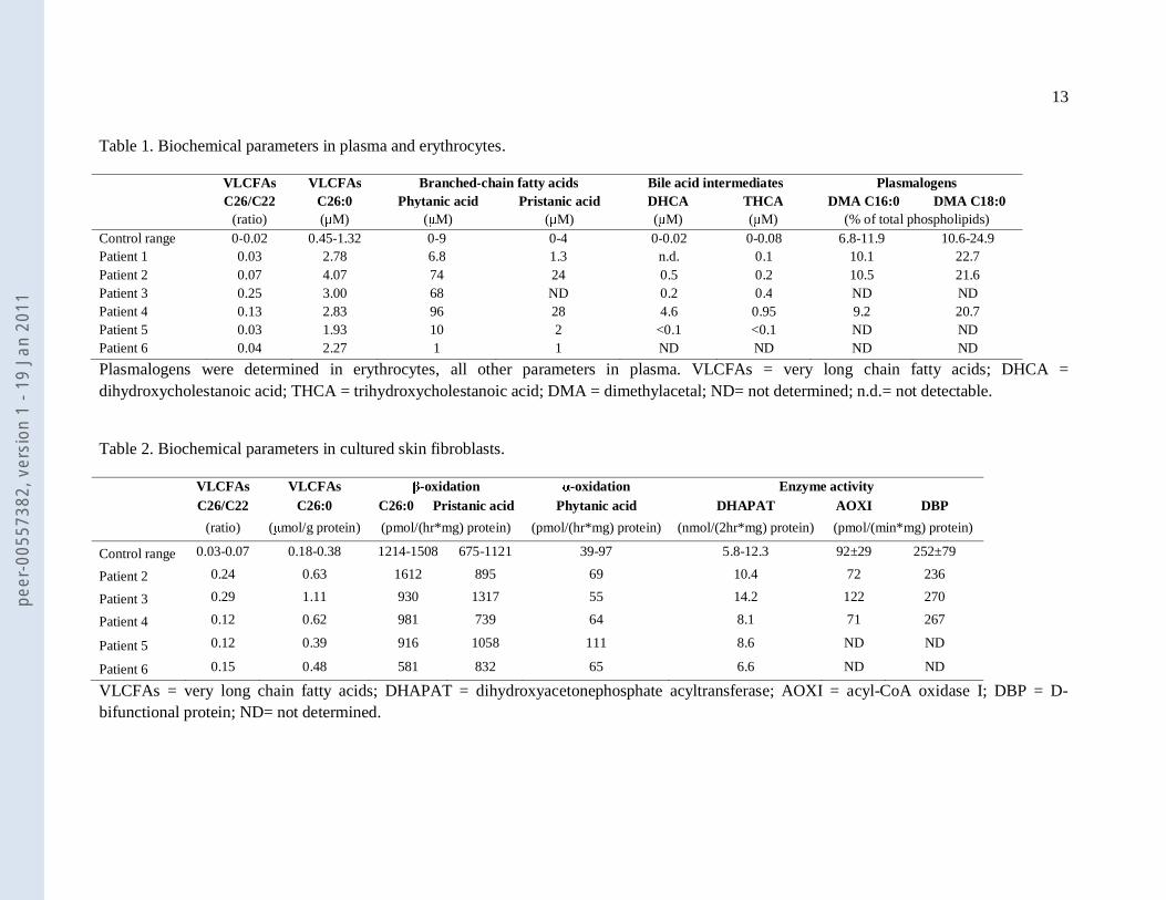
Table 1. Biochemical parameters in plasma and erythrocytes.
VLCFAs VLCFAs Branched-chain fatty acids Bile acid intermediates Plasmalogens C26/C22 C26:0 Phytanic acid Pristanic acid DHCA THCA DMA C16:0 DMA C18:0 (ratio) (μM) (μM) (μM) (μM) (μM) (% of total phospholipids) Control range 0-0.02 0.45-1.32 0-9 0-4 0-0.02 0-0.08 6.8-11.9 10.6-24.9 Patient 1 0.03 2.78 6.8 1.3 n.d. 0.1 10.1 22.7 Patient 2 0.07 4.07 74 24 0.5 0.2 10.5 21.6 Patient 3 0.25 3.00 68 ND 0.2 0.4 ND ND Patient 4 0.13 2.83 96 28 4.6 0.95 9.2 20.7 Patient 5 0.03 1.93 10 2 <0.1 <0.1 ND ND Patient 6 0.04 2.27 1 1 ND ND ND ND
Plasmalogens were determined in erythrocytes, all other parameters in plasma. VLCFAs = very long chain fatty acids; DHCA = dihydroxycholestanoic acid; THCA = trihydroxycholestanoic acid; DMA = dimethylacetal; ND= not determined; n.d.= not detectable.
Table 2. Biochemical parameters in cultured skin fibroblasts.
VLCFAs VLCFAs β-oxidation α-oxidation Enzyme activity C26/C22 C26:0 C26:0 Pristanic acid Phytanic acid DHAPAT AOXI DBP
(ratio) (μmol/g protein) (pmol/(hr*mg) protein) (pmol/(hr*mg) protein) (nmol/(2hr*mg) protein) (pmol/(min*mg) protein)
Control range 0.03-0.07 0.18-0.38 1214-1508 675-1121 39-97 5.8-12.3 92±29 252±79
Patient 2 0.24 0.63 1612 895 69 10.4 72 236
Patient 3 0.29 1.11 930 1317 55 14.2 122 270
Patient 4 0.12 0.62 981 739 64 8.1 71 267
Patient 5 0.12 0.39 916 1058 111 8.6 ND ND
Patient 6 0.15 0.48 581 832 65 6.6 ND ND
VLCFAs = very long chain fatty acids; DHAPAT = dihydroxyacetonephosphate acyltransferase; AOXI = acyl-CoA oxidase I; DBP = D-bifunctional protein; ND= not determined.
peer
-005
5738
2, v
ersi
on 1
- 19
Jan
201
1
14
Mutation analysis
To determine whether the patients had a novel variant of a PBD due to mutations in any of the
currently known 12 PEX genes, we sequenced all 12 PEX genes either in genomic DNA or in
cDNA prepared from the corresponding mRNAs. Unexpectedly, we identified five novel
apparent homozygous mutations in the PEX16 cDNAs (Table 3). The mutations were also
checked and appeared homozygous in genomic DNA. The PEX16 gene is localized at
chromosome 11p12-p11.2 and consists of 11 exons. In humans, two different mRNA variants
of PEX16 are produced as a result of alternative splicing, each with an alternate exon 11
(exon 11a and exon 11b). Both transcription variants are expressed in human fibroblasts, of
which variant 1 containing exon 11a is most abundant. We identified two missense mutations,
two small deletions and a large genomic deletion of exon 11a, which are all located in the
carboxy-terminal end of PEX16. The c.984delG in exon 11a in patient 1 and 2 results in a
frame shift and introduces a termination codon at amino acid position 357. Exon 11b is
unaffected in patient 1 and 2. The small deletion in patient 3 results in a deletion of a valine at
position 252. Patients 4 and 5 both have a missense mutation, leading to the amino acid
substitution of a proline to a threonine at position 289 (patient 4) and of a tyrosine to a
cysteine at position 331 (patient 5), respectively. The c.992A>G (p.Y331C) mutation in
patient 5 is located in exon 11a. Patient 6 has a large intragenic deletion in transcription
variant 1 comprising the last 468 base pairs of intron 10, the entire exon 11a and the first 80
base pairs of the 3’ flanking region of exon 11a and in transcription variant 2 comprising the
last 603 base pairs of intron 10, and the first 4 base pairs of exon 11b. Investigation of the
effect of this deletion on cDNA revealed 3 splice products encoding the following amino acid
sequences p.R318SfsX138, p.R318IfsX38 and p.E296DfsX33.
Table 3. PEX16 mutations identified.
Mutations
peer
-005
5738
2, v
ersi
on 1
- 19
Jan
201
1
15
Nucleotide Amino acid Exon Patient 1 + 2 c.984delG p.I330SfsX27 11 Patient 3 c.753_755delTGT p.V252del 8 Patient 4 c.865C>A p.P289T 9 Patient 5 c.992A>G p.Y331C 11 Patient 6 c.952+118_1011+80 p.R318SfsX138, p.R318IfsX38, p.E296DfsX33 11 Reference sequence of PEX16: GenBank accession number NM_004813.2. Nucleotide numbering starts at the adenine of the translation initiation codon ATG. Complementation assays
To confirm that the identified mutations in PEX16 are the underlying cause of the
peroxisomal abnormalities observed in fibroblasts of the patients, the cell lines were
transfected with wild type PEX16 cDNA, and PEX12 cDNA as a negative control. Expression
of wild type PEX16 cDNA in the cell lines of the unrelated patients (2-6) restored the number
of peroxisomes to the number found in control fibroblasts and also reduced the size of
peroxisomes to normal (Figure 3C). In contrast, expression of wild type PEX12 cDNA did not
complement the peroxisomal abnormalities in the patient cell lines (Figure 3D).
In addition, a fibroblast cell line with a complete PEX16-deficiency, resulting in the total
absence of peroxisomal structures, was transfected with wild type PEX16 cDNA, and the
mutant PEX16 cDNAs of patients 1-4. Expression of wild type PEX16 cDNA resulted in
more than 100-150 normal sized peroxisomes in nearly all (120 of the 130) transfected cells
(Figure 3E). However, expression of the PEX16 cDNAs containing either the c.865C>A, the
c.984delG or the c.753_755delTGT mutation, resulted in approximately 30-70 enlarged
peroxisomes in approximately 30% of the transfected cells while in the remaining 70% of the
transfected cells no restoration of peroxisomes was found.
peer
-005
5738
2, v
ersi
on 1
- 19
Jan
201
1
16
DISCUSSION
In the present study, we identified 6 patients including one sib pair with different defects in
PEX16 and an unexpected phenotype in skin fibroblasts. PEX16 was previously shown to be
involved in peroxisomal membrane protein import and in line with this role; no peroxisomal
remnants and a complete lack of peroxisomal functions were found in fibroblasts of the 3
PEX16-deficient patients reported in literature to date.[4, 6]
Despite the extensive diagnostic work up in patient 1-4 no definite diagnosis could be made
for a long time because the phenotypic presentation was highly unusual for a PBD. The
biochemical parameters in plasma of these patients showed elevated levels of VLCFAs,
phytanic acid, pristanic acid and the C27-bile acid intermediates but normal plasmalogen
levels in erythrocytes. Moreover, in fibroblasts catalase and ALDP immunofluorescence
revealed enlarged, import-competent peroxisomes which were reduced in number (Figure 2).
This phenotype is typical for peroxisomal single enzyme deficiencies, i.e. AOXI but
especially DBP deficiency. However, AOXI and DBP activities were completely normal in
these patients. Instead, a defect in PEX16 was identified (Table 3).
The identification of the PEX16 defect in patients 1-4 allowed a rapid diagnosis for patient 5
and 6. The fibroblasts of patients 5 and 6 were sent only recently to our laboratory for
diagnostic work up. Because the results of the biochemical tests in their fibroblasts were very
similar to the results obtained for patients 1-4, we suspected a possible defect in PEX16.
Subsequent mutation analysis indeed revealed mutations in the PEX16 gene of these patients.
This shows that the biochemical phenotype in fibroblasts is consistent. The biochemical
presentation in plasma is also similar for all examined patients, although there is some
peer
-005
5738
2, v
ersi
on 1
- 19
Jan
201
1
17
variability in the level of abnormality for both branched-chain fatty acids and bile acid
intermediates.
PEX16 contains two transmembrane domains (TMDs), consisting of amino acids 110-144 and
amino acids 222-243. Both the amino- and carboxy-terminal ends are exposed into the
cytosol.[17] Amino acids 59-219 are needed for binding to PEX19 and for sorting to the
peroxisomal membrane.[18] The mutations which we identified, two missense mutations and
3 deletions, appear to have a mild effect on the function of PEX16 and are not located in one
of the known functional domains of PEX16, but are all located in the carboxy-terminal end of
PEX16. The detected changes have not been reported previously as either mutations or
polymorphisms in the NCBI SNP database. To demonstrate that the mutations we identified
cause the phenotype observed in fibroblasts of these patients, we transfected the patient cell
lines with wild type PEX16 cDNA (transcript variant 1) and studied whether
complementation occurred. In addition, the effect of the mutated PEX16 cDNAs of patients 1-
4 on peroxisomal size and number was studied. These experiments showed that the mutant
PEX16 proteins are functional, but not to the same extent as wild type PEX16. The expression
of the wild-type PEX16 restored biogenesis of peroxisomes in nearly all cells whereas
expression of the mutant PEX16 cDNAs resulted in peroxisomes in approximately 30% of the
transfected cells. Possibly, the concentration needed to restore the biogenesis of peroxisomes
in all cells was not reached when using the mutated PEX16 cDNAs for transfection due to a
reduced stability of the mutant proteins. Interestingly, two transcription variants of PEX16
exist containing either exon 11a or exon 11b. Three homozygous mutations were identified in
exon 11 of which two only affected the transcript variant 1 with exon 11a (patient 1, 2 and 5)
and one affected both transcript variants (patient 6). The phenotype in fibroblasts was
undistinguishable for these different mutations, showing that mutations in transcript variant 1
peer
-005
5738
2, v
ersi
on 1
- 19
Jan
201
1
18
cause the phenotype of enlarged import-competent peroxisomes. The phenotype of a small
number of enlarged peroxisomes caused by the identified mutations in the PEX16 gene
suggests that PEX16 is involved in the morphology and division of peroxisomes in addition to
its involvement in membrane assembly.
The peroxisomes in the patients’ fibroblasts are able to import matrix and membrane proteins,
and this import is sufficient to keep the α- and β-oxidation rate at normal levels (Table 2).
However, it could very well be that in other organs such as the liver, peroxisomal functions
are more severely affected since the patients did accumulate peroxisomal metabolites in
plasma. This hypothesis is supported by studies in a liver biopsy from patient 3 which showed
parenchymal cells with peroxisomes devoid of the matrix enzymes catalase and alanine-
glyoxylate aminotransferase.[7]
The observed MRI appearances of progressive leukodystrophy and selective brain atrophy
merits some discussion. MR signal characteristics of abnormalities that are greater on T2 than
T1 weighted images have been previously described as a ‘hypomyelination’ pattern of
leukodystrophy[19]; however, these changes were seen developing in white matter that had
previously appeared normally myelinated on MRI. The observation of selective atrophy of the
corpus callosum and cerebellum in combination with this pattern of progressive
leukodystrophy is judged to be unique.
In summary, our results show that in cases with only very mild biochemical peroxisomal
abnormalities in fibroblasts, a PBD should not be excluded when peroxisomal metabolites in
peer
-005
5738
2, v
ersi
on 1
- 19
Jan
201
1

19
plasma are abnormal. Moreover, although PEX16 is involved in peroxisomal membrane
assembly, PEX16 defects can present with import-competent peroxisomes in fibroblasts. This
is important for future diagnostics of patients with a peroxisomal disorder.
peer
-005
5738
2, v
ersi
on 1
- 19
Jan
201
1
20
ACKNOWLEDGEMENTS
This study was supported by a grant from the “Prinses Beatrix Fonds” (MAR 03_0216), the
FP6 European Union Project “peroxisomes” (LSHG-CT-2004512018) and a grant from the
Netherlands Organisation for Scientific research (NWO grant 916.46.109). We thank Dr. M.
Pineda and Dr. M. Giros for referring their patients to us and we thank the families for their
permission to publish this article.
The Corresponding Author has the right to grant on behalf of all authors and does grant on
behalf of all authors, an exclusive licence (or non exclusive for government employees) on a
worldwide basis to the BMJ Publishing Group Ltd to permit this article (if accepted) to be
published in Journal of Medical Genetics and any other BMJPGL products and sublicences
such use and exploit all subsidiary rights, as set out in our licence
(http://group.bmj.com/products/journals/instructions-for-authors/licence-
forms).
Competing Interest: None to declare.
peer
-005
5738
2, v
ersi
on 1
- 19
Jan
201
1
21
REFERENCES
1 Gould SJ, Raymond GV, Valle D. The peroxisome biogenesis disorders. In: Scriver C.R., Beaudet A.L., Sly W.S., Valle D, eds. The metabolic and molecular bases of inherited disease.New York: McGraw-Hill, Inc. 2001: p. 2287-324.
2 Wanders RJ, Waterham HR. Peroxisomal disorders I: biochemistry and genetics of peroxisome biogenesis disorders. Clin Genet. 2005;672:107-33.
3 Poll-The BT, Saudubray JM, Ogier HA, et al. Infantile Refsum disease: an inherited peroxisomal disorder. Comparison with Zellweger syndrome and neonatal adrenoleukodystrophy. Eur J Pediatr. 1987;1465:477-83.
4 Honsho M, Tamura S, Shimozawa N, et al. Mutation in PEX16 is causal in the peroxisome-deficient Zellweger syndrome of complementation group D. Am J Hum Genet. 1998;636:1622-30.
5 Shimozawa N, Suzuki Y, Zhang Z, et al. Identification of PEX3 as the gene mutated in a Zellweger syndrome patient lacking peroxisomal remnant structures. Hum Mol Genet. 2000;913:1995-9.
6 Shimozawa N, Nagase T, Takemoto Y, et al. A novel aberrant splicing mutation of the PEX16 gene in two patients with Zellweger syndrome. Biochem Biophys Res Commun. 2002;2921:109-12.
7 Pineda M, Giros M, Roels F, et al. Diagnosis and follow-up of a case of peroxisomal disorder with peroxisomal mosaicism. J Child Neurol. 1999;147:434-9.
8 Dacremont G, Cocquyt G, Vincent G. Measurement of very long-chain fatty acids, phytanic and pristanic acid in plasma and cultured fibroblasts by gas chromatography. J Inherit Metab Dis. 1995;18 Suppl 1:76-83.
9 Dacremont G, Vincent G. Assay of plasmalogens and polyunsaturated fatty acids (PUFA) in erythrocytes and fibroblasts. J Inherit Metab Dis. 1995;18 Suppl 1:84-9.
10 Ofman R, Wanders RJ. Purification of peroxisomal acyl-CoA: dihydroxyacetonephosphate acyltransferase from human placenta. Biochim Biophys Acta. 1994;12061:27-34.
11 Wanders BJ, Denis SW, Dacremont G. Studies on the substrate specificity of the inducible and non-inducible acyl-CoA oxidases from rat kidney peroxisomes. J Biochem. 1993;1135:577-82.
12 van Grunsven EG, van Berkel E, Mooijer PA, et al. Peroxisomal bifunctional protein deficiency revisited: resolution of its true enzymatic and molecular basis. Am J Hum Genet. 1999;641:99-107.
13 Vreken P, van Lint AE, Bootsma AH, et al. Rapid stable isotope dilution analysis of very-long-chain fatty acids, pristanic acid and phytanic acid using gas chromatography-electron impact mass spectrometry. J Chromatogr B Biomed Sci Appl. 1998;7132:281-7.
peer
-005
5738
2, v
ersi
on 1
- 19
Jan
201
1
22
14 Wanders RJ, Denis S, Ruiter JP, et al. Measurement of peroxisomal fatty acid beta-oxidation in cultured human skin fibroblasts. J Inherit Metab Dis. 1995;18 Suppl 1:113-24.
15 Wanders RJ, van Roermund CW. Studies on phytanic acid alpha-oxidation in rat liver and cultured human skin fibroblasts. Biochim Biophys Acta. 1993;11673:345-50.
16 Wanders RJ, Dekker C, Ofman R, et al. Immunoblot analysis of peroxisomal proteins in liver and fibroblasts from patients. J Inherit Metab Dis. 1995;18 Suppl 1:101-12.
17 South ST, Gould SJ. Peroxisome synthesis in the absence of preexisting peroxisomes. J Cell Biol. 1999;1442:255-66.
18 Fransen M, Wylin T, Brees C, et al. Human pex19p binds peroxisomal integral membrane proteins at regions distinct from their sorting sequences. Mol Cell Biol. 2001;2113:4413-24.
19 Schiffmann R, van der Knaap MS. Invited article: an MRI-based approach to the diagnosis of white matter disorders. Neurology. 2009;728:750-9.
peer
-005
5738
2, v
ersi
on 1
- 19
Jan
201
1
23
FIGURE LEGENDS
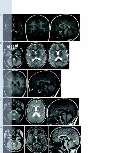
Figure 1. Axial T2-weighted, coronal and sagittal T1-weighted MRI brain images of patient 1
at the age of 6 years (A). Axial T2-weighted, coronal and sagittal T1-weighted MRI brain
images of patient 2 at the age of 3 years (B). Axial T2-weighted and sagittal T1-weighted
MRI brain images of patient 2 at the age of 11 years (C). Axial T2-weighted and sagittal T1-
weighted MRI brain images of a normal child, annotated to show the dorsal brainstem (white
arrow), posterior limb of the internal capsule (black arrow), corpus callosum (black arrow
head) and cerebellar vermis (white arrow head) (D). All the patient studies showed a pattern
of leukodystrophy where the abnormalities were more obvious on the T2-weighted images
and less obvious on the T1-weighted images. Atrophy was global but also particularly notable
in the corpus callosum and cerebellar vermis.
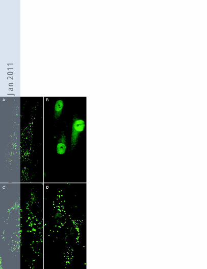
Figure 2. Catalase immunofluorescence in fibroblasts of a control subject (A), a patient with
classical Zellweger Syndrome (B), a patient with peroxisomal acyl-CoA oxidase I deficiency
(C), and patient 3 (D). The phenotype observed in patient 3 with a reduced number of
enlarged import-competent peroxisomes was similar to that observed in all five examined
patients.
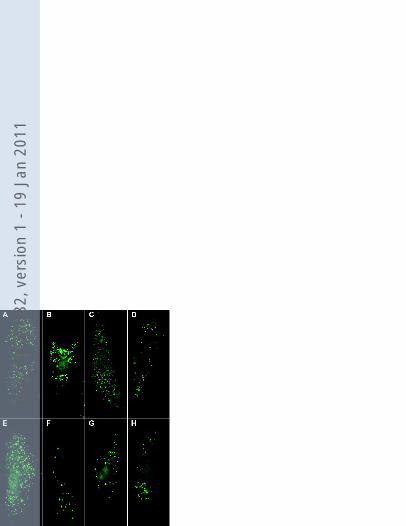
Figure 3. Catalase immunofluorescence in a control cell line (A), patient cell line 4 (B) and
patient cell line 4 transfected with PEX16 cDNA (C) and PEX12 cDNA (D). Expression of
wild type PEX16 cDNA resulted in restoration of number and size of peroxisomes in the
different patient cell lines. Three mutated PEX16 cDNA constructs harboring the mutations
identified in patients 1-4 (PEX16_753delTGT (F), PEX16_865C>A (G) and PEX16_984delG
(H) were expressed in a complete PEX16-deficient cell line and compared with expression of
wild type PEX16 cDNA (E). Restoration of peroxisomes was visualized by means of catalase
peer
-005
5738
2, v
ersi
on 1
- 19
Jan
201
1
24
immunofluorescence 4 days after transfection. Expression of wild type PEX16 cDNA
revealed full restoration of peroxisome formation in more than 95% of the transfected cells.
Expression of the mutated PEX16 cDNAs revealed 30-70 enlarged peroxisomes in
approximately 30% of the transfected cells.
peer
-005
5738
2, v
ersi
on 1
- 19
Jan
201
1
25
SUPPLEMENTED DATA
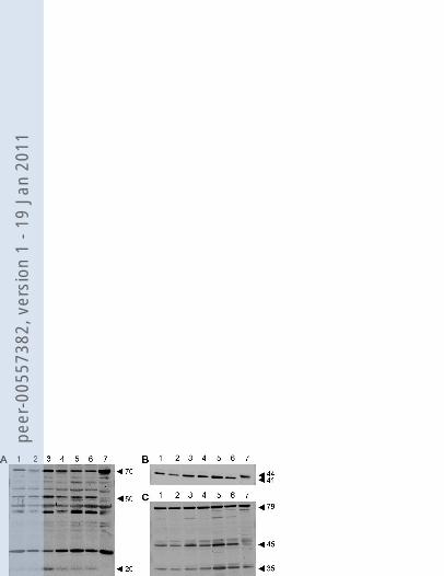
Supplemental Figure S1. Peroxisomal processing. Immunoblot analysis of the peroxisomal
matrix proteins AOXI (A), thiolase I (B) and DBP (C) in fibroblasts homogenates from a
control subject (lane 1), patient 1 (lane 2), patient 3 (lane 3), patient 4 (lane 4), patient 5 (lane
5), patient 6 (lane 6) and a ZS patient (lane 7). The molecular masses of the proteins in kDa
have been indicated (arrows).
Supplemental Table 1. Primer sets used for PEX16 mutation analysis.
Amplicon Primers Exon of gDNA Regions of cDNA
A [-21M13]-GAAGCAGGAAGGAGGGCG 1 and 2
[M13-Rev]-ATTCAGTCATAGCACAAGGTG
B [-21M13]-TGTGAGATCATGTTGGGGAG 3
[M13-Rev]-CTAAGATGGGAATACTCACAC
C [-21M13]-GTCAGAGAAGCTCCCTCCTAG 4 and 5
[M13-Rev]-TACTGTATTCATGCTGGTTGG
D [-21M13]-CCTGCTTGTAGTTCCCTTGAC 6, 7 and 8
[M13-Rev]-ATTATAGCAGAAAGCCCAGTG
E [-21M13]-ACATAGGCGGGGTGGCAG 9
[M13-Rev]-CCCGGACAACACACAGTGC
F [-21M13]-GCACGGTGGTCAGTGAAGG 10 and 11
[M13-Rev]-TATGGCTGCCGAGGCGAG
1 [-21M13]-TGTCGGTGCCGAGGGCAGGAT c.1-19_352
[M13-Rev]-AGCTGGATGAGGGCGATGACA
2 [-21M13]-TGTTCATGGAGATGGGAGCT c.281_721
[M13-Rev]-AAGAGCCAGGGTTTCCACGA
3 [-21M13]-TTTGTACATTGCCCGGCCGCT c.666_1111+59
[M13-Rev]-AGGGAGCCCCTCTTCCCTAAT
peer
-005
5738
2, v
ersi
on 1
- 19
Jan
201
1
peer
-005
5738
2, v
ersi
on 1
- 19
Jan
201
1
peer
-005
5738
2, v
ersi
on 1
- 19
Jan
201
1
peer
-005
5738
2, v
ersi
on 1
- 19
Jan
201
1
peer
-005
5738
2, v
ersi
on 1
- 19
Jan
201
1


