Embed Size (px)
Citation preview
ORIGINAL ARTICLE
IGFBP-3 hypermethylation-derived deficiency mediates cisplatin resistancein non-small-cell lung cancer
I Ibanez de Caceres1, M Cortes-Sempere2, C Moratilla2, R Machado-Pinilla2, V Rodriguez-Fanjul2,C Manguan-Garcıa2, P Cejas3, F Lopez-Rıos4,7, L Paz-Ares5,8, J de CastroCarpeno3, M Nistal6,C Belda-Iniesta3 and R Perona2
1Oncology Research Laboratory, Research Unit, FIB-La Paz University Hospital, Madrid, Spain; 2Instituto de InvestigacionesBiomedicas CSIC/UAM, Translational Oncology Unit CSIC/H La Paz, Madrid, Spain; 3Department of Medical Oncology, La PazUniversity Hospital, Translational Oncology Unit CSIC/H La Paz, Madrid, Spain; 4Department of Pathology, 12 de OctubreHospital, Madrid, Spain; 5Department of Medical Oncology, 12 de Octubre Hospital, Madrid, Spain and 6Department of Pathology,La Paz University Hospital, Madrid, Spain
Cisplatin-based chemotherapy is the paradigm of non-small-cell lung cancer (NSCLC) treatment; however, it alsoinduces de novo DNA-hypermethylation, a process that maybe involved in the development of drug-resistant phenotypesby inactivating genes required for drug-cytotoxicity. Byusing an expression microarray analysis, we aimed toidentify those genes reactivated in a set of two cisplatin(CDDP) resistant and sensitive NSCLC cell lines afterepigenetic treatment. Gene expression, promoter methyla-tion and CDDP-chemoresponse were further analyzed inthree matched sets of sensitive/resistant cell lines, 23 humancancer cell lines and 36 NSCLC specimens. Results revealedspecific silencing by promoter hypermethylation of IGFBP-3in CDDP resistant cells, whereas IGFBP-3 siRNA inter-ference, induced resistance to CDDP in sensitive cells(Po0.001). In addition, we found a strong correlationbetween methylation status and CDDP response in tumorspecimens (Po0.001). Thus, stage I patients, whose tumorsharbor an unmethylated promoter, had a trend towardsincreased disease-free survival (DFS). We report that a lossof IGFBP-3 expression, mediated by promoter-hypermethy-lation, results in a reduction of tumor cell sensitivity tocisplatin in NSCLC. Basal methylation status of IGFBP-3before treatment may be a clinical biomarker and a predictorof the chemotherapy outcome, helping to identify patientswho are most likely to benefit from CDDP therapy alone orin combination with epigenetic treatment.Oncogene (2010) 29, 1681–1690; doi:10.1038/onc.2009.454;published online 21 December 2009
Keywords: CDDP-resistance; IGFBP-3; NSCLC; hyper-methylation
Introduction
Non-small-cell lung cancer (NSCLC) comprises morethan 80% of lung cancers, with 1.2 million new casesworldwide each year (WCR, 2003; Jemal et al., 2008).Cisplatin (CDDP)-based chemotherapy remains thestandard treatment in combination with other chemo-therapeutic agents; however, NSCLC patients frequentlyexhibit chemotherapy resistance. Molecular mechanismsof drug resistance remain unclear and are believed tobe multifactorial, involving host factors, numerousmolecular events and genetic and epigenetic changes(Gottesman, 2002). In addition, chemotherapeutics in-duce epigenetic changes in the promoter area of specificgenes, altering expression and possibly underlyingresistance in many tumor types (Baker et al., 2005).
The aberrant DNA hypermethylation of CpG islandsin the gene promoter region is well-described as analternative mechanism for tumor suppressor genessilencing in cancer cells. This includes lung cancer cells,in which MGMT, p16, RARb, TIMP-3 and DAPKhave been reported as hypermethylated (Merlo et al.,1995; Esteller et al., 1999; Zochbauer-Muller et al.,2001). In addition, drug-induced DNA hypermethyla-tion could be a mechanism of tumor cell response tochemotherapy agents (Nyce, 1997; Strathdee et al., 1999;Shen et al., 2004; Zhang et al., 2004). In both lungadenocarcinoma (HTB-54) and rhabdomyosarcomahuman cells (CCl-136), pulse exposure to CDDP isassociated with drug-induced DNA hypermethylation(Nyce, 1989), an event that has also been reported in vivo(Koul et al., 2004). Therefore, one possible reason forthe development of chemoresistance in NSCLC mightbe the epigenetic inactivation of certain tumor suppres-sor gene as a consequence of chemotherapy treatment.This epigenetic silencing could be reverted by demethy-lating drugs and histone deacetylase inhibitors such as5 Aza-2deoxycytidine (5Aza-dC) and Trichostatine A(TSA). Both drugs act in synergy by depleting methyl-transferase activity (Baylin et al., 1998) and reversingthe formation of transcriptionally repressive chrom-atin structure (Marks et al., 2004). Thus, the associa-tion between promoter-demethylation of hMLH1 by
Received 6 June 2009; revised 27 July 2009; accepted 8 November 2009;published online 21 December 2009
Correspondence: Dr I Ibanez de Caceres, Oncology ResearchLaboratory, FIB-La Paz University Hospital, Instituto de Investiga-ciones Biomedicas Madrid, C/Arturo Duperier, 4, Madrid, 28029, Spain.E-mail: [email protected] address: Department of Pathology, H Clara Campal, Madrid,Spain8Current address: Department of Medical Oncology, H. V. del Rocıo,Sevilla, Spain
Oncogene (2010) 29, 1681–1690& 2010 Macmillan Publishers Limited All rights reserved 0950-9232/10 $32.00
www.nature.com/onc
pharmacological treatment and reversal of drug-resis-tance has been reported in human tumor xenografts(Plumb et al., 2000; Steele et al., 2009).
Many epigenetically silenced tumor suppressor geneinvolved in cancer-chemoresistance likely remain to beidentified as most studies have focused on a limitednumber of candidate genes (Strathdee et al., 1999; Shenet al., 2004). Therefore, a microarray-based screeningpairing the differential genetic profile of sensitive andCDDP-resistant cell lines with an epigenetic reactivationapproach has the advantage of a global analysis thatshould preferentially identify epigenetically silencedgenes as a result of acquired CDDP-chemoresistance.
In this study, we first established two CDDP-resistantcell lines derived from the sensitive H23 and H460NSCLC cell lines. Screening for genes differentiallyexpressed in those resistant cells compared with theirdrug-sensitive parent cells and re-expressed after 5Aza-dC and TSA treatments is a new approach that canprovide signatures for the identification of target genesfor chemotherapy profiling. This process may lead tofurther elucidation of the biology of treatment responsein NSCLC. We have identified a panel of genes showingthis pattern, and found the insulin-like growth factor-bindin protein-3 (IGFBP-3) gene specifically downregu-lated through promoter-hypermethylation in the resis-tant phenotypes.
The biological significance of IGFBP-3 is of greatimportance in controlling cell growth, transformation andsurvival. It is the main IGFBP-family member, physiolo-gically, IGFBP-3 binds IGF-I with stronger affinity thanthe specific IGF-I receptor (IGFI-R) to the cell mem-brane, blocking their interaction and, therefore, the IGF-Imitogenic and anti-apoptotic actions. It is induced bywild-type p53 and interacts with the TGF-b and EGFRpathways (Buckbinder et al., 1995; Rajah et al., 1997;Guix et al., 2008). IGFBP-3 has been recently reported bythe authors and others as a candidate tumor suppressorgene under epigenetic regulation in several tumor typesincluding lung, kidney and ovarian cancer (Chang et al.,2002b; Ibanez de Caceres et al., 2006; Wiley et al., 2006),and although its role in tumor progression has beenstudied, its function in acquired resistance to CDDP,chemotherapy response prediction as well as in drug-resistance phenotype identification remains to becharacterized. In this study we examined the promotermethylation profile of IGFBP-3 regarding sensitivity toCDDP in 27 human cancer cell lines and in a panel of 36NSCLC specimens. We report that a loss of IGFBP-3expression mediated by aberrant promoter hypermethyla-tion results in a reduction of tumor cell sensitivity toCDDP treatment in NSCLC.
Results
Establishment of NSCLC human cell lines resistantto CDDPWe established two human NSCLC cell lines resistant toCDDP, H23R and H460R, which were selected after afinal exposure to 0.2 and 0.5 mg/ml CDDP respectively,
showing approximately four times more drug resistancethan the matched parental cell line (4.07 and 3.89resistant index; Po0.001 and Po0.002) (SupplementaryFigures 2a, b and c). Both cell lines showed a similarCDDP resistant-index to 41R ovarian cancer cells (4.8;Po0.001) (Supplementary Figure 2c).
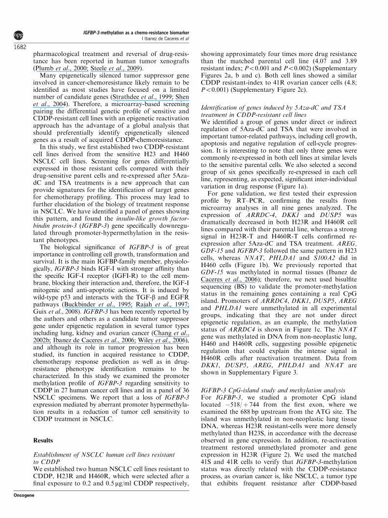
Identification of genes induced by 5Aza-dC and TSAtreatment in CDDP-resistant cell linesWe identified a group of genes under direct or indirectregulation of 5Aza-dC and TSA that were involved inimportant tumor-related pathways, including cell growth,apoptosis and negative regulation of cell-cycle progres-sion. It is interesting to note that only three genes werecommonly re-expressed in both cell lines at similar levelsto the sensitive parental cells. We also selected a secondgroup of six genes specifically re-expressed in each cellline, representing, as expected, significant inter-individualvariation in drug response (Figure 1a).
For gene validation, we first tested their expressionprofile by RT–PCR, confirming the results frommicroarray analyses in all nine genes analyzed. Theexpression of ARRDC-4, DKK1 and DUSP5 wasdramatically decreased in both H23R and H460R celllines compared with their parental line, whereas a strongsignal in H23R-T and H460R-T cells confirmed re-expression after 5Aza-dC and TSA treatment. AREG,GDF-15 and IGFBP-3 followed the same pattern in H23cells, whereas NNAT, PHLDA1 and S100A2 did inH460 cells (Figure 1b). We previously reported thatGDF-15 was methylated in normal tissues (Ibanez deCaceres et al., 2006); therefore, we next used bisulfitesequencing (BS) to validate the promoter-methylationstatus in the remaining genes containing a real CpGisland. Promoters of ARRDC4, DKK1, DUSP5, AREGand PHLDA1 were unmethylated in all experimentalgroups, indicating that they are not under directepigenetic regulation, as an example, the methylationstatus of ARRDC4 is shown in Figure 1c. The NNATgene was methylated in DNA from non-neoplastic lung,H460 and H460R cells, suggesting possible epigeneticregulation that could explain the intense signal inH460R cells after reactivation treatment. Data fromDKK1, DUSP5, AREG, PHLDA1 and NNAT areshown in Supplementary Figure 3.
IGFBP-3 CpG-island study and methylation analysisFor IGFBP-3, we studied a promoter CpG islandlocated !518/" 744 from the first exon, where weexamined the 688 bp upstream from the ATG site. Theisland was unmethylated in non-neoplastic lung tissueDNA, whereas H23R resistant-cells were more denselymethylated than H23S, in accordance with the decreaseobserved in gene expression. In addition, re-activationtreatment restored unmethylated promoter and geneexpression in H23R (Figure 2). We used the matched41S and 41R cells to verify that IGFBP-3-methylationstatus was directly related with the CDDP-resistanceprocess, as ovarian cancer is, like NSCLC, a tumor typethat exhibits frequent resistance after CDDP-based
IGFBP-3-methylation as a chemo-resistance biomarkerI Ibanez de Caceres et al
1682
Oncogene
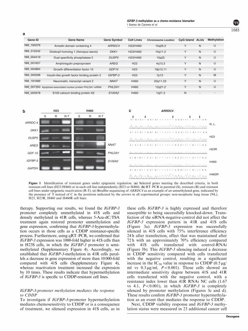
therapy. Supporting our results, we found the IGFBP-3promoter completely unmethylated in 41S cells anddensely methylated in 41R cells, whereas 5-Aza-dC/TSAtreatment again restored promoter unmethylation andgene expression, confirming that IGFBP-3-hypermethyla-tion occurs in those cells as a CDDP resistance-specificprocess. Furthermore, using qRT–PCR, we confirmed thatIGFBP-3 expression was 1000-fold higher in 41S cells thanin H23S cells, in which the IGFBP-3 promoter is semi-methylated (Supplementary Figure 4). Accordingly, weestablished that IGFBP-3-methylation in 41R cells paral-lels a decrease in gene expression of more than 10000-foldcompared with 41S cells (Supplementary Figure 4),whereas reactivation treatment increased the expressionby 10 times. These results indicate that hypermethylationof IGFBP-3 is specific to CDDP-resistant cell lines.
IGFBP-3-promoter methylation mediates the responseto CDDPTo investigate if IGFBP-3-promoter hypermethylationmediates chemosensitivity to CDDP or is a consequenceof treatment, we silenced expression in 41S cells, as in
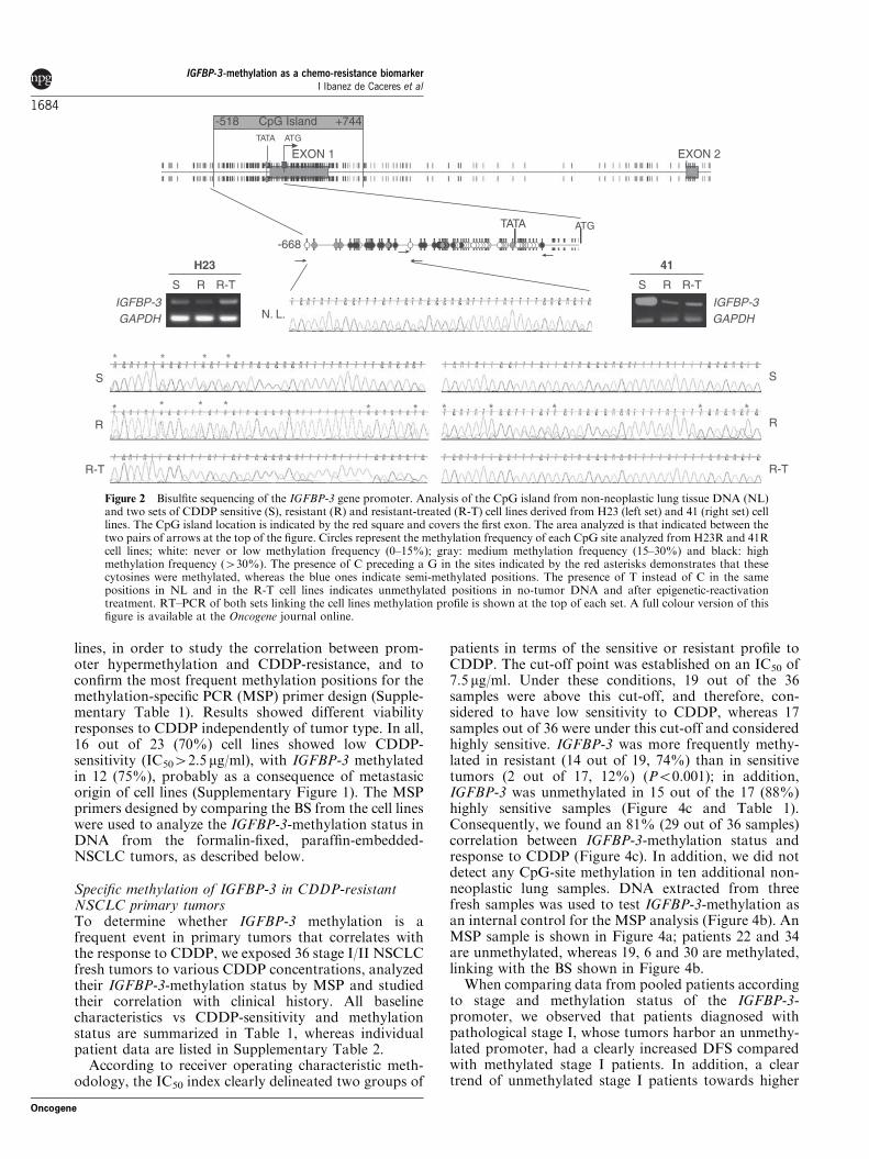
these cells IGFBP-3 is highly expressed and thereforesusceptible to being successfully knocked-down. Trans-fection of the siRNA-negative-control did not affect theIGFBP-3 expression pattern in 41R and 41S cells(Figure 3a). IGFBP-3 expression was successfullysilenced in 41S cells with 75% interference efficiency24 h after transfection, effect that was maintained after72 h with an approximately 70% efficiency comparedwith 41S cells transfected with control-RNAi(Figure 3b). This IGFBP-3 silencing induced a decreasein CDDP sensitivity compared with cells transfectedwith the negative control, resulting in a significantincrease in the IC50 value in response to CDDP (0.5 mg/ml vs 0.3 mg/ml, Po0.001). Those cells showed anintermediate sensitivity degree between 41S and 41Rcells transfected with the negative control, with aresistance index lower than 41R RNAi NC cells (1.67vs 4.1, Po0.001), in which IGFBP-3 is completelysilenced by promoter methylation (Figures 3c and d).These results confirm IGFBP-3 promoter hypermethyla-tion as an event that mediates the response to CDDP.
Next, CDDP viability response and IGFBP-3 methy-lation status were measured in 23 additional cancer cell
NM_183376
NM_012242
NM_004419
NM_001657
NM_004864
NM_000598
NM_181689
NM_007350
NM_005978
Arrestin domain containing 4
Dickkopf homolog 1 (Xenopus laevis)
Dual specificity phosphatase 5
Amphiregulin preproprotein
Growth differentiation factor 15
Insulin-like growth factor binding protein 3
Neuronatin, transcript variant 2
Apoptosis-associated nuclear protein PHLDA1 mRNA
S100 calcium binding protein A2
ARRDC4
DKK1
DUSP5
AREG
GDF15
IGFBP-3
NNAT
PHLDA1
S100A2
U
U
U
U
U
M
U
U
-
Gene NameGene ID Gene Symbol Methylation
15q26.3
10q11.2
10q25
4q13.3
19p13.11
7p13
20q11.23
12q21.2
1q21.3
Chromosome Location
H23/H460
H23/H460
H23/H460
H23
H23
H23
H460
H460
H460
Cell Lines
Y
Y
Y
Y
Y
Y
Y
Y
N
CpG Island
N
N
N
N
N
N
N
N
-
ALUs
ARRDC-4
AREG
GAPDH
IGFBP-3
GDF-15
DKK1
DUSP5
H23 H460 ARRDC4
NNAT
PHLDA1
S100A2
N. L.
H460
H460R
H23
H23R
S R-TRS R-TR
Figure 1 Identification of resistant genes under epigenetic regulation. (a) Selected genes meeting the described criteria, in bothresistant cell lines (H23/H460) or in each cell line independently (H23 or H460). (b) RT–PCR in parental (S), resistant (R) and resistantcell lines under epigenetic reactivation (R-T). (c) Bisulfite sequencing of ARRDC4 as an example of an unmethylated gene, indicated bythe presence of T instead of C in the positions indicated by the arrows in all experimental groups: non-neoplastic lung tissue (NL),H23, H23R, H460 and H460R cell lines.
IGFBP-3-methylation as a chemo-resistance biomarkerI Ibanez de Caceres et al
1683
Oncogene
lines, in order to study the correlation between prom-oter hypermethylation and CDDP-resistance, and toconfirm the most frequent methylation positions for themethylation-specific PCR (MSP) primer design (Supple-mentary Table 1). Results showed different viabilityresponses to CDDP independently of tumor type. In all,16 out of 23 (70%) cell lines showed low CDDP-sensitivity (IC5042.5 mg/ml), with IGFBP-3 methylatedin 12 (75%), probably as a consequence of metastasicorigin of cell lines (Supplementary Figure 1). The MSPprimers designed by comparing the BS from the cell lineswere used to analyze the IGFBP-3-methylation status inDNA from the formalin-fixed, paraffin-embedded-NSCLC tumors, as described below.
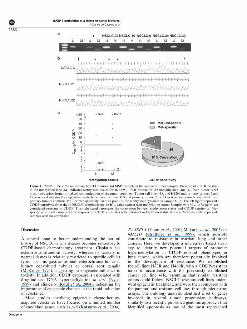
Specific methylation of IGFBP-3 in CDDP-resistantNSCLC primary tumorsTo determine whether IGFBP-3 methylation is afrequent event in primary tumors that correlates withthe response to CDDP, we exposed 36 stage I/II NSCLCfresh tumors to various CDDP concentrations, analyzedtheir IGFBP-3-methylation status by MSP and studiedtheir correlation with clinical history. All baselinecharacteristics vs CDDP-sensitivity and methylationstatus are summarized in Table 1, whereas individualpatient data are listed in Supplementary Table 2.
According to receiver operating characteristic meth-odology, the IC50 index clearly delineated two groups of
patients in terms of the sensitive or resistant profile toCDDP. The cut-off point was established on an IC50 of7.5 mg/ml. Under these conditions, 19 out of the 36samples were above this cut-off, and therefore, con-sidered to have low sensitivity to CDDP, whereas 17samples out of 36 were under this cut-off and consideredhighly sensitive. IGFBP-3 was more frequently methy-lated in resistant (14 out of 19, 74%) than in sensitivetumors (2 out of 17, 12%) (Po0.001); in addition,IGFBP-3 was unmethylated in 15 out of the 17 (88%)highly sensitive samples (Figure 4c and Table 1).Consequently, we found an 81% (29 out of 36 samples)correlation between IGFBP-3-methylation status andresponse to CDDP (Figure 4c). In addition, we did notdetect any CpG-site methylation in ten additional non-neoplastic lung samples. DNA extracted from threefresh samples was used to test IGFBP-3-methylation asan internal control for the MSP analysis (Figure 4b). AnMSP sample is shown in Figure 4a; patients 22 and 34are unmethylated, whereas 19, 6 and 30 are methylated,linking with the BS shown in Figure 4b.
When comparing data from pooled patients accordingto stage and methylation status of the IGFBP-3-promoter, we observed that patients diagnosed withpathological stage I, whose tumors harbor an unmethy-lated promoter, had a clearly increased DFS comparedwith methylated stage I patients. In addition, a cleartrend of unmethylated stage I patients towards higher
H23
S
N. L.
R
R-T
S
R
R-T
IGFBP-3GAPDH
41
IGFBP-3GAPDH
ATGTATA
-668
EXON 2TATA
EXON 1ATG
+744-518 CpG Island
* * **
*
*** **
S R-TRS R-TR
* **
* *
Figure 2 Bisulfite sequencing of the IGFBP-3 gene promoter. Analysis of the CpG island from non-neoplastic lung tissue DNA (NL)and two sets of CDDP sensitive (S), resistant (R) and resistant-treated (R-T) cell lines derived from H23 (left set) and 41 (right set) celllines. The CpG island location is indicated by the red square and covers the first exon. The area analyzed is that indicated between thetwo pairs of arrows at the top of the figure. Circles represent the methylation frequency of each CpG site analyzed from H23R and 41Rcell lines; white: never or low methylation frequency (0–15%); gray: medium methylation frequency (15–30%) and black: highmethylation frequency (430%). The presence of C preceding a G in the sites indicated by the red asterisks demonstrates that thesecytosines were methylated, whereas the blue ones indicate semi-methylated positions. The presence of T instead of C in the samepositions in NL and in the R-T cell lines indicates unmethylated positions in no-tumor DNA and after epigenetic-reactivationtreatment. RT–PCR of both sets linking the cell lines methylation profile is shown at the top of each set. A full colour version of thisfigure is available at the Oncogene journal online.
IGFBP-3-methylation as a chemo-resistance biomarkerI Ibanez de Caceres et al
1684
Oncogene
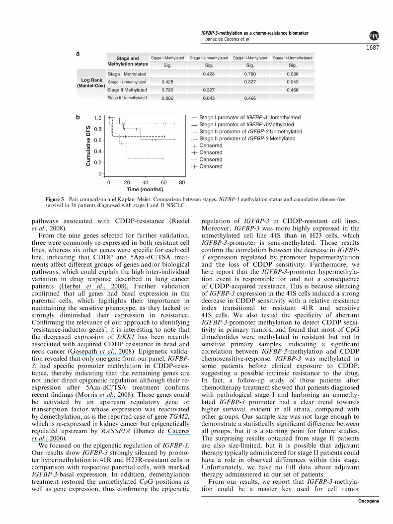
survival was evident in all strata (Figure 5b), butwithout statistical significance, mainly because of asmall sample size (Figure 5a). Surprisingly, for stage II
patients we observed the opposite scenario: patients thatharbor an unmethylated promoter had a lower DFSthan those methylated within the same stage.
Cel
l Via
bilit
y%
of U
ntre
ated
Con
trol
s
0
25
50
75
100
CDDP (µg/ml)0 0.5 1 1.5 2 41R RNAi NC
IC50 (µg/ml) ± SD CDDP RI P-value
41R RNAi NC41S RNAi IGFBP-341S RNAi NC
300
200
100
41R 41SM - -NC NC RNAi
RNAi
IGFBP-3
GAPDH
300
bp 200
100
41R 41SM NC NC
IGFBP-3
IGFBP-3
18% 100% 33%
GAPDH
300200
100
RNAi41R 41S
M NC NC IGFBP-3
bp
Time: 24h
Time: 72h
IGFBP-3
GAPDH
8% 100%25%bp
41S RNAi IGFBP-3
41S RNAi NC 0.3 ± 0.04
0.5 ± 0.07
1.2 ± 0.18 4.1
1.67
-
<0.001
<0.001
-
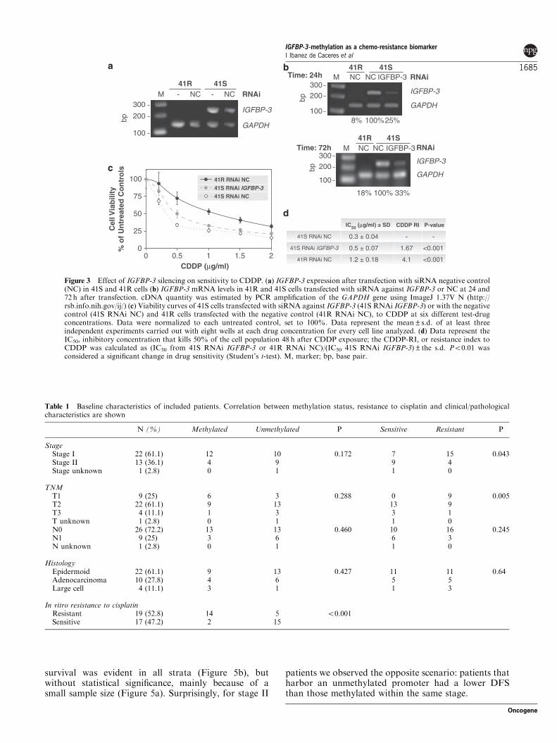
Figure 3 Effect of IGFBP-3 silencing on sensitivity to CDDP. (a) IGFBP-3 expression after transfection with siRNA negative control(NC) in 41S and 41R cells (b) IGFBP-3 mRNA levels in 41R and 41S cells transfected with siRNA against IGFBP-3 or NC at 24 and72 h after transfection. cDNA quantity was estimated by PCR amplification of the GAPDH gene using ImageJ 1.37V N (http://rsb.info.nih.gov/ij/) (c) Viability curves of 41S cells transfected with siRNA against IGFBP-3 (41S RNAi IGFBP-3) or with the negativecontrol (41S RNAi NC) and 41R cells transfected with the negative control (41R RNAi NC), to CDDP at six different test-drugconcentrations. Data were normalized to each untreated control, set to 100%. Data represent the mean±s.d. of at least threeindependent experiments carried out with eight wells at each drug concentration for every cell line analyzed. (d) Data represent theIC50, inhibitory concentration that kills 50% of the cell population 48 h after CDDP exposure; the CDDP-RI, or resistance index toCDDP was calculated as (IC50 from 41S RNAi IGFBP-3 or 41R RNAi NC)/(IC50 41S RNAi IGFBP-3)±the s.d. Po0.01 wasconsidered a significant change in drug sensitivity (Student’s t-test). M, marker; bp, base pair.
Table 1 Baseline characteristics of included patients. Correlation between methylation status, resistance to cisplatin and clinical/pathologicalcharacteristics are shown
N (%) Methylated Unmethylated P Sensitive Resistant P
StageStage I 22 (61.1) 12 10 0.172 7 15 0.043Stage II 13 (36.1) 4 9 9 4Stage unknown 1 (2.8) 0 1 1 0
TNMT1 9 (25) 6 3 0.288 0 9 0.005T2 22 (61.1) 9 13 13 9T3 4 (11.1) 1 3 3 1T unknown 1 (2.8) 0 1 1 0N0 26 (72.2) 13 13 0.460 10 16 0.245N1 9 (25) 3 6 6 3N unknown 1 (2.8) 0 1 1 0
HistologyEpidermoid 22 (61.1) 9 13 0.427 11 11 0.64Adenocarcinoma 10 (27.8) 4 6 5 5Large cell 4 (11.1) 3 1 1 3
In vitro resistance to cisplatinResistant 19 (52.8) 14 5 o0.001Sensitive 17 (47.2) 2 15
IGFBP-3-methylation as a chemo-resistance biomarkerI Ibanez de Caceres et al
1685
Oncogene
Discussion
A central issue to better understanding the naturalhistory of NSCLC is why disease becomes refractory toCDDP-based chemotherapy treatment. Cisplatin hasextensive antitumoral activity, whereas its toxicity innormal tissues is relatively restricted to specific cellulartypes such as gastrointestinal enterochromaffin cells,kidney convoluted tubules or dorsal root ganglia(McKeage, 1995), suggesting an epigenetic influence intoxicity. In addition, CDDP exposure is associated withdrug-induced DNA hypermethylation in vitro (Nyce,1989) and clinically (Koul et al., 2004), indicating theimportance of epigenetic changes in the rapid inductionof resistance.
Most studies involving epigenetic chemotherapy-acquired resistance have focused on a limited numberof candidate genes, such as p16 (Katsaros et al., 2004),
RASSF1A (Yoon et al., 2001; Makarla et al., 2005) orhMLH1 (Strathdee et al., 1999), which possiblycontribute to resistance in ovarian, lung and othercancers. Here, we developed a microarray-based strat-egy to identify new potential targets of promoterhypermethylation in CDDP-resistant phenotypes inlung cancer, which are therefore potentially involvedin the development of resistance. We establishedthe cell lines H23R and H460R, with a CDDP-resistantindex in accordance with the previously establishedcancer cell line 41R, assuming that similar resistantevents could follow. NSCLC-resistant cell lines under-went epigenetic treatment, and were then compared withthe parental and resistant cell lines through microarrayassays. The ontology analysis identified a set of genesinvolved in several tumor progression pathways,similarly to a recently published genomic approach thatidentified apoptosis as one of the most represented
– +
NSCLC.21
NSCLC.22
NSCLC.6
NSCLC.22 NSCLC.19 NSCLC.6 NSCLC.34 NSCLC.30MU MU MU MU MU MU MU
IC50
µg/
ml C
DD
P
Methylation Status
7.5
n=4
n=3
n=9
n=11
0
67
4
2
5
3
1
810203040
>9050
U M
81% Correlation
Met-UnspecificMet-specific
7
29
n=2
CDDP sensitivity
Figure 4 MSP of IGFBP-3 in primary NSCLC tumors. (a) MSP example in the analyzed tumor samples: Presence of a PCR productin the methylated lane (M) indicates methylated alleles for IGFBP-3. PCR product in the unmethylated lane (U) from tumor DNAmost likely arises from normal cell contamination of the tumor specimen. Tumor cell lines 41R and H1299 and primary tumors 5 and15 were used indistinctly as positive controls, whereas cell line 41S and primary tumors 21 y 29 as negative controls. (b) BS of threeprimary tumors confirms MSP primer specificity. Arrows point to the methylated cytosines in sample 6. (c) The left figure representsCDDP sensitivity from the 36 NSCLC samples using the IC50 value against their methylation status. Samples with IC50 X7.5mg/ml areconsidered resistant to CDDP. The right panel represents the correlation between methylation status and CDDP sensitivity. Met-specific represents samples whose response to CDDP correlates with IGFBP-3 methylation status, whereas Met-unspecific representssamples with no correlation.
IGFBP-3-methylation as a chemo-resistance biomarkerI Ibanez de Caceres et al
1686
Oncogene
pathways associated with CDDP-resistance (Riedelet al., 2008).
From the nine genes selected for further validation,three were commonly re-expressed in both resistant celllines, whereas six other genes were specific for each cellline, indicating that CDDP and 5Aza-dC/TSA treat-ments affect different groups of genes and/or biologicalpathways, which could explain the high inter-individualvariation in drug response described in lung cancerpatients (Herbst et al., 2008). Further validationconfirmed that all genes had basal expression in theparental cells, which highlights their importance inmaintaining the sensitive phenotype, as they lacked orstrongly diminished their expression in resistance.Confirming the relevance of our approach to identifying‘resistance-inductor-genes’, it is interesting to note thatthe decreased expression of DKK1 has been recentlyassociated with acquired CDDP resistance in head andneck cancer (Gosepath et al., 2008). Epigenetic valida-tion revealed that only one gene from our panel, IGFBP-3, had specific promoter methylation in CDDP-resis-tance, thereby indicating that the remaining genes arenot under direct epigenetic regulation although their re-expression after 5Aza-dC/TSA treatment confirmsrecent findings (Morris et al., 2008). Those genes couldbe activated by an upstream regulatory gene ortranscription factor whose expression was reactivatedby demethylation, as is the reported case of gene TGM2,which is re-expressed in kidney cancer but epigeneticallyregulated upstream by RASSF1A (Ibanez de Cacereset al., 2006).
We focused on the epigenetic regulation of IGFBP-3.Our results show IGFBP-3 strongly silenced by promo-ter hypermethylation in 41R and H23R-resistant cells incomparison with respective parental cells, with markedIGFBP-3-basal expression. In addition, demethylationtreatment restored the unmethylated CpG positions aswell as gene expression, thus confirming the epigenetic
regulation of IGFBP-3 in CDDP-resistant cell lines.Moreover, IGFBP-3 was more highly expressed in theunmethylated cell line 41S than in H23 cells, whichIGFBP-3-promoter is semi-methylated. Those resultsconfirm the correlation between the decrease in IGFBP-3 expression regulated by promoter hypermethylationand the loss of CDDP sensitivity. Furthermore, wehere report that the IGFBP-3-promoter hypermethyla-tion event is responsible for and not a consequenceof CDDP-acquired resistance. This is because silencingof IGFBP-3 expression in the 41S cells induced a strongdecrease in CDDP sensitivity with a relative resistanceindex transitional to resistant 41R and sensitive41S cells. We also tested the specificity of aberrantIGFBP-3-promoter methylation to detect CDDP sensi-tivity in primary tumors, and found that most of CpGdinucleotides were methylated in resistant but not insensitive primary samples, indicating a significantcorrelation between IGFBP-3-methylation and CDDPchemosensitive-response. IGFBP-3 was methylated insome patients before clinical exposure to CDDP,suggesting a possible intrinsic resistance to the drug.In fact, a follow-up study of those patients afterchemotherapy treatment showed that patients diagnosedwith pathological stage I and harboring an unmethy-lated IGFBP-3 promoter had a clear trend towardshigher survival, evident in all strata, compared withother groups. Our sample size was not large enough todemonstrate a statistically significant difference betweenall groups, but it is a starting point for future studies.The surprising results obtained from stage II patientsare also size-limited, but it is possible that adjuvanttherapy typically administered for stage II patients couldhave a role in observed differences within this stage.Unfortunately, we have no full data about adjuvanttherapy administered in our set of patients.
From our results, we report that IGFBP-3-methyla-tion could be a master key used for cell tumor
Stage I-Methylated Stage I-Unmethylated Stage II-Methylated Stage II-Unmethylated
Sig. Sig. Sig. Sig.
Stage I Methylated 0.428 0.780 0.086
Stage I Unmethylated 0.428 0.327 0.043
Stage II Methylated 0.780 0.327 0.466
Stage II Unmethylated 0.086 0.043 0.466
Stage andMethylation status
Log Rank(Mantel-Cox)
Cum
ulat
ive
DFS
0 20 40 60 80
0
0.4
0.2
0.8
0.6
1.0
Time (months)
Stage I promoter of IGFBP-3 MethylatedStage I promoter of IGFBP-3 Unmethylated
Stage II promoter of IGFBP-3 MethylatedStage II promoter of IGFBP-3 Unmethylated
CensoredCensored
CensoredCensored
Figure 5 Pair comparison and Kaplan–Meier. Comparison between stages, IGFBP-3 methylation status and cumulative disease-freesurvival in 36 patients diagnosed with stage I and II NSCLC.
IGFBP-3-methylation as a chemo-resistance biomarkerI Ibanez de Caceres et al
1687
Oncogene
progression in conjunction with its biological functions.Induction of IGFBP-3 gene expression by wild-type p53is associated with enhanced secretion of an active formof IGFBP-3 capable of inhibiting mitogenic signaling bythe IGF-I (Buckbinder et al., 1995). The CGH profile ofcell lines 41S and 41R indicate no changes in chromo-some 17, whereas p53 is located at (17p13) (Leyland-Jones et al., 1999), indicating that IGFBP-3 promotermethylation at the p53 regulatory element could causegene silencing resistant to p53 in those cell lines. TheIGF-I signaling pathway could mediate chemotherapyresistance of NSCLC cells through the activation ofAkt/mTOR-mediated synthesis of survival proteins,thus protecting NSCLC cells from apoptosis inducedby several drugs, as reported previously with drugs thatinhibit tyrosine-kinase receptors including EGFR orIGFIR (Morgillo et al., 2006, 2007). Those authorssuggest that resistance is caused by the activation ofalternative cell survival signaling mechanisms, becausethose drugs do not inhibit proliferation at dosessufficient to suppress EGFR activation. In fact, weobserved decreased expression of IGFBP-3 in chemore-sistant cell lines, therefore a higher amount of IGF-Ishould be available to join its own receptor and stronglyactivate the IGFIR pathway, inducing survival andmaintaining proliferation in CDDP-resistant cells. Inaddition, in a p53 and/or IGF-I-independent pathway,methylation could also be the mechanism responsiblefor inhibiting the apoptosis mediated by IGFBP-3through the activation of the TGF-b receptor TbR-V,(Rajah et al., 1997; Huang and Huang, 2005).
IGFBP-3-methylation has been correlated with clin-icopathological features indicative of poor prognosis inprostate and ovarian cancers (Wiley et al., 2006; Perryet al., 2007) and in the early stages of NSCLC patients(Chang et al., 2002a, 2002b), likely indicating that thosepatients have intrinsic resistance to CDDP. However,we know that those patients with unmethylated IGFBP-3in early stages could also progress towards a methylatedpromoter after CDDP treatment, thus showing acquiredresistance. A follow-up study after chemotherapy treat-ment measuring aberrant IGFBP-3 methylation in tumorDNA circulating in body fluids such as blood, bronchio-alveolar fluid or saliva could identify this situation whenCDDP-resistant cells arise, providing a non-invasive testto predict chemotherapy resistance.
In this study, we report that IGFBP-3 has an inverseeffect on the risk of NSCLC CDDP chemoresistancedevelopment. CDDP induces changes in IGFBP-3expression mediated by the acquisition of promoterhypermethylation, which promotes resistance to CDDPthrough various biological pathways. Therefore, basalmethylation status of the IGFBP-3 promoter beforechemotherapy treatment may be a clinical biomarkerpredictor of the chemotherapy outcome of NSCLCpatients, identifying those who are most likely to benefitfrom CDDP therapy. These results represent a newapplication of epigenetic cell control in chemotherapyresistance. This application involves the possible use ofnew highly sensitive and non-invasive tests based on thedetection of aberrant methylation in tumor-circulating
DNA (Carvalho et al., 2008), as well as the use of agentsthat reverse epigenetic changes. These agents haveshown promising results in a mouse model of lungcarcinogenesis and are being tested in lung cancerpatients (Belinsky et al., 2003)
Materials and methods
Cell culture and viability to CDDPA total of 23 human cancer cell lines (Supplementary Figure1), representing nine different cancer types, were purchasedfrom the ATCC (Manassas, VA, USA) or the ECACC (Sigma-Aldrich, Madrid, Spain) and cultured as recommended. TheCDDP-resistant variants H23R and H460R were establishedfollowing the methodology described previously (Plasenciaet al., 2006) and further detailed in Supplementary Materialsand Methods (Roninson, 2003; Chattopadhyay et al., 2006;Levina et al., 2008). To validate the results obtained from theresistant cell lines established in our laboratory, we also usedCDDP-sensitive and resistant ovarian cancer cell lines 41Mand 41MR, hereafter called 41S and 41R respectively, kindlyprovided by Dr Lloyd R Kelland (UK), and maintained inDMEM supplemented with 10% FBS.
NSCLC clinical samples and data collectionFresh and formalin-fixed, paraffin-embedded surgical speci-mens were obtained from 36 patients who had undergone acomplete resection (R0) for a histologically confirmed, earlyNSCLC. All patients had both a perioperative PET-CT scanshowing localized disease and a pathological confirmation ofstage I/II. In addition, an age of 18 years or older,intraoperative mediastinal-node dissection for reliable med-iastinal staging or biopsy of nodes at N3 without any evidenceof disease was inclusion criteria. Stage III, any involvement oftracheobronchial angle nodes (station 10), mixed histologicalfeatures and previous diagnosis of cancer within the last 5years were exclusion criteria. Histological slides obtained fromeach block were reviewed by an expert pathologist (M Nistal)to confirm the diagnosis and to guarantee at least 90%tumoral content. A total of 10 samples obtained frompulmonary biopsies with non-neoplastic lung pathology wereused as control tissues. Follow-up was carried out according tothe criteria used in the Medical Oncology Division from theUniversity Hospitals La Paz and 12 de Octubre, includingclinical assessments and thorax CT every 3 months for 2 yearsand every 6 months thereafter. Clinical, pathological andradiological data were recorded by an independent observer atthe H. La Paz and blinded for statistical analysis.
5Aza-2dC and TSA treatment5Aza-dC and TSA (Sigma-Aldrich) were stored as 5mM and330 mM stock solutions, respectively. For re-expression studies,resistant cell lines H23R, H460R and 41R were split to lowdensity and exposed to 5Aza-dC and TSA or to PBS andethanol (mock cells) as described previously (Ibanez deCaceres et al., 2006).
Oligonucleotide array hybridization and gene selectionParental H-23 and H-460 cell lines, derived CDDP-resistantcell lines and resistant cell lines treated with 5Aza-dC and TSAwere used for the oligonucleotide array hybridization. TotalRNA was isolated and purified as described (Ibanez de Cacereset al., 2006) and used for the microarray and RT–PCRanalysis. The microarray assay was carried out using the
IGFBP-3-methylation as a chemo-resistance biomarkerI Ibanez de Caceres et al
1688
Oncogene
Agilent gene expression platform 4X44 Whole HumanGenome Oligo Microarray Kit, representing the 41 000 knowngenes and transcripts in the human genome. Sample amplifica-tion, labeling and scanning procedures followed the Agilentmicroarray protocol and are further described in Supplemen-tary Materials and Methods.
Reverse transcription and qRT–PCRSemiquantitative and real-time RT–PCR assays were carriedout in all the experimental groups. In all, 5 mg of total RNAwas reverse transcribed using oligo (dT)24 primer and Super-script II reverse transcriptase (Invitrogen, Carlsbad, CA,USA). For quantitative real-time RT–PCR analysis, theamount of 1 mg of total RNA was retrotranscribed by High-Capacity cDNA Archive Kit (Applied Biosystems, Madrid,Spain). Next, each cDNA sample was analyzed in triplicateusing the ABI PRISM 7700 Sequence Detector (PE AppliedBiosystems, Madrid, Spain). Real-time PCR was carriedout using Taqman Universal PCR Master Mix (AppliedBiosystems, Madrid, Spain), containing ROX to normalizeemissions. Primers and probe for IGFBP-3 expression analysiswere purchased from Applied Biosystems (Assay ID:Hs00181211_m1). Relative gene expression quantificationwas calculated according to the comparative threshold cyclemethod (2 !DDCt) using GADPH and 18S rRNA as endogenouscontrol genes and the H23S and 41S cell lines as calibrators.Normalized expression values were determined as follows:10–(DCt sample!DCt calibrator), where DCt values were calculated bysubtracting the Ct value of the target gene from the value ofthe mean between endogenous control genes. PCR settings andprimer sequences are listed in Supplementary Materials andMethods and Supplementary Table 1.
siRNA transfectionThe 41S and 41R cell lines were transfected with 150nM IGFBP-3siRNA or negative control siRNA (HSS105266, Stealth SelectRNAi and 2935–200, stealth RNAi Negative Control, Invitrogen,Barcelona, Spain) according to manufacturer directions. For theviability assay, cells were seeded in 24-well dishes at 50000cells per well 24h after transfection with IGFBP-3 siRNA orsiRNA control, then treated with six different doses of CDDPfor an additional 48h and stained following the method describedpreviously (Chattopadhyay et al., 2006). Cell viability wasestimated relative to the density recorded over the sameexperimental group without drug exposure at same period oftime (24h following seeding of cells). Simultaneously, sameexperimental groups were seeded at 500000 cells per plate andharvested at same incubation periods, 24 and 72h after siRNAtransfection, as a time-course to test by RT–PCR the preservationof siRNA effects on IGFBP-3 expression.
BS and methylation-specific PCRDNA from human cancer cell lines, NSCLC primary speci-mens and non-neoplastic lung tissues were isolated, bisulfitemodified and then used for BS and MSP analysis as described(Ibanez de Caceres et al., 2006). PCR settings and primersequences for BS and MSP are listed in SupplementaryMaterials and Methods and Supplementary Table 1.
Culture and CDDP treatment of human cancer tissuesTo measure the sensitivity of the 36 primary NSCLC samplesto CDDP, fresh tumors were minced, passed through a nylonmesh and enzyme disaggregated with collagenase type II andhyaluronidase in Dulbeco’s modified Eagle’s medium nutrientmixture F-12/Ham media (Sigma-Aldrich) with antibiotics.Cells were resuspended in 96-well microtiter plates followed byexposure to various concentrations of CDDP as described inSupplementary Materials and Methods.
Statistical analysisDifferentially expressed genes from the microarray analysiswere selected using as a statistical method the t-test unpairedalgorithm with Benjamini Hochberg as the FDR correctionmethod for multiple testing corrections. Statistically significant(Po0.05 as adjusted P-value) genes were selected. Calculationswere carried out using the gene expression analysis softwareGeneSpring (further details in Oligonucleotide array hybridiza-tion and gene selection from Supplementary Materials andMethods).Receiver operating characteristic curves from NSCLC
patients were obtained to identify the relation betweensensitivity and resistance rate at a specific cut-off valueestablished according to the best combination of sensitivityand false-positive rate (1–specificity) (1;0).The Kaplan–Meier method was used to plot cumulative
disease-free survival curves for patients diagnosed with stage Iand unmethylated IGFBP-3 promoter, stage I methylated,stage II unmethylated and stage II methylated. Differenceswere compared with the log-rank method for every stratum.For patients without any evidence of relapse at the time ofanalysis, data on disease-free survival were censored at thetime of last contact. Disease-free survival was defined as thetime from surgery to clinical, radiological or histologicalevidence of relapse. Discrete variables (histology, T, N, stage,gender and methylation status at the IGFBP3 promoter andin vitro sensitivity/resistance to CDDP) were compared withthe w2 test and corrections with Fisher’s exact test were madewhen needed. Statistical significance was defined as Po0.05.Statistical analyses were done by using the SPSS software(version 17.0).
Conflict of interest
The authors declare no conflict of interest.
Acknowledgements
We gratefully acknowledge Javier Perez for the artwork and toJ Siegfried, for the English correction of the manuscript.Ibanez de Caceres was partially supported by the Fondo deInvestigacion Sanitaria (ISCIII) through the ‘Miguel Servet’program (CP 08/000689; PI-717). Note: Supported by FISPI06-1234, PI08-1485, PS09/00472 and Fundacion MedicaMutua Madrilena.
References
(2003). The World Cancer Report—the major findings. Cent Eur JPublic Health 11: 177–179.
Baker EK, Johnstone RW, Zalcberg JR, El-Osta A. (2005). Epigeneticchanges to the MDR1 locus in response to chemotherapeutic drugs.Oncogene 24: 8061–8075.
Baylin SB, Herman JG, Graff JR, Vertino PM, Issa JP. (1998).Alterations in DNA methylation: a fundamental aspect ofneoplasia. Adv Cancer Res 72: 141–196.
Belinsky SA, Klinge DM, Stidley CA, Issa JP, Herman JG, March THet al (2003). Inhibition of DNA methylation and histone
IGFBP-3-methylation as a chemo-resistance biomarkerI Ibanez de Caceres et al
1689
Oncogene
deacetylation prevents murine lung cancer. Cancer Res 63:7089–7093.
Buckbinder L, Talbott R, Velasco-Miguel S, Takenaka I, Faha B,Seizinger BR et al (1995). Induction of the growth inhibitorIGF-binding protein 3 by p53. Nature 377: 646–649.
Carvalho AL, Jeronimo C, Kim MM, Henrique R, Zhang Z,Hoque MO et al (2008). Evaluation of promoter hypermethylationdetection in body fluids as a screening/diagnosis tool for head andneck squamous cell carcinoma. Clin Cancer Res 14: 97–107.
Chang YS, Kong G, Sun S, Liu D, El-Naggar AK, Khuri FR et al(2002a). Clinical significance of insulin-like growth factor-bindingprotein-3 expression in stage I non-small cell lung cancer. ClinCancer Res 8: 3796–3802.
Chang YS, Wang L, Liu D, Mao L, Hong WK, Khuri FR et al(2002b). Correlation between insulin-like growth factor-bindingprotein-3 promoter methylation and prognosis of patients with stageI non-small cell lung cancer. Clin Cancer Res 8: 3669–3675.
Chattopadhyay S, Machado-Pinilla R, Manguan-Garcia C, Belda-Iniesta C, Moratilla C, Cejas P et al (2006). MKP1/CL100 controlstumor growth and sensitivity to cisplatin in non-small-cell lungcancer. Oncogene 25: 3335–3345.
Esteller M, Hamilton SR, Burger PC, Baylin SB, Herman JG. (1999).Inactivation of the DNA repair gene O6-methylguanine-DNAmethyltransferase by promoter hypermethylation is a commonevent in primary human neoplasia. Cancer Res 59: 793–797.
Gosepath EM, Eckstein N, Hamacher A, Servan K, von Jonquieres G,Lage H et al (2008). Acquired cisplatin resistance in the head-neckcancer cell line Cal27 is associated with decreased DKK1 expressionand can partially be reversed by overexpression of DKK1. Int JCancer 123: 2013–2019.
Gottesman MM. (2002). Mechanisms of cancer drug resistance. AnnuRev Med 53: 615–627.
Guix M, Faber AC, Wang SE, Olivares MG, Song Y, Qu S et al(2008). Acquired resistance to EGFR tyrosine kinase inhibitors incancer cells is mediated by loss of IGF-binding proteins. J ClinInvest 118: 2609–2619.
Herbst RS, Heymach JV, Lippman SM. (2008). Lung cancer. N Engl JMed 359: 1367–1380.
Huang SS, Huang JS. (2005). TGF-beta control of cell proliferation.J Cell Biochem 96: 447–462.
Ibanez de Caceres I, Dulaimi E, Hoffman AM, Al-Saleem T,Uzzo RG, Cairns P. (2006). Identification of novel target genes byan epigenetic reactivation screen of renal cancer. Cancer Res 66:5021–5028.
Jemal A, Siegel R, Ward E, Hao Y, Xu J, Murray T et al (2008).Cancer statistics, 2008. CA Cancer J Clin 58: 71–96.
Katsaros D, Cho W, Singal R, Fracchioli S, Rigault De La Longrais IA,Arisio R et al (2004). Methylation of tumor suppressor gene p16 andprognosis of epithelial ovarian cancer. Gynecol Oncol 94: 685–692.
Koul S, McKiernan JM, Narayan G, Houldsworth J, Bacik J,Dobrzynski DL et al (2004). Role of promoter hypermethylationin cisplatin treatment response of male germ cell tumors. MolCancer 3: 16.
Levina V, Marrangoni AM, DeMarco R, Gorelik E, Lokshin AE. (2008).Drug-selected human lung cancer stem cells: cytokine network,tumorigenic and metastatic properties. PLoS ONE 3: e3077.
Leyland-Jones B, Kelland LR, Harrap KR, Hiorns LR. (1999).Genomic imbalances associated with acquired resistance to plati-num analogues. Am J Pathol 155: 77–84.
Makarla PB, Saboorian MH, Ashfaq R, Toyooka KO, Toyooka S,Minna JD et al (2005). Promoter hypermethylation profile ofovarian epithelial neoplasms. Clin Cancer Res 11: 5365–5369.
Marks PA, Richon VM, Miller T, Kelly WK. (2004). Histonedeacetylase inhibitors. Adv Cancer Res 91: 137–168.
McKeage MJ. (1995). Comparative adverse effect profiles of platinumdrugs. Drug Saf 13: 228–244.
Merlo A, Herman JG, Mao L, Lee DJ, Gabrielson E, Burger PC et al(1995). 50 CpG island methylation is associated with transcriptionalsilencing of the tumour suppressor p16/CDKN2/MTS1 in humancancers. Nat Med 1: 686–692.
Morgillo F, Kim WY, Kim ES, Ciardiello F, Hong WK, Lee HY.(2007). Implication of the insulin-like growth factor-IR pathway inthe resistance of non-small cell lung cancer cells to treatment withgefitinib. Clin Cancer Res 13: 2795–2803.
Morgillo F, Woo JK, Kim ES, Hong WK, Lee HY. (2006).Heterodimerization of insulin-like growth factor receptor/epidermalgrowth factor receptor and induction of survivin expressioncounteract the antitumor action of erlotinib. Cancer Res 66:10100–10111.
Morris MR, Gentle D, Abdulrahman M, Clarke N, BrownM, KishidaT et al (2008). Functional epigenomics approach to identifymethylated candidate tumour suppressor genes in renal cellcarcinoma. Br J Cancer 98: 496–501.
Nyce J. (1989). Drug-induced DNA hypermethylation and drugresistance in human tumors. Cancer Res 49: 5829–5836.
Nyce JW. (1997). Drug-induced DNA hypermethylation: a potentialmediator of acquired drug resistance during cancer chemotherapy.Mutat Res 386: 153–161.
Perry AS, Loftus B, Moroose R, Lynch TH, Hollywood D,Watson RW et al (2007). In silico mining identifies IGFBP3 as anovel target of methylation in prostate cancer. Br J Cancer 96:1587–1594.
Plasencia C, Martinez-Balibrea E, Martinez-Cardus A, Quinn DI,Abad A, Neamati N. (2006). Expression analysis of genes involvedin oxaliplatin response and development of oxaliplatin-resistantHT29 colon cancer cells. Int J Oncol 29: 225–235.
Plumb JA, Strathdee G, Sludden J, Kaye SB, Brown R. (2000).Reversal of drug resistance in human tumor xenografts by 20-deoxy-5-azacytidine-induced demethylation of the hMLH1 gene promoter.Cancer Res 60: 6039–6044.
Rajah R, Valentinis B, Cohen P. (1997). Insulin-like growth factor(IGF)-binding protein-3 induces apoptosis and mediates the effectsof transforming growth factor-beta1 on programmed cell deaththrough a p53- and IGF-independent mechanism. J Biol Chem 272:12181–12188.
Riedel RF, Porrello A, Pontzer E, Chenette EJ, Hsu DS, Balakumaran Bet al (2008). A genomic approach to identify molecular pathwaysassociated with chemotherapy resistance.Mol Cancer Ther 7: 3141–3149.
Roninson IB. (2003). Tumor cell senescence in cancer treatment.Cancer Res 63: 2705–2715.
Shen DW, Su A, Liang XJ, Pai-Panandiker A, Gottesman MM.(2004). Reduced expression of small GTPases and hypermethylationof the folate binding protein gene in cisplatin-resistant cells. Br JCancer 91: 270–276.
Steele N, Finn P, Brown R, Plumb JA. (2009). Combined inhibitionof DNA methylation and histone acetylation enhances genere-expression and drug sensitivity in vivo. Br J Cancer 100: 758–763.
Strathdee G, MacKean MJ, Illand M, Brown R. (1999). A role formethylation of the hMLH1 promoter in loss of hMLH1 expressionand drug resistance in ovarian cancer. Oncogene 18: 2335–2341.
Wiley A, Katsaros D, Fracchioli S, Yu H. (2006). Methylation of theinsulin-like growth factor binding protein-3 gene and prognosis ofepithelial ovarian cancer. Int J Gynecol Cancer 16: 210–218.
Yoon JH, Dammann R, Pfeifer GP. (2001). Hypermethylation of theCpG island of the RASSF1A gene in ovarian and renal cellcarcinomas. Int J Cancer 94: 212–217.
Zhang P, Wang J, Gao W, Yuan BZ, Rogers J, Reed E. (2004). CHK2kinase expression is down-regulated due to promoter methylation innon-small cell lung cancer. Mol Cancer 3: 14.
Zochbauer-Muller S, Fong KM, Virmani AK, Geradts J, Gazdar AF,Minna JD. (2001). Aberrant promoter methylation of multiplegenes in non-small cell lung cancers. Cancer Res 61: 249–255.
Supplementary Information accompanies the paper on the Oncogene website (http://www.nature.com/onc)
IGFBP-3-methylation as a chemo-resistance biomarkerI Ibanez de Caceres et al
1690
Oncogene


