Embed Size (px)
Citation preview
Transcriptional activation of caspase-6 and -7 genes bycisplatin-induced p53 and its functional significance incisplatin nephrotoxicity
C Yang1, V Kaushal1, RS Haun2, R Seth1, SV Shah1 and GP Kaushal*,1,3
This study examined the role of cisplatin-induced p53 activation in regulation of caspases and cellular injury during cisplatinnephrotoxicity. The executioner caspase-6 and -7 but not caspase-3 were identified as transcriptional targets of p53 in cisplatininjury as revealed by chromatin immunoprecipitation, a reporter gene and electrophoretic mobility shift assays, and real-timePCR following overexpression and inhibition of p53. DNA binding by p53 involved the first introns of the human and mousecaspase-7 gene and the mouse caspase-6 gene. Studies in human kidney, breast, ovary, colon, and prostate tumor cell lines alsovalidated these findings. Treatment of p53 (�/�) cells with cisplatin did not induce caspase-6 and -7 expression and subsequentactivation. In caspase-3 (�/�) cells, inhibition of caspase-6 and -7 activations markedly prevented cisplatin-induced cell death.In an in vivo model of cisplatin nephrotoxicity inhibition of p53 activation by a p53 inhibitor suppressed transactivation of thecaspase-6 and -7 genes and prevented renal failure. p53 (�/�) mice were resistant to cisplatin nephrotoxicity as assessed byrenal function and histology. These studies provide first evidence for p53-dependent transcriptional control of the caspase-6 and-7 genes and its functional significance in cisplatin injury to renal cells and functional implication of cisplatin-induced p53induction in vitro and in vivo in cisplatin nephrotoxicity.Cell Death and Differentiation (2008) 15, 530–544; doi:10.1038/sj.cdd.4402287; published online 7 December 2007
One of the major side effects of cisplatin chemotherapy, usedfor several solid tumors, is nephrotoxicity or toxic acute kidneyinjury (AKI).1,2 Cisplatin preferentially accumulates in thekidney and induces damage to the kidney primarily in the S3segment of proximal tubules with some damage to distalnephrons.1,2 Although several targets of cisplatin in cells havebeen characterized, the primary biological target is generallyconsidered to be DNA.3 Cisplatin causes DNA damage due toits ability to form a platinum–DNA complex.3,4 The underlyingcellular response by which cisplatin-induced DNA damagetriggers cell death in renal tubular epithelial cells (RTECs) isnot fully understood. One of the major cellular responses toDNA damage inflicted by cisplatin and related cellularstresses in a variety of cells involves upregulation of p53protein, which is usually undetectable in normal cells.5,6 Uponactivation, p53 binds to specific DNA sequences that promotetranscription of target genes, including genes associated withapoptosis and cell cycle arrest,6–9 in a tissue- and cell-specificmanner.10,11
Upon receiving a proapoptotic stimulus, most if not all of thesignaling pathways of cell death converge into the activationof executioner caspase-3, -6, and -7. The initiator caspase-2,-8, -9, and -10, having long N-terminal prodomains, activatethe downstream executioner caspases with short N-terminalprodomains by proteolytic processing. Thus regulation of the
expression of executioner caspases is crucial prior to theexecution of cell death. Several studies including ours havepreviously demonstrated activation of executioner caspases(caspase-3/7 and -6) in both in vitro12,13 and in vivo14,15
models of cisplatin-induced AKI. Similarly, induction of p53transcription factor was demonstrated in RTECs in both invitro16–18 and in vivo19 studies of cisplatin AKI. Inhibiting theactivation of p53 protein by pifithrin-a, a pharmacologicalinhibitor of p53, blocked caspase activation.16–18 One of themechanisms whereby p53 can control caspase activation isby direct transcriptional regulation of their expression.However, transcriptional control of caspases and the func-tional significance of this regulation are not known in renalinjury. The expression of the caspase-1,20 -6,21 and -1022
genes has been shown previously to be transcriptionallycontrolled by p53 in vitro. Despite these studies, nocomprehensive study has been performed on transcriptionalregulation of caspases in any in vitro or in vivo models ofcellular injury.
In the present study, we have explored p53-dependenttranscriptional expression of the caspase genes and itsimplication in cisplatin nephrotoxicity. We have identifiedand characterized caspases that are transcriptionally respon-sive to p53 in both in vitro cell cultures of RTECs and in anin vivo experimental model of cisplatin nephrotoxicity. In
Received 29.3.07; revised 13.9.07; accepted 05.11.07; Edited by V De Laurenzi; published online 07.12.07
1John L McClellan Memorial Veterans Hospital, Central Arkansas Veterans Healthcare System and Department of Internal Medicine, University of Arkansas for MedicalSciences, Little Rock, AR 72205, USA; 2Department of Pathology, University of Arkansas for Medical Sciences, Little Rock, AR 72205, USA and 3Department ofBiochemistry, University of Arkansas for Medical Sciences, Little Rock, AR 72205, USA*Corresponding author: GP Kaushal, Department of Medicine, University of Arkansas for Medical Sciences, Slot 501, 4301 W. Markham Street, Little Rock, AR 72205,USA. Tel: 501/257 5834; Fax: 501/257 5827; E-mail: [email protected]: p53; caspase-6; caspase-7; transcription; cisplatin; apoptosisAbbreviations: AKI, acute kidney injury; ChIP, chromatin immunoprecipitation; PAS, periodic acid-Schiff stain; Pidd, p53-induced protein with a death domain; RTECs,renal tubular epithelial cells; siRNA, small interfering RNA; Z-DEVD-fmk, benzyloxycarbonyl-Asp-Glu-Val-Asp-amino-4-fluoromethylketone
Cell Death and Differentiation (2008) 15, 530–544& 2008 Nature Publishing Group All rights reserved 1350-9047/08 $30.00
www.nature.com/cdd
addition, we have explored the functional significance of p53and p53-induced caspase expression and their subsequentactivation in cisplatin injury in vitro in RTECs and in vivo incisplatin nephrotoxicity.
Results
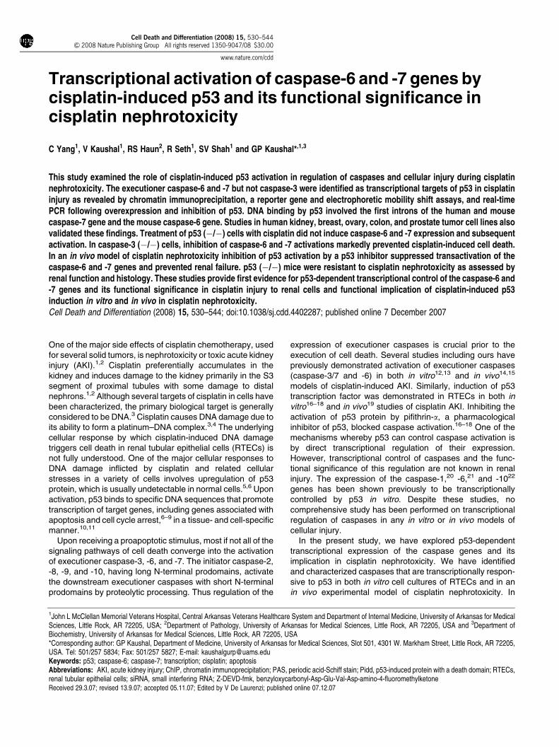
p53-dependent upregulation of mRNA and protein levelsof caspase-6 and -7 but not of caspase-3, -8, and -9. Thedependency of p53 in mRNA expression of caspases wasexplored by upregulating p53 or inhibiting p53. Treatment ofRTECs with cisplatin or transfection with Ad-p53 vectorexhibited a striking induction of p53 expression (Figure 1a).Control Ad-LacZ transfection did not induce p53 expression.The p53 small interfering RNA (siRNA) efficiently andspecifically silenced cisplatin-induced p53 expression(Figure 1b). Semiquantitative RT-PCR analysis revealed amarked increase in p53-dependent expression of thecaspase-6 and -7 genes but not of the caspase-3, -8, and-9 genes (Figure 1c). Overexpression of p53 by Ad-p53 or bycisplatin treatment upregulated caspase-6 and -7 mRNAlevels while pifithrin-a or p53 siRNA downregulated thesetranscripts (Figure 1c). The mRNA levels of caspase-3, -8,
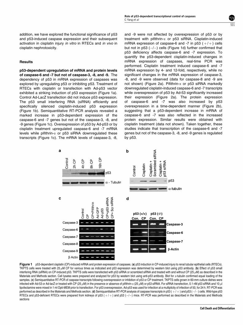
and -9 were not affected by overexpression of p53 or bytreatment with pifithrin-a or p53 siRNA. Cisplatin-inducedmRNA expression of caspase-6 and -7 in p53 (þ /þ ) cellsbut not in p53 (�/�) cells (Figure 1d) further confirmed thatp53 deficiency affects caspase-6 and -7 expression. Toquantify the p53-dependent cisplatin-induced changes inmRNA expression of caspases, real-time PCR wasperformed. Cisplatin treatment induced caspase-6 and -7mRNA expression by 4- and 12-fold, respectively, while nosignificant changes in the mRNA expression of caspase-3,-8, and -9 were observed (data for caspase-8 and -9 arenot shown) (Figure 2a). Pifithrin-a or p53 siRNA markedlydownregulated cisplatin-induced caspase-6 and -7 transcriptswhile overexpression of p53 by Ad-53 significantly increasedtheir expression (Figure 2a). The protein expressionof caspase-6 and -7 was also increased by p53overexpression in a time-dependent manner (Figure 2b),suggesting that a p53-dependent increase in mRNA ofcaspase-6 and -7 was also reflected in the increasedprotein expression. Similar results were obtained withcisplatin treatment (data not shown). Taken together, thesestudies indicate that transcription of the caspase-6 and -7genes but not of the caspase-3, -8, and -9 genes is regulatedby p53.
Con. Con.CP CPp53 (+/+)
Caspase-3
Caspase-6
Caspase-7
�-Actin
p53 (-/-)
Figure 1 p53-dependent cisplatin (CP)-induced mRNA and protein expression of caspases. (a) p53 induction in CP-induced injury to renal tubular epithelial cells (RTECs).TKPTS cells were treated with 25mM CP for various times as indicated and p53 expression was determined by western blot using p53 antibody. (b) Effect of p53 smallinterfering RNA (siRNA) on CP-induced p53. TKPTS cells were transfected with p53 siRNA or scrambled siRNA and treated with and without CP (25 mM) as described in theMaterials and Methods section. Cell lysates were prepared and analyzed for p53 by western blot using anti-p53 antibody. Blot for a-tubulin confirmed equal loading of thesamples. (c) Semiquantitative RT-PCR of caspase transcripts following overexpression or inhibition of p53 or CP treatment. TKPTS cells grown in 60-mm culture dishes wereinfected with Ad-53 or Ad-lacZ or treated with CP (25mM) in the presence or absence of pifithrin-a (25mM) or p53 siRNA. For siRNA transfection, 0.1 nM p53 siRNA and 10 mllipofectamine were mixed in 1 ml Opti-MEM prior to transfection. For p53 overexpression, Ad-p53 was used for infection at a multiplicity of infection of 50, for 24 h. RT-PCR wasperformed as described in the Materials and Methods section. (d) Semiquantitative RT-PCR analysis of caspase transcripts in p53 (þ /þ ) and p53 (�/�) cells. Wild-type p53RTECs and p53-deficient RTECs were prepared from kidneys of p53 (þ /þ ) and p53 (�/�) mice. RT-PCR was performed as described in the Materials and Methodssections
Role of p53-dependent transcriptional control of caspasesC Yang et al
531
Cell Death and Differentiation
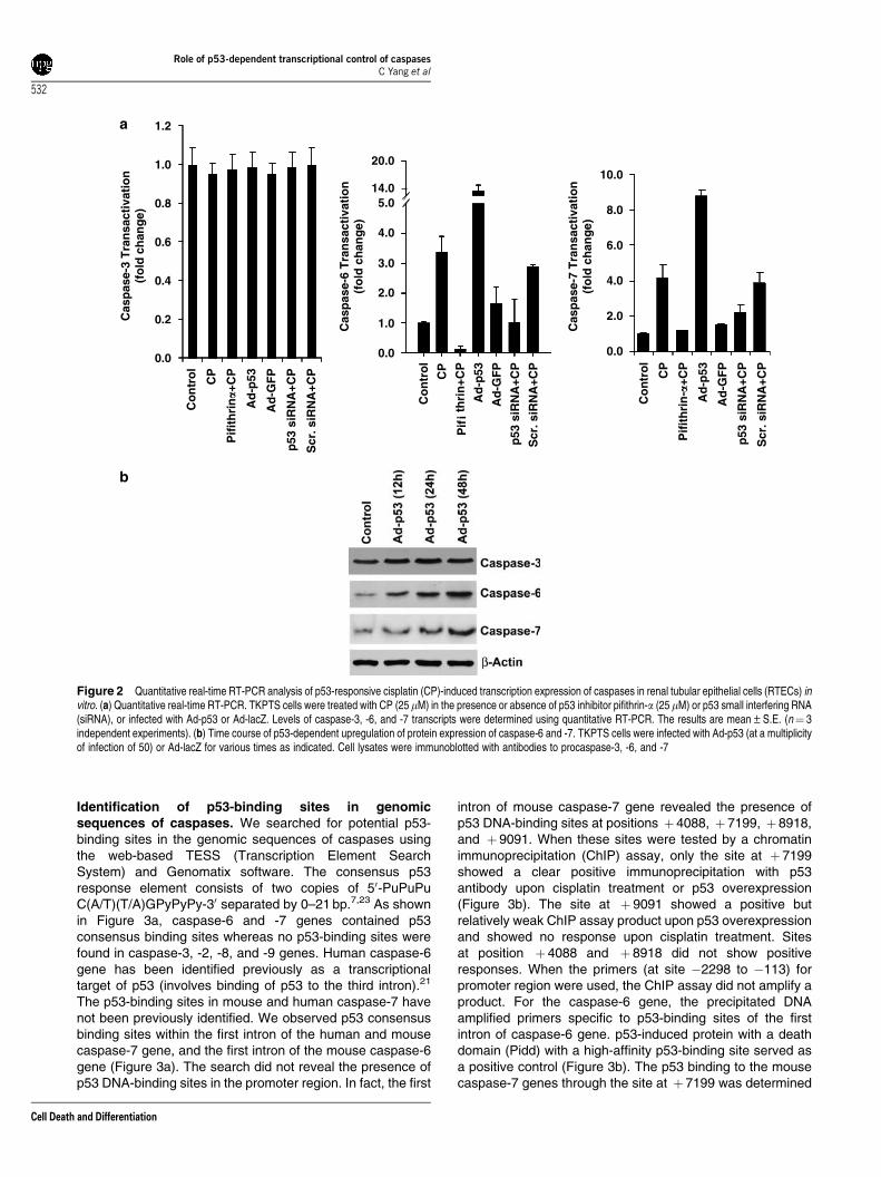
Identification of p53-binding sites in genomicsequences of caspases. We searched for potential p53-binding sites in the genomic sequences of caspases usingthe web-based TESS (Transcription Element SearchSystem) and Genomatix software. The consensus p53response element consists of two copies of 50-PuPuPuC(A/T)(T/A)GPyPyPy-30 separated by 0–21 bp.7,23 As shownin Figure 3a, caspase-6 and -7 genes contained p53consensus binding sites whereas no p53-binding sites werefound in caspase-3, -2, -8, and -9 genes. Human caspase-6gene has been identified previously as a transcriptionaltarget of p53 (involves binding of p53 to the third intron).21
The p53-binding sites in mouse and human caspase-7 havenot been previously identified. We observed p53 consensusbinding sites within the first intron of the human and mousecaspase-7 gene, and the first intron of the mouse caspase-6gene (Figure 3a). The search did not reveal the presence ofp53 DNA-binding sites in the promoter region. In fact, the first
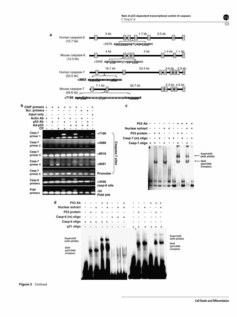
intron of mouse caspase-7 gene revealed the presence ofp53 DNA-binding sites at positions þ 4088, þ 7199, þ 8918,and þ 9091. When these sites were tested by a chromatinimmunoprecipitation (ChIP) assay, only the site at þ 7199showed a clear positive immunoprecipitation with p53antibody upon cisplatin treatment or p53 overexpression(Figure 3b). The site at þ 9091 showed a positive butrelatively weak ChIP assay product upon p53 overexpressionand showed no response upon cisplatin treatment. Sitesat position þ 4088 and þ 8918 did not show positiveresponses. When the primers (at site �2298 to �113) forpromoter region were used, the ChIP assay did not amplify aproduct. For the caspase-6 gene, the precipitated DNAamplified primers specific to p53-binding sites of the firstintron of caspase-6 gene. p53-induced protein with a deathdomain (Pidd) with a high-affinity p53-binding site served asa positive control (Figure 3b). The p53 binding to the mousecaspase-7 genes through the site at þ 7199 was determined
0.0
0.2
0.4
0.6
0.8
1.0
1.2
Co
ntr
ol
CP
Pif
ith
rin�+
CP
Ad
-p53
Ad
-GF
P
p53
siR
NA
+CP
Scr
. siR
NA
+CP
Cas
pas
e-3
Tra
nsa
ctiv
atio
n
(fo
ld c
han
ge)
Cas
pas
e-6
Tra
nsa
ctiv
atio
n
(fo
ld c
han
ge)
Cas
pas
e-7
Tra
nsa
ctiv
atio
n
(fo
ld c
han
ge)
Pif
iA
d-p
53
Co
ntr
ol
CP
thri
n+C
P
Ad
-GF
P
p53
siR
NA
+CP
Scr
. siR
NA
+CP
0.0
2.0
4.0
6.0
8.0
10.0
Pif
ith
rin
-�+C
P
Co
ntr
ol
Ad
-p53
Ad
-GF
P
p53
siR
NA
+CP
Scr
. siR
NA
+CP
CP
0.0
1.0
2.0
3.0
4.0
20.0
14.05.0
Figure 2 Quantitative real-time RT-PCR analysis of p53-responsive cisplatin (CP)-induced transcription expression of caspases in renal tubular epithelial cells (RTECs) invitro. (a) Quantitative real-time RT-PCR. TKPTS cells were treated with CP (25 mM) in the presence or absence of p53 inhibitor pifithrin-a (25mM) or p53 small interfering RNA(siRNA), or infected with Ad-p53 or Ad-lacZ. Levels of caspase-3, -6, and -7 transcripts were determined using quantitative RT-PCR. The results are mean±S.E. (n¼ 3independent experiments). (b) Time course of p53-dependent upregulation of protein expression of caspase-6 and -7. TKPTS cells were infected with Ad-p53 (at a multiplicityof infection of 50) or Ad-lacZ for various times as indicated. Cell lysates were immunoblotted with antibodies to procaspase-3, -6, and -7
Role of p53-dependent transcriptional control of caspasesC Yang et al
532
Cell Death and Differentiation
2 53 4 6 71
2 53 4 6 7
+7199 agacttgtcccacacgtggacacacacacaatcacaaggct
1
2 63 4 7 85
+3953 aggcatgcaccaccatgccc
1
2 53 4 6 71
2 53 4 6 7
+3456 agccttgcaatgcaggccttgccc
14 kb 4 kb 1.4 kb 1.1 kb
2 53 4 6 7
+7199 agacttgtcccacacgtggacacacacacaatcacaaggct
1 2 53 4 6 7
+7199 agacttgtcccacacgtggacacacacacaatcacaaggct
17.1 kb 28.7 kb 4.6 kb2.3 kb
2 63 4 7 85
+3953 aggcatgcaccaccatgccc
1 2 63 4 7 85
+3953 aggcatgcaccaccatgccc
1 2 63 4 7 85
+3953 aggcatgcaccaccatgccc
118.1 kb 23.4 kb 3.6 kb 2.9 kb
2 53 4 6 7
+5976 aggcaaggagtttgagacaagtct
Human caspase-6 (15.7 kb)
Human caspase-7 (52.5 kb)
Mouse caspase-6 (13.3 kb)
Mouse caspase-7(46.6 kb)
15 kb 1.7 kb 3.5 kb
+
- - + + - - - - - - - - - + + + + -
- - + - + - + -+ - +
+ - + - + - -
Scr. primers - -
- - - - - + + -- - - - - - - +
Casp
ase-7 sites
ChIP primers + + + + + - - +
Input onlyActin Ab
p53 AbAd-p53
CPCasp-7 primer 1
Casp-7 primer 2
Casp-7 primer 3
Casp-7 primer 4
Casp-7 primer 5
Casp-6 primers
Pidd primers
+7199
+4088
+8918
+9091
Promoter
+3456 casp-6 site
-54 Pidd site
- - -
-
-
-
-
-
(
+ + + - + + -
- + - + + - + - +
+ + - - - + + - -
- - + + - - - + +
- - - - - + + + +P53 Ab
Nuclear extract
P53 protein
Casp-7 oligo
Casp-7 (m) oligo
Supershift (with p53Ab)
Shift (p53-DNAComplex)
+ + + + + - - - - - - - -
- - - - -
-
+ - - - - - -
+ - +
+
- - - + - - + -
- + - + - + - - + - - +--
-
- - + + - - + - - - + + +
- - - - - - -
-
+
-
+
- + + + + + +
Supershift (with p53Ab)
Shift (p53-DNAcomplex)
Supershift (with p53Ab)
Shift (p53-DNAcomplex)
P53 AbNuclear extract
P53 protein
p21 oligo
Casp-6 (m) oligo
Casp-6 oligo
Figure 3 Continued
Role of p53-dependent transcriptional control of caspasesC Yang et al
533
Cell Death and Differentiation
by an electrophoretic mobility shift assay (EMSA) usingsynthetic oligonucleotides corresponding to the consensusbinding site. A clear shift in the oligonucleotide band followingthe incubation of the consensus site specific oligonucleotidesin the first intron of the mouse caspase-7 gene with nuclearextracts (prepared from cisplatin-treated RTECs) or purifiedrecombinant p53 protein (Figure 3c) indicated formation of ap53–DNA complex. A strong supershift observed on additionof p53 antibody confirmed the presence of p53 in the protein–
DNA complex (Figure 3c). Similar results for p53 binding tothe human caspase-7 gene were obtained (data not shown).For the binding of the mouse caspase-6 gene to p53, theoligonucleotides corresponding to the consensus sites in thefirst intron of the mouse caspase-6 gene were used(Figure 3d). p21 having high affinity p53-binding site wasused as a positive control. For the human caspase-6 gene,formation of p53–DNA complex has been shown previouslyby EMSA.21 In addition, the luciferase-reporter constructs for
0
2000
6000
10000
14000
p21 p21-m
p21
luci
fera
se a
ctiv
ity
0
200
400
600
800
Cas
pas
e-6
luci
fera
se a
ctiv
ity
0
2000
4000
6000
8000
Casp-7 Casp-7-mCasp-6 Casp-6-m
Cap
ase-
7 lu
cife
rase
act
ivit
y
Ad-LacZ
Ad-GFP+Ad-LacZ
Ad-p53+Ad-LacZ
Ad-LacZ
Ad-GFP+Ad-LacZ
Ad-p53+Ad-LacZ
Ad-LacZ
Ad-GFP+Ad-LacZ
Ad-p53+Ad-LacZ
Control
Cisplatin
Cisplatin+Pifithrin-�
Ad-p53
Ad-GFP
Cisplatin+Pifithrin-�
Ad-p53
Ad-GFP
MCF-7 PA-I
LNCapHEK-293
Control
Cisplatin
Caspase-7
Caspase-7
�-Actin
�-Actin
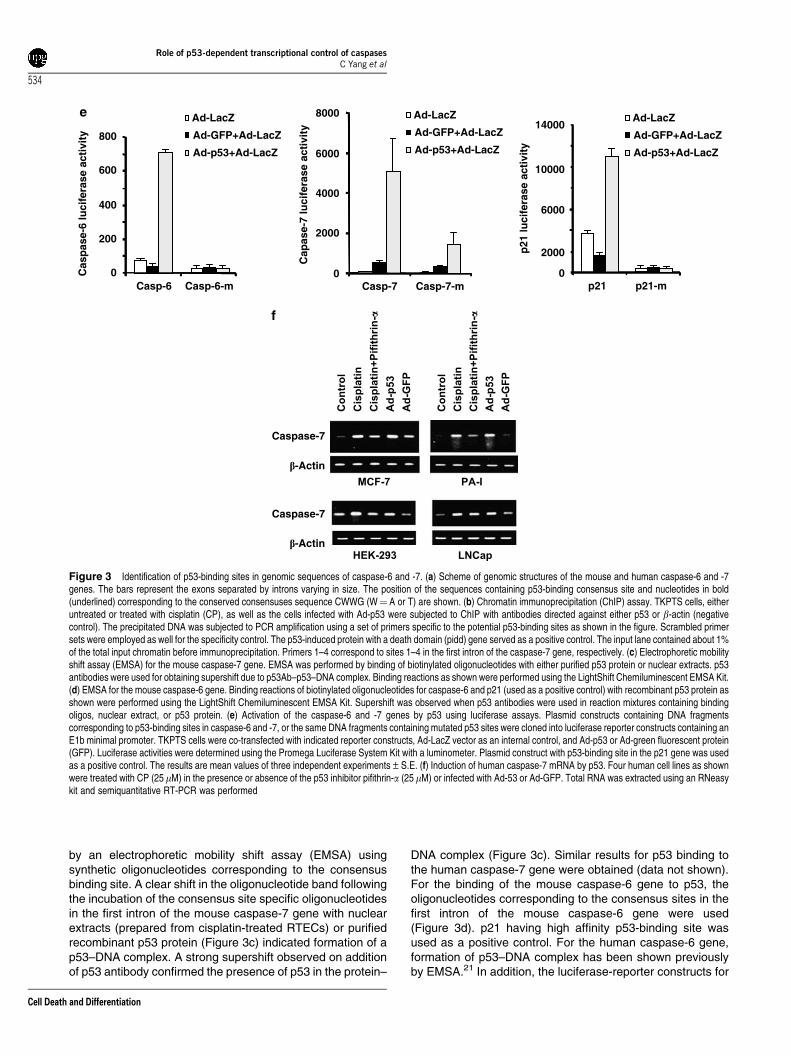
Figure 3 Identification of p53-binding sites in genomic sequences of caspase-6 and -7. (a) Scheme of genomic structures of the mouse and human caspase-6 and -7genes. The bars represent the exons separated by introns varying in size. The position of the sequences containing p53-binding consensus site and nucleotides in bold(underlined) corresponding to the conserved consensuses sequence CWWG (W¼ A or T) are shown. (b) Chromatin immunoprecipitation (ChIP) assay. TKPTS cells, eitheruntreated or treated with cisplatin (CP), as well as the cells infected with Ad-p53 were subjected to ChIP with antibodies directed against either p53 or b-actin (negativecontrol). The precipitated DNA was subjected to PCR amplification using a set of primers specific to the potential p53-binding sites as shown in the figure. Scrambled primersets were employed as well for the specificity control. The p53-induced protein with a death domain (pidd) gene served as a positive control. The input lane contained about 1%of the total input chromatin before immunoprecipitation. Primers 1–4 correspond to sites 1–4 in the first intron of the caspase-7 gene, respectively. (c) Electrophoretic mobilityshift assay (EMSA) for the mouse caspase-7 gene. EMSA was performed by binding of biotinylated oligonucleotides with either purified p53 protein or nuclear extracts. p53antibodies were used for obtaining supershift due to p53Ab–p53–DNA complex. Binding reactions as shown were performed using the LightShift Chemiluminescent EMSA Kit.(d) EMSA for the mouse caspase-6 gene. Binding reactions of biotinylated oligonucleotides for caspase-6 and p21 (used as a positive control) with recombinant p53 protein asshown were performed using the LightShift Chemiluminescent EMSA Kit. Supershift was observed when p53 antibodies were used in reaction mixtures containing bindingoligos, nuclear extract, or p53 protein. (e) Activation of the caspase-6 and -7 genes by p53 using luciferase assays. Plasmid constructs containing DNA fragmentscorresponding to p53-binding sites in caspase-6 and -7, or the same DNA fragments containing mutated p53 sites were cloned into luciferase reporter constructs containing anE1b minimal promoter. TKPTS cells were co-transfected with indicated reporter constructs, Ad-LacZ vector as an internal control, and Ad-p53 or Ad-green fluorescent protein(GFP). Luciferase activities were determined using the Promega Luciferase System Kit with a luminometer. Plasmid construct with p53-binding site in the p21 gene was usedas a positive control. The results are mean values of three independent experiments±S.E. (f) Induction of human caspase-7 mRNA by p53. Four human cell lines as shownwere treated with CP (25 mM) in the presence or absence of the p53 inhibitor pifithrin-a (25mM) or infected with Ad-53 or Ad-GFP. Total RNA was extracted using an RNeasykit and semiquantitative RT-PCR was performed
Role of p53-dependent transcriptional control of caspasesC Yang et al
534
Cell Death and Differentiation
0
500
1000
1500
2000
2500
3000
3500
Co
ntr
ol
Ad
-Lac
Z
CP
CP
+Pif
ith
rin
Ad
-p53
CP
+Ad
-p53
Cas
pas
e-6
acti
vity
(re
lati
ve u
nit
s)
∗0
200
400
600
800
1000
1200
1400
Co
ntr
ol
Ad
-Lac
Z
CP
CP
+Pif
ith
rin
Ad
-p53
CP
+Ad
-p53
Cas
pas
e-7
acti
vity
(re
lati
ve u
nit
s)
∗
∗∗
∗
∗∗
DAPIActiveCasp-6
ActiveCasp-3
DAPI+ActiveCasp-3&-6
PhaseContrast
PhaseContrastCasp-7DAPI Casp-3 Casp-3&-7
Controlp53(-/-)
Controlp53(+/+)
Cisplatinp53(-/-)
Cisplatinp53(+/+)
Controlp53(-/-)
Controlp53(+/+)
Cisplatinp53(-/-)
Cisplatinp53(+/+)
Figure 4 Continued
Role
ofp5
3-d
epen
den
ttran
scription
alcon
trolof
caspases
CYan
get
al
535
CellD
eathand
Differentiation
0.0
0.2
0.4
0.6
0.8
1.0
1.2
0h 12h 18h 24h 28h
Cel
l su
rviv
al (
Ab
sorb
. 570
nm
)
CP aloneCP+p53 siRNACP+casp-7 siRNACP+casp-6 siRNA
CP aloneCP+p53 siRNACP+casp-7 siRNACP+casp-6 siRNA
Caspase-3 (+/+) cells treated with CP
0h 12h 18h 24h 28h
Caspase-3 (-/-) cells treated with CP
∗∗
∗∗
∗
∗∗∗
∗∗∗∗∗∗
0.0
0.2
0.4
0.6
0.8
1.0
1.2
1.4
Cel
l su
rviv
al (
Ab
sorb
. 570
nm
)
∗∗
∗∗
∗∗
∗∗
∗∗∗∗
∗ ∗
Control CP 8h CP 12h
CP 18h CP 18h+zDEVD
CP 4h
CP 24h CP 24h+zDEVD
Caspase-3 (+/+) renal tubular cells
CP 18h+Pifithrin-α
CP 24h+Pifithrin-α
CP 18h+VEID
CP 24h+VEID
Control CP 8h CP 12h
CP 18h CP 18h+zDEVD
CP 4h
CP 24h CP 24h+zDEVD
Caspase-3 (-/-) renal tubular cells
CP 18h+Pifithrin-α
CP 24h+Pifithrin-α
CP 18h+VEID
CP 24h+VEID
0
20
40
60
80
100
Co
n
CP
4h
CP
8h
CP
12h
CP
18h
CP
18h
+DE
VD
CP
18h
+VE
ID
CP
18h
+Pif
ith
rin
CP
24h
CP
24h
+DE
VD
CP
24h
+VE
ID
CP
24h
+Pif
ith
rin
∗
∗
∗
∗
∗∗ ∗∗ ∗∗∗∗ ∗∗
∗∗
0
20
40
60
80
100
Co
n
CP
4h
CP
8h
CP
12h
CP
18h
CP
18h
+DE
VD
CP
18h
+VE
ID
CP
18h
+Pif
ith
rin
CP
24h
P 2
4h+D
EV
D
CP
24h
+VE
ID
CP
24h
+Pif
ith
rin
Cas
pas
e-3
(-/-
) ap
op
toti
c ce
lls (
%)
∗∗
∗
∗
∗∗ ∗∗ ∗∗∗∗ ∗∗
∗∗Cas
pas
e-3
(+/+
) ap
op
toti
c ce
lls (
%)
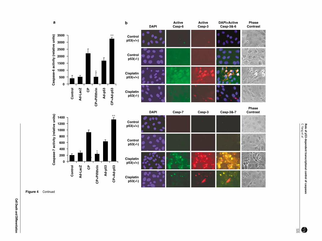
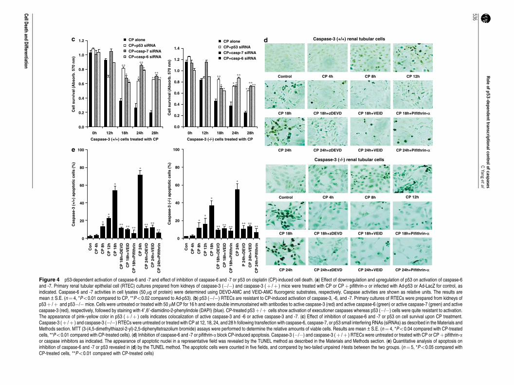
Figure 4 p53-dependent activation of caspase-6 and -7 and effect of inhibition of caspase-6 and -7 or p53 on cisplatin (CP)-induced cell death. (a) Effect of downregulation and upregulation of p53 on activation of caspase-6and -7. Primary renal tubular epithelial cell (RTEC) cultures prepared from kidneys of caspase-3 (�/�) and caspase-3 (þ /þ ) mice were treated with CP or CPþ pifithrin-a or infected with Ad-p53 or Ad-LacZ for control, asindicated. Caspase-6 and -7 activities in cell lysates (50 mg of protein) were determined using DEVD-AMC and VEID-AMC fluorogenic substrates, respectively. Caspase activities are shown as relative units. The results aremean±S.E. (n¼ 4, *Po0.01 compared to CP, **Po0.02 compared to Ad-p53). (b) p53 (�/�) RTECs are resistant to CP-induced activation of caspase-3, -6, and -7. Primary cultures of RTECs were prepared from kidneys ofp53þ /þ and p53�/� mice. Cells were untreated or treated with 50mM CP for 16 h and were double immunostained with antibodies to active caspase-3 (red) and active caspase-6 (green) or active caspase-7 (green) and activecaspase-3 (red), respectively, followed by staining with 40,60-diamidino-2-phenylindole (DAPI) (blue). CP-treated p53þ /þ cells show activation of executioner caspases whereas p53 (�/�) cells were quite resistant to activation.The appearance of pink–yellow color in p53 (þ /þ ) cells indicates colocalization of active caspase-3 and -6 or active caspase-3 and -7. (c) Effect of inhibition of caspase-6 and -7 or p53 on cell survival upon CP treatment.Caspase-3 (þ /þ ) and caspase-3 (�/�) RTECs were untreated or treated with CP at 12, 18, 24, and 28 h following transfection with caspase-6, caspase-7, or p53 small interfering RNAs (siRNAs) as described in the Materials andMethods section. MTT (3-(4,5-dimethylthiazol-2-yl)-2,5-diphenyltetrazolium bromide) assays were performed to determine the relative amounts of viable cells. Results are mean±S.E. (n¼ 4, *Po0.04 compared with CP-treatedcells, **Po0.01 compared with CP-treated cells). (d) Inhibition of caspase-6 and -7 or pifithrin-a block CP-induced apoptosis. Caspase-3 (�/�) and caspase-3 (þ /þ ) RTECs were untreated or treated with CP or CPþ pifithrin-aor caspase inhibitors as indicated. The appearance of apoptotic nuclei in a representative field was revealed by the TUNEL method as described in the Materials and Methods section. (e) Quantitative analysis of apoptosis oninhibition of caspase-6 and -7 or p53 revealed in (d) by the TUNEL method. The apoptotic cells were counted in five fields, and compared by two-tailed unpaired t-tests between the two groups. (n¼ 5, *Po0.05 compared withCP-treated cells, **Po0.01 compared with CP-treated cells)
Role
ofp5
3-d
epen
den
ttran
scription
alcon
trolof
caspases
CYan
get
al
536
CellD
eathand
Differentiation
caspase-6 and -7 containing the intronic regions that bindp53 were generated. These constructs were responsive tothe wild-type p53 binding but not to the constructs containinga mutated p53-binding site (Figure 3e). A p21 luciferase-reporter construct was used as a positive control (Figure 3e).Similar results for human caspase-6 and -7 were obtained(data not shown). These data indicate that the p53-bindingelements of the caspase-6 and -7 genes are capable ofrecruiting p53 to activate transcription of caspase-6 and -7genes.
Induction of caspase-7 expression by p53 in humancancer cell lines. The dependency of p53 in cisplatin-induced expression of endogenous caspase-7 mRNA wasalso explored by inhibiting as well as upregulating p53expression in human cell lines having an endogenous wild-type p53 gene. The human embryonic kidney cell line HEK-293 and three human cancer cell lines, MCF-7, PA-1, andLNCaP, were treated with cisplatin or transfected with a Ad-p53 vector or treated with p53 inhibitor pifithrin-a, and totalRNA was isolated to examine caspase-7 expression. All ofthese cell lines are known to induce wild-type p53 inresponse to genotoxic stress. These showed an efficientp53 gene transfer with 90–100% cells stained for greenfluorescent protein (GFP) when Ad-GFP was used as acontrol. The induction of p53 expression and inhibitionby pifithrin-a was monitored by western blot (data notshown). Semiquantitative RT-PCR revealed that caspase-7expression was significantly increased with either Ad-p53 orby cisplatin treatment and the expression was suppressed bypifithrin-a (Figure 3f).
p53 overexpression and/or cisplatin treatment resultsin enhanced activation of caspase -6 and -7 and a p53inhibitor or use of p53 (�/�) cells prevent caspaseactivation. We examined the possibility that p53-dependentincrease in the protein levels of procaspase-6 and -7 willresult in higher activation of these executioner caspases. Ourprevious study showed significant activation of caspase-3/7and -6 by cisplatin treatment.12 Caspase-3 and -7 utilize thesame substrate DEVD-AMC and therefore the activitiesdetermined by cleavage of DEVD-AMC are in factcontributed by both of these caspases. To specificallyexamine cisplatin-induced caspase-7 activation, we usedprimary cultures of caspase-3 (�/�) RTECs prepared fromthe kidneys of caspase-3 (�/�) mice. Caspase-6 and -7activities were significantly increased with cisplatin treatmentor overexpression of p53 (Figure 4a). A p53 inhibitor pifithrin-amarkedly prevented cisplatin-induced caspase-6 and -7activation. To examine further the specific role of p53 inactivation of executioner caspases, we prepared RTECs fromp53 (�/�) and p53 (þ /þ ) mice. Immunofluorescent studiesusing specific antibodies to the active forms of executionercaspases revealed that compared to p53 (þ /þ ) cells, p53(�/�) cells were effective in blocking activation of caspase-3,-6, and -7 (Figure 4b). These studies show that p53-deficientRTECs were quite resistant to activation of executionercaspases.
Inhibition of p53 or p53-dependent caspase-6 and -7activation significantly prevents and delays cisplatin-induced cell death. To investigate the potential functionalsignificance of p53-dependent caspase-6 and -7 activationby cisplatin we used an RNA interference (RNAi) approachfor the specific inhibition of caspase-6 and -7. As cisplatin-induced caspase-3 activation may also contribute to celldeath we used caspase-3 (�/�) RTECs for specific inhibitionof caspase-6 or -7. Inhibition of caspase-6 or -7 by theirrespective siRNA resulted in cell survival (as revealed byMTT (3-(4,5-dimethylthiazol-2-yl)-2,5-diphenyltetrazoliumbromide) assay) and delayed cisplatin-induced cell death(Figure 4c). However, p53 inhibition was more effective inpreventing cell death as compared to the inhibitors thatblocked caspase-6 and -7. Inhibition of caspase-6 and -7 orp53 was effective in preventing cisplatin-induced apoptosis(Figure 4d and e).
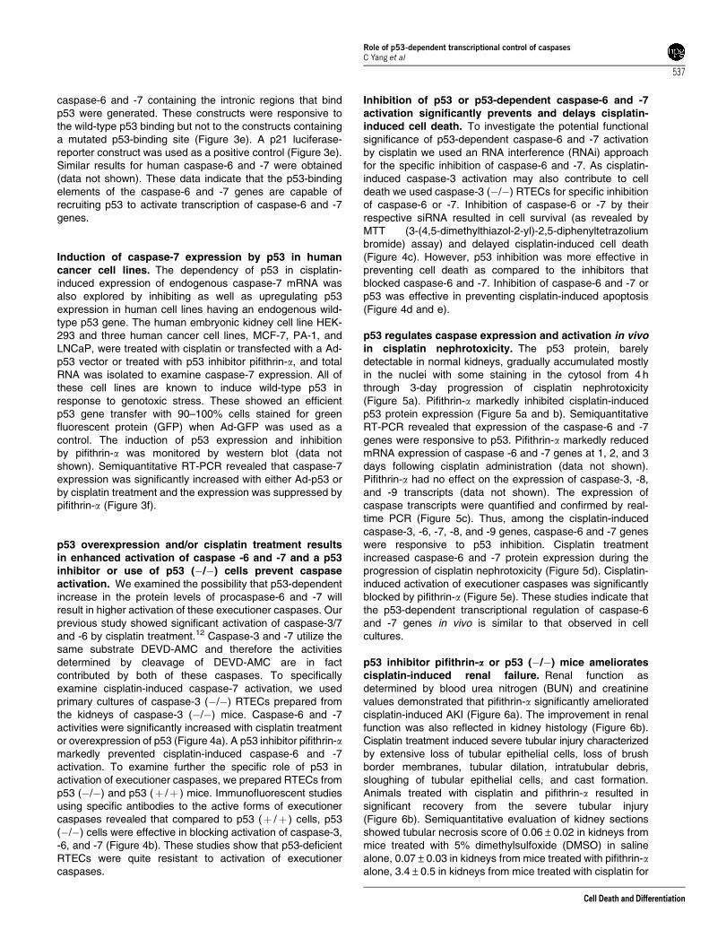
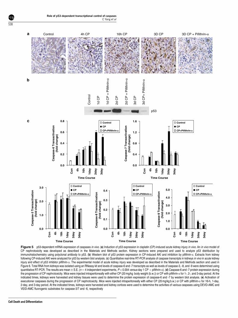
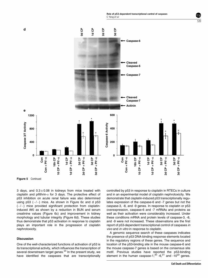
p53 regulates caspase expression and activation in vivoin cisplatin nephrotoxicity. The p53 protein, barelydetectable in normal kidneys, gradually accumulated mostlyin the nuclei with some staining in the cytosol from 4 hthrough 3-day progression of cisplatin nephrotoxicity(Figure 5a). Pifithrin-a markedly inhibited cisplatin-inducedp53 protein expression (Figure 5a and b). SemiquantitativeRT-PCR revealed that expression of the caspase-6 and -7genes were responsive to p53. Pifithrin-a markedly reducedmRNA expression of caspase -6 and -7 genes at 1, 2, and 3days following cisplatin administration (data not shown).Pifithrin-a had no effect on the expression of caspase-3, -8,and -9 transcripts (data not shown). The expression ofcaspase transcripts were quantified and confirmed by real-time PCR (Figure 5c). Thus, among the cisplatin-inducedcaspase-3, -6, -7, -8, and -9 genes, caspase-6 and -7 geneswere responsive to p53 inhibition. Cisplatin treatmentincreased caspase-6 and -7 protein expression during theprogression of cisplatin nephrotoxicity (Figure 5d). Cisplatin-induced activation of executioner caspases was significantlyblocked by pifithrin-a (Figure 5e). These studies indicate thatthe p53-dependent transcriptional regulation of caspase-6and -7 genes in vivo is similar to that observed in cellcultures.
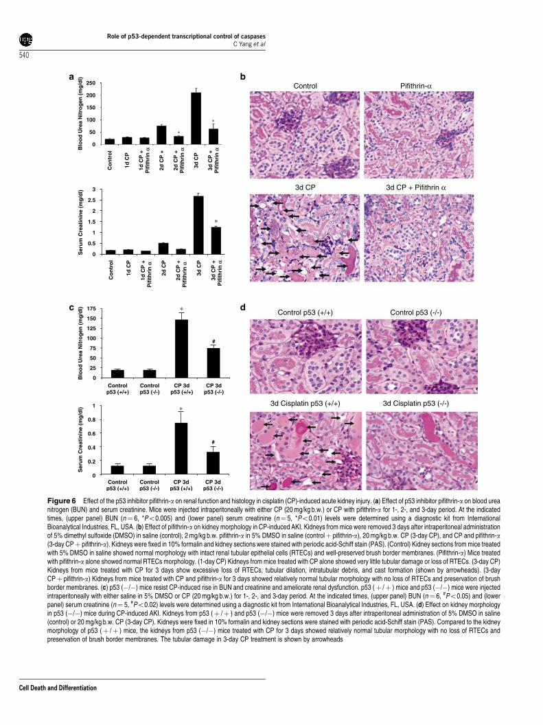
p53 inhibitor pifithrin-a or p53 (�/�) mice amelioratescisplatin-induced renal failure. Renal function asdetermined by blood urea nitrogen (BUN) and creatininevalues demonstrated that pifithrin-a significantly amelioratedcisplatin-induced AKI (Figure 6a). The improvement in renalfunction was also reflected in kidney histology (Figure 6b).Cisplatin treatment induced severe tubular injury characterizedby extensive loss of tubular epithelial cells, loss of brushborder membranes, tubular dilation, intratubular debris,sloughing of tubular epithelial cells, and cast formation.Animals treated with cisplatin and pifithrin-a resulted insignificant recovery from the severe tubular injury(Figure 6b). Semiquantitative evaluation of kidney sectionsshowed tubular necrosis score of 0.06±0.02 in kidneys frommice treated with 5% dimethylsulfoxide (DMSO) in salinealone, 0.07±0.03 in kidneys from mice treated with pifithrin-aalone, 3.4±0.5 in kidneys from mice treated with cisplatin for
Role of p53-dependent transcriptional control of caspasesC Yang et al
537
Cell Death and Differentiation
0.0
0.2
0.4
0.6
0.8
**
0.0
0.4
0.8
1.2
1.6
**
0.0
1.0
2.0
3.0
Cas
pas
e-3
Tra
nsa
ctiv
atio
n(f
old
ch
ang
e)
Cas
pas
e-8
Tra
nsa
ctiv
atio
n(f
old
ch
ang
e)
Co
n 4h 16h
1day
2day
3day
Time Course
Co
n 4h 16h
1day
2day
3day
Time Course
Co
n 4h 16h
1day
2day
3day
Time Course
Co
n 4h 16h
1day
2day
3day
Time Course
Co
n 4h 16h
1day
2day
3day
Time Course
0.0
0.1
0.2
0.3
0.4
Control
CP
CP+Pifithrin-α
Control
CP
CP+Pifithrin-α
Control
CP
CP+Pifithrin-α
Control
CP
CP+Pifithrin-α
Control
CP
CP+Pifithrin-α
0.0
1.0
2.0
3.0
4.0
5.0
Con
trol
1d C
P
1d C
P +
Pifi
thrin
-α
2d C
P
2d C
P +
Pifi
thrin
-α
3d C
P
3d C
P+
Pifi
thrin
-α
p53
Control 4h CP 16h CP 3D CP 3D CP + Pifithrin-α
Cas
pas
e-9
Tra
nsa
ctiv
atio
n(f
old
ch
ang
e)
Cas
pas
e-7
Tra
nsa
ctiv
atio
n(f
old
ch
ang
e)
Cas
pas
e-6
Tra
nsa
ctiv
atio
n(f
old
ch
ang
e)
Figure 5 p53-dependent mRNA expression of caspases in vivo. (a) Induction of p53 expression in cisplatin (CP)-induced acute kidney injury in vivo. An in vivo model ofCP nephrotoxicity was developed as described in the Materials and Methods section. Kidney sections were prepared and used to analyze p53 distribution byimmunohistochemistry using polyclonal antibody to p53. (b) Western blot of p53 protein expression in CP-induced AKI and inhibition by pifithrin-a. Extracts from kidneyfollowing CP-induced AKI were analyzed for p53 by western blot analysis. (c) Quantitative real-time RT-PCR analysis of caspase transcripts in kidneys in vivo in acute kidneyinjury and effect of p53 inhibitor pifithrin-a. The experimental model of acute kidney injury was developed as described in the Materials and Methods section and used inFigure 6. Total RNA from kidneys was isolated using an RNeasy kit and levels of caspase-6 and -7 transcripts as well as levels of caspase-3, -8, and -9 were determined usingquantitative RT-PCR. The results are mean±S.E. (n¼ 4 independent experiments, Po0.004 versus day 1 CPþ pifithrin-a). (d) Caspase-6 and -7 protein expression duringthe progression of CP nephrotoxicity. Mice were injected intraperitoneally with either CP (20 mg/kg body weight (b.w.)) or CP with pifithrin-a for 1-, 2-, and 3-day period. At theindicated times, kidneys were harvested and kidney tissues were used to determine the protein expression of caspase-6 and -7 by western blot analysis. (e) Activation ofexecutioner caspases during the progression of CP nephrotoxicity. Mice were injected intraperitoneally with either CP (20 mg/kg b.w.) or CP with pifithrin-a for 16-h, 1-day,2-day, and 3-day period. At the indicated times, kidneys were harvested and kidney cortices were used to determine the activities of various caspases using DEVD-AMC andVEID-AMC fluorogenic substrates for caspase-3/7 and -6, respectively
Role of p53-dependent transcriptional control of caspasesC Yang et al
538
Cell Death and Differentiation
3 days, and 0.3±0.08 in kidneys from mice treated withcisplatin and pifithrin-a for 3 days. The protective effect ofp53 inhibition on acute renal failure was also determinedusing p53 (�/�) mice. As shown in Figure 6c and d p53(�/�) mice provided significant protection from cisplatin-induced AKI as shown by a reduction in BUN and serumcreatinine values (Figure 6c) and improvement in kidneymorphology and tubular integrity (Figure 6d). These studiesthus demonstrate that p53 activation in response to cisplatinplays an important role in the progression of cisplatinnephrotoxicity.
Discussion
One of the well-characterized functions of activation of p53 isits transcriptional activity, which influences the transcription ofseveral downstream target genes.24 In the present study, wehave identified the caspases that are transcriptionally
controlled by p53 in response to cisplatin in RTECs in cultureand in an experimental model of cisplatin nephrotoxicity. Wedemonstrate that cisplatin-induced p53 transcriptionally regu-lates expression of the caspase-6 and -7 genes but not thecaspase-3, -8, and -9 genes. In response to cisplatin or p53overexpression, caspase-6 and -7 mRNAs and proteins aswell as their activation were considerably increased. Underthese conditions mRNA and protein levels of caspase-3, -8,and -9 were not increased. These observations are the firstreport of p53-dependent transcriptional control of caspases invivo and in vitro in response to cisplatin.
A genomic sequence search of these caspases indicatesthe presence of p53 DNA-binding response elements locatedin the regulatory regions of these genes. The sequence andlocation of the p53-binding site in the mouse caspase-6 andthe mouse caspase-7 genes is based on the consensus sitemotif. Previous studies have reported the p53-bindingelement in the human caspase-1,20 -6,21 and -1022 genes.
0d C
P
1d C
P
2d C
P
Caspase-6
CleavedCaspase-6
Actinin
CleavedCaspase-7
Caspase-7
3d C
P1200
800
400
0
Co
nC
on
+ P
if -
α α
16h
CP
16 C
P +
Pif
-α
1d C
P1d
CP
+ P
if -
α
2d C
P2d
CP
+ P
if -
α
3d C
P3d
CP
+ P
if -
α
Co
nC
on
+ P
if -
α
16h
CP
16 C
P +
Pif
-α
1d C
P1d
CP
+ P
if -
α
2d C
P
2d C
P +
Pif
-α
3d C
P
3d C
P +
Pif
-α
Cas
pas
e 3/
7 A
ctiv
ity
600
400
200
0
Cas
pas
e-6
Act
ivit
y
∗ ∗
**
∗ ∗
* *
Figure 5 Continued
Role of p53-dependent transcriptional control of caspasesC Yang et al
539
Cell Death and Differentiation
Co
ntr
ol
1d C
P
1d C
P +
Pif
ith
rin
αα
2d C
P
2d C
P +
Pif
ith
rin
α
3d C
P
3d C
P +
Pif
ith
rin
α
0
0.5
1
1.5
2
2.5
3
Ser
um
Cre
atin
ine
(mg
/dl)
∗
25
0
50
75
100
125
150
175
Controlp53 (+/+)
Controlp53 (-/-)
CP 3dp53 (+/+)
CP 3dp53 (-/-)
Blo
od
Ure
a N
itro
gen
(m
g/d
l) ∗
#
0
0.2
0.4
0.6
0.8
1
Controlp53 (+/+)
Controlp53 (-/-)
CP 3dp53 (+/+)
CP 3dp53 (-/-)
Ser
um
Cre
atin
ine
(mg
/dl)
#
∗
Control Pifithrin-α
3d CP + Pifithrin α3d CP
Control p53 (+/+) Control p53 (-/-)
3d Cisplatin p53 (-/-)3d Cisplatin p53 (+/+)
Blo
od
Ure
a N
itro
gen
(m
g/d
l)
∗
∗
3d C
P +
Pif
ith
rin
α
3d C
P
2d C
P +
Pif
ith
rin
α
2d C
P +
1d C
P +
Pif
ith
rin
α
1d C
P
Co
ntr
ol
250
200
150
100
50
0
Figure 6 Effect of the p53 inhibitor pifithrin-a on renal function and histology in cisplatin (CP)-induced acute kidney injury. (a) Effect of p53 inhibitor pifithrin-a on blood ureanitrogen (BUN) and serum creatinine. Mice were injected intraperitoneally with either CP (20 mg/kg b.w.) or CP with pifithrin-a for 1-, 2-, and 3-day period. At the indicatedtimes, (upper panel) BUN (n¼ 6, *Po0.005) and (lower panel) serum creatinine (n¼ 5, *Po0.01) levels were determined using a diagnostic kit from InternationalBioanalytical Industries, FL, USA. (b) Effect of pifithrin-a on kidney morphology in CP-induced AKI. Kidneys from mice were removed 3 days after intraperitoneal administrationof 5% dimethyl sulfoxide (DMSO) in saline (control), 2 mg/kg b.w. pifithrin-a in 5% DMSO in saline (controlþ pifithrin-a), 20 mg/kg b.w. CP (3-day CP), and CP and pifithrin-a(3-day CPþ pifithrin-a). Kidneys were fixed in 10% formalin and kidney sections were stained with periodic acid-Schiff stain (PAS). (Control) Kidney sections from mice treatedwith 5% DMSO in saline showed normal morphology with intact renal tubular epithelial cells (RTECs) and well-preserved brush border membranes. (Pifithrin-a) Mice treatedwith pifithrin-a alone showed normal RTECs morphology. (1-day CP) Kidneys from mice treated with CP alone showed very little tubular damage or loss of RTECs. (3-day CP)Kidneys from mice treated with CP for 3 days show excessive loss of RTECs, tubular dilation, intratubular debris, and cast formation (shown by arrowheads). (3-dayCPþ pifithrin-a) Kidneys from mice treated with CP and pifithrin-a for 3 days showed relatively normal tubular morphology with no loss of RTECs and preservation of brushborder membranes. (c) p53 (�/�) mice resist CP-induced rise in BUN and creatinine and ameliorate renal dysfunction. p53 (þ /þ ) mice and p53 (�/�) mice were injectedintraperitoneally with either saline in 5% DMSO or CP (20 mg/kg b.w.) for 1-, 2-, and 3-day period. At the indicated times, (upper panel) BUN (n¼ 6, #Po0.05) and (lowerpanel) serum creatinine (n¼ 5, #Po0.02) levels were determined using a diagnostic kit from International Bioanalytical Industries, FL, USA. (d) Effect on kidney morphologyin p53 (�/�) mice during CP-induced AKI. Kidneys from p53 (þ /þ ) and p53 (�/�) mice were removed 3 days after intraperitoneal administration of 5% DMSO in saline(control) or 20 mg/kg b.w. CP (3-day CP). Kidneys were fixed in 10% formalin and kidney sections were stained with periodic acid-Schiff stain (PAS). Compared to the kidneymorphology of p53 (þ /þ ) mice, the kidneys from p53 (�/�) mice treated with CP for 3 days showed relatively normal tubular morphology with no loss of RTECs andpreservation of brush border membranes. The tubular damage in 3-day CP treatment is shown by arrowheads
Role of p53-dependent transcriptional control of caspasesC Yang et al
540
Cell Death and Differentiation
Caspase-7 was shown to be induced in response to p53 in avariety of cell lines. A recent study showed that caspase-7 is ap53 responsive gene but the p53-binding site was notidentified.25 In these studies, it was suggested that thecaspase-7 gene may bind to p53 through the promoter regionwithin �1000 bp. However, no consensus binding site for p53was found in this region in the human or mouse caspase-7gene. The primers in the promoter region when used from theposition �2298 to �113 in ChIP assay did not amplify aproduct. Our studies showed that DNA binding by p53involved the first intron of the human and mouse caspase-7gene. We have found four p53 DNA-binding consensus sitesin the intron 1 of caspase-7 gene at positions þ 4088, þ 7199,þ 8918, and þ 9091. We demonstrate that, among these fourputative p53-binding sites, the site at þ 7199 is a functionalresponse element in response to cisplatin or p53 over-expression. However, it is possible that these sites collectivelymay have better response than the single site. In addition, wehave demonstrated p53-dependent transactivation of cas-pase-6 and -7 both in vitro in cell cultures and in vivo studies inkidneys from cisplatin-induced AKI.
We have shown that a high level of p53 protein induced bycisplatin exerts its effect in upregulation of mRNA and proteinlevels of procaspase-6 and -7. p53-dependent high activitiesof these caspases suggest enhanced activation of procas-pase-6 and -7. Thus, p53-dependent increased production ofprocaspase-6 and -7 may lower apoptotic threshold incisplatin injury by increased processing of these caspases.The functional significance of p53-dependent caspase-6and -7 activations in caspase-3 (�/�) cells revealed thatthese caspases significantly contribute to cell death incisplatin injury. Inhibition of caspase-6 by its specific inhibitorVEID-CHO and inhibition of caspase-7 by its inhibitorbenzyloxycarbonyl-Asp-Glu-Val-Asp-amino-4-fluoromethyl-ketone (Z-DEVD-fmk) in caspase-3 (�/�) cells providedmarked protection from cisplatin injury. In addition, blockingcisplatin-induced p53 induction prevented activation of thesecaspases and provided marked protection and delayedcisplatin-induced cell death. Recently, caspase-7 was alsoshown to be crucial to the mitochondrial events of apoptosisincluding AIF and cytochrome c release26 in addition to its roleas an executioner caspase. Thus, our studies in part revealthe mechanism whereby cisplatin is able to cause cell death inRTECs. In addition, caspase-120 and -1022 genes have beenshown previously to be transcriptional target of p53 but atpresent we do not know their transcriptional activation inresponse to cisplatin. Our previous study showed that theactivity of caspase-1 is not increased in response to cisplatinin kidney cells.12 Caspase-3 is predominately activated in ap53-dependent manner in cisplatin injury in RTECs12,13 andin vivo in cisplatin nephrotoxicity but, like initiator caspases, itwas not found to be a transcriptional target of p53. In fact, nop53-binding element was found in the caspase-3 gene.Cisplatin-induced caspase-3 activation can be regulated in atranscriptional-independent manner by multiple pathwaysmediated by p53. For example, p53 is known to transactivategenes that encode proapoptotic proteins of the Bcl-2family including Puma,27,28 Noxa,29 Bak,30 Bax,31 Bid,32 andp53AIP1,33 and recruitment of these proapoptotic moleculeson the mitochondrial surface can antagonize the antiapoptotic
Bcl-2 members to trigger the release of cytochrome c andsubsequent caspase-3 activation. Also, in a receptor-mediatedpathway a number of p53 target genes including DR4, DR5,and Fas34 can result in caspase-8 activation, which subse-quently can activate caspase-3. In addition, p53 has anextranuclear role to regulate transcription-independent cas-pase-3 activation and cell death. The extranuclear p53 candirectly activate Bax35 or directly binds to Bcl-xL and Bcl-2proteins to induce mitochondrial permeabilization and releasecytochrome c,36,37 which can promote caspase-3 activation. Inaddition to the direct transcriptional effect on caspases, theiractivators, and proapoptotic molecules of the Bcl-2 family, p53can induce transcriptional repression of antiapoptotic genes38
including Bcl-2 and surviving. Thus, direct upregulation ofcaspases and other proapoptotic molecules by p53 under-scores the importance of p53 in cisplatin-induced AKI.
A potent and highly specific pharmacological inhibitor of p53blocked cisplatin-induced caspase activation and amelioratedrenal dysfunction in an experimental model of cisplatin-induced AKI. Administration of pifithrin-a did not blockinduction of caspase-6 and -7 activation until 4 h to 1 day inan in vivo experiment. Subsequently, pifithrin-a effectivelyblocked caspase-6 and -7 activation at later periods. Unlikethe direct exposure of the drug to cells in vitro, to some extentlack of sufficient accessibility of pifithrin-a to the target tissuesin the in vivo experiments may also contribute in showing littleor no effect of pifithrin-a within a short time period. In addition,we demonstrate that p53 (�/�) mice were resistant tocisplatin-induced AKI. A previous study using a rat model ofischemia–reperfusion injury has shown that p53 protein is animportant mediator of renal cell apoptosis whereby inhibitionof p53 reduced ischemic injury in rats.39
In summary, we have identified caspase-6 and -7 but not -3,-8, and -9 as transcriptional targets of p53 in renal tubular cellsand the kidney cortex in response to cisplatin-induced AKI.We have characterized the p53-binding sites in the caspase-6and -7 genes. A p53-dependent increased production ofprocaspase-6 and -7 resulted in enhanced processing andactivation of these caspases in cisplatin injury. Inhibition ofp53 either by p53 inhibitor or using p53 (�/�) cells blockedactivation of executioner caspases and provided markedprotection from cisplatin-induced cell death in vitro in cellculture and preserved renal function and kidney histologyin vivo in cisplatin-induced AKI. p53 (�/�) mice amelioratedcisplatin-induced renal dysfunction. These studies identify animportant pathway by which cisplatin-induced p53 expressiontranscriptionally controls caspase-6 and -7 expression andAKI in cisplatin nephrotoxicity.
Materials and MethodsCell lines and reagents. RTECs used in the study were mouse kidneyproximal tubule epithelial cells (TKPTS) and porcine kidney proximal tubuleepithelial cells (LLC-PK1) and were cultured as described.12 The human cell linesHEK-293, MCF-7, LNCaP, and PA-1 were from ATCC and were cultured underconditions recommended by their depositors. The status of endogenous p53 gene inthese cells is wild type. Antibodies to p53 (clone 1C12), Ser-15-phosphorylated p53(clone 16G8), caspase-3 (clone 3G2), caspase-6 (catalog no. 9762), cleavedcaspase-6 (catalog no. 9761), caspase-7 (catalog no. 9492), and cleaved caspase-7 (catalog no. 9491 were obtained from Cell Signaling (Beverly, MA, USA). Actin(catalog no. sc-8432) and a-tubulin (catalog no. sc-8035) were from Santa CruzBiotechnology (Santa Cruz, CA, USA). Caspase substrates (4-aminomethyl
Role of p53-dependent transcriptional control of caspasesC Yang et al
541
Cell Death and Differentiation
coumarin-tagged), pancaspase inhibitor (benzyloxycarbonyl-Val-Ala-Asp-fluoromethylketone), caspase-3 inhibitor (Z-DEVD-fmk), and caspase-6 inhibitor(VEID-CHO) were obtained from Enzyme System Products (Livermore, CA, USA).Cell transfection reagents, primers and one-step RT-PCR kit were from Invitrogen(Carlsbad, CA, USA). SYBR Green PCR master mix for real-time PCR was fromApplied Biosystems and RNeasy RNA purification kit was from Qiagen (Chatsworth,CA, USA).
Induction of cisplatin-induced acute renal failure. Animal protocolwas approved by the Institutional Animal Care and Use Committee. Mice (SV 129, 8weeks old, n¼ 6) were given a single intraperitoneal injection of cisplatin (20 mg/kg b.w.) or cisplatin (20 mg/kg b.w.) plus pifithrin-a (2 mg/kg b.w.). Control animalswere administered 5% DMSO in saline. Animals were killed at 4 h, 16 h, 1 day,2 days, and 3 days. Kidneys were used for histology, RNA isolation (kidneys wereplaced in RNAlater reagent (Qiagen) and stored in liquid nitrogen), and caspaseassays. Blood was collected for BUN and serum creatinine level determinations.
Preparation of primary cultures of p53 (þ /þ ), p53(�/�),caspase-3 (�/�), and caspase (þ /þ ) RTECs. Primary cultures ofRTECs were prepared from kidneys of the knockout and wild-type mice. Caspase-3null mice were generously provided by Richard Flavell, PhD. p53 null mice wereobtained from Jackson Laboratories. All procedures for the preparation of primarycultures were performed under sterile conditions as described and used in ourprevious studies.18
Immunohistochemistry for p53. Kidney tissue was fixed in phosphate-buffered 4% formalin (pH 7.4) for 24 h and then embedded in paraffin. Sections(5mm) were cut, deparaffinized, incubated with the primary antibody, washed inPBS and incubated with secondary antibody conjugated with horseradishperoxidase.
Immunofluorescence staining for active caspase-3, -6, and-7. Cells were grown on sterile glass coverslips and treated with cisplatin for16 h. The cells were washed in PBS and fixed in 2% paraformaldehyde in PBS for15 min. After washing twice in PBS the cells were permeabilized for 1 h in blockingbuffer containing 1% BSA, 1% goat serum, 0.1% saponin, 1 mM CaCl2, 1 mMMgCl2, and 2 mM NaV2O5 in PBS. The cells were then incubated with rabbitanti-caspase-3 (active) antibody (1 : 200) and goat anti-caspase-6 (active) or goatanti-caspase-7 (active) antibody for 1 h in a 371C humidified incubator. After threewashes in washing buffer containing 1% BSA and 0.1% saponin in PBS the cellswere incubated at 371C in a humidified incubator for 1 h with 1 : 500 of Alexa fluor-conjugated secondary antibody (anti-mouse Alexa fluor 488 for caspase-6 or -7 andanti-rabbit Alexa fluor 594 for caspase-3) in blocking solution and again washedwith washing buffer. The nuclei were stained with 0.5mg/ml of 40,60-diamidino-2-phenylindole for 5 min and the cells were washed twice in washing buffer.Coverslips were then mounted on slides using antifade mounting media (MolecularProbes). Localization of active caspase-3, -6, and -7 and morphological changes ofthe nuclei were analyzed using a Zeiss Deconvolution microscope.
Caspase activity assay. Cells were harvested, washed twice in cold PBS,lysed, and assayed for caspase activities as described.12
Adenoviral infection of cells. Recombinant adenoviral vectors carryingp53, GFP, or lacZ, were generous gifts from Dr. Ruth Slack. The adenoviralinfectivity titers for TKPTS cells were determined as described.40 TKPTS cells wereinfected with adenoviral vectors at a multiplicity of infection of 50 plaque-formingunits/cell. The efficiency of the infection was confirmed by co-transfection ofAd-GFP or Ad-LacZ followed by staining with 5-bromo-4-chloro-3-indolyl-b-D-galactopyranoside (X-gal) for b-galactosidase expression. The cells were infectedwith adenovirus p53 or transfected with p53 siRNA 36 h prior to the treatment ofcisplatin.
Electrophoretic mobility shift assay. Oligonucleotides complimentary tothe consensus p53 DNA-binding site for the human p21, mouse caspase-6, mousecaspase-7, human caspase-7 (located in intron 1, Figure 3a), and human Piddgenes, and the mutated oligonucleotides for these sites (replacing conserved C andG to A and T, respectively, in each consensus half site) were synthesized(Invitrogen). The sequences of the wild-type and mutant oligonucleotides usedare as follows: mouse caspase-7 (WT), 50-AATTCTCGAGAGACTTGTCCCAC
ACGTGGACACACACACAATCACAAGGCTCTCGAGAATT; mouse caspase-7(Mutant), 50-AATTCTCGAGAGAACCTTCCCAC ACGTGGACACACACACAATCAACCTGCTCTCGAGAATT, mouse caspase-6 (WT), 50-AAT TCTCGAGAGCCTTGCAATGCAGGCCTTGCCCCTCGAGAATT; mouse caspase-6 (Mutant), 50-AATTCTCGAGAGCACCTCAATGCAGGCACCTCCCCTCGAGAATT. Oligonucleotideswere labeled with biotin at the 50-end using the labeling kit (Pierce). Thecomplementary oligonucleotides were annealed and ends were filled using theKlenow fragment of DNA polymerase to produce double-stranded oligonucleotides.Nuclear extracts from RTECs were prepared by high-salt extraction of nuclei usingthe Sigma CelLytic NUCLEAR extraction kit. Recombinant p53 protein and MCF-7nuclear extracts were from Active Motif. Binding reactions were performed withLightShift Chemiluminescent Kit according to manufacturer’s instructions. Forsupershift, the cocktail of p53 antibodies (Santa Cruz) were used. The reactionswere loaded on 4–20% TBE ready gel (Invitrogen), transferred to nylon membraneand biotinylated oligos were detected with streptavidin-linked horseradishperoxidase.
Luciferase assay. Luciferase reporter plasmids were constructed by cloningeither wild-type or mutant p53-binding elements of the mouse caspase-6 (þ 3456to þ 3479), mouse caspase-7 (þ 7198 to þ 7238), human caspase-7 (þ 3953 toþ 3972), and human p21 (�2281 to �2256) genes, containing a minimum E1bpromoter sequence, into the KpnI and HindIII site of pGL2 basic vector (Promega)with the coding region for firefly luciferase. The sequences of the syntheticoligonucleotides containing p53-binding sequences (shown in bold) and a minimumE1b promoter sequence and cloning sites KpnI and HindIII (Invitrogen) were thefollowing: mouse caspase-6, 50-GCGCGGTACCAGCCTTGCAATGCAGGCCTTGCCCGCTAGCGAGCTCAGGGTATATAATGAAGCTTGGCC-30; human caspase-750-GCGCGGTACCAGGCATGCACCACCATGCCCGCTAGCGCTAGCGAGCTCAGGGTATATAATGAAGCTTGGCCC; mouse caspase-7, 50-GCGCGGTACCAGACTTGTCCCACACGTGGACACACACACAATCACAAGGCTGCTAGCGAGCTCAGGGTATATAATGAAGCTTGGCC-30; p21, GCGCGGTACCGAACATGTCCCAACATGTTGGCTAGCGAGCTCAGGGTATATAATGAAGCTTGGCC-30. The mutatedoligonucleotides were synthesized after replacing conserved C and G to A and T,respectively.
TKPTS cells were infected with Ad-LacZ, Ad-p53, or Ad-GFP at a multiplicity ofinfection of 50. After 24 h, cells were transfected with 8 mg reporter plasmid DNAusing lipofectamine in Opti-MEM media and assayed for luciferase activity(Promega Luciferase Kit).
Chromatin immunoprecipitation. ChIP assays were performed usingChIP-IT express kit (Active Motif, CA, USA) essentially according to themanufacturer’s instructions. Briefly, cells were fixed with formaldehyde, lysed withlysis buffer and the suspension was centrifuged at 5000 r.p.m. to obtain the nuclearpellet. Nuclei were suspended in shearing buffer and sonicated on ice for fivepulses, each 20 s, using VirSonic 475. After centrifugation at 15 000 r.p.m., 100ml ofthe chromatin solution was incubated with p53 antibodies (p53 DO-1 and FL-393)and 25ml Protein G Magnetic Beads in ChIP buffer 1 at 41C for 16 h. For control,actin antibody was used. The immunoprecipitates were washed one time with800ml ChIP buffer 1 and two times with ChIP buffer 2. Beads were resuspendedwith 100ml of elution buffer and incubated at 651C for 2.5 h, and treated with 2 mlprotease K at 371C for 1 h. DNA recovered from the immunocomplex and inputmaterial were used for PCR using the following primers for p53-binding sites in thecaspase-7 gene: intron 1, site 1 (þ 4088), forward 50-TTCCCTCTCTCCCTCTTTCTG, reverse 50-GCAAGGCGTGCCTTTAAATA; site 2 (þ 7199), forward 50-GCGAGAGAGCCTGGCTAATA, reverse 50-AGAGATGCAGAGTGGCGTCT; site 3 (þ 8918),forward 50-ACTTTTTCAGGGGCCTGTGT, reverse 50-TCTCACCAGTCAGCTTGCTC; site 4 (þ 9091), forward 50-CCTCATTGAAGAAGGTGGACA, reverse 50-GACGTGCCTGTGTATGTTGG; caspase-7 promoter, forward 50-ACTGGAATCCGTGTGTGTGTG, reverse 50-ATGCGGCCGGGAGTCAGTCAAAC; Pidd gene,forward 50-GGTCCCCTACAGCCTACTCC, reverse 50-ATCACCCATGGAAGGTCCTC. After amplification PCR products were run on 3% agarose gel and wereanalyzed by ethidium bromide staining.
Semiquantitative reverse transcription PCR. Total RNA from TKPTScells or kidney cortex following treatments was prepared using an RNeasy Mini Kit(Qiagen). Approximately 1 mg of total RNA was used for reverse transcription andgene-specific amplification using the primer pairs shown in Table 1a. Primers forb-actin were used as an internal control.
Role of p53-dependent transcriptional control of caspasesC Yang et al
542
Cell Death and Differentiation
Quantitation of mRNA expression of caspases by real-timeRT-PCR. All primers for real-time RT-PCR were designed using OligoPerfect andD-LUX Designer Software (Invitrogen) following the guidelines based on sequencesfrom GenBank (Table 1b). After optimal primer concentrations were determined,10 ng of cDNA samples were used for the PCR using SYBR Green chemistry andanalyzed using a DNA Engine Opticon 2 Detection System (MJ Research). Cyclethreshold (Ct) values were obtained from the Opticon 2 system software and relativelevels of various genes were calculated and normalized to that of b-actin for eachsample.
RNA interference. RTECs were plated in a six-well plate with completemedium. When the cells were 60% confluent, old medium was replaced with freshmedium without serum and antibiotics. Commercially available siRNAs for p53(sc29436) and control (sc37007) from Santa Cruz Biotechnology Inc., and forcaspase-6 (ID no. 66079) and caspase-7 (ID no. 16007) from Ambion (Austin, TX,USA) were used in this study according to the manufacturer’s instructions with minormodification as we described previously.18
Cell viability and cell apoptosis. Inhibition of cell proliferation wasdetermined by the MTT (Roche Molecular Biochemicals, Laval, PQ) according to themanufacturer’s protocol. For the detection of apoptosis, cells were grown on glasscoverslips in six-well plates. The cells were treated with caspase inhibitors orpifithrin-a for 1 h prior to treatment with 50mM cisplatin for 12 h. Apoptosis was
detected following the TUNEL assay using a TdT-FragEL DNA fragmentationdetection kit (catalog no. QIA33, Calbiochem, La Jolla, CA, USA) according to themanufacturer’s instructions. Images were viewed and captured using an Olympusmicroscope with digital camera model DP71 and the apoptotic cells were counted infive fields and compared by two-tailed unpaired t-tests between the two groups.
Acknowledgements. This work was supported by the NIDDK fundedprogram project, project 2 (PI, GPK) (Program Director, SVS) and VA merit toGPK. We thank Ms. Cindy Reid for technical editing assistance.
1. Arany I, Safirstein RL. Cisplatin nephrotoxicity. Semin Nephrol 2003; 23: 460–464.2. Bonegio R, Lieberthal W. Role of apoptosis in the pathogenesis of acute renal failure.
Curr Opin Nephrol Hypertens 2002; 11: 301–308.3. Jamieson ER, Lippard SJ. Structure recognition and processing of cisplatin-DNA adducts.
Chem Rev 1999; 99: 2467–2498.4. Wang D, Lippard SJ. Cellular processing of platinum anticancer drugs. Nat Rev Drug
Discov 2005; 4: 307–320.5. El-Deiry WS. The role of p53 in chemosensitivity and radiosensitivity. Oncogene 2003; 22:
7486–7495.6. Vogelstein B, Lane D, Levine AJ. Surfing the p53 network. Nature 2000; 408: 307–310.7. Harris SL, Levine AJ. The p53 pathway: positive and negative feedback loops. Oncogene
2005; 24: 2899–2908.8. Vousden KH, Lu X. Live or let die: the cell’s response to p53. Nat Rev Cancer 2002; 2:
594–604.9. Wei CL, Wu Q, Vega VB, Chiu KP, Ng P, Zhang T et al. A global map of p53 transcription-
factor binding sites in the human genome. Cell 2006; 124: 207–219.10. Bouvard V, Zaitchouk T, Vacher M, Duthu A, Canivet M, Choisy-Rossi C et al. Tissue and
cell-specific expression of the p53-target genes: bax fas mdm2 and waf1/p21 before andfollowing ionising irradiation in mice. Oncogene 2000; 19: 649–660.
11. Fei P, Bernhard EJ, El-Deiry WS. Tissue-specific induction of p53 targets in vivo. CancerRes 2002; 62: 7316–7327.
12. Kaushal GP, Kaushal V, Hong X, Shah SV. Role and regulation of activation of caspases incisplatin-induced injury to renal tubular epithelial cells. Kidney Int 2001; 60: 1726–1736.
13. Tsuruya K, Ninomiya T, Tokumoto M, Hirakawa M, Masutani K, Taniguchi M et al. Directinvolvement of the receptor-mediated apoptotic pathways in cisplatin-induced renal tubularcell death. Kidney Int 2003; 63: 72–82.
14. Sheikh-Hamad D, Cacini W, Buckley AR, Isaac J, Truong LD, Tsao CC et al. Cellular andmolecular studies on cisplatin-induced apoptotic cell death in rat kidney. Arch Toxicol 2004;78: 147–155.
15. Faubel S, Ljubanovic D, Reznikov L, Somerset H, Dinarello CA, Edelstein CL. Caspase-1-deficient mice are protected against cisplatin-induced apoptosis and acute tubularnecrosis. Kidney Int 2004; 66: 2202–2213.
16. Cummings BS, Schnellmann RG. Cisplatin-induced renal cell apoptosis: caspase3-dependent and -independent pathways. J Pharmacol Exp Ther 2002; 302: 8–17.
17. Jiang M, Yi X, Hsu S, Wang CY, Dong Z. Role of p53 in cisplatin-induced tubular cellapoptosis: dependence on p53 transcriptional activity. Am J Physiol Renal Physiol 2004;287: F1140–F1147.
18. Seth R, Yang C, Kaushal V, Shah SV, Kaushal GP. p53-dependent caspase-2 activation inmitochondrial release of apoptosis-inducing factor and its role in renal tubular epithelial cellinjury. J Biol Chem 2005; 280: 31230–31239.
19. Megyesi J, Udvarhelyi N, Safirstein RL, Price PM. The p53-independent activation of transcriptionof p21 WAF1/CIP1/SDI1 after acute renal failure. Am J Physiol 1996; 271: F1211–F1216.
20. Gupta S, Radha V, Furukawa Y, Swarup G. Direct transcriptional activation of humancaspase-1 by tumor suppressor p53. J Biol Chem 2001; 276: 10585–10588.
21. MacLachlan TK, El-Deiry WS. Apoptotic threshold is lowered by p53 transactivation ofcaspase-6. Proc Natl Acad Sci USA 2002; 99: 9492–9497.
22. Rikhof B, Corn PG, El-Deiry WS. Caspase 10 levels are increased following DNA damagein a p53-dependent manner. Cancer Biol Ther 2003; 2: 707–712.
23. El-Deiry WS, Kern SE, Pietenpol JA, Kinzler KW, Vogelstein B. Definition of a consensusbinding site for p53. Nat Genet 1992; 1: 45–49.
24. Chipuk JE, Green DR. Dissecting p53-dependent apoptosis. Cell Death Differ 2006; 13:994–1002.
25. Joshi B, Rastogi S, Morris M, Carastro LM, DeCook C, Seto E et al. Differential regulationof human YY1 and caspase 7 promoters by prohibitin through E2F1 and p53 binding sites.Biochem J 2007; 401: 155–166.
26. Lakhani SA, Masud A, Kuida K, Porter Jr GA, Booth CJ, Mehal WZ et al. Caspases 3 and 7:key mediators of mitochondrial events of apoptosis. Science 2006; 311: 847–851.
27. Nakano K, Vousden KH. PUMA a novel proapoptotic gene is induced by p53. Mol Cell2001; 7: 683–694.
28. Yu J, Wang Z, Kinzler KW, Vogelstein B, Zhang L. PUMA mediates the apoptoticresponse to p53 in colorectal cancer cells. Proc Natl Acad Sci USA 2003; 100:1931–1936.
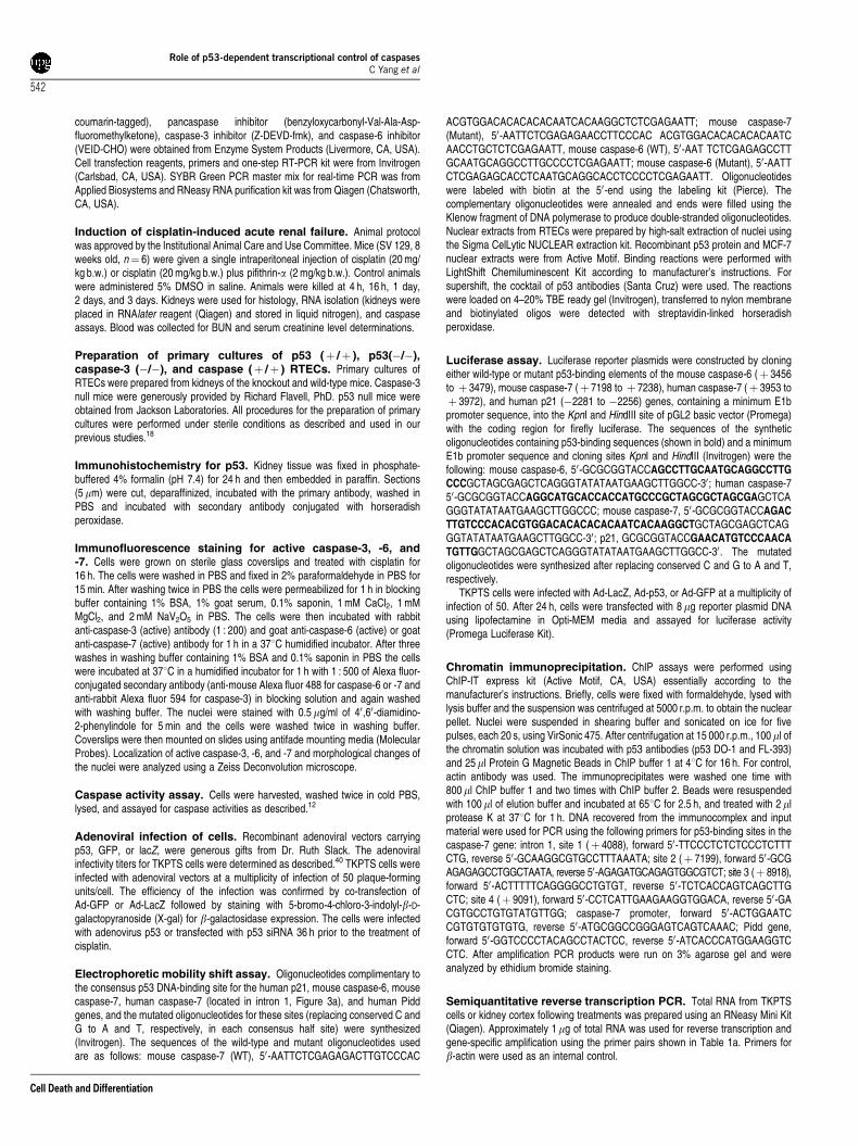
Table 1a Semiquantitative RT-PCR primers
Genes ofinterest
Primerposition
Primer sequence
Caspase-3 Sense (457) 5’-GGGCCTGTTGAACTGAAAAAAntisense
(698)50-CCGTCCTTTGAATTTCTCCA
Caspase-6 Sense (412) 50-TTCAGACGTTGACTGGCTTGAntisense
(562)50-TCCAGCTTGTCTGTCTGGTG
Caspase-7 Sense (850) 50-TTTGCTTACTCCACGGTTCCAntisense
(1055)50-GAGCATGGACACCATACACG
Caspase-8 Sense (1269) 50-GGCCTCCATCTATGACCTGAAntisense
(1502)50-TTCTTCACCGTAGCCATTCC
Caspase-9 Sense (1300) 50-GATGCTGTCCCCTATCAGGAAntisense
(1504)50-GGGACTGCAGGTCTTCAGAG
b-Actin Sense (471) 50-AGCCATGTACGTAGCCATCCAntisense
(697)50-TCTCAGCTGTGGTGGTGAAG
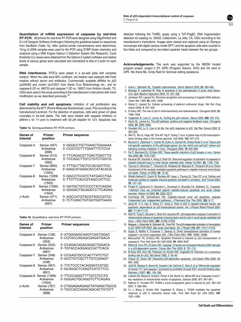
Table 1b Quantitative real-time RT-PCR primers
Genes ofinterest
Primerposition
Primer sequences
Caspase-3 Sense (128) 50-ATGGGAGCAAGTCAGTGGACAntisense
(264)50-CGTACCAGAGCGAGATGACA
Caspase-6 Sense (545) 50-CCAGACAGACAAGCTGGACAAntisense
(687)50-TGTACCAGGAGCCATTCACA
Caspase-7 Sense (348) 50-CCGAGTGCCCACTTATCTGTAntisense
(438)50-ACCTGTCGCTTTGTCGAAGT
Caspase-8 Sense (567) 50-TTCTCCCTACAGGGTCATGCAntisense
(691)50-GCAGGCTCAAGTCATCTTCC
Caspase-9 Sense (1399) 50-TTCCCAGGTTTTGTCTCCTGAntisense
(1504)50-GGGACTGCAGGTCTTCAGAG
b-Actin Sense (787) 50-CTGGAGAAGAGCTATGAGCTGCCGAntisense
(988)50-TGCCACCAGACAGCACTGTGTT
Role of p53-dependent transcriptional control of caspasesC Yang et al
543
Cell Death and Differentiation
29. Oda E, Ohki R, Murasawa H, Nemoto J, Shibue T, Yamashita T et al. Noxa a BH3-onlymember of the Bcl-2 family and candidate mediator of p53-induced apoptosis. Science2000; 288: 1053–1058.
30. Pearson AS, Spitz FR, Swisher SG, Kataoka M, Sarkiss MG, Meyn RE et al. Up-regulationof the proapoptotic mediators Bax and Bak after adenovirus-mediated p53 gene transfer inlung cancer cells. Clin Cancer Res 2000; 6: 887–890.
31. Miyashita T, Reed JC. Tumor suppressor p53 is a direct transcriptional activator of thehuman bax gene. Cell 1995; 80: 293–299.
32. Sax JK, Fei P, Murphy ME, Bernhard E, Korsmeyer SJ, El-Deiry WS. BID regulation by p53contributes to chemosensitivity. Nat Cell Biol 2002; 4: 842–849.
33. Oda K, Arakawa H, Tanaka T, Matsuda K, Tanikawa C, Mori T et al. p53AIP1 a potentialmediator of p53-dependent apoptosis and its regulation by Ser-46-phosphorylated p53.Cell 2000; 102: 849–862.
34. Wajant H. CD95L/FasL and TRAIL in tumour surveillance and cancer therapy. CancerTreat Res 2006; 130: 141–165.
35. Chipuk JE, Kuwana T, Bouchier-Hayes L, Droin NM, Newmeyer DD, Schuler M et al. Directactivation of Bax by p53 mediates mitochondrial membrane permeabilization andapoptosis. Science 2004; 303: 1010–1014.
36. Mihara M, Erster S, Zaika A, Petrenko O, Chittenden T, Pancoska P et al. p53 has a directapoptogenic role at the mitochondria. Mol Cell 2003; 11: 577–590.
37. Moll UM, Wolff S, Speidel D, Deppert W. Transcription-independent pro-apoptotic functionsof p53. Curr Opin Cell Biol 2005; 17: 631–636.
38. Ho J, Benchimol S. Transcriptional repression mediated by the p53 tumour suppressor.Cell Death Differ 2003; 10: 404–408.
39. Kelly KJ, Plotkin Z, Vulgamott SL, Dagher PC. P53 mediates the apoptotic response toGTP depletion after renal ischemia–reperfusion: protective role of a p53 inhibitor. J Am SocNephrol 2003; 14: 128–138.
40. Cregan SP, MacLaurin J, Gendron TF, Callaghan SM, Park DS, Parks RJ et al. Helper-dependent adenovirus vectors: their use as a gene delivery system to neurons. GeneTherapy 2000; 7: 1200–1209.
Role of p53-dependent transcriptional control of caspasesC Yang et al
544
Cell Death and Differentiation


