Embed Size (px)
Citation preview
Imaging the Vasculit ides
David S. Younger, MD, MPH, MSa,b,*
KEYWORDS
� Neuroimaging � Computerized tomography � Magnetic resonance imaging� Single-photon emission tomography � Positron emission tomography� Cerebral angiography
KEY POINTS
� Neuroimaging plays a vital role in the diagnosis of primary and secondary vasculiticdisorders.
� There are a multiplicity of neuroimaging options available to accurately describe the un-derlying clinical deficits of involved cases.
� Noninvasive neuroimaging modalities provide less risk and when interdigitated, form thebasis for a more conclusive understanding of the disease process.
� There are instances in which invasive cerebral angiography may be needed to image theintricate and at times, small involved vessels.
� Neuroradiologists should be included in the multidisciplinary team of physicians caring forpatients with vasculitides and in research to providemore sensitive and safe modalities forthe accurate diagnosis thereof.
INTRODUCTION
Vasculitis is a term used to describe a diverse spectrum of diseases characterized byinflammation of the blood vessels that may progress to ischemic injury of the centralnervous system (CNS) resulting in a myriad of focal and generalized neurologicsymptoms. The injury is usually secondary to mural changes resulting in vessel steno-sis or occlusion. Endothelial inflammation promotes intraluminal coagulation andthrombosis.1 Perivascular inflammatory changes and edema contribute to the patho-logic picture. Arterial and venous components may be involved separately or togetherand dural sinuses may be affected. Generalized inflammatory processes may producea secondary encephalitis or myelitis. The radiological aspects of CNS vasculitishave evolved in the past decade.2 No one single imaging modality is sufficient or
Disclosure Statement: The author has nothing to disclose.a Department of Neurology, Division of Neuro-Epidemiology, New York University School ofMedicine, New York, NY 10016, USA; b School of Public Health, City University of New York,New York, NY, USA* 333 East 34th Street, Suite 1J, New York, NY 10016.E-mail address: [email protected]: http://www.davidsyounger.com
Neurol Clin 37 (2019) 249–265https://doi.org/10.1016/j.ncl.2019.01.010 neurologic.theclinics.com0733-8619/19/ª 2019 Elsevier Inc. All rights reserved.
Younger250
preeminent; a combination of studies is typically required for a confident diagnosis ofCNS vasculitis.
OVERVIEW AND CLASSIFICATION
With an estimated worldwide incidence of 20 per million for eosinophilic granulomato-sis with polyangiitis (EGPA), also known as Churg-Strauss syndrome, 10 per million forgranulomatosis with polyangiitis (GPA) or Wegener granulomatosis, 2.6 per million forTakayasu arteritis (TAK), and 0.9 per million for polyarteritis nodosa (PAN),3,4 and onlya fraction of patients presenting with CNS involvement, it is important for clinicians tobe familiar with the clinical and neuroradiologic presentations of the vasculitides. Thisis especially important in young and middle-aged adults in whom the prevalence ofatherosclerotic disease is low.5–9
The historical aspects of the classification of the vasculitides are reviewed else-where in this issue. To illustrate the clinical importance placed on the radiologic man-ifestations of the PAN group of primary systemic vasculitides, the narrative shouldbegin with Citron and colleagues10 who described multiorgan arteritis of the CNS ina highly publicized report of 14 Los Angeles multidrug abusers with a common denom-inator of intravenous methamphetamine abuse by all but 2 patients, and exclusively by1. Acute vessel lesions included fibrinoid necrosis of the media and intima with infiltra-tion of polymorphonuclear cells, eosinophils, lymphocytes, and histiocytes, followedby vascular elastic and vascular smooth muscle destruction resulting in lesionsconsidered typical for PAN. Substantiation of necrotizing arteritis was present inonly 4 of the 14 patients. Citron and Peters11 responded to the criticism from Baden12
that he had not observed a causal relation between drug abuse and necrotizing arter-itis at the Office of Chief Medical Examiner of New York City for the past one-half cen-tury among thousands of autopsied drug abusers, with the countering opinion thatevidence of aneurysms noted in 13 of the 14 patients was ample proof of arteritis.The contribution of angiography to the designation of CNS vasculitis commenced
with the identification of angiographic beading and a sausagelike appearance of cere-bral vessels at sites of presumed arteritis first in 1964 by Hinck and coworkers13 in gi-ant cell arteritis (GCA). In 1983, Cupps and colleagues14 established the utility ofcerebral angiography in the diagnosis of histologically proven isolated angiitis or pri-mary angiitis of the CNS (PACNS). As giant cells and epithelioid cells usually foundat postmortem examination in such patients were an inconsistent finding in a menin-geal and brain biopsy and therefore considered unnecessary for antemortem diag-nosis, Moore and Cupps15 considered angiography necessary for the diagnosis ofPACNS. This prevailing opinion was shared by Calabrese andMallek,16 who proposedcriteria for the diagnosis of PACNS, and Hajj-Ali and Calabrese17 who later separatedPACNS from the reversible cerebral vasoconstrictive syndrome (RCVS), characterizedinstead by transient nonvasculitic narrowing of intracranial vessels.By 1990, Hunder and colleagues18 on behalf of the American College of Rheuma-
tology (ACR) noted that the goal in classification was to identify sets of sensitivecriteria that recognized a high proportion of patients with a particular form of vasculitis,while specifically excluding a high proportion of those with other diseases. Althoughhighly specific and sensitive classification criteria might prove useful in the depictionof patients for epidemiologic studies and therapeutic trials, such criteria might notnecessarily include the full spectrum of manifestations of a particular vasculitic dis-ease, which was instead the role of formal diagnostic criteria. Lie19 noted that althougha definitive diagnosis of vasculitis almost invariably required histologic documentation,the interpretation of a diagnostic tissue sample was subject to variables as diverse as
Imaging the Vasculitides 251
the pathologist’s experience, tissue selection, sample size, chronologic age of the dis-ease, and any prior treatment at the time of the biopsy. The angiographic appearanceof aneurysms or occlusions of visceral arteries not due to arteriosclerosis, fibromuscu-lar dysplasia, or other noninflammatory causes, were useful in the classification ofPAN20 with a sensitivity of 73.5% and specificity of 89.2%. The angiographic featuresof narrowing, aneurysm, or occlusion of the aorta or its primary branches, were usefulcriteria for the classification of TAK21 with sensitivities and specificities of 85.5% and81.2%, 20.3% and 95.9%, and 51.6% and 86.1%, respectively. In the same 1990 vol-ume of the journal, Arthritis and Rheumatism, the ACR Subcommittee on Classificationof Vasculitis noted no diagnostic features of angiography useful in the classificationcriteria of EGPA, GPA, hypersensitivity vasculitis, immunoglobulin A vasculitis (IgAV)and GCA.22–26
Jennette and colleagues27 held 2 Chapel Hill Consensus Conferences (CHCCs)beginning in 1994 with a 2012 revision,28 establishing the nomenclature or nosologyof systemic vasculitides. However, different from the ACRSubcommittee on Classifica-tion of Vasculitis,18 Jennette and colleagues27,28 incorporated prevailing knowledgeabout etiology, pathogenesis, pathology, demographics, and clinical manifestations,andusedamodel of thepredominant caliber of involved vessel that delineated the3ma-jor categoriesof systemic vasculitis including large-sizevessel vasculitis (LVV),medium-size vessel vasculitis (MVV), andsmall-sized vessel vasculitis (SVV) types, adding furtherdistinctionsas to thestructural and functional characteristicsofparticular vascularbeds,aswell as the known biochemical and functional properties that rendered them suscep-tible to vasculitic injury. With approximately 26 recognized vasculitides in the 2012revised CHCC, many of which demonstrate overlap in affected involved arteries,coupled with advances in the neuroradiologic techniques for discerning CNS involve-ment, there has been heightened interest in imaging the cerebral vasculature.Kuker29 differentiated the entities of extracranial LVV and intracranial MVV and SVV,
noting that vasculitic involvement of the internal carotid (ICA), common carotid (CCA),M1 and A1 segments of the middle (MCA) and anterior cerebral arteries (ACA), intra-cranial vertebral and basilar arteries, and P1 segment of the posterior cerebral artery(PCA), generally regarded as intracranial LVV, would instead be considered systemicMVV by 2012 Revised CHCC nomenclature.28 Moreover, vasculitic involvement alongarterial vessels distal to the MCA bifurcation, as well as communicating vessels suchas the anterior and posterior communicating arteries, were still considered MVV sys-temically although intracranial. They may not be demonstrable along with intracranialLVV byMRI, magnetic resonance angiography (MRA), or computed tomographic angi-ography (CTA), and may require conventional angiography (CA) for luminal irregularityto be visualized.The smallest muscular arteries and arterioles in the brain parenchyma, as well as the
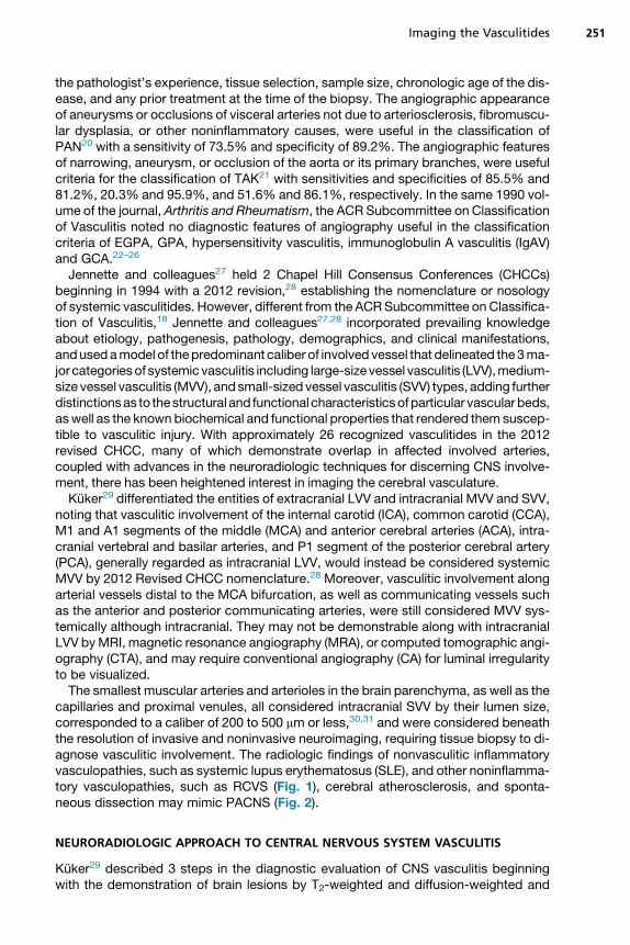
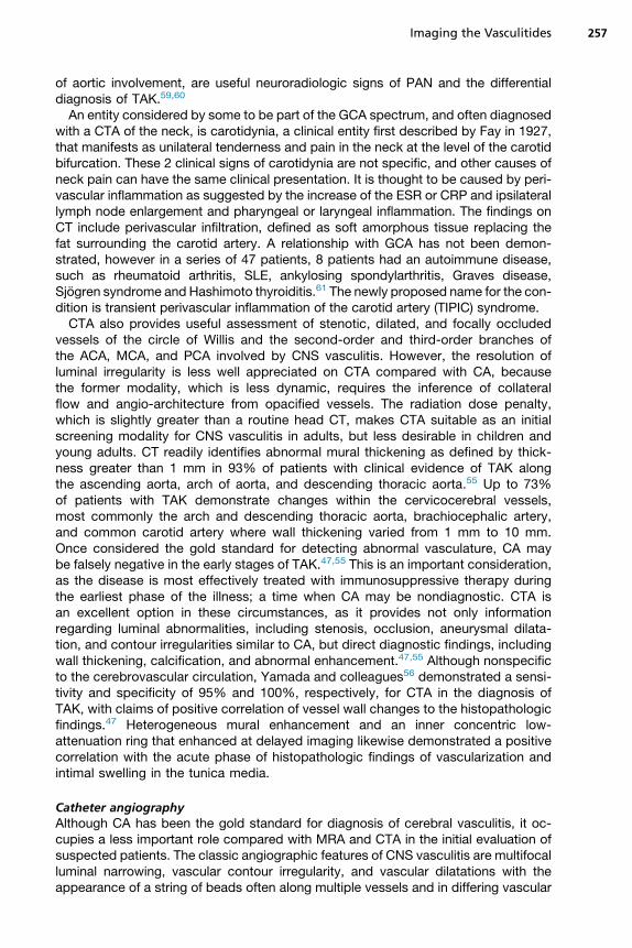
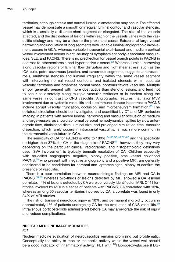
capillaries and proximal venules, all considered intracranial SVV by their lumen size,corresponded to a caliber of 200 to 500 mm or less,30,31 and were considered beneaththe resolution of invasive and noninvasive neuroimaging, requiring tissue biopsy to di-agnose vasculitic involvement. The radiologic findings of nonvasculitic inflammatoryvasculopathies, such as systemic lupus erythematosus (SLE), and other noninflamma-tory vasculopathies, such as RCVS (Fig. 1), cerebral atherosclerosis, and sponta-neous dissection may mimic PACNS (Fig. 2).
NEURORADIOLOGIC APPROACH TO CENTRAL NERVOUS SYSTEM VASCULITIS
Kuker29 described 3 steps in the diagnostic evaluation of CNS vasculitis beginningwith the demonstration of brain lesions by T2-weighted and diffusion-weighted and
Fig. 1. RCVS. (A) MRI FLAIR imaging on presentation demonstrates multifocal abnormalhyperintense signal within the bilateral hemispheric white matter, more prominent in theparietal and occipital lobes where it extends to the cortex. (B) T2-diffusion imaging demon-strates restricted diffusion consistent with acute ischemia. The white matter distributionwithin the left hemisphere straddles the anterior, middle, and posterior cerebral vascularterritories, a “watershed” distribution. (C) MRA at admission demonstrates short-segmentmultifocal narrowing within the distal bilateral vertebral arteries, the basilar artery, andbilateral middle and posterior cerebral vasculature (white arrows). (D) CTA demonstratesmoderate narrowing within the right PCA P2 segment and a more severe narrowing distallywithin the P3 parieto-occipital segment (left, black arrows). Mild narrowing is presentwithin the ACA A1 segment (right, black arrow). (E) CA confirms multifocal narrowingwithin the bilateral posterior cerebral arteries (black arrows). (F) Follow-up CTA (at presen-tation on the left, 5 months’ follow-up on the right) reveals complete resolution of the orig-inal findings (white arrows). (Courtesy of Adam Davis, MD New York, NY.)
Younger252
perfusion-weighted MRI, followed by the delineation of underlying vascular pathologyby 1.5-T MRA to study the entire course of the carotid and vertebral arteries, as well asthe circle of Willis. Time-of-flight (TOF) MRA sequences permit detection of more sub-tle stenoses and improve spatial resolution, as well as mural thickness in basal brainarteries with MRA source images; moreover, MRI may discern mural enhancement.Conventional angiography with digital subtraction (DSA) is used to evaluatemedium-sized and small brain vessels and the status of cerebral hemodynamicsand assessment of brain perfusion.Gomes32 divided available neuroimaging studies into 3 groups, including the brain
parenchyma, vessel lumen, and vessel wall. Parenchymal findings, although least spe-cific, were necessary to detect the presence of disease as well as to follow progres-sion and remission status. Vessel lumen and wall abnormalities, although highlysuggestive for systemic vasculitis when present, were considered nonspecific andinsensitive in the diagnosis of intracranial SVV.
Parenchymal Imaging
TheMRI findings of CNS vasculitis have been previously described,33–36 the common-est of which are T2/fluid-attenuated inversion recovery (FLAIR) hyperintense lesionssecondary to ischemia distributed throughout subcortical and deep white matter,the deep gray nuclei, and the cortices. The MCA territory is the commonest involved
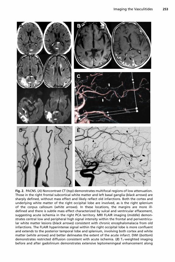
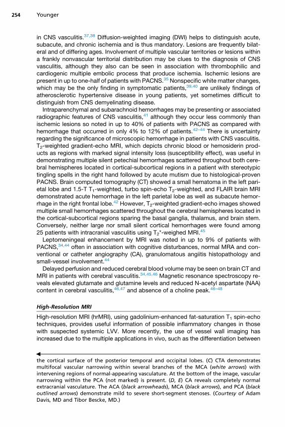
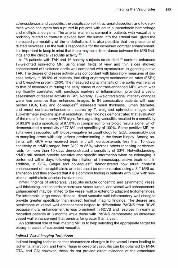
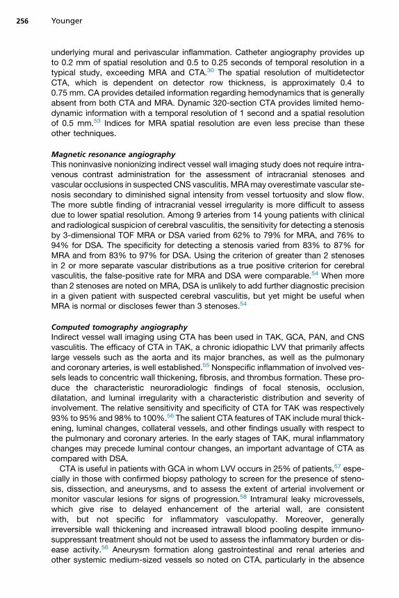
Fig. 2. PACNS. (A) Noncontrast CT (top) demonstrates multifocal regions of low attenuation.Those in the right frontal subcortical white matter and left basal ganglia (black arrows) aresharply defined, without mass effect and likely reflect old infarctions. Both the cortex andunderlying white matter of the right occipital lobe are involved, as is the right spleniumof the corpus callosum (white arrows). In these locations, the margins are more ill-defined and there is subtle mass effect characterized by sulcal and ventricular effacement,suggesting acute ischemia in the right PCA territory. MRI FLAIR imaging (middle) demon-strates central low and peripheral high signal intensity within the frontal and periventricu-lar white matter lesions (black arrows) consistent with chronic encephalomalacia from oldinfarctions. The FLAIR hyperintense signal within the right occipital lobe is more confluentand extends to the posterior temporal lobe and splenium, involving both cortex and whitematter (white arrows) and better delineates the extent of the acute infarct. DWI (bottom)demonstrates restricted diffusion consistent with acute ischemia. (B) T1-weighted imagingbefore and after gadolinium demonstrates extensive leptomeningeal enhancement along
Imaging the Vasculitides 253
Younger254
in CNS vasculitis.37,38 Diffusion-weighted imaging (DWI) helps to distinguish acute,subacute, and chronic ischemia and is thus mandatory. Lesions are frequently bilat-eral and of differing ages. Involvement of multiple vascular territories or lesions withina frankly nonvascular territorial distribution may be clues to the diagnosis of CNSvasculitis, although they also can be seen in association with thrombophilic andcardiogenic multiple embolic process that produce ischemia. Ischemic lesions arepresent in up to one-half of patients with PACNS.35 Nonspecific white matter changes,which may be the only finding in symptomatic patients,39,40 are unlikely findings ofatherosclerotic hypertensive disease in young patients, yet sometimes difficult todistinguish from CNS demyelinating disease.Intraparenchymal and subarachnoid hemorrhages may be presenting or associated
radiographic features of CNS vasculitis,41 although they occur less commonly thanischemic lesions so noted in up to 40% of patients with PACNS as compared withhemorrhage that occurred in only 4% to 12% of patients.42–44 There is uncertaintyregarding the significance of microscopic hemorrhage in patients with CNS vasculitis.T2-weighted gradient-echo MRI, which depicts chronic blood or hemosiderin prod-ucts as regions with marked signal intensity loss (susceptibility effect), was useful indemonstrating multiple silent petechial hemorrhages scattered throughout both cere-bral hemispheres located in cortical-subcortical regions in a patient with stereotypictingling spells in the right hand followed by acute mutism due to histological-provenPACNS. Brain computed tomography (CT) showed a small hematoma in the left pari-etal lobe and 1.5-T T1-weighted, turbo spin-echo T2-weighted, and FLAIR brain MRIdemonstrated acute hemorrhage in the left parietal lobe as well as subacute hemor-rhage in the right frontal lobe.42 However, T2-weighted gradient-echo images showedmultiple small hemorrhages scattered throughout the cerebral hemispheres located inthe cortical-subcortical regions sparing the basal ganglia, thalamus, and brain stem.Conversely, neither large nor small silent cortical hemorrhages were found among25 patients with intracranial vasculitis using T2*-weighed MRI.45
Leptomeningeal enhancement by MRI was noted in up to 9% of patients withPACNS,34,44 often in association with cognitive disturbances, normal MRA and con-ventional or catheter angiography (CA), granulomatous angiitis histopathology andsmall-vessel involvement.44
Delayed perfusion and reduced cerebral blood volumemay be seen on brain CT andMRI in patients with cerebral vasculitis.34,45,46 Magnetic resonance spectroscopy re-veals elevated glutamate and glutamine levels and reduced N-acetyl aspartate (NAA)content in cerebral vasculitis,46,47 and absence of a choline peak.46–48
High-Resolution MRI
High-resolution MRI (hrMRI), using gadolinium-enhanced fat-saturation T1 spin-echotechniques, provides useful information of possible inflammatory changes in thosewith suspected systemic LVV. More recently, the use of vessel wall imaging hasincreased due to the multiple applications in vivo, such as the differentiation between
=the cortical surface of the posterior temporal and occipital lobes. (C) CTA demonstratesmultifocal vascular narrowing within several branches of the MCA (white arrows) withintervening regions of normal-appearing vasculature. At the bottom of the image, vascularnarrowing within the PCA (not marked) is present. (D, E) CA reveals completely normalextracranial vasculature. The ACA (black arrowheads), MCA (black arrows), and PCA (blackoutlined arrows) demonstrate mild to severe short-segment stenoses. (Courtesy of AdamDavis, MD and Tibor Bescke, MD.)
Imaging the Vasculitides 255
atherosclerosis and vasculitis, the visualization of intracranial dissection, and to deter-mine which aneurysm has ruptured in patients with acute subarachnoid hemorrhageand multiple aneurysms. The arterial wall enhancement in patients with vasculitis isprobably related to contrast leakage from the lumen into the arterial wall, given theincreased permeability of the endothelium; it is also possible that the presence ofdilated neovessels in the wall is responsible for the increased contrast enhancement.It is important to keep in mind that there may be a discordance between the MRI find-ings and the clinical vasculitis activity.49
In 26 patients with TAK and 16 healthy subjects so studied,50 contrast-enhancedT1-weighted spin-echo MRI using small fields of view and thin slices showedenhancement of thickened aortic wall compared with myocardium, suggesting activeTAK. The degree of disease activity was concordant with laboratory measures of dis-ease activity in 88.5% of patients, including erythrocyte sedimentation rates (ESRs)and C-reactive protein (CRP). The measured signal intensity of the aortic wall relativeto that of myocardium during the early phase of contrast-enhanced MRI, which wassignificantly correlated with serologic markers of inflammation, provided a usefulassessment of disease activity in TAK. Notably, T2-weighted signal intensity changeswere less sensitive than enhanced images. In 64 consecutive patients with sus-pected GCA, Bley and colleagues51 assessed mural thickness, lumen diameter,and mural contrast-enhancement scores by T1-weighted spin-echo images withsub-millimeter in-plane spatial resolution. Their findings demonstrated that evaluationof the mural inflammatory MRI signs for diagnosing vasculitis resulted in a sensitivityof 80.6% and a specificity of 97.0%, in comparison to histologic results alone, whichdemonstrated a sensitivity of 77.8% and specificity of 100%. Some positive MRI re-sults were associated with biopsy-negative histopathology for GCA, presumably dueto sampling errors with skip lesions predominating in the tissue biopsy. Among pa-tients with GCA who received treatment with corticosteroids less than 10 days,sensitivity of hrMRI ranged from 81% to 85%, whereas others receiving corticoste-roids for more than 10 days demonstrated a sensitivity of 33%. Notwithstanding,hrMRI still should provide sensitive and specific information when neuroimaging isperformed within days following the initiation of immunosuppressive treatment. Inaddition, in GCA, Geiger and colleagues52 demonstrated how mural contrastenhancement of the ophthalmic arteries could be demonstrated using a 3-T MRI ex-amination and they showed that it is a common finding in patients with GCA with sus-picious ophthalmic arteries involvement.hrMRI findings of intracranial vasculitis include concentric and asymmetric vessel
wall thickening, an eccentric or narrowed vessel lumen, and vessel wall enhancement.Enhancement may be limited to the vessel wall or extend to adjacent leptomeninges.For intracranial large vessel disease, direct vascular wall inflammatory wall changesprovide greater specificity than indirect luminal imaging findings. The degree andpersistence of vessel wall enhancement helped to differentiate PACNS from RCVSbecause mural enhancement is less prominent in RCVS and resolves in nearly allrestudied patients at 3 months while those with PACNS demonstrate an increasedvessel wall enhancement that persists for greater than a year.An additional role of wall imaging MRI is to help selecting the appropriate target for
biopsy in cases of suspected vasculitis.
Indirect Vessel Imaging Techniques
Indirect imaging techniques that characterize changes in the vessel lumen leading toischemia, infarction, and hemorrhage in cerebral vasculitis can be obtained by MRA,CTA, and CA; however, these do not provide direct evidence of the associated
Younger256
underlying mural and perivascular inflammation. Catheter angiography provides upto 0.2 mm of spatial resolution and 0.5 to 0.25 seconds of temporal resolution in atypical study, exceeding MRA and CTA.30 The spatial resolution of multidetectorCTA, which is dependent on detector row thickness, is approximately 0.4 to0.75 mm. CA provides detailed information regarding hemodynamics that is generallyabsent from both CTA and MRA. Dynamic 320-section CTA provides limited hemo-dynamic information with a temporal resolution of 1 second and a spatial resolutionof 0.5 mm.53 Indices for MRA spatial resolution are even less precise than theseother techniques.
Magnetic resonance angiographyThis noninvasive nonionizing indirect vessel wall imaging study does not require intra-venous contrast administration for the assessment of intracranial stenoses andvascular occlusions in suspected CNS vasculitis. MRAmay overestimate vascular ste-nosis secondary to diminished signal intensity from vessel tortuosity and slow flow.The more subtle finding of intracranial vessel irregularity is more difficult to assessdue to lower spatial resolution. Among 9 arteries from 14 young patients with clinicaland radiological suspicion of cerebral vasculitis, the sensitivity for detecting a stenosisby 3-dimensional TOF MRA or DSA varied from 62% to 79% for MRA, and 76% to94% for DSA. The specificity for detecting a stenosis varied from 83% to 87% forMRA and from 83% to 97% for DSA. Using the criterion of greater than 2 stenosesin 2 or more separate vascular distributions as a true positive criterion for cerebralvasculitis, the false-positive rate for MRA and DSA were comparable.54 When morethan 2 stenoses are noted on MRA, DSA is unlikely to add further diagnostic precisionin a given patient with suspected cerebral vasculitis, but yet might be useful whenMRA is normal or discloses fewer than 3 stenoses.54
Computed tomography angiographyIndirect vessel wall imaging using CTA has been used in TAK, GCA, PAN, and CNSvasculitis. The efficacy of CTA in TAK, a chronic idiopathic LVV that primarily affectslarge vessels such as the aorta and its major branches, as well as the pulmonaryand coronary arteries, is well established.55 Nonspecific inflammation of involved ves-sels leads to concentric wall thickening, fibrosis, and thrombus formation. These pro-duce the characteristic neuroradiologic findings of focal stenosis, occlusion,dilatation, and luminal irregularity with a characteristic distribution and severity ofinvolvement. The relative sensitivity and specificity of CTA for TAK was respectively93% to 95% and 98% to 100%.56 The salient CTA features of TAK include mural thick-ening, luminal changes, collateral vessels, and other findings usually with respect tothe pulmonary and coronary arteries. In the early stages of TAK, mural inflammatorychanges may precede luminal contour changes, an important advantage of CTA ascompared with DSA.CTA is useful in patients with GCA in whom LVV occurs in 25% of patients,57 espe-
cially in those with confirmed biopsy pathology to screen for the presence of steno-sis, dissection, and aneurysms, and to assess the extent of arterial involvement ormonitor vascular lesions for signs of progression.58 Intramural leaky microvessels,which give rise to delayed enhancement of the arterial wall, are consistentwith, but not specific for inflammatory vasculopathy. Moreover, generallyirreversible wall thickening and increased intrawall blood pooling despite immuno-suppressant treatment should not be used to assess the inflammatory burden or dis-ease activity.56 Aneurysm formation along gastrointestinal and renal arteries andother systemic medium-sized vessels so noted on CTA, particularly in the absence
Imaging the Vasculitides 257
of aortic involvement, are useful neuroradiologic signs of PAN and the differentialdiagnosis of TAK.59,60
An entity considered by some to be part of the GCA spectrum, and often diagnosedwith a CTA of the neck, is carotidynia, a clinical entity first described by Fay in 1927,that manifests as unilateral tenderness and pain in the neck at the level of the carotidbifurcation. These 2 clinical signs of carotidynia are not specific, and other causes ofneck pain can have the same clinical presentation. It is thought to be caused by peri-vascular inflammation as suggested by the increase of the ESR or CRP and ipsilaterallymph node enlargement and pharyngeal or laryngeal inflammation. The findings onCT include perivascular infiltration, defined as soft amorphous tissue replacing thefat surrounding the carotid artery. A relationship with GCA has not been demon-strated, however in a series of 47 patients, 8 patients had an autoimmune disease,such as rheumatoid arthritis, SLE, ankylosing spondylarthritis, Graves disease,Sjogren syndrome and Hashimoto thyroiditis.61 The newly proposed name for the con-dition is transient perivascular inflammation of the carotid artery (TIPIC) syndrome.CTA also provides useful assessment of stenotic, dilated, and focally occluded
vessels of the circle of Willis and the second-order and third-order branches ofthe ACA, MCA, and PCA involved by CNS vasculitis. However, the resolution ofluminal irregularity is less well appreciated on CTA compared with CA, becausethe former modality, which is less dynamic, requires the inference of collateralflow and angio-architecture from opacified vessels. The radiation dose penalty,which is slightly greater than a routine head CT, makes CTA suitable as an initialscreening modality for CNS vasculitis in adults, but less desirable in children andyoung adults. CT readily identifies abnormal mural thickening as defined by thick-ness greater than 1 mm in 93% of patients with clinical evidence of TAK alongthe ascending aorta, arch of aorta, and descending thoracic aorta.55 Up to 73%of patients with TAK demonstrate changes within the cervicocerebral vessels,most commonly the arch and descending thoracic aorta, brachiocephalic artery,and common carotid artery where wall thickening varied from 1 mm to 10 mm.Once considered the gold standard for detecting abnormal vasculature, CA maybe falsely negative in the early stages of TAK.47,55 This is an important consideration,as the disease is most effectively treated with immunosuppressive therapy duringthe earliest phase of the illness; a time when CA may be nondiagnostic. CTA isan excellent option in these circumstances, as it provides not only informationregarding luminal abnormalities, including stenosis, occlusion, aneurysmal dilata-tion, and contour irregularities similar to CA, but direct diagnostic findings, includingwall thickening, calcification, and abnormal enhancement.47,55 Although nonspecificto the cerebrovascular circulation, Yamada and colleagues56 demonstrated a sensi-tivity and specificity of 95% and 100%, respectively, for CTA in the diagnosis ofTAK, with claims of positive correlation of vessel wall changes to the histopathologicfindings.47 Heterogeneous mural enhancement and an inner concentric low-attenuation ring that enhanced at delayed imaging likewise demonstrated a positivecorrelation with the acute phase of histopathologic findings of vascularization andintimal swelling in the tunica media.
Catheter angiographyAlthough CA has been the gold standard for diagnosis of cerebral vasculitis, it oc-cupies a less important role compared with MRA and CTA in the initial evaluation ofsuspected patients. The classic angiographic features of CNS vasculitis are multifocalluminal narrowing, vascular contour irregularity, and vascular dilatations with theappearance of a string of beads often along multiple vessels and in differing vascular
Younger258
territories, although ectasia and normal luminal diameter also may occur. The affectedvessel may demonstrate a smooth or irregular luminal contour and vascular stenosis,which is classically a discrete short segment or elongated. The size of the vesselsaffected, and the distribution of lesions within each of the vessels varies with the vas-culitic etiology and may be a clue to the proximate cause. Extracranial large vesselnarrowing and undulation of long segments with variable luminal angiographic involve-ment occurs in GCA, whereas variable intracranial skull–based and medium corticalvessel involvement occurs in anti-neutrophil cytoplasm antibody–associated vasculit-ides, SLE, and PACNS. There is no predilection for vessel branch points in PACNS incontrast to atherosclerosis and hypertensive disease.62 Whereas luminal narrowingalong vascular regions of laminar flow disruption and high shear stress, such as theICA bulb, petro-cavernous junction, and cavernous segments, suggests atheroscle-rosis, multifocal stenosis and luminal irregularity within the same vessel segmentwith intervening normal vessel contours, and isolated stenosis within separatevascular territories and otherwise normal vessel contours favors vasculitis. Multipleemboli generally present with more obstructive than stenotic lesions, and tend notto occur as discretely along multiple vascular territories or in tandem along thesame vessel in contrast to CNS vasculitis. Angiographic features that favor MVVinvolvement due to systemic vasculitis and autoimmune disease in contrast to PACNSinclude abrupt vascular truncation, occlusion, and microaneurysm formation.63 Thecollateral circulation should be investigated and quantified by CT and MR perfusionimaging in patients with severe luminal narrowing and vascular occlusion of mediumand large vessels, as should abnormal cerebral hemodynamics typified by slow anter-ograde flow, diminished distal luminal size, and prolonged circulation time. Vasculardissection, which rarely occurs in intracranial vasculitis, is much more common inthe extracranial vasculature in GCA.The sensitivity of CA for PACNS is 40% to 100%,34,35,38,40,62–66 and the specificity
no higher than 37% for CA in the diagnosis of PACNS67; however, they may varydepending on the particular clinical, radiographic, and histopathologic definitionsused. SVV involvement is typically beneath resolution of CA. Children especiallywith so-called angiography negative, biopsy positive, small-vessel childhoodPACNS,68 who present with negative angiography and a positive MRI, are generallyconsidered to be candidates for cerebral and leptomeningeal biopsy to confirm thepresence of vasculitis.There is a poor correlation between neuroradiologic findings on MRI and CA in
PACNS.38,63 Whereas two-thirds of lesions detected by MRI showed a CA lesionalcorrelate, 44% of lesions detected by CA were conversely identified on MRI. Of 41 ter-ritories involved by MRI in a series of patients with PACNS, CA correlated with 15%,whereas among 50 vascular territories involved by CA, a correlate was found in only34% of MRI studies.The risk of transient neurologic injury is 10%, and permanent morbidity occurs in
approximately 1% of patients undergoing CA for the evaluation of CNS vasculitis.69
Intravenous corticosteroids administered before CA may ameliorate the risk of injuryand reduce complications.
NUCLEAR MEDICINE IMAGE MODALITIESPET
Nuclear medicine evaluation of neurovasculitis remains promising but problematic.Conceptually the ability to monitor metabolic activity within the vessel wall shouldbe a good indicator of inflammatory activity. PET with 18Fluorodeoxyglucose (FDG-
Imaging the Vasculitides 259
PET) has been the best studied radionuclide for this indication, particularly in systemicLVV. One meta-analysis70 demonstrated a wide variability of diagnostic sensitivity forTAK ranging from 28% to 100%, whereas the range of specificity was 50% to 100%.Fused PET and CT images provide superior anatomic localization and improved sensi-tivity and specificity of 91% and 89% for TAK. A greater diagnostic sensitivity of 80%and specificity of 89% in those studied by FDG-PET were noted for GCA.71 Nonethe-less, the specificity of FDG-PET is degraded by the presence of atherosclerosis, asactive inflammatory plaques may produce false-positive findings.72 The utility ofFDG-PET for monitoring disease activity in these patients may be problematic.Some patients with TAK deemed inactive by clinical criteria may in fact demonstratebiopsy-proven active inflammation.73 Studies indicate that FDG-PET may be a sensi-tive and helpful diagnostic study for identifying these patients with subclinical activedisease, with 83% of patients with biopsy-proven GCA demonstrating positiveFDG-PET studies.74 The sensitivity and specificity of FDG-PET in the identificationof active vasculitic disease compared with clinical signs and laboratory criteria leadto respective rates of 100% and 89%.75 The fusion of MRI for morphology and volu-metric assessment, and FDG-PET for metabolic analysis of the brain, has been usedto investigate the autoimmune encephalitides, and may have a role in the investigationof CNS vasculitides.76
Single-Photon Emission Computed Tomography
Single-photon emission computed tomography (SPECT) uses multiplanar nuclearmedicine imaging for the investigation of regional CNS perfusion abnormalities. It pro-vides direct information about the pathophysiology and cerebral metabolism in cere-bral vasculitides at the level of capillary endothelium in the blood-brain barrier (BBB)microcirculation beyond the resolution of MRA, CTA, and CA.77,78
There are claims of the utility of brain SPECT imaging in the clinical diagnosisand management of cerebral vasculitis associated with SLE,79,80 Kawasaki dis-ease,81 IgAV,82 neurologic Behcet disease,83 GPA,84 and brain irradiation.85,86 Apartfrom the direct impact of vascular narrowing and occlusion resulting from necrotizingarteritis and vascular infiltration, other explanations for an abnormal brain SPECTinclude circulating immune complexes on the BBB,87 and neurotoxic effects of anti-bodies and brain antigenic targets,81 glial cell interactions,86 and pathogenic hyper-sensitivity responses to brain antigens released during vascular-mediated tissuenecrosis.86
The results of brain SPECT imaging were described in one patient with histologi-cally verified cerebral vasculitis.88 This 71-year-old man with later-proven granuloma-tous angiitis of the brain underwent Tc-99m hexamethylpropyleneamine oxime brainSPECT 3 weeks after onset of CNS disease. There was irregular radiotracer uptakethroughout both cerebral hemispheres with scattered multiple areas of hypoperfu-sion, further demonstrated in surface volumetric images. Postmortem examinationshowed fibrinoid necrosis, inflammatory cells, mainly lymphocytes, histiocytes, anda few multinucleated giant cells involving medium-to-small meningeal and paren-chymal vessels with intramural vascular deposits of amyloid, without systemicvasculitis.
Color Doppler Ultrasonography
Color Doppler ultrasonography and color duplex imaging provide direct imaging andevaluation of superficial arteries and their vessel walls. It has been most extensivelystudied in systemic LVV, such as GCA and TAK. It provides a high-resolution imagingof the walls of deep-seated vessels as compared with MRI, detecting wall thickness
Younger260
and edema, the latter of which produces a hypoechoic signal on color Doppler ultra-sonography as a halo sign. In a meta-analysis of 998 patients with 17 studies, thesensitivity of the halo sign for biopsy-proven GCA was only 75%, but specificitywas 83%. Concentric homogeneous mural thickening, stenosis, and occlusion ofthe aorta and brachiocephalic branches are typical ultrasonography features ofGCA and TAK,72,89,90 which may be differentiated from atherosclerotic disease bythe absence of plaque formation, concentric long segment involvement, and location.Ultrasonography revealed subtle mural changes characterized by a homogeneous,circumferential mid-echoic wall thickening within the subclavian and carotid arteriesin the early stages of TAK preceding abnormalities detected by CA,91 with overallgreater wall thickness of the CCA and ICA in the vasculitic vessels compared with con-trols. The CCA intima-to-medial thickness ratio was increased in patients with TAKcompared with normal controls,92 yielding respective sensitivity and specificity ratesof 82% and 70%.The wall diameters of common, frontal, and parietal division of the superficial tem-
poral artery were significantly greater in patients with GCA than in symptomatic pa-tients without the disease, as well as asymptomatic age-matched controls.93 Ahypoechoic halo surrounding a patent vessel lumen was found in 73% of patientswith biopsy-proven vasculitis, but not in symptomatic patients without GCA andasymptomatic controls. The histopathologic finding of mural cellular infiltration didnot correlate with a hypoechoic halo albeit attributed to edema. The halo disappearedat a mean of 16 days after effective treatment. Similar findings are present in the oc-cipital arteries,94 although the sensitivity is less when compared with the superficialtemporal arteries. The halo examination is useful for symptomatic patients presentingwith nuchal pain, occipital headache, or occipital scalp tenderness, especially whenoccipital artery involvement may be the only imaging manifestation of the disease.
SUMMARY
The neuroimaging evaluation of vasculitis may seem complex and nonspecific, partic-ularly for PACNS and the primary systemic autoimmune vasculitides. When all imagingmodalities, including those that provide parenchymal, luminal, and mural evaluation,are brought to bear on a given patient with suspected vasculitis, the entire constella-tion of findings typically brings clarity to the situation. When imaging is combined withthe clinical and laboratory results, this diagnostic triad becomes more predictableeven in the most difficult of clinical cases.
ACKNOWLEDGMENTS
The author acknowledges the assistance of Eytan Raz, MD, PhD, Department ofRadiology, Division of Neuro-Radiology, New York University School of Medicine,New York, NY, who provided valuable edits, advice, and figures (as noted) to thisarticle. Adam Davis, MD, formerly of the, Department of Radiology, Division ofNeuro-Radiology, New York University School of Medicine, New York, NY, providedimaging studies (as noted).
REFERENCES
1. Giannini C, Salvarani C, Hunder G, et al. Primary central nervous system vascu-litis: pathology and mechanisms. Acta Neuropathol 2012;123:759–72.
2. Wynne PJ, Younger DS, Khandji A, et al. Radiographic features of central nervoussystem vasculitis. Neurol Clin 1997;15:779–804.
Imaging the Vasculitides 261
3. Marsh EB, Zeiler SR, Levy M, et al. Diagnosing CNS vasculitis: the case againstempiric treatment. Neurologist 2012;18:233–8.
4. Weyand CM, Goronzy JJ. Giant-cell arteritis and polymyalgia rheumatica. AnnIntern Med 2003;139:505–15.
5. Adams HP, Kapelle J, Biller J, et al. Ischemic stroke in young adults. Iowa registryof stroke in young adults. Arch Neurol 1995;52:491–5.
6. Gemmete JJ, Davagnanam I, Toma AK, et al. Arterial ischemic stroke in children.Neuroimaging Clin N Am 2013;23:781–98.
7. Younger DS. Adult and childhood vasculitis of the nervous system. Chapter 14.In: Younger DS, editor. Motor disorders. 3rd edition. New York: David S. YoungerMD PC; 2013. p. 235–80.
8. Younger DS, Kass RM. Vasculitis and the nervous system. Neurol Clin 1997;15:737–58.
9. Cupps TR, Fauci AS. The vasculitides. Major Probl Intern Med 1981;21:1–5.
10. Citron BP, Halpern M, McCarron M, et al. Necrotizing angiitis associated withdrug abuse. N Engl J Med 1970;283:1003–11.
11. Citron B, Peters R. Angiitis in drug abusers [letter]. N Engl J Med 1971;284:111–3.
12. Baden M. Angiitis in drug abusers [letter]. N Engl J Med 1971;284:111.
13. Hinck V, Carter C, Rippey C. Giant cell (cranial) arteritis. A case with angio-graphic abnormalities. Am J Roentgenol Radium Ther Nucl Med 1964;92:769–75.
14. Cupps TR, Moore PM, Fauci AS. Isolated angiitis of the central nervous system.Prospective diagnostic and therapeutic experience. Am J Med 1983;74:97–105.
15. Moore PM, Cupps TR. Neurological complications of vasculitis. Ann Neurol 1983;14:155–67.
16. Calabrese HL, Mallek JA. Primary angiitis of the central nervous system: report of8 new cases, review of the literature, and proposal for diagnostic criteria. Medi-cine 1988;67:20–39.
17. Hajj-Ali RA, Calabrese LH. Central nervous system vasculitis. Curr Opin Rheuma-tol 2009;21:10–8.
18. Hunder GG, Arend WP, Bloch DA, et al. The American College of Rheumatology1990 criteria for the classification of vasculitis. Introduction. Arthritis Rheum 1990;33:1065–7.
19. Lie JT. Illustrated histopathologic classification criteria for selected vasculitis syn-drome. Arthritis Rheum 1990;33:1074–87.
20. Lightfoot RW Jr, Michel BA, Bloch DA, et al. The American College of Rheuma-tology 1990 criteria for the classification of polyarteritis nodosa. Arthritis Rheum1990;33:1088–93.
21. Arend WP, Michel BA, Bloch DA, et al. The American College of Rheumatology1990 criteria for the classification of Takayasu arteritis. Arthritis Rheum 1990;33:1129–34.
22. Masi AT, Hunder GG, Lie JT, et al. The American College of Rheumatology 1990criteria for the classification of Churg-Strauss syndrome (allergic granulomatosisand angiitis. Arthritis Rheum 1990;33:1094–100.
23. Leavitt RY, Fauci AS, Bloch DA, et al. The American College of Rheumatology1990 criteria for the classification of Wegener’s granulomatosis. Arthritis Rheum1990;33:1101–7.
24. Calabrese LH, Michel BEA, Bloch DA, et al. The American College of Rheuma-tology 1990 criteria for the classification of hypersensitivity vasculitis. ArthritisRheum 1990;33:1108–13.

Younger262
25. Mills JA, Michel BA, Bloch DA, et al. The American College of Rheumatology1990 criteria for the classification of Henoch-Schonlein purpura. Arthritis Rheum1990;33:1114–21.
26. Hunder GG, Bloch DA, Michel BA, et al. The American College of Rheumatology1990 criteria for the classification of giant cell arteritis. Arthritis Rheum 1990;33:1122–8.
27. Jennette JC, Falk RJ, Andrassy K, et al. Nomenclature of systemic vasculitides.Proposal of an international consensus conference. Arthritis Rheum 1994;37:187–92.
28. Jennette JC, Falk RJ, Bacon PA, et al. 2012 revised International Chapel Hillconsensus conference nomenclature of vasculitides. Arthritis Rheum 2013;65:1–11.
29. Kuker W. Cerebral vasculitis: imaging signs revisited. Neuroradiology 2007;49:471–9.
30. Kaufman TJ, Kallmes DF. Diagnostic cerebral angiography: archaic andcomplication-prone or here to stay for another 80 years? AJR Am J Roentgenol2008;190:1435–7.
31. Hajj-Ali RA, Calabrese LH. PACNS. Autoimmun Rev 2013;12:463–6.32. Gomes LJ. The role of imaging in the diagnosis of central nervous system vascu-
litis. Curr Allergy Asthma Rep 2010;10:163–70.33. Lie JT. Vasculitis associated with infectious agents. Curr Opin Rheumatol 1996;8:
26–9.34. Zuccoli G, Pipitone N, Haldipur A, et al. Imaging findings in primary central ner-
vous system vasculitis. Clin Exp Rheumatol 2007;29(Suppl 64):S104–9.35. Salvarani C, Brown RD Jr, Calamia KT, et al. Primary central nervous system
vasculitis: analysis of 101 patients. Ann Neurol 2007;62:442–51.36. Kuker W. Imaging of cerebral vasculitis. Int J Stroke 2007;2:184–90.37. Wasserman BA, Stone JH, Hellman, et al. Reliability of normal findings on MR im-
aging for excluding the diagnosis of vasculitis of the central nervous system. AJRAm J Roentgenol 2001;177:455–9.
38. Pomper M, Miller T, Stone J, et al. CNS vasculitis in autoimmune disease: MR im-aging findings and correlation with angiography. AJNR Am J Neuroradiol 1999;20:75–85.
39. Rossi CM, Comite G. The clinical spectrum of the neurological involvement invasculitides. J Neurol Sci 2009;285:13–21.
40. Neel A, Pangnoux C. Primary angiitis of the central nervous system. Clin ExpRheumatol 2009;27:S95–107.
41. Spitzer C, Mull M, Rohde V, et al. Non-traumatic cortical subarachnoid haemor-rhage: diagnostic work-up and etiological background. Neuroradiology 2005;47:525–31.
42. Ay H, Sahin G, Saatci I, et al. PACNS and silent cortical hemorrhages. AJNR Am JNeuroradiol 2002;23:1561–3.
43. Miller DV, Salvarani C, Hunder GG, et al. Biopsy findings in PACNS. Am J SurgPathol 2009;33:35–43.
44. Salvarani C, Brown RD Jr, Gene G. Adult primary central nervous system vascu-litis. Lancet 2012;380:767–77.
45. Kuker W, Gaertner S, Nagele T, et al. Vessel wall contrast enhancement: a diag-nostic sign of cerebral vasculitis. Cerebrovasc Dis 2008;26:23–9.
46. Muccio CF, DiBlasi A, Espositio G, et al. Perfusion and spectroscopy magneticresonance imaging in a case of lymphocytic vasculitis mimicking brain tumor.Pol J Radiol 2013;78:66–9.
Imaging the Vasculitides 263
47. Park MS, Marlin AE, Gaskill SJ. Angiography-negative primary angiitis of the cen-tral nervous system in childhood. J Neurosurg Pediatr 2014;13:62–7.
48. Sundgren PC, Jennings J, Attwood JT, et al. MRI and 2D-CSI MR spectroscopy ofthe brain in the evaluation of patients with acute onset of neuropsychiatric sys-temic lupus erythematosus. Neuroradiology 2005;47:576–85.
49. Mandell DM, Mossa-Basha M, Qiao Y. Vessel wall imaging Study Group of theAmerican Society of Neuroradiology. Intracranial vessel wall MRI: principlesand expert consensus recommendations of the American Society of Neuroradi-ology. AJNR Am J Neuroradiol 2017;38(2):218–29.
50. Choe YH, Han BK, Koh EM, et al. Takayasu’s arteritis: assessment of disease ac-tivity with contrast-enhanced MR imaging. AJR Am J Roentgenol 2000;175:505–51.
51. Bley TA, Uhl M, Carew J, et al. Diagnostic value of high-resolution MR imaging ingiant cell arteritis. AJNR Am J Neuroradiol 2007;28:1722–7.
52. Geiger J, Ness T, Uhl M, et al. Involvement of the ophthalmic artery in giant cellarteritis visualized by 3T MRI. Rheumatology (Oxford) 2009;48(5):537–41.
53. Brouwer PA, Bosman T, van Walderveen MA, et al. Dynamic 320-section CTangi-ography in cranial arteriovenous shunting lesions. AJNR Am J Neuroradiol 2010;31:767–70.
54. Demaerel P, De Ruyter N, Maes F, et al. Magnetic resonance angiography in sus-pected cerebral vasculitis. Eur Radiol 2004;14:1005–12.
55. Khandelwal N, Kalra N, Garg MK, et al. Multidetector CT angiography in Takaya-su’s arteritis. Eur J Radiol 2011;77:369–74.
56. Yamada I, Nakagawa T, Himeno Y, et al. Takayasu’s arteritis: evaluation of thethoracic aorta with CT angiography. Radiology 1998;209:103–9.
57. Kermani TA, Warrington KJ, Crowson CS, et al. Large-vessel involvement in giantcell arteritis: a population-based cohort study of the incidence-trends and prog-nosis. Ann Rheum Dis 2013;72:1989–94.
58. Weyand CM, Goronzy JJ. Giant-cell arteritis and polymyalgia rheumatica. N EnglJ Med 2014;4:371, 50-57.
59. Zhu FP, Luo S, Wang ZJ, et al. Takayasu arteritis: imaging spectrum at multidetec-tor CT angiography. Br J Radiol 2012;85:e1282–92.
60. Mnif N, Chaker M, Oueslati S, et al. Abdominal polyarteritis nodosa: angiographicfeatures. J Radiol 2004;85:635–8.
61. Lecler A, Obadia M, Savatovsky J, et al. TIPIC syndrome: beyond the myth of car-otidynia, a new distinct unclassified entity. AJNR Am J Neuroradiol 2017;38:1391–8.
62. Birnbaum J, Hellman DB. Primary angiitis of the central nervous system. ArchNeurol 2009;66:704–9.
63. Cloft HJ, Phillips CD, Dix JE, et al. Correlation of angiography and MR imaging incerebral vasculitis. Acta Radiol 1999;40:83–7.
64. Alrawi A, Trobe JD, Blaivas M, et al. Brain biopsy in primary angiitis of the centralnervous system. Neurology 1999;53:858–60.
65. Harris KG, Tran DD, Sickels WJ, et al. Diagnosing intracranial vasculitis: the rolesof MR and angiography. AJNR Am J Neuroradiol 1994;15:317–30.
66. Duna GF, Calabrese LH. Limitations of invasive modalities in the diagnosis of pri-mary angiitis of the central nervous system. J Rheumatol 1995;22:662–7.
67. Chu CT, Gray L, Goldstein LB, et al. Diagnosis of intracranial vasculitis: a multi-disciplinary approach. J Neuropathol Exp Neurol 1998;57:30–8.
Younger264
68. Benseler SM, deVeber G, Hawkins C, et al. Angiography-negative primary centralnervous system vasculitis in children: a newly recognized inflammatory centralnervous system disease. Arthritis Rheum 2005;52:2159–67.
69. Hellmann DB, Roubenoff R, Healy RA, et al. Central nervous system angiography:safety and predictors of a positive result in 125 consecutive patients evaluated forpossible vasculitis. J Rheumatol 1992;19:568–72.
70. Cheng Y, Lu N, Wang Z, et al. 18F-FDG-PET in assessing disease activity in Ta-kayasu’s arteritis: a meta-analysis. Clin Exp Rheumatol 2013;31:S22–7.
71. Besson FL, Arienti JJ, Bienvenu B, et al. Diagnostic performance of 18Fuorodeox-yglucose positron emission tomography in giant cell arteritis: a systematic reviewand meta-analysis. Eur J Nucl Med Mol Imaging 2011;38:1764–72.
72. Amezcua-Guerra LM, Pineda C. Imaging studies in the diagnosis and manage-ment of vasculitis. Curr Rheumatol Rep 2007;9:320–7.
73. Direskeneli H, Aydın SZ, Merkel PA. Assessment of disease activity and progres-sion in Takayasu’s arteritis. Clin Exp Rheumatol 2011;29:S86–91.
74. Blockmans D, de Ceuninck L, Vander-Schueren S, et al. Repetitive 18F-fluoro-deoxyglucose positron emission tomography in giant cell arteritis: a prospectivestudy of 35 patients. Arthritis Rheum 2006;55:131–7.
75. Karapolat I, Kalfa M, Keser G, et al. Comparison of 18F-FDG PET/CT findings withcurrent clinical disease status in patients with Takayasu’s arteritis. Clin Exp Rheu-matol 2013;31:S15–21.
76. Bacchi S, Franke K, Wewegama D, et al. Magnetic resonance and positron emis-sion tomography in anti-NMDA receptor encephalitis: a systemic review. J ClinNeurosci 2018;52:54–9.
77. Yuh WTC, Ueda T, Maley JE, et al. Diagnosis of microvasculopathy in CNS vascu-litis: value of perfusion and diffusion imaging. J Magn Reson Imaging 1999;10:310–3.
78. Masdeu JC, Brass LM, Holman BL, et al. Brain single-photon emission computedtomography. Neurology 1994;44:1970–7.
79. Meusser S, Rubbert A, Manger B, et al. 99m-Tc-HMPAO-SPECT in diagnosis ofearly cerebral vasculitis. Rheumatol Int 1996;16:37–42.
80. Zhang X, Zhu A, Zhang F, et al. Diagnostic value of single-photon-emissioncomputed tomography in severe central nervous system involvement of systemiclupus erythematosus: a case-control study. Arthritis Rheum 2005;53:845–9.
81. Sato T, Ushiroda Y, Oyama T, et al. Kawasaki disease-associated MERS: patho-logical insights from SPECT findings. Brain Dev 2012;34(34):605–8.
82. Suh J-S, Hahn W-H, Cho R-S, et al. A rare case of cerebral vasculitis in Henoch-Schonlein purpura with emphasis on the diagnostic value of magnetic resonanceangiography (MRA) and single-photon emission computed tomography (SPECT)given normal magnetic resonance imaging (MRI). Int J Dermatol 2010;49:803–5.
83. Sener RN. Neuro-Behcet’s disease: diffusion MR imaging and proton MR spec-troscopy. AJNR Am J Neuroradiol 2003;24:1612–4.
84. Marienhagen J, Geissler A, Lang B. High resolution single photon emissioncomputed tomography of the brain in Wegener’s granulomatosis. J Rheumatol1996;23:1828–30.
85. Groothuis DR, Mikhael MA. Focal cerebral vasculitis associated with circulatingimmune complexes and brain irradiation. Ann Neurol 1986;19:590–2.
86. Rottenberg DA, Chernik NL, Deck MEF, et al. Cerebral necrosis following radio-therapy of extracranial neoplasms. Ann Neurol 1977;1:339–57.

Imaging the Vasculitides 265
87. Faust TW, Chang EH, Kowal C, et al. Neurotoxic lupus antibodies alter brain func-tion through two distinct mechanisms. Proc Natl Acad Sci U S A 2010;107:18569–74.
88. Shih W-J, Wilson D, Stipp V, et al. Heterogeneous uptake on brain SPECT. SeminNucl Med 1999;29:85–8.
89. Gotway M, Araoz PA, Macedo TA, et al. Imaging findings in Takayasu’s arteritis.AJR Am J Roentgenol 2004;184:1945–50.
90. Kissin EY, Merkel PA. Diagnostic imaging in Takayasu’s arteritis. Curr Opin Rheu-matol 2004;16:31–7.
91. Schmidt WA, Nerenheim A, Seipelt E, et al. Diagnosis of early Takayasu’s arteritiswith sonography. Rheumatology (Oxford) 2002;41:496–502.
92. Seth S, Goyal NK, Jagia P, et al. Carotid intima–medial thickness as a marker ofdisease activity in Takayasu’s arteritis. Int J Cardiol 2006;108:385–90.
93. Schmidt WA, Kraft HE, Vorpahl K, et al. Color duplex ultrasonography in the diag-nosis of temporal arteritis. N Engl J Med 1997;337:1336–42.
94. Pfadenhauer K, Weber H. Giant cell arteritis of the occipital arteries: a prospec-tive color coded duplex sonography study in 78 patients. J Neurol 2003;250:844–9.


