Embed Size (px)
Citation preview


Immunity to Lawsonia intracellularis
vaccination in pigs
Mariana Gomes Nogueira
BVetMed, MAnimSc (Brazil)
Submitted to the University of Sydney in fulfilment of the requirements
for the degree of Doctor of Philosophy
Faculty of Veterinary Science
The University of Sydney
2013

III
“All our dreams can come true,
if we have the courage to pursue them”
Walt Disney

IV
STATEMENT OF ORIGINALITY
“The material in this thesis has not been previously submitted for a degree in any
university, and is original to the best of my knowledge. It does not contain material
previous published or written by any other person except where due
acknowledgement is made in the thesis itself.”
Mariana Gomes Nogueira
24/02/2014

V
ABSTRACT
Lawsonia intracellularis is the causative agent of proliferative enteropathy (PE).
PE is an important disease of weaner and grower pigs causing degrees of diarrhoea
and negative effects on feed intake and weight gain. In-feed antibiotics are routinely
used to control PE disease outbreaks. However, increasing restrictions to antibiotic
use is being implemented in various pig producing countries. Therefore, alternatives
to improve resistance while promoting growth performance are ideal. To limit the
establishment of infection and increase profitability of pig production, disease
prevention needs both nutritional and immunological strategies and well as effective
sanitary measures. Since 2006, in the Australian market, a live attenuated Lawsonia
intracellularis vaccine has been used to reduce clinical signs and PE lesions and
reduce L. intracellularis shedding in faeces. However, the systemic and local
immunological responses to a standard vaccine dose are poorly characterised. In the
absence of proof that vaccinated pigs are protected from PE, veterinarians are
unwilling to remove antibiotic medication for fear of acute haemorrhagic PE in adult
pigs. Additionally, feeding strategies as alternatives to antibiotics, such as beta-glucan
have been proposed as a possible option, but no study has investigated the effect of
phytase on immune responses in growing pigs. The research in this thesis addresses
studies to immune-based investigation of factors affecting the induction of immune
responses following vaccination with L. intracellularis. Results revealed that the use
of an oral standard and ten times dose of L. intracellularis vaccine protects pigs
against PE disease by reducing lesions, shedding in faeces and clinical signs.
However, intramuscular delivery also protected pigs against PE. Immunological
responses to L. intracellularis vaccination, particularly IgG and cytokines response
were observed after oral, intraperitoneal and intramuscular L. intracellularis
vaccination. However these were highly variable, highlighting the difficulties in
finding suitable biomarkers. The effect of adding S. cerevisae yeast beta-glucan and
microbial phytase to weaner diet affected on mucosal and systemic L. intracellularis
vaccination local immune response.

VI
ACKNOWLEDGEMENTS
I wish to thank the many people who have helped, encouraged, provided
financial support and advised me during my PhD.
I would like to express my sincere gratitude to my supervisor Prof. David Emery
and advisors Dr. Alison Collins and Dr. Stuart Wilkinson. Their guidance, support and
vast knowledge in the field of immunology, swine health and nutrition have made this
thesis far more than it would have been without their input. They have been an
invaluable source of knowledge, skills training, guidance and encouragement
throughout the entire period of my project. They have gone out of their way on
innumerable occasions to provide explanations, assistance, and support above and
beyond their call of duty. They also have given an enormous amount of their time to
reading and editing my draft material.
I would like to thank the Australian Research Council, New South Wales
Department of Primary Industries, The Sydney University and Boehringer Ingelheim
for their financial support for the vaccination trial project. A special thanks to Dr.
Roel Lising, Dr. Bernd Grosse, Dr. Oliver Duran, Dr. Jeremy Kroll and Dr. Mike Roof.
Thanks Boehringer Ingelheim for the invitation and funding tickets and
accommodation to the Boehringer Ingelheim Research & Development Meeting in
United States 2012.
I would like to acknowledge the assistance of veterinarian Dr. Hugo Dunlop for
his expertise in pig health and input for the on farm trial. Thank you to the farm
owner, manager and staff for the invaluable help during the trial. I would also like to
thank Patrick Daniel and Dr. Tony Fahy from the Department of Primary Industries
Pig Health Research Unit, Bendigo, Victoria for assistance with necropsy.
I would like to thank AB Vista for their generous financial support and
opportunity to run a trial with their feed additives, special thanks to Carrie Walk. Also
special thanks to the Sydney University Associate Professors Dr. Aaron Cowieson and
Dr. Navneet Dhand on their input on nutrition and statistical analysis to this research.

VII
Thank you to all of the wonderfully helpful Sydney University staff, with special
thanks to Greg McNamara with help with pigs. As well as I gratefully appreciate the
help from the Elizabeth Macarthur Institute staff, special thanks to Shayne Fell,
Jocelyn Gonsalves, Dr. Graeme Eamens, Lauren Wolley, Dr. Ian Marsh, Francesca
Galea, Dr. David Read and Dr. Rodney Reece.
I’d like to thank my Brazilian colleagues Juliana Calveyra and Dr. Patricia
Schwartz from Boehringer Ingelheim (Brazil) for supplying internal manuals and
papers. Also like to thank Prof. Roberto Guedes for permitting me to use his pictures
in this thesis.
I would like to acknowledge the Australian Pork Limited (APL) and Pork CRC,
especially Pat Mitchell for their generous invitation to present at Australasian Pig
Science Association (APSA) conference 2011 and funding for accommodation.
I would also like to express my immense gratitude to my best friend and
partner Matt for all the love, tolerance during periods of stress and the understanding
given during my absence. Daily phone calls and occasional weekend flights have been
part of our life in the last three years, and I’m excited about the future, wherever it
may lead, and I am grateful that you are going to be there with me. As well as, thank
you to my beloved Australian family David, Diane, Ben and Louise for extensive
emotional and financial support. Thanks Uncle Kim, it is definitely an advantage to
have a family member inside the University.
At last but not least, I’d like to thank my parents, Roberto and Aurea, and also
my sisters Aline and Caroline for the constant source of support – emotional, moral
and of course financial. I am honoured to be part of this family and this thesis would
certainly not have existed without them. Thank you! Obrigada!

VIII
Peer-reviewed publications
Nogueira, MG; Emery, D; Collins AM (2013). Impact of route of administration on the
mucosal immune response to Lawsonia intracellularis vaccine in pigs. Australian
Veterinary Journal. Submitted.
Nogueira, MG; Emery, D; Collins, AM; Walk, CL; Wilkinson, SJ; Cowieson, AJ (2013).
Dietary effects of Saccharomyces cerevisae β-glucan yeast on vaccine immune
response and performance of pigs. In: Manipulation in pig production IXV, p.113. ed.
Pluske, J & Pluske, J. Australasian Pig Science Association, Werribee, Australia.
Nogueira, MG; Emery, D; Collins, AM; Walk, CL; Wilkinson, SJ; Cowieson, AJ (2013).
The impact of reduced dietary phytate on immune responses and performance of pigs
In: Manipulation in pig production IXV, p.114. ed. Pluske, J & Pluske, J. Australasian Pig
Science Association, Werribee, Australia.
Nogueira, MG; Emery, D; Collins, AM; Dunlop, HG (2013). The effects of
intraperitoneal vaccination with Lawsonia intracellularis on immune responses in
weaner pigs. In: Manipulation in pig production IXV, p.201. ed. Pluske, J & Pluske, J.
Australasian Pig Science Association, Werribee, Australia.
Nogueira, MG; Collins, AM; Donahoo, M; Emery D. (2013) Immunological responses to
vaccination following experimental Lawsonia intracellularis virulent challenge.
Veterinary Microbiology. 164:131-138.
Nogueira, MG; Collins, AM; Emery, D. (2011). The immune response to a ten-times
oral or intramuscular immunisation with Lawsonia vaccine in weaner pigs. In:
Manipulation in pig production IV, p.47. eds van Barneveld, RJ. Australasian Pig
Science Association, Werribee, Australia.

IX
Non-reviewed publications
Nogueira, MG; Collins, AM; Emery D. (2012) Immune characterization to Lawsonia
vaccination in experimentally challenged pigs. 22nd International Pig Veterinary
Society Congress (IPVS) proceedings. p.595, Jeju, South Korea.
Nogueira, MG; Emery D.; Wilkinson, S; Collins, AM; (2012) Dietary effects of phytate
concentrations and β-glucan yeast immune stimulation to vaccination in pigs. Sydney
Emerging Infections and Biosecurity Institute Colloquium. 51p, Sydney, Australia.
Oral presentations
Nogueira MG. (2012) Immunological responses to Enterisol® Ileitis vaccination.
Boehringer Ingelheim Research & Development (BIRD) Meeting, Kansas City, U.S.
Nogueira MG, Collins AM, Emery D. (2011). The immune response to a ten-times oral
or intramuscular immunisation with Lawsonia vaccine in weaner pigs. 13th biennial
Conference of the Australasian Pig Science Association, Adelaide, Australia.
Nogueira MG. (2011). Infection and immunity to Lawsonia intracellularis in pigs.
APL/Pork CRC Student Workshop, Adelaide, Australia.

X
Contents
STATEMENT OF ORIGINALITY ____________________________________________________ IV
ABSTRACT _______________________________________________________________________ V
ACKNOWLEDGEMENTS __________________________________________________________ VI
Peer-reviewed publications ___________________________________________________________________________ VIII
LIST OF FIGURES ______________________________________________________________________________________ XIII
LIST OF TABLES________________________________________________________________________________________ XVI
LIST OF EQUATIONS ___________________________________________________________________________________ XVII
Chapter 1 Introduction __________________________________________________________________________________18
AIMS_____________ ________ _______________________________________________________________________________21
Chapter 2 Literature Review ____________________________________________________________________________22
Part 1- Proliferative enteropathy in pigs ___________________________________________________________23
Part 2- Porcine immunological responses to infection ___________________________________________37
Part 3- Principles of mucosal vaccination __________________________________________________________45
Part 4- Nutrition and mucosal immunology _______________________________________________________51
Chapter 3 General Methods and Materials _____________________________________________________________61
Chapter 4 Systemic and mucosal immune responses following vaccination and challenge with
Lawsonia intracellularis ________________________________________________________________________________73
Chapter 5 The effect of route of vaccination with Lawsonia intracellularis on immune responses in
weaner pigs______ ________________________________________________________________________________________97
Chapter 6 Effect of phytase and beta-glucan in feed on immune responses of growing pigs to
vaccination with Lawsonia intracellularis ___________________________________________________________ 111
Chapter 7 General discussion _________________________________________________________________________ 133
References ____________________________________________________________________ 138
Appendix I Clinical observation record _________________________________________ 160
Appendix II Published papers __________________________________________________ 162

XI
LIST OF ABREVIATIONS
ADG average daily weight gain
AID Apparent ileal digestibility coefficients
AU$ Australian dollar
BW bodyweight
CD Cell cluster of differentiation marker
cm centimetre
DNA deoxyribonucleic acid
dpi days post-inoculation/challenge
EDTA Ethylene diamine tetra acetic acid
ELISA Enzyme-linked immunosorbent assay
FCR Feed conversion ratio = feed intake divided by weight gain
FI Feed intake
FTU Phytase activity units
h hour
H&E Haematoxylin and eosin stain
HRP Horseradish peroxidase
IEC Intestinal epithelial cells
IFN-γ Interferon-gamma
Ig Immunoglobulin
IHC Immunohistochemistry
IL Interleukin
IM intramuscular
IP Intraperitoneal
IPMA Immunoperoxidase monolayer assay
kg kilogram
km kilometre
L litre
LP Lamina propria
LPS Lipopolysaccharide
m metre
mg milligram
min minute

XII
mL mililitre
M cells Microfold cell
mg Micrograms
MHC Major Histocompatibility complex
MLN Mesenteric lymph node
NK cells Natural killer cells
OD Optical density
PAMPS Pathogen Associated Molecular Patterns
PBS Phosphate buffered saline
PCR polymerase chain reaction
PLN Pre-scapular lymph node
PE Proliferative enteropathy
PHE Proliferative haemorrhagic enteropathy
PI Percent inhibition
PIA Porcine Intestinal Adenomatosis
PMBC peripheral mononuclear blood cells
PMN Polymorphonuclear cells
PP Peyer’s patchers
ppm part per million
REML Restricted maximum likelihood
rRNA ribosomal ribonucleic acid
sec second
SEM Standard error of the mean
sIgA Secretory immunoglobulin A
Taq Thermus aquaticus enzyme
Tc Cytotoxic T cell
TCID50 Tissue culture infective dose at 50%
TGF Tumor growth factor
Th T helper cell
TMB 3,3’5,5-tetramethylbenzidine
TNF Tumor necrosis factor
μL microlitre
μM micromolar

XIII
LIST OF FIGURES
Figure 1: Porcine small intestine. (1a): Proliferative haemorrhagic enteropathy (PHE). Observe
mucosa thickened and presence of blood in the lumen (arrow). (1b): Porcine intestinal
adenomatosis (PIA), with marked thickening of intestinal mucosal with small reddening area
(circle). (Photographs courtesy of Prof. Dr. Roberto M. C. Guedes, Department of Veterinary Clinic
and Surgery, Minas Gerais Federal University, Belo Horizonte, Brazil)...................................................... 32
Figure 2: Ileal section of a pig experimentally infected with L. intracellularis: H&E staining, left
figure (10 x magnifications) showing normal tissue with areas of characteristic PE lesions of crypt
enterocyte proliferation (arrows). Right figure the proliferation of enterocytes with loss of goblet
cells and exudate within crypt lumen (100 x mags.). ......................................................................................... 33
Figure 3: Primary and secondary antibody responses induced by a first and second vaccine
exposure. Note that the secondary response is faster and greater that the primary response and it
is specific for antigen A. .................................................................................................................................................. 39
Figure 4: The role of T helper (Th) cells in immunity. CD4+ Th cells play a multiple role as: Th1 cells
by secreting IL-2 and IFN-γ cytokine, Th2 by secreting IL-4 and IL-10 cytokines. As well as
stimulating a regulatory activity by releasing of TGF-β, IL-17 cytokines. ................................................. 42
Figure 5: Pig research unit at Department of Primary Industries (DPI-NSW), Australia. Right
figure shows internal room structure ready for the start of an experimental trial. ............................. 62
Figure 6: Collection of ileal mucosa secretion by scraping gently with sterile scalpel blade and
adding to sterile containers with PBS+ EDTA. ........................................................................................................ 65
Figure 7: Porcine Quantikine® Elisa kit used for cytokine evaluation. ........................................................ 68
Figure 8: Graphic representation of real-time PCR standards. ΔRn is the fluorescence signal
strength plotted against PCR cycle number. The blue line indicates the threshold cycle value
(CT=0.07032) at the initial point of the linear phase. ........................................................................................ 69
Figure 9: Small intestine HE staining, at 10x magnification with clear overall look at ileum villus
crypt and goblet cells. ..................................................................................................................................................... 70
Figure 10: Immunochemistry staining (40x magnification) with positive areas of L. intracellularis
antigen presence (brown) in ileal crypts. ............................................................................................................... 72
Figure 11: Delayed type hypersensitivity reaction test with intradermal injections of sterile
phosphate buffer saline (control) or L. intracellularis vaccine. ..................................................................... 80
Figure 12: Predicted means for weight at each time point post-vaccination (days). (■) Negative
Control (NC, blue), (■) ten-times oral (10xOR, red) and (■) ten-times intramuscular (10xIM,
green). ................................................................................................................................................................................... 81

XIV
Figure 13: Humoral immune response to L. intracellularis Bioscreen ELISA (a), Direct ELISA for IgG
(b). Lines correspond to the mean percentage of inhibition (PI) for (a) and mean titre for (b) for
each treatment: (▲) Negative control (green, n=4); (●) 10xOral (red, n=5); (■) 10xIM (Blue, n=6). *
Superscript next to lines identifies significant difference between treatment and control groups
(P<0.05). ............................................................................................................................................................................... 82
Figure 14: Humoral immune response to L. intracellularis IgA (a) and IgM (b). (▲) Negative
control (green, n=4); (●) 10xOral (red, n=5); (■) 10xIM (Blue, n=6). * Superscript next to lines
identifies significant difference between treatment and control groups (P<0.05). ............................... 82
Figure 15: The concentration of circulating (serum) cytokines (pg/mL) on days 0, 9 and 17 after L.
intracellularis vaccination within treatments: Negative control (green, n=4); 10xOral (red, n=5);
10xIM (blue, n=6). ............................................................................................................................................................. 83
Figure 16: Mean concentrations for cytokines (pg/ml) and IgG (PI value) and IgM (Titre), L.
intracellularis specific antibodies in (a) mucosal scrapings on days 9 (dark) and 17 (clear) and (b)
in serum on days 0 (dark), 9 (shade) and 17 (clear) after ten-times oral (10xOral), intramuscular
(10xIM) vaccination and unvaccinated (NC). Columns with a star on the top indicate statistical
difference (P<0.05). .......................................................................................................................................................... 84
Figure 17: Ileum section of a pig (#B53), in HE staining, after 9 days orally vaccinated (10xOR)
with L. intracellularis. ..................................................................................................................................................... 85
Figure 18: Predicted means for serum L. intracellularis-IgG (percentage inhibition, PI; left) and
IgA titres (right) on days 0, 7, 14 and 21 in each group after L. intracellularis virulent challenge
between groups ten-times oral (10xOR), one-time oral (1xOR) and intramuscular (1xIM), negative
control (NC) and positive control (PC). ..................................................................................................................... 86
Figure 19: Estimation of Lawsonia intracellularis per gram of faeces at each time point post-
infection for each group (♦, blue) Positive Control (PC); (■, red) 1x Oral; (▲, green) 1xIM, and (X,
purple) 10xOral. On the same sampling values without common superscripts (a-c) are
significantly different (P<0.05). .................................................................................................................................. 87
Figure 20: Small intestine section from a positive control pig (#Y67), unvaccinated, with
characteristic proliferation 21 days after Lawsonia intracellularis challenge (arrow). .................... 87
Figure 21: Ileal section from a pig (#G99) from group 1xIM presenting extensive crypt enterocytes
proliferation and loss of goblet cells, 21 days after Lawsonia intracellularis challenge (circle). .... 88
Figure 22: Proliferation of enterocytes in ileal crypt with loss of goblet cells and presence of
exudate in the lumen (40 x mags) from pig (#O75) orally vaccinated (1xOR) and challenged
(arrow).................................................................................................................................................................................. 88
Figure 23: Lawsonia intracellularis antigen staining (brown, 20x mag.) in ileal section by
immunohistochemistry from pig (#Y77), unvaccinated, from positive control group 21 days after
challenge. ............................................................................................................................................................................. 89

XV
Figure 24: Ileal section in 40 times magnification from pig (#Y77), unvaccinated and challenged
(positive control) with Lawsonia intracellularis antigen staining in the apical area of enterocytes
(brown). ................................................................................................................................................................................ 89
Figure 25: Mean percentage affected area (+ SD) in the ileum for each group as determined by
haematoxylin and eosin (shaded) and immunohistochemistry (clear) staining. a-c different letters
differ significantly (P<0.05).......................................................................................................................................... 90
Figure 26: On farm route of Lawsonia intracellularis (Li) vaccination. .................................................. 100
Figure 27: Local of intraperitoneal and intramuscular routes of vaccine administration
(Anonymous, 2010). ...................................................................................................................................................... 101
Figure 28: Predicted mean and standard errors for serum Lawsonia intracellularis IgG
percentage inhibition (P.I.) value on days 0, 8 and 17 post-vaccination with Enterisol® via
intramuscular (IM, ♦, red), intraperitoneal (IP, ●, green), oral ( , blue) and unvaccinated (xx,
purple) groups. On day 17 means with different superscripts differ significantly (*, P<0.05)...... 102
Figure 29: Lawsonia intracellularis specific IgG antibody, as percentage inhibition values (% PI
value) in ileal mucosa of individual pigs, 17 days after vaccination with L. intracellularis. The cut-
off value of 30% is highlighted (red line). ............................................................................................................ 103
Figure 30: Predicted means and standard errors for concentrations of Lawsonia intracellularis
IgG (percentage inhibition, PI, %) and IgA (Titre) in ileal mucosa secretions at day 17 post-
vaccination. * Significant differences between treatment groups vs. controls. .................................... 104
Figure 31- Mean and individual concentrations of cytokines Interferon-gamma (IFN-γ),
Interleukin-6 (IL-6), Interleukin-10 (IL-10), Tumour Necrosis factor-alpha (TNF-α) and
Transforming Growth factor-beta1 in ileal mucosal secretions. Significant differences (P<0.05)
between means for the unvaccinated and treated pigs is shown by the star superscript. ............... 106
Figure 32: Ileal section (40x mag.), showing visualization of goblets cells (arrows) within
enterocytes of pig (#B171) from G2 group, supplemented with β-glucan and L. intracellularis
(vaccinated) after 28 days post-vaccination....................................................................................................... 126
Figure 33: Mean goblet cells counts in ileal sections and mean from each group (G1-G8). ............. 127

XVI
LIST OF TABLES
Table 1:The mean of total P and proportion of phytate-P in total P and bioavailability of total P for
pigs in common feed ingredients a. ............................................................................................................................ 58
Table 2- The minimum detection dose range (MDD) recovery %, of porcine cytokines in plasma
according to the manufactures of the Quantikine® Elisa test kits (R&D Systems, US). ......................... 67
Table 3- Experiment design and groups in trial 2*. ............................................................................................ 76
Table 4- Groups selected for serum cytokine analysis and results correlated to respective
percentage affected area of PE lesions(IHC and HE) and numbers of L. intracellularis shedding (Li
g/faeces) on day 21 pi. .................................................................................................................................................... 78
Table 5- Mean weights from the left and right pre-scapular lymph nodes (PLN, grams) and delayed
type hypersensitivity (DTH) reactions measurements after L. intracellularis (Li) vaccination. ...... 81
Table 6- Average weight gains (kg) and approximate (±) standard errors (SE) of pigs between
periods pre-challenge, post-challenge and total period of trial (d0: vaccination; d21: L.
intracellularis challenge; d42: necropsy). .............................................................................................................. 86
Table 7- Serum cytokines concentrations (pg/mL) in individual pigs from groups (G) PE severe
infection (S), sub-clinical infection (SB) and negative controls (NC) and the correlations between
group*lesions*cytokines, group*Li-shedding-in-faeces*cytokines, group*ADG*cytokine. .................. 91
Table 9 – Experimental design and treatment groups.................................................................................... 114
Table 10- Ingredient and nutrient specification (g/kg) of the basal diet fed to grower pigs (as-fed
basis)................................................................................................................................................................................... 115
Table 11- Experimental days and sampling schedule ..................................................................................... 116
Table 12- Individual average daily weight gain (ADG, g/day), average daily feed intake (FI, g/day)
and feed weigh: gain ratio (FCR, g feed/g weight) over the 5 week trial (week 1=vaccination)
within groups. ................................................................................................................................................................. 120
Table 13- The apparent ileal digestibility coefficients of nitrogen (N) and minerals in pigs
vaccinated (V) and unvaccinated, with or without β-glucan yeast (+/- B) and phytase (+/- P)1,2. . 121
Table 14 – Mean concentrations for the main effects and interactions of Lawsonia intracellularis
IgG in serum, as percentage inhibition (PI, %), within groups vaccinated and unvaccinated, with
or without β-glucan yeast (+/- B) and phytase (+/- P). .................................................................................... 123
Table 15- The mean concentrations for the main effects and interactions of Lawsonia
intracellularis IgG (PI, %) and IgA (Titre) and cytokines (pg/mL) in ileal mucosa within groups
vaccinated and unvaccinated, with or without β-glucan yeast (+/- B) and phytase (+/- P). ............. 125

XVII
LIST OF EQUATIONS
Equation 1- Quantification of inocula ....................................................................................................................... 63
Equation 2- The percentage inhibition estimation equation for Bioscreen®. ........................................... 66

18
Chapter 1 Introduction
Diarrhoea and enteric diseases that principally affect young weaner and grower
pigs are significant problems facing the pork industry worldwide. They directly affect
the profitability of pig production, not only due to the death of some animals, but also
due to the negative impact on growth, feed conversion efficiency and increasing
production and medication costs incurred. To limit the establishment of infection and
safeguard the profitability of pig production as in-feed antibiotics are progressively
withdrawn, disease prevention needs both nutritional and immunological strategies
as well as effective sanitary measures. The research in this thesis investigates factors
affecting the induction of mucosal immune responses following vaccination with
Lawsonia intracellularis.
Proliferative enteropathy (PE) or ileitis, is caused by the obligate intracellular
bacterium Lawsonia intracellularis (Lawson et al., 1993). It is one of the most
important causes of diarrhoea in weaner and grower pigs (Jacobson, 2003). The
disease commonly causes a range of clinical signs from acute haemorrhage to chronic
diarrhoea (Lawson and Gebhart, 2000). Clinical and subclinical cases (with no
apparent clinical signs) are commonly accompanied by a decrease in feed intake,
slower rate of weight gain and poorer feed conversion rate (Collins et al., 2010b;
Paradis et al., 2012). The economic losses to pig producers are not only from poorer
performance but are also due to the costs of controlling the disease. Previous
economic models have estimated that PE costs pig producers around US$ 20 million
dollars annually or around AU$ 7.00 per pig (Holyoake et al., 1996; Bronsvoort et al.,
2001). However, these reports did not take into account the economic impact of
subclinical PE and the cost of controlling the disease. More recently, subclinical L.
intracellularis infection was estimated to reduce profitability by AU$ 8.33 per pig if
80% of the herd were affected, while clinical infection reduced profitability by AU$
13.0 per pig when as few as 16% of pigs are clinically affected (Holyoake et al.,
2010a). In contrast, the cost of controlling PE by vaccination (Enterisol® Ileitis,
Boehringer Ingelheim) was estimated to be AU$ 2.70 per pig.

19
Post-mortem diagnosis of PE relies on the combination of gross lesions and
histopathological lesions (e.g. stained with haematoxylin and eosin, HE or with a
monoclonal antibody against L. intracellularis). However, recovery from PE within
three to four weeks means that pathology is only a useful means of diagnosis in pre-
slaughter pigs or very sick animals. Ante-mortem diagnostic tests, such as serology
and PCR, are now routinely used for diagnosis, but are also useful for estimating the
prevalence of L. intracellularis infection in pig herds. The prevalence of L.
intracellularis in faeces (by PCR) has been reported to range from 20 to 75% in
positive herds of large pig producing countries such as the United States, Brazil and
Denmark (Chiriboga et al., 1999; Stege et al., 2004; Armbruster et al., 2007).
However, 4.5%, 30% and 37.6% of the herds in Spain, Taiwan and Norway were also
tested as PE positive, respectively (Chang et al., 1997; Pozo et al., 1998; Flo et al.,
2000). Positive faecal PCR results are usually indicative of L. intracellularis presence,
either currently or in recovery stages while high seroprevalence indicates that pigs
were previously exposed to L. intracellularis. Serological analysis by indirect
immunofluorescence (IFAT) or ELISA tests revealed that approximately 60 to 100%
of the farms in the United States, the European Union and Australia were positive for
L. intracellularis antibodies (Chouet et al., 2003; Class and Bilkei, 2004; Holyoake et
al., 2010b).
In Australia, PE is traditionally controlled in pig herds with in-feed antibiotic
medication (Holyoake et al., 2010b). Although antibiotics are effective in controlling
PE outbreaks, care needs to be exercised in the use of antibiotics to avoid the
emergence of bacterial resistance to antimicrobials. Over the last 2 decades the
European Union (EU) has increasingly restricted the use of antimicrobial growth
promoters in animal production. Currently, the United States is also expanding its
program of restricting the use of antibiotics while in Australia specific
recommendations for the appropriate use of antibiotic in food producing animals
have been established (JETACAR, 1999; USDA, 2007; NARMS, 2010). Therefore, in
order to prevent PE infection an alternative to antibiotics is required.
Such an alternative to L. intracellularis infection may be through vaccination of
pig herds with a commercially available oral live vaccine (Enterisol® Ileitis,
Boehringer Ingelheim). However, while this vaccine has been demonstrated to reduce
clinical signs and lesions of PE (Kroll et al., 2004; McOrist and Smits, 2007), the

20
systemic and local immune responses to a standard vaccine dose are poorly
characterised (Donahoo, 2009). In the absence of proof that vaccinated pigs are
protected from PE, some producers are unwilling to remove antibiotic medication for
fear of acute haemorrhagic PE in adult pigs. Therefore, in the first part of this
doctorate (Chapters 4 and 5) trials were designed with the objective being to
measure the immune response of vaccinated and challenged pigs in an effort to
identify immune markers for vaccination and protection.
Additionally, nutritional strategies have been suggested as alternatives to
antibiotic growth promoters (Gallois et al., 2009; Heo et al., 2013; Pluske, 2013). The
addition of β-glucan in pigs diets has been shown to improve growth performance
(Dritz et al., 1995; Decuypere et al., 1998), increase functional activity of
macrophages and neutrophils (Hiss and Sauerwein, 2003; Sonck et al., 2010) and
increase release of pro-inflammatory cytokines (Young et al., 2001; Xiao et al., 2004).
Conversely, some components of pig diets such as the phytate-P can reduce growth
performance. The addition of the enzyme phytase to pig diets can improve phytate-P
digestibility, nutrient absorption and increase pig growth rates (Selle and Ravindran,
2008). However, the information on its effects on the immune response to
vaccination is limited, but increases in serum lymphocyte numbers have been
observed when phytase was added to nutritionally marginal broiler diets (Liu et al.,
2008). It is possible that the increased bioavailability of nutrients in the gut (with
phytase) will allow additional nutrients to be redirected for immune cell growth and
replication. Thus, this trial was designed to observe the effect of phytase and beta-
glucan in weaner pigs diets on the immune response to L. intracellularis vaccination
and growth performance (Chapter 6).

21
AIMS
The work presented in this thesis is from a series of animal studies intended to
investigate the dynamics of immunity for L. intracellularis using a commercial vaccine
and its effects of nutritional additives. The first objective was to determine the
immunological responses of weaner pigs to a L. intracellularis vaccine at the local
intestine mucosa and systemically in serum. The second objective was to investigate
whether different routes and dose concentrations of a L. intracellularis vaccine
provide equivalent protection to pigs against a virulent L. intracellularis challenge.
Characterisation of an associated immune response that might predict successful
induction of protection was also a part of this objective. In a following study the aim
was to determine whether any immune response detected after different routes of
vaccination are correlated with successful induction of protection (prior to
challenge). A third study, investigated the effects of dietary yeast and phytase on the
local and systemic immune responses of pigs after vaccination with L. intracellularis.

22
Chapter 2 Literature Review
Part 1- Proliferative enteropathy in pigs _______________________________________________________________23
2.1 Background and Aetiology to proliferative enteropathy disease _______________________ 23
2.2 Clinical aspects of the proliferative enteropathy disease _________________________________ 23
2.3 Epidemiology _____________________________________________________________________________________ 25
2.3.1 Pig to pig transmission and risk factors _______________________________________________________ 25
2.3.2 Mechanical and vector transmission ___________________________________________________________ 27
2.3.3 Prevalence of PE _________________________________________________________________________________ 28
2.4 Lawsonia intracellularis entry and recovery ________________________________________________ 30
2.5 Pathology of the porcine proliferative enteropathies _____________________________________ 31
2.6 Diagnostic test for Lawsonia intracellularis infection _____________________________________ 33
2.7 Lawsonia intracellularis dynamics of infection _____________________________________________ 35
2.7.1 Infective doses ___________________________________________________________________________________ 35
2.7.2 Immunity to re-infection ________________________________________________________________________ 36
Part 2- Porcine immunological responses to infection ________________________________________________37
2.8 Innate and adaptive immunity to enteric pathogens in pigs _____________________________ 37
2.8.1 Antibody and cellular mediated immunity (CMI) _____________________________________________ 39
2.9 Immune responses to Lawsonia intracellularis vaccination and virulent challenge_ 42
Part 3- Principles of mucosal vaccination ______________________________________________________________45
2.10 Introduction to porcine mucosal vaccination _____________________________________________ 45
2.11 The maternal immunity interference on vaccination ____________________________________ 47
2.12 Route of vaccination on mucosal immune response _____________________________________ 48
2.13 Lawsonia intracellularis vaccines____________________________________________________________ 49
Part 4- Nutrition and mucosal immunology ___________________________________________________________51
2.14 Nutrition of the young pig _____________________________________________________________________ 51
2.14.1 Impact of Lawsonia intracellularis infection on digestion and absorption of nutrients___53
2.15 Effect of diet on immune stimulation _______________________________________________________ 54
2.15.1 β-glucan as immunomodulators for pigs _____________________________________________________ 55
2.15.2 Microbial phytase potential effect on immunity ____________________________________________ 57

23
Part 1- Proliferative enteropathy in pigs
2.1 Background and Aetiology to proliferative enteropathy
disease
Twenty years after the association of porcine proliferative enteropathies with
the presence of an intracellular bacterium within the ileal epithelial cells by electron
microscopy (Rowland and Lawson, 1973), the causative agent was successfully
cultured in vitro (Lawson et al., 1993). This intracellular bacterium, previously known
as Campylobacter-like organism, Ileal symbiont intracellularis, and Ileobacter
intracellularis was then established in a new genus Lawsonia and species
intracellularis (McOrist et al., 1995a). The final classification was achieved using
molecular taxonomic methods on the 16S rRNA gene, positioning the causal organism
(Lawsonia intracellularis) with 81% similarities to Desulfovibrionacea family from the
delta subdivision of Phylum Proteobacteria (Gebhart et al., 1993). It has unique
characteristics as a Gram-negative bacterium (around 1.5 µm in length and 0.34 µm
in width), with curved or sigmoid rod shape (Lawson et al., 1993), single and
unipolar flagellum, but no fimbriae or spores (McOrist et al., 1995a).
2.2 Clinical aspects of the proliferative enteropathy disease
Proliferative enteropathy has been reported in a number of animals, but clinical
signs differ between them. Most species are affected with diarrhoea and poor growth.
However, clinical signs such as fever, peripheral oedema and colic are present in
affected horses (Pusterla and Gebhart, 2009), but not in pigs or other affected
animals. While the clinical signs may slightly differ between horses and pigs, post-
mortem examinations show similar thickening of sections of the small intestinal wall
in both species (Vannucci et al., 2012a).
In pigs, proliferative enteropathy (PE) can affect animals from post weaning
until adult life. Clinical signs can range from acute to chronic and subclinical,
presenting as various degrees of diarrhoea and reduced growth rates (Ward and
Winkelman, 1990; Paradis et al., 2012). The acute presentation of proliferative
haemorrhagic enteropathy (PHE) affects mainly adult pigs between 4 and 12 months
of age (Ward and Winkelman, 1990). Pregnant sows may abort following infection

24
and mortality can reach 50% in severe outbreaks (Love et al., 1977; Mauch and
Bilkei, 2005). This haemorrhagic syndrome can start with pigs becoming pale and
having severe diarrhoea and malaena and if it is not treated may result in death (Love
and Love, 1979). However, sudden death may also occur without symptomatic signs
(Love and Love, 1979). Other pathologies during the finisher to adult phase can
present similar signs of severe diarrhoea such as swine dysentery (Brachyspira
hyodysenteriae) and Salmonellosis (Salmonella Typhimutrium) (Straw et al., 2006). In
addition, non-pathogenic occurrences can lead to haemorrhagic diarrhoea such as in
chronic intestinal bleedings (McOrist and Gebhart, 2006).
Porcine intestinal adenomatosis (PIA) commonly affects weaners and growers
observed as persistent chronic diarrhoea (Ward and Winkelman, 1990), but can
cause mortality (Rowland, 1975). PIA presents as reduced weight gains and non-
uniformity in weight among 6 to 12 weeks old pigs (Lawson and Gebhart, 2000).
Gogolewski et al., (1991) described a marked ill-thrift (weight gain less than half of
the weekly mean weight gain from non-infected pigs) and diarrhoea in grower pigs
affected with PIA. PIA needs to be differentiated from other diseases that cause
diarrhoea during the weaning and growing phase such as post-weaning diarrhoea
(Escherichia coli), Spirochetal colitis (Brachyspira pilosicoli) and Salmonellosis
(Jacobson, 2003).
Clinical signs of proliferative enteropathy have been reported to improve
gradually after a week (Yates et al., 1979), however, pigs can also progress to necrotic
enteritis and regional ileitis which can lead to death (Ward and Winkelman, 1990). In
necrotic enteritis the mucosa is destroyed resulting in extensive coagulative necrosis
of the epithelium (Rowland, 1975), with yellowish-grey lesions on the mucosal
surface (Rowland and Hutchings, 1978). The animals that survive this episode of
necrotic enteritis may progress to regional ileitis (Rowland, 1975; Lawson and
Gebhart, 2000). Regional ileitis is a progressive granulation tissue proliferation in the
lamina propria and submucosa (Rowland, 1975; 1978).
The subclinical form of PE is considered to be the most common in pigs, but is
difficult to recognize due to the absence of clinical signs. However, production
parameters are negatively affected, such as reduced feed intake, lower rate of daily

25
live weight gain and poor feed conversion efficiency (FCE)(Collins et al., 2010b;
Paradis et al., 2012). Poor growth in affected pigs results in an increased number of
days to slaughter (Brandt et al., 2010). The dose of L. intracellularis pigs are exposed
to will impact on the severity of clinical signs, including weight gain and FCE. In a
study by Paradis et al, (2012), six different groups of pigs were challenged with 10
fold dilutions of mucosal homogenate L. intracellularis between 104 to 108 and their
performance compared to uninfected controls. Excretion of L. intracellularis was
detected in all pigs, starting from 14 days post-challenge. Consistently poor
performance was observed in all challenged groups, even those given the lowest
dose, 104 L. intracellularis, with a 37% reduction in average daily weight gain and a
27% increase in FCE during the 21 day trial period relative to non-challenged pigs
(Paradis et al., 2012). Similarly, Collins et al., (2010b) reported a reduction in feed
intake and large variation in final body weight of pigs that were subclinically infected
(without diarrhoea and positive for Lawsonia-PCR and immunofluorescence antibody
test (IFAT)) after an experimental challenge with 5.9 x 10⁹ L. intracellularis compared
with an uninfected cohort.
2.3 Epidemiology
2.3.1 Pig to pig transmission and risk factors
The principal mode of transmission of Lawsonia intracellularis is direct contact
between affected and susceptible pigs (Jordan et al., 2004) and through the faecal-
oral route (Collins et al., 2000). Transmission also occurs through contact with L.
intracellularis contaminated environments (Collins et al., 2013), but may also be
transmitted by rodents (Collins et al., 2011). Pigs clinically affected with PE can shed
at least 3 x 10⁸ L. intracellularis per gram of faeces (Collins et al., 2011), so less than
one gram of infected faeces is required to infect naive pigs (Collins et al., 2001). The
infection of a single pig within a group or pen is likely to result in the infection of
susceptible pigs that are in contact, as sentinel pigs became infected 8 days after they
were housed in contact with pigs inoculated with 105 of pure culture L. intracellularis
(Jordan et al., 2004). Similarly, in a natural PHE outbreak, L. intracellularis infection
was transmitted between breeding stock to young adult pigs, where the movement of
breeding stock between units was performed (Love et al., 1977). L. intracellularis

26
infection was observed in naive pigs after they were introduced to dirty pens (Collins
et al., 2013) or dosed orally with 10g of faeces containing 106 to 107 L. intracellularis
from naturally infected pigs (Collins et al., 2000). In both of these studies it was
observed that L. intracellularis survived in faeces in contaminated pig pens for at
least 2 weeks at temperatures between 9°C and 18°C. Therefore, the combination of
pigs shedding large numbers of bacteria in faeces, the prolonged survival of the
bacteria in the environment, together with the small doses required to initiate an
infection strongly favours the transmission of infection within the herd.
Farm management factors such as animal grouping, feed management, buying
replacement stock, stocking density, age, and hygiene have been shown to influence
the risk of L. intracellularis infection (Smith et al., 1998; Bronsvoort et al., 2001; Stege
et al., 2001; Collins and Love, 2003). For instance, Collins and Love (2003) observed
the risk of L. intracellularis infection was six times greater in a continuous flow
management pig production system when compared to an all-in-all-out management
program. Additionally, an all-in-all-out management system has often been shown to
provide protective factors against other post-weaning intestinal pathogens infections
(Madec et al., 1998). Proper hygiene such as cleaning and disinfecting pens between
pig groups has been shown to eliminate L. intracellularis and prevent transmission of
infection to a second group of naive pigs introduced to cleaned pens (Collins et al.,
2013).
Smith et al. (1998) studied risk factors for PE using a postal survey of 319
British herds. Breeding herd size greater than 500 sows, concurrent enzootic
pneumonia, replacement boars from selected nucleus herds and use of slatted floors
above deep sunken pits were important factors associated with owner-reported PE
on the farm during the three year survey. However, the authors’ point out that
possibly the farm owners relied only on slatted floors to clean pens. Another study,
which included a questionnaire survey, production records and faecal PCR analysis,
demonstrated that the use of new buildings and recent mixing of pigs were
associated with PE outbreaks by grouping affected and susceptible pigs (Bane et al.,
2001). A cross sectional study in Danish pigs herds showed that the use of
commercial feed products increased the risk of L. intracellularis infection when
compared to batch production systems using home-mixed feed (Stege et al., 2001).

27
Bronsvoort et al., (2001) surveyed 184 herds in the United States by collecting
serum from breeding sows and grower to finisher pigs and testing them for L.
intracellularis antibodies (by IFAT) and linked the results with questionnaire data
from respective farms. Risk factors associated with PE outbreaks in breeding herds
included seropositivity to L. intracellularis during the grower-finisher phase (48.9%
positive herds), a continuous flow system for the farrowing unit, and younger sow
parity. On the other hand, risk factors for a PE outbreak in grower-finisher herds
included seropositive status of the breeding unit (66.9% positive herds), a high
number of pigs entering the facilities, the use of concrete slats as flooring, and
intensive indoor management.
2.3.2 Mechanical and vector transmission
Others sources of infection, such as mechanical and biological vectors may also
be of significant consequence for the re-introduction of L. intracellularis to new herds
(Jensen et al., 2005). The potential for L. intracellularis transmission via fomites like
boots, overalls, brooms and shovels has not been reported. However, transmission of
L. intracellularis infection has been reported despite preventive measures, such as
separate clothing and disinfecting footbaths being used (Winkelman et al., 1998;
Jordan et al., 2004).
Transmission of L. intracellularis infection is also likely via biological vectors
where wild and domestic animals shedding L. intracellularis in their faeces could
transmit the infection to naive pigs, if they come into contact with infected faeces.
Lesions of PE have been reported in the intestines of hamsters (Jonas et al., 1965),
rabbits (Hotchkiss et al., 1996), ferrets (Fox and Lawson, 1988), lambs (Cross et al.,
1973), horses (Duhamel and Wheeldon, 1982), guinea pigs (Muto et al., 1983), deer
(Cooper et al., 1997), macaques (Klein et al., 1999), dogs (Leblanc et al., 1993), cows,
giraffes and porcupines (Herbst et al., 2003). However, it is more likely that animals,
such as wild pigs, rodents, insects and birds, are possible vectors due to their close
contact with pig farms. Highlighting the potential for external vectors, Tomanova et
al., (2002) described the detection of L. intracellularis in 29.6% of ileal tissues from
wild pigs using nested PCR and 51.6% of these were seropositive for L. intracellularis
in the Czech Republic. In Australia, L. intracellularis antibodies have been detected in
feral pigs (91.5% of pigs tested were positive) within 10 km of two large scale

28
commercial piggeries in southern Queensland state (Pearson, 2012). On the other
hand, Pusterla et al., (2008) was not able to detect L. intracellularis infection in
Brewer’s blackbird (Euphagus cyanocephalus) found around livestock farms.
Experimental infections with L. intracellularis in chickens (Gallus gallus) and
sparrows (Passer domesticus) did not cause detectable histological lesions of PE
(Collins et al., 1999; Viott et al., 2013). However, proliferative lesions have been
detected in other avian species such as ostrich (Struthio camelus) and emu (Dromaius
novaehollandiae) (Cooper et al., 1997; Lemarchand et al., 1997).
The presence of L. intracellularis in the intestines of rodent captured on PE
positive farms implicates rodents as potential biological vectors (Friedman et al.,
2008). Collins et al., (2011) also reported L. intracellularis excretion from 70.6% of
rodents captured on PE positive pig farms, with up to 1010 organisms excreted per
gram of rat faeces. However, although these studies could not prove if rats were the
infective source for pigs or vice versa, it is clearly an important avenue for
contamination, particularly where rodents are in close contact with pigs. Similarly,
the potential mechanism of disease spread within herds through positive flies has
been raised since the detection of Lawsonia-DNA from 22% to 75% of all adult flies
collected from 14 seropositive pig farms in England (McOrist et al., 2011). The Musca
domestica (house fly) and Eristalis sp. (hoverfly or flower fly) were the most common
positive species captured among farms (and in closest proximity to pigs). However,
although the study did not determine the amount of viable DNA or the flight distance
of these flies, it indicates the potential for spread of L. intracellularis contamination
within herds.
2.3.3 Prevalence of PE
PE is an endemic disease that is widespread across every continent involved in
pork production and occurs in many different production systems. Prevalence is
determined either by the use of L. intracellularis-specific serology assays such as
indirect immunofluorescent antibody test (IFAT), immunoperoxidase monolayer
assay (IPMA) or by molecular assays such as the polymerase chain reaction (PCR)
(Guedes et al., 2002b; Jensen et al., 2005).

29
Infection prevalence differs depending on the sample and assay used. For
instance, the amplification of L. intracellularis DNA from faeces by PCR was
demonstrated between 14 and 40 days after 107 L. intracellularis challenge (Collins
and Love, 2007). While after the same challenge the detection of serum IgG
antibodies against L. intracellularis were observed two weeks after (28 days) and
persisted until 70 days post-challenge (Collins and Love, 2007). These assays may be
used as a tool to estimate the timing of infection (Guedes, 2008). Molecular testing
(PCR) has been reliably used to detect L. intracellularis DNA in actively infected pigs,
while the L. intracellularis specific antibodies indicate previous exposure to infection
(Jacobson et al., 2004).
In Australia, a cross-sectional study of finisher pigs from 63 herds across all
states determined that 100% of the herds tested were positive for L. intracellularis
antibodies (Holyoake et al., 2010b). Similarly, a longitudinal study of natural L.
intracellularis infection in five large Danish grower pig herds also revealed that
seroconversion had occurred in all herds, and that 75% of pigs examined by faecal
PCR were actively infected (Stege et al., 2004). Specific L. intracellularis serum
antibodies were also found in 100% of the grower-finished herds tested in Korea
(Lee et al., 2001) and 90.9% of 174 pig farms in United States (Armbruster et al.,
2007). In a US study, faecal shedding of L. intracellularis occurred most commonly in
grower and finisher pigs, with the reported prevalence of L. intracellularis infection
ranging from 8 to 67% of pigs positive (Armbruster et al., 2007).
The prevalence of lesions during slaughter from seropositive farms with
subclinical infection has been reported as 1.5% (Brandt et al., 2010). The low gross
lesion prevalence at slaughter age (at 26 weeks of age) was likely due to lesion
resolution, as severe histopathological changes were observed in five euthanized 8
weeks old pigs (Brandt et al., 2010). In more severe outbreaks of PHE, thickening of
the mucosa was observed in 72.8% of slaughter age pigs (van der Heijden et al.,
2004). Jensen et al., (1999) recommend monitoring of slaughter pigs (110 kg) by
visual and palpatory demonstration to identify pigs with increased thickening of the
ileum. These techniques correlate with the presence of L. intracellularis (by IHC), but
are not a reliable guide to the prevalence of PE in herds.

30
2.4 Lawsonia intracellularis entry and recovery
The presence of intracellular L. intracellularis bacteria within the apical
cytoplasm of enterocytes is highly correlated with proliferation of cells as observed
by electronic microscopy (Rowland, 1975; McOrist et al., 1995b). The preferential
localization of L. intracellularis in the ileum has been demonstrated by IHC (Boutrup
et al., 2010), but L. intracellularis can spread and colonise other sections of the
intestinal tract such as the jejunum, colon and caecum (Smith and Lawson, 2001;
Jensen et al., 2006). Possible factors for the preferential colonisation of the ileum
sections might include the presence of specific receptors, a favourable physiological
environment for L. intracellularis or simple mechanical reasons such as longer
exposure to the intestinal epithelium (Boutrup et al., 2010). The ability of some
Gram-negative intracellular bacteria, like Yersinia enterocolitica (Autenrieth and
Firsching, 1996) and Salmonella spp. (Clark et al., 1994) to exploit M cells (Microfold
cells) to penetrate the host epithelium, and the presence of early lesions of PE in the
mucosa overlying the Payer’s patches (PP) (Lomax et al., 1982) suggests that L.
intracellularis could also utilise a similar strategy. Alternatively, L. intracellularis
could enter crypt enterocytes directly by forming single membrane bound vacuoles
(McOrist et al., 2006a), as well as through loose membrane junctions (McOrist et al.,
1995b).
The identification of specific receptors or adhesins for Lawsonia intracellularis
remains speculative, with proposed entry mechanisms inferred by extrapolation
from other pathogens. McCluskey et al., (2002), identified an outer membrane
protein (OMP) Lawsonia surface antigen (LsaA) from cultured L. intracellularis and
from bacteria present in ileal tissues of infected animals. They demonstrated that the
LsaA gene was expressed in early infection and the protein was synthesized by L.
intracellularis during infection (McCluskey et al., 2002). In addition, the analysis of
the genetic sequence of L. intracellularis identified some homologous regions to
membrane factors of Yersinia sp. (Yop and LvrV) and these may indicate that the type
III secretion system (T3SS) is present in L. intracellularis and is expressed during
infection (Hueck, 1998; Alberdi et al., 2009). This secretion system is common in
enteropathogenic Gram negative bacteria, presenting as a needle-like protein
structure that has important pathogenic role by forming pores in the host cell

31
membrane, as well as evading host’s innate immunity by down-regulating
inflammation (Autenrieth and Firsching, 1996).
Under normal conditions, enterocytes lining the intestine divide by mitosis and
mature as they migrate from the crypt to the tip of the villus, where they are involved
in absorption of nutrients as they mature (Friendship, 1989). It is thought that L.
intracellularis preferentially adheres to the immature epithelial cells. However, the
mechanism by which L. intracellularis inhibits normal crypt cell differentiation is not
yet understood. During recovery from infection, the restoration of normal epithelium
occurs by elimination of the infected enterocytes and multiplication of uninfected
adjacent cells (McOrist et al., 1996; MacIntyre et al., 2003). Resolution of lesions
starts approximately three to four weeks after infection (Smith and McOrist, 1997;
Winkelman et al., 2002) and is dependent on the initial infection dose. Higher average
PE lesion length (171cm) 20 days after pigs were inoculated with 1010 L.
intracellularis was observed in comparison with lower length (5cm) lesions from pigs
inoculated with 108 L. intracellularis (5cm) (Guedes et al., 2003). The presence of L.
intracellularis antigen has been reported in the intestinal lumen following resolution
of L. intracellularis induced lesions where infected cells have been lysed and shed
(van der Heijden et al., 2004).
2.5 Pathology of the porcine proliferative enteropathies
The thickening of the intestinal mucosa is the most noticeable macroscopic
alteration of all forms of PE. Intestines affected with PHE may also have a solid clot of
blood in the intestinal lumen (Figure 1a), as well as thickening of mucosa and
oedema. In PIA cases, the macroscopic lesions can present as severe thickening of the
mucosa (“cerebroid aspect”) and relatively free from inflammation as shown in
Figure 1b. By comparison, enteric bacteria such as Escherichia coli can affect small
intestines and present severe areas of inflammation (Fairbrother and Gyles, 2012). In
more severe cases of coagulative necrotic enteritis, clearly defined fibrin deposits will
be present in the intestinal lumen (Love et al., 1977). In mild cases, the changes may
be subtly characterized by oedema associated with focal proliferative lesions.

32
1a) 1b)
Figure 1: Porcine small intestine. (1a): Proliferative haemorrhagic enteropathy (PHE). Observe
mucosa thickened and presence of blood in the lumen (arrow). (1b): Porcine intestinal
adenomatosis (PIA), with marked thickening of intestinal mucosal with small reddening area
(circle). (Photographs courtesy of Prof. Dr. Roberto M. C. Guedes, Department of Veterinary Clinic and
Surgery, Minas Gerais Federal University, Belo Horizonte, Brazil).
Lesions of PE at the ultra-structural level, show the presence of vibrio-shaped
intracellular bacteria in the apical cytoplasm of enterocytes, which can be visualised
with Warthin-Starry silver staining, monoclonal antibodies to L. intracellularis or
electron microscopy (Rowland, 1975; McOrist et al., 2006a). The villus-crypt
structures are elongated with proliferation of enterocytes (Figure 2), marked
reduction or loss of goblet cells and/or presence of exudate in the crypt lumen
(Lawson et al., 1993; Lawson and Gebhart, 2000). Compared with normal crypts,
which are a single layer of cells, affected crypts are often 5, 10 or more cells deep
(McOrist et al., 2006a). Electron microscopic studies of experimentally and naturally
infected pigs (Rowland, 1975; Love et al., 1977) have shown that highly infected
enterocytes usually have short, irregular microvilli compared with healthy animals.

33
Figure 2: Ileal section of a pig experimentally infected with L. intracellularis: H&E staining, left
figure (10 x magnifications) showing normal tissue with areas of characteristic PE lesions of
crypt enterocyte proliferation (arrows). Right figure the proliferation of enterocytes with loss of
goblet cells and exudate within crypt lumen (100 x mags.).
2.6 Diagnostic test for Lawsonia intracellularis infection
Diagnosis of PE by routine culture from faeces is not feasible because L.
intracellularis is an obligate intracellular bacterium that is fastidious in its culture
requirements (Lawson et al., 1993). The non-specific nature of clinical signs of PE
makes a differential diagnosis difficult. Other enteric infections such as Brachyspira
hyodysenteriae, Brachyspira pilosicoli, haemolytic E. coli and Salmonella spp. may also
cause diarrhoea and reduced weight gains in growing pigs (Jacobson, 2003).
Diagnosis of PE is based on a combination of gross lesions and histopathology
lesions associated with more specific immunohistochemistry. Although severe
lesions in the terminal jejunum and ileum are easily seen, the more common
moderate to mild lesions may be harder to detect (Guedes et al., 2002c). The
presence of proliferation of enterocytes on routine haematoxylin and eosin (H&E)
staining is a good indication of PE infection. However, in recovering pigs PE lesions
may not be present. Immunohistochemistry of intestinal tissues from the ileum using
an antibody specific for L. intracellularis antigen allows the visualization of the
bacteria within intestinal crypts early in the infection and in the lamina propria late
in the course of disease (Guedes and Gebhart, 2003a). However, the monitoring of PE

34
prevalence in herds through post-mortem tests of slaughter age pigs have the
tendency to be underestimated because most lesions in the infected pigs have
recovered by slaughter time (van der Heijden et al., 2004; Brandt et al., 2010).
Therefore, an ante-mortem diagnostic test, such as serology and PCR, specific for L.
intracellularis is beneficial for diagnosing L. intracellularis infection in pig herds.
An indirect fluorescent antibody test (IFAT) using L. intracellularis antigen from
PE affected mucosa or cultured bacteria has been used to detect IgM, IgA and IgG
responses in serum and mucosa of naturally and experimentally infected pigs
(Lawson et al., 1988; Guedes and Gebhart, 2003b; Donahoo, 2009). A modified IFAT
using cultured L. intracellularis and peroxidase labelled antibody (immunoperoxidase
monolayer assay, IPMA), was used to detect L. intracellularis specific serum IgG in
field and experimentally challenged pigs (Guedes et al., 2002b; Marsteller et al.,
2003). However, for larger numbers of diagnostic samples a high throughput
diagnostic technique is practical. Epidemiology studies have employed a commercial
ELISA (Bioscreen® Enterisol Ileitis ELISA, GmbH, Münster, Germany) to determine
the prevalence of L. intracellularis antibodies (Holyoake et al., 2010b; Jacobson et al.,
2011b; Collins et al., 2012). This blocking ELISA detects serum IgG antibodies to L.
intracellularis (Keller et al., 2006). To ensure high specificity, the blocking ELISA is a
direct sandwich ELISA with L. intracellularis specific monoclonal antibodies used to
bind L. intracellularis to wells. Specific antibodies in pig serum are captured with a
peroxidase-conjugated L. intracellularis monoclonal antibody (Keller et al., 2006).
This commercial ELISA is reported to have a sensitivity of 90.5% and specificity of
83% relative to an indirect fluorescent antibody test (IFAT) in pigs experimentally
challenged with L. intracellularis (Collins et al., 2012).
The polymerase chain reaction (PCR) assay is a diagnostic test to detect the
presence or absence of L. intracellularis DNA in faeces and tissues. Several studies
using PCR assays have been used to monitor the dynamics of disease in both
experimental and naturally infected pigs (Jones et al., 1993; Knittel et al., 1998). The
lowest quantity of L. intracellularis in faeces that conventional PCR has been reported
to detect is 103 L. intracellularis per gram of faeces (Jones et al., 1993). The diagnostic
sensitivity has been reported to be highly variable (between 36 and 100%) (Pedersen
et al., 2010). This may be due to sample quality and the presence of inhibitory factors

35
in faeces (Nathues and Beilage, 2008). However, this test is unable to quantify the
numbers of L. intracellularis in faecal samples. More recently real-time PCR (qPCR)
methods have been developed that allow quantification of L. intracellularis relative to
faecal standards seeded with known numbers of L. intracellularis (Lindecrona et al.,
2002; Nathues et al., 2009; Collins et al., 2011). In a previous study, Collins et al
(2011) used qPCR to detected between 104 and 108 L. intracellularis per gram of pig
faeces on farms. The correlation between quantification of bacterial load and
indication of clinical disease and histological findings have been studied by Pedersen
et al., (2012). They demonstrated a positive correlation between histopathology and
L. intracellularis numbers in faeces of pigs with diarrhoea.
2.7 Lawsonia intracellularis dynamics of infection
2.7.1 Infective doses
The dynamics and severity of L. intracellularis infection are closely related to
the initial challenge dose (Guedes et al., 2003; Collins and Love, 2007). The minimal
infective dose was evident in a study by Collins et al., (2001), where groups of weaner
pigs were challenged with either 103, 105, 107, 1010 L. intracellularis and compared
with a negative control group. Doses as low as 103 L. intracellularis induced faecal
shedding of the bacteria from day 26 until day 54 post-challenge (by PCR), even
though the serum L. intracellularis IgG response (IFAT) was delayed up to 56 days
after challenge. Similarly, pigs inoculated with 105 L. intracellularis by experimental
challenge (Collins and Love, 2007) or orally vaccinated (Guedes and Gebhart, 2003b)
also showed delayed bacterial faecal shedding (from 19 to 63 days) and delayed
serological responses (from 35 until 91 days) when compared with pigs inoculated
with higher doses of L. intracellularis (1010 and 109, respectively).
The earliest serological response to L. intracellularis was 14 days post challenge
and faecal shedding after one week in pigs inoculated with higher doses of 109 and
1010 L. intracellularis (Collins et al., 2001; Riber et al., 2011b). Higher doses (109) of L.
intracellularis from intestinal homogenates (Guedes et al., 2003; Collins et al., 2007)
and pure culture (Vannucci et al., 2012b) also induce clinical signs of PE including
diarrhoea and reduced weight gains from 14 to 21 days post-challenge. In moderate
36
challenges with 107 L. intracellularis, clinical signs of diarrhoea were still observed,
but with less severe diarrhoea over a shorter duration (day 19-21) (Collins and Love,
2007). Faecal shedding of L. intracellularis was observed between day 14 and day 40,
and antibodies between day 28 and day 70 after challenge (Collins et al., 2001; Collins
and Love, 2007). Four pigs challenged with a pure culture of L. intracellularis
containing 106 organisms remained clinically healthy throughout the trial but two
pigs had gross PE lesions and all four pigs had IHC evidence of L. intracellularis
antigen 22 days after challenge (McOrist et al., 1993).
2.7.2 Immunity to re-infection
A number of studies have demonstrated that once pigs recover from infection,
they are immune to re-infection with L. intracellularis (Collins and Love, 2007; Riber
et al., 2011b; Cordes et al., 2012). In one trial, by Collins and Love (2007), groups of
pigs challenged with 105 to 1010 L. intracellularis were monitored for 70 days to
determine when faecal shedding ceased. At 70 days post primary challenge, all
groups were given a second challenge with 1010 L. intracellularis. All previously
challenged pigs were protected from re-colonization regardless of the initial dose of
L. intracellularis. On the other hand, naive controls exhibited persistent faecal
shedding of L. intracellularis (from 7 to 23 days), diarrhoea and a serum IgG response
(Collins and Love, 2007). The replication of B and T cells, the production of antibodies
against L. intracellularis after first exposure, and the generation of memory cells have
been speculated as the mechanism to inactivate the antigen prior to entry and avoid
colonization after a second challenge (Collins and Love, 2007; Cordes et al., 2012).
Accumulation of secretory IgA has been demonstrated (by IHC) in the apical
cytoplasm of proliferating enterocytes, macrophage-granulocytes and in cell debris in
the crypt lumen of PE-affected pigs (McOrist et al., 1992). An increasing L.
intracellularis IgG serum immune response were also detected in re-challenged pigs
(Collins and Love, 2007). In a later re-challenge study, Riber et al., (2011b) initially
challenged pigs with 109 L. intracellularis from PE–affected mucosae followed by
antibiotic (tiamulin) treatment. Seven weeks post-challenge, pigs were re-challenged
with 1010 Lawsonia intracellularis. Pigs were protected from re-infection as
demonstrated by the absence of faecal shedding of L. intracellularis and low
concentrations of antigen in intestinal mucosa of re-inoculated pigs compared with

37
infection infected controls. In addition, no increases in acute phase protein (C-
reactive protein and haptoglobin) concentrations were observed. In a similar study,
whole blood IFN-γ concentrations increased ten-fold in re-challenged pigs, indicating
a memory recall immune response to L. intracellularis (Cordes et al., 2012).
Therefore, the protection against PE disease re-exposure provides the basis for
vaccine strategies formulation utilized to control PE disease.
Part 2- Porcine immunological responses to infection
2.8 Innate and adaptive immunity to enteric pathogens in pigs
Within the intestinal tract, the mucosal layer consists of secretory and
absorptive epithelium and is the primary physical barrier against external antigens.
Innate immunity is constitutive of an array of lectins, C-reactive proteins, β-defensins,
macroglobulins and complement system (Sauerwein et al., 2007). It is the first
differentiation between self from non- self products by recognition of conserved
microbial structures (pathogen associated molecular patterns, PAMPs)(Kumar et al.,
2009). The main leucocytes involved in this process are granulocytes (e.g.
neutrophils), phagocytes (monocytes and macrophages) and natural killer cells (NK).
These cells and also enterocytes possess membrane receptors called Toll-like
receptors (TLRs) to recognize PAMPs on infectious agents, such as the outer
membrane lipopolysaccharide (LPS) molecules in Gram-negative bacteria and β-
glucan from fungi and yeast (Abreu and Targan, 1996). Specifically, TLR subsets have
been reported to recognise extracellular bacterial components (TLR 1, 2, 4, 5, 6 and
10), bacterial flagellin (TLR 5) and intracellular components (TLR 3, 7, 8) in pigs
(Uenishi and Shinkai, 2009; Emery and Collins, 2011). The role of TLRs in immunity
has been elucidated by identifying functional single-nucleotide polymorphisms
(SNPs), TLRs and immunity against specific pathogens. For instance, Toka et al.,
(2009) demonstrated in vitro that TLR7 was associated with increased IFN-γ
production and activation of porcine NK cells against foot and mouth disease virus.

38
The consequence of binding TLRs to epithelial cells and phagocytosis of
antigens results in the secretion of a range of inflammatory cytokines (IFN-γ, IL-6,
TNF-α, IL-12) by T and B lymphocytes (Bailey, 2009; Abreu, 2010). For instance,
increasing movements of macrophages (by MIF), neutrophils (by IL-8) and dendritic
cells (CCL20) were expressed in ileal sections 3 h after pigs were orally challenge
with pathogenic Salmonella sp (Skjolaas et al., 2006). These pro-inflammatory
cytokines also stimulate the liver to produce acute-phase proteins, increase the
release of amino acids from muscle tissue, and may induce fever and loss of appetite
(Murtaugh et al., 2009). Eventually, migration of macrophages and dendritic cells
(antigen presenting cells, APC’s) into the lymphoid tissues (e.g. Payers patches and
lymph nodes) occurs with consequent antigen presentation to T and B lymphocytes
and activation of adaptive immunity.
Innate immunity is antigenic nonspecific and produces a short lived response.
On the other hand, while the adaptive immunity takes longer to be induced (5 to 7
days) it provides a more effective and antigenic specific response (Rothkötter et al.,
1999). The classical anamnestic response to adaptive immunity is by developing
antigenic memory and protection (and boost immunity) against re-exposure to the
same pathogen (Figure 3). The adaptive immune response enables the production of
antibodies against specific antigens by B lymphocytes (humoral immune response)
and the generation of cytotoxic and helper T lymphocytes in infected cells (cell-
mediated immune response) (Ogra et al., 2005; Abbas et al., 2010).

39
Figure 3: Primary and secondary antibody responses induced by a first and second vaccine
exposure. Note that the secondary response is faster and greater that the primary response and it
is specific for antigen A.
The presence of large clusters of immune cells (B and T lymphocytes), as well as
dendritic cells and macrophages, along the intestinal epithelium and lamina propria
(LP) have been described in pigs (Stokes et al., 1994; Burkey et al., 2009). In naive
animals, the specific immune response will initiate once antigen presenting cells
(APC’s) bind and present antigens to naive or sensitised T cells by expressing the
major histocompatibility complex (MHC) in organised lymphoid tissues (Stokes et al.,
1994; Ogra et al., 2005). APC’s and T cells in the mucosa can migrate to the
mesenteric lymph nodes (MLN’s) via the afferent lymphatic circulation, where clonal
expansion will occur and memory antigen-specific cells can be generated. In other
words, upon encountering with specific antigen, B cells are stimulated (by T cells) to
divide into plasma and memory cells (Moser and Leo, 2010). Sensitised cells then re-
circulate in the body via blood and fluids reaching the infection site. The pathogen is
then attacked by phagocytosis or cytotoxicity and apoptosis through local production
of cytokines (T cells) or antibodies (B cells) (Bailey, 2009; Moser and Leo, 2010).
2.8.1 Antibody and cellular mediated immunity (CMI)
Antibodies (immunoglobulin) are basically Y shaped proteins, produced by B
cells, to recognize and eliminate a given antigen. The antibody class (e.g. IgM, IgG,
IgD) is important to determining the capacity of a given antibody to reach the site of
infection and recruit the adequate effector mechanism (Janeway et al., 2001). The
most direct way in which antibodies can protect from pathogens or toxic products is

40
by binding to them and thereby blocking their attachment to the epithelium and
avoiding antigen colonization (Stokes et al., 1994). The distribution of different
antibodies classes within ileal section has been observed in an earlier study where
IgA (42%) and IgM (7.5%) and occasional IgG (2.5%) staining (IHC) was detected in
ileal crypt and lamina propria of adult pigs (Butler et al., 1981). After initial exposure
the antigen-antibodies, such as IgM and IgG are observed locally and systemically. For
instance, Taenia solium specific IgM response on intestinal mucus was observed by
Western blotting one week after pigs were infected with 10.000 Taenia eggs (Tsang
et al., 1991). Following IgM disappearance, IgG increases were observed after 3
weeks (Tsang et al., 1991). Similarly, anti- E. coli IgM antibody response in serum
increased and declined rapidly in the first 2 weeks after weaner pigs were challenged,
following by gradual increase in serum concentrations of IgG antibodies (Porter and
Hill, 1970). IgM possess high avidity and is effective in activating the complement
system (C3b)(Wellek et al., 1976). IgG antibody has a more amplified function such as
binding to antigen via agglutination and also opsonising pathogens, as well as
activating the complement system (Wellek et al., 1976). In addition, IgA presence
within fluids and membrane mucosa in adult pigs have been described (Lamm et al.,
1996) and are attributed to disease protection (Jemmott and McClelland, 1989). For
instance, the use of a dendrimeric peptide suspension protected pigs against
challenge with foot-and-mouth disease virus and induced high titres of specific virus
neutralizing antibodies and IgA response locally and systemically compared with
non-infected controls pigs (Cubillos et al., 2008).
The generation of cytotoxic and helper T lymphocytes in infected cells, includes
a series of mutual complex interaction to control infection as summarized in Figure 4.
CD4+ T cells subpopulation stimulate CD8+ cytotoxic T cells or macrophage to kill
cells through intracellular pathogens (Th1 pathway). The Th2 pathway is essential to
control immune activation, prevent auto-immune destruction and activate allergic
responses to parasites (Charerntantanakul and Roth, 2006; Moser and Leo, 2010).
Th1 and Th2 pathways are mostly mediated by the secretion of cytokines. CD4+ Th1
cells produce mainly IL-2, IFN- γ and TNF-α promoting cell-mediated immune
responses against intracellular pathogens, while the CD4+ Th2 subset produces IL-4,
IL-5, IL-6 and IL-10 cytokines activate the antibody specific production (IgE) by B

41
cells to aid in the clearance of extracellular bacteria and parasites (Ogra et al., 2005;
Emery and Collins, 2011). The IL-2, IFN- γ ,and TNF-α and also IL-1β mediate the
initial inflammatory response to the acute phase proteins, increases blood flow and
vascular permeability (Burke-Gaffeyy and Keenan, 1993). The expression of mRNA
encoding for IFN-γ and TNF-α in lung tissues have been demonstrated one day after
pigs were experimentally infected with porcine respiratory reproductive syndrome
(PRRS) virus (Shi et al., 2010). On the other hand, up-regulation of Th2-type genes
(CCR3, ARG1, MUC5AC, IL-4, IL-5, IL-13) in colonic sections was observed in pigs 5
weeks after Trichuris suis nematode infection (Kringel et al., 2006). However,
interactions are more complex than this, as for instance, in porcine B cells cultures,
the secretion of IgG isotypes (IgG1 and IgG2) in presence of Th1 and Th2 type
cytokines, confirmed that IL-10 promotes IgG1 and IFN-γ promotes IgG2 (Crawley et
al., 2003).
Transforming growth factor-β (TGF-β) controls the initiation and resolution of
inflammatory responses through the regulation of lymphocyte activation, promoting
T cell survival and inducing IgA class switching in B cells (Schluesener et al., 1990).
For instance, mice injected with anti-TGF-β1 in spleen and liver became susceptible
to listeriosis, whereas the administration of human TGF-β1 enhanced their
resistance, even though level of IFN-γ, TNF-α and IL-6 were reduced in these animals
(Nakane et al., 1996). Therefore, together with immunoregulatory T cells (T-reg) and
Th-17 cells, TGF-β is involved in the tolerance and regulation in the gut (Moser and
Leo, 2010). T-reg are CD4+T cells that secrete predominantly TGF-β and IL-10
(Fontenot and Rudensky, 2005) and contribute to dampening macrophage function
and maintaining homeostasis (Bailey, 2009). T-reg also express the transcription
factor FoxP3 and the surface receptor CD25 (the IL-2 receptor α-chain), through
involvement with NK, T and B cells, control the immune response against self or
exogenous antigen (Fontenot and Rudensky, 2005). Meanwhile, Th17 cells are
related to γδ-T cells and produce IL-17 and IL-22 which influence neutrophilia, tissue
remodelling and repair (Emery and Collins, 2011). CD4+ effector T cells and Th17
cells have been found in mucosa surfaces and act against bacterial and fungal
infections by secreting IL-17 and expressing CCR6 and IL-23R signalling receptors
(Weaver et al., 2007).

42
Figure 4: The role of T helper (Th) cells in immunity. CD4+ Th cells play a multiple role as: Th1
cells by secreting IL-2 and IFN-γ cytokine, Th2 by secreting IL-4 and IL-10 cytokines. As well as
stimulating a regulatory activity by releasing of TGF-β, IL-17 cytokines.
2.9 Immune responses to Lawsonia intracellularis vaccination
and virulent challenge
The L. intracellularis immune response is most probably initiated after the
pathogen is captured by dendritic cells and macrophages and presented to T-
lymphocytes in lymphoid tissues. Consequently, dendritic cells and monocytes
recruitment leads to activation of CD4+ Th1 cells and later production of secretory
IgA (Abreu, 2010). Humoral and cellular immune responses in pigs naturally or
experimentally infected with L. intracellularis have been measured by immunoassays
and gene expression studies (Collins et al., 2001; Guedes and Gebhart, 2003b; Collins
and Love, 2007; Jacobson et al., 2011a; Riber et al., 2011a; Cordes et al., 2012). In an
initial study using serological immunofluorescence tests with a crude filtered
Campylobacter–like antigen suspension, it was demonstrated that growing pigs with
naturally severe PHE lesions had predominantly IgM responses, which persisted for 8
weeks, with less marked IgG and IgA responses (Lawson et al., 1988). Similarly,
Holyoake et al., (1994), struggled to detect significant differences in IgG titres
between uninfected and challenged pigs (mucosal homogenate from PHE-affected
pigs) using an IgG ELISA test with whole L. intracellularis as the antigen. This work
was additionally hampered by the difficulty in identifying naive pigs for negative

43
control sera. A subsequent study was able to improve the sensitivity and specificity of
IgG detection, employing an immunofluorescent (IFAT) test based on L. intracellularis
co-cultured in IEC-18 mammalian cells (Knittel et al., 1998). The IFAT was able to
detect serum specific IgG antibodies in 90% of pigs 3 weeks after virulent challenge
with cultured L. intracellularis (Knittel et al., 1998). More recently, the reliability of a
commercial ELISA to detect Lawsonia intracellularis IgG antibodies in pigs serum
with subclinical PE after experimental challenge (109) has been also described during
the 35 days trial (Collins et al., 2012). Similarly, L. intracellularis specific IgG
antibodies have been detected (1:25 titre) in ileal mucosa 21 days after piglets were
vaccinated with attenuated L. intracellularis suspension (Donahoo, 2009).
Secretory IgA is promoted as an important defence mechanism against
enteropathogenic bacteria by blocking antigen adhesion to the mucosal epithelium
(Lawson et al., 1988; Jemmott and McClelland, 1989; Husband et al., 1996).
Accumulations of IgA have been demonstrated (by IHC) in the apical cytoplasm of
proliferating enterocytes in the crypt lumen and Peyer’s patches from pigs with
clinical cases of PIA and PHE (Lawson et al., 1979; McOrist et al., 1992). However,
minimal concentrations (1:4 titre) of L. intracellularis specific IgA (by IPMA) were
detected in intestinal lavage 22 days after 108 cultured L. intracellularis were given
orally to weaner pigs (Guedes and Gebhart, 2010). Similarly, total faecal IgA was also
minimal (<200ng/mg of faeces) in 10 week old pigs after primary (109) and re-
challenge (1010 L. intracellularis) inoculation (Cordes et al., 2012). Additionally, the L.
intracellularis specific IgA in serum increased slowly with peak 4 weeks (100 OD%)
after primary challenge, being undetectable after 7 weeks (<5 OD%) (Cordes et al.,
2012). However, significant increase in L. intracellularis specific IgA in serum of pigs
following 1010 L. intracellularis re-inoculation was not detectable (Cordes et al.,
2012).
Early lesions of PE contain few infiltrating inflammatory cells, indicating the
initial epithelial cell nature of the infection and the lack of an inflammatory stimulus
(McOrist et al., 1992). The lack of inflammation associated with non-haemorrhagic PE
in pigs is possibly due to the limited numbers of CD8+CD25+ T lymphocytes observed
within intraepithelial lymphocytes cells (IELs) and the lamina propria (McOrist et al.,
1992; MacIntyre et al., 2003). Descriptive immunocytological studies of intestinal

44
tissues sections of pigs clinically affected with PE reveal a mild infiltration of
cytotoxic CD8+ T cells, macrophages and B lymphocyte carrying MHC class II
structure 14 days after 108 L. intracellularis challenge (MacIntyre et al., 2003).
However, moderate infiltrations of CD8+CD25+T lymphocytes have been observed in
pigs suffering from severe PHE (McOrist et al., 1987), suggesting that more severe
cases can induce infiltration of cytotoxic cells. However, the expansion of CD8+ and
CD4+ cell populations (reduced ratio of non specific CD4:CD8 T cells) have been
observed in ileal samples 3 to 4 weeks after experimental L. intracellularis challenge
(109) (Cordes et al., 2012). In addition, high concentrations of IFN-γ were associated
with increased numbers of L. intracellularis specific IFN-γ producing CD8+ cells in the
serum of PE infected pigs (Cordes et al., 2012).
A significant role for IFN-γ in immunity against PE disease has been implicated
in mouse and pig challenge models (Smith et al., 2000; Guedes and Gebhart, 2003b).
Mice without IFN-γ receptor (IFN-γ R-) were substantially more susceptible to PE
disease outbreaks and more extensive lesions as observed by immunohistochemistry
compared with wild type mice (IFN-γ R+) after oral infection with 107 cultured L.
intracellularis (Smith et al., 2000). The generation of antigen-sensitised (CD4+ and
CD8+) lymphocytes and the production of L. intracellularis specific IFN-γ following
incubation of L. intracellularis antigen with peripheral blood mononuclear cells
(PBMC) has been reported in pigs within 14 days of challenge with 109 virulent L.
intracellularis in 12 weeks old pigs (Guedes and Gebhart, 2010; Cordes et al., 2012).
Guedes et al., (2003b) also detected L. intracellularis specific IFN-γ producing cells by
ELISPOT, in porcine PBMC until 13 weeks post-vaccination with 105 L. intracellularis.
In horses, IFN-γ gene expression in all vaccinated foals was significantly higher after
60 days following oral vaccine administration compared to IFN-γ gene expression in
control unvaccinated foals (Pusterla et al., 2012a). L. intracellularis as intracellular
organism stimulate IFN-γ, leading to the clearance of infection or imputing disease
severity (Cruz et al., 2006). However, cytokine gene expression was limited and did
not correlate with clinical signs, lesions or antibody response in naturally infected
PHE and PIA pigs (Jacobson et al., 2011a). But the gene encoding for insulin-like
growth factor binding protein 3 (IGFBP-3) was up-regulated in two pigs with

45
prominent mucosal proliferation (PIA and PHE) and also in L. intracellularis infected
McCoy cells (Oh et al., 2010). .
Delayed-type hypersensitivity (DTH) responses can be induced by a number of
intracellular bacteria, such as Brucella abortus, Listeria monocytogenes and
Mycobacterium bovis (Abourebyeh et al., 1992; Bercovich, 2000). The mechanism by
which DTH responses are observed is due to secondary exposure to the pathogen and
activation of antigen-specific Th1 T cells to secrete especially IL-2 and IFN-γ that will
mediate the hypersensitivity reaction (Abourebyeh et al., 1992). In a L. intracellularis
trial by Guedes et al, (2010), DTH were detected 20 days after pigs experimentally
infected with 108 cultured L. intracellularis. The DTH reactions were observed in a
dose dependent manner 24 hours after intradermal injections with 107 to 109 L.
intracellularis treated by formalin fixation, sonication or extracts of outer membrane
proteins (Guedes and Gebhart, 2010). In other diseases, such as Brucella abortus,
responses are typically within 48 to 72 hours in sensitized hosts no matter the dose
used (Bercovich, 2000).
Part 3- Principles of mucosal vaccination
2.10 Introduction to porcine mucosal vaccination
The activation, replication and differentiation of T and B cells lymphocytes
leading to the generation of memory cells which respond and provide protection
against specific pathogens are desirable for successful immunization (Meeusen et al.,
2004). Mucosal immunity refers literally to the immunity induced at the mucus-
covered epithelial surfaces typically present in the gastrointestinal, upper respiratory
and lower reproductive tracts (McGhee and Kiyono, 1994; Meeusen et al., 2004;
Sedgmen et al., 2004). Additionally, identifying an immune marker that correlates to
the protective mechanism to be induced by mucosal vaccines is ideal to facilitate
vaccine design (Emery and Collins, 2011). Therefore, for the specific formulation of a
vaccine, it is fundamental the information about the pathogen, such as how it infects
the cells, and how the immune system responds to it, as well as practical
considerations (e.g. costs of production and delivery). On the other hand, the
maternal antibodies may also need to be considered, since their interference has
46
been related to vaccination failure (Hodgins et al., 2004). Additionally, factors to be
considered in vaccine formulation include the administration route (parenteral vs.
oral), antigen dosage and formulation (killed vs. live), the presence of adjuvants
(saline vs. oil) (McGhee and Kiyono, 1994; Sedgmen et al., 2004). To induce
protective responses against (extracellular) tetanus toxin, high titres of systemic
antibody are required (Tregoning et al., 2005), while for mycobacterial infections
such as tuberculosis, macrophage activated cell-mediated immunity and for influenza
viruses, cytotoxic T cell responses and IFN-γ are important (Brodin et al., 2004).
The success of live vaccines has been attributed to a mimic of natural infection
without causing “severe” disease. Live vaccines are normally produced by attenuating
an agent from field disease outbreaks by continuous sub-cultivation or
chemical/radiation attenuation (Meeusen et al., 2004; Sedgmen et al., 2004). This
produces an ability of the antigen to replicate and produce subclinical disease
(depending dose) generating immune responses similar to the natural infection.
Examples of available live vaccines for pigs include the porcine reproductive and
respiratory syndrome (PRRS) vaccine (Porcilis® PRRS, MSD Animal Health) and
Salmonella Choleraesuis vaccine (Enterisol® SC-54, Boehringer Ingelheim). These
vaccines have been demonstrated to elicit a strong and rapid antibody and cellular
immune responses (dominated by CD8+ T cell) resulting in lifelong immunity (at least
3 months) with only one dose (Husa et al., 2009; Martelli et al., 2009). However, if an
attenuated live vaccine contains insufficient live organisms or replication is
compromised (e.g. temperature of refrigeration), it may not provide sufficient
stimulus to the immune system to induce a measurable response or immunity
(Meeusen et al., 2007).
Inactivated vaccines can contain antigen specific epitopes, fractions of protein,
toxoids, subunits, DNA or whole viruses or bacteria (Meeusen et al., 2004). The
immune response to an inactivated vaccines is mostly humoral (Watson, 1987),
except when they are added with specific adjuvants (e.g. Complete Freund's
adjuvant/CFA) (Holmgren et al., 2003). One of the benefits of using inactivated
vaccines is the ability to amplify the responses against specific pathogens strains by
combining different pathogens or different virulent factors in one unique suspension.
For instance, the Actinobacillus pleuropneumoniae (App) vaccine (Porcilis® APP, MSD

47
Animal Health) is based on the outer membrane protein (OMP) with an additional
three toxoids ApxI, ApxII and ApxII, that are produced by different strains found
around the world (Tumamao et al., 2004). Another commercially available
Clostridium perfringens Type C and Escherichia coli (K88, K99, 987P) bacterin
(Scourmune-C® , Merck Animal Health) for use in pregnant gilts and sows have been
shown to reduce neonatal diarrhoea in piglets when compared with unvaccinated
sows (Cunningham et al., 2005). However, during inactivated vaccine development
the addition of adjuvants or multiple immunisations are often required to achieve
sufficient stimulus to generate suitable and sustained protection (Pavot et al., 2012).
The use of recombinant DNA (or vector) vaccines is beneficial due to addition of
specific pathogen genes. For example, an inactivated vaccine containing a killed
baculovirus vector carrying a protective ORF2 antigen against Porcine Circovirus 2
(PCV2) is commercially available (Porcilis® PCV, Merck Animal Health). Live vector
and DNA vaccines have been shown to be effective against intracellular pathogens
(bacterial and viral) at mucosal surfaces (Fort et al., 2008), possibly because they
elicit intracellular antigen production and induce cell-mediated immunity which can
inactivate or destroy infected target cells or prevent pathogen replication.
2.11 The maternal immunity interference on vaccination
Non-responsiveness to vaccination in young piglets to active immunisation has
been previously associated with maternal immunity interference (Mengeling et al.,
1992; Lawhorn et al., 1994; Hodgins et al., 1999). The maternal antibody half-life for
L. intracellularis in serum has been estimated to be between 3 to 6 weeks in serum of
piglets (Holyoake et al., 1994; Guedes et al., 2002a). Investigation on the efficacy of
the L. intracellularis vaccine (Enterisol®Ileitis) when administered to 2 to 6 days old
suckling piglets (from sows that were naturally exposed to L. Intracellularis) and
challenged (108 organism) at 7 weeks of age, demonstrated the reduction on the
duration of L. intracellularis faecal shedding but did not reduce intestinal lesions
relative to non-vaccinated pigs (Donahoo, 2009). It is hypothesised that when live
vaccines are administered orally to suckling pigs the antigen are not readily available
due to neutralisation by maternal antibodies present in the intestinal tract of suckling
animals (Siegrist, 2003). Therefore, piglets active immunisation strategies are
normally delayed until passive immunity is declining but to not create a window of

48
opportunity for infecting diseases (around 4 to 6 weeks) (Emery and Collins, 2011).
Alternatively, strategies to avoid this interference include using inactivated vaccines
or alternative routes of vaccination such as passive vaccination (Emery and Collins,
2011; Chase and Lunney, 2012).
2.12 Route of vaccination on mucosal immune response
The ultimate goal of vaccination is to induce a specific immune response at the
local site of pathogen adherence and proliferation. Therefore, it is only logical that to
avoid mucosal pathogens colonization, a mucosal vaccine delivery is the most likely
to be successful in protecting against disease. The oral dosing is often related as the
main mucosal vaccination route (Curtiss et al., 1996; Mirchamsy et al., 1996; Husa et
al., 2009). This is based on the premise that the induction of IgA/IgG plasma cells will
occur following antigen uptake at inductive sites (e.g. Peyer’s patches) in the gut
mucosa (Mestecky et al., 2005; Roth, 2010). Pigs orally vaccinated with Salmonella
Typhimurium live attenuated vaccine showed reduced intestinal lesions and
increasing antigen specific IgG in serum after pigs were inoculated with 109 virulent
Salmonella sp. compared with the unvaccinated group (Husa et al., 2009). However,
alternative mucosal routes such as intranasal, intraperitoneal and intrarectal
vaccinations have been also successful in protecting livestock against mucosal
diseases (Muir et al., 1998; Pusterla et al., 2010; Riddle et al., 2011). Similar to oral
vaccination, intrarectal and intranasal routes induce IgA responses at the local site of
infection following antigen uptake within lymphoid follicles present at these sites
(Sedgmen et al., 2004). In humans, intranasal delivery with a Shigella flexneri 2a
lipopolysaccharide 50 subunit vaccine (LPS, IpaB, IpaC and IpaD) induced a fivefold
increase in the concentrations of antigen-specific intestinal faecal IgA and IgG (Riddle
et al., 2011). Similarly, intra-rectal vaccination using a live attenuated L.
intracellularis vaccine has been found to induce increased specific L. intracellularis
IgG responses in serum of horses (Pusterla et al., 2009).
In addition, the intraperitoneal route has also been demonstrated to induce
mucosal immune responses against specific pathogens. For instance, intraperitoneal
vaccination of a crude tetanus toxoids with either Freund’s adjuvants or oil-in-water
adjuvants demonstrated a markedly improved anti-tetanus IgA antibody responses in

49
jejunum of chickens compared with an orally vaccinated group (Muir et al., 1995). In
a subsequent study in broilers, the intraperitoneal vaccination against Salmonella sp.
produced protection and increased IgG and IgA serum responses that were
comparable to those from orally vaccinated birds (Muir et al., 1998). Antigens
delivered by intraperitoneal route act by reaching the serosal surface through
diffusing across the peritoneal membrane and are absorbed via the lymphatic system
and consequently drain to the mesenteric lymph nodes (Lukas et al., 1971). Pigs
vaccinated intraperitoneally at 30 days and 60 days old with formalin killed
Mycoplasma hyopneumoniae plus adjuvant had significantly fewer lung lesions and
increasing serum M. hyopneumoniae specific IgG response than the non-vaccinated
controls (Sheldrake et al., 1993).
By contrast, non-mucosal routes of vaccination have also been related to
vaccine protection against mucosal pathogens. For instance, Salmonella
Typhimurium autogenous bacterin reduced the rate of pigs positive to Salmonella sp.
shedding from 24% prior to intramuscular vaccination (d0) to 8.5% in marketing-age
pigs (d49) (Farzan and Friendship, 2010). Presumably the mechanism by which
intramuscular vaccination protects against mucosal pathogen is through the homing
of activated immune cells from somatic sites and lymph nodes to mucosal tissues
(Daynes et al., 1996), or more likely, by the diffusion of circulating antibody into
mucosal tissues. In L. intracellularis trials, Dale et al., (1997) observed a 98.5%
reduction in faecal L. intracellularis counts in four pigs that were vaccinated twice
intramuscularly, 3 weeks apart, with killed L. intracellularis in incomplete Freund's
adjuvant. The additional pigs were then inoculated with recombinant GroEL-Like
protein and exhibited reduced faecal bacteria compared to infected controls.
2.13 Lawsonia intracellularis vaccines
Recently, a patent (WO/2009/127684) have has been registered presenting an
inactivated Lawsonia intracellularis, Mycoplasma hyopneumoniae and porcine
circovirus (PCV2) vaccine (Jacobs et al., (2011); Intervet/Schering-Plough Animal
Health). This invention vaccine is constituted with inactivated Lawsonia
intracellularis whole cells (1.7 x 10⁸ cells/mL), PCV2 antigen with ORF2 expressed in
baculovirus and inactivated whole M. hyopneumoniae antigen in an oil-in-water

50
adjuvant. The whole cell Lawsonia intracellularis vaccine was derived from a live
isolate from a pig with PE and was inactivated by high temperature (100°C) and
0.01% beta-propiolactone (Jacobs et al., 2011). In the first experimental trial using
this vaccine, 2mL of the L. intracellularis whole antigen plus 19/21kD, 37kD and
50kD outer membrane proteins (OMP) suspension was given twice intramuscularly
(4 weeks apart) to 6 weeks old pigs. At 7 weeks post vaccination pigs were then
orally challenged with a 108 L. intracellularis mucosa homogenate. Results showed
higher L. intracellularis antibody concentrations (by IFAT) 4 weeks after vaccination
comparing with the unvaccinated pigs. After the L. intracellularis challenge, pigs
vaccinated showed significant reductions in L. intracellularis shedding and lesions
relative to control animals. However, this vaccine is not yet commercially available.
In the Australian market, a lyophilised Lawsonia intracellularis vaccine for pigs
(Enterisol® Ileitis, Boehringer Ingelheim Vetmedica) has been available since 2006.
The Enterisol® Ileitis is a live attenuated vaccine containing a L. intracellularis isolate
derived from the ileum of a Danish sow with acute PHE (Kroll et al., 2004). The L.
intracellularis isolate (B3903) was co-cultured in McCoy mouse fibroblast cells with
repeated subculturing for attenuation (Knittel and Roof, 1999). The culture is
harvested and the number of viable L. intracellularis is estimated using the dilution
assay to quantify the number of bacteria required to infect 50% of cell hosts and
induce pathological changes (TCID50). According to the Enterisol® Ileitis vaccine label
information, one dose of reconstituted vaccine contains a minimum concentration of
1x 104.9 TCID50 and a maximum of 1x 106.1 TCID50 L. intracellularis (Kroll and Roof,
2007).
The Enterisol® Ileitis vaccine is recommended for use in weaned pigs 3-4
weeks of age, and given via drinking water or drench gun (McOrist et al., 2006b).
Since it is a live vaccine it is recommended that antibiotics should be removed from
feed 2 days prior and 3 days post vaccination (Walter et al., 2005; McOrist et al.,
2006b). The manufacture’s also recommends, when giving the vaccine in drinking
water, adding skimmed milk or sodium thiosulphate solution as a stabilizer prior to
adding the vaccine (McOrist et al., 2006b). There is no withdrawal period prior to
slaughter. The L. intracellularis vaccine has been shown to protect 3 week old pigs
and older pigs against clinical signs and intestinal lesions (28 days) (Kroll et al., 2004;

51
McOrist and Smits, 2007). Increased herd uniformity and improved average daily
weight gains of individual pigs between 20 to 34 g per pig per day have also been
reported (Almond and Bilkei, 2006; Bak and Rathkjen, 2008). The onset of protection
occurs as early as 3-4 weeks post-vaccination and lasts for at least 17 weeks (Guedes
et al., 2003); however, a direct correlation between immune responses and the level
of protection have not been clearly established and is the focus of this thesis.
Part 4- Nutrition and mucosal immunology
2.14 Nutrition of the young pig
The basic physiology of the digestive system comprises the physical and
chemical break down of complex nutrients in feed into simple molecules and
absorption of these molecules across the intestinal epithelium (Yen, 2001). The
absorption of nutrients from the lumen of the gut begins with their transport, active
or passive, across the membrane of enterocytes lining the mucosal surface, followed
by passage across the cells or metabolism within cells and entry into the blood or
lymphatic system (Yen, 2001; Böhme, 2002). Concomitantly the presence of immune
cells, mucus and microbiota along intestinal epithelium act against attachment and
colonization of pathogens (Bailey et al., 2005). Therefore, an efficient physiology of
the gastrointestinal tract, such as motility, chemical digestion and absorption of
essential nutrients, is critical to ensure optimum health and maintain growth and
development of immunity. However, during post-partum and at weaning, piglets have
to cope with many environmental and nutritional changes (Pluske et al., 1997).
Initially the colostrum and milk intake in neonate piglets plays a crucial role in
the piglets survival and development by providing energy (Le Dividich et al., 2007)
and enabling maternal antibody immune transfer (Rooke and Bland, 2002).
Colostrum and milk have high fat (21.9% and 62%, respectively) and protein
(68.04% and 56%) contents (Lin et al., 2009). The intake of colostrum is directly
correlated with piglet survival rate and growth performance (Le Dividich et al.,
2007). Lin et al., (2009) observed that 98.3% of proteins in the colostrum were
utilised by neonatal piglets in the form of the amino acids; lysine, glutamine, leucine,

52
and threonine. Amino acids are known to be utilize in a range of processes, such as
the formation of secretory mucins, biosynthesis of amino acid, glutathione peptide
formation and nucleic acid composition (Halas et al., 2006; Stoll, 2006).
Weaning piglets around 21 to 35 days of age is part of the routine farm
management of intensive pig production systems. Weaning is a critical period in the
young pig’s life as they are abruptly forced to adapt to new nutritional,
immunological and psychological changes (Pluske et al., 1997). The innate and
adaptive immune system of weaner pigs develop while passive immunity from sows
is declining at weaning. Additionally, piglets have to adapt from sow’s milk that is
highly digestible to a dry and less digestible starch based diet containing complex
protein and carbohydrates that also include anti-nutritional factors (Williams, 2003;
Lalles et al., 2009). Dietary changes induce villus atrophy and crypt hyperplasia
leading to reduced nutrient absorption and changes in the digestive enzymes profiles
(Pluske et al., 1997). Therefore it is no surprise that increased pig morbidity and
mortality occurs post-weaning with increased pathogen attachment and proliferation
along the intestinal tract. Common endemic pathogens at this stage include E. coli,
Salmonella spp. and L. intracellularis and rotavirus (Jacobson, 2003). All of these
pathogens cause diarrhoea and reduced growth performance of piglets. Therefore,
strategies to overcome these post-weaning challenges are important. Feed antibiotic
growth promoters and/or mineral compounds such as zinc (ZnO) are known to be
beneficial in reducing disease outbreaks and mortality (Verstegen and Williams,
2002). However, due to food safety concerns and issues surrounding antibiotic
resistance in humans, alternative strategies and feed additives are being investigated
(de Lange et al., 2010; Kim et al., 2012; Heo et al., 2013; Pluske, 2013). These include
using diet composition to offset some of the nutritional challenges presented at
weaning (e.g. protein/amino acid content of diets)(de Lange et al., 2010) as well as
the supply of immunomodulators (e.g. β-glucans, spray-dried plasma)(Halas et al.,
2006); and addition of feed additives (e.g. ZnO, organic acids, prebiotics and
probiotics)(Pluske, 2013). For instance, the use of β-glucans to increase and
modulate immune cells, protect against disease and increase growth performance has
been discussed as a viable alternative to antibiotic use in mammals (Bohn and
BeMiller, 1995; Brown and Gordon, 2003; Gallois et al., 2009).

53
2.14.1 Impact of Lawsonia intracellularis infection on digestion and
absorption of nutrients
The small intestine is around 4m long in neonatal pigs, 12m in weaner pigs and
20m long in adult pigs (Yen, 2001). Morphologically, the presence of villi projecting
into the intestinal lumen are lined with epithelial cells covered with a continuous
single-layer of absorptive cells termed enterocytes (Böhme, 2002). Under normal
conditions, enterocyte cells actively divide by mitosis, migrate and mature along the
crypt-villus to the tip of the villi where they are involved in absorption of nutrients
across the intestinal wall and into the bloodstream (Friendship, 1989). Eventually,
mature cells are sloughed off into the lumen and replaced with new cells.
Histopathology of L. intracellularis infected pigs shows proliferation of
enterocytes and a marked reduction or loss of goblet cells (Lawson et al., 1993;
Lawson and Gebhart, 2000). Therefore, it is expected that disturbance can also lead
to reduction in nutrient absorption. Hamsters experimentally infected with 109 L.
intracellularis were associated with a reduction in the absorption of protein, amino
acids and Cl- and K- at 26 days post-challenge (Vannucci et al., 2010). The authors’
speculate that the effect of PE may have reduced expression or production of sodium-
dependent glucose transporter 1 (SGLT1) (Vannucci et al., 2010), therefore lowering
intestinal absorption of nutrients and redirecting water into the intestinal lumen.
Wong et al., (2009) also demonstrated impaired intestinal absorption of glucose in
four PE affected foals using an oral glucose absorption test. Although this test is not
specific, it clearly does suggest that L. intracellularis infection impairs the intestinal
absorption of nutrients. Supporting this notion, when compared to healthy pigs, PE
lesions in infected pigs resulted in an approximate 7% reduction in the amino acid
digestibility in the terminal ileum of pigs (Rowan and Lawrence, 1982). Similarly, PE
lesions have also been related to changes in enzyme activity of the brush border. For
instance, pigs infected with PE showed reduced concentrations of Mg-ATPase and
acid phosphatase activity in crypt cells (Eriksen and Landsverk, 1988). Similarly, PE
severity has been directly correlated to reduced activity of alkaline phosphatase and
amino peptidase enzymes in the ileum of pigs severely affected with PE compared
with control pigs (Collins et al., 2009). Alkaline phosphatase and amino-peptidase
enzymes have been demonstrated as essential in fat absorption and digestion of
amino acids, respectively (Danielsen et al., 1995; Hansen et al., 2007). Therefore, L.

54
intracellularis infection possibly leads to alteration in the intestinal morphology and
function, with possible impairment of nutrient digestion and absorption.
2.15 Effect of diet on immune stimulation
The immune system is dependent upon the redirection of dietary nutrients to
meet the demand of immune cells for growth and replication, and this impacts
directly on pig health and performance (Klasing, 2007). The deficiency of
macronutrients or micronutrients, such as zinc, selenium, iron, and antioxidant
vitamins can lead to clinical disease, and alter immunocompetence and increase the
risk of infection (Scrimshaw, 2007). Specifically, nutritional deficiencies can impair
phagocyte function of innate immunity and cytokine production, as well as adversely
affecting certain aspects of humoral and cell-mediated immunity (Klasing and Korver,
1997; Korver and Klasing, 1997; Kidd, 2004). Impairment of these responses can
compromise the integrity of the immune system, thereby increasing the animal’s
susceptibility to infection. For instance, Zn deficiency affects many lymphocyte
functions, antibody and cytokine synthesis (Peterson et al., 2008), recruitment of
naive T cells and reduces the resistance to infection (McMurray et al., 1990).
Therefore, nutritionists aim to feed pigs to optimize the supply of nutrients to
guarantee growth and performance, health as well as improve resistance to disease.
Antigen or cytokine receptor functions (such as CD79, CD21, CD23) on lymphocytes
and phagocytic cells can be influenced by nutrients that affect membrane structure or
transmembrane signalling functions (Scrimshaw and SanGiovanni, 1997; Scrimshaw,
2007). Kegley et al., (2001) fed 3 weeks old piglets with one of four corn-soybean
meal based treatment diets containing 0.16, 0.24, 0.32 or 0.40% available P to
investigate the effect on immune function. Pigs were challenged with
lipopolysaccharide (LPS) and the researchers demonstrated that increasing
supplemental dietary available P increased the production of lymphocytes 21 days
after challenge. In addition, dietary depletion of vitamin A in C57BL/6J mices
decreased lymphoid dendritic cells, memory CD8+ cells and CD4+ T lymphocytes in
spleen using a multicolour flow cytometry test (Duriancik and Hoag, 2010).
Therefore, this result suggests that diet could not only be a cofactor to infection

55
resistance but may also be able to potentially influence immune responses to
vaccination.
2.15.1 β-glucan as immunomodulators for pigs
Beta-glucans (β-glucan) are the most abundant polysaccharides found inside
cell walls of yeast, bacteria, cereals, seaweed and fungi (Bohn and BeMiller, 1995).
They contain D-glucose as structural components and, depending on the source, are
linked by either 1,3 or/and 1,6 β-glycosidic bonds (Sonck et al., 2011). β-glucans
extracted from the cell wall of Saccharomyces cerevisae yeast is the most common
source of commercial preparations for use in pigs’ diet (e.g. Macrogard®, Glucagen®,
EnergyPlus® and AntafermMG®). β-glucan derived from the Saccharomyces cerevisae
yeast is composed mainly of β-1-3-glucan linkages (around 85%) and about, 3% β-1-
6-glucan linkages (Manners et al., 1974). In contrast, the structure of cereal derived
glucans consist of β-1-3 and β-1-4 linkage, while bacteria presents only β-1-3
linkages (Bohn and BeMiller, 1995). Besides the differences in the type of linkage, β-
glucans can vary in solubility, molecular mass and polymer charge (Volman et al.,
2008). The high molecular weight of β-glucans from fungi directly activate
leukocytes, while low molecular weight β-glucans only modulate the response of cells
when they are stimulated with cytokines (Brown and Gordon, 2005). The type of
isolation of β-glucan, even if is derived from the same source, can influence the
lymphocyte responses (Sonck et al., 2010). Therefore, variation between β-glucans
will influence their physiological function and immune modulating effects. Sonck et
al., (2010) incubated pig peripheral mononuclear blood cells (PMBCs) with each of
seven β-glucans (100μg/mL) from different sources. Interestingly, the two products
originating from S. cerevisae stimulated proliferation of lymphocytes, neutrophils and
increased significantly concentrations of TNF-α and IL-1β (Sonck et al., 2010).
Conversely, β-glucan from the bacterium Agrobacterium biobar did not activate
monocytes or neutrophils, but stimulated lymphocyte proliferation and IL-10
cytokine production (Sonck et al., 2010). More recently the same authors, in a similar
experimental design demonstrated an enhancement of porcine dendritic cell (DCs)
maturation by S. cerevisae beta-glucan (Sonck et al., 2011). It is possible that the
extra-branching of the S. cerevisae (1, 3 and 1, 6) may increase the recognition by
dendritic cells and macrophage receptors and be an important determining factor on

56
their stimulatory capacity (Volman et al., 2008). Variation in β-glucan
supplementation responses can be also influenced by inclusion rate to diet. For
example, while pigs fed diets with 0.25% S. cerevisae β-glucan (Macrogard®)
increased average daily weight gain after 28 days, with no affect in cohort pigs
supplemented with 1% β-glucan (Dritz et al., 1995).
The activation of the mononuclear phagocyte system and protective
inflammatory cytokines by β-glucans in humans and pigs has been directly correlated
to innate immune responses (Decuypere et al., 1998; Brown and Gordon, 2001).
Human whole blood incubated with soluble yeast β-glucan showed an increase in the
production of pro-inflammatory cytokines, TNF-α, IL-6, IL-8 and monocyte tissue
factor (Adachi et al., 1994; Young et al., 2001). At least four receptors have been
identified for initial recognition of beta-glucans. With there being complement
receptor 3 (CR3), lactosylceramide, scavenger receptors and dectin-1 (Brown and
Gordon, 2001; Brown et al., 2003; Brown and Gordon, 2005). Between them, dectin-1,
a C type lectin was described as the most important receptor in pigs (Sonck et al.,
2009). Dectin-1 belongs to the large family of pattern recognition receptors (PRRs)
which identify conserved PAMPs (Brown and Gordon, 2001). These are expressed by
various antigen-presenting cells, such as dendritic cells, neutrophils, macrophages
and some T and B cells (Bohn and BeMiller, 1995), and modulate innate immunity by
increasing cytokine production (Adachi et al., 1994). And possibly could lead to
infection resistance. Glucan therapy by inoculating 1,3-β-D-glucan (150 mg/kg)
intraperitoneally in mice prior to a 108 E. coli challenge, showed increased blood
leukocyte activity and enhanced phagocytosis of E. coli (Williams et al., 1988).
In pigs, an increase in PRRS specific IFN-γ concentrations (by ELISPOT) was
detected in PBMCs of infected pigs after incubation with a β-1,3-1,6-glucan
preparation (50 μg/mL) (Xiao et al., 2004). These authors suggest that β-glucan
possibly enhanced Th1 specific immune responses, by increasing the maturational or
activation state of sensitised T cells, as the effect in vitro was delayed until 50 days
after infection (Xiao et al., 2004). In agreement with this notion, Shen et al (2009)
also observed an increase in CD4+ T cells and IFN-γ secretion in the gut mucosa 21
days after piglets were fed diets containing yeast culture (5g/ kg). However,
concentrations of IgM, IgA or CD4+ and CD8+ T-cells in ileal tissues (by IHC) of

57
finishing pigs were not modified by β-glucan supplementation at 0.03% or 0.3%
(Sauerwein et al., 2007). However, this may be related because a 2.5% β-glucan daily
dose, given to newborn piglets for two weeks caused a significantly increased
expression of TNF-α and IL-1β mRNA in ileal sections (Eicher et al., 2006).
Additionally, seven day old piglets supplemented with a β-glucan derived from
seaweed Laminaria digitata and Laminaria hyperborean, and yeast Saccharomyces
cerevisae (650g β-glucan per kg of diet) did not express any pro- or anti-
inflammatory cytokines markers in the ileal epithelial cells (Sweeney et al., 2012). On
the other hand, after an ex vivo LPS challenge, incubation of PBMC with Laminaria
digitata beta-glucan showed enhanced expression of the IL-8 cytokine marker. This
was also site-related, as the effect was reduced in the colon (Sweeney et al., 2012). All
of these findings imply that the effects of β-glucan on porcine immunity depends on
the source and processing (related to structure), dose and age of recipient, as well as
site of analysis / infection in the gut and the duration of feeding. Therefore it is
essential to decipher the literature carefully, but also essential to fully trial the effects
of supplementation in vaccination and disease models to have a relevant and
functional read-out.
2.15.2 Microbial phytase potential effect on immunity
Phytate salts (myo-inositol hexaphosphate; IP-6) occur naturally in plants and
serves as the storage form of phosphorus P (Sharpe et al., 1950). The primary storage
compartment of phytate in plants is seeds, which are used extensively in pig diets.
However, phytate-P has a low bioavailability in monogastric animals (Simons et al.,
1990). Different feed ingredients vary in their concentration of phytate-P as well as
the proportion of P that is present in phytate. For instance, the proportion of
phosphorus present as phytate-P ranges from 67 to 86% in barley, maize, oats,
canola, soybean, sorghum and wheat in Australian sourced pig and poultry feed
ingredients (Selle et al., 2003). Therefore the composition of the diet influences the
amount of available-P in the diet (Table 1).

58
Table 1:The mean of total P and proportion of phytate-P in total P and bioavailability of total P for pigs in common feed ingredients a.
Feed ingredient Samples
(n) Total P (g/Kg)
Phytate-P, proportion of
total P (%)
P bioavailability for pigs (%)
Barley 41 3.21 61.0 30.0
Maize 45 2.62 71.6 13.0 Sorghum 64 3.42 77.8 20.0 Wheat 97 3.07 71.6 49.0 Canola meal 28 9.72 66.4 21.0
Cottonseed meal 21 10.02 77.1 10.0
Soybean meal 89 6.49 59.9 27.0
Rice bran 37 17.82 79.5 25.0
Wheat bran 25 10.96 76.3 41.0
a Table adapted from Selle et al., 2011.
The main anti-nutritional effect of phytate occurs by interference with
digestibility and chelation to nutrients directly affecting the absorption and
utilization of nutrients (Simons et al., 1990; Selle et al., 2011). The formation of a
insoluble Ca5-K2-phytate complex occurs at pH 5 (Simons et al., 1990) and Cu2+, Pb2+,
Zn2+ phytate complexes have been observed in small intestines of pigs (Cigala et al.,
2010). In addition, phytate has been shown to negatively affect the activity of α-
amylase and trypsinogen (Deshpande and Cheryan, 1984; Caldwell, 1992).
The use of a phytase enzyme to break down phytate chelation releases nutrients
bound to phytate and potentially increases their digestion and utilization in the gut.
Phytase enzyme can be derived from intrinsic plant phytase (e.g. wheat), endogenous
mucosal phytase (e.g. phytase generated by small intestines of some animals), gut
microbiota or exogenous microbial phytase (e.g. feed enzyme) (Selle and Ravindran,
2008). The production of phytases, their characteristics, and manipulation of thermo-
tolerance and gastric resistance are beyond the scope of this thesis but have been
described in detail elsewhere (Ha et al., 2000; Pandey et al., 2001). Briefly, microbial
phytases have been demonstrated to degrade phytate-P in the acidic conditions of the
stomach and small intestine of pigs (Yi and Komegay, 1996), by a series of
dephosphorylation reactions and generation of a series of lower myo-inositol
phosphate esters, resulting in inositol and inorganic P (Schlemmer et al., 2001). The
addition of phytase to diets of monogastric animals has been shown to increase
availability of P (Cromwell et al., 1995) and as well as other minerals (Lei et al., 1993;

59
Adeola et al., 1995; Biehl and Baker, 1996). Cromwell et al. (1995) observed an
improvement in P absorption and an decrease of P excreted in faeces of growing pigs
fed with soybean meal supplemented with Aspergillus niger derived phytase (5,000
phytase units (FTU)/g). Similarly, Igbasan et al., (2000) demonstrated that two
phytases derived from Aspergillus niger and E. coli both improved the total
digestibility of P (33-34%) and Ca (18-20%) to similar extents in the intestines of
weaner pigs offered maize-soy diets. Weaner pigs fed with a corn soybean meal diet
supplemented with microbial phytase at 1350 FTU/g feed had improved
bioavailability of Zn comparing with control groups without supplemented phytase
(Lei et al., 1993). However, although the correlation with growth performance
improvement by phytase has been confirmed (Brana et al., 2006), the increased
bioavailability of protein and amino acids by adding phytase to diets is not as
consistent. Brana et al. (2006), compared the efficacy of phytases derived from A.
niger or E. coli on P in growing pigs fed inadequate maize-soy diets for 130 days. At
500 phytase units (FTU)/kg, both phytases improved weight gain (9.9%), feed
efficiency (8%) and fibular ash (6.5%). Bohlke et al., (2005) reported greater amino
acid digestibility in growing pigs fed low-phytate corn meal with phytase than
responses from pigs fed normal corn meal and the enzyme. On the other hand,
Johnston et al, (2004) and Woyengo et al. (2009) indicated that phytase had a limited
affect on the bioavailability of protein and amino acids at the terminal ileum of
weaner pigs.
Studies on the effects of phytase on immunological parameters and related to
nutrition are very limited. Theoretically, the increased bioavailability of chelated
nutrients should also increase the availability of nutrients for immune cell growth
and replication. Lower molecular weight inositol phosphatase added to human cell
lines (insulin secreting cells) increased Ca2+ channel activity by initiating the uptake
and phophorylation of glucose and regulation of adenosine triphosphate (ATP)
activity as well as increasing the bioavailability of calcium (Larsson et al., 1997).
Additionally, the free inositol IP6 molecule has been speculated to regulate cell
division and differentiation (Menniti et al., 1993) and to stimulate respiratory burst
and bacterial killing by releasing IL-8, TNF-α and IL-6 cytokines (Eggleton, 1999).
Broilers fed a high phytate diet (0.44% phytate P) supplemented with E. coli-derived

60
phytase (1,000 phytase units (FTU)/kg feed) had a 3-fold increase in numbers of
erythrocyte rosette forming cells at day 21 compared to the control group (high
phytate diet 0.44% P). The percentage of CD4+ and CD8+ T lymphocytes subsets were
also increased by phytase on day 28 after supplementation (Liu et al., 2008). These
effects could also lead to a protective response. As observed previously, broilers fed a
marginal diet supplemented with 500FTU/kg of phytase (0.35% P) and challenged
with Eimeria acervulina (1,000 oocysts) showed less lesions when compared with
infected birds fed control diets (Shaw et al., 2011). In addition, increased IL-17
mRNA gene expression in the duodenum in phytase supplemented broilers
correlated with decreased pathogenicity on day 18 after challenge (Shaw et al.,
2011). Ghahri et al., (2012) fed 14 to 42 day old broilers either an adequate or low
calcium and nonphytate P (Ca-nPP) diet supplemented with a microbial phytase
(600-1000 FTU/g feed, Natuphos®10000). They observed neutralising antibody
titres against Newcastle disease vaccine in the marginal but not the adequate C-nPP
diets, 14 and 42 days after vaccination (but not at other weekly intervals) (Ghahri et
al., 2012). A search of the literature failed to identify and research investigating the
effect of phytase on immune responses in growing pigs.

61
Chapter 3 General Methods and Materials
Contents
3.1 Housing conditions and husbandry ___________________________________________________________ 61
3.2 Vaccine preparation _____________________________________________________________________________ 62
3.3 Preparation of L. intracellularis challenge inoculum ______________________________________ 63
3.4 Clinical scores _____________________________________________________________________________________ 63
3.5 Sampling procedures ____________________________________________________________________________ 64
3.5.1 Blood and faeces _________________________________________________________________________________ 64
3.5.2 Tissue section ____________________________________________________________________________________ 64
3.5.3 Mucosa secretions _______________________________________________________________________________ 64
3.6 Laboratory procedures _________________________________________________________________________ 65
3.6.1 Lawsonia intracellularis competitive ELISA test ______________________________________________ 65
3.6.2 “In house” modified direct ELISA test __________________________________________________________ 66
3.6.3 Porcine cytokines sandwich ELISA _____________________________________________________________ 67
3.6.4 Real-time PCR detection of L. intracellularis in faeces _______________________________________ 68
3.6.5 Haematoxylin and eosin staining (HE)_________________________________________________________ 69
3.6.6 Immunohistochemistry (IHC) __________________________________________________________________ 70
3.7 Statistical analysis _______________________________________________________________________________ 72
3.1 Housing conditions and husbandry
Two experimental trials were conducted at Elizabeth Macarthur Agricultural
Institute research unit (EMAI, NSW-DPI, Menangle, Figure 5). The experimental
building has four rooms, with separated external and internal access and controlled
waste management. Room temperatures were monitored during trials to maintain
ideal ambient temperature by an automatic air conditioning system. The facility has a
concrete slatted-floor and pens were constructed using interlocking, portable metal
partitions and gates. Each pen was fitted with at least one nipple drinker and hopper
feeder.

62
Figure 5: Pig research unit at Department of Primary Industries (DPI-NSW), Australia. Right
figure shows internal room structure ready for the start of an experimental trial.
Biosecurity measures were instigated and followed prior to and during trials.
Each room and equipment were cleaned prior with a pressure hose and disinfected
with Virkon® (Antec International, Sudbury, UK) to eliminate any possible bacterial
and viral contamination. The protocols also minimised the risk of external spreading
of Lawsonia intracellularis into the rooms, between rooms and vice versa. Before
entry to the facility, personnel were required to change into a pair of protective
boots, for internal use only and to use clean overalls daily. Other protective
equipment (such as gloves, masks, glasses and earmuffs) was also used when
required. In addition, to reduce cross-contamination between pens with different
treatments, the sampling and treatment routines occurred in the order from the less
to the most contaminated pens.
3.2 Vaccine preparation
The lyophilized Enterisol® Ileitis vaccines (Boehringer Ingelheim Pty Ltd.) were
reconstituted according to the manufacturer’s instructions to either a standard (1x)
dose (104.9TCID50) or ten times this (105.9TCID50). Briefly, 100 mL of sterile diluent
was mixed toughly with lyophilized L. intracellularis antigen containing
approximately 104.9 TCID50 L. intracellularis organism. For the ten times dose the
antigen was diluted using 10 mL of sterile diluent and mixed (105.9 TCID50).
Vaccination occurred within 30 minutes of reconstitution and left over of vaccine was
discarded. The same batch vaccine was used for each experimental trial.

63
3.3 Preparation of L. intracellularis challenge inoculum
The inoculum of L. intracellularis was prepared using L. intracellularis infected
intestinal mucosa and quantified by Dr. Alison Collins at the EMAI-Microbiology
laboratory (Collins et al., 1996; Collins and Love, 2007). Briefly, the intestinal mucosa
derived from infected pigs with L. intracellularis (clinical PHE) was scraped and
homogenized gently in sterile PBS. The extracellular material was separated by
repeated washing in PBS and 0.5M EDTA and centrifugation until the supernatant
was clear. The supernatant was retrieved and stored on ice in a sterile container until
challenge. Prior to inoculation, the presence of the bacteria was confirmed by
modified Ziehl Neelsen (MZN) staining. The number of L. intracellularis was
calculated per dose by counting bacteria under the microscope after
immunofluorescence antibody (IFA) staining. The inoculum suspensions were
serially diluted with PBS from 1:2 to 1:64 and each dilution was fixed on slides. A L.
intracellularis monoclonal antibody (IG4) was diluted 1:200 in PBS and incubated
with the inoculum at 37°C for 30 minutes. Non-specific binding was removed by
washing slides in PBS, and the slides were then incubated with 1:50 dilution of sheep
anti-mouse IgG conjugated with FITC (Sigma, US). After incubation, slides were
washed in water and examined by fluorescent microscopy at 1000X magnification.
The number of fluorescing comma-shaped bacteria was counted for ten field
diameters at two different dilutions and in duplicate wells. The number of bacteria
per mL of inoculum was calculated using specific formula (Equation 1).
Equation 1- Quantification of inocula
Bacterial per mL = Mean number of bacteria per field diameter * dilution of inoculum
5.94* 10-6
3.4 Clinical scores
During experimental trials, animals were assessed daily recording their clinical
score: faecal consistency, body condition and behaviour (Appendix I). Faecal
consistency was scored as, 1=normal, 2= soft (semi-solid), 3= watery, 4= presence of
blood. Behaviour was scored as 1=normal, 2= moderately depressed, standing, 3=
severely depressed, lying down. Body condition score as 1=normal, 2= mildly gaunt
(thin, hips and backbone noticeable), severely gaunt (hips and backbone very
prominent).

64
3.5 Sampling procedures
3.5.1 Blood and faeces
Individual pigs were restrained in a cradle (pigs less than 15kgs) or by using
a nasal snare (and standing) depending on their size. Blood was captured by
venipuncture of the jugular vein using serum clot separator vacuum tubes
(Vacuette®, Greiner BioOne, Austria). After collecting around 5 mL of blood, samples
were left in 4°C cold room overnight and then centrifuged at 2000g for 20 minutes to
separate blood clot. Serum samples were then poured off to sterile individual
containers and stored frozen at -20°C until analysis. Around two grams of faecal
matter were individually collected directly from the rectum using clean gloves for
each animal. Samples were then stored in sterile containers and frozen at -20°C until
further processing.
3.5.2 Tissue section
Intestinal tissues were collected at necropsy from the ileum 10cm proximal
from the ileal caecal valve (ICV), the jejunum 40cm proximal to the ICV and the
caecum, 5cm distal to the ICV. Tissues were divided with one part kept in 10%
neutral-buffered formalin fixative and another in RNA stabilization suspension (RNA
later, Ambion®, Life Technologies, US) at room temperature until analysis. A third
tissue segment was snapped frozen using the fresh tissue freezing technique with
optimum cutting temperature (OCT) compound (Tissue-Tek®, Sakura, US). Tissues
were placed in a plastic mold containing OCT and quickly placed in a steel bowl on
the top of dry ice pellets. To the bowl, isopentane (2-methyl butane) was slowly
added and once frozen; the tissue moulds were stored in a -80°C freezer.
3.5.3 Mucosa secretions
Samples were collected 10cm from the ileal-caecal valve (ICV). The ileum
was slit longitudinally and the intestinal mucosa was gently scraped with a sterile
scalpel blade to collect the surface secretions. These were placed into 2 mL
phosphate-buffered saline (PBS, pH 7.2) containing 0.2M EDTA. Each scraping was
stored at -20°C. Prior to assay, mucosal scrapings were thawed and adjusted to a
protein concentration of 25mg/mL. In this method, each sample was thawed in ice
and transferred into sterile 2mL microfuge tubes containing approximately 200µL of

65
0.1 mm silicon beads (Biospec Products, Bartlesville, USA) for tissue disruption. Then,
samples were vortex for 1 min five times, to solubilise the antibody in the tissue.
Following extraction, samples were centrifuged at 11000g for 15 minutes at 6°C to
separate the cellular debris and tissue from the soluble protein. After the supernatant
was collected, the total protein concentration was estimated by reading the
absorbance of each sample diluted 1:100 in sterile PBS using spectrophotometer set
at 280nm (BioRad®, xMark™ Microplate Absorbance, US). All samples were
standardised to 25mg/mL of total protein using sterile PBS and re-frozen until the
day of analysis.
Figure 6: Collection of ileal mucosa secretion by scraping gently with sterile scalpel blade and
adding to sterile containers with PBS+ EDTA.
3.6 Laboratory procedures
3.6.1 Lawsonia intracellularis competitive ELISA test
Sera and mucosal scrapings were assayed for Lawsonia intracellularis
specific IgG antibody using a commercial competitive (blocking) ELISA kit
(Bioscreen® Ileitis, GmbH, Münster, Germany). The principle of the test consists of
serum and mucosa samples that contain L. intracellularis antibodies (mainly IgG) will
bind to the L. intracellularis antigen coated to the plates (Kroll et al., 2005). And after
a washing step in PBS, an anti- L. intracellularis monoclonal antibody (Mab)
peroxidase conjugate is added. The conjugate will bind only to the free antigen
epitopes (where Lawsonia-antibodies from the sample did not bind). After excess
conjugate is eliminated in a second wash step, the addition of a substrate may react

66
with peroxidise linked to Mab forming a colour reaction. In this case, the absence of
antibody will have an intense coloured reaction while the presence of anti-Lawsonia
antibodies in the samples diminishes the coloured reaction in proportion to the
antibody titre. The optical density (OD) of the wells was read bichromatically at 450
and 630nm using a microplate spectrophotometer (BioRad®, xMark™ Microplate
Absorbance, US). OD values were then used to validate run (negative controls ≥0.500,
and the percentage inhibition of positive controls are ≥ 40%) and to calculate the
percentage inhibition value (PI value) of the samples, determined by using the
following formula:
Equation 2- The percentage inhibition estimation equation for Bioscreen®.
Percentage
inhibition (PI) =
OD of negative control – OD of sample well *100
OD of negative control
According to manufacturer’s instructions, PI value greater than 30% were
interpreted as positive to L. intracellularis antibodies and value less than 20% were
negative. The sensitivity and specificity has been previously tested as 90.5% and
83%, respectively Collins et al..(2012).
3.6.2 “In house” modified direct ELISA test
Lawsonia intracellularis specific IgM, IgG and IgA titres were assayed using
an experimental modified direct ELISA developed specific for this project (Donahoo,
2009) and re-validated for each trial in this thesis. Samples calculated by plotting OD
values versus the dilution of standard controls the unknown samples were then
estimated using a regression equation from the standard curve in each plate. The re-
validation of the ELISA efficiency, the R2 (coefficient of determination) parameter was
calculated (R2≥0.98). Briefly, L. intracellularis coated plates (Bioscreen®Ileitis, GmnH,
Münster, Germany) were blocked with 1.5% w/v skim milk powder for 2h at room
temperature before washing three times in phosphate-buffered saline (PBS, pH 7.2)
plus 0.05% v/v Tween®20 (Sigma-Aldrich, US) washing buffer in plate washer
(BioTek® ELX405™, US). Serum or mucosal scrapings were serially diluted in
blocking solution before adding (100μl) to wells in the plate. Samples were gently
rocked for 1h at 37ºC before washing five times in washing solution. Subsequently,

67
labelled (HRP, Horseradish peroxidase) goat anti-porcine IgM, IgA and IgG
(AAI39P/AAI40P/AAI41F, AbD Serotec, UK) polyclonal conjugate were added
separately to relevant plates for 1.5 h at 37ºC before washing. The colour reaction
was developed by adding substrate (TMB, Sigma-Aldrich, US) for 10 min before the
reaction was stopped with 10μl of 2M sulphuric acid (Sigma-Aldrich, US). The optical
density of the wells was read at 450nm using a microplate spectrophotometer
(BioTek Instruments, Inc., US), with an established cut-off point of 0.250 OD, which
was 2-fold higher than the negative sample (<0.120 OD).
3.6.3 Porcine cytokines sandwich ELISA
The estimation of each cytokine, in mucosal scrapings or in serum, was
analysed using commercial sandwich ELISA kits (porcine Quantikine® ELISA, R&D
systems, Minneapolis, USA) according to manufacturer’s directions. Basically, this
assay is a quantitative sandwich immunoassay with a monoclonal antibody specific
for the respective cytokine, pre-coated to the plate. After the addition of samples,
control and standards, a second monoclonal antibody is added. The intensity of
colour measured is in proportion to the amount of cytokine bound in the initial step.
Using GraphPad®Prism software (v.4.02, 2004), the cytokine concentrations in
pictograms (pg)/mL were calculated by plotting the OD readings bichromatically at
450nm and 540nm against a titration curve produced from dilution of the standard
control provided with the kit. The significance assessed by manufactures is
summarized in the table below (Table 2).
Table 2- The minimum detection dose range (MDD) recovery %, of porcine cytokines in plasma
according to the manufactures of the Quantikine® Elisa test kits (R&D Systems, US).
Cytokines MDD (pg/mL) Recovery (%) Code
IFN-γ 2.7-11.2 98.2 PIF00
IL-6 0.68-4.3 96.5 P6000B
TNF-α 1.8-5.5 99.1 PTA00
TGF-β1 2.8-5.0 99.7 PMB100B
IL-10 1.7-15.4 94.8 P1000
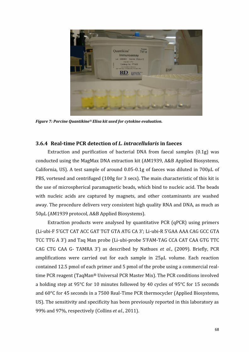
68
Figure 7: Porcine Quantikine® Elisa kit used for cytokine evaluation.
3.6.4 Real-time PCR detection of L. intracellularis in faeces
Extraction and purification of bacterial DNA from faecal samples (0.1g) was
conducted using the MagMax DNA extraction kit (AM1939, A&B Applied Biosystems,
California, US). A test sample of around 0.05-0.1g of faeces was diluted in 700μL of
PBS, vortexed and centrifuged (100g for 3 secs). The main characteristic of this kit is
the use of microspherical paramagnetic beads, which bind to nucleic acid. The beads
with nucleic acids are captured by magnets, and other contaminants are washed
away. The procedure delivers very consistent high quality RNA and DNA, as much as
50μL (AM1939 protocol, A&B Applied Biosystems).
Extraction products were analysed by quantitative PCR (qPCR) using primers
(Li-ubi-F 5’GCT CAT ACC GAT TGT GTA ATG CA 3’; Li-ubi-R 5’GAA AAA CAG GCC GTA
TCC TTG A 3’) and Taq Man probe (Li-ubi-probe 5’FAM-TAG CCA CAT CAA GTG TTC
CAG CTG CAA G- TAMRA 3’) as described by Nathues et al., (2009). Briefly, PCR
amplifications were carried out for each sample in 25μL volume. Each reaction
contained 12.5 pmol of each primer and 5 pmol of the probe using a commercial real-
time PCR reagent (TaqMan® Universal PCR Master Mix). The PCR conditions involved
a holding step at 95°C for 10 minutes followed by 40 cycles of 95°C for 15 seconds
and 60°C for 45 seconds in a 7500 Real-Time PCR thermocycler (Applied Biosystems,
US). The sensitivity and specificity has been previously reported in this laboratory as
99% and 97%, respectively (Collins et al., 2011).

69
Amplification was plotted for each sample to demonstrate the intersection
between the fluorescence reaction (ΔRn) and the PCR cycle number (Figure 8). Then
the CT value line was drawn at initial point of the linear phase, for better reliability
and to accurately estimate bacterial numbers (Nadkarni et al., 2002). The amount of
amplified product approximately doubles in each cycle. As the reaction proceeds,
enough amplified product accumulates to yield a detectable fluorescent signal
(threshold cycle, CT). And to quantify numbers of the unknown sample a range of
standards with known quantity were added to each run. Quantification standards
were used by seeding five negative faeces with 106, 105, 104, 103, 102 L. intracellularis
/ g respectively. And by plotting CT value versus the number of L. intracellularis
seeded, the unknown samples were then estimated using a regression equation from
the standard curve. To evaluate the PCR efficiency, the R2 parameter was calculated
to provide a good confidence value. When R2≤0.98, results were not considered and
the test repeated.
Figure 8: Graphic representation of real-time PCR standards. ΔRn is the fluorescence signal
strength plotted against PCR cycle number. The blue line indicates the threshold cycle value
(CT=0.07032) at the initial point of the linear phase.
3.6.5 Haematoxylin and eosin staining (HE)
The tissue sections were processed at the veterinary pathology diagnostic
services laboratory at University of Sydney. Briefly, the formalin fixed tissue was
embedded in paraffin and sectioned at 4 µm in duplicate. Slides were then dewaxed
in three steps with 100%, 95% and 75% ethanol and rehydrated with running tap

70
water. Tissue were then stained in Whitlock’s Haematoxylin for 3 minutes, then in
Scott’s Blueing solution (alkaline solution of Mg2SO4 and NaCO3) for 3 minutes, rinsed
under running tap water for 2 minutes and rinsed with 70% ethanol. Subsequently,
the sections were counterstained in alcoholic Eosin Y for 30 seconds, dehydrated
rapidly through 70%, 95% and 100% ethanol, cleared in xylol and mounted in DPX
mounting media and cover-slipped (Figure 9).
Figure 9: Small intestine HE staining, at 10x magnification with clear overall look at ileum villus
crypt and goblet cells.
3.6.6 Immunohistochemistry (IHC)
Formalin-fixed tissues were stained by immunohistochemistry (IHC) with
VPM53 Lawsonia intracellularis monoclonal antibody (Boehringer Ingelheim
Vetmedica, Ames, USA), and using the method described by Jensen et al., (1997). In
each batch of IHC, a positive and negative control section was included. Initially,
sections were dewaxed twice in xylene and once in absolute ethanol, following by
blocking solution containing methanol peroxidase (30% v/v hydrogen peroxidase)
for 10 minutes to inhibit endogenous peroxidase activity in the cells before being
washed in reverse osmosis (RO) water. Sections were incubated in pre-warmed

71
trypsin digest (0.2% w/v Trypsin 250, 0.1% w/v calcium chloride dehydrate made up
to 200mL with milliQ water) for 30 minutes at 37°C. Sections were washed twice in
RO water, once in PBS, drained and placed in humidity chambers. To inhibit non-
specific binding, 150μL of blocking solution (0.5% w/v bovine serum albumin-BSA,
1% v/v goat serum made up to volume with PBS) was added to each section and
incubated at room temperature for 20 minutes. A plastic coverslip was placed over
the top to ensure contact between the tissue section and the solution. Following
rinsing of slides in PBS to remove the coverslip and draining, the VPM53 monoclonal
antibody was diluted in PBS at 1:200. 15μL of antibody was applied to each slide with
a coverslip and incubated for one hour at 37°C. The coverslip were removed in PBS,
the slides flooded with PBS, drained and a HRP-labelled secondary antibody (Dako
Cytomation Envision, Dako, Carpinteria, US) was applied at 150μL per slide with a
coverslip and re-incubated for one hour at 37°C.
Chromagen working solutions were pre-prepared and stored. The 3-amino-
9-ethylcarbazole (AEC) was made into solution by adding 0.04g of AEC powder to
4mL of N, N-dimethylformamide (DMF) and was stored in a light-proof container at
room temperature. A 0.05M acetate buffer (5pH) was prepared by adding 0.82 grams
of anhydrous sodium acetate to 200mL of milliQ water. At the end of the incubation
period, the chromagen substrate was prepared by adding 150μL of AEC in DMF to 7.5
mL of acetate buffer. Coverslips were removed in PBS and drained, following by 150
μL chromagen applied with coverslip. Colour development and stoped after exactly
12 minutes by washing sections twice in RO water and removing the coverslip.
Section were stained in Mayer’s Haematoxylin for 3 minutes, washed twice in RO
water and set to drain. They were mounted with aqueous mounting solution (Dako,
Carpinteria, US), set to dry and placed in the dark until read (Figure 10).
The blinded slides were scored by one operator under 10x magnification light
microscope to estimate the proportion of intestinal crypt hyperplasia (H&E) and
presence of L. intracellularis -specific antigen (IHC) within crypts. A total of fifteen
fields was analysed for each section.

72
Figure 10: Immunochemistry staining (40x magnification) with positive areas of L. intracellularis
antigen presence (brown) in ileal crypts.
3.7 Statistical analysis
Statistical analysis was performed in GenStat 13th ed. (2010). Raw data were
initially screened with graphical observations and data summaries to locate overall
tendencies, outliers and sample distribution using Microsoft Office Excel 2007-2010.
Treatment differences were analysed by restricted maximum likelihood test (REML)
and testing residuals for normality distribution. The REML test (linear mixed model)
was chosen for its ability to analyse a wide range of data that involve more than one
source of error variation and unbalance designs (Steel, 1997). Variables that did not
fitted to the residual normality were log transformed to fulfil linearity assumptions.
Pens, pigs ID and room were included as random effects when required. Unpaired
variables were tested using Mann-Whitney U (Wilconox rank-sum) test correlations
within groups at a 95% confident level (p<0.05). Qualitative data were categorised
into binary data and then analysed using Generalized Mix Models (GLMM) for
repetitive measures and two-sample binomial test for single measure (GenStat).

73
Chapter 4 Systemic and mucosal immune responses
following vaccination and challenge with Lawsonia
intracellularis
(Modified from: Nogueira MG, Collins AM, Donahoo M, Emery, D; (2013). Immunological
responses to vaccination following experimental Lawsonia intracellularis virulent
challenge. Veterinary Microbiology, 164:131-138.)
4.1 Introduction _______________________________________________________________________________________ 73
4.2 Materials and Methods __________________________________________________________________________ 75
4.2.1 Animal Ethics _____________________________________________________________________________________ 75
4.2.2 Experimental design _____________________________________________________________________________ 75
4.2.3 Lawsonia vaccination ____________________________________________________________________________ 75
4.2.4 Ten times vaccination trial with challenge infection _________________________________________ 76
4.2.5 Antibodies and cytokines analysis _____________________________________________________________ 77
4.2.6 Correlation of serum cytokines with PE disease severity ____________________________________ 78
4.2.7 L. intracellularis real-time PCR _________________________________________________________________ 78
4.2.8 Histological lesion scores in intestinal tissue _________________________________________________ 78
4.2.9 Skin testing for delayed type-hypersensitivity ________________________________________________ 79
4.2.10 Statistical analysis ______________________________________________________________________________ 80
4.3 Results _____________________________________________________________________________________________ 80
4.3.1 Experiment 2a: Immune response to a ten times oral or intramuscular immunisation_ _ 80
4.3.2 Experiment 2b: Protective immunity to Proliferative Enteropathy ________________________ 85
4.3.3 Correlations between disease severity and serum cytokines _______________________________ 90
4.4 Discussion _________________________________________________________________________________________ 92
4.1 Introduction
The Gram-negative intracellular bacterium Lawsonia intracellularis causes
proliferative enteropathy (PE), characterized by diarrhoea and poor performance in
growing pigs and severe haemorrhagic diarrhoea in finisher and breeding animals
(Lawson and Gebhart, 2000). The most consistent macroscopic pathology of PE is
thickening of the intestinal mucosa which is associated histologically with
proliferation of immature enterocytes (Lawson and Gebhart, 2000). The clinical signs
and lesions can be controlled using a commercial oral live attenuated vaccine
(Enterisol®Ileitis, Boehringer Ingelheim) (Kroll et al., 2004; McOrist and Smits, 2007).
This vaccine contains an attenuated L. intracellularis isolate (B3903; 104.9

74
TCID50/dose) originally isolated from the ileum of a Danish pig with acute
proliferative haemorrhagic enteropathy (PHE) (Kroll et al., 2004). Protective
immunity against re-infection is also apparent in recovered pigs (Collins and Love,
2007; Riber et al., 2011b; Cordes et al., 2012).
A conventional immunological approach to induce mucosal immunity against
gut pathogens has involved oral vaccination or intraperitoneal inoculation (Muir et
al., 1998). However, where antibody provides mucosal protection, high
concentrations of serum IgG engendered by systemic vaccination have been effective
against some intracellular bacteria. For example, Salmonella enterica serovar
Typhimurium bacterin administrated intramuscularly (IM) reduced lesions and
shedding in naturally-infected pigs (Farzan and Friendship, 2010). Similarly, an
intramuscular L. intracellularis killed bacterin induced significant protection to PE
after virulent challenge (Dale et al., 1997), and in a patent description, protection was
induced after immunisation IM with killed L. intracellularis (Jacobs et al., 2011).
To identify immune responses which might indicate the successful induction of
protective immunity following vaccination with Enterisol® Ileitis, this study
measured local mucosal and systemic immune responses in the first 3 weeks after
immunisation. Since local mucosal responses were marginal following a conventional
single dose vaccination in a preliminary trial 1 (published in the paper Nogueira et al.,
2013, but not in this thesis), pigs in trial 2 were vaccinated orally with a ten times
dose (10x) of Enterisol®Ileitis. Local mucosal and systemic antibody and selected
cytokine responses were measured and compared with those from piglets given the
same dose of vaccine IM. To determine whether these responses could foreshadow
protection, the remaining vaccinated cohorts were challenged and immunity was
assessed by serological assays, reduction of clinical signs and intestinal lesions and
the duration and magnitude of bacterial shedding in faeces.

75
4.2 Materials and Methods
4.2.1 Animal Ethics
All animal experiments were performed according to the Australian Code of
Practice for the Care and Use of Animals for Scientific Purposes, and approved by the
University of Sydney and Elizabeth Macarthur Agricultural Institute Animal Ethics
Committees (M09/10).
4.2.2 Experimental design
For trials 1 and 2, a total of 40 and 62 Landrace x Large White pigs, respectively,
aged 3-4 weeks, were purchased from a commercial herd clinically and serologically
negative for L. intracellularis. Piglets were transferred to a controlled environment
research facility, with access to water and fed on weaner/grower diet (DE:
15.3MJ/kg; CP: 20%; Fibre: 2.5%; Vella Stock Feeds, Australia) with no medication.
Pigs were weighed (6.0±0.5kg) and randomly allocated into respective treatment
groups (Table 3; trial 2) housed in separate pens under strict quarantine conditions.
Pigs were monitored daily for body condition, clinical signs and abnormal behaviour.
4.2.3 Lawsonia vaccination
Prior to vaccination (d0) at 5 weeks of age, pigs were bled by jugular
venipuncture and faeces were collected from individual pigs to confirm the absence
of L. intracellularis or the presence of maternal antibodies that might interfere with
vaccination. The lyophilised vaccine (Enterisol®Ileitis, Boehringer Ingelheim Pty Ltd.,
SA-122A-323) was reconstituted according to the manufacturer’s instructions to
either standard (1x) dose (104.9TCID50) or ten times (105.9TCID50) concentrations.
Pigs received the appropriate vaccine dose orally in 2.0mL diluent by drenching gun
or by intramuscular inoculation (IM) in the right deltoid muscle (cervical area).
Negative and positive control pigs were not vaccinated.

76
Table 3- Experiment design and groups in trial 2*.
Experiment Group Li dose Route Li challenge N°
pigs
2a
10xOral 105.9 TCID50 Oral No 6
10xIM 105.9 TCID50 IM No 6
Control - - No 4
2b
PC - - Yes 10
1xOR 104.9 TCID50 Oral Yes 10
1x IM 104.9 TCID50 IM Yes 10
10xOR 105.9 TCID50 Oral Yes 10
NC - - No 6
*Li: Lawsonia intracellularis; IM: intramuscular; PC: Positive control; NC: Negative control.
4.2.4 Ten times vaccination trial with challenge infection
4.2.4.1 Experiment 2a: Analysis of immune responses after vaccination
Fifteen pigs aged 4 weeks were randomly assigned into three treatment groups:
six pigs in each of groups 1 and 2 were orally vaccinated and IM vaccinated,
respectively, with 10x dose of Enterisol®Ileitis vaccine and three pigs in group 3 were
not vaccinated. Sera were obtained from each pig by venipuncture of the jugular vein
before vaccination and necropsy. Seven pigs were randomly selected (3 from groups
1 and 2, and one from group 3) and euthanized on d9 post vaccination, and the
remainder on d17. At necropsy, the left and right pre-scapular lymph nodes were
weighed and blood taken from the jugular vein. Ileal and jejunal tissue was sampled
at 10cm and 1m from the proximal ileo-caecal valve (ICV), respectively, together with
adjacent mesenteric lymph nodes (MLN). Each tissue and MLN sample (ca 2x2mm)
was stored in 10% neutral-buffered formalin fixative until analysis. In the adjacent
segment, the intestinal mucosal secretions were collected by gently scraping the ileal
mucosa with a sterile scalpel and placed into 2ml phosphate-buffered saline (PBS, pH
7.2) containing 0.2M EDTA on ice. The scrapings were each stored at -20°C. Prior to
assay, mucosal scraping samples were thawed and adjusted to a protein level of
25mg of protein ml-1.
4.2.4.2 Experiment 2b: Immune responses after challenge infection
Forty-six pigs were randomly allocated into 5 treatment groups. Groups 1-3 (10
pigs each) were vaccinated with 1x oral (1xOR- group1), 1x IM (1xIM- group 2), 10x
oral (10xOR- group3), respectively, while 10 positive control pigs (PC- group 4)
remained unvaccinated. The vaccination procedures were the same as described for

77
experiment 2a. Six pigs were kept unvaccinated and unchallenged as negative
controls (NC- group 5). Four weeks after vaccination, each pig in groups 1-4 was
dosed orally with L. intracellularis infected intestinal mucosa inoculum prepared and
quantified as described elsewhere (Collins et al., 1996; Collins and Love, 2007).
Briefly, pigs were fasted overnight, sedated with 0.2mg/kg of azaperone (Stresnil®,
Fivet, Australia) and intubated with an oro-gastric tube to enable administration of
25mL of the bacterial suspension containing around 109 L. intracellularis. Pigs were
euthanized at a local abattoir 21 days post infection (21pi) where the gut was
collected for analysis.
Pig live weights were recorded before vaccination, at challenge and at necropsy
to determine the average daily live weight gain. Sera were collected from each pig on
days 0pi, 7pi, 14pi and 21pi while faecal samples from individual animals were
collected on days 0pi, 7pi, 10pi, 14pi, 17pi and 21pi for a quantitative L. intracellularis
PCR (qPCR). At necropsy, ileal samples were collected as for experiment 2a and
preserved in 10% buffered formalin prior to staining with H&E and
immunohistochemistry (IHC). Following histopathological analysis, 24 pigs were
selected from all groups according to the percentage affected area and Lawsonia
intracellularis shedding numbers. There were four negative controls, ten severely
affected and ten infected but without lesions, and also their sera from days 0pi and
21pi were tested for cytokines using porcine Quantikine® assay kits (Chapter 3).
4.2.5 Antibodies and cytokines analysis
Sera and mucosal scrapings were assayed in duplicate for the detection of L.
intracellularis -specific IgM, IgG, IgA and concentrations of the cytokines, interferon-
gamma (IFN-γ), interleukin-6 (IL-6), interleukin-10 (IL-10), tumor necrosis factor-
alpha (TNF-α) and transforming growth factor-beta1 (TGF-β1). L. intracellularis-
specific antibody was determined using a commercial competitive (blocking) ELISA
kit (Bioscreen®Ileitis, GmbH, Münster, Germany) at a positive- negative cut-off of
30% inhibition as recommended by the manufacturers. In addition, L. intracellularis-
specific IgM, IgG and IgA titres were assayed using an experimental modified direct
ELISA developed for this project (Donahoo, 2009). The quantity of each of the
cytokines IFN-γ, IL-6, IL-10, TNF-α and TGF-β1 in mucosal scrapings or serum was
analysed using commercial sandwich ELISA kits (porcine Quantikine® ELISA, R&D

78
systems, Minneapolis, USA) according to the manufacturer’s directions (Chapter 3).
The minimum limits of detection were 2.7 pg/mL for IFN-γ, 10 pg/mL for IL-6, 1.8
pg/mL for IL-10, 2.8 pg/mL for TNF-α and 19.5pg/mL for TGF-β1. Concentrations
were estimated by generating a logistic parameter curve using GraphPad®Prism
software (v.4.02, 2004).
4.2.6 Correlation of serum cytokines with PE disease severity
The 20 infected pigs were selected from challenged groups according to the
percentage affected area on IHC staining and L. intracellularis shedding in faeces
(from subclinical to severe infection, Table 4). Cytokine concentrations were
correlated with average weight gain, ileal lesions and quantities of Lawsonia
intracellularis shed on days 14pi, 17pi and 21pi.
Table 4- Groups selected for serum cytokine analysis and results correlated to respective percentage affected area of PE lesions(IHC and HE) and numbers of L. intracellularis shedding (Li g/faeces) on day 21 pi.
Groups IHC and HE Li shedding (g/ faeces)
No. samples
Severe infection >10% 104-106 10
Subclinical infection 0-5% 102-104 10
Negative controls 0% 0 4
*IHC: Immunohistochemistry expressed as percentage affected area; HE: Haematoxylin and eosin;
4.2.7 L. intracellularis real-time PCR
Extraction and purification of bacterial DNA from individual faecal samples
(0.1g) were made using the MagMax DNA extraction kit (A&B Applied Biosystems,
California, USA) (Chapter 3). Extraction products were analysed by real time PCR
(7500 Real-Time PCR System; Applied Biosystems) using primers and Taq Man probe
as described by Nathues et al., (2009). The sensitivity and specificity of the test in our
laboratory have been reported by Collins et al. (2011) as 99% and 97%, respectively .
4.2.8 Histological lesion scores in intestinal tissue
Formalin-fixed tissues were stained with haematoxylin and eosin (H&E) (Luna,
1968) and by immunohistochemistry (IHC) with VPM53 L. intracellularis monoclonal
antibody (Boehringer Ingelheim Vetmedica, Ames, USA) using the method described
by Jensen et al., (1997). The blinded slides were scored by one operator under 10x
magnification light microscope to estimate the proportion of intestinal crypt

79
hyperplasia (H&E) and presence of L. intracellularis -specific antigen (IHC) within
crypts. A total of fifteen fields was analysed for each section.
4.2.9 Skin testing for delayed type-hypersensitivity
Delayed type hypersensitivity reactions were examined in individual pigs 20
days after vaccination by intradermal (ID) injections of 0.2mL of sterile phosphate
buffer saline (PBS, pH 7.2) (control) or around 105 L. intracellularis in 0.2ml of 10x
Enterisol® Ileitis in two areas between nipples. The sites were examined at 24h and
48h and reactions were recorded as the diameter of erythema at the injection sites
(Figure 11).

80
Figure 11: Delayed type hypersensitivity reaction test with intradermal injections of sterile
phosphate buffer saline (control) or L. intracellularis vaccine.
4.2.10 Statistical analysis
Statistical differences were analysed among treatment groups and days post-
vaccination and challenge using Restricted Maximum Likelihood (REML, GenStat
Release 13th Ed., Oxford, UK) at a 95% confidence level (P<0.05). Individual pigs were
included as random effects in every analysis. Correlations between two unpaired
variables were analysed using the Mann Whitney U test.
4.3 Results
4.3.1 Experiment 2a: Immune response to a ten times oral or
intramuscular immunisation
Prior to vaccination, all pigs were PCR negative and seronegative for L.
intracellularis and did not exhibit diarrhoea or clinical signs of PE after vaccination.
Pigs gradually increased weights between days 0 and 17 post-vaccination, however,
without differences between treatments groups (Figure 12). There was no evidence
of gross or histopathological lesions of PE in ileal sections at necropsy (Figure 17). No
significant differences between weights from left and right pre-scapular lymph nodes
(P>0.05) and delayed type hypersensitivity reaction were minimal between animals
at 20 days after vaccination (Table 5).

81
Figure 12: Predicted means for weight at each time point post-vaccination (days). (■) Negative
Control (NC, blue), (■) ten-times oral (10xOR, red) and (■) ten-times intramuscular (10xIM,
green).
Table 5- Mean weights from the left and right pre-scapular lymph nodes (PLN, grams) and delayed type hypersensitivity (DTH) reactions measurements after L. intracellularis (Li) vaccination.
Treatment Left PLN (grams)
Right PLN (grams)
DTH control PBS (mm)
DTH Li vaccine (mm)
10xOR 1.39 1.33 - <0.5 10xIM 1.70 1.91 - <0.5
NC 1.51 1.12 - -
Increased concentrations of serum L. intracellularis IgG (direct) were detected
in the orally and IM vaccinated pigs compared with the control group 3 between d9
and d17 (P<0.001, Figure 13b). The serum IgG titres reached 85 (direct) and 24%
inhibition (Bioscreen®) at day 17, and while similar values were detected in both
vaccinated groups, they were significantly higher than control animals (33 and 7%
inhibition, respectively in the two assays) (Figure 13). The orally vaccinated group
produced serum IgM titres of 148 that was significantly higher (P=0.03) than those
expressed by pigs in the IM and control groups (62 and 55, respectively, Figure 14). L.
intracellularis -specific IgA titres in sera and in mucosal secretions from all pigs were
not different and all data titres less than 4 using the modified ELISA test.
In mucosal scrapings, higher titres of anti - L. intracellularis IgM on day 9 (270)
were found in ileal scrapings from pigs vaccinated orally (P<0.001) and IgG on day 17
(14%) for both oral and IM vaccinated animals (58 and 70, respectively) compared to
control group (Figure 16a). Serum cytokines exhibited a large variation within
animals from the same treatment group and no significant differences (P>0.05)
between treatments were apparent (Figure 15). In contrast, the concentrations of all
five cytokines (IFN-γ, IL-6, IL-10, TNF-α, TGF-β1) in mucosal secretions from oral
vaccinates significantly increased between days 9 and 17 over values in control and
5.7 6.7 7.5
9.4
6.0 7.4 8.1
10.2
6.1 7.6 8.4
10.4
0 6 9 17
kg
days

82
IM vaccinates (Figure 16). The quantity of TNF-α and TGF-β1 in mucosal scrapings
from oral vaccinates was significantly higher (P<0.05) than the concentrations found
in IM and control pigs on day 17.
a) Bioscreen® b) direct
Figure 13: Humoral immune response to L. intracellularis Bioscreen ELISA (a), Direct ELISA for
IgG (b). Lines correspond to the mean percentage of inhibition (PI) for (a) and mean titre for (b)
for each treatment: (▲) Negative control (green, n=4); (●) 10xOral (red, n=5); (■) 10xIM (Blue,
n=6). * Superscript next to lines identifies significant difference between treatment and control
groups (P<0.05).
a) IgA b) IgM
Figure 14: Humoral immune response to L. intracellularis IgA (a) and IgM (b). (▲) Negative
control (green, n=4); (●) 10xOral (red, n=5); (■) 10xIM (Blue, n=6). * Superscript next to lines
identifies significant difference between treatment and control groups (P<0.05).
0
10
20
30
40
0 3 6 9 12 15 18
PI
(%)
Days
0
20
40
60
80
100
120
0 3 6 9 12 15 18
Tit
re
Days
0.8
1.8
2.8
3.8
4.8
5.8
6.8
7.8
8.8
9.8
0 3 6 9 12 15 18
Titr
e
Days
0
50
100
150
200
250
300
350
400
0 3 6 9 12 15 18
Tit
re
Days
*
*
*
*

83
IL-6
IFN-gamma IL-10
TNF-alpha
120
60
50
40
100
30
20
9 17
10
0
60
0
20
17 9 0
70
40
80
0
days
pg/mL
days
pg/mL
Figure 15: The concentration of circulating (serum) cytokines (pg/mL) on days 0, 9 and 17 after L.
intracellularis vaccination within treatments: Negative control (green, n=4); 10xOral (red, n=5);
10xIM (blue, n=6).

84
a
b
Figure 16: Mean concentrations for cytokines (pg/ml) and IgG (PI value) and IgM (Titre), L. intracellularis specific antibodies in (a) mucosal scrapings on days 9 (dark) and 17 (clear) and (b) in serum on days 0 (dark), 9 (shade) and 17 (clear) after ten-times oral (10xOral), intramuscular (10xIM) vaccination and unvaccinated (NC). Columns with a star on the top indicate statistical difference (P<0.05).
0
5
10
15
20
25
30%
P.I.
10xOral 10xIM NC0
50
100
150
200
250
300
IgM
10xOral 10xIM NC
0
200
400
600
800
IFN
-ga
mm
a
10xOral 10xIM NC0
50
100
150
200
250
IL-6
10xOral 10xIM NC
0
50
100
150
200
TG
F-b
eta
1
10xOral 10xIM NC0
20
40
60
80
100
120
TN
F-a
lph
a
10xOral 10xIM NC
0
50
100
150
10xOral 10xIM NC
IgM
Tit
re

85
Figure 17: Ileum section of a pig (#B53), in HE staining, after 9 days orally vaccinated (10xOR)
with L. intracellularis.
4.3.2 Experiment 2b: Protective immunity to Proliferative Enteropathy
All 40 pigs appeared clinically normal after challenge and there were no
significant differences in average weight gain between groups over the 3 weeks
following vaccination or challenge and over the entire 6 weeks of the trial (Table 6).
Similar kinetics for the significant increases (P<0.001) in serum L. intracellularis IgG
and IgA titres were observed in challenged animals (groups 1-4) compared to
negative control animals in group 5 (Figure 18). There were no significant differences
(P>0.05) in antibody titres between challenged groups. At day 21pi, all infected pigs
were serologically positive for L. intracellularis -antibodies, with a mean P.I. of 48%.

86
Table 6- Average weight gains (kg) and approximate (±) standard errors (SE) of pigs between
periods pre-challenge, post-challenge and total period of trial (d0: vaccination; d21: L.
intracellularis challenge; d42: necropsy).
Treatment Pre-challenge
(d0-d21)
Post-challenge
(d21- d42)
Total period
(d0-d42)
Positive Control 6.67 ± 0.52 9.44 ± 0.72 16.12 ± 0.91
1xOR 6.09 ± 0.50 9.90 ± 0.68 16.00 ± 0.86
1xIM 6.28 ± 0.50 8.82 ± 0.68 15.10 ± 0.86
10xOR 6.76 ± 0.50 10.85 ± 0.68 17.61 ± 0.86
Negative Control 6.40 ± 0.50 8.64 ± 0.70 15.04 ± 0.86
P (<0.05) 0.877 0.206 0.272
Figure 18: Predicted means for serum L. intracellularis-IgG (percentage inhibition, PI; left) and
IgA titres (right) on days 0, 7, 14 and 21 in each group after L. intracellularis virulent challenge
between groups ten-times oral (10xOR), one-time oral (1xOR) and intramuscular (1xIM), negative
control (NC) and positive control (PC).
There was a range of pathology noted at necropsy (Figure 20) and determined
by subsequent histological (Figures 21 and 22) and immunohistological analyses
(Figures 23 and 24). Assessments of protection based on estimations of the numbers
of L. intracellularis shed in faeces and the % affected area in ileal sections revealed
that the vaccination significantly (P<0.001) reduced L. intracellularis shedding when
compared to positive control group. Pigs given the 10x oral vaccine were significantly
(P<0.05) more protected than the other 2 vaccinated groups (Figure 19 and 25).
0
10
20
30
40
50
PI v
alu
e (
%)
10xOR 1xIM 1xOR NC PC0
20
40
60
80
100
120
IgA
Tit
re
10xOR 1xIM 1xOR NC PC
Cut-off
Cut-off Cut-off
Cut-off

87
Figure 19: Estimation of Lawsonia intracellularis per gram of faeces at each time point post-
infection for each group (♦, blue) Positive Control (PC); (■, red) 1x Oral; (▲, green) 1xIM, and (X,
purple) 10xOral. On the same sampling values without common superscripts (a-c) are
significantly different (P<0.05).
Figure 20: Small intestine section from a positive control pig (#Y67), unvaccinated, with
characteristic proliferation 21 days after Lawsonia intracellularis challenge (arrow).
-1.00E+06
0.00E+00
1.00E+06
2.00E+06
3.00E+06
4.00E+06
5.00E+06
0 1 2 3 4 5 6 7 8 9 10 11 12 13 14 15 16 17 18 19 20 21 22 23
Law
son
ia/g
of
faec
es
days pos-inoculation (pi)
a
a
b
a
a
a a
b
b
b bc
c
c
b

88
Figure 21: Ileal section from a pig (#G99) from group 1xIM presenting extensive crypt enterocytes
proliferation and loss of goblet cells, 21 days after Lawsonia intracellularis challenge (circle).
Figure 22: Proliferation of enterocytes in ileal crypt with loss of goblet cells and presence of
exudate in the lumen (40 x mags) from pig (#O75) orally vaccinated (1xOR) and challenged
(arrow).

89
Figure 23: Lawsonia intracellularis antigen staining (brown, 20x mag.) in ileal section by
immunohistochemistry from pig (#Y77), unvaccinated, from positive control group 21 days after
challenge.
Figure 24: Ileal section in 40 times magnification from pig (#Y77), unvaccinated and challenged
(positive control) with Lawsonia intracellularis antigen staining in the apical area of enterocytes
(brown).
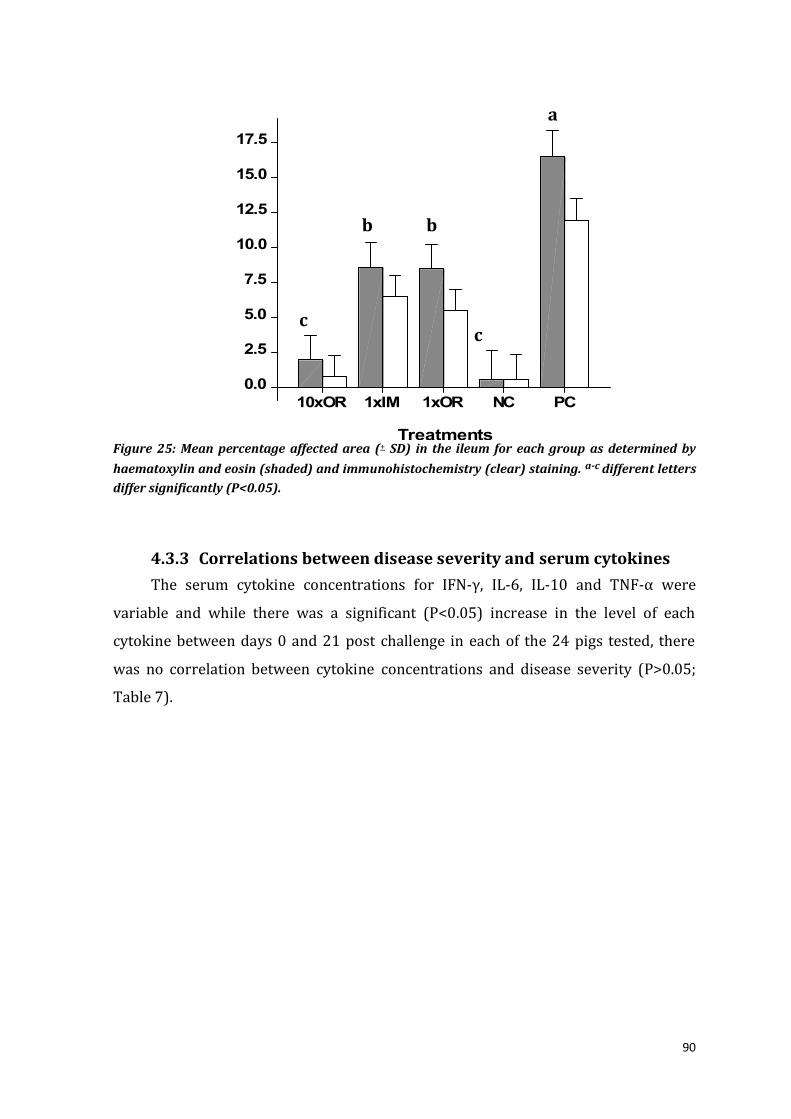
90
Figure 25: Mean percentage affected area (+ SD) in the ileum for each group as determined by
haematoxylin and eosin (shaded) and immunohistochemistry (clear) staining. a-c different letters
differ significantly (P<0.05).
4.3.3 Correlations between disease severity and serum cytokines
The serum cytokine concentrations for IFN-γ, IL-6, IL-10 and TNF-α were
variable and while there was a significant (P<0.05) increase in the level of each
cytokine between days 0 and 21 post challenge in each of the 24 pigs tested, there
was no correlation between cytokine concentrations and disease severity (P>0.05;
Table 7).
1xOR
12.5
15.0
17.5
PC
0.0
5.0
10.0
1xIM10xOR
2.5
NC
7.5
% a
ffe
cte
d a
rea
Treatments
a
b b
c c

91
Table 7- Serum cytokines concentrations (pg/mL) in individual pigs from groups (G) PE severe infection (S), sub-clinical infection (SB) and negative controls (NC) and the correlations between group*lesions*cytokines, group*Li-shedding-in-faeces*cytokines, group*ADG*cytokine.
G ID Lesions ADG
Li
IFN-γ pg/mL
IL-6 pg/mL
IL-10 pg/mL
TNF-α pg/mL
HE%
IHC%
kg g/f 0pi 21pi 0pi 21pi 0pi 21pi 0pi 21pi
S G99 35 10 22.2 105-6 49.9 12.5 9.4 7.2 < 44.5 44.6 87.9
S O81 31 15 16.0 105-6 9.1 70.9 16.6 50.9 < 50.4 < 149.1
S G64 27 10 14.8 105 18.1 27.5 11.5 58.4 9.5 10.2 68.8 112.4
S Y63 27 9 15.6 105 < 29.6 < 98.4 57.5 119.5 47.3 326.9
S Y68 24 2 17.7 104-5 33.3 51.8 8.3 8.8 < 30.0 51.9 92.3
S Y77 23 9 17.3 104-6 14.3 60.6 36.2 77.0 22.9 77.9 89.3 196.5
S Y70 20 2 18.4 105 27.5 23.7 12.6 21.4 11.7 15.3 83.6 92.8
S O54 19 7 18.4 104 126 133.9 12.1 60.1 < 16.9 18.0 60.8
S B81 14 0 22.8 105 31.2 46.3 9.4 19.1 8.7 21.1 21.9 55.2
S O100 11 1 14.5 106 29.3 3.2 3.6 58.4 < 79.0 28.4 109.9
S G100 10 5 17.2 106 87.6 1.4 9.4 14.8 23.1 89.2 52.9 111.4
Mean SEM
22 6 18.0 105 43 42 13 43 22 50 51 127
5.8 5 0.8 22.5 38.9 16.7 15.9 8.6 89.6 26.8 42.6
SB Y83 5 0 17.4 104 < < 43.4 20.8 47.6 54.48 28.4 58.9
SB Y65 2 0 9.7 105 5.1 29.6 7.2 75.4 < 17.56 48.7 363.2
SB Y71 3 0 17.1 105 23.7 1.4 11.0 9.9 < < 18.0 62.6
SB O77 0 0 14.2 105 31.2 46.3 9.9 9.4 23.8 38.5 21.0 206.2
SB G98 0 0 9.5 104 < < 8.8 9.4 < 79.2 33.3 90.8
SB Y73 2 0 17.5 104 27.5 12.5 90.8 4.1 45.3 78.4 38.7 96.7
SB G53 0 0 15.7 103 3.2 31.2 10.4 11.5 < < 30.7 157.7
SB O43 0 0 15.5 104 1.4 < 14.8 37.1 < 56.4 40.0 69.7
SB B98 0 0 17.9 102 12.5 20.1 16.4 74.2 < 44.4 84.6 71.6
SB O73 0 0 17.5 103 35.1 20.1 13.1 21.9 85 97.1 33.3 118.5
Mean SEM
1 0 15.0 104 17 23 23 27 50 58 38 130
0.2 0 0.5 11.9 19.7 26.7 20.0 30.5 32.8 29.6 52.9
NC B60 0 0 14.0 0 < 128.4 12.1 136.4 35.2 36.9 141 139.2
NC B71 0 0 16.1 0 18.1 89.5 14.2 16.4 < < 46.9 115.0
NC O88 0 0 11.3 0 23.7 133.4 24.7 45.1 < 47.2 37.3 84.6
NC W81 0 0 19.6 0 < 27.5 13.1 5.7 < 52.1 60.8 101.1
Mean SEM
P (<0.05)
0 0 15.0 0 21 95 16 51 35 45 72 110
0 0 0.6 0 12.6 92.6 9.8 57.8 28.1 42.5 94.5 16.9
<0.05 NS <0.05 NS NS NS NS NS NS NS NS
G* lesions G*Li
G*ADG
NS NS NS NS NS NS NS NS
NS NS NS NS NS NS NS NS
NS NS NS NS NS NS NS NS
NS: Not significant; Li: L. intracellularis; ADG: average daily weight gain; IHC: immunohistochemistry; HE: Haematoxylin and eosin;

92
4.4 Discussion
This study monitored and detected dose-dependent increases in mucosal and
serum IgM, IgG, IgA and cytokines following vaccination with Enterisol® Ileitis and
subsequent challenge. Dose-dependent induction of protection against virulent L.
intracellularis challenge was demonstrated by significant reduction in PE lesion
scores and the magnitude and duration of bacterial shedding in faeces in all
vaccinated pigs. As anticipated, oral vaccination induced local mucosal immunity with
increased IgG and cytokine responses and tended to generate more comprehensive
protection than IM vaccination.
The analysis of protective immune responses to L. intracellularis is an area of
considerable interest for the control of PE. Initial studies on the development of
humoral and cell-mediated immunity (involving analysis of IFN-γ responses) have
described similar kinetics to those found in the current study. Dose-dependency of
humoral responses is well documented. Following vaccination with 104.9 TCID50 L.
intracellularis, serum antibody responses were either undetectable (Kroll et al., 2004)
or restricted to modest concentrations of L. intracellularis -specific IgG in ileal
scrapings. Higher doses (105.9 TCID50 in this study) of vaccine or primary infection
with 105-1010 L. intracellularis induced detectable concentrations of L. intracellularis -
specific IgG within 14 days after inoculation (Collins and Love, 2007; Riber et al.,
2011b). Consistent with previous analyses (Guedes and Gebhart, 2010; Jacobs et al.,
2011), mucosal IgA responses were of lesser magnitude and while the highest serum
titres were generated by challenge control pigs (group 4), IgA titres were not boosted
substantially by challenge of immunised animals. This could be dose related, as IgA
accumulations have been demonstrated previously, by IHC, in apical cytoplasm of
proliferating enterocytes from pigs with clinical cases of PIA and PHE (Lawson et al.,
1979; McOrist et al., 1992). The timing and duration of humoral responses is also well
studied and relatively consistent. In the current study, a 10x dose oral vaccination
elicited increased concentrations of IgM, IgG in ileal mucosal scrapings and elevated
concentrations of IgM and IgG in serum. The systemic and local IgM immune
response after 9 days post vaccination precedes IgG (Lawson et al., 1988) and has
similar kinetics to increases in serum haptoglobin and C-reactive protein following
primary infection with L. intracellularis (Riber et al., 2011b). The induction of IgA
was not detected in this study after vaccination with 105.9 TCID50 L. intracellularis, but
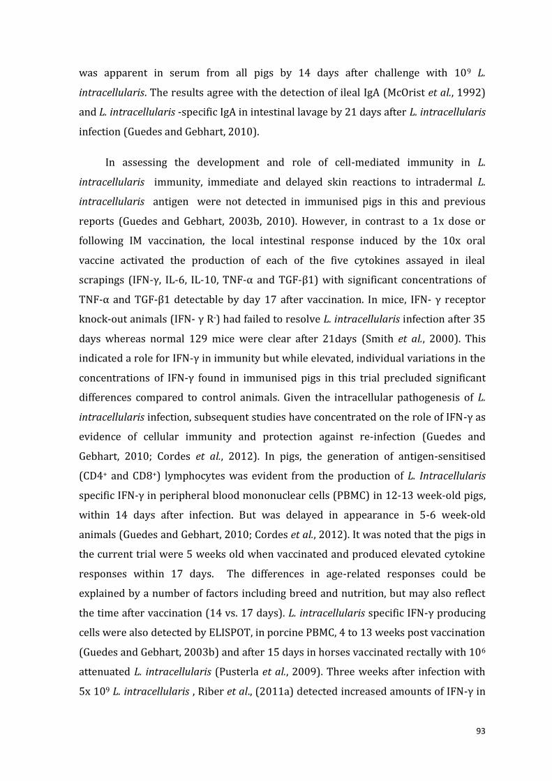
93
was apparent in serum from all pigs by 14 days after challenge with 109 L.
intracellularis. The results agree with the detection of ileal IgA (McOrist et al., 1992)
and L. intracellularis -specific IgA in intestinal lavage by 21 days after L. intracellularis
infection (Guedes and Gebhart, 2010).
In assessing the development and role of cell-mediated immunity in L.
intracellularis immunity, immediate and delayed skin reactions to intradermal L.
intracellularis antigen were not detected in immunised pigs in this and previous
reports (Guedes and Gebhart, 2003b, 2010). However, in contrast to a 1x dose or
following IM vaccination, the local intestinal response induced by the 10x oral
vaccine activated the production of each of the five cytokines assayed in ileal
scrapings (IFN-γ, IL-6, IL-10, TNF-α and TGF-β1) with significant concentrations of
TNF-α and TGF-β1 detectable by day 17 after vaccination. In mice, IFN- γ receptor
knock-out animals (IFN- γ R-) had failed to resolve L. intracellularis infection after 35
days whereas normal 129 mice were clear after 21days (Smith et al., 2000). This
indicated a role for IFN-γ in immunity but while elevated, individual variations in the
concentrations of IFN-γ found in immunised pigs in this trial precluded significant
differences compared to control animals. Given the intracellular pathogenesis of L.
intracellularis infection, subsequent studies have concentrated on the role of IFN-γ as
evidence of cellular immunity and protection against re-infection (Guedes and
Gebhart, 2010; Cordes et al., 2012). In pigs, the generation of antigen-sensitised
(CD4+ and CD8+) lymphocytes was evident from the production of L. Intracellularis
specific IFN-γ in peripheral blood mononuclear cells (PBMC) in 12-13 week-old pigs,
within 14 days after infection. But was delayed in appearance in 5-6 week-old
animals (Guedes and Gebhart, 2010; Cordes et al., 2012). It was noted that the pigs in
the current trial were 5 weeks old when vaccinated and produced elevated cytokine
responses within 17 days. The differences in age-related responses could be
explained by a number of factors including breed and nutrition, but may also reflect
the time after vaccination (14 vs. 17 days). L. intracellularis specific IFN-γ producing
cells were also detected by ELISPOT, in porcine PBMC, 4 to 13 weeks post vaccination
(Guedes and Gebhart, 2003b) and after 15 days in horses vaccinated rectally with 106
attenuated L. intracellularis (Pusterla et al., 2009). Three weeks after infection with
5x 10⁹ L. intracellularis , Riber et al., (2011a) detected increased amounts of IFN-γ in

94
the supernatant fluid after incubation of whole porcine blood with Lawsonia antigen
and the inflammatory cytokine, IL-18. In contrast, limited microarray studies of pigs
with varying concentrations of intestinal pathology did not reveal substantial
activation of cytokine genes (Jacobson et al., 2011a). While the synthesis of the three
“inflammatory” cytokines (IFN-γ from NK cells, IL-6, and TNF-α) would result from
local infection and serve to activate the acquired immune system, including IFN-γ-
producing T-cells and IgG production. The quantities of IL-10 and TGF-β1 protein
were also increased in the ileal scrapings. This may indicate induction of local
immunoregulation through T-reg cells (Chen et al., 2007; Bailey, 2009), and local
restoration, given the role of TGF-β1 in wound healing in pigs (Quaglino et al., 1991).
Increased concentrations of IL-10 and TGF-β1 are also consistent with detection of
insulin-like growth factor binding protein 3 (IGFBP-3) during the immune priming
following oral infection or vaccination with L. intracellularis (Jacobson et al., 2011a).
To induce protective immune responses at mucosal surfaces, oral vaccination
with live attenuated or killed/ subunit vaccines is the preferred protocol to block
pathogen attachment or to neutralize local virulence factors by specific IgA or IgG
antibody (Mestecky and McGhee, 1987; Husband et al., 1996). Attenuated live
vaccines are used to generate protective immunity against intracellular pathogens.
For L. intracellularis, oral administration has been effective, but rectal administration
of 106.3 TCID50 L. intracellularis generated L. intracellularis specific IgG detectable
responses in horses (Pusterla et al., 2009) and intraperitoneal (IP) vaccination
against Salmonella Typhimurium has effectively immunised livestock (Muir et al.,
1998). When the quantitative and qualitative aspect of L. intracellularis challenge was
determined by real-time PCR in faeces, the ten times oral vaccination (105.9 TCID50)
elicited the greatest protection but the 1x intramuscular and 1x oral vaccines were
also efficacious. Since L. Intracellularis enters and multiplies in immature (crypt)
enterocytes (Lawson and Gebhart, 2000), the 1x IM protocol suggests that serum
antibody can significantly reduce the number of L. Intracellularis available in the
gastrointestinal tract prior to cellular entry. Results with inactivated or subcellular L.
intracellularis vaccines would support this mechanism. Dale et al (1997) reported
that faecal L. intracellularis counts were reduced by 98.5% in four pigs vaccinated
twice intramuscularly (IM), 3 weeks apart, with killed L. intracellularis in incomplete

95
Freunds adjuvant. Additional pigs inoculated with recombinant GroEL-like protein
exhibited reduced faecal bacteria counts compared to infected controls (Dale et al.,
1997). In their patent, Jacobs et al., (2011) also reported the induction of significant
protection against L. intracellularis challenge after two IM inocula were given 4
weeks apart, of 2.8 x 10⁸ killed L. intracellularis in oil adjuvant. These researchers
reported significant reductions in L. intracellularis shedding after initial IM
vaccination of pigs with 50µg each of recombinant 19/21 and 37kDa outer
membrane proteins (OMPs) or putative LPS in adjuvant. Dose dependent protection
was achieved with vaccines containing killed organisms where 5 x 10⁷ L.
intracellularis elicited protection and 1.25 x 10⁷ L. intracellularis was ineffective
(Jacobs et al., 2011). These results and the fact that IM administration of Enterisol®
would be expected to kill the live L. intracellularis rapidly after inoculation, indicate
that with sufficient antigenic stimulus, antibodies against L. intracellularis cells and
components can protect against infection. Given the elevated serological titres at
challenge, this presumably occurs by preventing intracellular entry. That significant
mucosal protection can be induced by relatively small doses of live oral L.
intracellularis without substantial serological titres (Kroll et al., 2004) would indicate
both a dose-dependent and site-specific response.
In the search for readily accessible biomarkers for immunity to L. Intracellularis,
antibody responses have not been informative (Kroll et al., 2004; Cordes et al., 2012),
with IgA responses low and ephemeral (Cordes et al., 2012). Skin tests are also
unreliable (Guedes and Gebhart, 2010), as was found in this trial. In addition, the
measurement of cytokine concentrations in body fluids such as serum and mucosal
secretions is costly and is complicated by the dynamic responses to L. intracellularis
in individual pigs where the variations between individuals are often greater than the
responses within the treatment groups (Jacobson et al., 2011a; Cordes et al., 2012).
Unlike cases where clinical signs have been correlated with serum IFN-γ, IL-6 and
TNF- α in pigs experimentally infected with B. hyodysenteriae (Kruse et al., 2008),
pigs with clinical signs of diarrhoea after natural infection with L. intracellularis
showed high variability of TNF-α and TGF-β1 concentrations in sera and poor
cytokine expression in intestinal tissues (Jacobson et al., 2011a). In contrast, this
study documented increased TNF-α and TGF-β1 in mucosal secretions in pigs from

96
the 10x orally vaccinated group while individual variations in IFN-γ, IL-6 and IL-10
precluded significant results. However, in this trial, an attempt was made to define an
immune correlate between vaccination and protection. A sequential serological
analysis of antibody and TNF-α in 24 pigs with varying concentrations of immunity as
assessed by bacterial excretion and ileal histopathology did not reveal any significant
associations. The results concurred with previous reports (Jacobson et al., 2011a;
Cordes et al., 2012). These contrasting findings highlight the difficulties associated
with finding suitable systemic biomarkers which illuminate the immune status of
mucosal tissues.
In conclusion, this study was able to demonstrate the dose-dependent
correlation to increased local and systemic IgG and IgM responses after vaccination
and protection by reducing lesions and L. intracellularis shedding. Although
protective immunity was also elicited when Enterisol®Ileitis was administrated IM,
suggesting antibody-mediated neutralisation, the precise mechanisms and immune
correlates underscoring the more comprehensive protection following oral
vaccination are still not completely resolved.

97
Chapter 5 The effect of route of vaccination with
Lawsonia intracellularis on immune responses in weaner
pigs
(Modified from: Nogueira, MG; Emery, D; Collins AM (2013). Impact of route of administration
on the mucosal immune response to Lawsonia intracellularis vaccine in pigs. Australian
Veterinary Journal. Submitted.)
5.1 Introduction _______________________________________________________________________________________ 97
5.2 Methods ____________________________________________________________________________________________ 98
5.2.1 Code of Ethics ____________________________________________________________________________________ 98
5.2.2 Farm background information _________________________________________________________________ 99
5.2.3 Experimental design ____________________________________________________________________________ 100
5.3 Statistical analysis ______________________________________________________________________________102
5.4 Results ____________________________________________________________________________________________102
5.5 Discussion ________________________________________________________________________________________104
5.1 Introduction
Lawsonia intracellularis is an obligate intracellular Gram negative bacterium
that causes PE in pigs (McOrist et al., 1993). The major effects of PE infection in pigs
includes poor growth, diarrhoea or sudden death (Lawson and Gebhart, 2000). It is a
financially significant production problem estimated to reduce profitability from AU$
8 to 13.0 per pig (Holyoake et al., 2010a), requires continuous inclusion of antibiotics
in the feed of grower and finisher pigs on farms to prevent the occurrence of clinical
disease. The trial in the previous chapter has documented the humoral and cell
mediated immune responses after experimental exposure with pathogenic L.
intracellularis, and has been reported in other experimental and field trials (Guedes
et al., 2002a; Collins and Love, 2007). Oral administration of live L. intracellularis
vaccine reduces clinical signs and lesions of PE (Kroll et al., 2004), without generating
consistently detectable systemic immune responses. Without evidence of vaccine
induced protection, many veterinarians are unwilling to recommend the removal of
antibiotic medication for fear of outbreaks of the haemorrhagic form of PE. Oral
delivery of mucosal vaccines reliably induces immunity against enteric pathogens,

98
but protection has also been demonstrated after intraperitoneal (IP) or
intramuscular (IM) administration (Djordjevic et al., 1997; Muir et al., 1998).
As part of routine farm management, piglets are often vaccinated while they’re
still suckling to overcome the need to remove antibiotics for 5 days around
vaccination. In some herds, this leads to reduced vaccine efficacy because maternal
antibodies transferred to the piglet in the sow’s milk can inactivate the vaccine
(Hodgins et al., 1999). Efficacy data on intramuscular or intraperitoneal
administration of the vaccine may allow producers to vaccinate and protect weaner
pigs from ileitis without needing to remove antibiotics to treat other infections. In the
previous experimental challenge trial, protection was successfully induced by IM
vaccination against challenge with L. intracellularis (Nogueira et al., 2013 ). Potential
correlates for protection were identified, but their specificity in the field needed to be
tested. This is because weaner pigs face multiple challenges that would stimulate the
immune system at the same time as L. intracellularis vaccination, including the
introduction of solid feed and other infections such as haemolytic E. coli. Therefore,
efficient strategies to induce mucosal protection to a range of enteric diseases in
young pigs is necessary, especially to complement nutritional additives and to
replace the need for antibiotics in feed. To ensure that vaccines have effectively
immunised animals prior to exposure to pathogens, knowledge of the protective
immune response is also highly beneficial. In the search for an immune correlate with
protection, and to further define the interaction between the routes of inoculation,
this investigation compared local and systemic immune responses after oral, IM and
IP inoculation of L. intracellularis vaccine in a trial on farm.
5.2 Methods
5.2.1 Code of Ethics
This research was approved by the Animal Ethics Committees of the Elizabeth
Macarthur Agricultural Institute (Department of Primary Industries-DPI) and the
Sydney University (M12/03), Australia.

99
5.2.2 Farm health status and pre-screening
The trial was conducted on a commercial piggery located in the state of Victoria,
Australia between months of March and April of 2012. According to reports from the
farm veterinarian (Dr. Hugo Dunlop, Chris Richards & Associates), the commercial
farm consisted of 650 farrowing to finish batch unit, with an additional 2 grow-out
sites. Pre and post weaning mortality were below 10% and 3.5%, respectively. Gilts
and sows were routinely vaccinated against pathogenic neonatal E. coli, Glässers
Disease and Streptococcus suis at introduction to the herd and 3 weeks prior to
farrowing. The farm was free of Mycoplasma hyopneumoniae and Actinobacillus
pleuropneumoniae (sv. 1, 5, 7. 15). Historically, Brachyspira hyodysenteriae and
Ascaris suum have been present on this farm, but based on regular farm visits and
slaughter inspections of pigs there has been no clinical evidence of their presence for
the last 5 years. Clinical outbreaks of Glässer’s disease and Streptococcus suis
occasionally occur from 11 weeks of age; they are treated using water medication.
Post-weaning diarrhoea and PE infections has not been a significant problem of this
farm, however, preventive antibiotics are used at strategic times post-weaning. In the
routine management of the herds, progeny are normally vaccinated against
proliferative enteropathy and porcine circovirus type 2 three days prior to weaning.
Piglets are normally weaned at 22-24 days of age into an all-in-all-out room with
controlled ventilation until they are 8 weeks of age and then are moved into a
naturally ventilated grower facility until 16 weeks of age, where they stay until sale.
Prior to the trial, a pre-screening selection was performed to observe the
dynamics of Lawsonia intracellularis infection of this farm. Blood was collected from
12 to 16 weeks old pigs for serological analysis (Bioscreen® Elisa) and pen pooled
faeces from 24, 37, 53, 70 and 98 day old pigs were collected and screened to
estimate numbers of L. intracellularis in faeces (qPCR). Results indicated that at 12
weeks of age, 67% of the pigs tested were seropositive for L. intracellularis but this
prevalence had declined to 13.3% at 16 weeks of age. The presence of L.
intracellularis was observed in pooled faeces from 37, 53 and 70 days old pigs (from
103 to 104 L. intracellularis per gram of faeces).

100
5.2.3 Experimental design
Forty Landrace/ Large White crossbred piglets were selected at 21 days of age
(5 days pre-weaning). Grouping of piglets was performed by randomly selecting four
offspring from one sow and allocating one pig to each of the four treatment groups:
oral (OR), intramuscular (IM), intraperitoneal (IP) and unvaccinated control groups
(Figure 26). This was repeated for ten sows until each group had ten piglets. At 26
days of age (just after weaning), 10 pigs were orally vaccinated with 2.0 mL of a ten
times dose concentrate (105.9 TCID50) of L. intracellularis vaccine via drench gun
(Enterisol® Ileitis, Boehringer Ingelheim, US). A second group was given the same 2.0
mL dose IM via syringe in the right shoulder (21G needle) and the third group was
vaccinated IP through the inguinal region in the lower half of the abdomen using a
23G needle (d0) (Figure 27). The remaining 10 pigs were kept unvaccinated as a
negative control group. Routine herd management was continued with sows and
piglets given water and feed ad libitum. Antibiotics were removed from feed and
water 3 days prior and 2 days after vaccination.
Piglets Vaccination Vaccination
route N° of Li per dose N° pigs
1 Yes Oral 105.9 TCID50 10
2 Yes Intramuscular 105.9 TCID50 10
3 Yes Intraperitoneal 105.9 TCID50 10
4 No - - 10
Figure 26: On farm route of Lawsonia intracellularis (Li) vaccination.
101
Intraperitoneal Intramuscular
Figure 27: Local of intraperitoneal and intramuscular routes of vaccine administration
(Anonymous, 2010).
5.2.4 Sample collection and analyses
Blood was collected from individual pigs on day 0, 8 and 17 post-vaccination
(pi) for further serological analysis. Necropsy of the pigs was performed 17 days after
vaccination. All pigs were transported to Department of Primary Industries regional
laboratory necropsy room (Bendigo, Victoria), where pigs were sedated (Xylazil IM
1.5ml/pig) and euthanized (Lethobarb IV 1 mL/pig) by a DPI veterinarian. All post-
mortem sampling was conducted essentially as described in Chapters 3. Ileal tissue
sections were placed in 10% formalin for histopathology and mucosal scrapings were
collected and processed as previous described. Blood and mucosal secretions were
assessed for L. intracellularis specific IgG antibodies (as percentage inhibition, PI%)
using the Bioscreen® Ileitis ELISA (GmnH, Münster, Germany) and IgA was assessed
with an in-house modified direct ELISA. PI values for greater than 30% were
interpreted as positive for L. intracellularis antibodies and values less than 20% were
deemed negative. The quantities of IFN-γ, TNF-α, IL-6, IL-10 and TGF-β1 cytokines in
ileal secretions were analysed (Quantikine®ELISA kit; R&D Systems, US), with
minimum limits of detection for this trial in pg/mL were; 3.8 (IFN-γ); 2.7 (TNF-α);
15.3 (IL-6); 3.2 (IL-10); 2.8 (TGF-β1).

102
5.1 Statistical analysis
The IgA titres and cytokines concentrations (pg/mL) were estimated by
generating a logistic parameter curve using GraphPad®Prism software (2004).
Statistical analysis was performed in GenStat 13th ed. (2010) using restricted
maximum likelihood test (REML), testing each group vs. the controls; and also testing
differences between vaccinated groups. Sow parity and individual pigs were included
as random effects.
5.2 Results
5.2.1 Antibody responses
Prior to vaccination, all piglets were seronegative for L. intracellularis
antibodies. Increased Lawsonia intracellularis specific IgG in serum was detected in
each vaccinated group (Figure 28; P<0.05) between day 0 and 17 post-vaccination
(pv). By day 17 pv, pigs in the oral (28.9%), IP (11.7%) and IM (15.8%) vaccinated
groups had produced a significant increase in L. intracellularis IgG antibody
compared to the unvaccinated pigs (3.7%). However, only orally vaccinated pigs
(9/10) were above the seropositive cut-off (30% PI).
Figure 28: Predicted mean and standard errors for serum Lawsonia intracellularis IgG
percentage inhibition (P.I.) value on days 0, 8 and 17 post-vaccination with Enterisol® via
intramuscular (IM, ♦, red), intraperitoneal (IP, ●, green), oral ( , blue) and unvaccinated (xx,
purple) groups. On day 17 means with different superscripts differ significantly (*, P<0.05).
15
0
20
17
25
30
0
10
5
8
days
PI v
alu
e (
%)
*
*
*

103
In the ileal mucosa, the majority of pigs in all vaccinated groups were above the
cut-off value (PI: 30%) at day 17 pv, with positive values in oral (8/10), IP (8/10), IM
(7/10), but also unvaccinated pigs (6/10) for L. intracellularis IgG antibody (Figure
29). However, vaccinated pigs generated significant increases in L. intracellularis-IgG
concentrations in the ileal mucosa when compared with unvaccinated pigs at day 17
post-vaccination (Figure 30). In addition, oral and IP vaccination also generated
significant (P<0.05) mucosal increases in L. intracellularis IgA, but the humoral
immune response to IM vaccination was limited to increased IgG (Figure 30).
Figure 29: Lawsonia intracellularis specific IgG antibody, as percentage inhibition values
(% PI value) in ileal mucosa of individual pigs, 17 days after vaccination with L. intracellularis.
The cut-off value of 30% is highlighted (red line).
0
10
20
30
40
50
60
70
80
P51 P52 P53 P54 P55 P56 P57 P58 P59 P60
%
Pig_ID
Orally vaccinated
0
10
20
30
40
50
60
70
80
B151 B152 B153 B154 B155 B156 B157 B158 B159 B160
%
Pig_ID
IM vaccinated
0
10
20
30
40
50
60
70
80
Y1 Y2 Y3 Y4 Y5 Y6 Y7 Y8 Y9 Y10
%
Pig_ID
IP vaccinated
0
10
20
30
40
50
60
70
80
G101 G102 G103 G104 G105 G106 G107 G108 G109 G110
%
Pig_ID
Unvaccinated

104
IgG IgA
Figure 30: Predicted means and standard errors for concentrations of Lawsonia intracellularis
IgG (percentage inhibition, PI, %) and IgA (Titre) in ileal mucosa secretions at day 17 post-
vaccination. * Significant differences between treatment groups vs. controls.
5.2.1 Mucosal cytokine responses
The mucosal cytokines responses for individual pigs within groups are
summarized in the Figure 31. In this on farm study, piglets generated significant
(P<0.05) increases in mucosal concentrations of TNF-α after oral and IP
immunisation and TGF-β1 after oral vaccination (P<0.05).
IFN-γ
0
10
20
30
40
50
60
Oral IM IP Unvac.
PI
va
lue
(%
)
0
10
20
30
40
50
Oral IM IP Unvac.
Tit
re
IM
0
Oral
5
Unvac
10
15
20
25
IP
pg/
mL
* *
*
* *

105
TNF-α
IL-6
TGF-β1
Unvac
200
300
400
500
Oral
600
IM
700
0
100
IP
pg/
mL
IM
50
Oral
100
Unvac
150
200
250
300
IP
pg/
mL
Oral
50
75
100
Unvac
125
IP
150
175
0
IM
25
pg/
mL

106
IL-10
Figure 31- Mean and individual concentrations of cytokines Interferon-gamma (IFN-γ),
Interleukin-6 (IL-6), Interleukin-10 (IL-10), Tumour Necrosis factor-alpha (TNF-α) and
Transforming Growth factor-beta1 in ileal mucosal secretions. Significant differences (P<0.05)
between means for the unvaccinated and treated pigs is shown by the star superscript.
5.3 Discussion
This study demonstrated that systemic and local mucosal immune responses
after IP inoculation of the L. intracellularis vaccine paralleled those induced by oral
vaccination. L. intracellularis specific IgG titres in serum significantly increased (P
<0.05) from days 0 to 17 post vaccination in each vaccinated group. Oral dosing with
live or attenuated vaccines has been shown to induce protective immune responses
at mucosal surfaces which block pathogen attachment or neutralise local virulence
factors by specific IgA or IgG antibody, but IP immunisation has been equally
efficacious (Mestecky and McGhee, 1987; Husband et al., 1996). In this study, oral
vaccination consistently elicited the highest mucosal responses. The oral route is
often prescribed as the principal route to induce effective mucosal vaccination
(Curtiss et al., 1996; Mirchamsy et al., 1996; Husa et al., 2009) on the premise that the
induction of IgA/IgG plasma cells will occur following antigen uptake at inductive
sites (e.g. Peyer’s patches) in the gut mucosa (Mestecky et al., 2005; Roth, 2010). For
instance, pigs orally vaccinated with Salmonella Typhimurium live attenuated vaccine
showed reduced intestinal lesions and increasing antigen specific IgG in serum after
inoculation with 109 virulent Salmonella sp. compared with unvaccinated group
(Husa et al., 2009). However, it was also anticipated from previous reports (Hall et al.,
1989; Husband et al., 1996; Muir et al., 1998) that the serosal surface of the intestine
IM
0
Oral
20
Unvac
40
60
80
100
IP
pg/
mL

107
and draining mesenteric lymph nodes may be accessed by IP delivery. This happens
especially if the antigen is accompanied by an inflammatory adjuvant or bacterial
components, and induces mucosal immunity with secretion of bioactive products into
the intestinal lumen. Previously, IP vaccination with either Freund’s adjuvant or oil-
in-water emulsion adjuvants had been shown to protect pigs from lung lesions due to
Mycoplasma hyopneumoniae and Actinobacillus pleuropneumoniae (Hall et al., 1989;
Djordjevic et al., 1997). Administration of antigen IP have been reported as being
effective in stimulating IgA producing lymphocytes at Payer’s patches of pigs
(Sheldrake et al., 1993). In other species, IP vaccination of crude tetanus toxoids
preparation with either Freund’s adjuvants or oil-in-water adjuvants demonstrated a
markedly improve anti-tetanus IgA antibody responses in the jejunum of chickens
compared with orally vaccinated groups (Muir et al., 1995). Similar the results of our
study, when Muir et al., (1998) vaccinated chickens against Salmonella sp. IP, the IgG
and IgA serum responses were increased and comparable to oral vaccination and
induced protection against disease. In addition, other accessible mucosa, such as
intranasal and intrarectal approaches have been explored as alternative sites for
delivering mucosal vaccines. For instance, foals were seropositive for Lawsonia-
specific IgG (by IPMA) 21 days after intra-rectal administration of an attenuated live
vaccine of L. intracellularis (Enterisol®Ileitis) (Pusterla et al., 2010). In humans,
intranasal delivery with Shigella flexneri 2a lipopolysaccharide 50 subunit vaccine
(LPS, IpaB, IpaC and IpaD) increased by five-fold, the concentrations of antigen-
specific intestinal faecal IgA and IgG (Riddle et al., 2011). However, the requirement
for mucosal immunization to generate protective immunity against mucosal
pathogens it is not essential, as this and the previous trials have demonstrated that L.
intracellularis specific IgG immune responses were detectable at mucosal sites and in
serum following systemic delivery (IM). This is consistent with other studies where
pigs vaccinated intradermally with live attenuated porcine reproductive and
respiratory syndrome (PRRS) virus and then naturally exposed to a heterologous
European field strain showed reduced clinical signs and the development of cell-
mediated immunity (reactive NK cells, and antigen-specific γ/δ T and cytotoxic T
lymphocytes) in the blood (Martelli et al., 2009). Protection from mucosal challenges
with L.intracellularis has been also reported. For instance, Dale et al., (1997),
reduced faecal L.intracellularis counts by 98.5% in four pigs vaccinated twice IM, 3

108
weeks apart, with killed L. intracellularis in incomplete Freund's adjuvant (IFA). In
that study, additional pigs inoculated with recombinant GroEL-like protein also
exhibited reduced faecal bacteria counts compared to infected controls. Presumably
the mechanism by which IM vaccination protects against mucosal pathogens is
through the homing of activated immune cells from somatic sites and lymph nodes to
mucosal tissues (Daynes et al., 1996), or more likely, by the diffusion of circulating
antibody into mucosal tissues. As observed in the earlier study (Chapter 4), pigs
vaccinated IM elicited increased L. intracellularis IgG in the ileal mucosa providing by
protection against a challenge with 108 virulent L. intracellularis. In this study, the
majority of the pigs from all groups (including some in the unvaccinated group) were
seropositive for L. intracellularis IgG antibodies, however, higher titres were derived
in the vaccinated, compared to unvaccinated pigs.
In this on-farm study, ileal antibody and cytokine responses generated
following IP immunisation resembled those induced by oral, rather than IM routes of
vaccination. The quantities of the inflammatory cytokines IFN-γ and IL-6 (16 and 60
pg/mL) in the ileal secretions following oral vaccination were reduced by comparison
with the previous pen trial in Chapter 4 (750 and 225 pg/mL), whereas
concentrations of TNF-α and TGF-β1 were comparable. In this trial, significantly
(P<0.05) increased mucosal concentrations of TNF-α and TGF-β1 were exhibited
after oral and IP immunisation. Interestingly, cytokine concentrations were 2- to 200-
fold lower in unvaccinated pigs relative to their vaccinated cohorts. Differences in
immune responses between pen and farm trials are expected, as on farm, the
environment is less controlled and concomitant infections are expected. Caged hens
exhibited a significantly higher level of total antibody production (5.30 ± 0.23) for the
3 weeks after inoculation with killed Newcastle disease viral vaccine compared to
free-range hens (2.82 ± 0.26) (Hoffman et al., 2009). The prevalence of a soil fungus
infection Paracoccidiodes braziliensis was also increased in free-range chickens, as
measure by ELISA titres to the gp43 antigen (Oliveira et al., 2011). Although protein
concentrations of mucosal secretions were standardised between studies and pigs
did not show signs of diarrhoea and were healthy, the range of mucosal responses of
individual pigs does vary substantially in on-farm trials (Hall et al., 1989). However,
in this situation, the reduced responses for IFN-γ and IL-6 may reflect the use of

109
antibiotics, the different gastrointestinal microflora in these piglets or the timing of
sampling, but the aim was to highlight the response to the L. intracellularis vaccine.
Cytokines such as IFN-γ have been involved in protective immunity against
intracellular infections and this has been implicated for L. intracellularis. Specific IFN-
γ producing cells were detected by ELISPOT, in porcine peripheral blood
mononuclear cells (PBMC), 4 to 13 weeks after vaccination with L. intracellularis
(Guedes and Gebhart, 2003b). This method is more sensitive than analysis of mucosal
secretions as it measures the response of primed T-lymphocytes after a second
exposure to L. intracellularis antigen in vitro. Similarly, knockout mice for IFN-γ
receptor (IFN-γ R-) were substantially more susceptible to disease outbreaks and
more extensive lesions as observed by immunohistochemistry compared with wild
type mice (IFN-γ R+) after oral infection with 107 cultured L. intracellularis (Smith et
al., 2000). This implies a specific and direct protective effect of IFN-γ. In horses, IFN-γ
gene expression in all Enterisol® vaccinated foals was significantly higher, 60 days
following oral vaccine administration compared to IFN-γ gene expression in control
foals (Pusterla et al., 2012a). The specificity of the cytokine response to vaccination in
this trial was also supported by higher concentrations of TGF- β1, possibly related to
local mucosal repair and immune regulation resulting from the limited infection
associated with vaccine “take”. The TGF-β controls the initiation and resolution of
inflammatory responses through the regulation of lymphocytes activation, promoting
T cell survival and inducing IgA class switching in B cells (Schluesener et al., 1990). In
pigs, a transient decrease in TGF- β1 concentrations at the intestinal villi epithelium
was observed and associated with obvious intestinal villi atrophy and marked
reduction of mucosal digestive enzyme activities (Mei and Xu, 2005), suggesting that
changes would be expected with its intestinal infection like that caused by L.
intracellularis. Consistent with its’ anti-inflammatory and regulatory role, porcine
TGF- β1 suppressed human T-helper (Th)-cell activation through the inhibition of IL-
12 secretion and the induction of Th-cell apoptosis (Palmer et al., 2002).

110
The comparable spectrum of immune responses generated after vaccination
and the subsequent protection after challenge in the previous pen trial (Chapter 4),
suggests that significant protection against L. intracellularis would be anticipated in
each of the vaccinated groups in this study. The results from both trials would
indicate that if specific antibody responses can be detected following vaccination, a
protective level of immunity may have been induced against a moderate level of
challenge. However, this would be expected to wane with time as the interval
between vaccination and challenge increased.

111
Chapter 6 Effect of phytase and beta-glucan in feed on
immune responses of growing pigs to vaccination with
Lawsonia intracellularis
(Modified from a confidential report submitted to AB Vista)
6.1 Introduction ______________________________________________________________________________________111
6.2 Materials and Methods ________________________________________________________________________114
6.2.1 Animal Ethics ____________________________________________________________________________________ 114
6.2.2 Experimental design and diets ________________________________________________________________ 114
6.2.3 Pig housing and treatments ____________________________________________________________________ 116
6.2.4 Sampling schedule ______________________________________________________________________________ 116
6.2.5 Production parameters _________________________________________________________________________ 117
6.2.6 Sample analyses _________________________________________________________________________________ 117
6.2.7 Histological procedures ________________________________________________________________________ 118
6.2.8 Chemical analysis of diet and ileal digesta ___________________________________________________ 118
6.2.9 Statistical analysis ______________________________________________________________________________ 119
6.3 Results ____________________________________________________________________________________________119
6.3.1 Room temperature and clinical health ________________________________________________________ 119
6.3.2 Production performances ______________________________________________________________________ 119
6.3.3 Nutrient digestibility ___________________________________________________________________________ 121
6.3.4 Systemic immune responses ___________________________________________________________________ 122
6.3.5 Local mucosal immune responses_____________________________________________________________ 124
6.3.6 L. intracellularis shedding ______________________________________________________________________ 126
6.3.7 Pathology results ________________________________________________________________________________ 126
6.4 Discussion ________________________________________________________________________________________127
6.4.1 Potential effect of phytase on vaccination ____________________________________________________ 127
6.4.2 Diet effect on growth performance and digestibility ________________________________________ 129
6.4.3 Diet effect on general health ___________________________________________________________________ 130
6.4.4 Effect of β-glucan on L. intracellularis vaccination __________________________________________ 130
6.1 Introduction
In pig herds, the presence of proliferative enteropathy (PE) disease is
associated with a range of clinical signs from acute haemorrhage to chronic diarrhoea
and death (Lawson and Gebhart, 2000). The aetiologic agent is Lawsonia
intracellularis an obligate intracellular bacterium. Histologically, the lesions in

112
infected animals consist of proliferating immature enterocytes containing
intracellular L. intracellularis bacterial antigen (Lawson and Gebhart, 2000). Clinical
outbreaks and subclinical cases (with no apparent clinical signs) are also
accompanied by decreased feed intake, reduced weight gain and poorer feed
conversion efficiency (Paradis et al., 2012). PE is also associated with reduced activity
of digestive enzymes in the small intestine of pigs which may lead to decreased
digestion and absorption of proteins from the lumen of the intestine (Rowan and
Lawrence, 1982; Collins et al., 2009). Therefore, controlling PE is important to
improve productivity, reduce animal losses and increase herd profitability. PE is
traditionally controlled in pig herds with in-feed antibiotic medication which have
been effective in controlling PE outbreaks (Tzika et al., 2009). However, with a
complete ban on in-feed antibiotics in place inside the EU since 2006, pig producers
are seeking alternatives to in feed antibiotics to control pathogens in pigs. An
alternative to in-feed antibiotics for the control of PE is the commercially available
attenuated live L. intracellularis vaccine (Enterisol® Ileitis, Boehringer Ingelheim).
This vaccine has been shown to reduce the clinical signs and lesions of PE as well as
the amount of L. intracellularis that is shed in faeces (Kroll et al., 2004). The
Enterisol® Ileitis vaccine has been shown to increase herd uniformity and provide
additional weight gains of between 20 to 34 g per pig/day (Almond and Bilkei, 2006).
In the previous chapters, systemic and local immune responses were monitored and
detected after vaccination by various routes with a ten times vaccine dose (Chapters
4 and 5). The result showed an increase in L. intracellularis specific IgG antibody
response in serum and mucosa, which could not be detected following administration
of standard doses (Donahoo, 2009). The variability in local and systemic immune
responses negated the detection of an immune-marker which correlated with herd or
individual protection in pigs after vaccination. However, it was apparent that
detection of either specific antibody or significantly increased concentrations of IFN-
γ following oral or IM vaccination would indicate protection from a field challenge.
Pig producers are hesitant to remove preventive in-feed antibiotics without the
guarantee of protection against PE. Therefore it was reasoned that feed additives
which are proposed to boost immune responses could enhance the development of
mucosal immune responses to detectable and consistent concentrations.

113
Phytase is frequently added to pig diets to improve phytate-P (inositol
hexaphosphate, IP6) digestibility. However, other benefits to the use of phytase
include increased nutrient absorption, growth rates and possible enhanced
immunity(Selle and Ravindran, 2008). These results are hypothesised to be
associated with the extra phosphoric effects of phytase where the benefit to animals
fed phytase extends beyond the role of just liberating phytate-P (Liu et al., 2008;
Shaw et al., 2011). While the precise mechanism of action is unclear, phytase could
possibly increase immunity by releasing the nutrient-bound-phytate, increasing its
bioavailability and absorption and promote growth or proliferation of immune cells
(Scrimshaw, 2007).The inositol ring has also been shown to reduce pro-cancerous
intestinal cell proliferation by increasing pro-inflammatory cytokine production
(Wawszczyk et al., 2013) and inducing cellular apoptosis (Ferry et al., 2002) which
may influence the pathogenesis of PE. Increases in blood lymphocyte numbers have
been observed after 18 days when 500 FTU/kg of a microbial phytase was added to a
high phytate (0.44% P) broiler diets (Liu et al., 2008). From the available literature,
the effects of phytase on the mucosal immune responses in pigs have not been
examined and will be investigated in this chapter.
Another feed additive that has gained recent attention for its possible
immunological benefits for animals is beta glucan (β-glucans). They have been
identified as potential dietary supplements to offset the removal of antibiotic growth
promoters. Results in nursery pigs have shown improved growth performance and
increased production of lymphocytes when diets were supplemented with 5g/kg of
Saccharomyces cerevisae yeast culture (Shen et al., 2009). Glucans have been reported
to increase the phagocytic activity of macrophage and neutrophils in human and
porcine peripheral blood mononuclear cells (PBMCs) (Williams et al., 1986;
Sherwood et al., 1987; Sonck et al., 2010). However, depending on their source and
structure, the type of β-glucan used influences the immune responses of animals
(Sonck et al., 2011). For instance, reduced clinical signs and increased PRRS specific
IFN-γ in PBMCs incubated with high molecular β-1,3-1,6-glucan preparation were
observed 50 days after a challenge infection with PRRS virus (Xiao et al., 2004). Pigs
fed diets supplemented with 600 ppm β-glucan prior to an ex-vivo E. coli
lipopolysaccharide challenge showed increased cytokine production when compared

114
to intestinal tissues from non-supplemented pigs (Sweeney et al., 2012). The effect of
β-glucan on specific mucosal responses to vaccines such as L. intracellularis has not
been determined. Therefore, this trial assessed the individual and potential
synergistic effects of adding phytase and β-glucan to diets on immune responses of
pigs when given an attenuated L. intracellularis oral vaccine.
6.2 Materials and Methods
6.2.1 Animal Ethics
All procedures involving the use of animals were approved by the Elizabeth
Macarthur Agricultural Institute Animal Ethics Committee (M12-02), Australia.
6.2.2 Experimental design and diets
The experiment was conducted as a 2 x 2 x 2 factorial arrangement with
phytase, β-glucan and vaccination as the main factors. Ninety-nine entire, male,
Landrace X Large White crossbred weaner pigs were randomly allocated by weight
(6.9 ± 1.7kg; BW ± SE) to one of eight treatment groups (Table 9).
Table 8 – Experimental design and treatment groups.
Groups Phytase
(2000 FTU/kg) ß-Glucan (100ppm)
Vaccination (105.9TCID50)
N° of pigs
G1 Yes Yes Yes 12
G2 No Yes Yes 12
G3 Yes No Yes 12
G4 No No Yes 12
G5 Yes Yes No 12 G6 No Yes No 13
G7 Yes No No 13
G8 No No No 13
Total 99
Diets (Table 10) were offered in mash form without antibiotic growth
promoters and were formulated to meet or exceed the requirements for all nutrients
(NRC, 2012). Groups G1, G3, G5 and G7 had a third generation E. coli-phytase
(Quantum® Blue phytase, AB Vista, UK) added (mean analysed activity of 2,007
phytase units (FTU)/kg). The diets containing β-glucan (groups G1, G2 and G5, G6)
were supplemented at the rate of 1g/kg of feed (AB Vista Feed Ingredients,
Marlborough, UK) according to the manufacturer’s instructions. The β-glucan
preparation (AB Vista, UK) was extracted from baker’s yeast, Saccharomyces

115
cerevisae and isolated by methods described by Haldar et al., (2011). To calculate the
apparent ileal digestibility (AID) coefficients for crude nitrogen (N) and minerals, an
indigestible marker Celite 281 (Filchem Australia Pty Ltd, Australia) was added to
diets at a concentration of 20 g/kg.
Table 9- Ingredient and nutrient specification (g/kg) of the basal diet fed to grower pigs (as-fed basis).
Ingredient g/kg
Wheat (fine) 557.3
Soybean meal 153.3
Canola meal 80.0
Whey 80.0
Millrun (Manildra) 66.6
Spent oil 26.7
Dicalcium phosphate 13.3
Limestone (fine) 10.0
Lysine HCl 5.23
Vitamin-mineral premix1 2.52
Betafin (betaine) 2.00
L-threonine 1.22
Salt 1.1
Celite 20.0
Calculated composition2
Moisture, % 9.89
DM, % 89.6
CP 199.5
Fibre 36.6
Fat 49.5
Ca 9.35
Total P 7.23
Phytate-P 3.01
Ca:P 1.29
Cal/D:P 2.05
Total Sulphur 0.12
Threonine 8.40
Lysine 0.137
Methionne, 0.041
DE, MJ/kg 14.25
1 Vitamin premix provided per kilogram of diet: Mg, 20mg; Na, 18.2mg; Cl, 23.2mg; Fe, 257mg; Mn, 114mg; Cu,
198mg; Se, 714μg; Mo, 1.0mg; Co, 932μg; I, 125mg; Vitamin B6, 103mg; Vitamin B12, 25μg; niacin, 78mg; folic,
322 μg; biotin, 252mg; choline, 398μg; potassium, 6 mg; tyrosine, 6 mg; tryptophane, 23mg; arginine, 115mg;
isoleucine, 8.2mg; phenylalanine, 8.7mg. 2Composition was calculated on the requirements for weaner pigs
(NRC);

116
6.2.3 Pig housing and treatments
Pigs aged between 3 to 4 weeks were purchased from a commercial herd
(Young, NSW) clinically and serologically free of PE and housed in an
environmentally controlled research facility. Pigs were placed in groups of 3 to 4
animals in 4.0 x 4.5 m pens equipped with slatted-floors, a feeder and nipple drinker
to provide ad libitum access to feed and water, respectively. The temperature in the
room was controlled between 24 and 28°C as recommended by the Australian animal
welfare standard and guidelines (Animal Welfare Working Group, 2007). After one
week on the respective diets, pigs in G1, G2, G3 and G4 groups were orally vaccinated
(d 0) via drench gun with 2.0 mL of Lawsonia intracellularis vaccine containing 105.9
TCID50 organisms (Enterisol®Ileitis, Boehringer Ingelheim, US). The remaining
groups (G5-G8) were not vaccinated.
6.2.4 Sampling schedule
On days 0, 7 and 25 post-vaccination (pv), individual pigs were bled by jugular
venipuncture (5.0 mL) using serum clot separator vacuum tubes (Vacuette®, Geiner
Bio-one, Australia) for serological analysis. On these days, faeces (approximately 2 g)
were collected from individual pigs using clean gloves and stored in sterile containers
(Techno-Plas®, Australia) and frozen at -20 °C until further analysis (Table 11).
Table 10- Experimental days and sampling schedule -7 -6 0 1 7 8 15 22 25 28 29
Feed starts Vacc. Necropsy
Faeces √ √ √ Blood √ √ √
Feed intake √
W0 √
W1
√ W2
√
W3
√ W4
√
W5 √
W5
Weight √ √ √ √ √ √ √ Necropsy Ileal Mucosa Ileal Digesta Ileal section Carcass
weight
√ √
After 35 days of feed consumption (d 28 pv), pigs from all groups were
slaughtered at a local abattoir (Wollondilly, NSW) over two consecutive days. Entire
gastrointestinal tracts were collected, placed in individually-labelled bags and
transported on ice to Elizabeth Macarthur Agricultural Institute necropsy room for
tissue collection. Briefly, ileal digesta was obtained by massaging caudally from 2 m
to 10 cm anterior to the ileo-caecal valve (ICV) and placed in sterile containers and

117
kept at -20 °C until freeze-dried. Subsequently, a section of ileal tissue was cut and
around 1 cm segment stored in 10 % neutral-buffered formalin fixative for
histopathology. The adjacent segment was cut longitudinally and the intestinal
mucosal secretions were collected by gently scraping the ileal mucosa with a sterile
scalpel as previously described (Chapter 3).
6.2.5 Production parameters
Pigs were monitored daily for body condition, clinical signs of PE and abnormal
behaviour. Pigs were individually weighed in electronic scales (Adam Equipment,
South Africa) at the time of arrival and weekly during the trial to determine average
daily live weight gain (ADG). Weekly feed consumption was calculated as the
difference in the weight of feed supplied minus the feed remaining and was used to
estimate the average daily feed intake (ADFI) and feed conversion ratio (FCR). At
necropsy, the percentage dress weight for each pig was calculated as the hot standard
carcass weight (HSCW) divided by the final live weight prior to slaughter multiplied
by 100.
6.2.6 Sample analyses
Sera and mucosal scrapings were assayed in duplicate for the detection of L.
intracellularis specific antibodies (IgG) using a commercial blocking ELISA
(Bioscreen®Ileitis, GmbH, Münster, Germany). L. intracellularis-IgA was assayed using
an experimentally modified direct ELISA (as described in Chapter 3). The quantities
of cytokines IFN-γ, IL-6, IL-10, TNF-α and TGF-β1 in ileal mucosal secretions were
analysed using a commercial sandwich ELISA Kit (porcine Quantikine® ELISA, R&D
systems, Minneapolis, USA). The minimum limits of detection for this study were 2.0
pg/mL for IFN-γ, 1.7 pg/mL for IL-6, 17.2 pg/mL for IL-10, 1.7 pg/mL for TNF-α and
8.1 pg/mL for TGF-β1. Concentrations were estimated by generating a logistic
parameter curve using GraphPad®Prism software. To quantify bacterial excretion,
purification of bacterial DNA from faecal samples (0.1g) was performed with the
MagMax DNA extraction kit (A&B Applied Biosystems, California, USA). Extraction
products were analysed by real-time PCR (7500 Real-Time PCR System, Applied
Biosystems) using primers and Taq Man probe as described by Nathues et al., (2009).
The sensitivity and specificity of the test has been reported by Collins et al., (2011) as
99% and 97%, respectively.
118
6.2.7 Histological procedures
Formalin-fixed tissues were stained with haematoxylin and eosin (H&E) (Luna,
1968). The blinded slides were scored by one operator under 40x magnification light
microscope to count the number of goblet cell crypts using a manual cell counter
(Bantex, USA). A total of ten fields were analysed for each slide, with the first chosen
randomly and then every second field to the right. The presence of proliferative
affected area (%) was also noted and when positive an immunohistochemistry (IHC)
stain, using a VPM53 L. intracellularis monoclonal antibody (Boehringer Ingelheim,
US) was performed to confirm the presence of L. intracellularis -specific antigen
within crypts (Jensen et al., 1997).
6.2.8 Chemical analysis of diet and ileal digesta
The ileal digesta samples were freeze-dried and all samples were ground
(Retsch ZM 100, Retsch GmbH, Germany) to pass through a 0.5 mm screen prior to
chemical analyses. The nitrogen (N) contents of the minerals P, Ca, Cu, K, Mg, Mn, Na,
Sr, Fe and Zn were determined in feed and dried digesta samples. Nitrogen
concentration was determined by the Dumas method using a FP-428 nitrogen
analyser (method 968.06, LECO® Corporation, St. Joseph, MI, USA) as described by
Sweeney (1989). Samples were wet acid digested using nitric acid and hydrogen
peroxide prior to the determination of mineral concentration by Inductively Coupled
Plasma-Optical Emission Spectroscopy using a Perkin Elmer OPTIMA 7300 (Perkin
Elmer Inc, Waltham, MA, USA) at the Mark Wainwright analytical centre (The
University of New South Wales, Kensington, 2052). The acid insoluble ash component
of dried diets and ileal digesta samples were determined according to the method of
Siriwan et al., (1993). The apparent ileal digestibility (AID) coefficient of crude
protein (CP) and minerals were calculated as described by Ravindran et al., (2001).
Phytase activity was tested in the diet using an ELISA test kit (Envirologix, USA) as
designated by the ESC Standard Analytical Method (#SAM099, ESC laboratories, UK).
Phytase recovered in the diets was determined (Enzyme Services and Consultancy,
Ystrad Mynach, UK) according to the method of Engelen et al. (2001), modified to
determine activity specifically from the exogenous phytase. One phytase unit is
defined as the amount of enzyme required to liberate 1 mol of inorganic P/min from
phytic acid at pH 4.5 and 60 °C.
119
6.2.9 Statistical analysis
The experiment was a 2 x 2 x 2 factorial arrangement, with dietary phytase, β-
glucan and vaccination being the main factors. The data were subjected to
randomized complete block design using a linear mixed model procedure of
Restricted Maximum Likelihood (REML, GenStat Release 13th Ed, UK) with a 95%
confidence level (P<0.05) (Steel, 1997). Pigs were the experimental unit for all
immune responses and pathology lesions. Pen was the experimental unit for growth
performance and nutrient digestibility metrics. Clinical observation data were
categorized into binary data and then analysed using a two sample binomial test
comparison (Steel, 1997).
6.3 Results
6.3.1 Room temperature and clinical health
Overall, pigs were healthy during the entire trial. Occasional scouring was
observed and treated orally with 200mL electrolytes (Vytrate, Jurox, Rutherford,
Australia) for affected pigs. However, scouring was not attributed to any specific diet.
6.3.2 Production performances
Performance data is shown in Table 12. At the start of the trial (d-7) pigs
weighed an average of 8 kg ± 0.65 (kg ± SE) with no differences between the total
bodyweights in each randomised group. Overall, vaccinated pigs demonstrated lower
average weight gain during weeks 4 and 5 (694.6 and 746 g/day, respectively) when
compared with unvaccinated pigs (724.1 and 803.7 g/day).This might be due to the
fact that unvaccinated pigs had higher daily feed intake (1383 g/ day) than the
vaccinated groups (1184 g/ day).
Prior to slaughter, live bodyweight averaged 27.8 kg across all groups with pigs
from the group G2 (glucan and vaccinated) showing the lowest average weights (26.3
kg). This group (G2) also had the lowest hot standard carcass weights (HSCW) of 19.2
kg, but there were no significant differences across groups (20.5 kg). Similarly, no
differences were found for carcass dressing percentage. The respective carcass
dressing percentage for the 8 groups were 73.2% (G1), 72.9% (G2), 72.9% (G3),
74.1% (G4), 74.5% (G5), 72.8% (G6), 73.5% (G7), 72.5% (G8).

120
Table 11- Individual average daily weight gain (ADG, g/day), average daily feed intake (FI, g/day) and feed weigh: gain ratio (FCR, g feed/g weight) over the 5 week trial (week 1=vaccination) within groups.
Vaccinated groups Non vaccinated groups
G1
(+P+B) G2
(-P+B) G3
(+P-B) G4
(-P-B) G5
(+P+B) G6
(-P+B) G7
(+P-B) G8
(-P-B) SEM
P (<0.05)
Week 1
ADG 319.6 318.2 344.5 385.8 369.4 323.2 343.5 383.5 31.9 NS
FI 463.9 433.1 518.7 491.4 487.7 454.6 503.0 464.0 32.2 NS
FCR 1.46 1.37 1.5 1.29 1.32 1.41 1.47 1.23 0.08 NS
Week 2
ADG 465.2b 417.9a 471.2b 526.2c 473.7b 497.4bc 493.0bc 478.0b 38.6 0.034
FI 660.8a 625.3a 760.1c 715.1bc 677.6ab 663.7a 705.3b 688.3b 44.2 <0.001
FCR 1.42 1.51 1.61 1.37 1.43 1.34 1.44 1.45 0.08 NS
Week 3
ADG 611.9b 535.0a 579.2a 559.5a 609.5b 548.2a 603.3b 560.4a 43.2 0.015
FI 947.0c 829.7a 967.2c 916.7b 956.3c 922.2b 961.3c 925.7b 51.2 <0.001
FCR 1.56 1.55 1.67 1.64 1.57 1.7 1.61 1.65 0.07 NS
Week 4
ADG 717.9ab 664.3a 666.7a 729.8b 694.0a 745.8c 739.9bc 725.3b 37.2 0.027
FI 1114.4a 1078.5a 1124.8a 1143.5b 1151.5b 1186.4b 1251.4b 1146.4b 54.8 <0.001
FCR 1.58 1.63 1.69 1.57 1.66 1.59 1.7 1.58 0.06 NS
Week 5
ADG 840.9c 759.1b 752.4b 699.0a 773.2b 814.5c 847.1c 838.4c 23.1 <0.001
FI 1428.2 1342.3 1382.6 1377.6 1418.3 1486.3 1479.8 1386.1 162.2 NS
FCR 1.75a 1.77ab 1.83b 1.99b 1.84b 1.85b 1.76ab 1.67a 0.08 0.047
Overall main effects ADG FI FCR
Vaccinated
Vacc + 620.0 1137 1.84
Vacc - 647.8 1175 1.81
P (<0.05) NS NS NS
Phytase
Phytase + 641.5 1178 1.83
Phytase - 626.3 1134 1.81
P (<0.05) NS NS NS
Glucan
Glucan + 630.7 1137 1.81
Glucan - 637.0 1175 1.85
P(<0.05) NS NS NS
Interactions (P<0.05)
P*V NS NS NS
V*B NS NS NS
B*P NS NS NS
P*V*B NS NS NS 1 Means in the same row without common superscripts are significantly different (P<0.05); 2 NS: Not significant (P>0.05); P: Phytase; B: β-glucan; V: vaccination

121
6.3.3 Nutrient digestibility
The effects of dietary treatment on the apparent ileal digestibility (AID) of
nitrogen (N) and the minerals Ca, P, Mg, Na, K, Fe, Cu, Mn, Zn and Sr are presented in
Table 13. No main effects of phytase, β-glucan or vaccination were found. However,
three way interactions were found for the AID of Mg, K, Fe, and Cu with the highest
AID of Mg observed for the phytase supplemented and vaccinated groups G1 and G3.
Similarly, the highest AID of K was detected in phytase supplemented groups (G1, G3,
G5 and G7) and for AID of Fe the group G5 had the highest coefficient.
Table 12- The apparent ileal digestibility coefficients of nitrogen (N) and minerals in pigs vaccinated (V) and unvaccinated, with or without β-glucan yeast (+/- B) and phytase (+/- P)1,2.
Groups N Ca P Mg Na K Fe Cu Mn Zn Sr
G1 (+P+B) 0.48 0.78 0.61 0.48b -0.07 0.63ab 0.33a 0.28b 0.47 0.52 0.69
G2 (-P+B) 0.46 0.76 0.57 0.42a -0.07 0.55a 0.44b 0.22ab 0.41 0.49 0.60
G3 (+P-B) 0.54 0.79 0.63 0.50b -0.08 0.67b 0.25a 0.30b 0.38 0.43 0.54
G4 (-P-B) 0.52 0.76 0.59 0.43a -0.06 0.59a 0.36ab 0.24b 0.32 0.39 0.45
G5 (+P+B) 0.43 0.76 0.55 0.43a -0.03 0.68b 0.47b 0.19a 0.37 0.40 0.47
G6 (-P+B) 0.42 0.73 0.51 0.37a -0.09 0.57a 0.35a 0.13a 0.31 0.37 0.39
G7 (+P-B) 0.49 0.76 0.56 0.42a -0.10 0.72b 0.38ab 0.21ab 0.28 0.31 0.61
G8 (-P-B) 0.48 0.73 0.52 0.35a -0.06 0.61a 0.27a 0.15a 0.22 0.27 0.52
SEM 0.08 0.05 0.05 0.08 0.11 0.08 0.09 0.07 0.06 0.07 0.07
P(<0.05) NS NS NS 0.046 NS 0.045 0.032 0.038 NS NS 0.068
Vaccinated
Vacc + 0.49 0.78 0.65 0.48 0.10 0.71 0.30 0.29 0.31 0.31 0.57
Vacc - 0.44 0.77 0.62 0.47 0.14 0.69 0.37 0.28 0.27 0.29 0.58
P (<0.05) NS NS NS NS NS NS NS NS NS NS NS
Phytase
Phytase + 0.47 0.79 0.66 0.48 0.03 0.72 0.38 0.31 0.30 0.37 0.60
Phytase - 0.46 0.76 0.61 0.47 0.02 0.68 0.25 0.25 0.28 0.24 0.55
P (<0.05) NS NS NS NS NS NS NS NS NS NS NS
Glucan
Glucan + 0.44 0.79 0.64 0.50 0.03 0.70 0.36 0.28 0.30 0.33 0.60
Glucan - 0.50 0.77 0.63 0.45 0.02 0.70 0.35 0.29 0.28 0.27 0.55
P(<0.05) NS NS NS NS NS NS NS NS NS NS NS
Interactions (P<0.05)
P*V NS NS NS NS NS NS NS NS NS NS NS
V*B NS NS NS NS NS NS NS NS NS NS NS
B*P NS NS NS NS NS NS NS NS NS NS NS
P*V*B NS NS NS NS NS NS NS NS NS NS NS 1 Values in the same column without common superscripts are significant different (P<0.05); NS: Not significant (P>0.05); P: Phytase; B: β-glucan; V: vaccination
122
6.3.4 Systemic immune responses
Pigs were seronegative at day 0 and 7 post-vaccination (Table 14). After 25
days pv, vaccinated pigs had higher L. intracellularis-IgG responses then unvaccinated
pigs (PI: 35% and 8.8%, respectively). The highest percentage inhibitions (PI) for
Lawsonia intracellularis specific IgG were observed on day 25 in groups G1, G3 (41
and 35.5%) when compared to other two vaccinated groups (G2 and G4, 10.8% and
16.2%). Vaccinating pigs increased specific IgG concentrations when compared to
non-vaccinated pigs. In addition, a tendency for higher IgG responses were also
observed in phytase and β-glucan supplemented pigs, leading to a phytase-β-glucan-
vaccination interaction (Table 14).
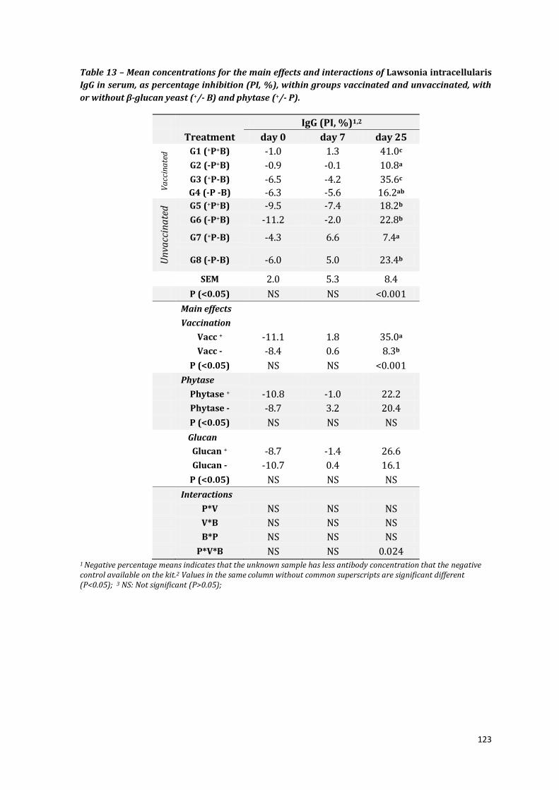
123
Table 13 – Mean concentrations for the main effects and interactions of Lawsonia intracellularis
IgG in serum, as percentage inhibition (PI, %), within groups vaccinated and unvaccinated, with
or without β-glucan yeast (+/- B) and phytase (+/- P).
IgG (PI, %)1,2
Treatment day 0 day 7 day 25
Va
ccin
ate
d G1 (+P+B) -1.0 1.3 41.0c
G2 (-P+B) -0.9 -0.1 10.8a
G3 (+P-B) -6.5 -4.2 35.6c G4 (-P -B) -6.3 -5.6 16.2ab
Un
vacc
ina
ted
G5 (+P+B) -9.5 -7.4 18.2b
G6 (-P+B) -11.2 -2.0 22.8b
G7 (+P-B) -4.3 6.6 7.4a
G8 (-P-B) -6.0 5.0 23.4b
SEM 2.0 5.3 8.4
P (<0.05) NS NS <0.001
Main effects
Vaccination
Vacc + -11.1 1.8 35.0a
Vacc - -8.4 0.6 8.3b
P (<0.05) NS NS <0.001
Phytase
Phytase + -10.8 -1.0 22.2
Phytase - -8.7 3.2 20.4
P (<0.05) NS NS NS
Glucan
Glucan + -8.7 -1.4 26.6
Glucan - -10.7 0.4 16.1
P (<0.05) NS NS NS
Interactions
P*V NS NS NS
V*B NS NS NS
B*P NS NS NS
P*V*B NS NS 0.024 1 Negative percentage means indicates that the unknown sample has less antibody concentration that the negative control available on the kit.2 Values in the same column without common superscripts are significant different (P<0.05); 3 NS: Not significant (P>0.05);

124
6.3.5 Local mucosal immune responses
Local immune responses are summarized in Table 15. In unvaccinated groups,
β-glucan supplementation substantially increased concentrations of IFN-γ, TNF-α and
TGF- β1 (group 6) compared to un-supplemented group 8 while phytase had no
significant effect (group 7), unless animals were vaccinated (group 3, Table 15).
Vaccinated pigs had higher mucosal IgG immune concentrations than unvaccinated
pigs at day 28 pv (PI: 21% and 13.4%, respectively). Pigs supplemented with phytase
generated higher L. intracellularis specific IgG concentrations in the ileal mucosal
scrapings compared to non-supplemented pigs (PI: 24.7% and 10.3%, respectively).
In addition, pigs fed β-glucan had higher mucosal IgG antibody responses than non-
supplemented pigs (20.4 and 14.6 pg/ml, respectively). Similarly, mucosal IgA titres
were increased in pigs supplemented with β-glucan (22.3 pg/mL) when compared
with the non-supplemented cohort (16.7 pg/mL). Interactions between phytase and
vaccination were observed, as vaccinating pigs had increased L. intracellularis IgG
responses when compared to non-vaccination and titres were further increased by
adding phytase to the pig diets.
Cytokine immune responses revealed that vaccinated pigs produced
significantly higher mucosal concentrations of TNF-α, IL-6 and IL-10 (TNF-α, 20.4; IL-
6, 18.9 and IL-10, 85.5) than unvaccinated animals (TNF-α, 9.3: IL-6, 14.3 and IL-10,
67.0) (Table 15). Vaccinated pigs also tended to have higher concentrations of
mucosal IFN-γ and TGF- β1 in mucosal secretions than control animals (IFN-γ; 113.2
vs. 87.4 pg/mL, respectively; TGF- β1; 143.3 vs. 103.1 pg/mL, respectively). Pigs
supplemented with β-glucan also had higher concentrations of IFN-γ (147.1pg/ml)
and TNF-α (20.6) cytokines in ileal mucosal scrapings when compared with
unsupplemented groups (IFN-γ =53.2 and TNF-α TNF=8.9 pg/ml). L. intracellularis
vaccination increased TNF-α response in ileal mucosa when compared with
unvaccinated pigs and a further increased response was observed when β-glucan was
added, leading to a vaccination-β-glucan interaction. In addition, pigs consuming
diets with added phytase generated higher IFN-γ concentrations (118. 8 pg/mL) in
mucosal secretions compared to pigs without phytase in the diet (81.4pg/mL). These
results and also the increasing IFN-γ responses associated with supplementation of
phytase in pigs feed resulted in a phytase-β-glucan interaction.

125
Table 14- The mean concentrations for the main effects and interactions of Lawsonia
intracellularis IgG (PI, %) and IgA (Titre) and cytokines (pg/mL) in ileal mucosa within groups
vaccinated and unvaccinated, with or without β-glucan yeast (+/- B) and phytase (+/- P).
Cytokines1,2 Li-Antibody
Groups IFN-γ TNF-α TGF-β1 IL-10 IL-6 IgG IgA
Va
ccin
ate
d G1 (+P+B) 199.3c 26.,3 292.8c 80.4 21.2 36.78a 22.52
G2 (-P+B) 121.7c 26.7 78.1b 84.2 19.7 24.18b 17.23
G3 (+P-B) 65.7a 14.2 145.1c 60.3 18.2 16.39c 17.75
G4 (-P -B) 66.1a 14.6 55.4a 63.8 16.7 9.75d 16.88
Un
vacc
ina
ted
G5 (+P+B) 174.6c 15.0 54.7a 87.1 16.8 20.48b 21.22
G6 (-P+B) 174.5c 15.5 79.2b 90.6 15.3 18.26bc 27.92
G7 (+P-B) 41.0a 3.0 60.5a 66.9 13.7 8.98d 15.28
G8 (-P-B) 97.0b 3.4 65.1a 70.5 12.3 6.69d 17.23
SEM 20.5 8.9 17.6 14.1 6.3 4.8 7.2
P (<0.05) <0.001 NS <0.001 NS NS <0.001 NS
Main factor
Vaccination
Vacc + 113.2 20.4a 144.3 85.5a 18.9a 21.78a 20.4
Vacc - 87.4 9.3b 107.1 67.0b 14.3b 13.47b 18.59
P (<0.05) NS 0.02 NS 0.04 0.04 <0.001 NS
Phytase
Phytase + 118.8 14.6 139.9 74.1 17.3 24.79a 19.93
Phytase - 81.4 14.8 120.4 77.9 15.8 10.35b 19.11
P (<0.05) NS NS NS NS NS 0.002 NS
Glucan
Glucan + 147.1 20.6 116.6 72.5 18.2 20.42a 22.34a
Glucan - 53.2 8.9 143.0 79.4 15.0 14.63b 16.76b
P (<0.05) <0.001 0.006 NS NS NS <0.001 0.03
Interactions
P*V NS NS NS NS NS 0.045 NS
V*B NS 0.004 NS NS NS NS NS
B*P 0.008 NS NS NS NS NS NS
P*V*B NS NS NS NS NS NS NS 1 Values in the same column without common superscripts are significant different (P<0.05); 2 NS: Not significant (P>0.05);

126
6.3.6 L. intracellularis shedding
At day 0, pigs were not shedding Lawsonia intracellularis. However, by day 25
two pigs from room 1 (pen G2 and G4) and two pigs from room 4 (pen G6 and G7)
were shedding between 5.9x10³ and 3.1x10³ L. intracellularis per gram of faeces.
However, ileal sections were negative for L. intracellularis antigen staining (IHC) from
these animals at necropsy after 28 days pv. Although strict quarantine procedures
were followed it may be possible that several pigs may have been recently infected
on-farm prior to transport and this resulted in low shedding, no lesions and a relative
low L intracellularis IgG response in the serum.
6.3.7 Pathology results
Pathology results from goblet cells counts (Figure 32) showed small differences
between groups (Figure 33). Stained ileal sections from groups G1 (112.1), G2
(116.3) and G5 (111.9) showed the highest per field counts of goblet cells per field,
and the lowest counts were found in groups G3 (91.5), G4 (100.4) and G5 (105.5).
Figure 32: Ileal section (40x mag.), showing visualization of goblets cells (arrows) within
enterocytes of pig (#B171) from G2 group, supplemented with β-glucan and L. intracellularis
(vaccinated) after 28 days post-vaccination.

127
Figure 33: Mean goblet cells counts in ileal sections and mean from each group (G1-G8).
6.4 Discussion
In this study, the supplementation of 100 ppm Saccharomyces cerevisae yeast
beta-glucan and 2,000 FTU/kg microbial phytase to weaner diets increased the
mucosal immune response to an attenuated Lawsonia intracellularis vaccine, as
evidenced by the significant increases in L. intracellularis specific IgG and the
cytokines, IFN-γ and TNF-α in ileal mucosa. From the available literature, this is the
first study to evaluate the response to L. intracellularis vaccination in pig’s fed diets
supplemented with both these additives. Although, the effect of β-glucan on humoral
and cellular immune cells is well studied, the effect of microbial phytase on immune
response in pigs is relatively limited. As expected, vaccination enhanced the
concentrations of L. intracellularis specific IgG in the serum and mucosa and these
serological results are consistent with previous findings reported in Chapters 4 and 5.
6.4.1 Potential effect of phytase on vaccination
In this study, pigs consuming diets with phytase generated higher L.
intracellularis specific IgG concentrations in mucosal scrapings compared with pigs
fed the diets without phytase. This increase may possibly be associated with the
activation of the innate immunity due to increased nutritional availability. Phytate
can interfere with digestibility and chelate to nutrients, directly affecting its
220
180
140
100
G8
60
G7G6G5G4
160
G3
80
G2G1
200
120
Num
ber
of g
oble
t cel
ls

128
absorption and utilization of nutrients (Simons et al., 1990). Therefore, the use of an
enzyme phytase to break down phytate is used to release nutrients bound to phytate
and potentially increase their digestion and utilization in the gut. It is possible that
the increased bioavailability of nutrients in the gut (with phytase) will allow
additional nutrients to be redirected for immune cell growth and replication. For
instance, Zn its known to affect the recruitment of naïve T cells and cytokines
synthesis (Peterson et al., 2008). As well as, the free inositol molecule has been
speculated to somehow up-regulate cell division and differentiation (Menniti et al.,
1993) and to stimulate respiratory burst, bacterial killing and recovery by releasing
IL-8, TNF-α and IL-6 and TGF-β1 cytokines (Eggleton, 1999). In the current study we
observed higher pro-inflammatory cytokine IFN-γ concentrations in ileal mucosa
(118. 8 pg/mL) in pigs fed diets that were supplemented with phytase than pigs fed
the un-supplemented diet (81.4pg/mL). Pro-inflammatory cytokines have been
shown to orchestrate the mechanisms responsible for supply of nutrients for
proliferation of lymphocyte and macrophage population during periods of immune
challenge (Klasing and Korver, 1997; Spurlock, 1997). Liu et al., (2008) evaluated the
effect of phytase (500 FTU/kg) on the immune responses of broilers that were
vaccinated with Newcastle disease virus (NDV) and fed a high phytate diet (0.44%
phytate P). Their results showed that phytase supplementation increase intestinal
secretory IgA, as well as increased CD3+CD4+ and CD3+CD8+ T lymphocyte numbers
in serum on day 28 after vaccination (Liu et al., 2008).
The CD4+ Th1 cells produce pro-inflammatory IL-2, IFN-γ and TNF-α cytokines
promoting cell mediated immune responses against intracellular pathogens, whereas
CD8+ T mediate cytotoxic killing of infected cells (Emery and Collins, 2011). However,
mucosa scrapings from the duodenum, jejunum and ileum of birds fed diets
supplemented with 2,000 FTU/ kg did not express cytokines (IL1β and IL-6) nor
innate immunity receptors TLR 2 and TLR 4 genes (Olukosi et al., 2013). In their
study, birds were not challenged or vaccinated. Therefore it is plausible that if any
changes in cytokine or innate immunity receptors did occur these may have been too
subtle to detect. Shaw et al., (2011) reported that birds fed 500FTU/kg phytase added
to broilers diets following an Eimeria sp. challenge had no effect on oocyst shedding.
However, the average lesion scores of infected birds fed phytase were reduced in the
caecum by 18 days post challenge. In addition, cytokine IL-17 sRNA expression in the

129
duodenum was increased in phytase supplemented broilers. IL-17 and T-reg cells are
normally involved in the inflammatory regulation in the gut which accompanies
neutrophil recruitment and activity against pathogens (Basso et al., 2009), as well as
promoting secretion of cytokines TGF-β and IL-10 by CD4+ Th2 cells (Emery and
Collins, 2011). Since IL-17 is produced from T-reg T-cells, and activates production of
TGF-β, these results may concur with our findings that phytase supplemented pigs
exhibited a trend to higher TGF-β concentrations (139pg/mL) than non-
supplemented animals (120 pg/mL). Therefore, the results in this trial could be
related to local gut restoration and wound healing as a consequence of the vaccine
“stimulation” in the gut lumen.
6.4.2 Diet effect on growth performance and digestibility
This study did not observe any increases in growth performance (ADFI, ADG
and FCR) of pigs supplemented with phytase or β-glucan. Similarly, previous studies
observed minimal effects on pig performance (FI, ADG and FCR) when pigs were fed
β-glucan (0.25%) from either Laminaria digitata or Saccharomyces cerevisae β-glucan
(Boling et al., 2000; Woyengo et al., 2008). The observations of growth performance
effects of β-glucan could also be related to the source, the length and dose of
supplementation. The available commercial E. coli phytases increased growth
performances (ADG) by 33% when given to nursery pigs until 3 weeks (Jones et al.,
2010). Similarly, supplementing E. coli-phytase (2,500 FTU/kg) to P deficient diets
(0.15%P) had increased ADG and FCR in weaner pigs during the 28 days trial (Veum
et al., 2006). However, modest changes on ADG and FCR were observed in weaner
pigs supplemented with 2,000 FTU/kg of E. coli derived phytase added to P deficient
corn based diets (0.53% P) (Beaulieu et al., 2007). Possibly subtle differences in these
phytase responses on growth performance could be due to differences in
experimental designs and the effects are more pronounced when dietary deficiencies
are corrected.
In this study, no main effects of phytase, β-glucan or vaccination were found in
nutrient digestibility in pigs. However, three way interactions were found for the AID
of Mg, K, Fe, and Cu with the highest AID of Mg observed for the phytase
supplemented and vaccinated groups G1 and G3. Madrid et al., (2013) investigated
the effect of microbial phytase (500 FTU/kg) addition to low total phosphorus diet on

130
apparent faecal mineral digestibility. Results demonstrated that pigs supplemented
with phytase had improved growth performance, however, increases in mineral
digestibility retention was limited to P and Cu (39% and 33%, respectively). Adeola
et al. (1995) also observed that supplemental phytase did not affect the retention, as
percentage of intake, of either Zn or Mg.
6.4.3 Diet effect on general health
During this study, pigs were overall healthy and with occasional scouring, but
without lesions and overall without L. intracellularis shedding after vaccination. The
anti-nutritive compound phytate has been related to increase mucin excretion and it
is suggested that the enzyme phytase could reverse this effect (Onyango et al., 2009;
Selle et al., 2012). The quantity and quality of mucin produced by goblet cells, within
the intestinal lumen has been directly correlated to protection against mechanical
and chemical damage and pathogen invasions (Belley et al., 1999). Therefore, while
excessive mucin excretion may compromise intestinal mucosa integrity, the opposite
is also true, the absence of mucin could lead to increased pathogen attachment (Kim
and Ho, 2010). Walk et al.,(2011) observed no effect of in feed E. coli 6-phytase (1,000
FTU/kg) and Eimeria sp. vaccination on the number of goblet cells in the duodenum
of birds 7 days post-vaccination. Similarly, in this study, no effect of numbers of
goblet cells within the ileal mucosa was observed by histopathology. A tendency for
increased number of goblet cells in β-glucan supplemented pigs was observed in ileal
mucosa (mean 111) compared with the un-supplemented cohort (100). Smith et al.,
(2011) demonstrated that dietary supplementation of 300ppm of seaweed derived
Laminarin sp. in pigs led to significant increases in MUC2 and MUC4 gene expression
in ileum. The mechanisms by which glucans influence mucin production are not yet
understood, but it has been speculated that alterations in the intestinal microbiota
could influence mucin synthesis and secretion (Enss et al., 1994; Rice et al., 2005).
6.4.4 Effect of β-glucan on L. intracellularis vaccination
The activation of the mononuclear phagocyte system and protective
inflammatory cytokines by β-glucans in humans and pigs has been directly correlated
with innate immune responses (Decuypere et al., 1998; Brown and Gordon, 2001).
Human whole blood incubated with soluble yeast β-glucan demonstrated an increase

131
in the production of pro-inflammatory cytokines TNF-α, IL-6, IL-8 and monocyte
tissue factor (Adachi et al., 1994; Young et al., 2001). Similarly, Sonck et al., (2010)
observed proliferation of lymphocytes, neutrophils and monocytes, with TNF-α and
IL-1β production after porcine PMBCs were incubated with S. cerevisae β-glucans
(200 μg/mL). In the current study, pigs fed β-glucan had raised mucosal L.
intracellularis IgG antibody responses when compared with non-supplemented pigs
(20.4 and 14.6 pg/ml, respectively). Mucosal IgA (22.3 and 16.7 pg/mL)
concentrations were similar. The presence of immunoglobulin A and G antibodies in
the intestinal lumen is often associated with increased protection against enteric
pathogens (Jemmott and McClelland, 1989; Husband et al., 1996). Purified β-glucan
from the same source (AB Vista), produced higher haemagglutination inhibition (HI)
titres against Newcastle disease 14 days post-challenge in yeast supplemented
broilers, compared with un-supplemented animals (Haldar et al., 2011). However, the
effects of β-glucans on immunity are not reliably repeatable and so their ability to act
against specific diseases cannot be predicted with any certainty. The administration
of 90 mg/kg of purified yeast (1,3/1,6)-D-glucan to neonatal piglets that were
vaccinated against human influenza, had no effect on intestinal or immune
development of T cell phenotypes or cytokine gene expression and did not improve
the antibody response to vaccination during the 21 day trial (Hester et al., 2012).
Conversely, Stuyven et al., (2009), observed that in-feed β-glucan (Macrogard®,
500g/ton) protected piglets against ETEC-F4 (E. coli) infection, and induced
increased concentrations of F4 specific serum antibody. However, further research is
required to specifically associate the effect S. cerevisae yeast β-glucan to proliferative
enteropathy protection in pigs.
Dendritic cell- associated C type lectin-1 (dectin-1) has been implicated as the
pattern recognition receptor mostly expressed in porcine monocytes and other APCs
that bind β-glucans (Sonck et al., 2009). The subsequent activation of NF-κB in
monocytes and linkage of TLR2, results in enhanced phagocytosis, oxidative burst
and cytokine production (Brown and Gordon, 2001; Sonck et al., 2009). These are in
agreement to our findings of higher concentrations of IFN-γ (147.1pg/ml) and TNF-α
(20.6) cytokines in ileal mucosa from pigs supplemented with β-glucan compared
with un-supplemented groups (IFN=53.2 and TNF=8.9 pg/ml). Possibly yeast

132
activation of the innate immune function could lead to greater induction of acquired
immunity and protection against specific diseases. For instance, reduced clinical signs
and increased PRRS specific IFN-γ in PBMCs incubated with a high molecular β-1,3-
1,6-glucan preparation in vitro was observed 50 days after a challenge infection with
PRRS virus (Xiao et al., 2004). However, the β-glucan structure and chemical
composition can modulate their effects on growth performance, health and immunity
(Sonck et al., 2011). Additionally, variations in the immune responses studied could
be also be related to the dose of supplementation. Concentrations of IgM, IgA or CD4+
and CD8+ T-cells in ileal tissues (by IHC) of finishing pigs were not modified by the
addition of S. cerevisae β-glucan (at 0.03% and 0.3%, AntafermMG®) (Sauerwein et
al., 2007). However when 2.5% S. cerevisae β-glucan (EnergyPlus®) was given to 7
days old piglets, an increased in TNF-α and IL-β1 mRNA expression in ileal tissues
was detected (Eicher et al., 2006).
In conclusion, the supplementation of Saccharomyces cerevisae yeast beta-
glucan and microbial phytase added to weaner diet affected the mucosal and
systemic Lawsonia intracellularis local immune response after vaccination. This study
showed that L. intracellularis vaccination with an attenuated infection to growing
pigs may be influenced by diet. It is possible that dietary components influence the
efficacy of concomitantly innate and acquired immune responses against specific
pathogen and protection. However, further research is required to confirm these
finding and relate to specific disease protection.

133
Chapter 7 General discussion
As the current limitations on the use of in-feed antibiotics become more
widely accepted and implemented, sanitary methods and vaccination assume a
greater importance to maintain the productivity and profitability of pig herds.
Currently, control of proliferative enteropathy relies on antibiotic medication and
vaccination. As pressures to reduce antimicrobial growth promoters continue, the
use of vaccination is considered the best alternative to protect herds from disease.
The results in this thesis provide novel information on the effects of vaccine dose
response, route of immunisation and in-feed additives on the systemic and local
mucosal responses to vaccination with an attenuated bacterium. The experimental
design also addressed whether an immune response could be confidently correlated
with successful vaccination (i.e. the magnitude of the responses is predictive of
protective immunity). The investigations in this thesis have expanded the limited
information available in the immunological responses to the attenuated Lawsonia
intracellularis vaccine Enterisol® ileitis in pigs.
If they are to remove in-feed antibiotics and rely on vaccines to prevent disease,
producers need to be assured that pigs are protected after vaccination. To define the
protective immune responses and the adequate level of protection induced in
experimental and on-farm vaccination with ten-times dose of L. intracellularis (105.9
TCID50) were performed. The dose reliably induces detectable local and systemic
antibody and cytokine responses (Chapters 4 and 5). From the data in chapter 4, the
induction of these responses indicates that protective immunity is generated. Since
sampling of mucosal responses is not practical, the detection of Lawsonia-specific
serum antibodies is a feasible indicative of protective immunity after infection
(Collins and Love, 2007). Additionally, using a standard dose of L. intracellularis (104.9
TCID50) induced low concentrations of L. intracellularis antibody in the ileal mucosa
and did not stimulate detectable concentrations of mucosal cytokines (Nogueira et al.,
2013 ). So, from the results in this thesis and from other approaches such as gene
expression (Jacobson et al., 2011a) and flow cytometry (Cordes et al., 2012), a
reliable immune correlate or biomarker for on-farm use could not be recommended.
The dose-dependent induction of protection against virulent L. intracellularis
challenge was demonstrated by a significant reduction of PE lesion scores and the
134
magnitude and duration of bacterial shedding in faeces in all vaccinated pigs.
Previous studies have related the lower initial oral challenge dose (103 L.
intracellularis) with minimal clinical expression and delayed bacterial shedding in
faeces and reduced histological changes when compared with higher doses (1010 L.
intracellularis) (Collins et al., 2001).
Additionally, there were significant variations in the concentrations of
responses depending on the individual pig, experiment or herd (pen or on-farm) and
the timing of the sample collection. Variation in antibody titres generated after
natural infection is well-described as being affected by sampling time and original
infective dose (related to severity of the primary infection) (in chapters 4 and 5;
Guedes and Gebhart, 2003b; Collins and Love, 2007). Timing of sampling is critical,
especially when attempts to account for individual pig variation require a quantum of
animals to be sacrificed to determine mucosal responses. The studies included in this
thesis was limited to sampling prior to 21 days after vaccination (days 7, 9-10 or 17)
since previous reports documented detectable serum responses by this time (Kroll et
al., 2004; Walter et al., 2005). It was gratifying to detect significant differences in
mucosal cytokine responses at 9 and 17 days after vaccination, even though the
spectrum of responses appeared to reflect the dynamics of immune activity. Here,
some pigs appeared by day 17 to be resolving pathology (higher IL-10) while others
remained activated with increased TNF-α and IFN-γ. The Lawsonia intracellularis
specific IgG concentrations in ileal mucosal secretions also ranged from 14% and
45% in experimental and on farm trials after 17 days post-vaccination; and around
21% after 28 days post- vaccination.
L. intracellularis pathogenesis in the intestinal mucosa is not understood, but
individual differences in the gut microflora of these piglets could explain some
variability in infection rates and immune reactivity (Molbak et al., 2008). The
description of the proportion of microbial and L. intracellularis populations in the
pigs in this study was thwarted by technical difficulties in optimizing a 16S microbial
population real-time PCR. However, given the effects of L. intracellularis on carcass
characteristics (Collins et al., 2009; Collins et al., 2010a), it would be a useful adjunct
to this work in attempting to define the effects of infection on gastrointestinal
microflora and vice-versa.

135
As part of routine farm management, piglets are often vaccinated while they’re
still suckling to overcome the need to remove antibiotics for a week around
vaccination. In some herds, this leads to reduced vaccine efficacy because maternal
antibodies transferred to the piglet in the sow’s milk can inactivate the vaccine.
Efficacy data on alternative sites for delivering mucosal vaccines, such as intranasal,
intraperitoneal and intrarectal vaccination may allow producers to vaccinate and
protect weaner pigs from ileitis without needing to remove antibiotics used to treat
other infections. This thesis demonstrated the success of intramuscular (IM)
vaccination and noted the systemic and local immunological responses after
intraperitoneal (IP) inoculation of the L. intracellularis vaccine paralleled those
induced by oral vaccination. These IM and IP routes would presumably act by
neutralising antibody and blocking L. intracellularis attachment and colonisation. In
horses, intra-rectal vaccination with Enterisol® Ileitis induced high titres (120) of L.
intracellularis IgG responses by IPMA (Pusterla et al., 2012b). The intraperitoneal
route of vaccination has been previously used to protect against disease in pig herds
(Sheldrake et al., 1993; Djordjevic et al., 1997). An intraperitoneal vaccination against
Salmonella sp. observed protection and increased IgG and IgA serum responses that
were comparable to those in orally vaccinated birds (Muir et al., 1998). As noted in
chapter 5, the comparable spectrum of immune responses generated after
vaccination and the subsequent protection after challenge seen in Chapter 4 data
strongly suggested that significant protection against L. intracellularis would occur
after intraperitoneal vaccination. In fact, anecdotal industry personal communication
would indicate that this may occur.
It was highly likely that the inoculation of L. intracellularis intramuscular or
intraperitoneal would probably kill the organism quickly. With inactivated or subunit
vaccines (or killed attenuated vaccines), the addition of an adjuvant to the L.
intracellularis vaccine would possibly induce greater mucosal immune responses and
further protection. As observed previously, killed L. intracellularis in incomplete
Freunds adjuvant vaccine given intramuscularly to pigs reduced faecal L.
intracellularis counts by 98.5% (Dale et al., 1997). Additionally, whereas L.
intracellularis oral live vaccine is very efficient in protecting pigs against proliferative
enteropathy disease and increasing growth performances, an inactivated killed

136
vaccine could also induce protection. One of the benefits of using inactivated vaccines
is the ability to amplify the responses against specific pathogens strains by combining
different pathogens in one unique suspension. However, type of antigen to be used,
cost of production and practicability of administration needs to be also considered.
Recently, a patent has been registered presenting an inactivated Lawsonia
intracellularis, Mycoplasma hyopneumoniae and PCV2 combo vaccine given
intramuscularly. These initial studies demonstrated to be positive in protecting pigs
against PE disease (reducing lesions and clinical signs), however, still information
regarding field efficiency is needed.
Another approach to the replacement of in-feed antibiotics is to uniformly and
non-specifically, heighten the immune “competence” to combat pathogens within pig
herds by in-feed additives. Chapter 6 was the first approach to evaluate the effect of
Saccharomyces cerevisae derived β-glucan and microbial phytase on the responses to
vaccination with Lawsonia intracellularis. Any synergic effect of immunomodulators
in feed to amplify responses after oral vaccination is particularly appealing given the
significance of gastrointestinal pathogens on pig production. This type of approach
and its effect on productivity has been an area of increasing research in international
pig production (Gallois et al., 2009; Heo et al., 2013). In this study, pigs consuming a
weaner diet with added β-glucan raised mucosal L. intracellularis specific IgG
antibody responses when compared with non-supplemented pigs, again indicating
immunity against L. intracellularis challenge (in line with chapter 4). However,
results from one trial are not sufficient and further investigation would be required
to make predictive assertions about the interaction of diet and their application
towards improving mucosal immunity in pigs. The effects of β-glucan are mediated
through innate immunity by receptors-mediated interactions with macrophages,
neutrophils and/or monocytes (Brown and Gordon, 2005), and were supported from
the study in Chapter 6 which also found higher concentrations of IFN-γ and TNF-α in
ileal secretions.
Phytases have been used in poultry and pigs herds to degrade plant phytate-P
to release P, which would otherwise be expelled in the faeces. And since phytate is
137
able to chelate many cations, its hydrolysis has been associated with improved
mineral utilization. Pigs consuming a weaner diet plus phytase generated higher L.
intracellularis specific IgG concentrations in ileal mucosa secretions compared with
pigs eating the standard diet. Again, comparing titres with those reported in chapter
4, it would anticipate protection against L. intracellularis disease in all groups
vaccinated. The detection of specific IgG as an indicator of vaccine stimulation and the
generation of protective immunity, possibly the supplementation with β-glucan could
increase the antibody response generated by a standard dose of Enterisol® Ileitis to a
detectable level and provide the relevant immune biomarker. The continual
activation of the innate immune system in a state of readiness by feed additives (such
as β-glucan in chapter 6) could possibly require greater energy input and the
enhanced inflammatory sensitivity may detract from weight gains. A better method
might be to increase the efficiency of digestion with phytase which did not promote
inflammatory cytokine production (group 7 pigs in chapter 6) and enhance the
availability of nutrients for immunity without compromising growth.
The implementation of strategies to induce the development of immunity to L.
intracellularis by vaccination, as part of a farm health plan, leads to improved animal
health, welfare and productivity while reducing reliance on antibiotics for preventing
disease outbreaks. The results in this thesis have provided confirmation that
alternative routes of vaccination as well as different approaches to nutritional
strategies could provide alternative means to augment the induction of mucosal
immune responses after vaccination.

138
References
Abbas, A, Lichtman, A, Pillai, S, 2010, Cellular and Molecular Immunology, 6th: Updated Edition Edition. Saunders Elsevier, Philadelphia, USA, 566 p.
Abourebyeh, H, Naher, H, Hahn, H, 1992, Delayed-type hypersensitivity (DHT)- Studies on necrotizing DTH reaction against Listerial antigen in the skin of guinea pigs. Zentralblatt Fur Bakteriologie-International Journal of Medical Microbiology Virology Parasitology and Infectious Diseases 276, 530-539.
Abreu, M, Targan, S, 1996, Regulation of T cells in the intestinal mucosa. Current Opinion in Gastroenterology 12, 569-576.
Abreu, M, 2010, Toll-like receptor signalling in the intestinal epithelium: how bacterial recognition shapes intestinal function. Nature reviews 10, 131-144.
Adachi, Y, Okazaki, M, Ohno, N, Yadomae, T, 1994, Enhancement of cytokine production by macrophages stimulated with 1-3-beta-D-glucan Grifolan (GRN) isolated from Grifola-frondosa. Biological & Pharmaceutical Bulletin 17, 1554-1560.
Adeola, O, Lawrence, B, Sutton, A, Cline, T, 1995, Phytase induced changes in mineral utilization in zinc supplemented diets for pigs. Journal of Animal Science 73, 3384-3391.
Alberdi, M, Watson, E, McAllister, G, Harris, J, Paxton, E, Thomson, J, Smith, D, 2009, Expression by Lawsonia intracellularis of type III secretion system components during infection. Veterinary Microbiology 139, 298-303.
Almond, P, Bilkei, G, 2006, Effects of oral vaccination against Lawsonia intracellularis on growing-finishing pig's performance in a pig production unit with endemic porcine proliferative enteropathy (PPE). Deutsche Tierarztliche Wochenschrift 113, 232-235.
Animal Welfare Working Group 2007. Model Code of Practice for the Welfare of Animals (Pigs), Department of Agriculture Fisheries and Forestry, ed. (Camberra, Australian Government Publishing Service), p. 40.
Anonymous 2010. Injection Reference for Swine (http://www.extension.org/pages/27257/injection-reference-for-swine).
Armbruster, G, Deen, J, Gebhart, C, Pelger, G, Keffaber, K, Parks, C 2007. Review of Lawsonia intracellularis seroprevalence screening in the United States -June 2003 to July 2006. In 38th Annual Meeting of the American Association of Swine Veterinarians, pp. 231-233.
Autenrieth, I, Firsching, R, 1996, Penetration of M cells and destruction of Peyer's patches by Yersinia enterocolitica: an ultrastructural and histological study. Journal of Medical Microbiology 44, 285-294.
Bailey, M, Haverson, K, Inman, C, Harris, C, Jones, P, Corfield, G, Miller, B, Stokes, C, 2005, The influence of environment on development of the mucosal immune system. Veterinary Immunology and Immunopathology 108, 189-198.
Bailey, M, 2009, The mucosal immune system: Recent developments and future directions in the pig. Developmental and Comparative Immunology 33, 375-383.
Bak, H, Rathkjen, P, 2008. Effect of vaccination against ileitis in herds with continuous production. In: 20th International Pig Veterinary Society Congress Durban, South Africa, p. 237.
139
Bane, D, Neumann, E, Gebhart, C, Gardner, I, Norby, B, 2001, Porcine proliferative enteropathy: a case-control study in swine herds in the United States. Journal of Swine Health and Production 9, 155-158.
Basso, A, Cheroutre, H, Mucida, D, 2009, More stories on Th17 cells. Cell research 19, 399-411.
Bearson, S, Allen, H, Bearson, B, Looft, T, Brunelle, B, Kich, J, Tuggle, C, Bayles, D, Alt, D, Levine, U, Stanton, T, 2013, Profiling the gastrointestinal microbiota in response to Salmonella: Low versus high Salmonella shedding in the natural porcine host. Infection, Genetics and Evolution 16, 330-340.
Beaulieu, A, Bedford, M, Patience, J, 2007, Supplementing corn or corn-barley diets with an E. coli derived phytase decreases total and soluble P output by weanling and growing pigs. Canadian Journal of Animal Science 87, 353-364.
Belley, A, Keller, K, Göttke, M, Chadee, K, Göettke, M, 1999, Intestinal mucins in colonization and host defense against pathogens. The American Journal of Tropical Medicine and Hygiene 60, 10-15.
Bercovich, Z, 2000, The use of skin delayed-type hypersensitivity as an adjunct test to diagnose brucellosis in cattle: A review. Veterinary Quarterly 22, 123-130.
Biehl, R, Baker, D, 1996, Efficacy of supplemental 1 alpha-hydroxycholecalciferol and microbial phytase for young pigs fed phosphorus- or amino acid-deficient corn-soybean meal diets. Journal of Animal Science 74, 2960-2966.
Bohlke, R, Thaler, R, Stein, H, 2005, Calcium, phosphorus, and amino acid digestibility in low-phytate corn, normal corn, and soybean meal by growing pigs. Journal of Animal Science 83, 2396-2403.
Böhme, H, 2002, Digestive physiology of pigs, Vol 97. CABI Publishing, UK, 123-124 pp.
Bohn, J, BeMiller, J, 1995, (1->3)-beta-D-glucans as biological response modifiers: A review of structure-functional activity relationships. Carbohydrate Polymers 28, 3-14.
Boling, S, Webel, D, Mavromichalis, I, Parsons, C, Baker, D, 2000, The effects of citric acid on phytate-phosphorus utilization in young chicks and pigs. Journal of Animal Science 78, 682-689.
Boutrup, T, Boesen, H, Boye, M, Agerholm, J, Jensen, T, 2010, Early Pathogenesis in Porcine Proliferative Enteropathy caused by Lawsonia intracellularis. Journal of Comparative Pathology 143, 101-109.
Brana, D, Ellis, M, Castaneda, E, 2006, Effect of novel phytase on growth performance, bone ash and mineral digestibility in nursery and grower finisher pigs. Journal of Animal Science 84, 1839-1849.
Brandt, D, Kaim, U, Baumgartner, W, Wendt, M, 2010, Evaluation of Lawsonia intracellularis infection in a group of pigs in a subclinically affected herd from weaning to slaughter. Veterinary Microbiology.
Brodin, P, Majlessi, L, Brosch, R, Smith, D, Bancroft, G, Clark, S, Williams, A, Leclerc, C, Cole, S, 2004, Enhanced protection against tuberculosis by vaccination with recombinant Mycobacterium microti vaccine that induces T cell immunity against region of difference antigens. Journal of Infectious Diseases 190, 115-122.
Bronsvoort, M, Norby, B, Bane, D, Gardner, I, 2001, Management factors associated with seropositivity to Lawsonia intracellularis in US swine herds. Journal of Swine Health and Production 9, 285-290.

140
Brown, G, Gordon, S, 2001, Immune recognition: A new receptor for [beta]-glucans. Nature 413, 36-37.
Brown, G, Gordon, S, 2003, Fungal beta-glucans and mammalian immunity. Immunity 19, 311-315.
Brown, G, Herre, J, Williams, D, Willment, J, Marshall, A, Gordon, S, 2003, Dectin-1 mediates the biological effects of beta-glucans. Journal of Experimental Medicine 197, 1119-1124.
Brown, G, Gordon, S, 2005, Immune recognition of fungal beta-glucans. Cellular microbiology 7, 471-479.
Burke-Gaffeyy, A, Keenan, A, 1993, Modulation of human endothelial cell permeability by combinations of the cytokines interleukin-1 α/β, tumor necrosis factor-α and interferon-γ. Immunopharmacology 25, 1-9.
Burkey, T, Skjolaas, K, Minton, J, 2009, Board-invited review:Porcine mucosal immunity of the gastrointestinal tract. Journal of Animal Science 87, 1493-1501.
Butler, J, Klobasa, F, Werhahn, E, 1981, The differential localization of IgA, IgM and IgG in the gut of suckled neonatal piglets. Veterinary Immunology and Immunopathology 2, 53-65.
Caldwell, R, 1992, Effect of Calcium and phytic acid on the activation of trypsinogen and the stability of trypsin. Journal of Agricultural and Food Chemistry 40, 43-46.
Chang, W, Wu, C, Wu, Y, Kao, Y, Pan, M, 1997, Prevalence of Lawsonia intracellularis in swine herds in Taiwan. Veterinary Record 141, 103-104.
Charerntantanakul, W, Roth, J, 2006, Biology of porcine T lymphocytes. Animal Health Research Reviews 7, 81-96.
Chase, C, Lunney, J, 2012, Immune system, In: Zimmerman, J, Locke, K, Ramirez, A (Eds.) Disease of swine John Wiley & sons, Hoboken, NJ, USA, p. 227p.
Chen, W, Perruche, S, Li, J, 2007, CD4(+)CD25(+) T regulatory cells and TGF-beta in mucosal immune system: The good and the bad. Current Medicinal Chemistry 14, 2245-2249.
Chiriboga, A, Guimaraes, W, Vanetti, M, Araujo, E, 1999, Detection of Lawsonia intracellularis in faeces of swine from the main producing regions in Brazil. Canadian Journal of Microbiology 45, 230-234.
Chouet, S, Prieto, C, Mieli, L, Veenhuizen, M, McOrist, S, 2003, Patterns of exposure to Lawsonia intracellularis infection on European pig farms. The Veterinary record 152, 14-17.
Cigala, R, Crea, F, De Stefano, C, Lando, G, Milea, D, Sammartano, S, 2010, Electrochemical Study on the Stability of Phytate Complexes with Cu2+, Pb2+, Zn2+, and Ni2+: A Comparison of Different Techniques. Journal of Chemical and Engineering Data 55, 4757-4767.
Clark, M, Jepson, M, Simmons, N, Hirst, B, 1994, Preferential interaction of Salmonella Typhimurium with mouse Peyer's patch M cells. Research in Microbiology 145, 543-552.
Class, M, Bilkei, G, 2004, Seroprevalence of antibodies against Lawsonia intracellularis among growing pigs raised in indoor versus outdoor units. Journal of the American Veterinary Medicine Association 225, 1905-1907.
Collins, A, Swift, I, Monckton, R, 1996, Replication of Australian porcine isolates of Ileal symbiont intracellularis in tissue culture. Veterinary Microbiology 49, 249-255.

141
Collins, A, Love, R, Jasni, S, McOrist, S, 1999, Attempted infection of mice, rats and chickens by porcine strains of Lawsonia intracellularis. Australian Veterinary Journal 77, 120-122.
Collins, A, Love, R, Pozo, J, Smith, S, McOrist, S, 2000, Studies on the ex vivo survival of Lawsonia intracellularis. Swine Health Production 8, 211-215.
Collins, A, Dijik, M, Vu, N, Pozo, J, Love, R, 2001. Immunity to Lawsonia intracellularis. . In: 28th Allen D. Leman Conference, St. Paul, USA, pp. 127-132.
Collins, A, Love, D, 2003, Risk factors associated with Lawsonia intracellularis infection., In: Paterson, JE (Ed.) Manipulating Pig Production IX Australian Pig Science Association, Werribee, Victoria, Australia, p. 28.
Collins, A, Barchia, I, Gonsalves, J, Fell, S, Eamens, G, 2007, Reducing survival and transmission of Lawsonia intracellularis in conventional piggeries, In: Paterson, J, Barker, J (Eds.) Manipulating pig production XI. . Australasian Pig Science Association, Werribee, Victoria, p. 125.
Collins, A, Love, R, 2007, Re-challenge of pigs following recovery from proliferative enteropathy. Veterinary Microbiology 120, 381-386.
Collins, A, Bolsius, N, van Straaten, J, Fell, S, 2009, Intestinal thickening caused by sub-clinical ileitis can be visualised by computerised tomography in live pigs, In: van Barneveld, R (Ed.) Manipulating Pig Production XII. Australasian Pig Science Association, Werribee, Victoria, p. p.130.
Collins, A, Fell, S, Straaten, J, Bolsius, N, 2010a. Reduced muscle growth in pigs sub-clinically affected with proliferative enteropathy. In: 21st International Pig Veterinary Socity (IPVS) Congress, Vancouver, Canada, p. 234.
Collins, A, Fell, S, Straaten, J, Bolsius, N, Penrose, L, 2010b. Increased variation in pig weight and reduced feed intake in pigs sub-clinically affected with proliferative enteropathy. In: 21st International Pig Veterinary Society (IPVS) Congress, Vancouver, Canada, p. 690.
Collins, A, Fell, S, Pearson, H, Toribio, J, 2011, Colonisation and shedding of Lawsonia intracellularis in experimentally inoculated rodents and in wild rodents on pig farms. Veterinary Microbiology 150, 384-388.
Collins, A, Gonsalves, J, Fell, S, Barchia, I, 2012, Comparison of a commercial ELISA with an indirect fluorescent antibody test to detect antibodies to Lawsonia intracellularis in experimentally challenged pigs. Australian Veterinary Journal 90, 97-99.
Collins, A, Fell, S, Barchia, I, 2013, Cleaning and disinfection with Virkon S significantly reduces Lawsonia intracellularis survival and transmission to naive pigs. Journal of Swine Health and Production 21, 144-147.
Cooper, D, Swanson, D, Barns, S, Gebhart, C, 1997, Comparison of the 16S Ribosomal DNA Sequences from the Intracellular Agents of Proliferative Enteritis in a Hamster, Deer, and Ostrich with the Sequence of a Porcine Isolate of Lawsonia intracellularis. International journal of systematic bacteriology 47, 635-639.
Cordes, H, Riber, U, Jensen, T, Jungersen, G, 2012, Cell-mediated and humoral immune responses in pigs following primary and challenge-exposure to Lawsonia intracellularis. Veterinary Research 43, 9.
Crawley, A, Raymond, C, Wilkie, B, 2003, Control of immunoglobulin isotype production by porcine B-cells cultured with cytokines. Veterinary Immunology and Immunopathology 91, 141-154.

142
Cromwell, G, Coffey, R, Parker, G, Monegue, H, Randolph, J, 1995, Efficacy of a recombinant-derived phytase in improving the bioavailability of phosphorus in corn-soybean meal diets for pigs. Journal of Animal Science 73, 2000-2008.
Cross, R, Smith, C, Parker, C, 1973, Terminal ileitis in Lambs. Journal of American Veterinary Medicine Association 162, 564-566.
Cruz, A, Khader, S, Torrado, E, Fraga, A, Pearl, J, Pedrosa, J, Cooper, A, Castro, A, 2006, Cutting edge: IFN-gamma regulates the induction and expansion of IL-17-producing CD4 T cells during mycobacterial infection. Journal of Immunology 177, 1416-1420.
Cubillos, C, de la Torre, B, Jakab, A, Clementi, G, Borrás, E, Bárcena, J, Andreu, D, Sobrino, F, Blanco, E, 2008, Enhanced Mucosal Immunoglobulin A Response and Solid Protection against Foot-and-Mouth Disease Virus Challenge Induced by a Novel Dendrimeric Peptide. Journal of Virology 82, 7223-7230.
Cunningham, G, Ettinger, R, Aasted, B, 2005. Evaluation of efficacy of Escherichia coli bacterin in controlling neonatal diarrhea in pigs (Scourmune) under field conditions. . In: American Association of Swines Veterinarians Annual Meeting, pp. 239-240.
Curtiss, R, Schodel, F, Potts, A, Kelly, S, DeGrandi, P, 1996, Oral and rectal immunization of adult female volunteers with a recombinant attenuated Salmonella typhi vaccine strain. Infection and Immunity 64, 5219-5224.
Dale, C, Strugnell, R, Lew, A, Hasse, D, Sinistaj, M, Panaccio, M, 1997, Vaccination against Proliferative Enteropathy in pigs., In: Cranwell, P (Ed.) Manipulating Pig Production VI. Australian Pig Science Association, Werribee, Victoria, p. 182.
Danielsen, E, Hansen, G, Nielschristiansen, L, 1995, Localization and biosynthesis of aminopeptidase-n in pig fatel small intestine. Gastroenterology 109, 1039-1050.
Daynes, R, Enioutina, E, Butler, S, Mu, H, McGee, Z, Araneo, B, 1996, Induction of common mucosal immunity by hormonally immunomodulated peripheral immunization. Infection and Immunity 64, 1100-1109.
de Lange, C, Pluske, J, Gong, J, Nyachoti, C, 2010, Strategic use of feed ingredients and feed additives to stimulate gut health and development in young pigs. Livestock Science, 124-134.
Decuypere, J, Dierick, N, Boddez, S, 1998, The potentials for immunostimulatory substances beta-1,3/1,6 glucans in pig nutrition. Journal of Animal and Feed Sciences 7, 259-265.
Deshpande, S, Cheryan, M, 1984, Effects of Phytic Acid, Divalent Cations, and Their Interactions on α-Amylase Activity. Journal of Food Science 49, 516-519.
Djordjevic, S, Eamens, G, Romalis, L, Nicholls, P, Taylor, V, Chin, J, 1997, Serum and mucosal antibody responses and protection in pigs vaccinated against Mycoplasma hyopneumoniae with vaccines containing a denatured membrane antigen pool and adjuvant. Australian Veterinary Journal 75, 504-511.
Donahoo, M, 2009. Studies on the immunisation of young pigs against Lawsonia intracellularis. The Sydney University, Sydney, Australia.
Dritz, S, Shi, J, Kielian, T, Goodband, R, Nelssen, J, Tokach, M, Chengappa, M, Smith, J, Blecha, F, 1995, Influence of dietary Beta-glucan on growth-performace, nonspecific immunity, and resistance to Streptococcus suis infection in weanling pigs. Journal of Animal Science 73, 3341-3350.
143
Duhamel, G, Wheeldon, E, 1982, Intestinal Adenomatosis in a Foal. Veterinary Pathology Online 19 447-450.
Duriancik, D, Hoag, K, 2010, Vitamin A deficiency alters splenic dendritic cell subsets and increases CD8(+)Gr-1(+) memory T lymphocytes in C57BL/6J mice. Cellular Immunology 265, 156-163.
Eggleton, P, 1999, Effect of IP6 on human neutrophil cytokine production and cell morphology. Anticancer Res 19, 3711-3715.
Eicher, S, McKee, C, Carroll, J, Pajor, E, 2006, Supplemental vitamin C and yeast cell wall β-glucan as growth enhancers in newborn pigs and as immunomodulators after an endotoxin challenge after weaning. Journal of Animal Science 84, 2352-2360.
Emery, DL, Collins, A, 2011, Development of the gastrointestinal ecosystem and mucosal immunity in the neonatal pig, In: van Barneveld, R (Ed.) Manipulating pig production XIII. Australasian Pig Science Association Werribee, Victoria, pp. 11-30.
Enss, M, Schmidt-Wittig, U, Honer, K, Kownatzki, R, Gartner, K, 1994, Mechanical challenge causes alterations of rat colonic mucosa and released mucins. Alterations of mucosa and mucins. J Exp Anim Sci 36, 128-140.
Eriksen, K, Landsverk, T, 1988. Altered patterns of cell differentiation in intestinal adenomatosis of swine. In: 10th International Pig Veterinary Society Congress (IPVS), Rio de Janeiro, Brazil., 1988, pp. 135-135.
Fairbrother, J, Gyles, C, 2012, Colibacilosis, In: Zimmerman, J, Karriker, L, Ramirez, A, Schwartz, K, Stevenson, G (Eds.) Diseases of Swine. Wiley-Blackwell, Hoboken, NJ, USA, p. 723.
Farzan, A, Friendship, R, 2010, A clinical field trial to evaluate the efficacy of vaccination in controlling Salmonella infection and the association of Salmonella-shedding and weight gain in pigs. Canadian Journal of Veterinary Research 74, 258-263.
Ferry, S, Matsuda, M, Yoshida, H, Hirata, M, 2002, Inositol hexakisphosphate blocks tumor cell growth by activating apoptotic machinery as well as by inhibiting the Akt/NFkappaB-mediated cell survival pathway. Carcinogenesis 23, 2031-2041.
Flo, H, Bergsjo, B, Grove, S, 2000. The prevalence of Lawsonia intracellularis in Norwegian swine herds. In: 16th International Pig Veterinary Society Congress, Melbourne, Australia, p. 67.
Fontenot, J, Rudensky, A, 2005, A well adapted regulatory contrivance: regulatory T cell development and the forkhead family transcription factor Foxp3. Nature Immunology 6, 331-337.
Fort, M, Sibila, M, Allepuz, A, Mateu, E, Roerink, F, Segales, J, 2008, Porcine circovirus type 2 (PCV2) vaccination of conventional pigs prevents viremia against PCV2 isolates of different genotypes and geographic origins. Vaccine 26, 1063-1071.
Fox, J, Lawson, G, 1988, Campylobacter-like omega intracellular antigen in proliferative colitis in ferrets. Laboratory Animal Science 38, 34-36.
Friedman, M, Bednar, V, Klimes, J, Smola, J, Mrlik, V, Literak, I, 2008, Lawsonia intracellularis in rodents from pig farms with the occurrence of porcine proliferative enteropathy. Letters of Applied Microbiology 47, 117-121.
Friendship, R, 1989, Physiology of intestinal digestion and absorption, In: Bock, HD, Eggum, BO, Low, AG, Simon, O, Zebrowska, T (Eds.) Protein metabolism in

144
farm animals: Evolution, Digestion, Absroption, and Metabolism. Oxford University Press, Oxford, UK, pp. 218-272.
Gallois, M, Rothkotter, H, Bailey, M, Stokes, C, Oswald, I, 2009, Natural alternatives to in-feed antibiotics in pig production: can immunomodulators play a role? Journal of Animal Bioscience 3, 1644-1661.
Gebhart, C, Barns, S, McOrist, S, Lin, G, Lawson, G, 1993, Ileal symbiont intracellularis, an obligate intracellular bacterium of porcine intestines showing a relationship to Desulfovibrio species. International journal of systematic bacteriology 43, 533-538.
Ghahri, H, Rostami, D, Zandiyeh, M, Abbasi, R, 2012, The effects of phytase on performance, serum mineral concentrations, enzyme activities and immune function of broilers fed nutritionally marginal diets. Middle East Journal of Scientific Research 11, 1481-1490.
Gogolewski, R, Cook, R, Batterham, E, 1991, Suboptimal growth associated with porcine intestinal adenomatosis in pigs in nutritional studies Australian Veterinary Journal 68, 406-408.
Guedes, R, Gebhart, C, Armbruster, G, Roggow, B, 2002a, Serologic follow-up of a repopulated swine herd after an outbreak of proliferative hemorrhagic enteropathy. Canadian Journal of Veterinary Research 66, 258-263.
Guedes, R, Gebhart, C, Deen, J, Winkelman, N, 2002b, Validation of an immunoperoxidase monolayer assay as a serologic test for porcine proliferative enteropathy. Journal of Veterinary Diagnostic Investigation 14, 528-530.
Guedes, R, Gebhart, C, Winkelman, N, Mackie-Nuss, R, Marsteller, T, Deen, J, 2002c, Comparison of different methods for diagnosis of porcine proliferative enteropathy. Canadian Journal of Veterinary Research 66, 99-107.
Guedes, R, Gebhart, C, 2003a, Onset and duration of fecal shedding, cell-mediated and humoral immune responses in pigs after challenge with a pathogenic isolate or attenuated vaccine strain of Lawsonia intracellularis. Veterinary Microbiology 91, 135-145.
Guedes, R, Gebhart, C, 2003b, Preparation and characterization of polyclonal and monoclonal antibodies against Lawsonia intracellularis. Journal of Veterinary Diagnostic Investigation 15, 438-446.
Guedes, R, Winkelman, N, Gebhart, C, 2003, Relationship between the severity of porcine proliferative enteropathy and the infectious dose of Lawsonia intracellularis. Veterinary Record 153, 432-433.
Guedes, R, 2008, Known facts about the epidemiology of porcine proliferative enteropathy. Thai Journal of Veterinary Medicine 38, 9-17.
Guedes, R, Gebhart, C, 2010, Evidence of cell-mediated immune response and specific local mucosal immunoglobulin (Ig) A production against Lawsonia intracellularis in experimentally infected swine. Can J Vet Res 74, 97-101.
Ha, N, Oh, B, Shin, S, Kim, H, Oh, T, Kim, Y, Choi, K, Oh, B, 2000, Crystal structures of a novel, thermostable phytase in partially and fully calcium-loaded states. Nature Structural Biology 7, 147-153.
Halas, V, Kovacs, M, Babinszky, L, 2006, Impact of nutrient supply on the immune functions in pig. Magyar Allatorvosok Lapja 128, 533-543.
Haldar, S, Ghosh, T, Toshiwati, S, Bedford, M, 2011, Effects of yeast (Saccharomyces cerevisiae) and yeast protein concentrate on production performance of

145
broiler chickens exposed to heat stress and challenged with Salmonella enteritidis. Animal Feed Science and Technology 168, 61-71.
Hall, W, Molitor, T, Joo, H, Pijoan, C, 1989, Comparison of protective immunity and inflamatory responses of pigs following immunization with different Actinobacillus pleuropneumoniae preparations with and without adjuvants. Veterinary Immunology and Immunopathology 22, 175-186.
Hansen, G, Niels-Christiansen, L, Immerdal, L, Nystrom, B, Danielsen, E, 2007, Intestinal alkaline phosphatase: selective endocytosis from the enterocyte brush border during fat absorption. American Journal of Physiology-Gastrointestinal and Liver Physiology 293, 1325-G1332.
Heo, J, Opapeju, F, Pluske, J, Kim, J, Hampson, D, Nyachoti, C, 2013, Gastrointestinal health and function in weaned pigs: a review of feeding strategies to control post-weaning diarrhoea without using in-feed antimicrobial compounds. Journal of Animal Physiology and Animal Nutrition 97, 207-237.
Herbst, W, Hertrampf, B, Schmitt, T, 2003, Diagnosis of Lawsonia intracellularis using the polymerase chain reaction (PCR) in pigs with and without diarrhea and other animal species. Deutsche Tierärztliche Wochenschrift 110, 361-364.
Hester, S, Comstock, S, Thorum, S, Monaco, M, Pence, B, Woods, J, Donovan, S, 2012, Intestinal and Systemic Immune Development and Response to Vaccination Are Unaffected by Dietary (1,3/1,6)-β-d-Glucan Supplementation in Neonatal Piglets. Clinical and Vaccine Immunology 19, 1499-1508.
Hiss, S, Sauerwein, H, 2003, Influence of dietary ß-glucan on growth performance, lymphocyte proliferation, specific immune response and haptoglobin plasma concentrations in pigs. Journal of Animal Physiology and Animal Nutrition 87, 2-11.
Hodgins, D, Kang, S, deArriba, L, Parreno, V, Ward, L, Yuan, L, To, T, Saif, L, 1999, Effects of maternal antibodies on protection and development of antibody responses to human rotavirus in gnotobiotic pigs. Journal of Virology 73, 186-197.
Hodgins, D, Shewen, P, Dewey, C, 2004, Influence of age and maternal antibodies on antibody responses of neonatal piglets vaccinated against Mycoplasma hyopneumoniae. Journal of Swine Health and Production 12, 10-16.
Hoffman, J, Arbona, D, Anderson, K, 2009, A comparison of humoral function in response to a killed Newcastle vaccine challenge in caged vs. free-range Hy-Line Brown Layers. Poultry Science Supplemment 88, 174-178.
Holmgren, J, Czerkinsky, C, Eriksson, K, Mharandi, A, 2003, Mucosal immunisation and adjuvants: a brief overview of recent advances and challenges. Vaccine 21 Suppl 2, 89-95.
Holyoake, P, Cutler, R, Caple, I, Monckton, R, 1994, Enzyme linked immunisorbent assay for measuring ileal symbiont intracellularis specific immunoglobulin G response in sera of pigs. Journal of Clinical Microbiology 32, 1980-1985.
Holyoake, P, Mullan, B, Cutler, R, 1996, Simulation of the economic impact of proliferative enteritis on pig production in Australia. Australian Veterinary Journal 73, 89-92.
Holyoake, P, Collins, A, Mullan, B, 2010a. Simulation of the economic impact of Lawsonia intracellularis infection. In: 21st International Pig Veterinary Society (IPVS) Congress, Vancouver, Canada, p. 233.

146
Holyoake, P, Emery, D, Gonsalves, J, Donahoo, M, Collins, A, 2010b, Prevalence of antibodies to Lawsonia intracellularis in pig herds in Australia. Australian Veterinary Journal 88, 186-188.
Hotchkiss, C, Shames, B, Perkins, S, Fox, J, 1996, Proliferative enteropathy of rabbits - the intracellular Campylobacter-like organism is closely related to Lawsonia intracellularis. Laboratory Animal Science 46, 623-627.
Hueck, C, 1998, Type III protein secretion systems in bacterial pathogens of animals and plants. Microbiology and Molecular Biology Reviews 62, 379-388.
Husa, J, Edler, R, Walter, D, Holck, J, Saltzman, R, 2009, A comparison of the safety, cross-protection, and serologic response associated with two commercial oral Salmonella vaccines in swine. Journal of Swine Health and Production 17, 10-21.
Husband, A, Kramer, D, Bao, S, Sutherland, R, Beagley, K, 1996, Regulation of mucosal IgA responses in vivo: cytokines and adjuvants. Veterinary Immunology and Immunopathology 54, 179-186.
Igbasan, F, Männer, K, Miksch, G, Borriss, R, Farouk, A, Simon, O, 2000, Comparative studies on the in vitro properties of phytases from various microbial origins. Archiv für Tierernaehrung 53, 353-373.
Jacobs, A, Vermeij, P, Segers, R, Schrier, C 2011. Vaccine for protection against Lawsonia intracellularis, Mycoplasma hyopneumoniae and porcine circovirus. In http://www.freepatentsonline.com/EP1586646B1.html (USA, Intervet/Schering-Plough Animal Health), p. 8.
Jacobson, M, 2003. Enteric diseases in pigs from weaning to slaughter. Veterinary Medicine Doctorate (PhD). Sveriges Lantbruksuniversitet (Sweden), Sweden.
Jacobson, M, Aspan, A, Konigsson, A, af Segerstad, C, Wallgren, P, Fellstrom, C, Jensen-Waern, M, Gunnarson, A, 2004, Routine diagnostics of Lawsonia intracellularis performed by PCR, serological and post mortem examination, with special emphasis on sample preparation methods for PCR. Veterinary Microbiology 102, 189-201.
Jacobson, M, Andersson, M, Lindberg, R, Fossum, C, Jensen-Waern, M, 2011a, Microarray and cytokine analyses of field cases of pigs with diarrhoea. Veterinary Microbiology 153, 307-314.
Jacobson, M, Wallgren, P, Nordengrahn, A, Merza, M, Emanuelson, U, 2011b, Evaluation of a blocking ELISA for the detection of antibodies against Lawsonia intracellularis in pig sera. Acta Veterinaria Scandinavica 53.
Janeway, CJ, Travers, P, Walport, M, 2001, The recognition and effector mechanisms of adaptive immunity, In: Immunobiology: The Immune System in Health and Disease. Garland Science, New York. USA.
Jemmott, J, McClelland, D, 1989, Secretory IgA as a measure of resistance to infectious disease Behavioral Medicine 15, 63-71.
Jensen, T, Mller, K, Leser, T, Jorsal, S, 1997, Comparison of histology, immunohistochemistry and polymerase chain reaction for detection of Lawsonia intracellularis in natural porcine proliferative enteropathy. European Journal of Veterinary Pathology 3, 115.
Jensen, T, Moller, K, Christensen, G, Leser, T, Jorsal, S, 1999, Monitoring ileitis and Lawsonia intracellularis in abattoir pigs. Veterinary Record 145, 613-615.
Jensen, T, Vigre, H, Sorensen, V, Moller, K, 2005, Naturally acquired Lawsonia intracellularis infection in pigs studied from weaning to slaughter by indirect
147
immunofluorescence antibody test and polymerase chain reaction on faeces. Res Vet Sci 79, 93-98.
Jensen, T, Christensen, B, Boye, M, 2006, Lawsonia intracellularis infection in the large intestines of pigs. APMIS 114, 255-264.
JETACAR 1999. The use of antibiotics in food-producing animals: antibiotic-resistant bacteria in animals and humans. In Poultry Science Supplemment (Camberra, Australia, Commonwealth Department of Agriculture, Fisheries and Forestry), p. 249.
Johnston, SL, Williams, SB, Southern, LL, Bidner, TD, Bunting, LD, Matthews, JO, Olcott, BM, 2004, Effect of phytase addition and dietary calcium and phosphorus levels on plasma metabolites and ileal and total-tract nutrient digestibility in pigs. Journal of Animal Science 82, 705-714.
Jonas, AM, Tomita;, Wyand, 1965, Enzootic intestinal adenocarcinoma in hamsters. . Journal of American Veterinary Medicine Association 147, 1102-1108.
Jones, C, Tokach, M, Dritz, S, Ratliff, B, Horn, N, Goodband, R, DeRouchey, J, Sulabo, R, Nelssen, J, 2010, Efficacy of different commercial phytase enzymes and development of an available phosphorus release curve for Escherichia coli-derived phytases in nursery pigs. Journal of Animal Science 88, 3631-3644.
Jones, G, Ward, G, Murtaugh, M, Lin, G, Gebhart, C, 1993, Enhanced detection of intracellular organism of swine proliferative enteritis, ileal symbiont intracellularis, in feces by polymerase chain-reaction. Journal of Clinical Microbiology 31, 2611-2615.
Jordan, D, Knitted, J, Schwartz, K, Roof, M, Hoffman, L, 2004, A Lawsonia intracellularis transmission study using a pure culture inoculated seeder-pig sentinel model. Veterinary Microbiology 104, 83-90.
Kegley, E, Spears, J, Auman, S, 2001, Dietary phosphorus and an inflammatory challenge affect performance and immune function of weanling pigs. Journal of Animal Science 79, 413-419.
Keller, C, Schoeder, H, Ohlinger, V, 2006. A new blocking ELISA kit for the detection of antibodies specific to Lawsonia intracellularis in porcine blood samples. In: 4th International Veterinary Vaccines and Diagnostics Conference, Oslo, Norway.
Kidd, MT, 2004, Nutritional modulation of immune function in broilers. Poultry Science 83, 650-657.
Kim, J, Hansen, C, Mullan, B, Pluske, J, 2012, Nutrition and pathology of weaner pigs: Nutritional strategies to support barrier function in the gastrointestinal tract. Animal Feed Science and Technology 173, 3-16.
Kim, Y, Ho, S, 2010, Intestinal Goblet Cells and Mucins in Health and Disease: Recent Insights and Progress. Current Gastroenterology Reports 12, 319-330.
Klasing, K, Korver, D, 1997, Leukocytic Cytokines Regulate Growth Rate and Composition Following Activation of the Immune System. Journal of Animal Science 75, 58-67.
Klasing, K, 2007, Nutrition and the immune system. British Poultry Science 48, 525-537.
Klein, E, Gebhart, C, Duhamel, D, 1999, Fatal outbreaks of proliferative enteritis caused by Lawsonia intracellularis in young colony-raised rhesus macaques. Journal of Medicine Primatology 28, 11-18.
Knittel, J, Jordan, D, Schwartz, K, Janke, B, Roof, M, McOrist, S, Harris, D, 1998, Evaluation of antemortem polymerase chain reaction and serologic methods

148
for detection of Lawsonia intracellularis-exposed pigs. American Journal of Veterinary Research 59, 722-726.
Knittel, J, Roof, M 1999. Lawsonia intracellularis cultivation, anti-Lawsonia intracellularis vaccines and diagnostic agents. In http://patentscope.wipo.int/search/en/WO1996039629 (USA, Boehringer Ingelheim/ NOBL Laboratories, Inc.), p. 14.
Korver, DR, Klasing, KC, 1997, Dietary fish oil alters specific and inflammatory immune responses in chicks. Journal of Nutrition 127, 2039-2046.
Kringel, H, Iburg, T, Dawson, H, Aasted, B, Roepstorff, A, 2006, A time course study of immunological responses in Trichuris suis infected pigs demonstrates induction of a local type 2 response associated with worm burden. International Journal for Parasitology 36, 915-924.
Kroll, J, Roof, M, McOrist, S, 2004, Evaluation of protective immunity in pigs following oral administration of an avirulent live vaccine of Lawsonia intracellularis. American Journal of Veterinary Research 65, 559-565.
Kroll, J, Eichmeyer, M, Schaeffer, M, McOrist, S, Harris, D, Roof, M, 2005, Lipopolysaccharide-based enzyme-linked immunosorbent assay for experimental use in detection of antibodies to Lawsonia intracellularis in pigs. Clinical Diagnosis Laboratory Immunology 12, 693-699.
Kroll, J, Roof, M 2007. Lawsonia vaccine and methods of use thereof, Boehringer Ingelheim Vetmedica, I, ed. (United States), p. 14.
Kruse, R, Essen-Gustavsson, B, Fossum, C, Jensen-Waern, M, 2008, Blood concentrations of the cytokines IL-1beta, IL-6, IL-10, TNF-alpha and IFN-gamma during experimentally induced swine dysentery. Acta Veterinaria Scandinavica 50, 32-39.
Kumar, H, Kawai, T, Akira, S, 2009, Toll-like receptors and innate immunity. Biochemical and Biophysical Research Communications 388, 621-625.
Lalles, J, Bosi, P, Janczyk, P, Koopmans, S, Torrallardona, D, 2009, Impact of bioactive substances on the gastrointestinal tract and performance of weaned piglets. Journal of animal bioscience 3, 1625-1643.
Lamm, M, Nedrud, J, Kaetzel, C, Mazanec, M, 1996, New Insights into Epithelial Cell Function in Mucosal Immunity: Neutralization of Intracellular Pathogens and Excretion of antigens by IgA, In: Martin, FK, Hiroshi, K (Eds.) Essentials of Mucosal Immunology. Academic Press, San Diego, pp. 141-149.
Larsson, O, Barker, C, Holm, A, Carlqvist, H, Michell, R, Bertorello, A, Nilsson, T, Honkanen, R, Mayr, G, Zwiller, J, Berggren, P, 1997, Inhibition of Phosphatases and Increased Ca21 Channel Activity by Inositol Hexakisphosphate. Science 278, 471-474.
Lawhorn, B, McConnell, S, Kit, M, Kit, S, 1994, Vaccination of newborn pigs in the presence of low levels of pseudorabies colostral antibodies. Vaccine 12, 601-606.
Lawson, G, Rowland, A, Roberts, L, Fraser, G, McCartney, E, 1979, Proliferative Hemorrhagic Enteropathy. Res Vet Sci 27, 46-51.
Lawson, G, McOrist, S, Rowland, A, McCartney, E, Roberts, L, 1988, Serological Diagnosis of the porcine proliferative enteropathies- implications for etiology and epidemiology. Veterinary Record 122, 554-557.
Lawson, G, McOrist, S, Jasni, S, Mackie, R, 1993, Intracellular bacteria of porcine proliferative enteropathy: cultivation and maintenance in vitro. Journal of Clinical Microbiology 31, 1136-1142.

149
Lawson, G, Gebhart, C, 2000, Proliferative enteropathy. Journal of Comparative Pathology 122, 77-100.
Le Dividich, J, Marion, J, Thomas, F, 2007, Energy and nitrogen utilisation of sow colostrum and milk by the piglet. Canadian Journal of Animal Science 87, 571-577.
Leblanc, B, Ettinger, R, Randolph, J, 1993, Hyperplastic Gastritis with Intraepithelial Campylobacter-like Organisms in a Beagle Dog. Veterinary Pathology Online 30, 391-394.
Lee, S, Kim, T, Park, S, Song, C, Chang, H, Yeh, J, Park, H, Lee, J, 2001, Prevalence of porcine proliferative enteropathy and its control with tylosin in Korea. Journal of Veterinary Science, 209-212.
Lei, X, Ku, P, Miller, E, Ullrey, D, Yokoyama, T, 1993, Supplemental microbial phytase improves bioavailability of dietary zinc to weanling pigs. Journal of Nutrition 123, 1117-1123.
Lemarchand, T, Tully, T, Shane, S, Duncan, D, 1997, Intracellular Campylobacter-like organisms associated with rectal prolapse and proliferative enteroproctitis in emus (Dromaius novaehollandiae). Veterinary Pathology 34, 152-156.
Lin, C, Mahan, D, Wu, G, Kim, S, 2009, Protein digestibility of porcine colostrum by neonatal pigs. Livestock Science 121, 182-186.
Lindecrona, R, Jensen, T, Andersen, P, Moller, K, 2002, Application of a 5' nuclease assay for detection of Lawsonia intracellularis in fecal samples from pigs. Journal of Clinical Microbiology 40, 984-987.
Liu, N, Ru, Y, Cowieson, A, Li, FD, Cheng, X, 2008, Effects of phytate and phytase on the performance and immune function of broilers fed nutritionally marginal diets. Poultry Science 87, 1105-1111.
Lomax, L, Glock, R, Hogan, J, 1982, Porcine proliferative enteritis (intestinal adenomatosis)- Field studies. Veterinary Medicine & Small Animal Clinician 77, 1777-1787.
Love, D, Love, R, 1979, Pathology of proliferative haemorrhagic enteropathy in pigs. Veterinary Pathology 16, 41-48.
Love, R, Love, D, Edwards, M, 1977, Proliferative haemorrhagic enteropathy in pigs. Veterinary Record 100, 65-68.
Lukas, G, Brindle, S, Greengar, P, 1971, Route of absoprtion of intraperitoneally administred compounds. Journal of Pharmacology and Experimental Therapeutics 178, 562.
Luna, L, 1968, Manual of histologic staining methods of the Armed Forces Institute of Pathology. McGraw-Hill, New York, U.S.A.
MacIntyre, N, Smith, D, Shaw, D, Thomson, J, Rhind, S, 2003, Immunopathogenesis of experimentally induced proliferative enteropathy in pigs. Veterinary Pathology 40, 421-432.
Madec, F, Bridoux, N, Bounaix, S, Jestin, A, 1998, Measurement of digestive disorders in the piglet at weaning and related risk factors. Preventive Veterinary Medicine 35, 53-72.
Madrid, J, Martínez, S, López, C, Hernández, F, 2013, Effect of phytase on nutrient digestibility, mineral utilization and performance in growing pigs. Livestock Science 154, 144-151.
Manners, D, Masson, A, Patterson, J, 1974, The structure of a β-(1-3)-D-glucan from yeast cell walls. Biochemistry Journal 137, 417.

150
Marsteller, T, Armbruster, G, Bane, D, Gebhart, C, Muller, R, Weatherford, J, Thacker, B, 2003, Monitoring the prevalence of Lawsonia intracellularis IgG antibodies using serial sampling in growing and breeding swine herds. Journal of Swine Health and Production 11, 127-130.
Martelli, P, Gozio, S, Ferrari, L, Rosina, S, De Angelis, E, Quintavalla, C, Bottarelli, E, Borghetti, P, 2009, Efficacy of a modified live porcine reproductive and respiratory syndrome virus (PRRSV) vaccine in pigs naturally exposed to a heterologous European (Italian cluster) field strain: Clinical protection and cell-mediated immunity. Vaccine 27, 3788-3799.
Mauch, C, Bilkei, G, 2005, Reproductive performance of gilts following an outbreak of acute proliferative enteropathy due to Lawsonia intracellularis. The Veterinary Journal 170, 128-131.
McCluskey, J, Hannigan, J, Harris, J, Wren, B, Smith, D, 2002, LsaA, an antigen involved in cell attachment and invasion, is expressed by Lawsonia intracellularis during infection in vitro and in vivo. Infection and Immunity 70, 2899-2907.
McGhee, J, Kiyono, H, 1994, Effective mucosal immunity. Current concepts for vaccine delivery and immune response analysis. International journal of technology assessment in health care 10, 93-106.
McMurray, D, Bartow, R, Mintzer, C, Hernandezfrontera, E, 1990, Micronutrient status and immune function in tuberculosis. Ann N Y Acad Sci 587, 59-69.
McOrist, S, Boid, R, Lawson, G, McConnell, I, 1987, Monoclonal-antibodies to intracellular Campylobacter-like organisms of the porcine proliferative enteropathies. Veterinary Record 121, 421-422.
McOrist, S, MacIntyre, N, Stokes, C, Lawson, G, 1992, Immunocytological responses in porcine proliferative enteropathies. Infection Immunity 60, 4184-4191.
McOrist, S, Jasni, S, Mackie, R, MacIntyre, N, Neef, N, Lawson, G, 1993, Reproduction of porcine proliferative enteropathy with pure cultures of ileal symbiont intracellularis. Infection and Immunity 61, 4286-4292.
McOrist, S, Gebhart, C, Boid, R, Barns, S, 1995a, Characterization of Lawsonia intracellularis gen. nov., sp. nov., the obligately intracellular bacterium of porcine proliferative enteropathy. International Journal of Systematic and Evolutionary Microbiology 45, 820-825.
McOrist, S, Jasni, S, Mackie, R, Berschneider, H, Rowland, A, Lawson, G, 1995b, Entry of bacterium ileal symbiont intracellularis into cultured enterocytes and its subsequent release. Res Vet Sci 59, 255-260.
McOrist, S, Roberts, L, Jasni, S, Rowland, A, Lawson, G, Gebhart, C, Bosworth, B, 1996, Developed and resolving lesions in porcine proliferative enteropathy: Possible pathogenetic mechanisms. Journal of Comparative Pathology 115, 35-45.
McOrist, S, Gebhart, C, 2006, Proliferative enteropathies, In: Straw, BE, Zimmermann, JJ, D'Allaire, S, Taylor, DJ (Eds.) Disease of Swine. Blackwell, Iowa, USA, pp. 727-738.
McOrist, S, Gebhart, C, Bosworth, B, 2006a, Evaluation of porcine ileum models of enterocyte infection by Lawsonia intracellularis. Canadian Journal of Veterinary Research 70, 155-159.
McOrist, S, Hardge, T, Gebhart, C, Ohlinger, V, Pohlenz, J, Keller, C, Voets, H, Walter, D 2006b. Technical Manual 3.0: Porcine Proliferative Enteropathy, Boehringer Ingelheim Vetmedica, I, ed. (St. Paul, USA).

151
McOrist, S, Smits, R, 2007, Field evaluation of an oral attenuated Lawsonia intracellularis vaccine for porcine proliferative enteropathy (ileitis). Veterinary Record 161, 26-28.
McOrist, S, Blunt, R, Gebhart, C, 2011, Pig-associated Lawsonia intracellularis in various on-farm dipterous fly stages. Journal of Swine Health and Production 19, 277-283.
Meeusen, E, Scheerlinck, J, Wattegedera, S, Entrican, G, 2004, Advances in mucosal vaccination. Animal Health Research Reviews 5, 209-217.
Meeusen, E, Walker, J, Peters, A, Pastoret, P, Jungersen, G, 2007, Current Status of Veterinary Vaccines. Clinical Microbiology Reviews 20, 489-510.
Mei, J, Xu, R, 2005, Transient changes of transforming growth factor-beta expression in the small intestine of the pig in association with weaning. British Journal of Nutrition 93, 37-45.
Mengeling, W, Lager, K, Volz, D, Brockmeier, S, 1992, Effect of various vaccination procedures on shedding, latency, and reactivation of attenuated and virulent pseudorabies virus in swine. American Journal of Veterinary Research 53, 2164-2173.
Menniti, F, Oliver, K, Putney, J, Shears, S, 1993, Inositol phosphates and cell signaling: new views of InsP5 and InsP6. Trends Biochem Sci 18, 53-56.
Mestecky, J, McGhee, J, 1987, Immunoglobulin A (IgA): Molecular and Cellular Interactions Involved in IgA Biosynthesis and Immune Response, In: Frank J. Dixon, KFALEH, Jonathan, WU (Eds.) Advances in Immunology. Academic Press, pp. 153-245.
Mestecky, J, Lamm, M, McGhee, J, Bienenstock, J, Mayer, L, Strober, W, 2005, Mucosal Immunology Third Edition.
Mirchamsy, H, Manhouri, H, Hamedi, M, Ahourai, P, Fateh, G, Hamzeloo, Z, 1996, Stimulating role of toxoids-laden liposomes in oral immunization against diphtheria and tetanus infections. Biologicals 24, 343-350.
Molbak, L, Johnsen, K, Boye, M, Jensen, T, Johansen, M, Moller, K, Leser, T, 2008, The microbiota of pigs influenced by diet texture and severity of Lawsonia intracellularis infection. Veterinary Microbiology 128, 96-107.
Moser, M, Leo, O, 2010, Key concepts in immunology. Vaccine 28, Supplement 3, C2-C13.
Muir, W, Bryden, W, Husband, A, 1995, Intraperitoneal immunization promotes local intestinal immunity in chickens. Avian Pathology 24, 679-692.
Muir, W, Bryden, W, Husband, A, 1998, Evaluation of the efficacy of intraperitoneal immunization in reducing Salmonella Typhimurium infection in chickens. Poultry Science 77, 1874-1883.
Murtaugh, M, Johnson, C, Xiao, Z, Scamurra, R, Zhou, Y, 2009, Species specialization in cytokine biology: Is interleukin-4 central to the TH1–TH2 paradigm in swine? Developmental & Comparative Immunology 33, 344-352.
Muto, T, Noguchi, Y, Suzuki, K, Zaw, KM, 1983, Adenomatous intestinal hyperplasia in guinea pigs associated with Campylobacter-like bacteria. Japanese journal of medical science & biology 36, 337-342
Nadkarni, M, Martin, FE, Jacques, N, Hunter, N, 2002, Determination of bacterial load by real-time PCR using a broad-range (universal) probe and primers set. Microbiology 148, 257-266.
Nakane, A, Asano, M, Sasaki, S, Nishikawa, S, Miura, T, Kohanawa, M, Minagawa, T, 1996, Transforming growth factor beta is protective in host resistance against

152
Listeria monocytogenes infection in mice. Infection and Immunity 64, 3901-3904.
NARMS, NARM 2010. Executive report, http://www.fda.gov/AnimalVeterinary/SafetyHealth/AntimicrobialResistance, ed. (Washington, United States).
Nathues, H, Beilage, E, 2008, Diagnosis of Lawsonia intracellularis infection in pigs after vaccination or antimicrobial treatment. Deutsch Tierärztliche Wochenschrift. 115, 404-409.
Nathues, H, Holthaus, K, Beilage, E, 2009, Quantification of Lawsonia intracellularis in porcine faeces by real-time PCR. Journal of Applied Microbiology 107, 2009-2016.
Nogueira, M, Collins, A, Donahoo, M, Emery, D, 2013 Immunological responses to vaccination following experimental Lawsonia intracellularis virulent challenge in pigs. Veterinary Microbiology 31, 131-138.
NRC, 2012, Nutrient Requirement of swine, 11th Revised Edition.Washington, DC, U.S.
Ogra, P, Mestecky, J, Lamm, M, Strober, W, McGhee, J, Bienenstock, J, 2005, Mucosal Immunology Third Edition. Academic Press, Burlington, xiii p.
Oh, Y, Lee, J, McOrist, S, 2010, Microarray analysis of differential expression of cell cycle and cell differentiation genes in cells infected with Lawsonia intracellularis. Veterinary Journal 184, 340-345.
Oliveira, G, Silveira, L, Itano, E, Soares, R, Freire, R, Watanabe, M, Camargo, Z, Ono, M, 2011, Serological Evidence of Paracoccidioides brasiliensis Infection in Chickens from Parana and Mato Grosso do Sul States, Brazil. Mycopathologia 171, 197-202.
Olukosi, O, Kong, C, Fru-Nji, F, Ajuwon, K, Adeola, O, 2013, Assessment of a bacterial 6-phytase in the diets of broiler chickens. Poultry Science 92, 2101-2108.
Onyango, EM, Asem, E, Adeola, O, 2009, Phytic acid increases mucin and endogenous amino acid losses from the gastrointestinal tract of chickens. British Journal of Nutrition 101, 836-842.
Palmer, E, Beilfuss, B, Nagai, T, Semnani, R, Badylak, S, van Seventer, G, 2002, Human helper T cell activation and differentiation is suppressed by porcine small intestinal submucosa. Tissue Engineering 8, 893-900.
Pandey , A, Szakacs, G, Soccol, C, Rodriguez-Leon, J, Soccol, V, 2001, Production, purification and properties of microbial phytases. Bioresource Technology 77, 203-214.
Paradis, M, Gebhart, C, Toole, D, Vessie, G, Winkelman, N, Bauer, S, Wilson, J, McClure, C, 2012, Subclinical ileitis: Diagnostic and performance parameters in a multi-dose mucosal homogenate challenge model. Journal of Swine Health and Production 20, 137-142.
Pavot, V, Rochereau, N, Genin, C, Verrier, B, Paul, S, 2012, New insights in mucosal vaccine development. Vaccine 30, 142-154.
Pearson, H, 2012. Understanding and mitigating the risk of pathogen transmission from wild animals to domestic pigs in Australia. Veterinary Science doctorate (PhD). The Sydney University, Sydney, Australia.
Pedersen, K, Holyoake, P, Stege, H, Nielsen, J, 2010, Diagnostic performance of different fecal Lawsonia intracellularis-specific polymerase chain reaction assays as diagnostic tests for proliferative enteropathy in pigs: a review. Journal of Veterinary Diagnostic Investigation 22, 487-494.

153
Pedersen, K, Stahl, M, Guedes, R, Angen, O, Nielsen, J, Jensen, T, 2012, Association between faecal load of Lawsonia intracellularis and pathological findings of proliferative enteropathy in pigs with diarrhoea. BMC Veterinary Research 8.
Peterson, D, Scrimgeour, A, McClung, J, Koutsos, E, 2008, Moderate zinc restriction affects intestinal health and immune function in lipopolysaccharide-challenged mice. Journal of Nutritional Biochemistry 19, 193-199.
Pluske, J, Hampson, D, Williams, I, 1997, Factors influencing the structure and function of the small intestine in the weaned pig: a review. Livestock Production Science 51, 215-236.
Pluske, J, 2013, Feed- and feed additives-related aspects of gut health and development in weanling pigs. Journal Animal Science and Biotechnology 4, 1-5.
Porter, P, Hill, I, 1970, Serological changes in immunoglobulins IgG, IgA, and IgM and E.coli antibodies in yong pig. Immunology 18, 565-&.
Pozo, J, Carvajal, A, De Arriba, M, Lanza, I, Rubio, P, 1998. Presence of Lawsonia intracellularis as an aetiologic agent in enteric disorders in Spanish swine herds. In: 15th International Pig Veterinary Society, Birmingham, England, p. 416.
Pusterla, N, Mapes, S, Rejmanek, D, Gebhart, C, 2008, Detection of Lawsonia intracellularis by Real-time PCR in the Feces of Free-living Animals from Equine Farms with Documented Occurrence of Equine Proliferative Enteropathy. Journal of Wildlife Diseases 44, 992-998.
Pusterla, N, Gebhart, C, 2009, Equine proliferative enteropthy caused by Lawsonia intracellularis. Equine Veterinary Education 21, 415-419.
Pusterla, N, Hilton, H, Wattanaphansak, S, Collier, J, Mapes, S, Stenbom, R, Gebhart, C, 2009, Evaluation of the humoral immune response and fecal shedding in weanling foals following oral and intra-rectal administration of an avirulent live vaccine of Lawsonia intracellularis. Veterinary Journal 182, 458-462.
Pusterla, N, Jackson, R, Mapes, S, Noland, J, Stenbom, R, Gebhart, C, 2010, Lawsonia intracellularis: humoral immune response and fecal shedding in weanling foals following intra-rectal administration of frozen-thawed or lyophilized avirulent live vaccine. Veterinary Journal 186, 110-112.
Pusterla, N, Mapes, S, Gebhart, C, 2012a, Lawsonia intracellularis-specific interferon γ gene expression by peripheral blood mononuclear cells in vaccinated and naturally infected foals. The Veterinary Journal 192, 249-251.
Pusterla, N, Vannucci, F, Mapes, S, Nogradi, N, Collier, J, Hill, J, Difrancesco, M, White, A, Akana, N, Simonek, G, Gebhart, C, 2012b, Efficacy of an avirulent live vaccine against Lawsonia intracellularis in the prevention of proliferative enteropathy in experimentally infected weanling foals. American Journal of Veterinary Research 73, 741-746.
Quaglino, D, Nanney, L, Ditesheim, J, Davidson, J, 1991, Transforming growth-factor-beta stimulates wound-healing and modulates extracellular matrix gene expression in pig skin-incisional wound model. Journal of Investigative Dermatology 97, 34-42.
Ravindran, V, Selle, PH, Ravindran, G, Morel, PCH, Kies, AK, Bryden, WL, 2001, Microbial phytase improves performance, apparent metabolizable energy, and ileal amino acid digestibility of broilers fed a lysine-deficient diet. Poultry Science 80, 338-344.

154
Riber, U, Boesen, H, Jakobsen, J, Nguyen, L, Jungersen, G, 2011a, Co-incubation with IL-18 potentiates antigen-specific IFN-g response in a whole-blood stimulation assay for measurement of cell-mediated immune responses in pigs experimentally infected with Lawsonia intracellularis. Veterinary Immunology and Immunopathology 139, 257-263.
Riber, U, Cordes, H, Boutrup, T, Jensen, T, Heegaard, P, Jungersen, G 2011b. Primary infection protects pigs against re-infection with Lawsonia intracellularis in experimental challenge studies. In Veterinary Microbiology, pp. 406-414.
Rice, P, Adams, E, Ozment-Skelton, T, Gonzalez, A, Goldman, M, Lockhart, B, Barker, L, Breuel, K, DePonti, W, Kalbfleisch, J, et al., 2005, Oral delivery and gastrointestinal absorption of soluble glucans stimulate increased resistance to infectious challenge. Journal of Pharmacology and Experimental Therapeutics 314, 1079-1086.
Riddle, M, Kaminski, R, Williams, C, Porter, C, Baqar, S, Kordis, A, Gilliland, T, Lapa, J, Coughlin, M, Soltis, C, et al., 2011, Safety and immunogenicity of an intranasal Shigella flexneri 2a Invaplex 50 vaccine. Vaccine 29, 7009-7019.
Rooke, J, Bland, I, 2002, The acquisition of passive immunity in the new-born piglet. Livestock Production Science 78, 13-23.
Roth, J 2010. Principles of immunology and vaccination. In Boehringer Ingelheim IPVS Symposium (Vancouver, Canada, Boehringer Ingelheim), p. 11.
Rothkötter, R, Möllhoff, G, Pabst, O, 1999, The Influence of Age and Breeding Conditions on the Number and Proliferation of Intraepithelial Lymphocytes in Pigs. Scandinavian Journal of Immunology 50, 31-38.
Rowan, T, Lawrence, T, 1982, Amino acid digestibility in pigs with signs of porcine intestinal adenomatosis. Veterinary Record 110, 306-307.
Rowland, A, Lawson, G, 1973, Intestinal haemorrhage syndrome in the pig. The Veterinary record 93, 402-404.
Rowland, A, 1975, Porcine intestinal adenomatosis: a possible relationship with necrotic enteritis, regional ileitis and proliferative haemorrhagic enteropathy. Veterinary Record 97, 178-181.
Rowland, A, Hutchings, D, 1978, Necrotic enteritis and regional ileitis in pigs at slaughter. Veterinary Record 103, 338-339.
Sauerwein, H, Schmitz, S, Hiss, S, 2007, Effects of a dietary application of a yeast cell wall extract on innate and acquired immunity, on oxidative status and growth performance in weanling piglets and on the ileal epithelium in fattened pigs. Journal of Animal Physiology and Animal Nutrition, 369–380.
Schlemmer, U, Jany, KD, Berk, A, Schulz, E, Rechkemmer, G, 2001, Degradation of phytate in the gut of pigs - Pathway of gastrointestinal inositol phosphate hydrolysis and enzymes involved. Archives of Animal Nutrition 55, 255-280.
Schluesener, H, Jung, S, Salvetti, M, 1990, Susceptibility and resistance of human autoimmune T cell activation to the immunoregulatory effects of transforming growth factor (TGF) β1, β2 and β1.2. Journal of Neuroimmunology 28, 271-276.
Scrimshaw, N, SanGiovanni, J, 1997, Synergism of nutrition, infection, and immunity: An overview. American Journal of Clinical Nutrition 66, S464-S477.
Scrimshaw, N, 2007, Prologue: historical introduction. Immunonutrition in health and disease. The British journal of nutrition 98 Suppl 1, S3-4.
Sedgmen, B, Meeusen, E, Lofthouse, S, 2004, Alternative routes of mucosal immunization in large animals. Immunology and Cell Biology 82, 10-16.
155
Selle, P, Walker, A, Bryden, W, 2003, Total and phytate-phosphorus contents and phytase activity of Australian-sourced feed ingredients for pigs and poultry. Australian Journal of Experimental Agriculture 43, 475-479.
Selle, P, Ravindran, V, 2008, Phytate-degrading enzymes in pig nutrition. Livestock Science 113, 99-122.
Selle, P, Ravindran, V, Cowieson, A, Bedford, M, 2011, Phytate and Phytase, In: Bedford, MRPGG (Ed.) Enzymes in Farm Animal Nutrition. pp. 160-205.
Selle, P, Cowieson, A, Cowieson, N, Ravindran, V, 2012, Protein-phytate interactions in pig and poultry nutrition: a reappraisal. Nutrition Research Reviews 25, 1-17.
Sharpe, L, Peacock, W, Cooke, R, Harris, R, 1950, The effect of phytate and other food factors on iron absorption. Journal of Nutrition 41, 433-446.
Shaw , A, van Ginkel, F, Macklin, K, Blake, J, 2011, Effects of phytase supplementation in broiler diets on a natural Eimeria challenge in naive and vaccinated birds. Poultry Science 90, 781-790.
Sheldrake, R, Romalis, L, Saunders, M, 1993, Serum and mucosal antibody responses against Mycoplasma hyopneumoniae following intraperitoneal vaccination and challenge of pigs with Mycoplasma hyopneumoniae. Res Vet Sci 55, 371-376.
Shen, Y, Piao, X, Kim, S, Wang, L, Liu, P, Yoon, I, Zhen, Y, 2009, Effects of yeast culture supplementation on growth performance, intestinal health, and immune response of nursery pigs. Journal of Animal Science 87, 2614-2624.
Sherwood, E, Williams, D, McNamee, R, Jones, E, Browder, I, Diluzio, N, 1987, In Vitro tumoricidal activity of resting and glucan activated Kupffer cells. Journal of Leukocyte Biology 42, 69-75.
Shi, K, Guo, X, Ge, X, Liu, Q, Yang, H, 2010, Cytokine mRNA expression profiles in peripheral blood mononuclear cells from piglets experimentally co-infected with porcine reproductive and respiratory syndrome virus and porcine circovirus type 2. Veterinary Microbiology 140, 155-160.
Siegrist, C, 2003, Mechanisms by which maternal antibodies influence infant vaccine responses: review of hypotheses and definition of main determinants. Vaccine 21, 3406-3412.
Simons, P, Versteegh, H, Jongbloed, A, Kemme, P, Slump, P, Bos, K, Wolters, M, Beudeker, R, Verschoor, G, 1990, Improvement of phosphorus availability by microbial phytase in broilers and pigs. British Journal of Nutrition 93, 137-152.
Siriwan, P, Bryden, WL, Mollah, Y, Annison, EF, 1993, Measurement of endogenous amino-acid losses in poultry. British Poultry Science 34, 939-949.
Skjolaas, K, Burkey, T, Dritz, S, Minton, J, 2006, Effects of Salmonella enterica serovars Typhimurium (ST) and Choleraesuis (SC) on chemokine and cytokine expression in swine ileum and jejunal epithelial cells. Veterinary Immunology and Immunopathology 111, 199-209.
Smith, A, O'Doherty, J, Reilly, P, Ryan, M, Bahar, B, Sweeney, T, 2011, The effects of laminarin derived from Laminaria digitata on measurements of gut health: selected bacterial populations, intestinal fermentation, mucin gene expression and cytokine gene expression in the pig. British Journal of Nutrition 105, 669-677.
Smith, D, Mitchell, S, Nash, T, Rhind, S, 2000, Gamma interferon influences intestinal epithelial hyperplasia caused by Lawsonia intracellularis infection in mice. Infection and Immunity 68, 6737-6743.

156
Smith, D, Lawson, G, 2001, Lawsonia intracellularis: getting inside the pathogenesis of proliferative enteropathy. Veterinary Microbiology 82, 331-345.
Smith, S, McOrist, S, 1997, Development of persistent intestinal infection and excretion of Lawsonia intracellularis by piglets. Res Vet Sci 62, 6-10.
Smith, S, McOrist, S, Green, L, 1998, Questionnaire survey of proliferative enteropathy on British pig farms. Veterinary Record 142, 690-693.
Sonck, E, Stuyven, E, Goddeeris, B, Cox, E, 2009, Identification of the Porcine C-type lectin dectin-1. Veterinary Immunology and Immunopathology 130, 131-134.
Sonck, E, Stuyven, E, Goddeeris, B, Cox, E, 2010, The effect of beta-glucans on porcine leukocytes. Veterinary Immunology and Immunopathology 135, 199-207.
Sonck, E, Devriendt, B, Goddeeris, B, Cox, E, 2011, Varying Effects of Different beta-Glucans on the Maturation of Porcine Monocyte-Derived Dendritic Cells. Clinical and Vaccine Immunology 18, 1441-1446.
Spurlock, M, 1997, Regulation of metabolism and growth during immune challenge: an overview of cytokine function. Journal of Animal Science 75, 1773-1783.
Steel, R, 1997, Principles and procedures of statistics: a biometrical approach 3rd Edition. WCB, New York.
Stege, H, Jensen, T, Moller, K, Baekbo, P, Jorsal, S, 2001, Risk factors for intestinal pathogens in Danish finishing pig herds. Preventive Veterinary Medicine 50, 153-164.
Stege, H, Jensen, T, Møller, K, Vestergaard, K, Baekbo, P, Jorsal, S, 2004, Infection dynamics of Lawsonia intracellularis in pig herds. Veterinary Microbiology 104, 197-206.
Stokes, C, Bailey, M, Wilson, A, 1994, Immunology of the porcine gastrointestinal tract. Veterinary Immunology and Immunopathology 43, 143-150.
Stoll, B, 2006, Intestinal uptake and metabolism of threonine: nutritional impact. Advances in Pork Production 17, 257-263.
Straw, B, CE;, D, Wilson, M, 2006, Differencial diagnosis of disease, In: Straw, BE (Ed.) Disease of Swine. Blackwell Publishing, Iowa, USA, pp. 241-248.
Stuyven, E, Cox, E, Vancaeneghem, S, Arnouts, S, Deprez, P, Goddeeris, B, 2009, Effect of beta-glucans on an ETEC infection in piglets. Veterinary Immunology and Immunopathology 128, 60-66.
Sweeney, RA, 1989, Generic combustion method for determination of crude protein in feeds: collaborative study. Journal - Association of Official Analytical Chemists 72, 770-774.
Sweeney, T, Collins, C, Reilly, P, Pierce, K, Ryan, M, O'Doherty, J, 2012, Effect of purified beta-glucans derived from Laminaria digitata, Laminaria hyperborea and Saccharomyces cerevisiae on piglet performance, selected bacterial populations, volatile fatty acids and pro-inflammatory cytokines in the gastrointestinal tract of pigs. The British journal of nutrition 108, 1226-1234.
Toka, F, Nfon, C, Dawson, H, Golde, W, 2009, Accessory-Cell-Mediated Activation of Porcine NK Cells by Toll-Like Receptor 7 (TLR7) and TLR8 Agonists. Clinical and Vaccine Immunology 16, 866-878.
Tomanova, K, Bartak, P, Smola, J, 2002, Detection of Lawsonia intracellularis in wild pigs in the Czech Republic. Veterinary Record 151, 765-767.
Tregoning, J, Clare, S, Bowe, F, Edwards, L, Fairweather, N, Qazi, O, Nixon, P, Maliga, P, Dougan, G, Hussell, T, 2005, Protection against tetanus toxin using a plant-based vaccine. European Journal of Immunology 35, 1320-1326.

157
Tsang, V, Pilcher, J, Zhou, W, Boyer, A, Kamango-Sollo, E, Rhoads, M, Murrell, K, Schantz, P, Gilman, R, 1991, Efficacy of the immunoblot assay for cysticercosis in pigs and modulated expression of distinct IgM/ IgG activities to Taenia solium antigens in experimental infections. Veterinary Immunology and Immunopathology 29, 69-78.
Tumamao, J, Bowles, R, van den Bosch, H, Klaasen, H, Fenwick, B, Blackall, P, 2004, An evaluation of the role of antibodies to Actinobacillus pleuropneumoniae serovar 1 and 15 in the protection provided by sub-unit and live streptomycin-dependent pleuropneumonia vaccines. Australian Veterinary Journal 82, 773-780.
Tzika, E, Papatsiros, V, Kyriakis, S, Alexopoulos, C, Lymberopoulos, A, Kyriakis, C, 2009, Efficacy of in-feed valnemulin hydrochloride for the treatment and control of ileitis in weaning and growing pigs. Journal of Applied Animal Research 35, 181-184.
Uenishi, H, Shinkai, H, 2009, Porcine Toll-like receptors: The front line of pathogen monitoring and possible implications for disease resistance. Developmental & Comparative Immunology 33, 353-361.
USDA 2007. Antimicrobial Resistance Issues In Animal Agriculture., USDA:APHIS:VS, ed. (Fort Collins, CO, USA., Center for Epidemiology and Animal Health).
van der Heijden, H, Bakker, J, Elbers, A, Vos, J, Weyns, A, de Smet, M, McOrist, S, 2004, Prevalence of exposure and infection of Lawsonia intracellularis among slaughter-age pigs. Res Vet Sci 77, 197-202.
Vannucci, F, Borges, E, de Oliveira, J, Guedes, R, 2010, Intestinal absorption and histomorphometry of Syrian hamsters (Mesocricetus auratus) experimentally infected with Lawsonia intracellularis. Veterinary Microbiology 145, 286-291.
Vannucci, F, Foster, D, Gebhart, C, 2012a, Comparative Transcriptional Analysis of Homologous Pathogenic and Non-Pathogenic Lawsonia intracellularis Isolates in Infected Porcine Cells. PloS one 7, 467-478.
Vannucci, F, Pusterla, N, Mapes, S, Gebhart, C, 2012b, Evidence of host adaptation in Lawsonia intracellularis infections. Veterinary Research 43, 53-54.
Verstegen, M, Williams, B, 2002, Alternatives to the use of antibiotics as growth promoters for monogastric animals. Animal Biotechnology 13, 113-127.
Veum, T, Bollinger, D, Buff, C, Bedford, M, 2006, A genetically engineered Escherichia coli phytase improves nutrient utilization, growth performance, and bone strength of young swine fed diets deficient in available phosphorus. Journal of Animal Science 84, 1147-1158.
Viott, A, França, S, Vannucci, F, Cruz Jr, E, Costa, M, Gebhart, C, Guedes, R, 2013, Infection of sparrows (Passer domesticus) and different mice strains with Lawsonia intracellularis. Pesquisa Veterinaria Brasileira 33, 372-378.
Volman, J, Ramakers, J, Plat, J, 2008, Dietary modulation of immune function by beta-glucans. Physiology & Behavior 94, 276-284.
Walk, CL, Cowieson, AJ, Remus, JC, Novak, CL, McElroy, AP, 2011, Effects of dietary enzymes on performance and intestinal goblet cell number of broilers exposed to a live coccidia oocyst vaccine. Poultry Science 90, 91-98.
Walter, D, Gebhart, C, Kroll, J, Holck, J, Chittick, W, 2005, Optimizing vaccination timing with Enterisol (R) ileitis, 241-244 pp.
Ward, G, Winkelman, N, 1990, Recognizing the 3 forms of proliferative enteritis in swine. Veterinary Medicine 85, 197-&.

158
Watson, D, 1987, Serological response of sheep to live and killed Staphylococcus aureus vaccines. Vaccine 5, 275-278.
Wawszczyk, J, Orchel, A, Kapral, M, Weglarz, L, 2013, Role of protein tyrosine kinase in the effect of IP6 on IL-8 secretion in intestinal epithelial cells. Acta Poloniae Pharmaceutica 70, 79-86.
Weaver, C, Hatton, R, Mangan, P, Harrington, L, 2007, IL-17 family cytokines and the expanding diversity of effector T cell lineages. Annu Rev Immunol 25, 821-852.
Wellek, B, Hahn, H, Opferkuch, W, 1976, Opsonizing activities of IgG, IgM antibodies and the C3b inactivator-cleaved third component of complement in macrophage phagocytosis. Agents Actions 6, 260-262.
Williams, D, Sherwood, E, Browder, I, McNamee, R, Jones, E, Rakinic, J, Diluzio, N, 1988, Effect of glucan on neutrophil dynamics and immune function in Escherichia coli peritonitis. Journal of Surgical Research 44, 54-61.
Williams, I, 2003, Growth of the weaned pig, In: Pluske, JR, Le Dividich, J, Verstegen, MWA (Eds.) Weaning the pig: Concepts and consequences. Wageningen Academic Publishers, Wageningen, Netherlands, pp. 17-35.
Williams, J, Topley, N, Alobaidi, H, Harber, M, 1986, Activation of human polymorphonuclear leukocytes by particulate zymosan is related to both its major carbohydrate components- glucan and mannan. Immunology 58, 117-124.
Winkelman, N, Pauling, G, Bagg, R, Dick, C, Paradis, M, Brennan, J, Wilson, J, 1998. Use of a challenge model to measure the impact of subclinical porcine proliferative enteritis on growth performance in pigs. In: American Association of Swine Practitioners annual meetting, Des Moines, USA, pp. 209-211.
Winkelman, N, Crane, J, Elfring, G, Kratzer, D, Meeuwse, D, Dame, K, Buckham, S, Gebhart, C, 2002, Lincomycin-medicated feed for the control of porcine proliferative enteropathy (ileitis) in swine. . Journal of Swine Health and Production 10 107-111.
Wong, D, Alcott, C, Sponseller, B, Young, J, Sponseller, B, 2009, Impaired Intestinal Absorption of Glucose in 4 Foals with Lawsonia intracellularis Infection. Journal of Veterinary Internal Medicine 23, 940-944.
Woyengo, T, Sands, J, Guenter, W, Nyachoti, C, 2008, Nutrient digestibility and performance responses of growing pigs fed phytase- and xylanase-supplemented wheat-based diets. Journal of Animal Science 86, 848-857.
Woyengo, T, Cowieson, A, Adeola, O, Nyachoti, C, 2009, Ileal digestibility and endogenous flow of minerals and amino acids: responses to dietary phytic acid in piglets. The British journal of nutrition 102, 428-433.
Xiao, Z, Trincado, C, Murtaugh, M, 2004, beta-glucan enhancement of T cell IFN gamma response in swine. Veterinary Immunology and Immunopathology 102, 315-320.
Yates, W, Clark, E, Osborne, A, Enweani, C, Radostits, O, Theede, A, 1979, Proliferative hemorrhagic enteropathy in swine- outbreak and review of the literature. Canadian Veterinary Journal 20, 261-268.
Yen, JT, 2001, Anatomy of the digestive system and nutritional physiology., In: Lewis, A, Southern, LL (Eds.) Swine Nutrition. CRC Press, Boca Raton, Florida, pp. 31-64.
Yi, E, Komegay, T, 1996, Sites of phytase activity in the gastrointestinal of young pigs. Animal Feed Science Technology 61, 361-368.

159
Young, S, Ye, J, Frazer, D, Shi, X, Castranova, V, 2001, Molecular mechanism of tumor necrosis factor-alpha production in 1 -> 3-beta-glucan (Zymosan)-activated macrophages. Journal of Biological Chemistry 276, 20781-20787.

160
Appendix I Clinical observation record_______________________

161
Clinical Observation Record Study Number: Room #
Group Pig ID Diarrhoea Score Behaviour Body Condition Score
1 2 3 4 1 2 3 4 1 2 3
1 2 3 4 1 2 3 4 1 2 3
1 2 3 4 1 2 3 4 1 2 3
1 2 3 4 1 2 3 4 1 2 3
1 2 3 4 1 2 3 4 1 2 3
1 2 3 4 1 2 3 4 1 2 3
1 2 3 4 1 2 3 4 1 2 3
1 2 3 4 1 2 3 4 1 2 3
1 2 3 4 1 2 3 4 1 2 3
1 2 3 4 1 2 3 4 1 2 3
1 2 3 4 1 2 3 4 1 2 3
1 2 3 4 1 2 3 4 1 2 3
1 2 3 4 1 2 3 4 1 2 3
1 2 3 4 1 2 3 4 1 2 3
Diarrhoea Score Behaviour Body Condition 1 = none 1 = normal 1 = normal 2 = semi-solid, no blood 2 = slight to moderately depressed,
will stand 2 = mild to moderately gaunt (thin)
3 = watery, no blood/dark faeces
3 = severely depressed or recumbent
3 = severely gaunt
4 = blood tinged faeces, loose or formed
4 = excitable
Note: If an animal is dead, record “found dead” in comments column and assign total score of 20 Observed by________________________________ Date _________________
Recorded by _______________________________ Date _____________________

162
Appendix II Published papers_________________________________


