Embed Size (px)
Citation preview
1
Immunohistochemical analysis to assess the effects of
static cold storage versus hypothermic machine
perfusion on porcine hearts
Pepijn van der Putten
Supervisor: Selma Kaffka genaamd Dengler
15-10-2021
2
Abstract Background
Because of population ageing and a high prevalence of overweight, heart failure is a growing global
health issue. Currently heart transplantion (HTx) is the gold standard treatment, but this treatment is
paired with a high waiting-list mortality, partially caused by a shortage of donor hearts. One way to
increase the donor pool would be to change the preservation of the donor hearts. A novel way to
preserve donor hearts is hypothermic machine perfusion. In this study, the effects of the currently
used preservation method static, cold storage and hypothermic machine perfusion on
slaughterhouse porcine hearts have been compared using immunohistochemical analyses.
Methods
Slaughterhouse porcine hearts were removed and preserved for 4 hours using either static cold
storage (SCS) or hypothermic machine perfusion (HMP) followed by 4 hours of normothermic
machine perfusion (NMP). At the end of the experiment, tissue samples were collected and
aphosphotungstic acid-haematoxylin (PTAH) staining was used to score contraction band necrosis
and erythrocyte extravasation. An active caspase-3 staining was performed to score apoptosis.
Results
After normalising the data for the survival time of the hearts, The SCS hearts showed significantly
higher contraction band necrosis compared to HMP hearts.
Conclusion
HMP shows great potential to improve the donor pool. In this study we discovered that more
contraction band necrosis is present in hearts that are preserved using SCS compared to HMP.
Further studies should include a higher sample size and more stainings could be performed to
further elucidate the effects of SCS and HMP on porcine hearts.
3
Plain language summary Heart failure is a growing health issue. Currently most treatment is focussed on slowing down the
progression of the disease and there is only one curative treatment at the moment, namely heart
transplantion. Due to a low number of donor hearts, there is a high waiting-list mortality. A way to
improve the donor pool would be to change the way the heart is preserved. Currently hearts are
preserved using a method called static cold storage (SCS). During this preservation, the hearts are
stored on ice. In this study we compared this preservation to a novel method of preservation named
hypothermic machine perfusion (HMP) using slaughterhouse porcine hearts. Using HMP, there is a
continuous flow through the heart, which leads to a decrease in harmful substances. Using
microscopy analysis we discovered that more damage occurred to the hearts that were preserved
using SCS than to the hearts that were preserved using HMP.
Introduction Heart failure (HF) is a growing global health issue, with a current estimate of over 64 million people
diagnosed. The incidence of HF still continues to rise due to population ageing and a high prevalence
of overweight (1). Treatment is focused on decelerating the disease using various pharmacological
agents for instance with angiotensin converting enzyme (ACE) inhibitors and angiotensin receptor
blockers (ARB), beta blockers and spironolactone (2). For patients with end-stage HF, heart transplantation (HTx) remains the gold standard treatment.
However, the demand for donor hearts exceeds the number of hearts suitable for transplantation
(3). The donor heart shortage makes stringent regulations on eligible patients necessary and the
shortage results in a waiting-list mortality up to 15% over a period of five years (4). A desirable way
to reduce the donor shortage, would be to expand the donor pool.
The preservation of donor hearts is one of the limiting factors for expanding the donor pool.
Currently, static cold storage (SCS) is used to preserve donor hearts. During SCS, the heart is
perfused with a preservation solution, which leads to cardiac arrest, after which the heart is stored
on ice. During SCS metabolism is converted to anaerobic respiration, which leads to the production
of lactate and a decreased intracellular pH (5). This decrease in pH disregulates the Na+/H+
exchanger and the Na+K+ exchanger and the 2Na+H+ exchanger with as final result a intracellular Ca2+
overload leading to cell swelling. When the heart is reperfused after transplantation, reactive oxygen
species (ROS) are generated due to xanthine oxidase, Nicotinamide adenine dinucleotide phosphate
(NADPH) oxidase and nitric oxide synthase (NOS) induction as well as the reactivation of the electron
transport chain (6). The generated ROS can cause cell damage and even cell death by activating
metalloproteinases and calpains and by the opening of the mitochondrial permeability transition
pore (MPTP). This event is known as ischemic reperfusion injury (IRI) and causes more damage to the
heart following ischemia (5).
A novel way to preserve a donor heart is hypothermic machine perfusion (HMP). Using HMP, the
donor heart is continuously being held in a cardioplegic state, thereby limiting the oxygen demand of
the cardiomyocytes, similar to SCS. In addition, the affluent as a result of continuous flow leads to a
decrease in harmful substances. This decreases the ischemia time and reduces IRI. HMP may also be
used to resuscitate hearts after circulatory death (DCD), thereby increasing the donor pool (7). The
aim of this study is to compare the effect of 4 hour SCS to HMP on the heart of slaughterhouse pigs
using an in vivo porcine model.
4
Methods Slaughterhouse animals
Pig hearts were harvested within five minutes after being sacrificed by means of electrical stunning.
A parasternal incision was made in the thorax and the heart and lungs were removed en-bloc. After
harvesting, the heart was immediately topologically cooled. Next, the pericardial sac was opened,
the pulmonary artery was transected under the bifurcation and the aorta was transected under the
first supra-aortic vessel (8). The heart was then isolated and prepared as described in a study
performed by Hannon et al (9). Immediately after removal, the aorta was cannulated and 2 L of
heparinized modified St. Thomas 2 crystalloid cardioplegic solution (Table 1) was administered
through the coronary arteries at a mean pressure of 80-100 mmHg and a temperature of 4 ˚C (10).
Preservation
After the cardioplegic solution was added, the hearts were preserved for 4 hours either using SCS or
HMP. In total 12 hearts were included in this study. 6 hearts were preserved with SCS and the other
6 with HMP.
Hypothermic machine perfusion
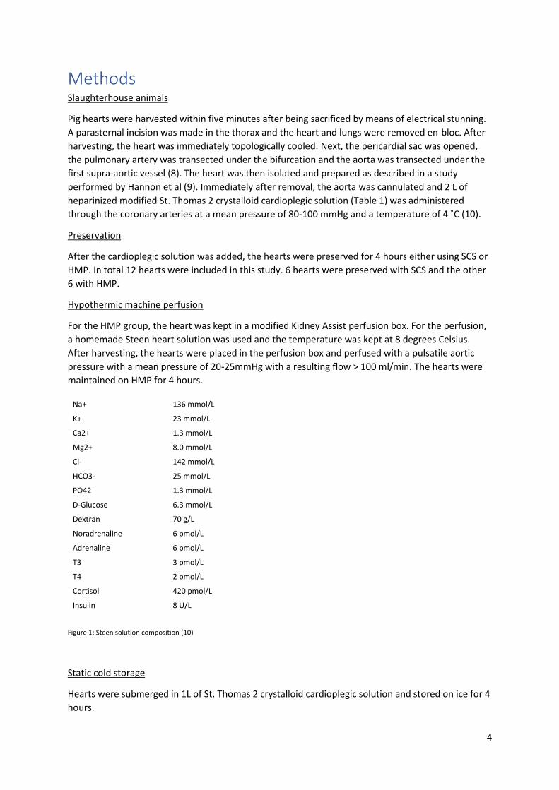
For the HMP group, the heart was kept in a modified Kidney Assist perfusion box. For the perfusion,
a homemade Steen heart solution was used and the temperature was kept at 8 degrees Celsius.
After harvesting, the hearts were placed in the perfusion box and perfused with a pulsatile aortic
pressure with a mean pressure of 20-25mmHg with a resulting flow > 100 ml/min. The hearts were
maintained on HMP for 4 hours.
Na+ 136 mmol/L
K+ 23 mmol/L
Ca2+ 1.3 mmol/L
Mg2+ 8.0 mmol/L
Cl- 142 mmol/L
HCO3- 25 mmol/L
PO42- 1.3 mmol/L
D-Glucose 6.3 mmol/L
Dextran 70 g/L
Noradrenaline 6 pmol/L
Adrenaline 6 pmol/L
T3 3 pmol/L
T4 2 pmol/L
Cortisol 420 pmol/L
Insulin 8 U/L
Figure 1: Steen solution composition (10)
Static cold storage
Hearts were submerged in 1L of St. Thomas 2 crystalloid cardioplegic solution and stored on ice for 4
hours.
5
Normothermic testing
After preservation, all hearts were reperfused ex-vivo using the PhysioHeartTM platform. For the
reperfusion, 4500 mL diluted whole blood of normothermic, heparinized oxygenated blood was
used, which was supplemented with glucose and insulin. First, the hearts were perfused for 60
minutes in Langendorff mode, followed by 3 hours of working heart mode perfusion. Atrial pressure
and aortic pressure were kept at respectively 10-20 mmHg and 60-100 mmHg. A glucose and insulin
mixture was added to keep the blood glucose level between 5-7 mmol/L and a pH of 7.40 was
maintained by adding sodium bicarbonate. In order to measure the cardiac output, an ultrasound
flow probe (SonoTT™ Clamp-On Transducer, em-tec GmbH, Finning, Germany) was placed after the
afterload, and another ultrasound probe was placed after the pulmonary artery to measure the
coronary flow (8).
Sample Collection
At the end of the experiment, perfusion was terminated and left and right ventricle samples were
collected and preserved in 4% formaldehyde and subsequently embedded in paraffin. A microtome
cutting instrument was used to cut slices with a thickness of 3 mu from the formalin-fixed, paraffin-
embedded tissue samples. Several stainings were performed to assess the functionality of the
hearts.
Stainings
Prior to staining, all slides were depariffinized in xylene 3 times, ethanol 100% 2 times and alcohol
70% and finally washed in demineralized water.
Haematoxylin and Eosin
To assess general tissue morphology, a haematoxylin and eosin (H&E) staining was performed. The
haematoxylin stains the nuclei blue and the cytoplasm and ECM pink. First the slides were
deparaffinized using xylene. After deparaffination, the slides were incubated for 5 minutes in
haematoxylin. Hereafter, the slides were washed in tap water for 10 minutes and subsequently
incubated in eosin for 1 minute. Finally, the slides were washed using demineralized water for 15
seconds and dried on a heater at 60 degrees Celsius.
Activate Caspase-3
In order to evaluate apoptosis, an activate caspase-3 staining was performed.
First the slides were deparaffinized in xylene and subsequently washed in ethanol. Hereafter, the
slides were washed in demineralized water and boiled for 20 minutes in 30 mL EDTA + 1500 mL
demineralized water (pH 9).
When the slides were cooled down, they were again washed in demineralized water and
subsequently washed in PBS 3 times for 1 minute. After this, 150 microliter Active Caspase-3
antibody diluted 1:200 in PBS/BSA Azide was added to the slides and the slides were incubated
overnight at 5 degrees Celsius. Next, the slides were incubated for 30 minutes at room temperature
and washed with PBS 3x for 15 minutes, after which the secondary antibody Bright Vision Rabbit AP
was added to the slides. Hereafter, the slides were incubated for 30 minutes at room temperature
and subsequently washed with PBS 3 times for 1 minute. Subsequently, Dako Liquid Permanent Red
Chromogen was added to the slides and after a 10 minute incubation the slides were washed with
6
demineralized. Hereafter, the slides were incubated in 1:3 diluted hematoxylin for 15 seconds and
washed in tap water for 10 minutes and finally dried on a heating plate at 60 degrees Celsius.
Phosphotungstic Acid Haematoxylin
A Phosphotungstic Acid Haematoxylin (PTAH) stain was performed to assess contraction band
necrosis and erythrocyte extravasation. First, the slides were deparaffinized in xylene 3 times,
ethanol 100% 2 times and alcohol 70% and finally washed in demineralized water. Subsequently,
ammonium iron (III) sulphate was added to the slides, followed by 30 minutes of incubation.
Hereafter, the slides were washed in demineralized water for 1 minute, after which oxalic acid (5%)
was added to the slides. After 10 minutes of incubation, the slides were again washed for 1 minute
using demineralized water after which phosphotungstic acid was added to the slides. Hereafter the
slides were incubated at 60 degrees in a laboratory oven for 3 hours. Finally, the slides were
dehydrated using 70% alcohol and 100% alcohol 2 times, after which they were washed in xylene 3
times.
Scoring and statistical analysis
For the analysis of the PTAH staining, the contraction band necrosis was scored based on the
percentage of contraction band necrosis over the whole slide. For this, a 4 class scoring system was
used. I 0-10% positive staining, II 10-20% positive staining, III 20-50% positive staining and IV >50%
positive staining. The extravasation of erythrocytes was scored based on a 3 class system: I: none to
minor extravasation, II: reasonable extravasation, III: severe extravasation. The scoring of the active
caspase-3 stained slides was performed by counting all positive stained cells on the whole slide and
normalized to the size of the slides by first multiplying the amount of cells with the size of the slide
and subsequently dividing it with the average slide size. Scoring was done blindly and the PTAH
slides were also scored by a second observer. Images of the slides were taken using cellSens
Dimension.
The obtained data underwent statistical analysis using the Mann-Whitney U test using IBM SPSS
Statistics.
7
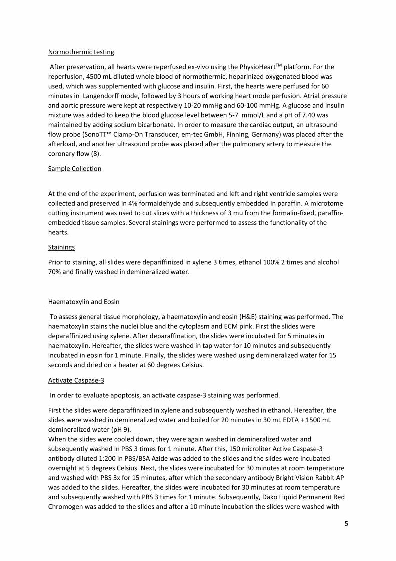
Results Microscopy images of contraction band necrosis and erythrocyte extravasation
Assessment of the slides with the microscope revealed that contraction band necrosis and
erythrocyte extravasation were present in both the SCS hearts ( figure 2 a,c) and the HMP hearts
(figure 2 b,d).
Overall however, notably more contraction band necrosis and erythrocyte extravasation were seen
in the SCS hearts.
Figure 2: Comparison of contraction band necrosis and erythrocyte extravasation using PTAH staining. More contraction band necrosis was
generally seen in the SCS hearts (a) compared to the HMP hearts (b). Erythrocyte extravasation was also mostly in the SCS hearts (c) than
in the HMP hearts (d).
8
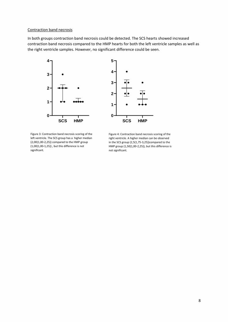
Contraction band necrosis
In both groups contraction band necrosis could be detected. The SCS hearts showed increased
contraction band necrosis compared to the HMP hearts for both the left ventricle samples as well as
the right ventricle samples. However, no significant difference could be seen.
SCS HMP
0
1
2
3
4
Contraction band necrosis LV
Contraction band necrosis
SCS HMP
0
1
2
3
4
5
Contraction band necrosis RV
Contraction band necrosis
Figure 3: Contraction band necrosis scoring of the
left ventricle. The SCS group has a higher median
(2,00(1,00-2,25)) compared to the HMP group
(1,00(1,00-1,25)) , but this difference is not
significant.
Figure 4: Contraction band necrosis scoring of the
right ventricle. A higher median can be observed
in the SCS group (2,5(1,75-3,25))compared to the
HMP group (1,50(1,00-2,25)), but this difference is
not significant.
9
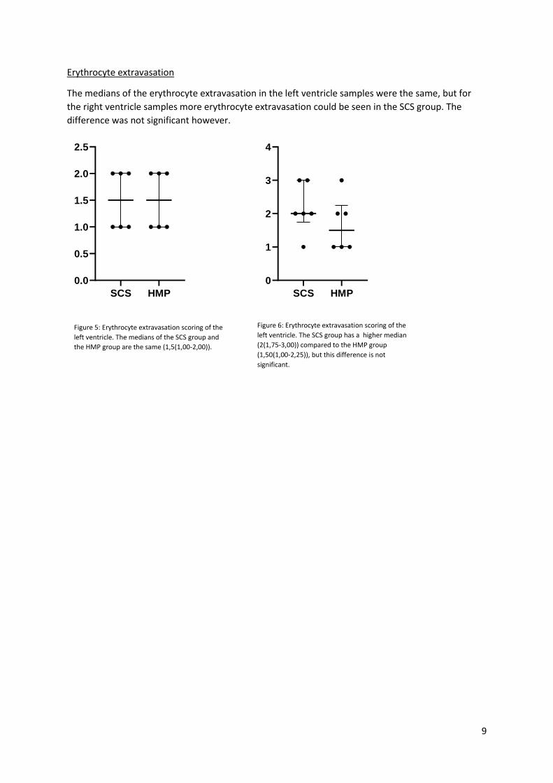
Erythrocyte extravasation
The medians of the erythrocyte extravasation in the left ventricle samples were the same, but for
the right ventricle samples more erythrocyte extravasation could be seen in the SCS group. The
difference was not significant however.
SCS HMP
0.0
0.5
1.0
1.5
2.0
2.5
Erythrocyte extravasation LV
Erythrocyte extravasation
SCS HMP
0
1
2
3
4
Erythrocyte extravasation RV
Erythrocyte extravasation
Figure 5: Erythrocyte extravasation scoring of the
left ventricle. The medians of the SCS group and
the HMP group are the same (1,5(1,00-2,00)).
Figure 6: Erythrocyte extravasation scoring of the
left ventricle. The SCS group has a higher median
(2(1,75-3,00)) compared to the HMP group
(1,50(1,00-2,25)), but this difference is not
significant.
10
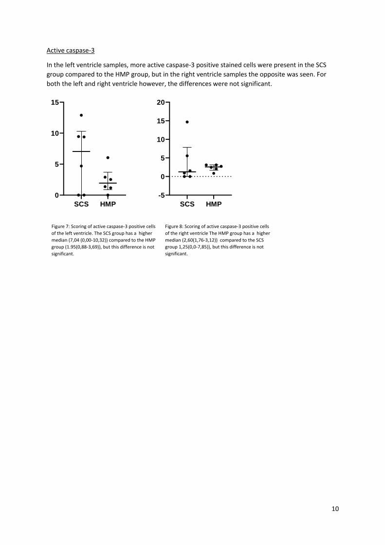
Active caspase-3
In the left ventricle samples, more active caspase-3 positive stained cells were present in the SCS
group compared to the HMP group, but in the right ventricle samples the opposite was seen. For
both the left and right ventricle however, the differences were not significant.
SCS HMP
0
5
10
15
Active Caspase-3 LV
Active Caspase-3
SCS HMP
-5
0
5
10
15
20
Active Caspase-3
Active Caspase-3
Figure 7: Scoring of active caspase-3 positive cells
of the left ventricle. The SCS group has a higher
median (7,04 (0,00-10,32)) compared to the HMP
group (1.95(0,88-3,69)), but this difference is not
significant.
Figure 8: Scoring of active caspase-3 positive cells
of the right ventricle The HMP group has a higher
median (2,60(1,76-3,12)) compared to the SCS
group 1,25(0,0-7,85)), but this difference is not
significant.
11
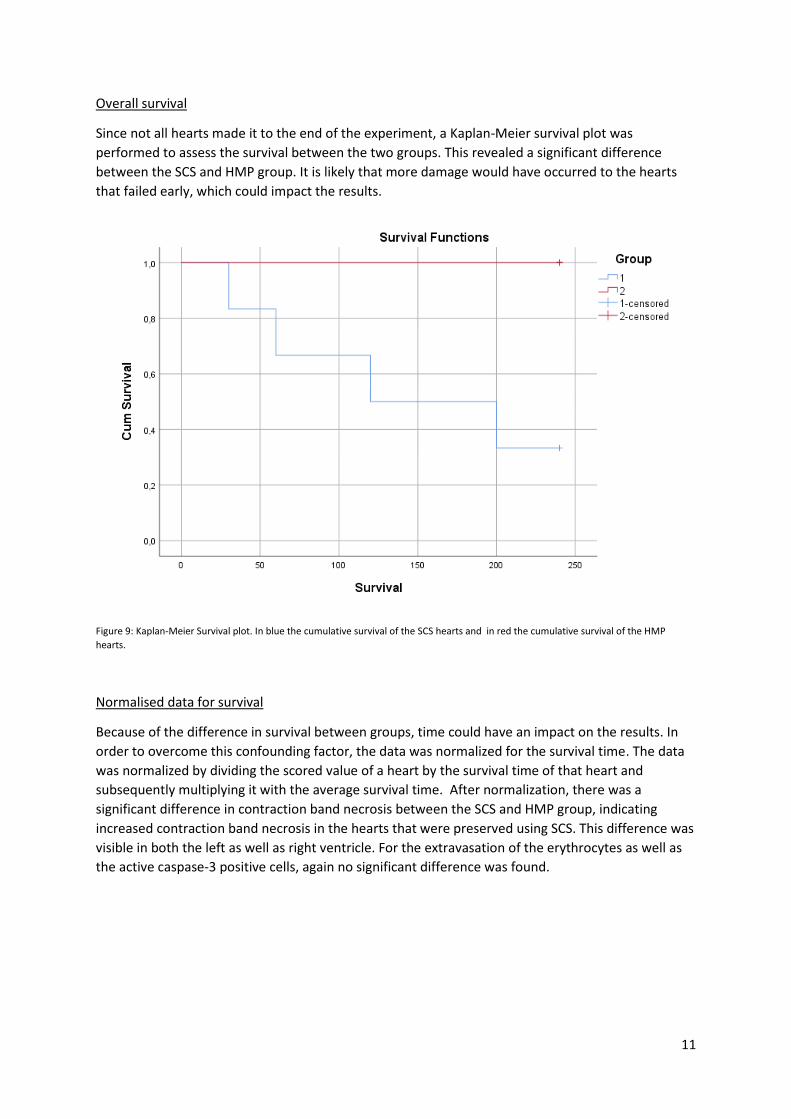
Overall survival
Since not all hearts made it to the end of the experiment, a Kaplan-Meier survival plot was
performed to assess the survival between the two groups. This revealed a significant difference
between the SCS and HMP group. It is likely that more damage would have occurred to the hearts
that failed early, which could impact the results.
Figure 9: Kaplan-Meier Survival plot. In blue the cumulative survival of the SCS hearts and in red the cumulative survival of the HMP
hearts.
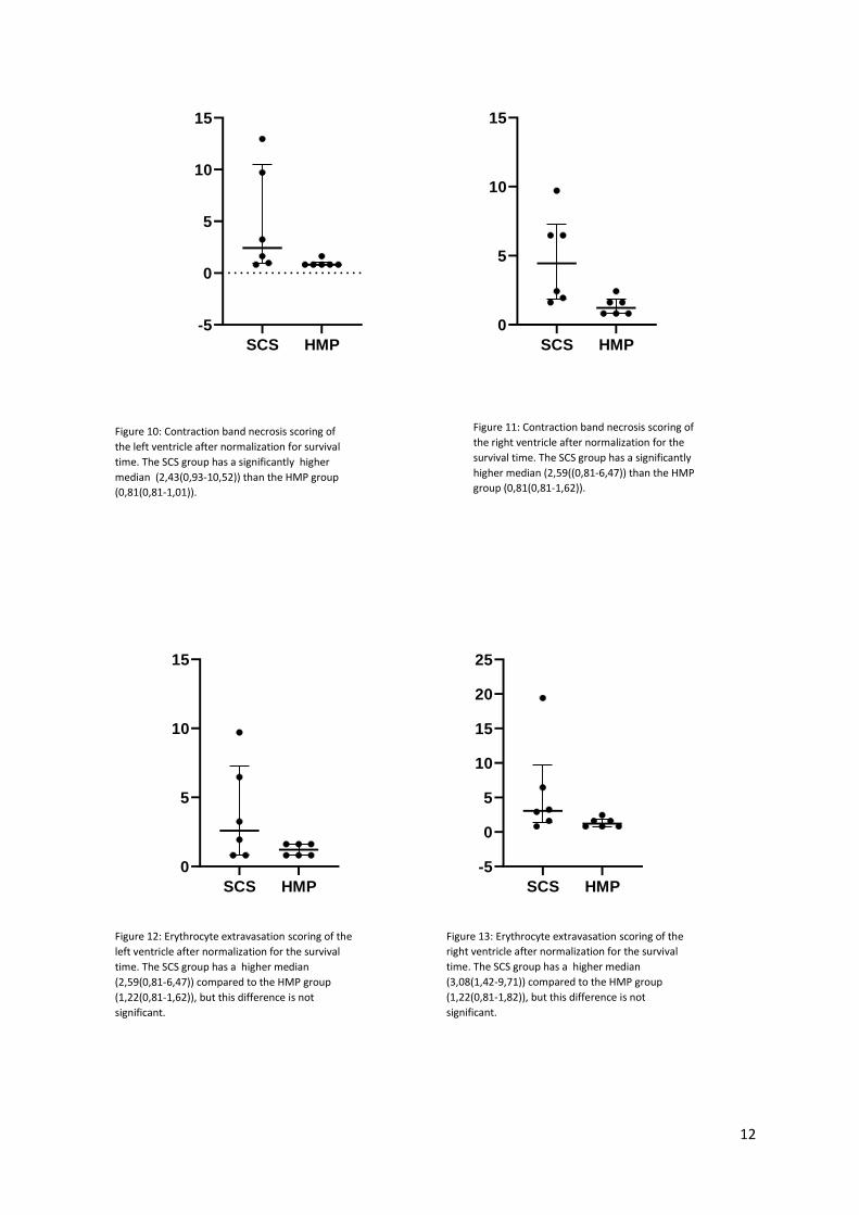
Normalised data for survival
Because of the difference in survival between groups, time could have an impact on the results. In
order to overcome this confounding factor, the data was normalized for the survival time. The data
was normalized by dividing the scored value of a heart by the survival time of that heart and
subsequently multiplying it with the average survival time. After normalization, there was a
significant difference in contraction band necrosis between the SCS and HMP group, indicating
increased contraction band necrosis in the hearts that were preserved using SCS. This difference was
visible in both the left as well as right ventricle. For the extravasation of the erythrocytes as well as
the active caspase-3 positive cells, again no significant difference was found.
12
SCS HMP
-5
0
5
10
15
Contraction band necrosis / Survival LV
Contraction band necrosis / Survival
SCS HMP
0
5
10
15
Contraction band necrosis / Survival RV
Contraction band necrosis / Survival
SCS HMP
0
5
10
15
Erythrocyte extravasation / Survival LV
Erythrocyte extravasation / Survival
SCS HMP
-5
0
5
10
15
20
25
Eryhtrocyte extravasation / Survival RV
Erythrocyte extravasation / Survival
Figure 10: Contraction band necrosis scoring of
the left ventricle after normalization for survival
time. The SCS group has a significantly higher
median (2,43(0,93-10,52)) than the HMP group
(0,81(0,81-1,01)).
Figure 11: Contraction band necrosis scoring of
the right ventricle after normalization for the
survival time. The SCS group has a significantly
higher median (2,59((0,81-6,47)) than the HMP
group (0,81(0,81-1,62)).
Figure 12: Erythrocyte extravasation scoring of the
left ventricle after normalization for the survival
time. The SCS group has a higher median
(2,59(0,81-6,47)) compared to the HMP group
(1,22(0,81-1,62)), but this difference is not
significant.
Figure 13: Erythrocyte extravasation scoring of the
right ventricle after normalization for the survival
time. The SCS group has a higher median
(3,08(1,42-9,71)) compared to the HMP group
(1,22(0,81-1,82)), but this difference is not
significant.
13
SCS HMP
-10
0
10
20
30
40
Active Caspase-3 / Survival LV
Active Caspase-3 / Survival
SCS HMP
-5
0
5
10
15
Active Caspase-3 / Survival RV
Active Caspase-3 / Survival
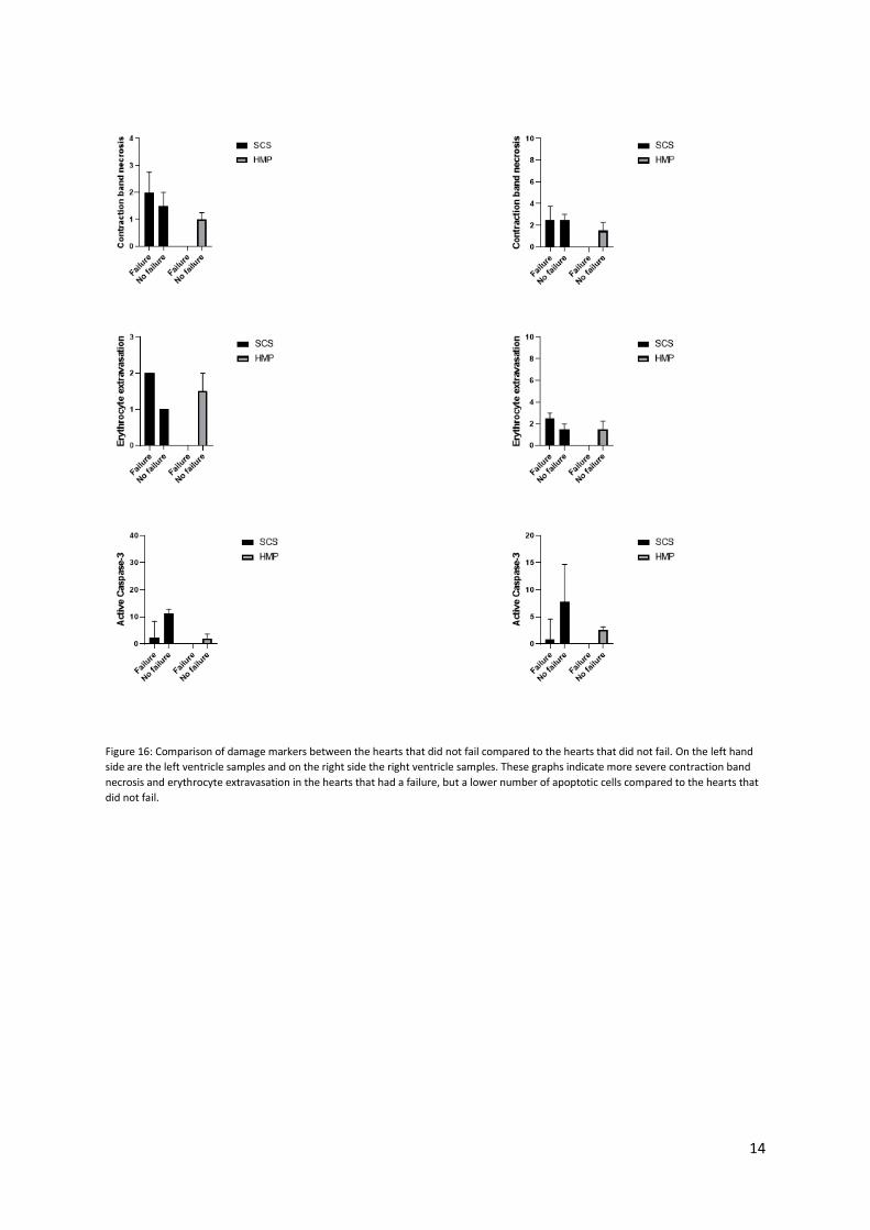
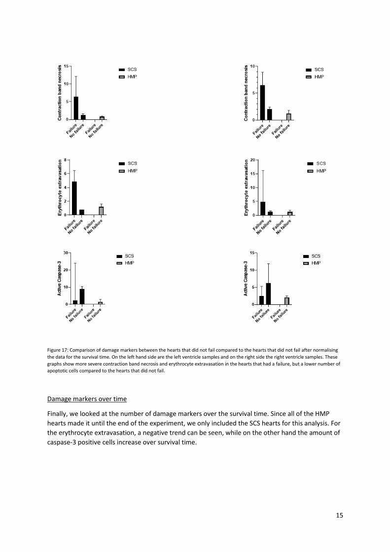
Damage markers between failing and non-failing hearts
We also compared the damage markers between the hearts that didn’t meet the inclusion criterium
of >3L CO until the end of the experiment and the hearts that did meet it. Figure 16 shows the
difference between the groups before normalization for survival time and figure 17 after
normalization for survival time. Both figures show that more severe contraction band necrosis and
erythrocyte extravasation can be seen in hearts that had a failure, but a lower number of apoptotic
cells compared to the hearts that did not fail.
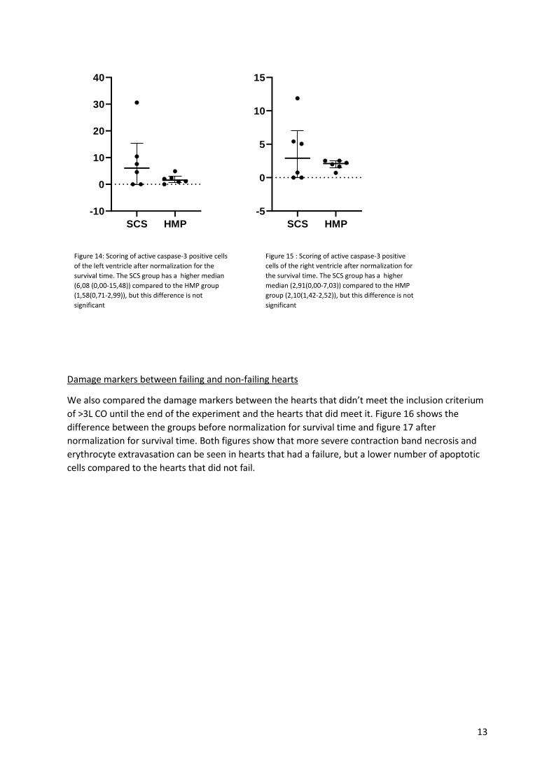
Figure 14: Scoring of active caspase-3 positive cells
of the left ventricle after normalization for the
survival time. The SCS group has a higher median
(6,08 (0,00-15,48)) compared to the HMP group
(1,58(0,71-2,99)), but this difference is not
significant
Figure 15 : Scoring of active caspase-3 positive
cells of the right ventricle after normalization for
the survival time. The SCS group has a higher
median (2,91(0,00-7,03)) compared to the HMP
group (2,10(1,42-2,52)), but this difference is not
significant
14
Figure 16: Comparison of damage markers between the hearts that did not fail compared to the hearts that did not fail. On the left hand
side are the left ventricle samples and on the right side the right ventricle samples. These graphs indicate more severe contraction band
necrosis and erythrocyte extravasation in the hearts that had a failure, but a lower number of apoptotic cells compared to the hearts that
did not fail.
15
Figure 17: Comparison of damage markers between the hearts that did not fail compared to the hearts that did not fail after normalising
the data for the survival time. On the left hand side are the left ventricle samples and on the right side the right ventricle samples. These
graphs show more severe contraction band necrosis and erythrocyte extravasation in the hearts that had a failure, but a lower number of
apoptotic cells compared to the hearts that did not fail.
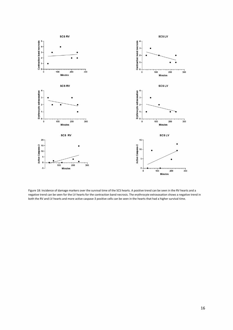
Damage markers over time
Finally, we looked at the number of damage markers over the survival time. Since all of the HMP
hearts made it until the end of the experiment, we only included the SCS hearts for this analysis. For
the erythrocyte extravasation, a negative trend can be seen, while on the other hand the amount of
caspase-3 positive cells increase over survival time.
16
Figure 18: Incidence of damage markers over the survival time of the SCS hearts. A positive trend can be seen in the RV hearts and a
negative trend can be seen for the LV hearts for the contraction band necrosis. The erythrocyte extravasation shows a negative trend in
both the RV and LV hearts and more active caspase-3 positive cells can be seen in the hearts that had a higher survival time.

17
Discussion This study was performed to assess the effects of SCS and HMP on cardiac function. While no
significant difference can be seen between the two storage methods on apoptosis and erythrocyte
extravasation, we did observe a significant difference in contraction band necrosis in both the left
and right ventricle after normalizing the data for the overall survival time of the hearts.
In this study apoptosis was scored using an anti-active caspase-3 staining. The primary advantage of
active caspase-3 staining is that once caspase-3 is cleaved, apoptosis is at a point of no return. This
greatly reduces the number of false positives. Moreover, this staining is known to work well with
formalin-fixed, paraffin-embedded tissues, and unlike TUNEL, which is heavily dependent on fixation
conditions and pre-treatment procedures, activate caspase-3 staining does not require special pre-
treatment procedures (11). Additionally, TUNEL stainings can provide false positives since it also
detects DNA damage by other causes than apoptosis. However, we were informed by the
pathologist that it is unlikely for apoptosis to occur in the timespan of our experiment, which is also
supported by literature (12). This could explain the low number of apoptotic cells counted.
Initially we opted to score the contraction band necrosis and erythrocyte extravasation using the
H&E staining, because the staining is relatively simple to perform and gives a comprehensive
overview of the tissue. These were however difficult to notice with this staining, due to a lack of
experience of the observers. Therefore, we decided to score contraction band necrosis and
erythrocyte extravasation using the PTAH staining. For the scoring of apoptosis, we corrected the
amount of positively stained cells for the size of the sample, because the samples had varying sizes,
which could have an impact on the results. A different way to correct the scoring would be to count
all the cells. Due to a lack of time, we were unable to perform this. Furthermore, we decided to
normalize the data because the exclusion criterium of >3.0L CO led to selection bias and this could
have impacted the results.
Future experiments should include a higher sample size to obtain more reliable results and more
stainings could be carried out to assess the difference in damage between hearts stored with SCS
and HMP. Possible stainings would first of all include Galectin-3 (GAL-3). GAL-3 is a beta galactoside
binding lectin and it has been shown in mice that GAL-3 levels are increased after IRI (13) Secondly, a
Masson’s Trichrome staining could be performed to assess the infarcted tissue(14). Finally, IRI has
been shown to cause the production of tumour necrosis factor-α (TNF- α) by the injured tissue (15).
A TNF-α staining could be performed to compare the amount of IRI between hearts preserved using
SCS and hearts preserved using HMP.
Conclusion
The aim of this study was to compare and assess the effects of SCS and HMP on cardiac function. On
apoptosis and erythrocyte extravasation we did not find any significant changes. However, HMP
appeared to cause less heart band necrosis. Necrosis causes inflammation in its surroundings and is
therefore regarded as highly damaging. In the case of cardiac tissue, this might lead to irreversible
damage. Therefore, based on this study we conclude that HMP is the preferred choice of heart
tissue preservation. Importantly, because this study has significant limitations as described above, it
is important that more research is done regarding this subject.
18
References (1) Groenewegen A, Rutten FH, Mosterd A, Hoes AW. Epidemiology of heart failure. European
journal of heart failure 2020 Aug;22(8):1342-1356. (2) Shah A, Gandhi D, Srivastava S, Shah KJ, Mansukhani R. Heart Failure: A Class Review of Pharmacotherapy. P&T (Lawrenceville, N.J.) 2017 Jul;42(7):464-472. (3) Portable Normothermic Cardiac Perfusion System in Donation After Cardiocirculatory Death: A Health Technology Assessment. Ontario health technology assessment series 2020;20(3):1-90. (4) Roest S, Kaffka Genaamd Dengler, S E, van Suylen V, van der Kaaij, N P, Damman K, van Laake LW, et al. Waiting list mortality and the potential of donation after circulatory death heart transplantations in the Netherlands. Netherlands heart journal 2021 Feb;29(2):88-97. (5) Hausenloy DJ, Yellon DM. Myocardial ischemia-reperfusion injury: a neglected therapeutic target. The Journal of clinical investigation 2013 Jan;123(1):92-100. (6) Neri M, Riezzo I, Pascale N, Pomara C, Turillazzi E. Ischemia/Reperfusion Injury following Acute Myocardial Infarction: A Critical Issue for Clinicians and Forensic Pathologists. Mediators of inflammation 2017 Feb 13,;2017:7018393-14. (7) Ou R, Lim YW, Choong JW, Esmore DS, Salamonsen RF, McLean C, et al. Low-Flow Hypothermic Crystalloid Perfusion Is Superior to Cold Storage During Prolonged Heart Preservation. Transplantation proceedings 2014;46(10):3309-3313. (8) B. Kappler. Process and quality control in use of isolated beating porcine slaughterhouse hearts; 2020. (9) Hannon, J.P. (Letterman Army Institute of Research, Presidio of San Francisco, CA), Bossone CA, Wade CE. Normal physiological values for conscious pigs used in biomedical research. Laboratory animal science (Chicago) 1990 May;40(3):293-298. (10) Steen S, Paskevicius A, Liao Q, Sjöberg T. Safe orthotopic transplantation of hearts harvested 24 hours after brain death and preserved for 24 hours. Scandinavian cardiovascular journal : SCJ 2016 May 3,;50(3):193-200. (11) Duan WR, Garner DS, Williams SD, Funckes-Shippy CL, Spath IS, Blomme EA. Comparison of immunohistochemistry for activated caspase-3 and cleaved cytokeratin 18 with the TUNEL method for quantification of apoptosis in histological sections of PC-3 subcutaneous xenografts. The Journal of pathology 2003 Feb;199(2):221-228. (12) Masters TN, Fokin AA, Schaper J, Pool L, Gong G, Robicsek F. Changes in the preserved heart that limit the length of preservation. The Journal of heart and lung transplantation 2002;21(5):590-599. (13) Al-Salam S, Hashmi S. Myocardial Ischemia Reperfusion Injury: Apoptotic, Inflammatory and Oxidative Stress Role of Galectin-3. Cellular physiology and biochemistry 2018 Oct;50(3):1123-1139. (14) Kim YS, Kim NY, Bae I, Park JK, Park DS, Shim JW, et al. Novel porcine model of acute myocardial infarction using polyethylene terephthalate. Journal of Biomedical Translational Research 2019 Jun;20(2):44-52. (15) Formigli L, Manneschi LI, Nediani C, Marcelli E, Fratini G, Zecchi Orlandini S, et al. Are macrophages involved in early myocardial reperfusion injury? The Annals of thoracic surgery 2001 May;71(5):1596-1602.


