Embed Size (px)
Citation preview
https://helda.helsinki.fi
Immunomodulating Therapies in Acute Myocarditis and
Recurrent/Acute Pericarditis
Ammirati, Enrico
2022-03-07
Ammirati , E , Bizzi , E , Veronese , G , Groh , M , Van de Heyning , C M , Lehtonen , J , de
Chambrun , M P , Cereda , A , Picchi , C , Trotta , L , Moslehi , J J & Brucato , A 2022 , '
Immunomodulating Therapies in Acute Myocarditis and Recurrent/Acute Pericarditis ' ,
Frontiers in Medicine , vol. 9 , 838564 . https://doi.org/10.3389/fmed.2022.838564
http://hdl.handle.net/10138/343335
https://doi.org/10.3389/fmed.2022.838564
cc_by
publishedVersion
Downloaded from Helda, University of Helsinki institutional repository.
This is an electronic reprint of the original article.
This reprint may differ from the original in pagination and typographic detail.
Please cite the original version.
SYSTEMATIC REVIEWpublished: 07 March 2022
doi: 10.3389/fmed.2022.838564
Frontiers in Medicine | www.frontiersin.org 1 March 2022 | Volume 9 | Article 838564
Edited by:
Silvia Piantoni,
ASST-Spedali Civili and University of
Brescia, Italy
Reviewed by:
Bernhard Maisch,
University of Marburg, Germany
Kazuko Tajiri,
University of Tsukuba, Japan
*Correspondence:
Enrico Ammirati
Antonio Brucato
Specialty section:
This article was submitted to
Rheumatology,
a section of the journal
Frontiers in Medicine
Received: 17 December 2021
Accepted: 28 January 2022
Published: 07 March 2022
Citation:
Ammirati E, Bizzi E, Veronese G,
Groh M, Van de Heyning CM,
Lehtonen J, Pineton de Chambrun M,
Cereda A, Picchi C, Trotta L,
Moslehi JJ and Brucato A (2022)
Immunomodulating Therapies in Acute
Myocarditis and Recurrent/Acute
Pericarditis. Front. Med. 9:838564.
doi: 10.3389/fmed.2022.838564
Immunomodulating Therapies inAcute Myocarditis andRecurrent/Acute PericarditisEnrico Ammirati 1*, Emanuele Bizzi 2, Giacomo Veronese 3, Matthieu Groh 4,5,
Caroline M. Van de Heyning 6, Jukka Lehtonen 7, Marc Pineton de Chambrun 8,9,10,
Alberto Cereda 11, Chiara Picchi 2, Lucia Trotta 2, Javid J. Moslehi 12 and
Antonio Brucato 2,13*
1De Gasperis Cardio Center and Transplant Center, Niguarda Hospital, Milano, Italy, 2 Internal Medicine, Fatebenefratelli
Hospital, Milano, Italy, 3Department of Health Sciences, University of Milano-Bicocca, Monza, Italy, 4National Reference
Center for Hypereosinophilic Syndromes, CEREO, Suresnes, France, 5Department of Internal Medicine, Hôpital Foch,
Suresnes, France, 6Department of Cardiology, Antwerp University Hospital, and GENCOR Research Group, Antwerp
University, Antwerp, Belgium, 7Department of Cardiology, Heart and Lung Center, Helsinki University Hospital, Helsinki,
Finland, 8 Sorbonne Université, Assistance Publique-Hôpitaux de Paris (APHP), Hôpital La Pitié-Salpêtrière, Service de
Médecine Intensive-Réanimation, Paris, France, 9 Sorbonne Université, APHP, Hôpital de la Pitié-Salpêtrière, Service de
Médecine Interne 2, Centre de Référence National Lupus et SAPL et Autres Maladies Auto-immunes et Systémiques Rares,
Paris, France, 10 Sorbonne Université, INSERM, UMRS_1166-ICAN, ICAN, Paris, France, 11Cardiovascular Department,
Association Socio Sanitary Territorial Santi Paolo e Carlo, Milano, Italy, 12 Section of Cardio-Oncology and Immunology,
Division of Cardiology and the Cardiovascular Research Institute, University of California, San Francisco, San Francisco, CA,
United States, 13Department of Biomedical and Clinical Sciences “Luigi Sacco,” Fatebenefratelli Hospital, University of
Milano, Milano, Italy
The field of inflammatory disease of the heart or “cardio-immunology” is rapidly
evolving due to the wider use of non-invasive diagnostic tools able to detect and
monitor myocardial inflammation. In acute myocarditis, recent data on the use of
immunomodulating therapies have been reported both in the setting of systemic
autoimmune disorders and in the setting of isolated forms, especially in patients with
specific histology (e.g., eosinophilic myocarditis) or with an arrhythmicburden. A role
for immunosuppressive therapies has been also shown in severe cases of coronavirus
disease 2019 (COVID-19), a condition that can be associated with cardiac injury
and acute myocarditis. Furthermore, ongoing clinical trials are assessing the role of
high dosage methylprednisolone in the context of acute myocarditis complicated by
heart failure or fulminant presentation or the role of anakinra to treat patients with
acute myocarditis excluding patients with hemodynamically unstable conditions. In
addition, the explosion of immune-mediated therapies in oncology has introduced new
pathophysiological entities, such as immune-checkpoint inhibitor-associated myocarditis
and new basic research models to understand the interaction between the cardiac
and immune systems. Here we provide a broad overview of evolving areas in
cardio-immunology. We summarize the use of new imaging tools in combination
with endomyocardial biopsy and laboratory parameters such as high sensitivity
troponin to monitor the response to immunomodulating therapies based on recent
evidence and clinical experience. Concerning pericarditis, the normal composition
of pericardial fluid has been recently elucidated, allowing to assess the actual
presence of inflammation; indeed, normal pericardial fluid is rich in nucleated cells,
protein, albumin, LDH, at levels consistent with inflammatory exudates in other
Ammirati et al. Immunomodulating Therapies in Myocarditis/Pericarditis
biological fluids. Importantly, recent findings showed how innate immunity plays a pivotal
role in the pathogenesis of recurrent pericarditis with raised C-reactive protein, with
inflammasome and IL-1 overproduction as drivers for systemic inflammatory response.
In the era of tailored medicine, anti-IL-1 agents such as anakinra and rilonacept have
been demonstrated highly effective in patients with recurrent pericarditis associated with
an inflammatory phenotype.
Keywords: acute myocarditis, pericarditis, immunosuppressive therapy, eosinophilic myocarditis, COVID-19,
cardiac sarcoidosis, corticosteroids, anti-IL-1 therapy
INTRODUCTION
The field of inflammatory disease of the heart or“cardio-Immunology” is rapidly evolving thanks to the wideruse of non-invasive diagnostic tools able to detect and monitormyocardial inflammation, such as cardiac magnetic resonanceimaging (CMRI) and fluorodeoxyglucose positron emissiontomography (FDG-PET) (1). In acute myocarditis (AM), recentdata on the use of immunomodulating therapies have beenreported both in the setting of systemic autoimmune disordersand in the setting of isolated forms, especially in patients withspecific histology (i.e., eosinophilic myocarditis, giant cellmyocarditis [GCM] or cardiac sarcoidosis [CS]) or characterizedby an arrhythmic burden (2). We elucidate the rationale totest the use of immunomodulating therapies in patients withlymphocytic AM. In addition, AM has also emerged as acomplication in the setting of coronavirus disease 2019 (COVID-19), mRNA vaccine (3–7), and immune checkpoint inhibitors(ICI) (8–10). Here, we summarize the clinical approach towardthe use of immunosuppressive therapies in these specific settings.Finally, we propose the use of new imaging tools in combinationwith endomyocardial biopsy (EMB) and laboratory parameterssuch as high sensitivity troponin to monitor the response toimmunomodulating therapies based on recent evidence andclinical experience.
In the second section of this review, we examine the rationaleand the evidence of immunosuppression in pericarditis. Wehighlight recent findings defining a pivotal role for innateimmunity in the pathogenesis of recurrent pericarditis withraised C-reactive protein (CRP), focusing on the emerging roleof anti-IL-1 agents (i.e., anakinra and rilonacept) for this subsetof patients with recurrent pericarditis.
LYMPHOCYTIC MYOCARDITIS
Lymphocytic AM is the most common histologic subset reportedin AM cohorts (11). Due to the fact that in the setting ofsuspected AM, histologic diagnosis is more often recommendedin specific scenarios (e.g., acute heart failure [HF], presence ofventricular arrhythmias (VA) or II/III-degree atrio-ventricularblock [AVB]) (1, 12), the prevalence of lymphocytic AM isfrequently estimated on cohorts of complicated AM. Froma recent international retrospective case collection of AMpresenting with left ventricular (LV) systolic dysfunction, theprevalence of lymphocytic AM has been estimated to be
∼72%, being the most frequently diagnosed form both infulminant myocarditis [FM], a clinical entity defined by theneed of circulatory support, and non-FM (11). The etiologyof lymphocytic AM is broad and includes heterogeneouspathogens, drugs or autoimmune-mediated injury in thesetting of systemic inflammatory diseases (10, 13, 14). Therole of viruses in myocarditis etiology has been historicallyrecognized, with Parvovirus (PV)B-19, adenoviruses, HumanHerpesvirus (HHV)-6, enteroviruses being the most commonagents identified in themyocardium of patients with AM (15, 16).Whether viruses have a direct or indirect causal relationship inclinical myocarditis etiology has been a matter of great debatethroughout the years with expert opinions varying according tothe evidence of the moment (17). The controversy matters as ithas been stated that the presence of specific viruses in the heartmay be a contraindication to the use of immunosuppression (18).A growing body of literature indicates that viruses, particularlyPVB-19 and HHV6, may be found in a large proportion ofpatients who do not have myocarditis, questioning their directcausal role in the pathogenesis of myocarditis (19, 20). Of note,PVB-19 was the only virus identified in patients with lymphocyticFM in an international registry (21). Except for enteroviruses(22, 23), such as coxsackievirus, whose ability to cause directmyocardial damage has been demonstrated and seems morecommon in newborns/infants (24), most of the available evidencesuggests that virus-triggered immune-mediated reactions arethe principal cause of cardiomyocyte injury (1). Respiratoryviruses, such as influenza and coronaviruses, are examples ofcommon viruses that can trigger immune-mediated lymphocyticmyocarditis with no evidence of viral genome in the myocardium(25, 26). Molecular mimicry between viral and cardiac antigensis suspected to be a key mechanism of myocardial injury invirus-triggered AM (27, 28). Furthermore, the concept thatFM may resemble the presentation of a high-grade cellularrejection observed after heart transplantation (HTx) is recentlyemerging. These findings may suggest that the identification ofviruses in the setting of AM may not represent an absolutecontraindication to immunosuppression (29). At present, the roleof a routine viral genome search on EMB in guiding patientmanagement and immunosuppression therapy in patients withAM remains unknown (17). This concept holds true especiallyin FM where early immunosuppression may be crucial todamper the inflammatory process sustaining AM. However,most studies focusing on immunomodulation have includedpatients with chronic inflammatory cardiomyopathy with HF
Frontiers in Medicine | www.frontiersin.org 2 March 2022 | Volume 9 | Article 838564
Ammirati et al. Immunomodulating Therapies in Myocarditis/Pericarditis
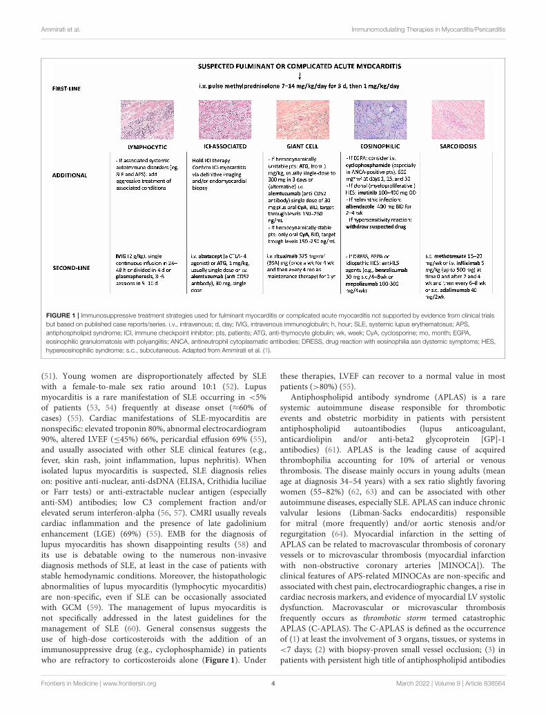
symptoms for more than 6 months rather than those with afulminant or complicated course (30–32). Though not supportedby evidence from randomized clinical trials, recommendationsfor immunosuppression exist in the setting of complicated AMbased on case series, expert opinions, and pathophysiologicalconsiderations (1) (Figure 1). The American Heart Association(AHA) suggests that, if a high suspicion for immune-mediatedFM exists, pulse steroid therapy (i.e., 1 g of methylprednisolone)should be administered urgently, before biopsy-confirmeddiagnosis or further diagnostic testing (33). Intravenous (IV)immunoglobulin (IG) (at a dose ranging from 0.5 g to 1 g/kg)is frequently used in pediatric lymphocytic myocarditis withevidence of some benefits in terms of functional recovery andsurvival, but the experience in adults has been limited (34, 35).Even though not standardized, maintenance therapy with lowdose steroids often in combination with mycophenolate mofetil,cyclosporine, azathioprine (AZA) as steroid-sparing drugs maybe used in those patients showing poor functional recoveryassociated with persistence of troponin release or any evidenceof residual myocardial inflammation (30, 36). StandardizedCorticosteroid therapy (IV methylprednisolone 200–400mg ordexamethasone 20–40mg) qd for 3–5 days and then graduallydown titrated and weaned in 7–10 days, and IVIG 10–20 g qdfor 3–5 days followed by 10 g for another 3–5 days has beendescribed from a Chinese registry of 138 FM and has beenassociated with improved survival (37). According to severalresearchers, even though robust evidence is substantially lackingin the setting of AM, high viral loads may contraindicatethe use of immunosuppression in favor of treatment withantiviral drugs or with agents boosting the native immuneresponse (e.g., interferon-β) (38). Lymphocytic AM can also beassociated with systemic autoimmune or inflammatory disorders(e.g., systemic lupus erythematosus [SLE], inflammatory boweldisorders, COVID-19) (39). The Lombardy registry of AMreported that 7.2% of patients had associated autoimmune orsystemic disorders, being more frequent in patients presentingwith complicated AM (40). The identification of the myocarditis-associated condition is essential to initiate disease-specifictreatments. IV corticosteroids have been successfully used incases of SARS-CoV-2 related FM, suggesting the relevance of thesystemic inflammatory response in determining cardiac injury inCOVID-19, even though more evidence is needed (41, 42).
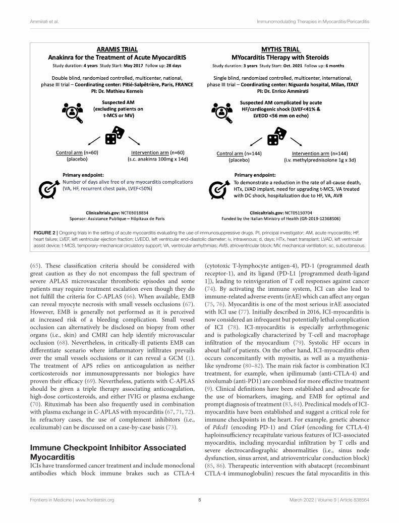
Ongoing TrialsAnakinra is the recombinant form of the naturally occurringinterleukin 1α (IL-1Rα) and blocks the activity of both IL-1α and IL-1β. The Anakinra vs. Placebo for the Treatment ofAcute Myocarditis (ARAMIS) trial (ClinicalTrials.gov identifier:NCT03018834) is a double-blind randomized clinical trial testingthe superiority of anakinra in addition to standard of care,defined as the maximum tolerated dosage of any beta-blockersand angiotensin receptor blockade in acute myocarditis. TheARAMIS trial has completed the randomization phase and willdirectly assess the role of the IL-1 immune innate pathwayin the setting of AM. The rationale of blocking the (IL-1β)pathway in myocarditis relies on prior studies that suggestedthe central role of the Nucleotide-binding domain (NACHT)
and Leucine-rich repeat (LRR) and Pyrin domain (PYD) (NLR)containing protein 3 (NLRP3) inflammasome predominatelyexpressed in macrophages (43–45). Despite anecdotal evidence,ARAMIS will directly test this concept and the results areexpected by the end of 2022 (46, 47). This double-blindedFrench study has assessed 120 patients with symptomatic AMdefined by elevated cardiac troponin (at least 1.5-fold upper thenormal reference limit) and CMRI consistent with myocarditisperformed within 72 h after admission (Figure 2). Patients in thetreatment arm received a daily subcutaneous dose of anakinra100mg during the hospitalization including an angiotensin-converting-enzyme inhibitor (ACE-i) and a beta-blocker. Theprimary endpoint of this study is the number of days alivefree of any myocarditis complications including (1) VA, (2)HF, (3) recurrent chest pain requiring medication, (4) leftventricular ejection fraction (LVEF) <50%, up to 28 days afterrandomization. This trial has also a sub-study that has assessedACE-i continuation or discontinuation after 1 month in patientswith normal LVEF that are followed for 1 year. This trialexcluded the patients with the poorest outcome, specifically thoseon mechanical ventilation or temporary mechanical circulatorysupports (t-MCS). To address specifically patients with FM oracute HF the MYocarditis THerapy with Steroids (MYTHS)trial (ClinicalTrials.gov identifier: NCT05150704) will randomize288 patients with FM (need for inotropes and/or t-MCS) orAM complicated by HF and severely impaired LVEF (<41%)to pulsed corticosteroid therapy (methylprednisolone 1 g IVqd for 3 days) on top of standard therapy and maximalsupportive care vs. placebo (Figure 2). The combined primaryendpoint is defined as the time from randomization to thefirst event occurring within 6 months including (1) all-causedeath, or (2) HTx, or (3) long-term left-ventricular assistancedevice (LVAD) implant, or (4) need for an upgrading of thet-MCS, or (5) a ventricular tachycardia (VT)/fibrillation (VF)treated with direct current (DC) shock (excluding VT/VF inpatients on t-MCS other than intra-aortic balloon pump [IABP]),or (6) first rehospitalization due to HF or VA, or advancedAVB. The trial started the enrollment in October 2021 andthe estimated duration is ∼3–4 years. The rationale for theMYTHS trial is based on clinical practice. Indeed, several caseseries and case reports support the effectiveness of high dosagecorticosteroids (48–50).
SPECIFIC SUBSET OF MYOCARDITIS
Myocarditis in Systemic LupusErythematosus and AntiphospholipidAntibody SyndromeSLE is a rare disease (prevalence 48–350 per 100,000 individuals)in which the immune system attacks healthy cells and tissues.Immune system activation is characterized by exaggeratedB/T cell responses and loss of tolerance against self-antigens.Production and defective elimination of antibodies, tissuedeposition of immune complexes, and complement and cytokineactivation contribute to clinical manifestations ranging fromjoint and skin inflammation to life-threatening organ damage
Frontiers in Medicine | www.frontiersin.org 3 March 2022 | Volume 9 | Article 838564
Ammirati et al. Immunomodulating Therapies in Myocarditis/Pericarditis
FIGURE 1 | Immunosuppressive treatment strategies used for fulminant myocarditis or complicated acute myocarditis not supported by evidence from clinical trials
but based on published case reports/series. i.v., intravenous; d, day; IVIG, intravenous immunoglobulin; h, hour; SLE, systemic lupus erythematosus; APS,
antiphospholipid syndrome; ICI, immune checkpoint inhibitor; pts, patients; ATG, anti-thymocyte globulin; wk, week; CyA, cyclosporine; mo, month; EGPA,
eosinophilic granulomatosis with polyangiitis; ANCA, antineutrophil cytoplasmatic antibodies; DRESS, drug reaction with eosinophilia asn dystemic symptoms; HES,
hypereosinophilic syndrome; s.c., subcutaneous. Adapted from Ammirati et al. (1).
(51). Young women are disproportionately affected by SLEwith a female-to-male sex ratio around 10:1 (52). Lupusmyocarditis is a rare manifestation of SLE occurring in <5%of patients (53, 54) frequently at disease onset (≈60% ofcases) (55). Cardiac manifestations of SLE-myocarditis arenonspecific: elevated troponin 80%, abnormal electrocardiogram90%, altered LVEF (≤45%) 66%, pericardial effusion 69% (55),and usually associated with other SLE clinical features (e.g.,fever, skin rash, joint inflammation, lupus nephritis). Whenisolated lupus myocarditis is suspected, SLE diagnosis relieson: positive anti-nuclear, anti-dsDNA (ELISA, Crithidia luciliaeor Farr tests) or anti-extractable nuclear antigen (especiallyanti-SM) antibodies; low C3 complement fraction and/orelevated serum interferon-alpha (56, 57). CMRI usually revealscardiac inflammation and the presence of late gadoliniumenhancement (LGE) (69%) (55). EMB for the diagnosis oflupus myocarditis has shown disappointing results (58) andits use is debatable owing to the numerous non-invasivediagnosis methods of SLE, at least in the case of patients withstable hemodynamic conditions. Moreover, the histopathologicabnormalities of lupus myocarditis (lymphocytic myocarditis)are non-specific, even if SLE can be occasionally associatedwith GCM (59). The management of lupus myocarditis isnot specifically addressed in the latest guidelines for themanagement of SLE (60). General consensus suggests theuse of high-dose corticosteroids with the addition of animmunosuppressive drug (e.g., cyclophosphamide) in patientswho are refractory to corticosteroids alone (Figure 1). Under
these therapies, LVEF can recover to a normal value in mostpatients (>80%) (55).
Antiphospholipid antibody syndrome (APLAS) is a raresystemic autoimmune disease responsible for thromboticevents and obstetric morbidity in patients with persistentantiphospholipid autoantibodies (lupus anticoagulant,anticardiolipin and/or anti-beta2 glycoprotein [GP]-1antibodies) (61). APLAS is the leading cause of acquiredthrombophilia accounting for 10% of arterial or venousthrombosis. The disease mainly occurs in young adults (meanage at diagnosis 34–54 years) with a sex ratio slightly favoringwomen (55–82%) (62, 63) and can be associated with otherautoimmune diseases, especially SLE. APLAS can induce chronicvalvular lesions (Libman-Sacks endocarditis) responsiblefor mitral (more frequently) and/or aortic stenosis and/orregurgitation (64). Myocardial infarction in the setting ofAPLAS can be related to macrovascular thrombosis of coronaryvessels or to microvascular thrombosis (myocardial infarctionwith non-obstructive coronary arteries [MINOCA]). Theclinical features of APS-related MINOCAs are non-specific andassociated with chest pain, electrocardiographic changes, a rise incardiac necrosis markers, and evidence of myocardial LV systolicdysfunction. Macrovascular or microvascular thrombosisfrequently occurs as thrombotic storm termed catastrophicAPLAS (C-APLAS). The C-APLAS is defined as the occurrenceof (1) at least the involvement of 3 organs, tissues, or systems in<7 days; (2) with biopsy-proven small vessel occlusion; (3) inpatients with persistent high title of antiphospholipid antibodies
Frontiers in Medicine | www.frontiersin.org 4 March 2022 | Volume 9 | Article 838564
Ammirati et al. Immunomodulating Therapies in Myocarditis/Pericarditis
FIGURE 2 | Ongoing trials in the setting of acute myocarditis evaluating the use of immunosuppressive drugs. PI, principal investigator; AM, acute myocarditis; HF,
heart failure; LVEF, left ventricular ejection fraction; LVEDD, left ventricular end-diastolic diameter; iv, intravenous; d, days; HTx, heart transplant; LVAD, left ventricular
assist device; t-MCS, temporary-mechanical circulatory support; VA, ventricular arrhythmias; AVB, atrioventricular block; MV, mechanical ventilation; sc, subcutaneous.
(65). These classification criteria should be considered withgreat caution as they do not encompass the full spectrum ofsevere APLAS microvascular thrombotic episodes and somepatients may require treatment escalation even though they donot fulfill the criteria for C-APLAS (66). When available, EMBcan reveal myocyte necrosis with small vessels occlusions (67).However, EMB is generally not performed as it is perceivedat increased risk of a bleeding complication. Small vesselocclusion can alternatively be disclosed on biopsy from otherorgans (i.e., skin) and CMRI can help identify microvascularocclusion (68). Nevertheless, in critically-ill patients EMB candifferentiate scenario where inflammatory infiltrates prevailsover the small vessels occlusions or it can reveal a GCM (1).The treatment of APS relies on anticoagulation as neithercorticosteroids nor immunosuppressants nor biologics haveproven their efficacy (69). Nevertheless, patients with C-APLASshould be given a triple therapy associating anticoagulation,high-dose corticosteroids, and either IVIG or plasma exchange(70). Rituximab has been also frequently used in combinationwith plasma exchange in C-APLAS with myocarditis (67, 71, 72).In refractory cases, the use of complement inhibitors (i.e.,eculizumab) can be discussed on a case-by-case basis (73).
Immune Checkpoint Inhibitor AssociatedMyocarditisICIs have transformed cancer treatment and include monoclonalantibodies which block immune brakes such as CTLA-4
(cytotoxic T-lymphocyte antigen-4), PD-1 (programmed deathreceptor-1), and its ligand (PD-L1 [programmed death-ligand1]), leading to reinvigoration of T cell responses against cancer(74). By activating the immune system, ICI can also lead toimmune-related adverse events (irAE) which can affect any organ(75, 76). Myocarditis is one of the most serious irAE associatedwith ICI use (77). Initially described in 2016, ICI-myocarditis isnow considered an infrequent but potentially lethal complicationof ICI (78). ICI-myocarditis is especially arrhythmogenicand is pathologically characterized by T-cell and macrophageinfiltration of the myocardium (79). Systolic HF occurs inabout half of patients. On the other hand, ICI-myocarditis oftenoccurs concomitantly with myositis, as well as a myasthenia-like syndrome (80–82). The main risk factor is combination ICItreatment, for example, when ipilimumab (anti-CTLA-4) andnivolumab (anti-PD1) are combined for more effective treatment(9). Clinical definitions have been established and advocate forthe use of biomarkers, imaging, and EMB for optimal andprompt diagnosis of treatment (83, 84). Preclinical models of ICI-myocarditis have been established and suggest a critical role forimmune checkpoints in the heart. For example, genetic absenceof Pdcd1 (encoding PD-1) and Ctla4 (encoding for CTLA-4)haploinsufficiency recapitulate various features of ICI-associatedmyocarditis, including myocardial infiltration by T cells andsevere electrocardiographic abnormalities (i.e., sinus nodedysfunction, sinus arrest, and atrioventricular conduction block)(85, 86). Therapeutic intervention with abatacept (recombinantCTLA-4 immunoglobulin) rescues the fatal myocarditis in this
Frontiers in Medicine | www.frontiersin.org 5 March 2022 | Volume 9 | Article 838564
Ammirati et al. Immunomodulating Therapies in Myocarditis/Pericarditis
mouse model, providing mechanistic support for inhibition ofT cell co-stimulation mediated by CTLA-4 as a treatment forICI-associated myocarditis. Anecdotal evidence supports theuse of abatacept in severe cases of ICI-myocarditis (87). ICI-induced myocarditis affects elder patients (median age of 65years) with more comorbidities compared with non-ICI-inducedmyocarditis (median age between 30 and 40 years) (40, 88–90). One of the largest case series of 122 patients with ICI-associated myocarditis had early onset of symptoms (median30 days after initial exposure to ICI), and up to 50% of deaths(9). A systematic analysis of the World Health Organizationpharmacovigilance database confirmed a 32.5% of mortality inpatients who had myocarditis associated with the administrationof ICIs with a median time-to-onset of 33 days (10). Theincreased reports of cases in the last years are perhaps consistentwith growing recognition of this new clinical syndrome, as wellas the more widespread use of ICIs. High-dose IV corticosteroidsand withdrawal of ICI are considered the first-line therapy (1, 91,92), while alemtuzumab (anti-CD52 antibody), antithymocyteglobulin (anti-CD3 antibody), and abatacept (a CTLA-4 agonist)have been proposed in corticosteroid-resistant forms (Figure 1)(87, 93, 94). Retrospective data suggest that earlier (within thefirst 24 h) and high doses (501–1,000 mg/day) of corticosteroidslead to an improved outcome (95). Prompt diagnosis andimmediate treatment of ICI-myocarditis becomes a critical issueamong the cardio-oncology population, as indications for ICIincrease for various cancer types. In 2021, nearly 50% of cancerpatients are eligible for ICI treatment. In many cases, ICIs arecombined with other cancer therapies with their own inherentcardiotoxicities (96–98). In addition, long-term cardiovasculareffects of ICI become an important consideration as a growingnumber of cancer patients respond to therapy (99–101). Finally,the emergence of ICI-myocarditis has opened new avenuesfor more fundamental investigation about the role of immunecheckpoints (e.g., PD-1/PD-L1 signaling) in other forms ofinflammatory heart disease (102, 103). These issues need to bea focus of future investigations.
Ventricular Arrhythmias and MyocarditisAM can be complicated by VA. Specifically, ∼40% of patientspresenting with life-threatening VA can experience a recurrenceat a median time of 8 months based on a recent internationalregistry including 156 patients (104). Factors associated witharrhythmic recurrence were initial presentation with sustainedVT, LGE involving ≥2 myocardial segments, and absenceof T2-weighted short-tau inversion recovery (STIR) signalsuggestive for residual edema on CMRI (104). In this registry, 98patients underwent EMB showing in the large majority of casesa lymphocytic myocarditis (88.8%). An immunosuppressivetherapy was initiated in 21% of cases and there was nodifference in the use of immunosuppressive therapy betweenpatients who subsequently experience an arrhythmic recurrencevs. those who did not (104). A second registry of 185 patientswith VA (including VF/VT, non-sustained VT, and Lown’s≥2 premature ventricular complexes [PVC]) and myocarditisconfirmed a 30% of recurrence of malignant VA at 2 years(105). Another study evaluated 58 patients with histologically
proven lymphocytic myocarditis and VA as above describedwho underwent immunosuppressive therapy vs. a matchedpopulation of 58 cases not treated with immunosuppressiveagents (2). Immunosuppressive therapy in most patients wasa combination of prednisone 1 mg/kg for 6 month and AZA2 mg/kg for 1 year. Alternatively, mycophenolate mofetil atdosage of 1–3 g/day was used instead of AZA. At 24-monthfollow-up, no significant differences in VF/VT occurrence wereobserved in patients treated with immunosuppressive agentsvs. those who did not (10 vs. 17%, respectively, p = 0.42),even if patients who were treated with immunosuppressiveagents showed a significant reduction in the PVC burden(2). Another prospective registry included 107 symptomaticpatients with >5,000 PVCs/24 h without ischemic etiology whounderwent a combination of laboratory testing, FDG-PET scan,CMRI and EMB (106). A positive FDG-PET scan consistentwith cardiac inflammation was observed in up to 51% ofpatients and CS was the final diagnosis in 24% of patientswith positive FDG-PET scan. Patients with signs consistent withmyocarditis started an immunosuppressive therapy (prednisone40mg for 3 months) alone or in combination with catheterablation, showing an optimal response in 67% of cases. Optimalresponse was defined as a reduction in the PVC burden>80% and negative FDG-PET scan at follow up. Furthermore,patients with LV systolic dysfunction showed an improvementin 37% of cases with a mean increase in LVEF of 13%(106). Although these studies are promising, the lack ofrandomization vs. a control group, the absence of reports ofside effects and the fact that the immunosuppression therapydid not significantly reduce VF/VT or cardiovascular deathcannot routinely support the use of corticosteroids in themanagement of patients with myocarditis complicated by VAor frequent PVC. Specific randomized trials are required toassess whether immunosuppression can ameliorate myocardialinflammation and reduce the risk of major VA. In addition, VAis especially a hallmark of ICI-myocarditis. In an internationalregistry of patients with ICI-myocarditis, consisting of 147patients, a total of 22 (15.0%) patients experienced 1 ormore life-threatening ventricular arrhythmia episodes, including16/147 (10.9%) sustained ventricular tachycardia, 4/147 (2.7%)ventricular fibrillation, and 2/147 (1.4%) torsade de pointes (107).
COVID-19 Associated Acute MyocarditisCardiac injury with release of troponin has been observed quiteoften in patients who were hospitalized with COVID-19 (108),nevertheless cases of well-characterized AM are anecdotal (3).Data on clinically suspected AM complicated by acute HFamong hospitalized patients with COVID-19 suggests a 0.12%incidence (109). Nevertheless, good data on the incidence ofAM are still lacking. It has been recognized that asymptomaticforms of AM associated with severe acute respiratory syndromecoronavirus 2 (SARS-CoV-2) exposure range between 0.3 and 3%based on a CMRI diagnosis. This population has been largelystudied among athletes who underwent systematic cardiac tests(ECG, troponin assessment, or transthoracic echocardiography)and, when clinically indicated, CMRI (110–112). It mustbe acknowledged that proportionally, individuals with mild
Frontiers in Medicine | www.frontiersin.org 6 March 2022 | Volume 9 | Article 838564
Ammirati et al. Immunomodulating Therapies in Myocarditis/Pericarditis
COVID-19 related symptoms have a higher likelihood ofsigns of myocardial inflammation compared with asymptomaticindividuals. Patients with cardiac tests consistent with AMshould be advised not to practice vigorous physical activitiesin the 3–6 months following SARS-CoV-2 exposure if theyhave preserved LVEF, whereas if patients have reduced LVEF,patients should initiate specific HF therapies (113), while thereis no indication for immunosuppression. Patients complainingof cardiac symptoms or signs associated with COVID-19 anddiagnostic findings consistent with AM can be further dividedbetween those with COVID-19 associated AM with concurrentpneumonia and those without pneumonia (isolated COVID-19myocarditis). Delayed-onset AM has been described after SARS-CoV-2 exposure and typically these patients can present withhigh titer of SARS-CoV-2-specific antibodies and recent historyconsistent with COVID-19 in the absence of SARS-CoV-2 byRT-PCR on a nasopharyngeal swab. Delayed-onset myocarditisis thought to be triggered by SARS-CoV-2 induced immune-mediated reactions. Immunomodulating therapies include non-steroidal anti-inflammatory drugs (NSAIDs) to relieve chestpain, low dosage of colchicine in case of associated pericardialinvolvement. Corticosteroids are generally used in patients withdelayed onset AM that present an associated hyperinflammatorystatus (114, 115). In severe COVID-19 AM presenting as FM,EMB can be deemed necessary with the aim to differentiateAM from sepsis-induced acute cardiomyopathy, especiallyin patients with hyperinflammatory status. Identification ofinflammatory infiltrates in the myocardium could supportthe empirical use of immunosuppressive drugs (33), even if,diffuse inflammatory infiltrates have been rarely seen (116).Hyperinflammatory status and acute HF/cardiogenic shock inwhich a predominant septic state has been excluded couldbe treated with immunosuppressive treatments, as suggestedby small series where intravenous corticosteroids have beenassociated with a favorable prognosis (114, 115). This conditionhas been termed multisystem inflammatory syndrome in adults(MIS-A) and is often associated with a delayed onset ofmyocarditis. The condition is usually associated with highlevels of inflammatory biomarkers and ferritin (117). TheMultisystem inflammatory syndrome in children (MIS-C)presents overlapping characteristics with myocarditis in adults(118). It has been that although a third of patients with MIS-Ccan require a t-MCS, but none died in a series of 35 childrenwho were treated with IVIG plus a third with the addition ofcorticosteroids (119). Finally, patients with concurrent severemyocarditis, pneumonia, and respiratory insufficiency shouldreceive corticosteroids (120). A review article that collected dataon 38 published cases of COVID-19 associated AM reporteduse of corticosteroids in 34% of cases and a mortality of 15%(121), even if larger series are needed to better understandoptimal therapies.
mRNA COVID19 Vaccine-Related AcuteMyocarditisThe association between vaccine administration and the onsetof myocarditis is supported by several case reports, case series,
and at the level of the national health care system (4–7, 122–124). The United States Vaccine Adverse Event Reporting System(VAERS), even if subject to bias, also revealed a clear signal forvaccine-associated myocarditis with nearly 1,300 cases reportedfrom more than 350 million doses in the United States (8).Most cases have been reported in young men, thus, for 18–24-year-old males, the expected prevalence of vaccine-associatedmyocarditis is ∼3 cases per 100,000 doses (0.003%) based onVAERS data (8). Nationwide observational data confirmed aCOVID-19 vaccine-associated myocarditis at ∼3 per 100,000patients (0.003%) vs. ∼11 per 100,000 patients (0.01%) for acuteCOVID-19 myocarditis (125). An analysis conducted in Englandrevealed that the increased risk of myocarditis associated with thetwo mRNA vaccines was present only in those younger than 40years (6).
Historically, the vaccine that is most associated withmyocarditis is the anti-smallpox (10, 126). Smallpox vaccine wasassociated with eosinophilic myocarditis, while almost all thepresent cases of mRNA COVID-19 vaccine are not associatedwith eosinophilia. We revised 90 cases published of mRNACOVID-19 vaccinemyocarditis up to the end of August 2021 (seeSupplementary Tables 1, 2), and we summarized major features,and anti-inflammatory and immunomodulatory drugs used. Themedian age at presentation was 25 years (interquartile range17–27), in agreement with a median age observed in VAERS (8),with a marked male prevalence (93%). Even if a higher numberof BNT162b2-related myocarditis is reported, disproportionalityanalyses using the Bayesian information component, revealeda higher likelihood of association between mRNA 1273 andmyocarditis (126). In 90% of cases, myocarditis occurs after thesecond dose, following a median time of 3 days between thelast dose and symptoms’ onset, including chest pain (observedin 96% of cases) generally preceded by fever (in 85%). Allthese findings suggest an immune-mediated reaction relatedto vaccine administration. AM is generally not severe. Whileelectrocardiographic abnormalities are present in 77% of cases,diagnostic tools revealed only a slight reduction in the LVEF(mean value of 53%) with a pericardial effusion observed in 14%of cases. Information on anti-inflammatory/immunomodulatorytherapy was available for 56 of 90 patients (62.2%). In 38 outof 56 patients, the administered drugs were reported as follows:aspirin, NSAIDs, corticosteroids, IVIG, colchicine, and anakinra.Patients who received anti-inflammatory/immunomodulatorytherapy did not differ in relation with age (23 ± 9 vs. 29 ± 19years, p-value 0.10) and LVEF on the first echocardiogram (53 ±11 vs. 53 ± 13%, p-value 0.90). The use of immunosuppressivetherapy was similar in the adult and pediatric populations (39.5vs. 44.4%, p-value 0.72). Overall, NSAIDs (including aspirin)were the most used drugs (23/56 patients, 41.1%), and aspirinwas used only in 3 out of 56 patients (5.4%). Corticosteroidswere used in 19 of 56 patients (33.9%), IVIG in 12 patients(21.4%), colchicine in 15 patients (26.8%), and anakinra inonly 2 patients. Most of the time, immunosuppressive agentswere used in combination. NSAIDs were used together with thecorticosteroids in 5 patients. IVIG along with corticosteroids wasused in 11 patients, including 10 pediatric patients. NSAIDs alongwith colchicine were used in 11 of 56 patients (19.6%). Prognosis
Frontiers in Medicine | www.frontiersin.org 7 March 2022 | Volume 9 | Article 838564
Ammirati et al. Immunomodulating Therapies in Myocarditis/Pericarditis
is considered favorable, with only three (3.3%) deaths reportedout of 90 patients, a figure in line with the one observed inAM patients in pre-COVID19 era (40). These data are largelyconsistent with a series of 139 adolescents (all with age<21 years)with suspected AM within 30 days of COVID-19 vaccination (7).In fact, the male prevalence was 91%, symptoms started a medianof 2 days after vaccination, and the most common symptom waschest pain (99%) (7). Again, NSAIDs were the most used drugs in81% of cases, followed by corticosteroids (22%) and IVIG (22%),while colchicine was administered in 8% (7). No patient died orrequired a t-MCS.
EOSINOPHILIC MYOCARDITIS
Eosinophils have widespread procoagulant effects, including theproduction of tissue factor (127), oxidation of phospholipids(128) (both of which activate the intrinsic pathway), therelease of platelet-activating factor (129), reactive oxygenspecies, and eosinophil extracellular traps (130). Moreover,activated eosinophils are potent producers of vasospasticmediators (including histamine, leukotrienes C4 and D4 andprostaglandin D2) and are able to modulate mast cell functions(131). Lastly, the shedding of both cytotoxic granules and pro-inflammatory mediators (i.e., tumor necrosis factor [TNF]-α,IL-1 and IL-6) are contributing factors of endothelial injuryand procoagulant state (132). Eosinophil-mediated toxicitycan lead to protean cardiovascular manifestations, includingvenous thromboembolism (133), eosinophilia-related coronaryvasospasm (134), thromboangiitis obliterans-like disease (135),eosinophilic coronaritis, systemic eosinophilic vasculitis (136),eosinophilic myocarditis (137), and Loeffler cardiomyopathy,a chronic inflammatory cardiomyopathy (1, 138). The naturalhistory of eosinophil-related heart involvement involvesthree successive (and potentially overlapping) phases: (1)AM, due to eosinophilic infiltration of the endocardium,that can be either asymptomatic or lead to acute HF or FM(137). High troponin levels, LV systolic dysfunction, andsubendocardial LGE pattern on CMRI can be observed (2, 137) athrombotic stage characterized by the occurrence of ventricularthrombi and the risk of systemic embolism; (3) a fibrotic stage,characterized by endomyocardial fibro-thrombosis that can leadto restrictive cardiomyopathy (i.e., Loeffler cardiomyopathy)and/or atrioventricular valvular disease (139). The diagnosisof eosinophilic myocarditis is usually straightforward in thepresence of hypereosinophilia, increased cardiac troponin, andCMRI consistent with subendocardial inflammation (137). EMBcan be considered when the initial presentation is characterizedby cardiogenic shock (1, 33), or CMRI findings are atypical(i.e., subepicardial LGE) or when absolute eosinophil countsare within the normal range (which has been reported in up to25% of patients with biopsy-proven eosinophilic myocarditis)(137, 139). Conversely, EMB is at risk of thromboembolism ifventricular thrombi are present, and can yield false-negativefindings when endomyocardial fibrosis is prominent andeosinophil infiltration has partially or completely vanished(138). Eosinophil-related heart involvement can be encounteredwithin the full spectrum of eosinophil-associated diseases (137),including drug hypersensitivity (even in the absence of skin
manifestations) (10), parasitic infections (namely toxocariasis,trichinosis, filarial infections or sarcocystosis), aspirin-exacerbated respiratory disease, eosinophilic granulomatosiswith polyangiitis (EGPA, formerly Churg-Strauss syndrome),hypereosinophilic syndromes (HES) (mainly idiopathic andFIP1L1-PDGFRA-associated HES, formerly chronic eosinophilicleukemia) and high-grade hematological malignancies [e.g.,Hodgkin and angioimmunoblastic T-cell lymphomas, as well asB-cell acute lymphoblastic lymphoma with t (5, 14) (q31;q32);IGH-IL3 rearrangement (140). In a review of 179 cases ofbiopsy-proven eosinophilic myocarditis, the main identifiedcauses were drug hypersensitivity, EPGA, HES and parasiticinfection, accounting for 34%, 13% and 8% of cases, respectively,while 36% of cases were idiopathic or eosinophilic myocarditiswith undefined cause (137).
Heart involvement is the leading cause of death inpatients with EGPA and is more frequent in antineutrophilcytoplasmic antibodies (ANCA)-negative patients (141). Ofnote, the differential diagnosis between ANCA-negative EGPAand HES is a frequent diagnostic and therapeutic dilemma.The European Respiratory Society and European Federationof Internal Medicine-endorsed Task Force suggested restrictionin the use of EGPA to patients with eosinophilic asthma whotest positive for ANCA and/or who exhibit genuine featuresof vasculitis (either biopsy-proven or clinical surrogates) (142).Likewise, in a retrospective analysis of 166 patients withblood eosinophilia >1,000/mm3 and systemic manifestations,it was recently suggested that serum CRP levels could be areliable biomarker able to distinguish EGPA from idiopathicHES, with low (i.e., < 36 mg/L) levels being suggestive ofidiopathic HES rather than EGPA (143). A workup to identifyassociated systemic disorders should be performed in all patientswith eosinophilic myocarditis. The workup should includetesting for ANCA (positive in 10–40% of EGPA patients),serological testing for toxocariasis (which has a broad geographicdistribution), ova and parasite tests (while further serologiesfor parasitic infections are generally guided by the patient’scountry of origin, travel history and dietary habits), serumvitamin B12 and tryptase levels (which are sensitive for thediagnosis of myeloid variant HES), total IgE levels (whichare suggestive of reactive polyclonal eosinophilia mediatedby IL-5, when elevated), lactate dehydrogenase (suggestiveof lymphoma), thoraco-abdominopelvic CT scan (seeking forextra-cardiac eosinophil-related organ involvements as well asunderlying solid or hematological malignancies). Furthermore,brain CT or brain MRI should be performed when embolicstroke is suspected in patients with eosinophilic myocarditis orLoeffler cardiomyopathy (144). Additionally, testing for FIP1L1-PDGFRA fusion gene should be performed in selected cases whenclinical (e.g., male sex, splenomegaly), biologic (e.g., high B12vitamin and/or tryptase levels) features and/or primary resistanceto steroids are observed (145). Polymerase chain reaction testingfor specific viruses (e.g., Herpesviridae, especially HHV 6)and the RegiSCAR scoring system can be useful in patientswith suspected Drug Reaction with Eosinophilia and SystemicSymptoms (DRESS) (146). Additional imaging, endoscopic andhistologic investigations are usually performed on a case-by-case basis after first-line investigations. In a retrospective
Frontiers in Medicine | www.frontiersin.org 8 March 2022 | Volume 9 | Article 838564
Ammirati et al. Immunomodulating Therapies in Myocarditis/Pericarditis
series of 19 patients with biopsy-proven myocarditis withfulminant presentation, the rate of either cardiac death or hearttransplantation at 60 days was up to 26% (11).
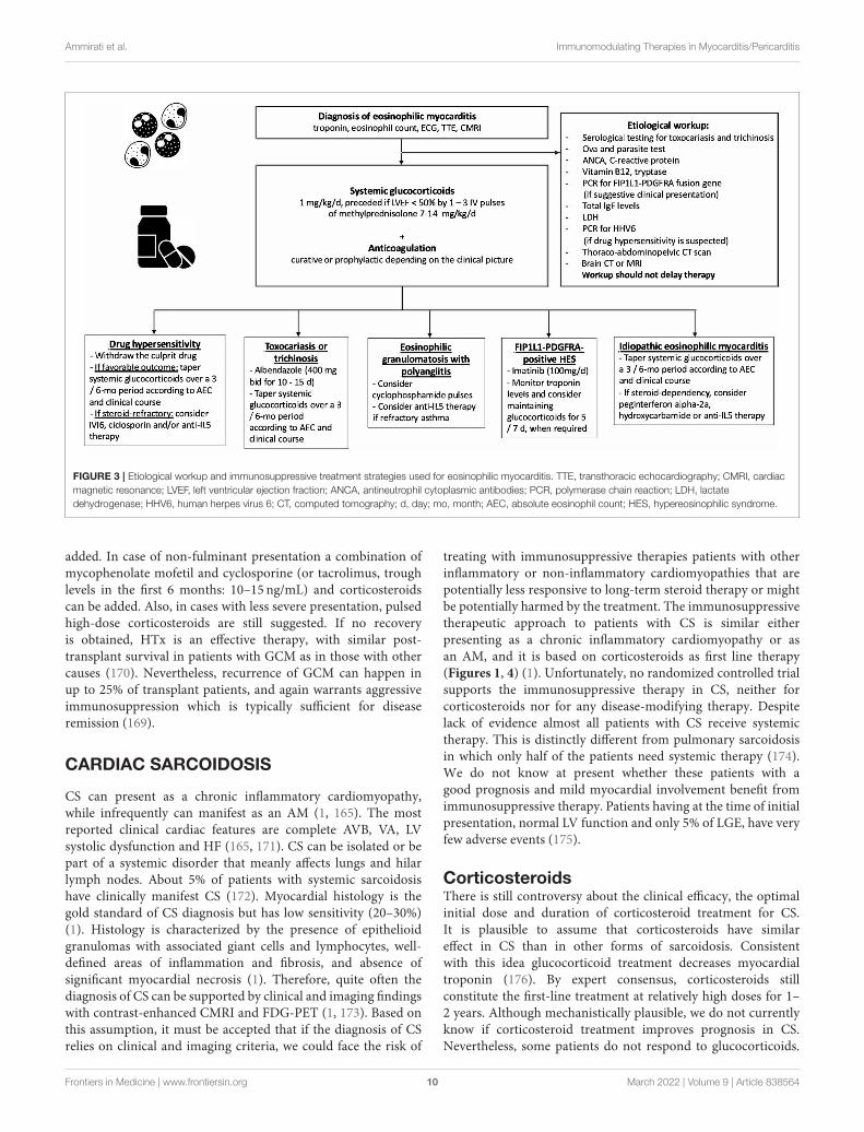
The cornerstone of the treatment relies on systemicglucocorticoids, starting dose: 1 mg/kg qd, preceded in case ofsevere LV systolic dysfunction by intravenous pulses of 7.5–15 mg/kg of methylprednisolone for 1–3 days (Figures 1, 3)(137, 139). In patients at risk of strongyloidiasis (owingto their past travel history), concomitant prescription of asingle dose of ivermectin (200 µg/kg) is warranted to preventStrongyloides stercoralis hyperinfection. When toxocariasis ortrichinosis are evidenced, a 10/15-day course of albendazole(400mg bid) is warranted (147). Likewise, in patients withevidence of intracavitary thrombus, anticoagulation should beinitiated (while prophylactic anticoagulation is mandatory inall other patients until absolute eosinophil counts normalize).The diagnoses of myeloid variant HES, DRESS or EGPAshould be suspected and investigated accordingly, after 2–4 days of corticosteroid-refractory eosinophilia. Specifically,the treatment of FIP1L1-PDGFRA-positive HES relies on thetyrosine kinase inhibitor imatinib (100 mg/d), and eosinophilsgenerally plummet within days after imatinib initiation (145).Yet, transient worsening of HF after onset of imatinib has beenreported, likely due to treatment-induced lysis of eosinophils(148). Conversely, IVIG and/or cyclosporine are the mostcommon drugs used for the treatment of corticosteroid-refractory DRESS (149, 150), yet benralizumab (a humanizedafucosylated monoclonal antibody that targets IL-5 receptorα) is on the rise in this setting (151). Historically, besidessystemic corticosteroids, the treatment of EGPA-associatedeosinophilic myocarditis complicated by severe HF relies oncyclophosphamide pulses (152, 153), yet it should be emphasizedthat there is no data proving that adding cyclophosphamidepulses to steroids improves outcomes. Whatever the underlyingdisorder, the aim is to quickly and persistently normalizeeosinophil count (< 500/mm3). Of note, both in EGPA (154–156) and in FIP1L1-PDGFRA-negative HES (157, 158) targetingIL-5 has emerged as clinically relevant. Anti-IL-5 agents, suchas mepolizumab and benralizumab are likely to become gamechangers and tend to replace the use of disease-modifyinganti-rheumatic drugs (i.e., AZA, methotrexate, peginterferonalpha-2a and hydroxycarbamide), even if trials are needed. Incase of persistent eosinophilia and subsequent occurrence ofendomyocardial fibrosis, heart surgery with resection of fibroticendocardium (endomyocardectomy) combined with valve repairor replacement can be considered (159). Finally, in case ofrefractory end-stage HF, orthotopic heart transplantation hasbeen reported to be safe and feasible in both EGPA and HES(160, 161).
GIANT CELL MYOCARDITIS
GCM is a rare but often fatal form of AM. The pathophysiologyof GCM is thought to be a T-cell mediated autoimmune processleading to diffuse or multifocal inflammatory infiltrate, includinglymphocytes with multinucleated giant cells, and definitive
diagnosis requires EMB. An immune-mediated mechanismin the etiology of GCM is further supported by the factthat no nucleic acids from viruses implicated in myocarditiswere detected in cardiac tissue samples from 9 patients withGCM (162).
However, the characteristic giant cells can take 1–2 weeks toappear, therefore, while EMB in the first few days of the illnessmay suggest myocarditis, it may render a false negative result forGCM; for this reason, EMB repetition can increase sensitivity inGCM diagnosis (163). It has been estimated to occur at a rateof 1 case per 200 patients with AM and constitutes about 10%of FM (11, 13). GCM affects men and women equally with amedian age at onset between 43 and 53 years. Association withother autoimmune disorders has been observed in about 20%of cases, especially autoimmune thyroiditis and inflammatorybowel disease (59). Recent data where RNA-Sequencing (RNA-Seq) was applied to a small series of GCM cases reveals adistinct transcriptomic signature for GCM compared to otherforms of myocarditis (164). Specifically, it has been observeddownregulation of pathways involved in muscle contraction,ion homeostasis, and cardiac conduction, potentially explainingthe typical patient presentation with acute heart failure andarrhythmias) (164).
Clinically, GCM generally presents with rapid hemodynamicdeterioration (FM), VA, and at times bradyarrhythmia. The rateof death or HTx has been estimated at 81% at 3 years from theinitial admission when GCM presents specifically as FM (11);whereas a 73% mortality rate at 5 years has been estimated morerecently considering all GCM (165). It is characterized by thelack of spontaneous recovery on t-MCS which more commonlyoccurs in FM. Prolonged use of intravascular microaxial pumpand VA-ECMO has been reported (166–168). Pharmacologictreatment includes multi-drug immunosuppression thattypically involves combinations of anti-T-cell drugs (i.e.,antithymocyte globulin, muromonab and cyclosporine) andhigh dose corticosteroids. No standardized protocols exist,though several regimens have been proposed in recent reviewarticles (1, 169). Clinically relevant, immunosuppressivetherapy should be initiated promptly. Treatment with anti–T-lymphocyte–based and calcineurin inhibitor therapy canlead to clinical remission in up to two-thirds of patients, inparticular in those not requiring t-MCS (163, 168). The initialapproach may vary based on the clinical presentation. In caseof FM, antithymocyte globulin (dose raging from 1 mg/kg to300mg in the first 3 days) associated with pulsed high-dosecorticosteroids (generally 1 g methylprednisolone per 3 days) ispreferred; even if alternative protocols including alemtuzumab(an anti-CD52 antibody; at dose of 15mg per 2 days) insteadof antithymocyte globulin have been reported. Cyclosporineis then added and titrated to trough levels of 150 to 250 ng/Las maintenance therapy. There is a variable rate of LVEFrecovery without transplant. Dosage of oral prednisone afterthe acute phase is generally 1 mg/kg in the 1st months withsubsequent slow tapering over 1 year, while cyclosporine isgenerally maintained >2 years, with a target plasma throughlevel of 80–100 ng/L. AZA at 1–2 mg/kg/day divided into 2 dailydoses or mycophenolate mofetil (500–1,000mg BID) can be
Frontiers in Medicine | www.frontiersin.org 9 March 2022 | Volume 9 | Article 838564
Ammirati et al. Immunomodulating Therapies in Myocarditis/Pericarditis
FIGURE 3 | Etiological workup and immunosuppressive treatment strategies used for eosinophilic myocarditis. TTE, transthoracic echocardiography; CMRI, cardiac
magnetic resonance; LVEF, left ventricular ejection fraction; ANCA, antineutrophil cytoplasmic antibodies; PCR, polymerase chain reaction; LDH, lactate
dehydrogenase; HHV6, human herpes virus 6; CT, computed tomography; d, day; mo, month; AEC, absolute eosinophil count; HES, hypereosinophilic syndrome.
added. In case of non-fulminant presentation a combination ofmycophenolate mofetil and cyclosporine (or tacrolimus, troughlevels in the first 6 months: 10–15 ng/mL) and corticosteroidscan be added. Also, in cases with less severe presentation, pulsedhigh-dose corticosteroids are still suggested. If no recoveryis obtained, HTx is an effective therapy, with similar post-transplant survival in patients with GCM as in those with othercauses (170). Nevertheless, recurrence of GCM can happen inup to 25% of transplant patients, and again warrants aggressiveimmunosuppression which is typically sufficient for diseaseremission (169).
CARDIAC SARCOIDOSIS
CS can present as a chronic inflammatory cardiomyopathy,while infrequently can manifest as an AM (1, 165). The mostreported clinical cardiac features are complete AVB, VA, LVsystolic dysfunction and HF (165, 171). CS can be isolated or bepart of a systemic disorder that meanly affects lungs and hilarlymph nodes. About 5% of patients with systemic sarcoidosishave clinically manifest CS (172). Myocardial histology is thegold standard of CS diagnosis but has low sensitivity (20–30%)(1). Histology is characterized by the presence of epithelioidgranulomas with associated giant cells and lymphocytes, well-defined areas of inflammation and fibrosis, and absence ofsignificant myocardial necrosis (1). Therefore, quite often thediagnosis of CS can be supported by clinical and imaging findingswith contrast-enhanced CMRI and FDG-PET (1, 173). Based onthis assumption, it must be accepted that if the diagnosis of CSrelies on clinical and imaging criteria, we could face the risk of
treating with immunosuppressive therapies patients with otherinflammatory or non-inflammatory cardiomyopathies that arepotentially less responsive to long-term steroid therapy or mightbe potentially harmed by the treatment. The immunosuppressivetherapeutic approach to patients with CS is similar eitherpresenting as a chronic inflammatory cardiomyopathy or asan AM, and it is based on corticosteroids as first line therapy(Figures 1, 4) (1). Unfortunately, no randomized controlled trialsupports the immunosuppressive therapy in CS, neither forcorticosteroids nor for any disease-modifying therapy. Despitelack of evidence almost all patients with CS receive systemictherapy. This is distinctly different from pulmonary sarcoidosisin which only half of the patients need systemic therapy (174).We do not know at present whether these patients with agood prognosis and mild myocardial involvement benefit fromimmunosuppressive therapy. Patients having at the time of initialpresentation, normal LV function and only 5% of LGE, have veryfew adverse events (175).
CorticosteroidsThere is still controversy about the clinical efficacy, the optimalinitial dose and duration of corticosteroid treatment for CS.It is plausible to assume that corticosteroids have similareffect in CS than in other forms of sarcoidosis. Consistentwith this idea glucocorticoid treatment decreases myocardialtroponin (176). By expert consensus, corticosteroids stillconstitute the first-line treatment at relatively high doses for 1–2 years. Although mechanistically plausible, we do not currentlyknow if corticosteroid treatment improves prognosis in CS.Nevertheless, some patients do not respond to glucocorticoids.
Frontiers in Medicine | www.frontiersin.org 10 March 2022 | Volume 9 | Article 838564
Ammirati et al. Immunomodulating Therapies in Myocarditis/Pericarditis
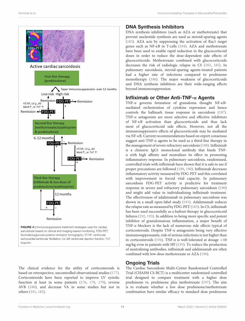
FIGURE 4 | Immunosuppressive treatment strategies used for cardiac
sarcoidosis based on clinical and imaging-based monitoring. FDG-PET,
fluorodeoxyglucose positron emission tomography; VT/VF, ventricular
tachycardia/ventricular fibrillation; LV, left ventricular ejection fraction; TnT,
troponin.
The clinical evidence for the utility of corticosteroids isbased on retrospective, uncontrolled observational studies (177).Corticosteroids have been reported to improve LV systolicfunction at least in some patients (176, 178, 179), reverseAVB (180), and decrease VA in some studies but not inothers (181, 182).
DNA Synthesis InhibitorsDNA synthesis inhibitors (such as AZA or methotrexate) thatprevent nucleotide synthesis are used as steroid-sparing agents(183). AZA acts by suppressing the activation of Rac1 targetgenes such as NF-κB in T-cells (184). AZA and methotrexatehave been used to enable rapid reduction in the glucocorticoiddoses in order to reduce the dose-dependent side effects ofglucocorticoids. Methotrexate combined with glucocorticoidsdecreases the risk of radiologic relapse in CS (183, 185). Inpulmonary sarcoidosis, steroid-sparing agents-treated patientshad a higher rate of infections compared to prednisonemonotherapy (186). The major weakness of glucocorticoidsand DNA synthesis inhibitors are their wide-ranging effectsbeyond immunosuppression.
Infliximab or Other Anti-TNF-α AgentsTNF-α governs formation of granuloma thought NF-κB-mediated orchestration of cytokine expression and hencecontrols the hallmark tissue response in sarcoidosis (187).TNF-α antagonists are more selective and effective inhibitorsof NF-κB activation than glucocorticoids and thus lackmost of glucocorticoid side effects. However, not all theimmunosuppressive effects of glucocorticoids may be mediatedvia NF-κB. Current recommendations based on expert consensussuggest anti-TNF-α agents to be used as a third-line therapy inthemanagement of severe refractory sarcoidosis (188). Infliximabis a chimeric IgG1 monoclonal antibody that binds TNF-α with high affinity and neutralizes its effect in promotinginflammatory response. In pulmonary sarcoidosis, randomized,controlled trials with infliximab have shown that it is safe to use ifproper precautions are followed (189, 190). Infliximab decreasesinflammatory activity measured by FDG-PET and this correlatedwith improvement in forced vital capacity. In pulmonarysarcoidosis FDG-PET activity is predictive for treatmentresponse in severe and refractory pulmonary sarcoidosis (190)and might add value in individualizing infliximab treatment.The effectiveness of adalimumab in pulmonary sarcoidosis wasshown in a small open-label study (191). Adalimumab reducesthe relapse rate as measured by FDG-PET (183). In CS, infliximabhas been used successfully as a bailout therapy in glucocorticoidfailures (192, 193). In addition to being more specific and potentinhibitor of granulomatous inflammation, a major benefit ofTNF-α blockers is the lack of numerous side effects typical ofcorticosteroids. Despite TNF-α antagonists being very effectiveimmunosuppressants, risk of serious infections is not higher thanin corticosteroids (194). TNF-α is well-tolerated at dosage <10mg/kg even in patients with HF (195). To reduce the productionof neutralizing antibodies, infliximab and adalimumab are oftencombined with low-dose methotrexate or AZA (196).
Ongoing TrialsThe Cardiac Sarcoidosis Multi-Center Randomized ControlledTrial (CHASM CS-RCT) is a multicenter randomized controlledtrial designed to compare treatment with a higher doseprednisone vs. prednisone plus methotrexate (197). The aimis to evaluate whether a low dose prednisone/methotrexatecombination have similar efficacy to standard dose prednisone
Frontiers in Medicine | www.frontiersin.org 11 March 2022 | Volume 9 | Article 838564
Ammirati et al. Immunomodulating Therapies in Myocarditis/Pericarditis
leading to an improvement in the quality of life, as a result of areduced burden of side effects. Eligible subjects will have activeclinically manifest CS with advanced conduction system disease,non-sustained or sustained VA, LV or right ventricular systolicdysfunction. The primary endpoint is a measure of myocardialfibrosis/scar, summed perfusion rest score on FDG-PET scanafter 6 months from randomization.
IMAGING TO GUIDEIMMUNOSUPPRESSIVE THERAPY INMYOCARDITIS AND CARDIACSARCOIDOSIS
Echocardiography is routinely performed in patients withsuspected AM to evaluate LV systolic and diastolic functionand the presence of pericardial effusion. However, its roleto guide therapy is limited, since it does not allow tissuecharacterization. CMRI has emerged as a powerful non-invasivediagnostic tool for the assessment of edema, inflammation andfibrosis (198). According to the Updated Lake Louise Criteria,AM can accurately be diagnosed if both edema and myocardialinjury (necrosis or fibrosis) are demonstrated by, respectively,T2-weighted (STIR or T2-mapping) and T1-weighted imaging(T1 mapping or LGE) (198). In healed myocarditis, residualscar can be depicted by LGE (with or without elevated focalT1-values), while persistence of edema, as assessed by T2-weighted imaging, suggests active inflammation. Moreover,CMRI is the gold standard for quantification of ventricularvolumes and function. In this respect, CMRI can be usedto select patients who might benefit from immunosuppressivetherapy, as well as to evaluate the impact of treatment onmyocardial function, ongoing inflammation and scar formation.Furthermore, assessment of the disease stage of myocarditisis especially relevant for patients with myocarditis and drug-refractory VT, as recent data show a high recurrence ratepost VT ablation if signs of active myocarditis are present onEMB or CMRI (199). Importantly, the Lake Louis Criteria areless accurate in detecting active myocarditis in the context ofsystemic immune-mediated diseases (200, 201), making CMRIless suitable to guide therapy in this setting. In sarcoidosis, thepresence of LGE is a sensitive marker of cardiac involvement,but assessment of active inflammation by T2-weighted imagingis not well-validated. However, extensive LGE (>20% LVmass) isassociated with a poor prognosis and absence of LV recovery afterimmunosuppressive therapy with corticosteroids (202). In thisrespect, CMRI is mainly used for diagnosis and prognosticationin CS.
New advances in the field of CMRI include the enhancementof ultrasmall superparamagnetic particles of iron oxide (USPIO),which are nanoparticles that are taken up by monocytes andmacrophages, to directly visualize cardiovascular inflammatoryprocesses (203). A pre-clinical study in a rat model withexperimental auto-immune myocarditis showed that USPIO-enhanced CMRI outperformed conventional CMRI regardingthe detection of myocardial inflammatory cellular infiltrates(204), but the only study in humans failed to show a differencebetween patients with AM (n = 9) and healthy volunteers
(n = 10) (205). Therefore, there is currently no role in clinicalpractice for USPIO-enhanced CMRI in the diagnosis or follow-up of patients with myocarditis.
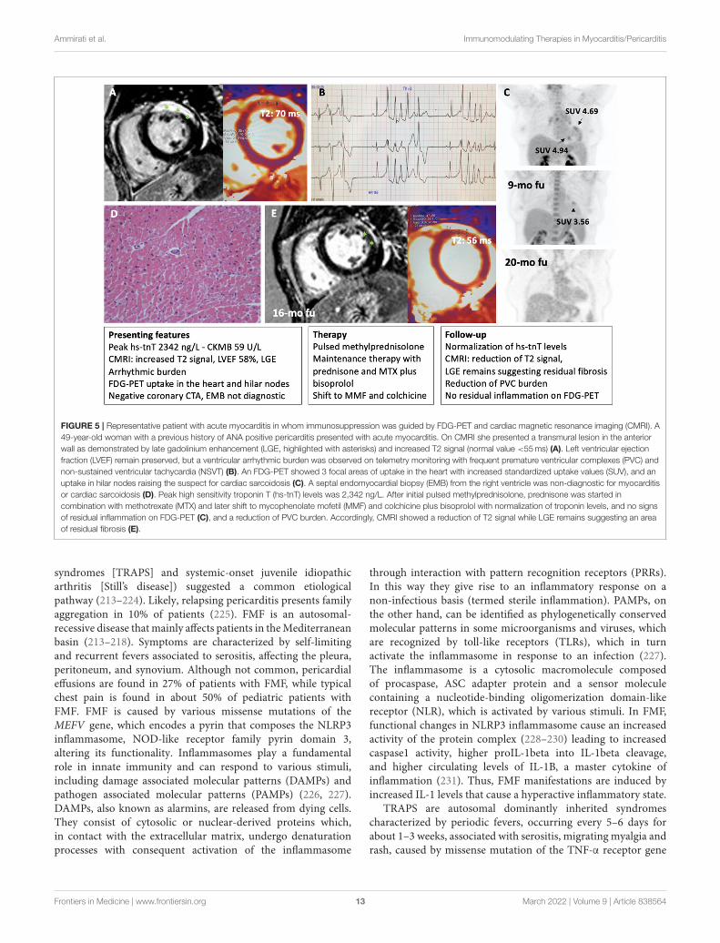
FDG-PET can detect T cells, macrophages, or granulocytesthat infiltrate the myocardium, either as non-specific responseto cell injury or as primary lesion in CS by an enhancedglucose metabolism after a carbohydrate-free diet. FDG-PET isrecommended by several guidelines in patients with suspectedactive CS (172, 206), in fact, it can reveal hypermetabolicmediastinal and hilar lymph nodes differentiating CS from otherautoimmune disease with cardiac involvement (e.g., vasculitis).Since FDG uptake correlates well with the level of granulomatousinflammation, it is assumed that immunosuppression shouldbe up titrated in patients with increased metabolic activity onFDG-PET after steroid therapy has been initiated (207), whilea dose reduction can be considered in patients with reducedFDG uptake. A recent study by Ning et al. (208) showed thatserial FDG-PET in patients with CS altered patient managementin most cases, resulting in complete weaning or significanttapering of prednisolone in 48 and 20%, respectively (Figure 4),while outcome was generally favorable. FDG-PET can be alsoconsidered as an alternative non-invasive diagnostic tool inhemodynamically stable patients with contraindication to CMRIor in patients with suspected autoimmune disease to guideimmunosuppression (Figure 5) (1).
NEW INSIGHTS ON PERICARDITIS
Pathologies of the pericardium are a heterogeneous group,spanning fromminimal pericardial effusion, often asymptomatic,to incessant multidrug-resistant pericarditis (209). Acutepericarditis is diagnosed based on two of the following criteria(210): chest pain, pericardial rubbing, typical changes in theelectrocardiogram, with new and widespread ST elevation orPR depression in the acute phase, and pericardial effusion,which is generally mild. Increased CRP levels can support thediagnosis. The natural history of acute pericarditis can vary.In most cases, it can be self-limiting with complete resolutionof the symptoms, whereas in some cases it can relapse. Thedevelopment of relapses increases by up to 50% in patientswho have received corticosteroid therapy for symptomaticcontrol of the first episode. Some patients can develop incessantpericarditis, a pericarditis whose symptoms continue withoutinterruption even for months (210). The etiology of pericarditischanges considerably depending on the geographic regions(211). In developing countries, pericarditis is often secondaryto tuberculosis (212). On the other hand, in developedcountries, pericarditis is more often idiopathic, secondaryto autoinflammatory or autoimmune processes or followingpericardial injury such radiotherapy or cardiac surgery (211).
The Autoinflammatory Processes inRecurrent PericarditisClinical and laboratory similarities between relapsingpericarditis and some autoinflammatory disorders (i.e.,familial Mediterranean fever [FMF], cryopyrin-associatedperiodic syndromes [CAPS], TNF receptor associated periodic
Frontiers in Medicine | www.frontiersin.org 12 March 2022 | Volume 9 | Article 838564
Ammirati et al. Immunomodulating Therapies in Myocarditis/Pericarditis
FIGURE 5 | Representative patient with acute myocarditis in whom immunosuppression was guided by FDG-PET and cardiac magnetic resonance imaging (CMRI). A
49-year-old woman with a previous history of ANA positive pericarditis presented with acute myocarditis. On CMRI she presented a transmural lesion in the anterior
wall as demonstrated by late gadolinium enhancement (LGE, highlighted with asterisks) and increased T2 signal (normal value <55ms) (A). Left ventricular ejection
fraction (LVEF) remain preserved, but a ventricular arrhythmic burden was observed on telemetry monitoring with frequent premature ventricular complexes (PVC) and
non-sustained ventricular tachycardia (NSVT) (B). An FDG-PET showed 3 focal areas of uptake in the heart with increased standardized uptake values (SUV), and an
uptake in hilar nodes raising the suspect for cardiac sarcoidosis (C). A septal endomyocardial biopsy (EMB) from the right ventricle was non-diagnostic for myocarditis
or cardiac sarcoidosis (D). Peak high sensitivity troponin T (hs-tnT) levels was 2,342 ng/L. After initial pulsed methylprednisolone, prednisone was started in
combination with methotrexate (MTX) and later shift to mycophenolate mofetil (MMF) and colchicine plus bisoprolol with normalization of troponin levels, and no signs
of residual inflammation on FDG-PET (C), and a reduction of PVC burden. Accordingly, CMRI showed a reduction of T2 signal while LGE remains suggesting an area
of residual fibrosis (E).
syndromes [TRAPS] and systemic-onset juvenile idiopathicarthritis [Still’s disease]) suggested a common etiologicalpathway (213–224). Likely, relapsing pericarditis presents familyaggregation in 10% of patients (225). FMF is an autosomal-recessive disease thatmainly affects patients in theMediterraneanbasin (213–218). Symptoms are characterized by self-limitingand recurrent fevers associated to serositis, affecting the pleura,peritoneum, and synovium. Although not common, pericardialeffusions are found in 27% of patients with FMF, while typicalchest pain is found in about 50% of pediatric patients withFMF. FMF is caused by various missense mutations of theMEFV gene, which encodes a pyrin that composes the NLRP3inflammasome, NOD-like receptor family pyrin domain 3,altering its functionality. Inflammasomes play a fundamentalrole in innate immunity and can respond to various stimuli,including damage associated molecular patterns (DAMPs) andpathogen associated molecular patterns (PAMPs) (226, 227).DAMPs, also known as alarmins, are released from dying cells.They consist of cytosolic or nuclear-derived proteins which,in contact with the extracellular matrix, undergo denaturationprocesses with consequent activation of the inflammasome
through interaction with pattern recognition receptors (PRRs).In this way they give rise to an inflammatory response on anon-infectious basis (termed sterile inflammation). PAMPs, onthe other hand, can be identified as phylogenetically conservedmolecular patterns in some microorganisms and viruses, whichare recognized by toll-like receptors (TLRs), which in turnactivate the inflammasome in response to an infection (227).The inflammasome is a cytosolic macromolecule composedof procaspase, ASC adapter protein and a sensor moleculecontaining a nucleotide-binding oligomerization domain-likereceptor (NLR), which is activated by various stimuli. In FMF,functional changes in NLRP3 inflammasome cause an increasedactivity of the protein complex (228–230) leading to increasedcaspase1 activity, higher proIL-1beta into IL-1beta cleavage,and higher circulating levels of IL-1B, a master cytokine ofinflammation (231). Thus, FMF manifestations are induced byincreased IL-1 levels that cause a hyperactive inflammatory state.
TRAPS are autosomal dominantly inherited syndromescharacterized by periodic fevers, occurring every 5–6 days forabout 1–3 weeks, associated with serositis, migrating myalgia andrash, caused by missense mutation of the TNF-α receptor gene
Frontiers in Medicine | www.frontiersin.org 13 March 2022 | Volume 9 | Article 838564
Ammirati et al. Immunomodulating Therapies in Myocarditis/Pericarditis
(219–222, 232). Previous studies reported an incidence of acutepericarditis in 7% of patients with TRAPS, while 25% of thesepatients reported chest pain with characteristics that resemblestypical pericarditis pain (222). There are also oligosymptomaticforms of TRAPS, caused by mutations in TNFRSF1A, andcharacterized by delayed onset in which pericarditis can bethe only manifestation (221). All these observations shedlight on the inflammasome, and the hyperproduction of IL-1 in relapsing pericarditis. Similarly, to what observed in theabove-mentioned autoinflammatory disorders, in patients withrelapsing pericarditis physical injuries via DAMPs as well asinfectious agents via PAMPs’ pathways can elicit inflammasomehyperactivity and IL-1 overproduction.
Pericarditis as an Autoimmune ProcessPericarditis can also be a complication of various autoimmunediseases, including SLE, rheumatoid arthritis (RA), Sjogren’ssyndrome, Behcet’s disease, chronic inflammatory bowel diseasesand vasculitis, including giant cell arteritis or ANCA-associatedvasculitis (233). In SLE, pericarditis is common, affecting ∼50%of patients, and generally occurs during disease flares. Pericarditisis usually associated with other serositis, malar rash, arthritis andleukopenia. The severity of pericarditis correlates with multipleserosal involvement. SLE therapies are normally effective (234–236). In RA, about 30% of patients have asymptomatic pericardialeffusion on echocardiography, but <10% of cases developsymptomatic pericarditis. The incidence of pericarditis in RApatients is higher in those with more severe forms of RA, andhigher levels of rheumatoid factor and anti-cyclic citrullinatedpeptide antibodies (237). Pericarditis can also be the initialsign of a new autoimmune disorder; thus, workup shouldbe prompted after the first episode. Nevertheless, testing forantibodies in all patients with pericarditis is not recommended inthe absence of signs or symptoms consistent with an autoimmunedisorder (210).
Pericarditis of Uncertain Classification(Post-cardiac Injury)Myocardial infarction, radiotherapy, cardiac surgery or evenminor procedures such as the positioning of pacemakerleads, or radiofrequency ablations can cause pericardial layers’inflammation. Oxidative stress, cell death or tissue damage canproduce the release of autoantigens and, due to altered expressionor post-translational modifications, these autoantigens couldtrigger tolerance break after epitope spreading (238). Theprevalence of anti-nuclear antibodies is 43% in patients withrelapsing pericarditis, while it is 10% in healthy individuals.Similarly, anti-heart antibodies and anti-intercalated diskantibodies are found in 67.5% of patients with relapsingpericarditis (210). The presence of these autoantibodies couldbe explained by the release of autoantigens by physical tissueinjury, then the exposure of autoantigens would trigger a T/B-cellautoimmune response. Alternatively, these autoantibodies can bejust an epiphenomenon. Myocardial injury can cause the releaseof DAMPs and the consequent activation of the inflammasomewith IL-1 overproduction. This hypothesis is corroborated bygood response to anti-IL-1 drugs in patients with relapsing
pericarditis secondary to myocardial or pericardial mechanicalinjury (239).
Pericarditis as a Systemic Disorder WithPleuro-Pulmonary InvolvementDiseases of the pericardium can be isolated or be part of asystemic condition associated a striking increase in CRP levels,erythrocyte sedimentation rate (ESR) values and neutrophilia(240–242). Approximately 53% of cases have associated pleuro-pulmonary involvement, 9% have hepatic involvement and5% have peritoneal involvement (242). These conditions areobserved more frequently in the pediatric population. ChestCT scan generally shows bilateral pleural effusion with areasof pulmonary atelectasis. Misdiagnosis with pneumonia canlead to antibiotic therapies, especially at the onset whenpericardial effusion is mild.When final diagnosis of pericarditis isreached, NSAIDs (e.g., Ibuprofen 600mg tid) and corticosteroidtherapy can improve the condition. Too rapid steroid taperingcan lead to pericarditis recurrence and a corticosteroid-dependent condition.
Pericardial EffusionPericardial effusion can be isolated or frequently associated withan underlying pericarditis (243). The symptoms span from absentor mild to severe, especially in case of rapid formation. Thepericardium tends to adapt better to slowly progressing effusions,while it tends to give compression phenomena when the effusiondevelops abundantly and rapidly.
Pericardial effusion can result by pericarditis, edematoussyndromes including HF and kidney failure, cancer, infectiousdiseases (i.e., tuberculosis), serositis and autoimmune diseases,and hypothyroidism (3, 212, 244, 245), even if idiopathicpericardial effusion can often occur. A pericardial effusion isdefined as chronic when it lasts for more than 3 months andsevere when it exceeds 20mm in thickness. Among 100 patientswith severe (>20mm), and chronic (>3 months) idiopathicpericardial effusion, 44 patients were asymptomatic, while 56presented with symptoms, of these 28 presented with dyspnea;33 patients had diabetes mellitus (246). One subset of patientswas symptomatic with a higher age, more likely to be diabetic,with hypertension, chronic obstructive pulmonary disease andatrial fibrillation; whereas a second subset was generallyasymptomatic, younger without significant comorbidities. Aftera mean follow-up of 50 months, no pathology that couldexplain the pericardial effusion was identified and completeregression of the effusion was observed in 39%. Adverse eventswere observed in 38 patients, of which 8 developed cardiactamponade (2.2%/year). Among the 100 patients, 30 underwentpericardiocentesis, 12 underwent pericardial windowing and3 underwent pericardiotomy. Patients who underwent someinvasive procedure presented worse outcomes in terms of relapseor complications than untreated patients. This study seems toemphasize that the risk of developing cardiac tamponade is quitelow and therapeutic strategies should be tailored on an individualbasis based on symptoms. An echocardiographic evaluationevery 3–6 months is recommended for the follow-up of thesepatients, while invasive techniques such as pericardiocentesis or
Frontiers in Medicine | www.frontiersin.org 14 March 2022 | Volume 9 | Article 838564
Ammirati et al. Immunomodulating Therapies in Myocarditis/Pericarditis
pericardiotomy, if separated from specific symptoms, are notrecommended (246). Furthermore, we recently showed that achronic pericardial effusion is present in 37% of subjects withpectus excavatum, with the size of effusion being related to theanatomical severity of the condition, and these effusions have agood prognosis (247). Thus, in presence of chronic pericardialeffusion not related to pericarditis, often with normal or near-normal serum CRP, we do not recommend any therapy, inparticular, we avoid immunosuppressive therapies since thereis no evidence of benefit. Low-dose corticosteroids might beconsidered in few selected patients on a case-by-case basis, butat present no literature deals with this topic. A study reportedgood efficacy and safety of intrapericardial triamcinolone inpatients affected by autoreactive pericarditis with pericardialeffusion (248): the use of an intrapericardial route may avoidthe typical side effects of the systemic use of corticosteroids.Thus, intrapericardial use of triamcinolone remains a viabletherapeutic option for patients with pericarditis and pericardialeffusion. Anti-inflammatory or immunosuppressive therapiesare often started because the analysis of pericardial fluid isconsidered suggestive of inflammation, based on the Light’scriteria validated for the evaluation of pleural fluid. Data froma recent study determined the reference values of analytes andcells in pericardial fluid (249). Specifically, proteins are 1.7–4.6g/dl, albumin 1.19–3.06 g/dl, LDH 141–2613 UI/L, total proteinin pericardial fluid/serum ratio 0.29–0.83, LDH in pericardialfluid/serum ratio 0.4–42.99. According to the Light’s criteria(250), pleural fluid is defined as inflammatory when at least oneof the following criteria is satisfied: fluid/serum protein ratio>0.5, fluid/serum LDH ratio >0.6, and fluid LDH >2/3 of theupper limit for serum levels. The new reference values observedin this population should lead to a reappraisal concerning theclassification of pericardial fluid as exudate or transudate basedon Light’s criteria. Efforts should be taken to stop interpretingpericardial fluid as an exudate or transudate based on evaluationtools that are not validated for this type of fluid, given the risk ofmisinterpreting non-inflammatory effusions into inflammatoryexudates. Elevated LDH found in physiological pericardial fluidmight be caused by the release of LDH bymesothelial cells, whichare particularly abundant in normal pericardial fluid (249).
COVID-19 Associated and mRNACOVID-19 Vaccine-Related AcutePericarditisBased on a retrospective cohort study, of 718.365 patients withCOVID-19, 10.706 (1.5%) developed new-onset pericarditis. Six-month all-cause mortality was 15.5% (n = 816) for pericarditisand 6.7% (n = 356) in matched controls (p < 0.0001),odds ratio 2.55 (95% CI: 2.24–2.91) (251). At present, only 2published studies focused the attention toward anti-COVID-19 vaccine-related acute pericarditis. Barda et al. reported inIsrael an incidence of 26 cases out of 884.828 vaccinatedindividuals (3/100.000) vs. 18 out of 884.828 unvaccinatedcontrols (2/100.000); RR 1.27 (p = non-significant) (124). Diazet al. described 37 cases in US, with an incidence of 1.8/100.000(252). The mean monthly number of cases of pericarditis during
the prevaccine period was 49.1 (95% CI, 46.4–51.9) vs. 78.8 (95%CI, 70.3–87.9) during the vaccine period (P < 0.001). A total of15 cases occurred after the first dose and 22 after the second dose;27 out of 37 subjects were males and median age was 59 years;13 were admitted to the hospital (median stay, 1 day), none tointensive care. No patient died.
THERAPY OF PERICARDITIS
NSAIDsNSAIDs represent the first line of therapy, exerting their actionboth on the pathogenesis of pericarditis and on the controlof symptoms. Understanding the role of inflammasome in thepathogenesis of relapsing pericarditis explains their effectiveness(Figure 6). Numerous NSAIDs are used for relapsing pericarditistherapy, including ibuprofen, indomethacin and acetylsalicylicacid (ASA). All these NSAIDs are recommended in high dosesas the first line of pericarditis treatment by the European Societyof Cardiology (Figure 7). The duration of treatment is variable,but in any case, prolonged (210, 253).
ColchicineThe rationale behind the use of colchicine in pericarditis arisesfrom the observation of good efficacy results in the controlof serositis during FMF and subsequently in the control ofpericarditis associated with other serositis during FMF (254).Colchicine performs its functions by inhibiting the activation ofpore formation carried out by P2X2 and P2X7 receptors, thatconcur to the activation of inflammasome, and by inhibitingNACHT-LRRPYD-containing protein 3 inflammasome (255).The combined use of NSAIDs and colchicine has producedpositive results on pericarditis in numerous clinical trials, wheretheir use has favored both the control of symptoms and theprevention of relapses (255–261). Colchicine during relapsingpericarditis should be administered early without loading doseand its dosage might be adapted to the patient’s weight: ingeneral, we start with a dose of 0.5mg per day and, if tolerated,the dose is then increased to 0.5mg BID or 1mg OD, basedon compliance and tolerability. The most frequent side effectsare gastrointestinal, with diarrhea that occurs mainly at thebeginning of therapy in ∼10%. The dosage of colchicine canpossibly be reduced in patients with this type of disorder.
CorticosteroidsThe use of corticosteroids in pericarditis remains controversial.If they find their indication for the forms of pericarditis causedby autoimmune processes or in the forms resistant to thecombined therapy of NSAIDs and colchicine, the probabilityof generating dependence for the control of symptoms is high(210). Many patients will experience a recurrence of pericarditisupon discontinuation of corticosteroid therapy, especially whencorticosteroid tapering is too rapid, thus leading to a realdependence on corticosteroids and the risk of a prolonged use(253). Chronic use of corticosteroids is burdened by numerousside effects, including weight gain, osteoporosis and possiblevertebral collapse, diabetes mellitus and Cushing’s syndrome(262). For this reason, the use of corticosteroids should be
Frontiers in Medicine | www.frontiersin.org 15 March 2022 | Volume 9 | Article 838564
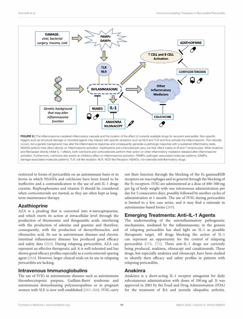
Ammirati et al. Immunomodulating Therapies in Myocarditis/Pericarditis
FIGURE 6 | The inflammasome-mediated inflammatory cascade and the location of the effect of currently available drugs for recurrent pericarditis. Non-specific
triggers such as structural damage or microbial agents may interact with specific receptors such as NLR and TLR and thus activate the inflammasome. This naturally
occurs, but a genetic background may alter the inflammasome response and consequently generate a pathologic response with a sustained inflammatory state.
NSAIDs perform their effect directly on inflammasome activation. Azathioprine and corticosteroids carry out their effect mainly on B and T lymphocytes. While Anakinra
and Rilonacept directly inhibit IL-1 effects, both colchicine and corticosteroids perform their action on other inflammatory mediators released after inflammasome
activation. Furthermore, colchicine also exerts an inhibitory effect on inflammasome activation. PAMPs, pathogen-associated molecular patterns; DAMPs,
damage-associated molecular patterns; TLR, toll-like receptor; NLR, NOD-like Receptor; NSAIDs, non-steroidal antiinflammatory drugs.
restricted to forms of pericarditis on an autoimmune basis or informs in which NSAIDs and colchicine have been found to beineffective and a contraindication to the use of anti-IL-1 drugscoexists. Bisphosphonates and vitamin D should be consideredwhen corticosteroids are started, as they are often kept as long-term maintenance therapy.
AzathioprineAZA is a prodrug that is converted into 6-mercaptopurine,and which exerts its action at intracellular level through theproduction of thioinosinic and thioguanilic acids, interferingwith the production of adenine and guanine and therefore,consequently, with the production of deoxyribonucleic andribonucleic acid. Its use in autoimmune diseases and chronicintestinal inflammatory diseases has produced good efficacyand safety data (263). During relapsing pericarditis, AZA canrepresent an effective therapeutic aid: it is well-tolerated and hasshown good efficacy profiles especially as a corticosteroid-sparingagent (264). However, larger clinical trials on its use in relapsingpericarditis are lacking.
Intravenous ImmunoglobulinsThe use of IVIG in autoimmune diseases such as autoimmunethrombocytopenic purpura, Guillain-Barré syndrome andautoimmune demyelinating polyneuropathies or in pregnantwomen with SLE is now well-established (265–268). IVIG carry
out their function through the blocking of the Fc-gammaRIIBreceptors on macrophages and in general through the blocking ofthe Fc receptors. IVIG are administered at a dose of 400–500mgper kg of body weight with one intravenous administration perday for 5 consecutive days, possibly followed by another cycles ofadministration at 1 month. The use of IVIG during pericarditisis limited to a few case series, and it may find a rationale inautoimmune-based forms (269).
Emerging Treatments: Anti-IL-1 AgentsThe understanding of the autoinflammatory pathogeneticmechanisms, mediated by the inflammasome, in the genesisof relapsing pericarditis has shed light on IL-1 as possibletherapeutic target. All drugs blocking the action of IL-1can represent an opportunity for the control of relapsingpericarditis (270, 271). Three anti-IL-1 drugs are currentlybeing produced, anakinra, rilonacept and canakinumab. Thesedrugs, but especially anakinra and rilonacept, have been studiedto identify their efficacy and safety profiles in patients withrelapsing pericarditis.
AnakinraAnakinra is a short-acting IL-1 receptor antagonist for dailysubcutaneous administration with doses of 100mg qd. It wasapproved in 2001 by the Food and Drug Administration (FDA)for the treatment of RA and juvenile idiopathic arthritis.
Frontiers in Medicine | www.frontiersin.org 16 March 2022 | Volume 9 | Article 838564
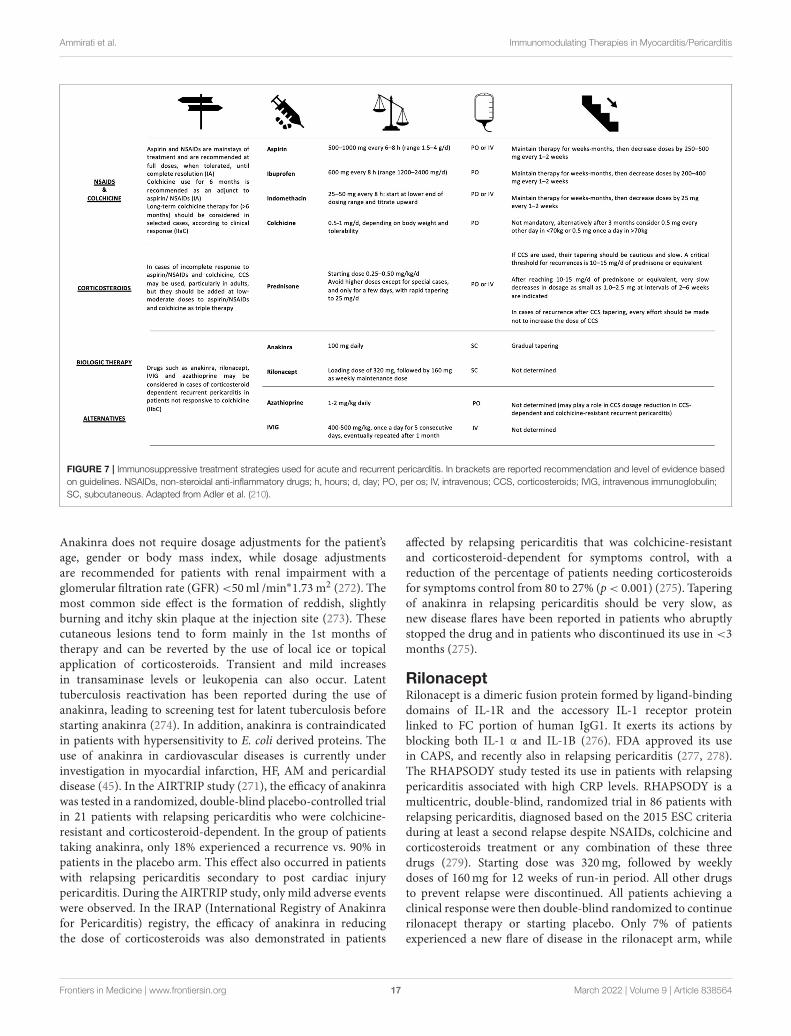
Ammirati et al. Immunomodulating Therapies in Myocarditis/Pericarditis
FIGURE 7 | Immunosuppressive treatment strategies used for acute and recurrent pericarditis. In brackets are reported recommendation and level of evidence based
on guidelines. NSAIDs, non-steroidal anti-inflammatory drugs; h, hours; d, day; PO, per os; IV, intravenous; CCS, corticosteroids; IVIG, intravenous immunoglobulin;
SC, subcutaneous. Adapted from Adler et al. (210).
Anakinra does not require dosage adjustments for the patient’sage, gender or body mass index, while dosage adjustmentsare recommended for patients with renal impairment with aglomerular filtration rate (GFR)<50ml /min∗1.73 m2 (272). Themost common side effect is the formation of reddish, slightlyburning and itchy skin plaque at the injection site (273). Thesecutaneous lesions tend to form mainly in the 1st months oftherapy and can be reverted by the use of local ice or topicalapplication of corticosteroids. Transient and mild increasesin transaminase levels or leukopenia can also occur. Latenttuberculosis reactivation has been reported during the use ofanakinra, leading to screening test for latent tuberculosis beforestarting anakinra (274). In addition, anakinra is contraindicatedin patients with hypersensitivity to E. coli derived proteins. Theuse of anakinra in cardiovascular diseases is currently underinvestigation in myocardial infarction, HF, AM and pericardialdisease (45). In the AIRTRIP study (271), the efficacy of anakinrawas tested in a randomized, double-blind placebo-controlled trialin 21 patients with relapsing pericarditis who were colchicine-resistant and corticosteroid-dependent. In the group of patientstaking anakinra, only 18% experienced a recurrence vs. 90% inpatients in the placebo arm. This effect also occurred in patientswith relapsing pericarditis secondary to post cardiac injurypericarditis. During the AIRTRIP study, only mild adverse eventswere observed. In the IRAP (International Registry of Anakinrafor Pericarditis) registry, the efficacy of anakinra in reducingthe dose of corticosteroids was also demonstrated in patients
affected by relapsing pericarditis that was colchicine-resistantand corticosteroid-dependent for symptoms control, with areduction of the percentage of patients needing corticosteroidsfor symptoms control from 80 to 27% (p< 0.001) (275). Taperingof anakinra in relapsing pericarditis should be very slow, asnew disease flares have been reported in patients who abruptlystopped the drug and in patients who discontinued its use in <3months (275).
RilonaceptRilonacept is a dimeric fusion protein formed by ligand-bindingdomains of IL-1R and the accessory IL-1 receptor proteinlinked to FC portion of human IgG1. It exerts its actions byblocking both IL-1 α and IL-1B (276). FDA approved its usein CAPS, and recently also in relapsing pericarditis (277, 278).The RHAPSODY study tested its use in patients with relapsingpericarditis associated with high CRP levels. RHAPSODY is amulticentric, double-blind, randomized trial in 86 patients withrelapsing pericarditis, diagnosed based on the 2015 ESC criteriaduring at least a second relapse despite NSAIDs, colchicine andcorticosteroids treatment or any combination of these threedrugs (279). Starting dose was 320mg, followed by weeklydoses of 160mg for 12 weeks of run-in period. All other drugsto prevent relapse were discontinued. All patients achieving aclinical response were then double-blind randomized to continuerilonacept therapy or starting placebo. Only 7% of patientsexperienced a new flare of disease in the rilonacept arm, while
Frontiers in Medicine | www.frontiersin.org 17 March 2022 | Volume 9 | Article 838564
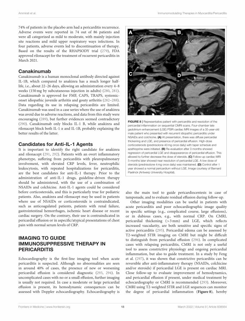
Ammirati et al. Immunomodulating Therapies in Myocarditis/Pericarditis
74% of patients in the placebo arm had a pericarditis recurrence.Adverse events were reported in 74 out of 86 patients andwere all categorized as mild to moderate, with mainly injectionsite reactions and mild upper respiratory ways infections. Infour patients, adverse events led to discontinuation of therapy.Based on the results of the RHAPSODY trial (279), FDAapproved rilonacept for the treatment of recurrent pericarditis inMarch 2021.
CanakinumabCanakinumab is a human monoclonal antibody directed againstIL-1B, which compared to anakinra has a much longer half-life, i.e., about 22–26 days, allowing an administration every 4–8weeks (150mg by subcutaneous injection in adults) (280, 281).Canakinumab is approved for FMF, CAPS, TRAPS, systemic-onset idiopathic juvenile arthritis and gouty arthritis (282–288).Data regarding its use in relapsing pericarditis are limited.Canakinumab was used in a case series where the use of anakinrawas avoid due to adverse reactions, and data from this study wereencouraging (289), but further evidences seemed contradictory(290). Canakinumab only blocks IL-1 B, while anakinra andrilonacept block both IL-1 α and IL-1B, probably explaining thebetter results of the latter.
Candidates for Anti-IL-1 AgentsIt is important to identify the right candidate for anakinraand rilonacept (291, 292). Patients with an overt inflammatoryphenotype, suffering from pericarditis with pleuropulmonaryinvolvement, with elevated CRP levels, fever, neutrophilicleukocytosis, with repeated hospitalizations for pericarditis,are the best candidates for anti-IL-1 therapy. Prior to theadministration of anti-IL-1 drugs, guideline-driven therapyshould be administered, with the use of a combination ofNSAIDs and colchicine. Anti-IL-1 agents could be consideredbefore corticosteroids, and this is particularly true for pediatricpatients. Also, anakinra and rilonacept may be used in patientswhere use of NSAIDs or corticosteroids is contraindicated,such as anticoagulated patients, patients with renal failure,gastrointestinal hemorrhages, ischemic heart disease or recentcardiac surgery. On the contrary, their use is contraindicated inpericardial effusion or in aspecific/atypical presentations of chestpain with normal serum levels of CRP.
IMAGING TO GUIDEIMMUNOSUPPRESSIVE THERAPY INPERICARDITIS
Echocardiography is the first-line imaging tool when acutepericarditis is suspected. Although no abnormalities are seenin around 40% of cases, the presence of new or worseningpericardial effusion is considered diagnostic (293, 294). Inuncomplicated cases with no or a small effusion, further imagingis usually not required. In case a moderate or large pericardialeffusion is present, its hemodynamic consequences can beassessed with Doppler echocardiography. Echocardiography is
FIGURE 8 | Representative patient with pericarditis and resolution of the
pericardial inflammation on sequential CMRI scans. Four-chamber late
gadolinium enhancement (LGE) PSIR cardiac MRI images of a 35-year-old
male patient who presented with recurrent idiopathic pericarditis under
NSAIDs and colchicine. (A) At presentation, there was diffuse pericardial
thickening and LGE, and presence of pericardial effusion. High-dose
corticosteroids (prednisolone 40mg once daily) with taper schedule and
azathioprine were initiated. (B) Re-evaluation after 3 months showed
regression of pericardial LGE and disappearance of pericardial effusion. This
allowed to further decrease the dose of steroids. (C) Follow-up cardiac MRI
5-months later showed near resolution of pericardial LGE. A low dose of
steroids (prednisolone 4mg once daily) was maintained. (D) Control after 1
year showed a normal pericardium without LGE. Image courtesy of Bernard
Paelinck (Antwerp University Hospital).
also the main tool to guide pericardiocentesis in case oftamponade, and to evaluate residual effusion during follow-up.
Other imaging modalities can be useful in patients withacute pericarditis and poor echocardiographic image qualityin specific settings (e.g., complicated course, large effusions),or in dubious cases, e.g., with normal CRP. On CMRI,pericardial thickening (>3mm) and LGE, which reflectsincreased vascularity, are both sensitive and specific signs ofactive pericarditis (295). Pericardial edema can be assessed byT2-weighted STIR imaging on CMRI but might be difficultto distinguish from pericardial effusion (296). In complicatedcases with relapsing pericarditis, CMRI is not only a usefultool to assess constrictive physiology and ongoing pericardialinflammation, but also to guide treatment. In a study by Fenget al. (297), it was shown that constrictive pericarditis can bereversible after anti-inflammatory therapy (NSAIDs, colchicineand/or steroids) if pericardial LGE is present on cardiac MRI.Close follow-up to evaluate improvement of hemodynamics,and pericardial effusion if present, under medical treatment byechocardiography or CMRI is recommended (293). Moreover,CMRI using T2-weighted STIR and LGE sequences can monitorthe degree of pericardial inflammation (Figure 8), hereby
Frontiers in Medicine | www.frontiersin.org 18 March 2022 | Volume 9 | Article 838564
Ammirati et al. Immunomodulating Therapies in Myocarditis/Pericarditis
providing important information for the clinician before anti-inflammatory or immunosuppressive medication is tapered off(296). Cardiac CT can be used to detect pericardial effusionand pericardial inflammation after contrast but implies ionizingradiation. In chronic forms of constrictive pericarditis, cardiacCT is particularly useful to assess pericardial calcificationsfor planning of a pericardiotomy. FDG-PET has been usedto visualize pericardial inflammation and metabolic activity(298), but it is seldom performed in clinical practice and isnot recommended by current guidelines (293). Nevertheless, asmall study in 16 patients showed that a high FDG uptake(SUVmax >3.0) predicted reversibility of constrictive pericarditiswith steroid treatment (299). In this respect, FDG-PET may beuseful in patients with non-CMRI-conditional devices to guideimmunosuppressive therapy, but further studies are needed toevaluate whether it provides incremental value to CMRI.
CONCLUSIONS
While therapies for patients with acute and recurrent pericarditisare mainly evidence-based, almost no trials are available for AM,thus immunosuppression in this setting is generally based onexpert consensus. Thus, an impelling need of clinical research is
to evaluate which immunosuppressive agent can be effective toimprove the outcome of patients with AM and the characteristicsof patients who can benefit more by immunosuppression.Thus, well-powered multicenter randomized trials are neededto test this hypothesis. In parallel, large prospective registriescan better define the main determinants of outcome, evenif large retrospective studies consistently demonstrated thatpresentation of AM complicated by reduced LVEF, HF,VA, AVB, or cardiogenic shock are associated with pooroutcome (11, 40).
AUTHOR CONTRIBUTIONS
EA, EB, GV, MG, CV, JL, MP, AC, CP, LT, JM, and ABwrote the draft. EA, EB, GV, MG, JM, and AB revised themanuscript. All authors contributed to the article and approvedthe submitted version.
SUPPLEMENTARY MATERIAL
The Supplementary Material for this article can be foundonline at: https://www.frontiersin.org/articles/10.3389/fmed.2022.838564/full#supplementary-material
REFERENCES
1. Ammirati E, Frigerio M, Adler ED, Basso C, Birnie DH, Brambatti
M, et al. Management of acute myocarditis and chronic inflammatory
cardiomyopathy: an expert consensus document. Circulation Heart
Fail. (2020) 13:e007405. doi: 10.1161/CIRCHEARTFAILURE.120.
007405
2. Peretto G, Sala S, De Luca G, Marcolongo R, Campochiaro C, Sartorelli
S, et al. Immunosuppressive therapy and risk stratification of patients
with myocarditis presenting with ventricular arrhythmias. JACC Clin
Electrophysiol. (2020) 6:1221–34. doi: 10.1016/j.jacep.2020.05.013
3. Inciardi RM, Lupi L, Zaccone G, Italia L, RaffoM, Tomasoni D, et al. Cardiac
involvement in a patient with coronavirus disease 2019 (COVID-19). JAMA
Cardiol. (2020) 5:819–24. doi: 10.1001/jamacardio.2020.1096
4. Larson KF, Ammirati E, Adler ED, Cooper LT, Hong KN, Saponara G, et
al. Myocarditis after BNT162b2 and mRNA-1273 vaccination. Circulation.
(2021) 144:506–8. doi: 10.1161/CIRCULATIONAHA.121.055913
5. Ammirati E, Cavalotti C, Milazzo A, Pedrotti P, Soriano F, Schroeder
JW, et al. Temporal relation between second dose BNT162b2
mRNA Covid-19 vaccine and cardiac involvement in a patient with
previous SARS-COV-2 infection. Int J Cardiol Heart Vasc. (2021)
34:100774. doi: 10.1016/j.ijcha.2021.100774
6. Patone M, Mei XW, Handunnetthi L, Dixon S, Zaccardi F, Shankar-
Hari M, et al. Risks of myocarditis, pericarditis, and cardiac arrhythmias
associated with COVID-19 vaccination or SARS-CoV-2 infection. Nat Med.
(2021). doi: 10.1038/s41591-021-01630-0. [Epub ahead of print].
7. Truong DT, Dionne A, Muniz JC, McHugh KE, Portman MA, Lambert
LM, et al. Clinically suspected myocarditis temporally related to COVID-19
vaccination in adolescents and young adults. Circulation. (2021) 145:345–
56. doi: 10.1161/CIRCULATIONAHA.121.056583
8. Bozkurt B, Kamat I, Hotez PJ. Myocarditis with
COVID-19 mRNA Vaccines. Circulation. (2021) 144:471–
84. doi: 10.1161/CIRCULATIONAHA.121.056135
9. Salem JE, Manouchehri A, MoeyM, Lebrun-Vignes B, Bastarache L, Pariente
A, et al. Cardiovascular toxicities associated with immune checkpoint
inhibitors: an observational, retrospective, pharmacovigilance study. Lancet
Oncol. (2018) 19:1579–89. doi: 10.1016/S1470-2045(18)30608-9
10. Nguyen LS, Cooper LT, Kerneis M, Funck-Brentano C, Silvain J, Brechot
N, et al. Systematic analysis of drug-associated myocarditis reported in
the World Health Organization pharmacovigilance database. Nat Commun.
(2022) 13:25. doi: 10.1038/s41467-021-27631-8
11. Ammirati E, Veronese G, Brambatti M, Merlo M, Cipriani M, Potena L,
et al. Fulminant versus acute nonfulminant myocarditis in patients with
left ventricular systolic dysfunction. J Am Coll Cardiol. (2019) 74:299–
311. doi: 10.1016/j.jacc.2019.04.063
12. Cooper LT, Baughman KL, Feldman AM, Frustaci A, Jessup
M, Kuhl U, et al. The role of endomyocardial biopsy in the
management of cardiovascular disease: a scientific statement from the
American Heart Association, the American College of Cardiology,
and the European Society of Cardiology. Circulation. (2007)
116:2216–33. doi: 10.1161/CIRCULATIONAHA.107.186093
13. Ammirati E, Veronese G, Bottiroli M, Wang DW, Cipriani M, Garascia
A, et al. Update on acute myocarditis. Trends Cardiovasc Med. (2020)
31:370–9. doi: 10.1016/j.tcm.2020.05.008
14. Tschöpe C, Ammirati E, Bozkurt B, Caforio ALP, Cooper LT,
Felix SB, et al. Myocarditis and inflammatory cardiomyopathy:
current evidence and future directions. Nat Rev Cardiol. (2020)
18:169–93. doi: 10.1038/s41569-020-00435-x
15. Bock CT, Klingel K, Kandolf R. Human parvovirus B19-associated
myocarditis. N Engl J Med. (2010) 362:1248–9. doi: 10.1056/NEJMc0911362
16. Bowles NE, Ni J, Kearney DL, Pauschinger M, Schultheiss HP,
McCarthy R, et al. Detection of viruses in myocardial tissues by
polymerase chain reaction. Evidence of adenovirus as a common
cause of myocarditis in children and adults. J Am Coll Cardiol. (2003)
42:466–72. doi: 10.1016/S0735-1097(03)00648-X
17. Veronese G, Ammirati E, Chen C, Klingel K, Suzuki M, Okumura
T, et al. Management perspectives from the 2019 Wuhan international
workshop on fulminant myocarditis. Int J Cardiol. (2021) 324:131–
8. doi: 10.1016/j.ijcard.2020.10.063
18. Caforio AL, Pankuweit S, Arbustini E, Basso C, Gimeno-Blanes J, Felix SB,
et al. Current state of knowledge on aetiology, diagnosis, management, and
therapy of myocarditis: a position statement of the European Society of
Cardiology Working Group on Myocardial and Pericardial Diseases. Eur
Heart J. (2013) 34:2636–48. doi: 10.1093/eurheartj/eht210
Frontiers in Medicine | www.frontiersin.org 19 March 2022 | Volume 9 | Article 838564
Ammirati et al. Immunomodulating Therapies in Myocarditis/Pericarditis
19. Verdonschot J, Hazebroek M, Merken J, Debing Y, Dennert R, Brunner-La
Rocca HP, et al. Relevance of cardiac parvovirus B19 in myocarditis and
dilated cardiomyopathy: review of the literature. Eur J Heart Fail. (2016)
18:1430–41. doi: 10.1002/ejhf.665
20. Stewart GC, Lopez-Molina J, Gottumukkala RV, Rosner GF, Anello MS,
Hecht JL, et al. Myocardial parvovirus B19 persistence: lack of association
with clinicopathologic phenotype in adults with heart failure. Circ Heart Fail.
(2011) 4:71–8. doi: 10.1161/CIRCHEARTFAILURE.110.958249
21. Veronese G, Ammirati E, Brambatti M, Merlo M, Cipriani M, Potena L, et al.
Viral genome search in myocardium of patients with fulminant myocarditis.
Eur J Heart Fail. (2020) 22:1277–80. doi: 10.1002/ejhf.1738
22. Fung G, Luo H, Qiu Y, Yang D, McManus B. Myocarditis. Circ Res. (2016)
118:496–514. doi: 10.1161/CIRCRESAHA.115.306573
23. Klingel K, Selinka HC, Huber M, Sauter M, Leube M, Kandolf R.
Molecular pathology and structural features of enteroviral replication.
Toward understanding the pathogenesis of viral heart disease. Herz. (2000)
25:216–20. doi: 10.1007/s000590050009
24. Matsuura H, Ichida F, Saji T, Ogawa S, Waki K, Kaneko M, et al. Clinical
features of acute and fulminant myocarditis in children- 2nd nationwide
survey by Japanese society of pediatric cardiology and cardiac surgery. Circ J.
(2016) 80:2362–8. doi: 10.1253/circj.CJ-16-0234
25. Ammirati E, Varrenti M, Veronese G, Fanti D, Nava A, Cipriani M, et al.
Prevalence and outcome of patients with acute myocarditis and positive
viral search on nasopharyngeal swab. Eur J Heart Fail. (2021) 23:1242–
5. doi: 10.1002/ejhf.2247
26. Bratincsák A, El-Said HG, Bradley JS, Shayan K, Grossfeld PD,
Cannavino CR. Fulminant myocarditis associated with pandemic
H1N1 influenza A virus in children. J Am Coll Cardiol. (2010)
55:928–9. doi: 10.1016/j.jacc.2010.01.004
27. Gil-Cruz C, Perez-Shibayama C, De Martin A, Ronchi F, van
der Borght K, Niederer R, et al. Microbiota-derived peptide
mimics drive lethal inflammatory cardiomyopathy. Science. (2019)
366:881–6. doi: 10.1126/science.aav3487
28. Anzai A, Mindur JE, Halle L, Sano S, Choi JL, He S, et al. Self-
reactive CD4(+) IL-3(+) T cells amplify autoimmune inflammation in
myocarditis by inciting monocyte chemotaxis. J Exp Med. (2019) 216:369–
83. doi: 10.1084/jem.20180722
29. Tschope C, Elsanhoury A, Schlieker S, Van Linthout S, Kuhl U.
Immunosuppression in inflammatory cardiomyopathy and parvovirus B19
persistence. Eur J Heart Fail. (2019) 21:1468–9. doi: 10.1002/ejhf.1560
30. Frustaci A, Russo MA, Chimenti C. Randomized study on the
efficacy of immunosuppressive therapy in patients with virus-negative
inflammatory cardiomyopathy: the TIMIC study. Eur Heart J. (2009)
30:1995–2002. doi: 10.1093/eurheartj/ehp249
31. Chen HS, Wang W, Wu SN, Liu JP. Corticosteroids for
viral myocarditis. Cochrane Database Syst Rev. (2013)
2013:Cd004471. doi: 10.1002/14651858.CD004471.pub3
32. Cheng CY, Cheng GY, Shan ZG, Baritussio A, Lorenzoni G, Tyminska
A, et al. Efficacy of immunosuppressive therapy in myocarditis: A
30-year systematic review and meta analysis. Autoimmun Rev. (2021)
20:102710. doi: 10.1016/j.autrev.2020.102710
33. Kociol RD, Cooper LT, Fang JC, Moslehi JJ, Pang PS, Sabe MA, et al.
Recognition and initial management of fulminant myocarditis: a scientific
statement from the American Heart Association. Circulation. (2020)
141:e69–92. doi: 10.1161/CIR.0000000000000745
34. Drucker NA, Colan SD, Lewis AB, Beiser AS, Wessel DL, Takahashi M, et al.
Gamma-globulin treatment of acute myocarditis in the pediatric population.
Circulation. (1994) 89:252–7. doi: 10.1161/01.CIR.89.1.252
35. Robinson J, Hartling L, Vandermeer B, Klassen TP.
Intravenous immunoglobulin for presumed viral myocarditis
in children and adults. Cochrane Database Syst Rev. (2015)
2015:Cd004370. doi: 10.1002/14651858.CD004370.pub3
36. Mason JW, O’Connell JB, Herskowitz A, Rose NR, McManus BM,
Billingham ME, et al. A clinical trial of immunosuppressive therapy for
myocarditis. The Myocarditis Treatment Trial Investigators. N Engl J Med.
(1995) 333:269–75. doi: 10.1056/NEJM199508033330501
37. Zhou N, Zhao Y, Jiang J, Shen L, Li J, Wan J, et al. Impact of mechanical
circulatory support and immunomodulation therapy on outcome of patients
with fulminant myocarditis: Chinese registry of fulminant myocarditis.
Signal Transduct Target Ther. (2021) 6:350. doi: 10.1038/s41392-021-00700-6
38. Schultheiss HP, Piper C, Sowade O, Waagstein F, Kapp JF, Wegscheider
K, et al. Betaferon in chronic viral cardiomyopathy (BICC) trial: Effects of
interferon-β treatment in patients with chronic viral cardiomyopathy. Clin
Res Cardiol. (2016) 105:763–73. doi: 10.1007/s00392-016-0986-9
39. Cooper LT. Myocarditis. N Engl J Med. (2009) 360:1526–
38. doi: 10.1056/NEJMra0800028
40. Ammirati E, Cipriani M, Moro C, Raineri C, Pini D, Sormani
P, et al. Clinical presentation and outcome in a contemporary
cohort of patients with acute myocarditis. Circulation. (2018)
138:1088–99. doi: 10.1161/CIRCULATIONAHA.118.035319
41. Mele D, Flamigni F, Rapezzi C, Ferrari R. Myocarditis in COVID-
19 patients: current problems. Intern Emerg Med. (2021) 16:1123–
9. doi: 10.1007/s11739-021-02635-w
42. Mehta P, McAuley DF, Brown M, Sanchez E, Tattersall RS, Manson JJ.
COVID-19: consider cytokine storm syndromes and immunosuppression.
Lancet. (2020) 395:1033–4. doi: 10.1016/S0140-6736(20)30628-0
43. Lim BK, Choe SC, Shin JO, Ho SH, Kim JM, Yu SS, et al. Local expression
of interleukin-1 receptor antagonist by plasmid DNA improves mortality
and decreases myocardial inflammation in experimental coxsackieviral
myocarditis. Circulation. (2002) 105:1278–81. doi: 10.1161/circ.105.11.
1278
44. Toldo S, Kannan H, Bussani R, Anzini M, Sonnino C, Sinagra G, et al.
Formation of the inflammasome in acute myocarditis. Int J Cardiol. (2014)
171:e119–21. doi: 10.1016/j.ijcard.2013.12.137
45. Abbate A, Toldo S, Marchetti C, Kron J, Van Tassell BW,
Dinarello CA. Interleukin-1 and the Inflammasome as Therapeutic
Targets in Cardiovascular Disease. Circ Res. (2020) 126:1260–
80. doi: 10.1161/CIRCRESAHA.120.315937
46. Cavalli G, Pappalardo F, Mangieri A, Dinarello CA, Dagna L, Tresoldi M.
Treating life-threatening myocarditis by blocking interleukin-1. Crit Care
Med. (2016) 44:e751–4. doi: 10.1097/CCM.0000000000001654
47. Bello F, Marchi A, Prisco D, Olivotto I, Emmi G. Antiarrhythmic
efficacy of anakinra in a young patient with autoimmune
lymphocytic myocarditis. Rheumatology (Oxford). (2020)
59:e88–90. doi: 10.1093/rheumatology/keaa207
48. Veronese G, Cipriani M, Bottiroli M, Garascia A, Mondino M, Pedrotti P, et
al. Fulminant myocarditis triggered by OC43 subtype coronavirus: a disease
deserving evidence-based care bundles. J Cardiovasc Med (Hagerstown).
(2020) 21:529–31. doi: 10.2459/JCM.0000000000000989
49. Ammirati E, Cipriani M, Lilliu M, Sormani P, Varrenti M, Raineri
C, et al. Survival and left ventricular function changes in fulminant
versus nonfulminant acute myocarditis. Circulation. (2017) 136:529–
45. doi: 10.1161/CIRCULATIONAHA.117.026386
50. Turgeon PY, Massot M, Beaupré F, Belzile D, Beaudoin J, Bernier M, et al.
Effect of acute immunosuppression on left ventricular recovery andmortality
in fulminant viral myocarditis: a case series and review of literature. CJC
Open. (2021) 3:292–302. doi: 10.1016/j.cjco.2020.10.017
51. Kiriakidou M, Ching CL. Systemic lupus erythematosus. Ann Intern Med.
(2020) 172:Itc81–96. doi: 10.7326/AITC202006020
52. Barber MRW, Drenkard C, Falasinnu T, Hoi A, Mak A, KowNY, et al. Global
epidemiology of systemic lupus erythematosus. Nat Rev Rheumatol. (2021)
17:515–32. doi: 10.1038/s41584-021-00668-1
53. Jia E, Geng H, Liu Q, Xiao Y, Zhang Y, Xie J, et al. Cardiac manifestations
of Han Chinese patients with systemic lupus erythematosus: a retrospective
study. Ir J Med Sci. (2019) 188:801–6. doi: 10.1007/s11845-018-1934-7
54. Tanwani J, Tselios K, Gladman DD, Su J, Urowitz MB. Lupus myocarditis: a
single center experience and a comparative analysis of observational cohort
studies. Lupus. (2018) 27:1296–302. doi: 10.1177/0961203318770018
55. Thomas G, Cohen Aubart F, Chiche L, Haroche J, Hié M, Hervier B,
et al. Lupus myocarditis: initial presentation and longterm outcomes
in a multicentric series of 29 patients. J Rheumatol. (2017) 44:24–
32. doi: 10.3899/jrheum.160493
56. Mathian A, Mouries-Martin S, Dorgham K, Devilliers H, Yssel H, Garrido
Castillo L, et al. Ultrasensitive serum interferon-α quantification during SLE
remission identifies patients at risk for relapse. Ann Rheum Dis. (2019)
78:1669–76. doi: 10.1136/annrheumdis-2019-215571
Frontiers in Medicine | www.frontiersin.org 20 March 2022 | Volume 9 | Article 838564
Ammirati et al. Immunomodulating Therapies in Myocarditis/Pericarditis
57. Mathian A, Mouries-Martin S, Dorgham K, Devilliers H, Barnabei L, Ben
Salah E, et al. Monitoring disease activity in systemic lupus erythematosus
with single-molecule array digital enzyme-linked immunosorbent assay
quantification of serum interferon-α. Arthritis Rheumatol. (2019) 71:756–
65. doi: 10.1002/art.40792
58. Guglin M, Smith C, Rao R. The spectrum of lupus myocarditis: from
asymptomatic forms to cardiogenic shock. Heart Fail Rev. (2021) 26:553–
60. doi: 10.1007/s10741-020-10054-w
59. Cooper LT Jr, Berry GJ, Shabetai R. Idiopathic giant-cell
myocarditis–natural history and treatment. Multicenter giant cell
myocarditis study group investigators. N Engl J Med. (1997)
336:1860–6. doi: 10.1056/NEJM199706263362603
60. Fanouriakis A, Kostopoulou M, Alunno A, Aringer M, Bajema I,
Boletis JN, et al. 2019 update of the EULAR recommendations for the
management of systemic lupus erythematosus. Ann Rheum Dis. (2019)
78:736–45. doi: 10.1136/annrheumdis-2019-215089
61. Garcia D, Erkan D. Diagnosis and management of the antiphospholipid
syndrome. N Engl J Med. (2018) 378:2010–21. doi: 10.1056/NEJMra1705454
62. Duarte-García A, Pham MM, Crowson CS, Amin S, Moder KG, Pruthi RK,
et al. The epidemiology of antiphospholipid syndrome: a population-based
study. Arthritis Rheumatol. (2019) 71:1545–52. doi: 10.1002/art.40901
63. Cervera R, Piette JC, Font J, Khamashta MA, Shoenfeld Y, Camps MT, et al.
Antiphospholipid syndrome: clinical and immunologic manifestations and
patterns of disease expression in a cohort of 1,000 patients. Arthritis Rheum.
(2002) 46:1019–27. doi: 10.1002/art.10187
64. Lenz CJ, Mankad R, Klarich K, Kurmann R, McBane RD. Antiphospholipid
syndrome and the relationship between laboratory assay positivity and
prevalence of non-bacterial thrombotic endocarditis: A retrospective cohort
study. J Thromb Haemost. (2020) 18:1408–14. doi: 10.1111/jth.14798
65. Asherson RA, Cervera R, de Groot PG, Erkan D, Boffa MC, Piette JC, et al.
Catastrophic antiphospholipid syndrome: international consensus statement
on classification criteria and treatment guidelines. Lupus. (2003) 12:530–
4. doi: 10.1191/0961203303lu394oa
66. Pineton de Chambrun M, Larcher R, Pène F, Argaud L, Demoule
A, Jamme M, et al. CAPS criteria fail to identify most severely-ill
thrombotic antiphospholipid syndrome patients requiring intensive care
unit admission. J Autoimmun. (2019) 103:102292. doi: 10.1016/j.jaut.2019.
06.003
67. Hucker WJ, Chatzizisis YS, Steigner ML, Winters GL,
Kirshenbaum JM. Myocardial catastrophe: a case of sudden,
severe myocardial dysfunction. Circulation. (2014) 130:854–
62. doi: 10.1161/CIRCULATIONAHA.113.007417
68. Sacré K, Brihaye B, Hyafil F, Serfaty JM, Escoubet B, Zennaro MC, et al.
Asymptomatic myocardial ischemic disease in antiphospholipid syndrome:
a controlled cardiac magnetic resonance imaging study. Arthritis Rheum.
(2010) 62:2093–100. doi: 10.1002/art.27488
69. Pineton de Chambrun M, Larcher R, Pène F, Argaud L, Mayaux J,
Jamme M, et al. In-hospital mortality-associated factors in patients with
thrombotic antiphospholipid syndrome requiring ICU admission. Chest.
(2020) 157:1158–66. doi: 10.1016/j.chest.2019.11.010
70. Rodríguez-Pintó I, Espinosa G, Erkan D, Shoenfeld Y, Cervera R.
The effect of triple therapy on the mortality of catastrophic anti-
phospholipid syndrome patients. Rheumatology (Oxford). (2018) 57:1264–
70. doi: 10.1093/rheumatology/key082
71. Tulai IM, Penciu OM, Raut R, Rudinskaya A. Catastrophic
antiphospholipid syndrome presenting as congestive heart failure in
a patient with thrombotic microangiopathy. Tex Heart Inst J. (2019)
46:48–52. doi: 10.14503/THIJ-17-6472
72. Berman H, Rodríguez-Pintó I, Cervera R, Morel N, Costedoat-
Chalumeau N, Erkan D, et al. Rituximab use in the catastrophic
antiphospholipid syndrome: descriptive analysis of the CAPS
registry patients receiving rituximab. Autoimmun Rev. (2013)
12:1085–90. doi: 10.1016/j.autrev.2013.05.004
73. Tinti MG, Carnevale V, Inglese M, Molinaro F, Bernal M, Migliore A,
et al. Eculizumab in refractory catastrophic antiphospholipid syndrome: a
case report and systematic review of the literature. Clin Exp Med. (2019)
19:281–8. doi: 10.1007/s10238-019-00565-8
74. Baik AH, Oluwole OO, Johnson DB, Shah N, Salem JE, Tsai KK, et al.
Mechanisms of cardiovascular toxicities associated with immunotherapies.
Circ Res. (2021) 128:1780–801. doi: 10.1161/CIRCRESAHA.120.315894
75. Johnson DB, Reynolds KL, Sullivan RJ, Balko JM, Patrinely JR, Cappelli
LC, et al. Immune checkpoint inhibitor toxicities: systems-based approaches
to improve patient care and research. Lancet Oncol. (2020) 21:e398–
404. doi: 10.1016/S1470-2045(20)30107-8
76. Moslehi JJ, Salem JE, Sosman JA, Lebrun-Vignes B, Johnson DB. Increased
reporting of fatal immune checkpoint inhibitor-associated myocarditis.
Lancet. (2018) 391:933. doi: 10.1016/S0140-6736(18)30533-6
77. Moslehi J, Lichtman AH, Sharpe AH, Galluzzi L, Kitsis RN. Immune
checkpoint inhibitor-associated myocarditis: manifestations and
mechanisms. J Clin Invest. (2021) 131:e145186. doi: 10.1172/JCI145186
78. Johnson DB, Balko JM, Compton ML, Chalkias S, Gorham J, Xu Y, et al.
Fulminant myocarditis with combination immune checkpoint blockade. N
Engl J Med. (2016) 375:1749–55. doi: 10.1056/NEJMoa1609214
79. Hu JR, Florido R, Lipson EJ, Naidoo J, Ardehali R, Tocchetti CG, et al.
Cardiovascular toxicities associated with immune checkpoint inhibitors.
Cardiovasc Res. (2019) 115:854–68. doi: 10.1093/cvr/cvz026
80. Anquetil C, Salem JE, Lebrun-Vignes B, Johnson DB, Mammen
AL, Stenzel W, et al. Immune checkpoint inhibitor-associated
myositis: expanding the spectrum of cardiac complications of
the immunotherapy revolution. Circulation. (2018) 138:743–
5. doi: 10.1161/CIRCULATIONAHA.118.035898
81. Johnson DB, Manouchehri A, Haugh AM, Quach HT, Balko JM, Lebrun-
Vignes B, et al. Neurologic toxicity associated with immune checkpoint
inhibitors: a pharmacovigilance study. J Immunother Cancer. (2019)
7:134. doi: 10.1186/s40425-019-0617-x
82. Allenbach Y, Anquetil C, Manouchehri A, Benveniste O, Lambotte
O, Lebrun-Vignes B, et al. Immune checkpoint inhibitor-induced
myositis, the earliest and most lethal complication among
rheumatic and musculoskeletal toxicities. Autoimmun Rev. (2020)
19:102586. doi: 10.1016/j.autrev.2020.102586
83. Lehmann LH, Cautela J, Palaskas N, Baik AH, Meijers WC, Allenbach Y, et
al. Clinical strategy for the diagnosis and treatment of immune checkpoint
inhibitor-associated myocarditis: a narrative review. JAMA Cardiol. (2021)
6:1329–37. doi: 10.1001/jamacardio.2021.2241
84. Bonaca MP, Olenchock BA, Salem JE, Wiviott SD, Ederhy S, Cohen A, et al.
Myocarditis in the setting of cancer therapeutics: proposed case definitions
for emerging clinical syndromes in cardio-oncology. Circulation. (2019)
140:80–91. doi: 10.1161/CIRCULATIONAHA.118.034497
85. Wei SC, Meijers WC, Axelrod ML, Anang NAS, Screever EM,
Wescott EC, et al. A genetic mouse model recapitulates immune
checkpoint inhibitor-associated myocarditis and supports a
mechanism-based therapeutic intervention. Cancer Discov. (2021)
11:614–25. doi: 10.1158/2159-8290.CD-20-0856
86. Asnani A, Moslehi JJ, Adhikari BB, Baik AH, Beyer AM, de Boer RA, et
al. Preclinical models of cancer therapy-associated cardiovascular toxicity:
a scientific statement from the american heart association. Circ Res. (2021)
129:e21–34. doi: 10.1161/RES.0000000000000473
87. Salem JE, Allenbach Y, Vozy A, Brechot N, Johnson DB, Moslehi JJ, et al.
Abatacept for severe immune checkpoint inhibitor-associated myocarditis.
N Engl J Med. (2019) 380:2377–9. doi: 10.1056/NEJMc1901677
88. Mahmood SS, Fradley MG, Cohen JV, Nohria A, Reynolds KL, Heinzerling
LM, et al.Myocarditis in patients treated with immune checkpoint inhibitors.
J Am Coll Cardiol. (2018) 71:1755–64. doi: 10.1016/j.jacc.2018.02.037
89. Pradhan R, Nautiyal A, Singh S. Diagnosis of immune checkpoint inhibitor-
associated myocarditis: A systematic review. Int J Cardiol. (2019) 296:113–
21. doi: 10.1016/j.ijcard.2019.07.025
90. Veronese G, Ammirati E. Differences in clinical presentation and outcome
between immune checkpoint inhibitor-associated myocarditis and classical
acute myocarditis: Same disease, distinct challenges to face. Int J Cardiol.
(2019) 296:124–6. doi: 10.1016/j.ijcard.2019.08.038
91. Bermas BL, Zaha VG. Mending broken hearts: a new treatment
paradigm for immune checkpoint inhibitor-induced myocarditis.
Circulation. (2021) 143:767–9. doi: 10.1161/CIRCULATIONAHA.120.
052307
Frontiers in Medicine | www.frontiersin.org 21 March 2022 | Volume 9 | Article 838564
Ammirati et al. Immunomodulating Therapies in Myocarditis/Pericarditis
92. Palaskas N, Lopez-Mattei J, Durand JB, Iliescu C, Deswal A.
Immune checkpoint inhibitor myocarditis: pathophysiological
characteristics, diagnosis, and treatment. J Am Heart Assoc. (2020)
9:e013757. doi: 10.1161/JAHA.119.013757
93. Jain V, Mohebtash M, Rodrigo ME, Ruiz G, Atkins MB, Barac A.
Autoimmune myocarditis caused by immune checkpoint inhibitors
treated with antithymocyte globulin. J Immunother. (2018) 41:332–
5. doi: 10.1097/CJI.0000000000000239
94. Esfahani K, Buhlaiga N, Thebault P, Lapointe R, Johnson NA, Miller WH.
Alemtuzumab for immune-related myocarditis due to PD-1 therapy. N Engl
J Med. (2019) 380:2375–6. doi: 10.1056/NEJMc1903064
95. Zhang L, Zlotoff DA, Awadalla M, Mahmood SS, Nohria A,
Hassan MZO, et al. Major adverse cardiovascular events and
the timing and dose of corticosteroids in immune checkpoint
inhibitor-associated myocarditis. Circulation. (2020) 141:2031–
4. doi: 10.1161/CIRCULATIONAHA.119.044703
96. Sheng CC, Amiri-Kordestani L, Palmby T, Force T, Hong CC, Wu JC,
et al. 21st century cardio-oncology: identifying cardiac safety signals in
the era of personalized medicine. JACC Basic Transl Sci. (2016) 1:386–
98. doi: 10.1016/j.jacbts.2016.05.008
97. Moslehi JJ. Cardiovascular toxic effects of targeted cancer therapies. N Engl J
Med. (2016) 375:1457–67. doi: 10.1056/NEJMra1100265
98. Groarke JD, Cheng S, Moslehi J. Cancer-drug discovery and
cardiovascular surveillance. N Engl J Med. (2013) 369:1779–
81. doi: 10.1056/NEJMp1313140
99. Tevaarwerk A, Denlinger CS, Sanft T, Ansbaugh SM, Armenian S, Baker KS,
et al. Survivorship, Version 1.2021. J Natl Compr Canc Netw. (2021) 19:676–
85. doi: 10.6004/jnccn.2021.0028
100. Patrinely JR, Young AC, Quach H, Williams GR, Ye F, et al. Survivorship
in immune therapy: Assessing toxicities, body composition and health-
related quality of life among long-term survivors treated with antibodies to
programmed death-1 receptor and its ligand. Eur J Cancer. (2020) 135:211–
20. doi: 10.1016/j.ejca.2020.05.005
101. Moslehi J. The cardiovascular perils of cancer survivorship. N Engl J Med.
(2013) 368:1055–6. doi: 10.1056/NEJMe1215300
102. Bracamonte-BaranW,Gilotra NA,WonT, Rodriguez KM, TalorMV,Oh BC,
et al. Endothelial stromal PD-L1 (programmed death ligand 1) modulates
CD8(+) T-cell infiltration after heart transplantation. Circ Heart Fail. (2021)
14:e007982. doi: 10.1161/CIRCHEARTFAILURE.120.007982
103. Choudhary A, Brinkley DM, Besharati S, Meijers WC, Atkinson JB,
Amancherla K, et al. PD-L1 (programmed death ligand 1) as a marker of
acute cellular rejection after heart transplantation. Circ Heart Fail. (2021)
14:e008563. doi: 10.1161/CIRCHEARTFAILURE.121.008563
104. Gentile P, Merlo M, Peretto G, Ammirati E, Sala S, Della Bella P, et al. Post-
discharge arrhythmic risk stratification of patients with acute myocarditis
and life-threatening ventricular tachyarrhythmias. Eur J Heart Fail. (2021)
23:2045–54. doi: 10.1002/ejhf.2288
105. Peretto G, Sala S, Rizzo S, Palmisano A, Esposito A, De Cobelli F, et al.
Ventricular arrhythmias in myocarditis: characterization and relationships
with myocardial inflammation. J Am Coll Cardiol. (2020) 75:1046–
57. doi: 10.1016/j.jacc.2020.01.036
106. Lakkireddy D, Turagam MK, Yarlagadda B, Dar T, Hamblin M, Krause
M, et al. Myocarditis causing premature ventricular contractions: insights
from the MAVERIC Registry. Circ Arrhythm Electrophysiol. (2019)
12:e007520. doi: 10.1161/CIRCEP.119.007520
107. Power JR, Alexandre J, Choudhary A, Ozbay B, Hayek S,
Asnani A, et al. Electrocardiographic manifestations of immune
checkpoint inhibitor myocarditis. Circulation. (2021) 144:1521–
3. doi: 10.1161/CIRCULATIONAHA.121.055816
108. Li C, Jiang J, Wang F, Zhou N, Veronese G, Moslehi JJ, et al. Longitudinal
correlation of biomarkers of cardiac injury, inflammation, and coagulation
to outcome in hospitalized COVID-19 patients. J Mol Cell Cardiol. (2020)
147:74–87. doi: 10.1016/j.yjmcc.2020.08.008
109. Alvarez-Garcia J, Jaladanki S, Rivas-Lasarte M, Cagliostro M, Gupta A, Joshi
A, et al. New heart failure diagnoses among patients hospitalized for COVID-
19. J Am Coll Cardiol. (2021) 77:2260–2. doi: 10.1016/j.jacc.2021.03.006
110. Daniels CJ, Rajpal S, Greenshields JT, Rosenthal GL, Chung EH,
Terrin M, et al. Prevalence of clinical and subclinical myocarditis in
competitive athletes with recent SARS-CoV-2 infection: results from
the big ten COVID-19 cardiac registry. JAMA Cardiol. (2021) 6:1078–
87. doi: 10.1001/jamacardio.2021.2065
111. Moulson N, Petek BJ, Drezner JA, Harmon KG, Kliethermes
SA, Patel MR, et al. SARS-CoV-2 cardiac involvement in
young competitive athletes. Circulation. (2021) 144:256–
66. doi: 10.1161/CIRCULATIONAHA.121.054824
112. Martinez MW, Tucker AM, Bloom OJ, Green G, DiFiori JP,
Solomon G, et al. Prevalence of inflammatory heart disease among
professional athletes with prior COVID-19 infection who received
systematic return-to-play cardiac screening. JAMA Cardiol. (2021)
6:745–52. doi: 10.1001/jamacardio.2021.0565
113. Bozkurt B, Colvin M, Cook J, Cooper LT, Deswal A, Fonarow GC,
et al. Current diagnostic and treatment strategies for specific dilated
cardiomyopathies: a scientific statement from the american heart association.
Circulation. (2016) 134:e579–646. doi: 10.1161/CIR.0000000000000455
114. Chau VQ, Giustino G, Mahmood K, Oliveros E, Neibart E,
Oloomi M, et al. Cardiogenic shock and hyperinflammatory
syndrome in young males with COVID-19. Circ Heart Fail. (2020)
13:e007485. doi: 10.1161/CIRCHEARTFAILURE.120.007485
115. Hekimian G, Kerneis M, Zeitouni M, Cohen-Aubart F, Chommeloux
J, Brechot N, et al. Coronavirus disease 2019 Acute myocarditis and
multisystem inflammatory syndrome in adult intensive and cardiac care
units. Chest. (2021) 159:657–62. doi: 10.1016/j.chest.2020.08.2099
116. Basso C, Leone O, Rizzo S, De Gaspari M, van der Wal AC, Aubry MC,
et al. Pathological features of COVID-19-associated myocardial injury: a
multicentre cardiovascular pathology study. Eur Heart J. (2020) 41:3827–
35. doi: 10.1093/eurheartj/ehaa664
117. Bajaj R, Sinclair HC, Patel K, Low B, Pericao A, Manisty C, et al. Delayed-
onset myocarditis following COVID-19. Lancet Respir Med. (2021) 9:e32–
e4. doi: 10.1016/S2213-2600(21)00085-0
118. Most ZM, Hendren N, Drazner MH, Perl TM. Striking similarities of
multisystem inflammatory syndrome in children and a myocarditis-like
syndrome in adults: overlapping manifestations of COVID-19. Circulation.
(2021) 143:4–6. doi: 10.1161/CIRCULATIONAHA.120.050166
119. Belhadjer Z, Méot M, Bajolle F, Khraiche D, Legendre A, Abakka S, et al.
Acute heart failure in multisystem inflammatory syndrome in children in
the context of global SARS-CoV-2 pandemic. Circulation. (2020) 142:429–
36. doi: 10.1161/CIRCULATIONAHA.120.048360
120. Group RC, Horby P, Lim WS, Emberson JR, Mafham M, Bell JL, et al.
Dexamethasone in hospitalized patients with Covid-19.N Engl J Med. (2021)
384:693–704. doi: 10.1056/NEJMoa2021436
121. Castiello T, Georgiopoulos G, Finocchiaro G, Claudia M, Gianatti A,
Delialis D, et al. COVID-19 and myocarditis: a systematic review
and overview of current challenges. Heart Fail Rev. (2022) 27:251–
61. doi: 10.1007/s10741-021-10087-9
122. Mevorach D, Anis E, Cedar N, Bromberg M, Haas EJ, Nadir E, et al.
Myocarditis after BNT162b2 mRNA vaccine against Covid-19 in Israel. N
Engl J Med. (2021) 385:2140–9. doi: 10.1056/NEJMoa2109730
123. Witberg G, Barda N, Hoss S, Richter I, Wiessman M, Aviv Y, et al.
Myocarditis after Covid-19 vaccination in a large health care organization.
N Engl J Med. (2021) 385:2132–9. doi: 10.1056/NEJMoa2110737
124. Barda N, Dagan N, Ben-Shlomo Y, Kepten E, Waxman J, Ohana R, et al.
Safety of the BNT162b2 mRNA Covid-19 vaccine in a nationwide setting. N
Engl J Med. (2021) 385:1078–90. doi: 10.1056/NEJMoa2110475
125. Dagan N, Barda N, Kepten E, Miron O, Perchik S, Katz MA, et al. BNT162b2
mRNA Covid-19 vaccine in a nationwide mass vaccination setting. N Engl J
Med. (2021) 384:1412–23. doi: 10.1056/NEJMoa2101765
126. Kerneis M, Bihan K, Salem JE. COVID-19 vaccines and myocarditis. Arch
Cardiovasc Dis. (2021) 114:515–7. doi: 10.1016/j.acvd.2021.06.001
127. Moosbauer C, Morgenstern E, Cuvelier SL, Manukyan D, Bidzhekov
K, Albrecht S, et al. Eosinophils are a major intravascular
location for tissue factor storage and exposure. Blood. (2007)
109:995–1002. doi: 10.1182/blood-2006-02-004945
128. Uderhardt S, Ackermann JA, Fillep T, Hammond VJ, Willeit J, Santer P,
et al. Enzymatic lipid oxidation by eosinophils propagates coagulation,
hemostasis, and thrombotic disease. J Exp Med. (2017) 214:2121–
38. doi: 10.1084/jem.20161070
Frontiers in Medicine | www.frontiersin.org 22 March 2022 | Volume 9 | Article 838564
Ammirati et al. Immunomodulating Therapies in Myocarditis/Pericarditis
129. Ojima-Uchiyama A, Masuzawa Y, Sugiura T, Waku K, Fukuda T, Makino
S. Production of platelet-activating factor by human normodense and
hypodense eosinophils. Lipids. (1991) 26:1200–3. doi: 10.1007/BF02536531
130. Marx C, Novotny J, Salbeck D, Zellner KR, Nicolai L, Pekayvaz K, et
al. Eosinophil-platelet interactions promote atherosclerosis and stabilize
thrombosis with eosinophil extracellular traps. Blood. (2019) 134:1859–
72. doi: 10.1182/blood.2019000518
131. Galdiero MR, Varricchi G, Seaf M, Marone G, Levi-Schaffer
F, Marone G. Bidirectional mast cell-eosinophil interactions in
inflammatory disorders and cancer. Front Med (Lausanne). (2017)
4:103. doi: 10.3389/fmed.2017.00103
132. Khoury P, Grayson PC, Klion AD. Eosinophils in vasculitis:
characteristics and roles in pathogenesis. Nat Rev Rheumatol. (2014)
10:474–83. doi: 10.1038/nrrheum.2014.98
133. Réau V, Vallée A, Terrier B, Plessier A, Abisror N, Ackermann F, et al. Venous
thrombosis and predictors of relapse in eosinophil-related diseases. Sci Rep.
(2021) 11:6388. doi: 10.1038/s41598-021-85852-9
134. Groh M, Pineton de Chambrun M, Georges JL, Panel K, Lefèvre G, Kahn
JE, et al. Recurrent cardiac arrest due to eosinophilia-related coronary
vasospasm successfully treated by benralizumab. J Allergy Clin Immunol
Pract. (2021) 9:3497–9.e1. doi: 10.1016/j.jaip.2021.04.067
135. Rohmer J, Groh M, Samson M, London J, Jachiet M, Rouzaud D, et al. Distal
ischemia as the initial presentation of hypereosinophilic syndrome-related
arterial involvement: A case study and literature review. Autoimmun Rev.
(2019) 18:828–30. doi: 10.1016/j.autrev.2019.06.004
136. Lefèvre G, Leurs A, Gibier JB, Copin MC, Staumont-Sallé D,
Dezoteux F, et al. “Idiopathic eosinophilic vasculitis”: another side
of hypereosinophilic syndrome? A comprehensive analysis of 117
cases in asthma-free patients. J Allergy Clin Immunol Pract. (2020)
8:1329–40.e3. doi: 10.1016/j.jaip.2019.12.011
137. Brambatti M, Matassini MV, Adler ED, Klingel K, Camici PG, Ammirati
E. Eosinophilic myocarditis: characteristics, treatment, and outcomes. J Am
Coll Cardiol. (2017) 70:2363–75. doi: 10.1016/j.jacc.2017.09.023
138. Ammirati E, Sirico D, Brevetti L, Scudiero L, Artioli D, Pedrotti P, et al.
The key clues to reach the diagnosis of Loeffler endomyocardial fibrosis
associated with eosinophilic granulomatosis with polyangiitis. J Cardiovasc
Med (Hagerstown). (2017) 18:831–2. doi: 10.2459/JCM.0000000000000496
139. Bondue A, Carpentier C, Roufosse F. Hypereosinophilic
syndrome: considerations for the cardiologist. Heart. (2022)
108:164–71. doi: 10.1136/heartjnl-2020-317202
140. Fournier B, Balducci E, Duployez N, Clappier E, Cuccuini W, Arfeuille C, et
al. B-ALL With t(5;14)(q31;q32); IGH-IL3 rearrangement and eosinophilia:
a comprehensive analysis of a peculiar IGH-rearranged B-ALL. Front Oncol.
(2019) 9:1374. doi: 10.3389/fonc.2019.01374
141. Pagnoux C, Groh M. Optimal therapy and prospects for new
medicines in eosinophilic granulomatosis with polyangiitis
(Churg-Strauss syndrome). Expert Rev Clin Immunol. (2016)
12:1059–67. doi: 10.1080/1744666X.2016.1191352
142. Cottin V, Bel E, Bottero P, Dalhoff K, Humbert M, Lazor R, et al.
Revisiting the systemic vasculitis in eosinophilic granulomatosis with
polyangiitis (Churg-Strauss): A study of 157 patients by the Groupe
d’Etudes et de Recherche sur les Maladies Orphelines Pulmonaires
and the European Respiratory Society Taskforce on eosinophilic
granulomatosis with polyangiitis (Churg-Strauss). Autoimmun Rev.
(2017) 16:1–9. doi: 10.1016/j.autrev.2016.09.018
143. Leurs A, Chenivesse C, Lopez B, Gibier JB, Clément G, Groh M, et
al. C-Reactive protein as a diagnostic tool in differential diagnosis of
hypereosinophilic syndrome and antineutrophil cytoplasmic antibody-
negative eosinophilic granulomatosis with polyangiitis. J Allergy
Clin Immunol Pract. (2019) 7:1347–51.e3. doi: 10.1016/j.jaip.2018.
10.002
144. Tennenbaum J, Groh M, Venditti L, Campos-Gazeau F, Chalayer E, De
Broucker T, et al. FIP1L1-PDGFRA-associated hypereosinophilic syndrome
as a treatable cause of watershed infarction. Stroke. (2021) 52:e605–
e9. doi: 10.1161/STROKEAHA.121.034191
145. Rohmer J, Couteau-Chardon A, Trichereau J, Panel K, Gesquiere C, Ben
Abdelali R, et al. Epidemiology, clinical picture and long-term outcomes
of FIP1L1-PDGFRA-positive myeloid neoplasm with eosinophilia: Data
from 151 patients. Am J Hematol. (2020) 95:1314–23. doi: 10.1002/ajh.
25945
146. Kardaun SH, Sidoroff A, Valeyrie-Allanore L, Halevy S, Davidovici
BB, Mockenhaupt M, et al. Variability in the clinical pattern
of cutaneous side-effects of drugs with systemic symptoms:
does a DRESS syndrome really exist? Br J Dermatol. (2007)
156:609–11. doi: 10.1111/j.1365-2133.2006.07704.x
147. Ma G, Holland CV, Wang T, Hofmann A, Fan CK, Maizels
RM, et al. Human toxocariasis. Lancet Infect Dis. (2018)
18:e14–e24. doi: 10.1016/S1473-3099(17)30331-6
148. Pitini V, Arrigo C, Azzarello D, La Gattuta G, Amata C, Righi M, et al. Serum
concentration of cardiac Troponin T in patients with hypereosinophilic
syndrome treated with imatinib is predictive of adverse outcomes. Blood.
(2003) 102:3456–7. doi: 10.1182/blood-2003-07-2393
149. Husain Z, Reddy BY, Schwartz RA. DRESS syndrome: Part II.
Management and therapeutics. J Am Acad Dermatol. (2013)
68:709.e1-9. doi: 10.1016/j.jaad.2013.01.032
150. Nguyen E, Yanes D, Imadojemu S, Kroshinsky D. Evaluation of cyclosporine
for the treatment of DRESS syndrome. JAMA Dermatol. (2020) 156:704–
6. doi: 10.1001/jamadermatol.2020.0048
151. Schmid-Grendelmeier P, Steiger P, Naegeli MC, Kolm I, Lang
CCV, Maverakis E, et al. Benralizumab for severe DRESS in
two COVID-19 patients. J Allergy Clin Immunol Pract. (2021)
9:481–3.e2. doi: 10.1016/j.jaip.2020.09.039
152. Groh M, Pagnoux C, Baldini C, Bel E, Bottero P, Cottin V, et al. Eosinophilic
granulomatosis with polyangiitis (Churg-Strauss) (EGPA) Consensus Task
Force recommendations for evaluation and management. Eur J Intern Med.
(2015) 26:545–53. doi: 10.1016/j.ejim.2015.04.022
153. Yates M, Watts RA, Bajema IM, Cid MC, Crestani B, Hauser T,
et al. EULAR/ERA-EDTA recommendations for the management
of ANCA-associated vasculitis. Ann Rheum Dis. (2016) 75:1583–
94. doi: 10.1136/annrheumdis-2016-209133
154. Wechsler ME, Akuthota P, Jayne D, Khoury P, Klion A, Langford CA, et al.
Mepolizumab or placebo for eosinophilic granulomatosis with polyangiitis.
N Engl J Med. (2017) 376:1921–32. doi: 10.1056/NEJMoa1702079
155. Guntur VP, Manka LA, Denson JL, Dunn RM, Dollin YT, Gill M, et
al. Benralizumab as a steroid-sparing treatment option in eosinophilic
granulomatosis with polyangiitis. J Allergy Clin Immunol Pract. (2021)
9:1186–93.e1. doi: 10.1016/j.jaip.2020.09.054
156. Canzian A, Venhoff N, Urban ML, Sartorelli S, Ruppert AM, Groh M,
et al. Use of biologics to treat relapsing and/or refractory eosinophilic
granulomatosis with polyangiitis: data from a european collaborative study.
Arthritis Rheumatol. (2021) 73:498–503. doi: 10.1002/art.41534
157. Roufosse F, Kahn JE, Rothenberg ME, Wardlaw AJ, Klion AD, Kirby SY,
et al. Efficacy and safety of mepolizumab in hypereosinophilic syndrome:
A phase III, randomized, placebo-controlled trial. J Allergy Clin Immunol.
(2020) 146:1397–405. doi: 10.1016/j.jaci.2020.08.037
158. Kuang FL, Legrand F, Makiya M, Ware J, Wetzler L, Brown T, et al.
Benralizumab for PDGFRA-negative hypereosinophilic syndrome. N Engl J
Med. (2019) 380:1336–46. doi: 10.1056/NEJMoa1812185
159. Schneider U, Jenni R, Turina J, Turina M, Hess OM. Long-term follow up
of patients with endomyocardial fibrosis: effects of surgery. Heart. (1998)
79:362–7. doi: 10.1136/hrt.79.4.362
160. Fassnacht F, Roumier M, Fouret P, Levy V, Varnous S, Russel S, et al.
Successful heart transplantation for unreversible endomyocardial fibrosis
related to FIP1L1-PDGFRA chronic eosinophilic leukemia. Transplantation.
(2015) 99:e176–7. doi: 10.1097/TP.0000000000000939
161. Groh M, Masciocco G, Kirchner E, Kristen A, Pellegrini C, Varnous S, et
al. Heart transplantation in patients with eosinophilic granulomatosis with
polyangiitis (Churg-Strauss syndrome). J Heart Lung Transplant. (2014)
33:842–50. doi: 10.1016/j.healun.2014.02.023
162. Heidecker B, Williams SH, Jain K, Oleynik A, Patriki D, Kottwitz J, et al.
Virome sequencing in patients with myocarditis. Circ Heart Fail. (2020)
13:e007103. doi: 10.1161/CIRCHEARTFAILURE.120.007103
163. Kandolin R, Lehtonen J, Salmenkivi K, Raisanen-Sokolowski A, Lommi J,
Kupari M. Diagnosis, treatment, and outcome of giant-cell myocarditis in
the era of combined immunosuppression. Circ Heart Fail. (2013) 6:15–
22. doi: 10.1161/CIRCHEARTFAILURE.112.969261
Frontiers in Medicine | www.frontiersin.org 23 March 2022 | Volume 9 | Article 838564
Ammirati et al. Immunomodulating Therapies in Myocarditis/Pericarditis
164. Amancherla K, Qin J, Wang Y, Axelrod ML, Balko JM, Schlendorf KH,
et al. RNA-sequencing reveals a distinct transcriptomic signature for giant
cell myocarditis and identifies novel druggable targets. Circ Res. (2021)
129:451–3. doi: 10.1161/CIRCRESAHA.121.319317
165. Nordenswan HK, Lehtonen J, Ekstrom K, Raisanen-Sokolowski
A, Mayranpaa MI, Vihinen T, et al. Manifestations and outcome
of cardiac sarcoidosis and idiopathic giant cell myocarditis
by 25-year nationwide cohorts. J Am Heart Assoc. (2021)
10:e019415. doi: 10.1161/JAHA.120.019415
166. Ma JI, Ammirati E, Brambatti M, Adler E. Biventricular intravascular
microaxial blood pumps and immunosuppression as a bridge to recovery
in giant cell myocarditis. J Am Coll Cardiol Case Rep. (2020) 2:1484–
8. doi: 10.1016/j.jaccas.2020.05.107
167. Ammirati E, Oliva F, Belli O, Bonacina E, Pedrotti P, Turazza
FM, et al. Giant cell myocarditis successfully treated with
antithymocyte globuline and extracorporeal membrane oxygenation
for 21 days. J Cardiovasc Med (Hagerstown). (2016) 17(Suppl.
2):e151–3. doi: 10.2459/JCM.0000000000000250
168. Montero S, Aissaoui N, Tadie JM, Bizouarn P, Scherrer V, Persichini R,
et al. Fulminant giant-cell myocarditis on mechanical circulatory support:
Management and outcomes of a French multicentre cohort. Int J Cardiol.
(2018) 253:105–12. doi: 10.1016/j.ijcard.2017.10.053
169. Bang V, Ganatra S, Shah SP, Dani SS, Neilan TG, Thavendiranathan P, et al.
Management of patients with giant cell myocarditis: JACC review topic of the
week. J Am Coll Cardiol. (2021) 77:1122–34. doi: 10.1016/j.jacc.2020.11.074
170. Elamm CA, Al-Kindi SG, Bianco CM, Dhakal BP, Oliveira GH.
Heart transplantation in giant cell myocarditis: analysis of the
united network for organ sharing registry. J Card Fail. (2017)
23:566–9. doi: 10.1016/j.cardfail.2017.04.015
171. Yafasova A, Fosbol EL, Schou M, Gustafsson F, Rossing K, Bundgaard H, et
al. Long-term adverse cardiac outcomes in patients with sarcoidosis. J Am
Coll Cardiol. (2020) 76:767–77. doi: 10.1016/j.jacc.2020.06.038
172. Birnie DH, Sauer WH, Bogun F, Cooper JM, Culver DA, Duvernoy CS,
et al. HRS expert consensus statement on the diagnosis and management
of arrhythmias associated with cardiac sarcoidosis. Heart Rhythm. (2014)
11:1305–23. doi: 10.1016/j.hrthm.2014.03.043
173. Rosenbaum AN, Kolluri N, Elwazir MY, Kapa S, Abou Ezzeddine OF,
Bois JP, et al. Identification of a novel presumed cardiac sarcoidosis
category for patients at high risk of disease. Int J Cardiol. (2021) 335:66–
72. doi: 10.1016/j.ijcard.2021.04.022
174. Baughman RP, Judson MA, Teirstein A, Yeager H, Rossman M, Knatterud
GL, et al. Presenting characteristics as predictors of duration of treatment in
sarcoidosis. QJM. (2006) 99:307–15. doi: 10.1093/qjmed/hcl038
175. Ekstrom K, Lehtonen J, Hanninen H, Kandolin R, Kivisto S, Kupari M.
Magnetic resonance imaging as a predictor of survival free of life-threatening
arrhythmias and transplantation in cardiac sarcoidosis. J Am Heart Assoc.
(2016) 5:3040. doi: 10.1161/JAHA.115.003040
176. Kandolin R, Lehtonen J, Airaksinen J, Vihinen T, Miettinen H,
Kaikkonen K, et al. Usefulness of cardiac troponins as markers of
early treatment response in cardiac sarcoidosis. Am J Cardiol. (2015)
116:960–4. doi: 10.1016/j.amjcard.2015.06.021
177. Sadek MM, Yung D, Birnie DH, Beanlands RS, Nery PB. Corticosteroid
therapy for cardiac sarcoidosis: a systematic review. Can J Cardiol. (2013)
29:1034–41. doi: 10.1016/j.cjca.2013.02.004
178. Yazaki Y, Isobe M, Hiroe M, Morimoto S, Hiramitsu S, Nakano T, et al.
Prognostic determinants of long-term survival in Japanese patients with
cardiac sarcoidosis treated with prednisone. Am J Cardiol. (2001) 88:1006–
10. doi: 10.1016/S0002-9149(01)01978-6
179. Kandolin R, Lehtonen J, Airaksinen J, Vihinen T, Miettinen H, Ylitalo
K, et al. Cardiac sarcoidosis: epidemiology, characteristics, and outcome
over 25 years in a nationwide study. Circulation. (2015) 131:624–
32. doi: 10.1161/CIRCULATIONAHA.114.011522
180. Kato Y, Morimoto S, Uemura A, Hiramitsu S, Ito T, Hishida H. Efficacy
of corticosteroids in sarcoidosis presenting with atrioventricular block.
Sarcoidosis Vasc Diffuse Lung Dis. (2003) 20:133–7.
181. Fussner LA, Karlstedt E, Hodge DO, Fine NM, Kalra S, Carmona
EM, et al. Management and outcomes of cardiac sarcoidosis: a 20-year
experience in two tertiary care centres. Eur J Heart Fail. (2018) 20:1713–
20. doi: 10.1002/ejhf.1319
182. Futamatsu H, Suzuki J, Adachi S, Okada H, Otomo K, Ohara T, et al.
Utility of gallium-67 scintigraphy for evaluation of cardiac sarcoidosis
with ventricular tachycardia. Int J Cardiovasc Imaging. (2006) 22:443–
8. doi: 10.1007/s10554-005-9043-x
183. Rosenthal DG, Parwani P, Murray TO, Petek BJ, Benn
BS, De Marco T, et al. Long-term corticosteroid-sparing
immunosuppression for cardiac sarcoidosis. J Am Heart Assoc. (2019)
8:e010952. doi: 10.1161/JAHA.118.010952
184. Tiede I, Fritz G, Strand S, Poppe D, Dvorsky R, Strand D, et al. CD28-
dependent Rac1 activation is the molecular target of azathioprine in
primary human CD4+ T lymphocytes. J Clin Invest. (2003) 111:1133–
45. doi: 10.1172/JCI16432
185. Ballul T, Borie R, Crestani B, Daugas E, Descamps V, Dieude P, et al.
Treatment of cardiac sarcoidosis: A comparative study of steroids and
steroids plus immunosuppressive drugs. Int J Cardiol. (2019) 276:208–
11. doi: 10.1016/j.ijcard.2018.11.131
186. Vorselaars ADM, Wuyts WA, Vorselaars VMM, Zanen P, Deneer VHM,
Veltkamp M, et al. Methotrexate vs. azathioprine in second-line therapy of
sarcoidosis. Chest. (2013) 144:805–12. doi: 10.1378/chest.12-1728
187. Broos CE, van Nimwegen M, Hoogsteden HC, Hendriks RW, Kool M,
van den Blink B. Granuloma formation in pulmonary sarcoidosis. Front
Immunol. (2013) 4:437. doi: 10.3389/fimmu.2013.00437
188. Drent M, Cremers JP, Jansen TL, Baughman RP. Practical eminence
and experience-based recommendations for use of TNF-α inhibitors in
sarcoidosis. Sarcoidosis Vasc Diffuse Lung Dis. (2014) 31:91–107.
189. Baughman RP, Drent M, Kavuru M, Judson MA, Costabel U, du Bois
R, et al. Infliximab therapy in patients with chronic sarcoidosis and
pulmonary involvement. Am J Respir Crit Care Med. (2006) 174:795–
802. doi: 10.1164/rccm.200603-402OC
190. Vorselaars AD, Crommelin HA, Deneer VH, Meek B, Claessen AM, Keijsers
RG, et al. Effectiveness of infliximab in refractory FDG PET-positive
sarcoidosis. Eur Respir J. (2015) 46:175–85. doi: 10.1183/09031936.00227014
191. Sweiss NJ, Noth I, Mirsaeidi M, Zhang W, Naureckas ET, Hogarth DK, et
al. Efficacy results of a 52-week trial of adalimumab in the treatment of
refractory sarcoidosis. Sarcoidosis Vasc Diffuse Lung Dis. (2014) 31:46–54.
192. Kandolin R, Lehtonen J, MIDFIN. Infliximab in cardiac sarcoidosis. Eur
Heart J. (2017) 38(suppl_1):12498. doi: 10.1111/joim.12498
193. Harper LJ, McCarthy M, Ribeiro Neto ML, Hachamovitch R, Pearson K,
Bonanno B, et al. Infliximab for refractory cardiac sarcoidosis. Am J Cardiol.
(2019) 124:1630–5. doi: 10.1016/j.amjcard.2019.07.067
194. Grijalva CG, Chen L, Delzell E, Baddley JW, Beukelman T, Winthrop KL,
et al. Initiation of tumor necrosis factor-α antagonists and the risk of
hospitalization for infection in patients with autoimmune diseases. JAMA.
(2011) 306:2331–9. doi: 10.1001/jama.2011.1692
195. Chung ES, Packer M, Lo KH, Fasanmade AA, Willerson JT. Randomized,
double-blind, placebo-controlled, pilot trial of infliximab, a chimeric
monoclonal antibody to tumor necrosis factor-alpha, in patients with
moderate-to-severe heart failure: results of the anti-TNF Therapy Against
Congestive Heart Failure (ATTACH) trial. Circulation. (2003) 107:3133–
40. doi: 10.1161/01.CIR.0000077913.60364.D2
196. Pascual-Salcedo D, Plasencia C, Ramiro S, Nuño L, Bonilla G, Nagore D,
et al. Influence of immunogenicity on the efficacy of long-term treatment
with infliximab in rheumatoid arthritis. Rheumatology (Oxford). (2011)
50:1445–52. doi: 10.1093/rheumatology/ker124
197. Birnie D, Beanlands RSB, Nery P, Aaron SD, Culver DA, DeKemp RA, et al.
Cardiac sarcoidosis multi-center randomized controlled trial (CHASM CS-
RCT). Am Heart J. (2020) 220:246–52. doi: 10.1016/j.ahj.2019.10.003
198. Ferreira VM, Schulz-Menger J, Holmvang G, Kramer CM, Carbone I,
Sechtem U, et al. Cardiovascular magnetic resonance in nonischemic
myocardial inflammation: expert recommendations. J Am Coll Cardiol.
(2018) 72:3158–76. doi: 10.1016/j.jacc.2018.09.072
199. Peretto G, Sala S, Basso C, Rizzo S, Radinovic A, Frontera A, et al.
Inflammation as a predictor of recurrent ventricular tachycardia after
ablation in patients with myocarditis. J Am Coll Cardiol. (2020) 76:1644–
56. doi: 10.1016/j.jacc.2020.08.012
Frontiers in Medicine | www.frontiersin.org 24 March 2022 | Volume 9 | Article 838564
Ammirati et al. Immunomodulating Therapies in Myocarditis/Pericarditis
200. Peretto G, Sala S, De Luca G, Campochiaro C, Sartorelli S, Cappelletti AM,
et al. Impact of systemic immune-mediated diseases on clinical features and
prognosis of patients with biopsy-proved myocarditis. Int J Cardiol. (2019)
280:110–6. doi: 10.1016/j.ijcard.2018.11.104
201. Mavrogeni S, Schwitter J, van Rossum A, Nijveldt R, Aletras A, Kolovou G,
et al. Cardiac magnetic resonance imaging in myocardial inflammation in
autoimmune rheumatic diseases: An appraisal of the diagnostic strengths
and limitations of the Lake Louise criteria. Int J Cardiol. (2018) 252:216–
9. doi: 10.1016/j.ijcard.2017.11.032
202. Ise T, Hasegawa T, Morita Y, Yamada N, Funada A, Takahama H, et
al. Extensive late gadolinium enhancement on cardiovascular magnetic
resonance predicts adverse outcomes and lack of improvement in LV
function after steroid therapy in cardiac sarcoidosis.Heart. (2014) 100:1165–
72. doi: 10.1136/heartjnl-2013-305187
203. Merinopoulos I, Gunawardena T, Stirrat C, Cameron D, Eccleshall
SC, Dweck MR, et al. Diagnostic applications of ultrasmall
superparamagnetic particles of iron oxide for imaging myocardial
and vascular inflammation. JACC Cardiovasc Imaging. (2021)
14:1249–64. doi: 10.1016/j.jcmg.2020.06.038
204. Moon H, Park HE, Kang J, Lee H, Cheong C, Lim YT,
et al. Noninvasive assessment of myocardial inflammation
by cardiovascular magnetic resonance in a rat model of
experimental autoimmune myocarditis. Circulation. (2012)
125:2603–12. doi: 10.1161/CIRCULATIONAHA.111.075283
205. Stirrat CG, Alam SR, MacGillivray TJ, Gray CD, Dweck MR, Dibb K, et al.
Ferumoxytol-enhanced magnetic resonance imaging in acute myocarditis.
Heart. (2018) 104:300–5. doi: 10.1136/heartjnl-2017-311688
206. Terasaki F, Azuma A, Anzai T, Ishizaka N, Ishida Y, Isobe M, et al. JCS 2016
guideline on diagnosis and treatment of cardiac sarcoidosis- digest version.
Circ J. (2019) 83:2329–88. doi: 10.1253/circj.CJ-19-0508
207. Shelke AB, Aurangabadkar HU, Bradfield JS, Ali Z, Kumar
KS, Narasimhan C. Serial FDG-PET scans help to identify
steroid resistance in cardiac sarcoidosis. Int J Cardiol. (2017)
228:717–22. doi: 10.1016/j.ijcard.2016.11.142
208. Ning N, Guo HH, Iagaru A, Mittra E, Fowler M, Witteles R. Serial cardiac
FDG-PET for the diagnosis and therapeutic guidance of patients with cardiac
sarcoidosis. J Card Fail. (2019) 25:307–11. doi: 10.1016/j.cardfail.2019.
02.018
209. Maisch B, Seferovic PM, Ristic AD, Erbel R, Rienmüller R, Adler Y, et
al. Guidelines on the diagnosis and management of pericardial diseases
executive summary; The Task force on the diagnosis and management of
pericardial diseases of the European society of cardiology. Eur Heart J. (2004)
25:587–610. doi: 10.1016/j.ehj.2004.02.002
210. Adler Y, Charron P, Imazio M, Badano L, Baron-Esquivias G, Bogaert J, et
al. 2015 ESC Guidelines for the diagnosis and management of pericardial
diseases: The Task Force for the Diagnosis and Management of Pericardial
Diseases of the European Society of Cardiology (ESC)Endorsed by: The
European Association for Cardio-Thoracic Surgery (EACTS). Eur Heart J.
(2015) 36:2921–64. doi: 10.1093/eurheartj/ehv318
211. Imazio M, Brucato A, Derosa FG, Lestuzzi C, Bombana E,
Scipione F, et al. Aetiological diagnosis in acute and recurrent
pericarditis: when and how. J Cardiovasc Med (Hagerstown). (2009)
10:217–30. doi: 10.2459/JCM.0b013e328322f9b1
212. Chang SA. Tuberculous and infectious pericarditis. Cardiol Clin. (2017)
35:615–22. doi: 10.1016/j.ccl.2017.07.013
213. Perricone C, Katz D, Ciccacci C, Ceccarelli F, Valesini G, Shoenfeld Y, et al.
The heart matters: contribution of genetic factors in recurrent pericarditis.
Isr Med Assoc J. (2019) 21:487–90.
214. Alsarah A, Alsara O, Laird-Fick HS. Cardiac manifestations
of familial mediterranean fever. Avicenna J Med. (2017)
7:158–63. doi: 10.4103/ajm.AJM_78_17
215. Hintenberger R, Falkinger A, Danninger K, Pieringer H. Cardiovascular
disease in patients with autoinflammatory syndromes. Rheumatol Int. (2018)
38:37–50. doi: 10.1007/s00296-017-3854-7
216. Erken E, Erken E. Cardiac disease in familialMediterranean fever.Rheumatol
Int. (2018) 38:51–8. doi: 10.1007/s00296-017-3853-8
217. Imazio M, Brucato A, Pluymaekers N, Breda L, Calabri G, Cantarini
L, et al. Recurrent pericarditis in children and adolescents: a
multicentre cohort study. J Cardiovasc Med (Hagerstown). (2016)
17:707–12. doi: 10.2459/JCM.0000000000000300
218. Kilic A, Varkal MA, Durmus MS, Yildiz I, Yildirim ZN, Turunc G, et al.
Relationship between clinical findings and genetic mutations in patients
with familial Mediterranean fever. Pediatr Rheumatol Online J. (2015)
13:59. doi: 10.1186/s12969-015-0057-1
219. Gaggiano C, Vitale A, Obici L, Merlini G, Soriano A, Viapiana O, et al.
Clinical features at onset and genetic characterization of pediatric and adult
patients with TNF-α receptor-associated periodic syndrome (TRAPS): A
series of 80 cases from the AIDA network. Mediators Inflamm. (2020)
2020:8562485. doi: 10.1155/2020/8562485
220. Navallas M, Inarejos Clemente EJ, Iglesias E, Rebollo-Polo
M, Zaki FM, Navarro OM. Autoinflammatory diseases in
childhood, part 1: monogenic syndromes. Pediatr Radiol. (2020)
50:415–30. doi: 10.1007/s00247-019-04536-9
221. Cantarini L, Rigante D, Merlini G, Vitale A, Caso F, Lucherini OM,
et al. The expanding spectrum of low-penetrance TNFRSF1A gene
variants in adults presenting with recurrent inflammatory attacks: clinical
manifestations and long-term follow-up. Semin Arthritis Rheum. (2014)
43:818–23. doi: 10.1016/j.semarthrit.2013.12.002
222. Cantarini L, Lucherini OM, Brucato A, Barone L, Cumetti D, Iacoponi
F, et al. Clues to detect tumor necrosis factor receptor-associated periodic
syndrome (TRAPS) among patients with idiopathic recurrent acute
pericarditis: results of a multicentre study. Clin Res Cardiol. (2012) 101:525–
31. doi: 10.1007/s00392-012-0422-8
223. Welzel T, Kuemmerle-Deschner JB. Diagnosis and management of the
cryopyrin-associated periodic syndromes (CAPS): what do we know today?
J Clin Med. (2021) 10(1). doi: 10.3390/jcm10010128
224. Insalaco A, Prencipe G, Buonuomo PS, Ceccherini I, Bracaglia C, Pardeo
M, et al. A novel mutation in the CIAS1/NLRP3 gene associated with an
unexpected phenotype of cryopyrin-associated periodic syndromes.Clin Exp
Rheumatol. (2014) 32:123–5. doi: 10.1186/1546-0096-11-S2-P189
225. Brucato A, Brambilla G. Recurrent idiopathic pericarditis: familial
occurrence. Int J Cardiol. (2005) 102:529. doi: 10.1016/j.ijcard.2004.06.012
226. Zong Z, Zhang Z, Wu L, Zhang L, Zhou F. The functional deubiquitinating
enzymes in control of innate antiviral immunity. Adv Sci (Weinh). (2021)
8:2002484. doi: 10.1002/advs.202002484
227. Jiang L, Shao Y, Tian Y, Ouyang C, Wang X. Nuclear
alarmin cytokines in inflammation. J Immunol Res. (2020)
2020:7206451. doi: 10.1155/2020/7206451
228. Yang X, Lin G, Han Z, Chai J. Structural biology of NOD-like receptors. Adv
Exp Med Biol. (2019) 1172:119–41. doi: 10.1007/978-981-13-9367-9_6
229. Sundaram B, Kanneganti TD. Advances in understanding activation
and function of the NLRC4 inflammasome. Int J Mol Sci. (2021)
22:1048. doi: 10.3390/ijms22031048
230. Vong CT, Tseng HHL, Yao P, Yu H, Wang S, Zhong Z, et al.
Specific NLRP3 inflammasome inhibitors: promising therapeutic
agents for inflammatory diseases. Drug Discov Today. (2021)
26:1394–408. doi: 10.1016/j.drudis.2021.02.018
231. Dinarello CA. Overview of the IL-1 family in innate inflammation and
acquired immunity. Immunol Rev. (2018) 281:8–27. doi: 10.1111/imr.12621
232. Camprubí D, Mitjavila F, Arostegui JI, Corbella X. Efficacy of anakinra
in an adult patient with recurrent pericarditis and cardiac tamponade as
initial manifestations of tumor necrosis factor receptor-associated periodic
syndrome due to the R92Q TNFRSF1A variant. Int J Rheum Dis. (2017)
20:510–4. doi: 10.1111/1756-185X.13029
233. Gawrysiak W, Skrypnik K, Suliburska J, Skrypnik D, Bogdanski P. [Cardiac
complications in rheumatoid arthritis, systemic lupus erythematosus and
systemic sclerosis]. Przegl Lek. (2017) 74:179–82.
234. Ward NKZ, Linares-Koloffon C, Posligua A, Gandrabur L, Kim
WY, Sperber K, et al. Cardiac manifestations of systemic lupus
erythematous: an overview of the incidence, risk factors, diagnostic
criteria, pathophysiology and treatment options. Cardiol Rev. (2022)
30:38–43. doi: 10.1097/CRD.0000000000000358
235. Dein E, Douglas H, Petri M, Law G, Timlin H. Pericarditis in lupus. Cureus.
(2019) 11:e4166. doi: 10.7759/cureus.4166
236. Ryu S, Fu W, Petri MA. Associates and predictors of pleurisy or pericarditis
in SLE. Lupus Sci Med. (2017) 4:e000221. doi: 10.1136/lupus-2017-000221
Frontiers in Medicine | www.frontiersin.org 25 March 2022 | Volume 9 | Article 838564
Ammirati et al. Immunomodulating Therapies in Myocarditis/Pericarditis
237. El Hasbani G, Masri BK, Rebeiz AG, Uthman I. Recurrent pericarditis as
an initial presentation of rheumatoid arthritis. Am J Med. (2020) 133:e50–
1. doi: 10.1016/j.amjmed.2019.07.038
238. Lazaros G, Oikonomou V, Oikonomou E, Aznaouridis K, Vlachopoulos C,
Vogiatzi G, et al. Recurrence of pericardial effusion after pericardiocentesis:
does catheter-induced acute pericardial inflammation play a role? Am J Med
Sci. (2021) 361:676–8. doi: 10.1016/j.amjms.2020.10.012
239. Tombetti E, Mulè A, Tamanini S, Matteucci L, Negro E, Brucato A, et al.
Novel pharmacotherapies for recurrent pericarditis: current options in 2020.
Curr Cardiol Rep. (2020) 22:59. doi: 10.1007/s11886-020-01308-y
240. Andreis A, Imazio M, Casula M, Avondo S, Brucato A. Recurrent
pericarditis: an update on diagnosis and management. Intern Emerg Med.
(2021) 16:551–8. doi: 10.1007/s11739-021-02639-6
241. Wu MA, Costedoat-Chalumeau N, Maestroni S, Brucato A. Acute
pericarditis or a systemic disease with pleuropulmonary involvement? Intern
Emerg Med. (2019) 14:731–3. doi: 10.1007/s11739-019-02057-9
242. Lazaros G, Antonopoulos AS, Imazio M, Solomou E, Lazarou E,
Vassilopoulos D, et al. Clinical significance of pleural effusions
and association with outcome in patients hospitalized with a
first episode of acute pericarditis. Intern Emerg Med. (2019)
14:745–51. doi: 10.1007/s11739-019-02041-3
243. Imazio M, Lazaros G, Brucato A. [Ten questions about pericardial effusion].
G Ital Cardiol. (2018) 19:339–45. doi: 10.1714/2922.29367
244. Chahine J, Ala CK, Gentry JL, Pantalone KM, Klein AL. Pericardial
diseases in patients with hypothyroidism. Heart. (2019) 105:1027–
33. doi: 10.1136/heartjnl-2018-314528
245. Ghosh AK, Crake T, Manisty C, Westwood M. Pericardial disease
in cancer patients. Curr Treat Options Cardiovasc Med. (2018)
20:60. doi: 10.1007/s11936-018-0654-7
246. Imazio M, Lazaros G, Valenti A, De Carlini CC, Maggiolini S, Pivetta E, et
al. Outcomes of idiopathic chronic large pericardial effusion. Heart. (2019)
105:477–81. doi: 10.1136/heartjnl-2018-313532
247. Conte E, Agalbato C, Lauri G, Mushtaq S, Carollo C, Bonomi A, et al.
Prevalence and prognosis of pericardial effusion in patients affected by
pectus excavatum: A case-control study. Int J Cardiol. (2021) 344:179–
83. doi: 10.1016/j.ijcard.2021.10.005
248. Maisch B, Ristic AD, Pankuweit S. Intrapericardial treatment of
autoreactive pericardial effusion with triamcinolone; the way to avoid
side effects of systemic corticosteroid therapy. Eur Heart J. (2002)
23:1503–8. doi: 10.1053/euhj.2002.3152
249. Buoro S, Tombetti E, Ceriotti F, Simon C, Cugola D, Seghezzi M, et al.
What is the normal composition of pericardial fluid?Heart. (2021) 107:1584–
90. doi: 10.1136/heartjnl-2020-317966
250. Light RW. The Light criteria: the beginning and why they are useful 40 years
later. Clin Chest Med. (2013) 34:21–6. doi: 10.1016/j.ccm.2012.11.006
251. Buckley BJR, Harrison SL, Fazio-Eynullayeva E, Underhill P, Lane
DA, Lip GYH. Prevalence and clinical outcomes of myocarditis and
pericarditis in 718,365 COVID-19 patients. Eur J Clin Invest. (2021)
51:e13679. doi: 10.1111/eci.13679
252. Diaz GA, Parsons GT, Gering SK, Meier AR, Hutchinson IV, Robicsek A.
Myocarditis and pericarditis after vaccination for COVID-19. JAMA. (2021)
326:1210–2. doi: 10.1001/jama.2021.13443
253. Imazio M, Brucato A, Maestroni S, Cumetti D, Dominelli A, Natale
G, et al. Prevalence of C-reactive protein elevation and time course
of normalization in acute pericarditis: implications for the diagnosis,
therapy, and prognosis of pericarditis. Circulation. (2011) 123:1092–
7. doi: 10.1161/CIRCULATIONAHA.110.986372
254. Liantinioti G, Argyris AA, Protogerou AD, Vlachoyiannopoulos P. The role
of colchicine in the treatment of autoinflammatory diseases.Curr PharmDes.
(2018) 24:690–4. doi: 10.2174/1381612824666180116095658
255. Leung YY, Yao Hui LL, Kraus VB. Colchicine–Update on mechanisms
of action and therapeutic uses. Semin Arthritis Rheum. (2015) 45:341–
50. doi: 10.1016/j.semarthrit.2015.06.013
256. Imazio M, Brucato A, Trinchero R, Spodick D, Adler Y.
Colchicine for pericarditis: hype or hope? Eur Heart J. (2009)
30:532–9. doi: 10.1093/eurheartj/ehn608
257. Markel G, Imazio M, Brucato A, Adler Y. Colchicine for the prevention of
recurrent pericarditis. Isr Med Assoc J. (2008) 10:69–72.
258. Lazaros G, Imazio M, Brucato A, Vlachopoulos C, Lazarou E, Vassilopoulos
D, et al. The role of colchicine in pericardial syndromes. Curr Pharm Des.
(2018) 24:702–9. doi: 10.2174/1381612824666180116101823
259. Imazio M, Brucato A, Cemin R, Ferrua S, Maggiolini S, Beqaraj F, et al. A
randomized trial of colchicine for acute pericarditis. N Engl J Med. (2013)
369:1522–8. doi: 10.1056/NEJMoa1208536
260. Imazio M, Brucato A, Cemin R, Ferrua S, Belli R, Maestroni S, et al.
Colchicine for recurrent pericarditis (CORP): a randomized trial. Ann Intern
Med. (2011) 155:409–14. doi: 10.7326/0003-4819-155-7-201110040-00359
261. Imazio M, Belli R, Brucato A, Cemin R, Ferrua S, Beqaraj F, et al. Efficacy
and safety of colchicine for treatment of multiple recurrences of pericarditis
(CORP-2): a multicentre, double-blind, placebo-controlled, randomised
trial. Lancet. (2014) 383:2232–7. doi: 10.1016/S0140-6736(13)62709-9
262. Hayashi R, Wada H, Ito K, Adcock IM. Effects of glucocorticoids
on gene transcription. Eur J Pharmacol. (2004) 500:51–
62. doi: 10.1016/j.ejphar.2004.07.011
263. SandbornWJ. A review of immunemodifier therapy for inflammatory bowel
disease: azathioprine, 6-mercaptopurine, cyclosporine, and methotrexate.
Am J Gastroenterol. (1996) 91:423–33.
264. Vianello F, Cinetto F, Cavraro M, Battisti A, Castelli M, Imbergamo S, et al.
Azathioprine in isolated recurrent pericarditis: a single centre experience. Int
J Cardiol. (2011) 147:477–8. doi: 10.1016/j.ijcard.2011.01.027
265. Eftimov F, Winer JB, Vermeulen M, de Haan R, van Schaik IN.
Intravenous immunoglobulin for chronic inflammatory demyelinating
polyradiculoneuropathy. Cochrane Database Syst Rev. (2013)
2013:Cd001797. doi: 10.1002/14651858.CD001797.pub3
266. Hughes RA, Swan AV, van Doorn PA. Intravenous immunoglobulin
for Guillain-Barré syndrome. Cochrane Database Syst Rev. (2014)
2014:Cd002063. doi: 10.1002/14651858.CD002063.pub6
267. Salib M, Clayden R, Clare R, Wang G, Warkentin TE, Crowther MA, et al.
Difficulties in establishing the diagnosis of immune thrombocytopenia: An
agreement study. Am J Hematol. (2016) 91:E327–9. doi: 10.1002/ajh.24404
268. Kaneko Y, Nimmerjahn F, Ravetch JV. Anti-inflammatory activity of
immunoglobulin G resulting from Fc sialylation. Science. (2006) 313:670–
3. doi: 10.1126/science.1129594
269. Imazio M, Lazaros G, Picardi E, Vasileiou P, Carraro M, Tousoulis D, et al.
Intravenous human immunoglobulins for refractory recurrent pericarditis:
a systematic review of all published cases. J Cardiovasc Med (Hagerstown).
(2016) 17:263–9. doi: 10.2459/JCM.0000000000000260
270. Lazaros G, Imazio M, Brucato A, Vassilopoulos D, Vasileiou P, Gattorno
M, et al. Anakinra: an emerging option for refractory idiopathic recurrent
pericarditis: a systematic review of published evidence. J Cardiovasc Med
(Hagerstown). (2016) 17:256–62. doi: 10.2459/JCM.0000000000000266
271. Brucato A, Imazio M, Gattorno M, Lazaros G, Maestroni S, Carraro M, et al.
Effect of anakinra on recurrent pericarditis among patients with colchicine
resistance and corticosteroid dependence: The AIRTRIP randomized clinical
trial. JAMA. (2016) 316:1906–12. doi: 10.1001/jama.2016.15826
272. Yang BB, Baughman S, Sullivan JT. Pharmacokinetics of anakinra in subjects
with different levels of renal function. Clin Pharmacol Ther. (2003) 74:85–
94. doi: 10.1016/S0009-9236(03)00094-8
273. Kaiser C, Knight A, Nordström D, Pettersson T, Fransson J, Florin-
Robertsson E, et al. Injection-site reactions upon Kineret (anakinra)
administration: experiences and explanations. Rheumatol Int. (2012) 32:295–
9. doi: 10.1007/s00296-011-2096-3
274. Goletti D, Petrone L, Ippolito G, Niccoli L, Nannini C, Cantini F.
Preventive therapy for tuberculosis in rheumatological patients undergoing
therapy with biological drugs. Expert Rev Anti Infect Ther. (2018) 16:501–
12. doi: 10.1080/14787210.2018.1483238
275. Imazio M, Andreis A, De Ferrari GM, Cremer PC, Mardigyan V, Maestroni
S, et al. Anakinra for corticosteroid-dependent and colchicine-resistant
pericarditis: The IRAP (International Registry of Anakinra for Pericarditis)
study. Eur J Prev Cardiol. (2020) 27:956–64. doi: 10.1177/2047487319879534
276. Radin A, Marbury T, Osgood G, Belomestnov P. Safety and
pharmacokinetics of subcutaneously administered rilonacept in patients
with well-controlled end-stage renal disease (ESRD). J Clin Pharmacol.
(2010) 50:835–41. doi: 10.1177/0091270009351882
277. Hoffman HM, Throne ML, Amar NJ, Sebai M, Kivitz AJ, Kavanaugh
A, et al. Efficacy and safety of rilonacept (interleukin-1 Trap) in
Frontiers in Medicine | www.frontiersin.org 26 March 2022 | Volume 9 | Article 838564
Ammirati et al. Immunomodulating Therapies in Myocarditis/Pericarditis
patients with cryopyrin-associated periodic syndromes: results from two
sequential placebo-controlled studies. Arthritis Rheum. (2008) 58:2443–
52. doi: 10.1002/art.23687
278. Hoffman HM, Throne ML, Amar NJ, Cartwright RC, Kivitz AJ,
Soo Y, et al. Long-term efficacy and safety profile of rilonacept in
the treatment of cryopryin-associated periodic syndromes: results of
a 72-week open-label extension study. Clin Ther. (2012) 34:2091–
103. doi: 10.1016/j.clinthera.2012.09.009
279. Klein AL, Imazio M, Cremer P, Brucato A, Abbate A, Fang F, et al. Phase 3
trial of interleukin-1 trap rilonacept in recurrent pericarditis. N Engl J Med.
(2021) 384:31–41. doi: 10.1056/NEJMoa2027892
280. Chioato A, Noseda E, Colin L, Matott R, Skerjanec A, Dietz AJ.
Bioequivalence of canakinumab liquid pre-filled syringe and reconstituted
lyophilized formulations following 150mg subcutaneous administration: a
randomized study in healthy subjects. Clin Drug Investig. (2013) 33:801–
8. doi: 10.1007/s40261-013-0127-4
281. Chakraborty A, Tannenbaum S, Rordorf C, Lowe PJ, Floch D, Gram H,
et al. Pharmacokinetic and pharmacodynamic properties of canakinumab,
a human anti-interleukin-1β monoclonal antibody. Clin Pharmacokinet.
(2012) 51:e1–18. doi: 10.2165/11599820-000000000-00000
282. Sahin A, Derin ME, Albayrak F, Karakas B, Karagöz Y. Assessment of
effectiveness of anakinra and canakinumab in patients with colchicine-
resistant/unresponsive familial Mediterranean fever. Adv Rheumatol. (2020)
60:12. doi: 10.1186/s42358-020-0117-1
283. Feist E, Quartier P, Fautrel B, Schneider R, Sfriso P, Efthimiou P, et al. Efficacy
and safety of canakinumab in patients with Still’s disease: exposure-response
analysis of pooled systemic juvenile idiopathic arthritis data by age groups.
Clin Exp Rheumatol. (2018) 36:668–75.
284. Landmann EC, Walker UA. Pharmacological treatment options for
cryopyrin-associated periodic syndromes. Expert Rev Clin Pharmacol. (2017)
10:855–64. doi: 10.1080/17512433.2017.1338946
285. Gattorno M, Obici L, Cattalini M, Tormey V, Abrams K,
Davis N, et al. Canakinumab treatment for patients with active
recurrent or chronic TNF receptor-associated periodic syndrome
(TRAPS): an open-label, phase II study. Ann Rheum Dis. (2017)
76:173–8. doi: 10.1136/annrheumdis-2015-209031
286. Ruperto N, Brunner HI, Quartier P, Constantin T, Wulffraat N, Horneff G,
et al. Two randomized trials of canakinumab in systemic juvenile idiopathic
arthritis. N Engl J Med. (2012) 367:2396–406. doi: 10.1056/NEJMoa1205099
287. Ridker PM, Everett BM, Thuren T, MacFadyen JG, Chang WH, Ballantyne
C, et al. Antiinflammatory therapy with canakinumab for atherosclerotic
disease. N Engl J Med. (2017) 377:1119–31. doi: 10.1056/NEJMoa1707914
288. Çakan M, Karadag S G, Ayaz NA. Canakinumab in colchicine resistant
familial Mediterranean fever and other pediatric rheumatic diseases. Turk
J Pediatr. (2020) 62:167–74. doi: 10.24953/turkjped.2020.02.001
289. Kougkas N, Fanouriakis A, Papalopoulos I, Bertsias G, Avgoustidis N,
Repa A, et al. Canakinumab for recurrent rheumatic disease associated-
pericarditis: a case series with long-term follow-up. Rheumatology (Oxford).
(2018) 57:1494–5. doi: 10.1093/rheumatology/key077
290. Signa S, D’Alessandro M, Consolini R, Miniaci A, Bustaffa M, Longo C, et
al. Failure of anti Interleukin-1 β monoclonal antibody in the treatment of
recurrent pericarditis in two children. Pediatr Rheumatol Online J. (2020)
18:51. doi: 10.1186/s12969-020-00438-5
291. Imazio M, Andreis A, Piroli F, Lazaros G, Gattorno M, Lewinter
M, et al. Anti-interleukin 1 agents for the treatment of recurrent
pericarditis: a systematic review and meta-analysis. Heart. (2021) 27:1324–
30. doi: 10.1093/eurheartj/ehab724.1829
292. Andreis A, Imazio M, Giustetto C, Brucato A, Adler Y, De Ferrari GM.
Anakinra for constrictive pericarditis associated with incessant or recurrent
pericarditis. Heart. (2020) 106:1561–5. doi: 10.1136/heartjnl-2020-316898
293. Cosyns B, Plein S, Nihoyanopoulos P, Smiseth O, Achenbach S,
Andrade MJ, et al. European Association of Cardiovascular Imaging
(EACVI) position paper: Multimodality imaging in pericardial disease.
Eur Heart J Cardiovasc Imaging. (2015) 16:12–31. doi: 10.1093/ehjci/
jeu128
294. Jain V, Chhabra G, Chetrit M, Bansal A, Berglund F, Montanè B, et al.
Role of non-invasive multimodality imaging in autoimmune pericarditis.
Eur Heart J Cardiovasc Imaging. (2021) 22:1228–40. doi: 10.1093/ehjci/je
ab131
295. Taylor AM, Dymarkowski S, Verbeken EK, Bogaert J. Detection
of pericardial inflammation with late-enhancement cardiac
magnetic resonance imaging: initial results. Eur Radiol. (2006)
16:569–74. doi: 10.1007/s00330-005-0025-0
296. Chetrit M, Xu B, Kwon DH, Ramchand J, Rodriguez RE, Tan CD, et al.
Imaging-guided therapies for pericardial diseases. JACC Cardiovasc Imaging.
(2020) 13:1422–37. doi: 10.1016/j.jcmg.2019.08.027
297. Feng D, Glockner J, Kim K, Martinez M, Syed IS, Araoz P,
et al. Cardiac magnetic resonance imaging pericardial late
gadolinium enhancement and elevated inflammatory markers
can predict the reversibility of constrictive pericarditis after
antiinflammatory medical therapy: a pilot study. Circulation. (2011)
124:1830–7. doi: 10.1161/CIRCULATIONAHA.111.026070
298. Xu B, Huang SS, Jellis C, Flamm SD. Diagnosis of active pericarditis
by positron emission tomography (PET)/cardiac magnetic resonance
(CMR) imaging. Eur Heart J. (2018) 39:179. doi: 10.1093/eurheartj/
ehx629
299. Chang SA, Choi JY, Kim EK, Hyun SH, Jang SY, Choi JO, et al.
[(18)F]Fluorodeoxyglucose PET/CT predicts response to steroid
therapy in constrictive pericarditis. J Am Coll Cardiol. (2017)
69:750–2. doi: 10.1016/j.jacc.2016.11.059
Conflict of Interest: EA received a grant from the Italian Ministry of Health (GR-
2019-12368506). AB: Institution received funding from Kiniksa Pharmaceuticals,
Ltd., as an investigative site; unrestricted research grant from SOBI and
ACARPIA; travel and accommodation for advisory committee from SOBI and
Kiniksa. JM has served on advisory boards for Bristol Myers Squibb, Pfizer,
Takeda, Audentes, Deciphera, Janssen, ImmunoCore, Myovant, Cytokinetics,
AstraZeneca, ProteinQure, and Pharmacyclics. JM was supported by the National
Institutes of Health (R01HL141466, R01HL155990, and R01HL156021). MG
received consulting fees from Astrazeneca and GlaxoSmithKline.
The remaining authors declare that the research was conducted in the absence of
any commercial or financial relationships that could be construed as a potential
conflict of interest.
Publisher’s Note: All claims expressed in this article are solely those of the authors
and do not necessarily represent those of their affiliated organizations, or those of
the publisher, the editors and the reviewers. Any product that may be evaluated in
this article, or claim that may be made by its manufacturer, is not guaranteed or
endorsed by the publisher.
Copyright © 2022 Ammirati, Bizzi, Veronese, Groh, Van de Heyning, Lehtonen,
Pineton de Chambrun, Cereda, Picchi, Trotta, Moslehi and Brucato. This is an
open-access article distributed under the terms of the Creative Commons Attribution
License (CC BY). The use, distribution or reproduction in other forums is permitted,
provided the original author(s) and the copyright owner(s) are credited and that the
original publication in this journal is cited, in accordance with accepted academic
practice. No use, distribution or reproduction is permitted which does not comply
with these terms.
Frontiers in Medicine | www.frontiersin.org 27 March 2022 | Volume 9 | Article 838564


