Embed Size (px)
Citation preview

Copyright copy 2015 Cognizant Communication Corporation
CT-1317 Cell Transplantation Early Epub provisional acceptance 02102015 1
DOI 103727096368915X687543 CT-1317 Accepted 02122015 for publication in ldquoCell Transplantationrdquo
Immunoregulatory effects of Mesenchymal Stem Cell-derived Extracellular Vesicles on T
lymphocytes
Andrea Del Fattore1 Rosa Luciano
1 Luisa Pascucci
2 Bianca Maria Goffredo
3 Ezio Giorda
4 Margherita
Scapaticci5 Alessandra Fierabracci
6 Maurizio Muraca
7
1Regenerative Medicine Bambino Gesugrave Childrenrsquos Hospital 00146 Rome Italy
2Department of
Veterinary Medicine University of Perugia 06126 Perugia Italy 3Department of Laboratory Bambino
Gesugrave Childrenrsquos Hospital 00146 Rome Italy 4Immunology Unit Bambino Gesugrave Childrenrsquos Hospital
00146 Rome Italy 5
Laboratory Medicine Figlie di San Camillo Hospital 31100 Treviso Italy
6Autoimmunity Laboratory Immunology and Pharmacotherapy Area Bambino Gesugrave Childrenrsquos
Hospital 00146 Rome Italy 7Department of Womenrsquos and Childrenrsquos Health University of Padova
35128 Padova Italy
Running Title Immunoregulatory effects of extracellular vesicles
Copyright copy 2015 Cognizant Communication Corporation
CT-1317 Cell Transplantation Early Epub provisional acceptance 02102015 2
Corresponding Author
Maurizio Muraca MD PhD
Department of Womenrsquos and Childrenrsquos Health
University of Padova
Via Giustiniani 3
35128 Padova Italy
Email muracaunipdit
Tel +39 0498213574
Copyright copy 2015 Cognizant Communication Corporation
CT-1317 Cell Transplantation Early Epub provisional acceptance 02102015 3
ABSTRACT
The immunomodulatory activity of mesenchymal stem cells (MSCs) is largely mediated by paracrine
factors We have recently shown that the immunosuppressive effects of MSCs on B lymphocytes in
peripheral blood mononuclear cell (PBMC) culture can be reproduced by extracellular vesicles (EVs)
isolated from MSC culture supernatants Here we investigated the effect of bone marrow-derived MSC-
EVs on T cells on PBMC cultures stimulated with aCD3CD28 beads Stimulation increased the number
of proliferating CD3+ cells as well as of T regulatory cells (Treg) Co-culture with MSCs inhibited the
proliferation of CD3+ cells with no significant changes in apoptosis Addition of MSC-EVs to PBMCs
did not affect proliferation of CD3+ cells but induced the apoptosis of CD3+ cells and of the CD4+ sub-
population and increased the proliferation and the apoptosis of Treg Moreover MSC-EV treatment
increased the TregTeff ratio and the immunosuppressive cytokine IL-10 concentration in culture
medium The activity of indoleamine 23-dioxygenase (IDO) an established mediator of MSC
immunosuppressive effects was increased in supernatants of PBMCs co-cultured with MSCs but was not
affected by the presence of MSC-EVs MSC-EVs demonstrate immunomodulatory effects on T cells in
vitro However these effects and the underlying mechanisms appear to be different from those exhibited
by their cells of origin
Key words Extracellular vesicles Mesenchymal stemstromal cells Regulatory T cells
Immunomodulation
Copyright copy 2015 Cognizant Communication Corporation
CT-1317 Cell Transplantation Early Epub provisional acceptance 02102015 4
INTRODUCTION
Mesenchymal stromal cells (MSCs) represent a non-hematopoietic population of undifferentiated cells
with the hallmark properties of self-renewal and the ability to differentiate into adipogenic chondrogenic
and osteogenic cells (44) Several studies have documented the effects of MSCs on the immune system
These cells exhibit low expression of class II major histocompatibility complex (MHCII) and
costimulatory molecules CD40 CD40L CD80 CD86 on cell surface (10) Their ability to modulate the
activity of T and B lymphocytes has been the object of intensive investigation
(61516223235455657) MSCs suppress both T and B lymphocytes by an apoptotic mechanism
associated with the activity of indoleamine 23-dioxygenase (IDO) (40) It is now recognized that MSCs
exert their biological effects largely by paracrine mechanisms including the secretion of extracellular
vesicles (EVs) (7192231) EVs are composed of a lipid bilayer including transmembrane proteins and
enclosing cytoplasmic components (14) They represent a heterogeneous population including exosomes
and microvesicles that differ in their size origin and antigenic composition (14) Exosomes are lt100 nm
in diameter and are released by exocytosis of multivesicular bodies Microvesicles are similar structures
with larger diameter (100-1000 nm) generated by regulated buddingblebbing of the plasma membrane
EVs are able to stimulate the target cells by binding or fusing with the plasma membrane and transferring
organelles (including mitochondria) (29) protein and nucleic acids (231239) Recent studies suggest
that EVs isolated from culture media of MSCs exhibit immunomodulatory activity In a previous work (9)
we observed that the inhibitory effect of MSCs on B cells could be reproduced by MSC-EVs treatment
We showed that MSC-EVs are able to inhibit the proliferation the differentiation and Ig production of
Copyright copy 2015 Cognizant Communication Corporation
CT-1317 Cell Transplantation Early Epub provisional acceptance 02102015 5
peripheral blood mononuclear cells (PBMCs) following CpG treatment similarly to their cells of origin
Here we compared the immunomodulatory properties of MSCs and MSC-EVs on human T lymphocytes
stimulated with antiCD3CD28 beads At variance with our previous work on B cells we observed that in
the present experimental setup MSCs and MSC-EVs exert different effects on T lymphocyte function
MSC-EVs appear to be potent inducers of regulatory T cells (Treg) a characteristic which could have
important therapeutic implications
MATERIAL AND METHODS
MSC culture
Commercially available human bone marrow MSCs (BM-MSCs) (Lonza Basel Switzerland) were plated
in polystyrene vented tissue culture flasks (Becton Dickinson USA) at a density of 4x10³cellscm
2 with
Mesencult basal medium (StemCell Technologies Vancouver BC Canada) supplemented with 10
ultracentrifuged fetal bovine serum (FBS Gibco Grand Island NY USA) 100Uml penicillin and 100
μgml streptomycin (Gibco Grand Island NY USA) Cultures were incubated at 37degC in a humidified
atmosphere containing 5 CO2
Peripheral blood mononuclear cells isolation
Blood samples from 11 healthy donors (6 males and 5 females age ranges between 19 and 40 years old)
were recruited at the Blood Transfusion Center of Bambino Gesugrave Childrenrsquos Hospital Rome Italy All
subjects provided written informed consent Blood mononuclear cells were prepared from 5-10 ml sodium
Copyright copy 2015 Cognizant Communication Corporation
CT-1317 Cell Transplantation Early Epub provisional acceptance 02102015 6
heparinized blood diluted 11 in phosphate buffered saline (PBS) solution (Gibco Grand Island NY
USA) Diluted blood was then layered over Histopaque 1077 (Histopaque Sigma-Aldrich Chemical co
St Louis Mo USA) solution centrifuged at 400 g for 30 min lsquolsquoBuffy coatrsquorsquo cells thus isolated were
collected and washed twice with PBS solution then centrifuged at the same speed for 15 min The
protocol involving the use of human material was approved by the Ethical Committee of the Bambino
Gesugrave Childrens Hospital
Extracellular vesicle isolation
MSC-EVs were isolated with a modification of the procedure of Lamparski et al (37) as described
previously (9) Cultures of MSCs at 90 confluence were used for the isolation of EVs The conditioned
media used were rescued after 7 days of culture and centrifuged at 1000 g for 20 min to remove the
debris To improve EV recovery 15 ml of supernatant was concentrated by centrifugation for 30 min at
2800 g in sterile hydrated 100 kDa MWCO Amicon Ultra Centrifugal filter (Millipore Bedford MA
USA) to a volume of 150-200 μl The concentrated medium was diluted in 8 ml of phosphate buffered
saline (PBS Lonza Verviers Belgium) in polyallomer tubes (Beckman Coulter Milan Italy) then ultra-
centrifuged at 100000 g at 4degC for 1h At the end of the procedure 2 ml from the bottom of the tubes
were collected and concentrated by centrifuging for 30 min at 2800 g in a sterile 100 kDa MWCO
Amicon Ultra Centrifugal filter (Millipore) to a volume of 15-20 microl
Tunable Resistive Pulse Sensing analysis of EVs
Copyright copy 2015 Cognizant Communication Corporation
CT-1317 Cell Transplantation Early Epub provisional acceptance 02102015 7
Particle size distribution and concentration of EVs was analyzed by Tunable Resistive Pulse Sensing
(TRPS) a high resolution technique that measures the change in electrical resistance in a pore as a
particle passes through it (IZON Science Oxford UK) EVs passing through a pore are detected as a
transient change in ionic current flow that is approximately proportional to the volume of EVs (5154)
Freshly isolated MSC-EVs were shipped at 4-8 degC and analysed within 48 hours
Electron microscopy examination of EVs
EVs collected from the culture medium of MSC were morphologically evaluated by transmission (TEM)
and scanning electron microscopy (SEM) Several drops of EV suspension (about 20 microl each one) were
placed on Parafilm (Bemis Neenah WI USA) Formvar coated copper grids (Electron Microscopy
Sciences Hatfield PA USA) were placed over them in a moist chamber for one hour at room
temperature Grids were then briefly washed in 01 M cacodylate buffer (CB Sigma-Aldrich Chemical
co St Louis Mo USA) pH 73 fixed for 10 minutes with 25 glutaraldehyde (Fluka St Louis MO
USA) in CB and contrasted with 2 uranyl acetate (Electron Microscopy Sciences Hatfield PA USA)
They were finally air dried and observed under a Philips EM 208 transmission electron microscope
equipped with a digital camera (University Centre for Electron Microscopy CUME Perugia Italy) For
SEM analysis EVs adherent to formvar coated copper grids were fixed as described for TEM The grids
were attached on metal stubs coated with chrome (Quorum Technologies Ltd Laughton Lewes UK) to
a thickness of 10 nm and examined with a ZEISS - LEO 1525 (Laboratorio Universitario di
Nanomateriali University of Perugia Perugia Italy)
Copyright copy 2015 Cognizant Communication Corporation
CT-1317 Cell Transplantation Early Epub provisional acceptance 02102015 8
Confocal microscopy analysis
All antibodies were purchased from BD MSC-EVs were labeled with PKH26 (Sigma-Aldrich Chemical
co St Louis MO USA) according to the manufacturerrsquos instructions Briefly the isolated EVs were
incubated with the dye for 5 minutes at room temperature After labeling EVs were washed with PBS and
ultracentrifuged at 100000 g for 1 h at 4 degC Supernatant was discarded and the visible red pellet was
resuspended in 20 microl PBS 5x10⁵ PBMCs were incubated with PKH26-labeled EVs rinsed in PBS fixed
in 4 formaldehyde (Sigma-Aldrich Chemical co St Louis MO USA) blocked with PBSBSA (5)
for 30 min and single-labeled with the anti-CD3 (110 clone SK7) FITC anti-CD19 (110 clone HIB19)
APC and anti-CD56 (110 clone B159) FITC conjugated antibodies Antibodies were diluted in
PBSbovine serum albumin (BSA Sigma-Aldrich Chemical co St Louis MO USA) (1) and
incubated for 40 minutes Nuclei were counterstained with 1 μgml Hoechst 33342 (Invitrogen Molecular
Probes Eugene OR USA) Confocal imaging was performed on an Olympus Fluoview FV1000 confocal
microscope equipped with FV10-ASW version 20 software Multi Ar (458ndash488 and 515 nm) 2acute HeNe
(543 and 633 nm) and 405-nm diode lasers using a 60acute (135 NA oil) objective
Co-culture of PBMCs with MSCs or with MSC-EVs
MSCs were plated in 96-multiwell flat bottom culture plates (Corning-Costar Celbio Milan Italy) at the
density of 5x10⁴ cellswell and cultured in Mesencult basal medium supplemented with FBS (10) After
Copyright copy 2015 Cognizant Communication Corporation
CT-1317 Cell Transplantation Early Epub provisional acceptance 02102015 9
6 hours for cell adhesion the medium was aspirated and replaced with fresh PBMCs at 5x10⁵ cellswell
corresponding to a ratio of MSCsPBMC 110 In order to evaluate cell proliferation and differentiation
PBMCs were pre-labeled with 05 microM 5-chloromethylfluorescein diacetate (CMFDA CellTracker
Invitrogen Molecular Probes OR USA) according with manufacturersquos guidelines and co-cultured with
MSCs in RPMI 1640 medium (BioWhittaker Lonza Belgium) supplemented with 10 ultracentrifuged
FBS T cell stimulation was achieved by adding Dynabeads Human T-activator CD3CD28 beads
(Invitrogen) at a bead-to-cell-ratio of 150 We used such suboptimal anti-CD3 antiCD28 beadcell ratio
because preliminary tests using the standard recommended 11 ratio generated a maximal level of total T
cell and Treg stimulation making more difficult to reveal the immunomodulatory activity of MSCs and of
MSC-EVs Five day non-adherent PBMCs (including T cells) were rescued from culture medium washed
in PBS and analyzed by Fluorescent activated cell analysis (FACS Canto II BD Biosciences Sunnyvale
CA USA) The effect of MSC-EVs was studied with the same procedure and time course by adding
fresh EV preparation (20 microl containing 46x108 particles as determined by TRPS analysis) 1h and 24h
after seeding the PBMCs in the absence of MSCs The EV concentration was chosen according to the
number of particles produced by 2x10⁵ seeded MSCs
Flow cytometry analysis
All antibodies were purchased from BD At the end of the experiments PBMCs were harvested from
culture plates centrifuged at 300 g for 5 min and resuspended in PBSFBS (2) Single cell suspensions
were incubated in the dark for 20 min at 4degC with directly conjugated monoclonal antibodies directed
against the following human surface molecules CD3 (140 Alexa Fluor 700-conjugated clone UCHT1)
Copyright copy 2015 Cognizant Communication Corporation
CT-1317 Cell Transplantation Early Epub provisional acceptance 02102015 10
CD8 (190 APC Cy7 conjugated clone SK1) CD4 (15 CyChrome conjugated clone L200) CD25 (15
PE-conjugated clone 2A3) CD127 (15 allophycocyanin-conjugated APC clone 40131111)
CD4+CD25-CD127high and CD4+CD25+CD127low cells were considered T effector cells (Teff) and
Treg cells respectively (4) To analyse the expression of FoxP3 positive cells cells were fixed and
permeabilized using FoxP3 buffer salt kit (BD CytofixCytoperm 51-2090KZ) according with the
manufacturersquos guideline (20) Single cell suspensions were incubated in the dark for 30 minutes at room
temperature with FITC-monoclonal antibody (clone 259DC7) directed against the human Foxp3
After labeling cells were washed twice in PBSFBS (2) and data were acquired with a FACS Canto II
(BD) Flow cytometer profiles were analyzed using FACSDiva software (BD Biosciences San Jose CA
USA) A minimum of 20000 events were collected per dataset
For the detection of apoptosis cultured PBMC were analyzed by Annexin V staining The cells were
centrifuged at 300 g for 10 min and incubated with the antibody mix previously described After washing
with PBS 5microl of Annexin V FITC (FITC-conjugated Calbiochem Darmstadt Germany) were added to a
final volume of 500 microl of Annexin V Binding Buffer 1X according with the manufacturersquos guideline
After 15 min of incubation in the dark at room temperature the samples were analyzed by flow
cytometry A minimum of 20000 events were collected per dataset
EV flow cytometry analysis was reported previously by our group (9) EVs were isolated with the
procedure described above and stained with Annexin V in conjunction with vital dye 7-AAD
(PerCPconjugated BD) After incubation the EV samples were transferred to Troucount tubes (BD)
containing calibration beads in order to gate the EVs by morphological parameters
Copyright copy 2015 Cognizant Communication Corporation
CT-1317 Cell Transplantation Early Epub provisional acceptance 02102015 11
Cytokine quantification
The IL-10 quantification was performed in conditioned media from PBMC cultures using the
FluoCytomix Analyte Detection kit (eBioscience San Diego CA) according to the manufacturer
instructions
Quantification of IDO activity
Tryptophan and kynurenine levels in conditioned media of stimulated PBMCs treated with or without
BM-MSCs or MSC-EVs were measured by reverse-phase HPLC (Agilent Technologies 1200
Waldbronn Germany) Sample were analyzed using a C18HPH ProteColreg HPLC column (SGE
Analytical Science Australia) and a double-pump HPLC apparatus equipped with spectrophotometric and
fluorescence detectors (Agilent Technologies) Briefly 200 μL sample aliquots were diluted with 200 μL
potassium phosphate buffer (005 molL pH=60) (Sigma-Aldrich Chemical co St Louis MO USA)
containing 3-nitro-L-tyrosine (100 μmolL) (Sigma-Aldrich Chemical co St Louis MO USA) as
internal standard Proteins were precipitated with 50 μL of 2 molL trichloroacetic acid (Sigma-Aldrich
Chemical co St Louis MO USA) and vials were immediately vortex-mixed and centrifuged for 10
minutes at 13000 g One-hundred-fifty μL of the supernatants were transferred into micro-vials and
placed into the auto-sampling device (Agilent Technologies 1200) Tryptophan was detected by a
fluorescence detector at an excitation wavelength of 285 nm and an emission wavelength of 365 nm
Copyright copy 2015 Cognizant Communication Corporation
CT-1317 Cell Transplantation Early Epub provisional acceptance 02102015 12
Kynurenine and nitrotyrosine were detected by recording UV absorbance at a wavelength of 360 nm The
concentrations of kynurenine and tryptophan were calculated according to the peak height and were
compared both with 3-nitro-L-tyrosine as internal standard and with reference curves built with increasing
concentrations of L-tryptophan (10 to 30 μmolL) and kynurenine (10 to 30 μmolL)
Statistical analysis
Normal distribution was tested using the Kolmogorov-Smirnov test the unpaired t test or Mann-Whitney
U test were used to evaluate the difference between stimulated PBMC and unstimulated cells
Comparisons between groups were performed with one-way analysis of variance (ANOVA) with
Bonferronirsquos multiple comparison test for data normal distribution for all groups except for IDO activity
which was compared with Kruskal-Wallis test with Dunns Multiple Comparison test for data non normal
distribution Results were analyzed using the GraphPad Prism software version number 5 (San Diego
California USA) Results are expressed as meanplusmnstandard deviation (SD) A result with plt005 was
considered statistically significant
RESULTS
Electron microscopic analysis of MSC-EVs
Copyright copy 2015 Cognizant Communication Corporation
CT-1317 Cell Transplantation Early Epub provisional acceptance 02102015 13
At transmission electron microscopy analysis MSC-EVs were mainly round-shaped ranging in size from
30 to 150 nm but the majority was smaller than 100 nm They were isolated or less frequently aggregated
in small clusters and showed a peripheral limiting membrane surrounding a homogeneous electron-lucent
to moderately electron dense content At scanning electron microscopy isolated EVs revealed the same
characteristics showing a round shape and overlapping dimensions (Figure 1) Flow cytometry analysis
of MSC-EVs was reported previously (9)
TRPS analysis of MSC-EVs
TRPS analysis indicated that the diameter of most EVs was less than 150 nm (Figure 2) It should be
noticed that the lower limit of detection of TRPS is ~ 40 nm thus smaller particles can only be detected
by electron microscopy Size distribution was similar between samples with a mode size of 760plusmn35 nm
Raw mean particle concentration at the end of the isolation procedure measured by TRPS analysis was
40 x 1010plusmn25 x 10
10 particlesml
Confocal analysis of MSC-EV association with stimulated PBMCs
In order to evaluate the association of MSC-EVs with PBMCs an immunofluorescence analysis of cells
stimulated with aCD3CD28 beads and pre-incubated for 1h with PKH26 stained EVs was performed
with a panel of fluorescently labeled antibodies directed to CD3 CD19 and CD56 The colocalization
Copyright copy 2015 Cognizant Communication Corporation
CT-1317 Cell Transplantation Early Epub provisional acceptance 02102015 14
analysis revealed a consistent association of EVs with CD3-positive cells (Figure 3) whereas only
sporadic association was observed with CD19- and CD56-positive cells
MSCs and MSC-EVs effects on T cell proliferation
Anti-CD3CD28 costimulation in 5 day cultures significantly increased the number of proliferating CD3+
cells (6541plusmn4685 vs 152plusmn177 plt00001) as well as of the CD4+ T cells (4524plusmn3638 vs 81plusmn54
p=0002) Stimulation also significantly increased the population of CD4+CD25+CD127low Treg
(1314plusmn778 vs 7plusmn11 p=00001)
Five day co-culture with MSCs inhibited the proliferation of CD3+ (1266plusmn1100 vs 6541plusmn4685 plt005)
(Figure 4A) A non-significant effect was observed on the proliferation of CD4+ (1004plusmn892 vs
4524plusmn3631 p=ns) (Figure 4B) and Treg cells (206plusmn137 vs 1314plusmn778 p=ns) (Figure 4C)
Addition of MSC-EVs to PBMCs did not affect the proliferation of both the CD3+ (10986plusmn5163 vs
6541plusmn4685 p=ns) (Figure 4A) and of the CD4+ (7139plusmn3679 vs 4524plusmn3638 p=ns) (Figure 4B) cells as
assessed after 5 days of culture However addition of MSC-EVs induced a 22 fold increase in the
proliferation of CD4+CD25+CD127low cells (3068plusmn1862 vs 1314plusmn778 plt005) (Figure 4C)
Identity of Treg cells was confirmed by the expression of FoxP3+ within CD4+CD25+CD127low
population (Figure 5 upper panels) MSC-EV treatment increased the number of
CD4+CD25+CD127lowFoxp3+ cells (1329plusmn115 vs 752plusmn342 plt005) (Figure 5 lower panel)
Copyright copy 2015 Cognizant Communication Corporation
CT-1317 Cell Transplantation Early Epub provisional acceptance 02102015 15
MSCs and of MSC-EVs effects on apoptosis of activated T cells
Co-culture with MSCs did not induce apoptosis either in the total CD3+ cell population (242plusmn178 vs
278plusmn110 p=ns) (Figure 6A) or in the T cell subpopulations CD4+ (297plusmn387 vs 279plusmn252 p=ns) (Figure
6B) and Treg (5plusmn5 vs 19plusmn11 p=ns) (Figure 6C)
On the contrary as shown in Figure 6A MSC-EVs increased the apoptosis of the total CD3+ population
(972plusmn625 vs 278plusmn110 plt005) of CD4+ cells (1135plusmn890 vs 279plusmn252 plt005) (Figure 5B) and of Treg
(134plusmn85 vs 19plusmn11 plt001) (Figure 6C)
MSCs and of MSC-EVs effect on the TregTeff ratio
The ratio of Treg to Teff cells was increased by MSC-EVs (2441plusmn1941 vs 0488plusmn0238 plt001) (Figure
7) while it was not affected by MSCs (0207plusmn0112 vs 0488plusmn0238 p=ns)
MSC-EVs induction of the immunosuppressive cytokine IL-10
MSC-EVs treatment almost doubled the concentration of IL-10 (216plusmn22 vs 115plusmn53 pgml p=0037) in
culture medium while no difference was observed with MSC co-culture (75plusmn110 vs 115plusmn53 p=ns)
(Figure 8)
MSC-EVs IDO independent activity
Copyright copy 2015 Cognizant Communication Corporation
CT-1317 Cell Transplantation Early Epub provisional acceptance 02102015 16
As shown in Figure 9 IDO activity was demonstrated by low levels of tryptophan associated with
increased kynurenine in media of PBMCs co-cultured with MSCs Conversely no IDO activity was
detected in conditioned medium of PBMC treated with MSC-EVs
DISCUSSION
MSCs were shown to modulate T cell proliferation and activation in different experimental protocols
Antigen-reactive T cells exposed to MSCs fail to efficiently progress through cell cycle (51826) When
human MSCs were added in co-cultures with purified subpopulations of immune cells the cytokine
secretion profile of dendritic cells naiumlve and effector T helper 1 (Th1) and Th2 was altered resulting in a
more anti-inflammatory phenotype (1) Work by Beyth et al (6) supports an immunoregulatory
mechanism wherein MSCs inhibit T cells indirectly by contact-dependent induction of regulatory Antigen
Presenting Cells (APCs) with T-cell-suppressive properties Additional studies suggest that T cell
inhibition is not antigen-specific but acting through HLA both on primary and secondary responses (35)
MSCs secrete soluble immune suppressive molecules (182853) Upregulation of intracellular pathways
such as IDO-mediated tryptophan catabolism by MSCs (40) results in the accumulation of toxic
kynurenines with inhibition of T cell proliferation Up-regulation of stress response pathways such as
inducible nitric-oxide synthetase (4952) also variably contribute to MSC-induced immune suppression
with notable species differences During MSC-mediated immunomodulation proinflammatory cytokines
Copyright copy 2015 Cognizant Communication Corporation
CT-1317 Cell Transplantation Early Epub provisional acceptance 02102015 17
have been shown to play a key role provoking MSCs to express iNOS (in rodents) or IDO (in humans)
associated with T cell suppression (48)
In their clinical application for the treatment of immune disorders however administrated MSCs might
encounter insufficient proinflammatory cytokines or a biased cytokine milieu in vivo and insufficient
levels of proinflammatory cytokines or iNOSIDO activity would actually render MSCs immune-
enhancing (48) Another important mechanism determining the immunomodulatory effect of MSCs was
described by Waterman et al (55) According to these authors MSCs can polarize to a proinflammatory or
to an immunosuppressive phenotype depending respectively on Toll-like receptor (TLR) 4 or TLR3
priming thus again depending on the characteristics of the inflammatory environment
Thus MSCs appear to respond to environmental signals possibly resulting in unpredictable opposite
behaviours in vivo Actually MSCs could promote the proliferation of suboptimally activated T cells
(3850) These findings suggest that it is important to be aware of the potential differential effects of
cytokines or drugs on the expression and activity of IDO when applying MSCs in the treatment of
disease as they are a critical switch that determines the immunomodulatory fate of MSCs
In the present experimental setup MSCs significantly inhibited the proliferation of activated T cells
without inducing apoptosis a finding also reported by other investigators (111726364258) Inhibition
of CD3+ cells also included the Treg population Thus in the described conditions MSCs did not induce
Treg proliferation A similar lack of induction was observed in a different in vitro setup (35) MSC-EVs
exerted a quite different effect since they induced T cell apoptosis without significantly suppressing cell
proliferation However MSC-EVs strongly induced Treg proliferation The TregTeff ratio was enhanced
Copyright copy 2015 Cognizant Communication Corporation
CT-1317 Cell Transplantation Early Epub provisional acceptance 02102015 18
by MSC-EVs indicating a net immunosuppressive effect confirmed by increased concentrations of the
anti-inflammatory cytokine IL-10 in culture medium The observed increased Treg apoptosis probably
resulting from an accelerated progression through cell cycle since it is associated with increased
proliferation in this cell population
The comparison between the effects of MSCs and MSC-EVs should be taken with caution because of the
variable results obtained following induction of T lymphocytes with different stimuli and with different
co-cultures procedures For instance Prevosto et al (46) evaluated the regulatory CD4+ or CD8+ activity
of lymphocytes harvested in co-cultures of PBMC with MSC This lymphocyte population contained a
variable proportion of CD25+ cells termed regulatory cells that strongly inhibited lymphocyte
proliferation induced by secondary mixed lymphocyte reaction (MLR) or anti-CD3 or
Phytohemagglutinin
The present experimental setup with anti-CD3CD28 beads involves a co-stimulation pathway We used a
suboptimal bead-to-cell ratio because we observed that with the standard 11 ratio the immunomodulatory
effect of MSCs and of MSC-EVs was largely blunted In theory the present procedure at appropriate
suboptimal anti-CD3CD28 beadratio might reproduce more ldquophysiologicalrdquo conditions to explore the
immunomodulatory activity of MSCs and MSC-derived products However one cannot exclude that
different stimuli could lead to different results as reported by other authors exploring the
immunomodulatory effects of MSCs on T cells (46) Interestigly similarly to our findings Mokarizadeh
et al (41) found that MSC-EVs induced apoptosis of activated T cells generation of Tregs and expression
Copyright copy 2015 Cognizant Communication Corporation
CT-1317 Cell Transplantation Early Epub provisional acceptance 02102015 19
of anti-inflammatory cytokines in an animal model of demyelinating autoimmune disease of central
nervous system
As discussed above MSCs need to be activated by proinflammatory cytokines present in the
microenvironment in order to exhibit their immunosuppressive effects The increased IDO activity in the
supernatant of MSCPBMC co-cultures indicates that such an activation took place On the contrary IDO
activity was absent following addition of MSC-EVs Collectively our findings in the present
experimental setup suggest that even if both MSCs and MSC-EVs exert immunosuppressive effects on T
cell their mechanisms of action are different Interestingly MSC-EVs express Galectin-1 and PD-L1
(2533) two molecules also expressed on MSC surface (3043) Galectin-1 an endogenous leptin has
been shown to induce apoptosis of activated T cells (47) and to promote the generation of Treg (8) PD-
L1 a negative costimulatory molecule for PD-1 also promotes Treg proliferation and function (232427)
Moreover MSC-EVs express TGF-β (41) a well-known inducer of Treg (132143)
Results of our previous work on B cells suggested that the immunomodulatory activity of MSCs was
mostly conveyed by EVs since these microparticles could reproduce the effects observed with their cells
of origin in CpG-stimulated PBMCs (9) However the divergent effects of MSCs and MSC-EVs on T
cells stimulated with aCD3CD28 are at present unexplained MSCs produce a variety of
immunomodulatory molecules depending on the microenvironment the net effect likely resulting from a
combination of different signals including EVs Since EVs are not whole living cells but rather represent
cell products carrying specific paracrine signals one could expect these effects to be less dependent on
the environment and thus more predictable Indeed MSC-EVs are attracting increasing interest since they
Copyright copy 2015 Cognizant Communication Corporation
CT-1317 Cell Transplantation Early Epub provisional acceptance 02102015 20
might represent a more convenient therapeutic tool with respect to their cells of origin Interestingly a
case of successful treatment with MSC-EVs in a patient with steroid-resistant GVHD was recently
reported (34) However additional work both in vitro and in vivo is needed in order to better understand
both the potency and the mechanisms of action of this novel potential immunosuppressive tool
ACKNOWLEDGEMENTS
The work was supported by the Italian Ministry of Health We thank Stefania Petrini for the confocal
analysis
DISCLOSURE OF INTEREST
The authors declare no conflict of interest in the conduction of this study A patent entitled
ldquoMesenchymal stromal cell-derived microvesicles as immunosuppressive agentsrdquo has been submitted
(PTC28702) InventorsApplicants Maurizio Muraca and Alessandra Fierabracci International filling
date 26 july 2012 National date 28 july 2011
Copyright copy 2015 Cognizant Communication Corporation
CT-1317 Cell Transplantation Early Epub provisional acceptance 02102015 21
REFERENCES
1 Aggarwal S Pittenger M F Human mesenchymal stem cells modulate allogeneic immune cell
responses Blood 1051815-1822 2005
2 Al-Nedawi K Meehan B Kerbel R S Allison A C Rak J Endothelial expression of
autocrine VEGF upon the uptake of tumor-derived microvesicles containing oncogenic EGFR
Proc Natl Acad Sci USA 1063794-3799 2009
3 Al-Nedawi K Meehan B Micallef J Lhotak V May L Guha A Rak J Intercellular
transfer of the oncogenic receptor EGFRvIII by microvesicles derived from tumour cells Nat
Cell Biol 10619-624 2008
4 Baraut J Grigore E I Jean-Louis F Khelifa S H Durand C Verrecchia F Farge D
Michel L Peripheral blood regulatory T cells in patients with diffuse systemic sclerosis (SSc)
before and after autologous hematopoietic SCT a pilot study Bone Marrow Transplant 49349-
354 2014
5 Benvenuto F Ferrari S Gerdoni E Gualandi F Frassoni F Pistoia V Mancardi G
Uccelli A Human mesenchymal stem cells promote survival of T cells in a quiescent state Stem
Cells 251753-1760 2007
6 Beyth S Borovsky Z Mevorach D Liebergall M Gazit Z Aslan H Galun E
Rachmilewitz J Human mesenchymal stem cells alter antigen-presenting cell maturation and
induce T-cell unresponsiveness Blood 1052214-2219 2005
Copyright copy 2015 Cognizant Communication Corporation
CT-1317 Cell Transplantation Early Epub provisional acceptance 02102015 22
7 Biancone L Bruno S Deregibus M C Tetta C Camussi G Therapeutic potential of
mesenchymal stem cell-derived microvesicles Nephrol Dial Transplant 273037-3042 2012
8 Blois S M Ilarregui J M Tometten M Garcia M Orsal A S Cordo-Russo R Toscano
M A Bianco G A Kobelt P Handjiski B Tirado I Markert U R Klapp B F Poirier
F Szekeres-Bartho J Rabinovich G A Arck P C A pivotal role for galectin-1 in
fetomaternal tolerance Nat Med 131450-1457 2007
9 Budoni M Fierabracci A Luciano R Petrini S Di Ciommo V Muraca M The
immunosuppressive effect of mesenchymal stromal cells on B lymphocytes is mediated by
membrane vesicles Cell Transplant 22369-379 2013
10 Chamberlain G Fox J Ashton B Middleton J Concise review mesenchymal stem cells
their phenotype differentiation capacity immunological features and potential for homing Stem
Cells 252739-2749 2007
11 Chang C J Yen M L Chen Y C Chien C C Huang H I Bai C H Yen B L Placenta-
derived multipotent cells exhibit immunosuppressive properties that are enhanced in the presence
of interferon-gamma Stem Cells 242466-2477 2006
12 Chen C Skog J Hsu C H Lessard R T Balaj L Wurdinger T Carter B S Breakefield
X O Toner M Irimia D Microfluidic isolation and transcriptome analysis of serum
microvesicles Lab Chip 10505-511 2010
13 Chen W Jin W Hardegen N Lei K J Li L Marinos N McGrady G Wahl S M
Conversion of peripheral CD4+CD25- naive T cells to CD4+CD25+ regulatory T cells by TGF-
beta induction of transcription factor Foxp3 J Exp Med 1981875-1886 2003
Copyright copy 2015 Cognizant Communication Corporation
CT-1317 Cell Transplantation Early Epub provisional acceptance 02102015 23
14 Cocucci E Racchetti G Meldolesi J Shedding microvesicles artefacts no more Trends Cell
Biol 1943-51 2009
15 Comoli P Ginevri F Maccario R Avanzini M A Marconi M Groff A Cometa A
Cioni M Porretti L Barberi W Frassoni F Locatelli F Human mesenchymal stem cells
inhibit antibody production induced in vitro by allostimulation Nephrol Dial Transplant
231196-1202 2008
16 Corcione A Benvenuto F Ferretti E Giunti D Cappiello V Cazzanti F Risso M
Gualandi F Mancardi G L Pistoia V Uccelli A Human mesenchymal stem cells modulate
B-cell functions Blood 107367-372 2006
17 Cuerquis J Romieu-Mourez R Francois M Routy J P Young Y K Zhao J Eliopoulos
N Human mesenchymal stromal cells transiently increase cytokine production by activated T
cells before suppressing T-cell proliferation effect of interferon-gamma and tumor necrosis
factor-alpha stimulation Cytotherapy 16191-202 2014
18 Di Nicola M Carlo-Stella C Magni M Milanesi M Longoni P D Matteucci P Grisanti
S Gianni A M Human bone marrow stromal cells suppress T-lymphocyte proliferation induced
by cellular or nonspecific mitogenic stimuli Blood 993838-3843 2002
19 Doorn J Moll G Le Blanc K van Blitterswijk C de Boer J Therapeutic applications of
mesenchymal stromal cells paracrine effects and potential improvements Tissue Eng Part B Rev
18101-115 2012
Copyright copy 2015 Cognizant Communication Corporation
CT-1317 Cell Transplantation Early Epub provisional acceptance 02102015 24
20 Eastaff-Leung N Mabarrack N Barbour A Cummins A Barry S Foxp3+ regulatory T
cells Th17 effector cells and cytokine environment in inflammatory bowel disease J Clin
Immunol 3080-89 2010
21 Fantini M C Becker C Monteleone G Pallone F Galle P R Neurath M F Cutting edge
TGF-beta induces a regulatory phenotype in CD4+CD25- T cells through Foxp3 induction and
down-regulation of Smad7 J Immunol 1725149-5153 2004
22 Fierabracci A Del Fattore A Luciano R Muraca M Teti A Muraca M Recent Advances
in Mesenchymal Stem Cell Immunomodulation the Role of Microvesicles Cell Transplant
24(2)133-151 2015
23 Francisco L M Salinas V H Brown K E Vanguri V K Freeman G J Kuchroo V K
Sharpe A H PD-L1 regulates the development maintenance and function of induced regulatory
T cells J Exp Med 2063015-3029 2009
24 Freeman G J Long A J Iwai Y Bourque K Chernova T Nishimura H Fitz L J
Malenkovich N Okazaki T Byrne M C Horton H F Fouser L Carter L Ling V
Bowman M R Carreno B M Collins M Wood C R Honjo T Engagement of the PD-1
immunoinhibitory receptor by a novel B7 family member leads to negative regulation of
lymphocyte activation J Exp Med 1921027-1034 2000
25 Garin M I Chu C C Golshayan D Cernuda-Morollon E Wait R Lechler R I Galectin-
1 a key effector of regulation mediated by CD4+CD25+ T cells Blood 1092058-2065 2007
26 Glennie S Soeiro I Dyson P J Lam E W F Dazzi F Bone marrow mesenchymal stem
cells induce division arrest anergy of activated T cells Blood 1052821-2827 2005
Copyright copy 2015 Cognizant Communication Corporation
CT-1317 Cell Transplantation Early Epub provisional acceptance 02102015 25
27 Guleria I Khosroshahi A Ansari M J Habicht A Azuma M Yagita H Noelle R J
Coyle A Mellor A L Khoury S J Sayegh M H A critical role for the programmed death
ligand 1 in fetomaternal tolerance J Exp Med 202231-237 2005
28 Highfill S L Kelly R M OShaughnessy M J Zhou Q Xia L Panoskaltsis-Mortari A
Taylor P A Tolar J Blazar B R Multipotent adult progenitor cells can suppress graft-versus-
host disease via prostaglandin E2 synthesis and only if localized to sites of allopriming Blood
114693-701 2009
29 Islam M N Das S R Emin M T Wei M Sun L Westphalen K Rowlands D J Quadri
S K Bhattacharya S Bhattacharya J Mitochondrial transfer from bone-marrow-derived
stromal cells to pulmonary alveoli protects against acute lung injury Nat Med 18759-765 2012
30 Kadri T Lataillade J J Doucet C Marie A Ernou I Bourin P Joubert-Caron R Caron
M Lutomski D Proteomic study of Galectin-1 expression in human mesenchymal stem cells
Stem Cells Dev 14204-212 2005
31 Katsuda T Kosaka N Takeshita F Ochiya T The therapeutic potential of mesenchymal stem
cell-derived extracellular vesicles Proteomics 131637-1653 2013
32 Keating A Mesenchymal stromal cells new directions Cell Stem Cell 10709-716 2012
33 Kilpinen L Impola U Sankkila L Ritamo I Aatonen M Kilpinen S Tuimala J Valmu
L Levijoki J Finckenberg P Siljander P Kankuri E Mervaala E Laitinen S
Extracellular membrane vesicles from umbilical cord blood-derived MSC protect against ischemic
acute kidney injury a feature that is lost after inflammatory conditioning J Extracell Vesicles
221927 2013
Copyright copy 2015 Cognizant Communication Corporation
CT-1317 Cell Transplantation Early Epub provisional acceptance 02102015 26
34 Kordelas L Rebmann V Ludwig A K Radtke S Ruesing J Doeppner T R Epple M
Horn P A Beelen D W Giebel B MSC-derived exosomes a novel tool to treat therapy-
refractory graft-versus-host disease Leukemia 28970-973 2014
35 Krampera M Cosmi L Angeli R Pasini A Liotta F Andreini A Santarlasci V
Mazzinghi B Pizzolo G Vinante F Romagnani P Maggi E Romagnani S Annunziato
F Role for interferon-gamma in the immunomodulatory activity of human bone marrow
mesenchymal stem cells Stem Cells 24386-398 2006
36 Krampera M Glennie S Dyson J Scott D Laylor R Simpson E Dazzi F Bone marrow
mesenchymal stem cells inhibit the response of naive and memory antigen-specific T cells to their
cognate peptide Blood 1013722-3729 2003
37 Lamparski H G Metha-Damani A Yao J Y Patel S Hsu D H Ruegg C Le Pecq J B
Production and characterization of clinical grade exosomes derived from dendritic cells J
Immunol Methods 270211-226 2002
38 Li W Ren G Huang Y Su J Han Y Li J Chen X Cao K Chen Q Shou P Zhang
L Yuan Z R Roberts A I Shi S Le A D Shi Y Mesenchymal stem cells a double-edged
sword in regulating immune responses Cell Death Differ 191505-1513 2012
39 Mathivanan S Simpson R J ExoCarta A compendium of exosomal proteins and RNA
Proteomics 94997-5000 2009
40 Meisel R Zibert A Laryea M Gobel U Daubener W Dilloo D Human bone marrow
stromal cells inhibit allogeneic T-cell responses by indoleamine 23-dioxygenase-mediated
tryptophan degradation Blood 1034619-4621 2004
Copyright copy 2015 Cognizant Communication Corporation
CT-1317 Cell Transplantation Early Epub provisional acceptance 02102015 27
41 Mokarizadeh A Delirezh N Morshedi A Mosayebi G Farshid A A Mardani K
Microvesicles derived from mesenchymal stem cells potent organelles for induction of
tolerogenic signaling Immunol Lett 14747-54 2012
42 Nazarov C Lo Surdo J Bauer S R Wei C H Assessment of immunosuppressive activity of
human mesenchymal stem cells using murine antigen specific CD4 and CD8 T cells in vitro Stem
Cell Res Ther 4128 2013
43 Pedemonte E Benvenuto F Casazza S Mancardi G Oksenberg J R Uccelli A
Baranzini S E The molecular signature of therapeutic mesenchymal stem cells exposes the
architecture of the hematopoietic stem cell niche synapse BMC Genomics 865 2007
44 Pittenger M F Mackay A M Beck S C Jaiswal R K Douglas R Mosca J D
Moorman M A Simonetti D W Craig S Marshak D R Multilineage potential of adult
human mesenchymal stem cells Science 284143-147 1999
45 Plumas J Chaperot L Richard M J Molens J P Bensa J C Favrot M C Mesenchymal
stem cells induce apoptosis of activated T cells Leukemia 191597-1604 2005
46 Prevosto C Zancolli M Canevali P Zocchi MR Poggi A Generation of CD4+ or CD8+
regulatory T cells upon mesenchymal stem cell-lymphocyte interaction Haematologica 92881-
8882007
47 Rabinovich G A Alonso C R Sotomayor C E Durand S Bocco J L Riera C M
Molecular mechanisms implicated in galectin-1-induced apoptosis activation of the AP-1
transcription factor and downregulation of Bcl-2 Cell Death Differ 7747-753 2000
Copyright copy 2015 Cognizant Communication Corporation
CT-1317 Cell Transplantation Early Epub provisional acceptance 02102015 28
48 Ren G Chen X Dong F Li W Ren X Zhang Y Shi Y Concise review mesenchymal
stem cells and translational medicine emerging issues Stem Cells Transl Med 151-58 2012
49 Ren G Zhang L Zhao X Xu G Zhang Y Roberts A I Zhao R C Shi Y
Mesenchymal stem cell-mediated immunosuppression occurs via concerted action of chemokines
and nitric oxide Cell Stem Cell 2141-150 2008
50 Renner P Eggenhofer E Rosenauer A Popp F C Steinmann J F Slowik P Geissler E
K Piso P Schlitt H J Dahlke M H Mesenchymal stem cells require a sufficient ongoing
immune response to exert their immunosuppressive function Transplant Proc 412607-2611
2009
51 Roberts G S Kozak D Anderson W Broom M F Vogel R Trau M Tunable
nanomicropores for particle detection and discrimination scanning ion occlusion spectroscopy
Small 62653-2658 2010
52 Sato K Ozaki K Oh I Meguro A Hatanaka K Nagai T Muroi K Ozawa K Nitric
oxide plays a critical role in suppression of T-cell proliferation by mesenchymal stem cells Blood
109228-234 2007
53 Selmani Z Naji A Zidi I Favier B Gaiffe E Obert L Borg C Saas P Tiberghien P
Rouas-Freiss N Carosella E D Deschaseaux F Human leukocyte antigen-G5 secretion by
human mesenchymal stem cells is required to suppress T lymphocyte and natural killer function
and to induce CD4+CD25highFOXP3+ regulatory T cells Stem Cells 26212-222 2008
54 van der Meel R Krawczyk-Durka M van Solinge W W Schiffelers R M Toward routine
detection of extracellular vesicles in clinical samples Int J Lab Hematol 36244-253 2014
Copyright copy 2015 Cognizant Communication Corporation
CT-1317 Cell Transplantation Early Epub provisional acceptance 02102015 29
55 Waterman R S Tomchuck S L Henkle S L Betancourt A M A new mesenchymal stem
cell (MSC) paradigm polarization into a pro-inflammatory MSC1 or an Immunosuppressive
MSC2 phenotype PLoS One 5e10088 2010
56 Yan Z Zhuansun Y Chen R Li J Ran P Immunomodulation of mesenchymal stromal cells
on regulatory T cells and its possible mechanism Exp Cell Res 32465-74 2014
57 Yi T Song S U Immunomodulatory properties of mesenchymal stem cells and their therapeutic
applications Arch Pharm Res 35213-221 2012
58 Zheng Z H Li X Y Ding J Jia J F Zhu P Allogeneic mesenchymal stem cell and
mesenchymal stem cell-differentiated chondrocyte suppress the responses of type II collagen-
reactive T cells in rheumatoid arthritis Rheumatology 4722-30 2008
Copyright copy 2015 Cognizant Communication Corporation
CT-1317 Cell Transplantation Early Epub provisional acceptance 02102015 30
FIGURE LEGENDS
Figure 1 Electron microscopy analysis of extracellular vesicles isolated from MSC medium
At TEM isolated EVs were round-shaped and displayed a diameter ranging from 30 to about 150 nm
They were solitary or aggregated in small clumps and appeared translucent due to their homogeneous
electron-lucent content At SEM (Scanning Electron Microscopy) isolated EVs displayed the same
features with regard to size and shape Left panel TEM Scale bar 100 nm Right panel SEM Scale bar
20 nm
Figure 2 TRPS analysis of MSC-EVs
Particle size distribution obtained from TRPS analysis of MSC-EVs The graph shows the size
distribution of three different EV samples The mean EV diameter was 1020plusmn25 nm with a mode size of
760plusmn35 nm
Figure 3 Confocal microscopy of the association between MSC-EVs and stimulated PBMCs
PBMCs stimulated with aCD3CD28 and incubated for 1h with PKH26 labeled-EVs (red) were
immunolabeled with antibody against CD3 conjugated to FITC (green) Nuclei were counterstained with
Hoechst (blue) Several PKH26-labeled EVs are associated with a CD3+ binucleated cell
Figure 4 MSC-EVs increase the proliferation of regulatory T cells
Copyright copy 2015 Cognizant Communication Corporation
CT-1317 Cell Transplantation Early Epub provisional acceptance 02102015 31
Upper panels representative cytometric analysis performed on A) CD3+CMFDA+ B) CD4+CMFDA+
or C) CD4+CD25+CD127lowCMFDA+ cells in basal conditions of aCD3CD28 stimulation (PBS)
after addition of BM-MSC or MSC-EVs Lower panels number of proliferating CMDFA positive cells
Graphs show individual data and mean (line) Comparisons between groups were performed with one-
way analysis of variance with Bonferronirsquos multiple comparison test plt005
Figure 5 MSC-EVs increase the number of Foxp3+ cells
Upper panels representative density plots of Foxp3+ cells within CD4+CD25+CD127low population in
basal conditions or after aCD3CD28 stimulation treated with PBS BM-MSC or MSC-EVs Lower panel
number of CD4+CD25+CD127lowFoxp3+ cells in stimulated PBMC treated with PBS BM-MSC or
MSC-EVs Graphs show individual data and mean (line) Comparisons between groups were performed
with one-way analysis of variance with unpaired t test plt005
Figure 6 MSC-EVs increase the apoptosis of total CD3+ and CD4+ cells
Upper panels representative cytometric analysis performed on A) CD3+AnnexinV+ B)
CD4+AnnexinV+ or C) CD4+CD25+CD127lowAnnexinV+ cells in basal conditions of aCD3CD28
stimulation (PBS) after addition of BM-MSC or MSC-EVs Lower panels number of the apoptotic
annexin positive cells Graphs show individual data and mean (line) Comparisons between groups were
performed with one-way analysis of variance with Bonferronirsquos multiple comparison test plt005
Copyright copy 2015 Cognizant Communication Corporation
CT-1317 Cell Transplantation Early Epub provisional acceptance 02102015 32
Figure 7 MSC-EVs increased the TregTeff ratio
Upper panels representative density plots Cytometric analysis of stimulated PBMCs cultured with or
without MSCs or MSC-EVs CD4+CD25+CD127low cells were gated in Treg while CD4+CD25-
CD127high were gated in Teff In the lower panel the ratio of TrefTeff was evaluated Lower graph
shows individual data and mean (line) Comparisons between groups were performed with one-way
analysis of variance with Bonferronirsquos multiple comparison test plt001
Figure 8 MSC-EVs treatment increased the levels of the anti-inflammatory cytokine IL-10
The levels of the anti-inflammatory cytokine IL10 were evaluated in conditioned media of stimulated
PBMC treated with or without BM-MSC or MSC-EVs Results are mean+SD of at least 3 independent
experiments Comparisons between groups were performed with one-way analysis of variance with
Bonferronirsquos multiple comparison test plt005
Figure 9 IDO independent activity of MSC-EVs
The levels of tryptophan and kynurenine were measured by reverse-phase HPLC in the conditioned
medium of stimulated PBMC treated with or without BM-MSC or MSC-EVs Graphs show individual
Copyright copy 2015 Cognizant Communication Corporation
CT-1317 Cell Transplantation Early Epub provisional acceptance 02102015 33
data and mean (line) Comparisons between groups were performed with Kruskal-Wallis test with Dunns
multiple comparison test plt001
Copyright copy 2015 Cognizant Communication Corporation
CT-1317 Cell Transplantation Early Epub provisional acceptance 02102015 34
Figure 1
Copyright copy 2015 Cognizant Communication Corporation
CT-1317 Cell Transplantation Early Epub provisional acceptance 02102015 35
Figure 2
Copyright copy 2015 Cognizant Communication Corporation
CT-1317 Cell Transplantation Early Epub provisional acceptance 02102015 36
Figure 3
Copyright copy 2015 Cognizant Communication Corporation
CT-1317 Cell Transplantation Early Epub provisional acceptance 02102015 37
Figure 4
Copyright copy 2015 Cognizant Communication Corporation
CT-1317 Cell Transplantation Early Epub provisional acceptance 02102015 38
Figure 5
Copyright copy 2015 Cognizant Communication Corporation
CT-1317 Cell Transplantation Early Epub provisional acceptance 02102015 39
Figure 6
Copyright copy 2015 Cognizant Communication Corporation
CT-1317 Cell Transplantation Early Epub provisional acceptance 02102015 40
Figure 7
Copyright copy 2015 Cognizant Communication Corporation
CT-1317 Cell Transplantation Early Epub provisional acceptance 02102015 41
Figure 8
Copyright copy 2015 Cognizant Communication Corporation
CT-1317 Cell Transplantation Early Epub provisional acceptance 02102015 42
Figure 9

Copyright copy 2015 Cognizant Communication Corporation
CT-1317 Cell Transplantation Early Epub provisional acceptance 02102015 5
peripheral blood mononuclear cells (PBMCs) following CpG treatment similarly to their cells of origin
Here we compared the immunomodulatory properties of MSCs and MSC-EVs on human T lymphocytes
stimulated with antiCD3CD28 beads At variance with our previous work on B cells we observed that in
the present experimental setup MSCs and MSC-EVs exert different effects on T lymphocyte function
MSC-EVs appear to be potent inducers of regulatory T cells (Treg) a characteristic which could have
important therapeutic implications
MATERIAL AND METHODS
MSC culture
Commercially available human bone marrow MSCs (BM-MSCs) (Lonza Basel Switzerland) were plated
in polystyrene vented tissue culture flasks (Becton Dickinson USA) at a density of 4x10³cellscm
2 with
Mesencult basal medium (StemCell Technologies Vancouver BC Canada) supplemented with 10
ultracentrifuged fetal bovine serum (FBS Gibco Grand Island NY USA) 100Uml penicillin and 100
μgml streptomycin (Gibco Grand Island NY USA) Cultures were incubated at 37degC in a humidified
atmosphere containing 5 CO2
Peripheral blood mononuclear cells isolation
Blood samples from 11 healthy donors (6 males and 5 females age ranges between 19 and 40 years old)
were recruited at the Blood Transfusion Center of Bambino Gesugrave Childrenrsquos Hospital Rome Italy All
subjects provided written informed consent Blood mononuclear cells were prepared from 5-10 ml sodium
Copyright copy 2015 Cognizant Communication Corporation
CT-1317 Cell Transplantation Early Epub provisional acceptance 02102015 6
heparinized blood diluted 11 in phosphate buffered saline (PBS) solution (Gibco Grand Island NY
USA) Diluted blood was then layered over Histopaque 1077 (Histopaque Sigma-Aldrich Chemical co
St Louis Mo USA) solution centrifuged at 400 g for 30 min lsquolsquoBuffy coatrsquorsquo cells thus isolated were
collected and washed twice with PBS solution then centrifuged at the same speed for 15 min The
protocol involving the use of human material was approved by the Ethical Committee of the Bambino
Gesugrave Childrens Hospital
Extracellular vesicle isolation
MSC-EVs were isolated with a modification of the procedure of Lamparski et al (37) as described
previously (9) Cultures of MSCs at 90 confluence were used for the isolation of EVs The conditioned
media used were rescued after 7 days of culture and centrifuged at 1000 g for 20 min to remove the
debris To improve EV recovery 15 ml of supernatant was concentrated by centrifugation for 30 min at
2800 g in sterile hydrated 100 kDa MWCO Amicon Ultra Centrifugal filter (Millipore Bedford MA
USA) to a volume of 150-200 μl The concentrated medium was diluted in 8 ml of phosphate buffered
saline (PBS Lonza Verviers Belgium) in polyallomer tubes (Beckman Coulter Milan Italy) then ultra-
centrifuged at 100000 g at 4degC for 1h At the end of the procedure 2 ml from the bottom of the tubes
were collected and concentrated by centrifuging for 30 min at 2800 g in a sterile 100 kDa MWCO
Amicon Ultra Centrifugal filter (Millipore) to a volume of 15-20 microl
Tunable Resistive Pulse Sensing analysis of EVs
Copyright copy 2015 Cognizant Communication Corporation
CT-1317 Cell Transplantation Early Epub provisional acceptance 02102015 7
Particle size distribution and concentration of EVs was analyzed by Tunable Resistive Pulse Sensing
(TRPS) a high resolution technique that measures the change in electrical resistance in a pore as a
particle passes through it (IZON Science Oxford UK) EVs passing through a pore are detected as a
transient change in ionic current flow that is approximately proportional to the volume of EVs (5154)
Freshly isolated MSC-EVs were shipped at 4-8 degC and analysed within 48 hours
Electron microscopy examination of EVs
EVs collected from the culture medium of MSC were morphologically evaluated by transmission (TEM)
and scanning electron microscopy (SEM) Several drops of EV suspension (about 20 microl each one) were
placed on Parafilm (Bemis Neenah WI USA) Formvar coated copper grids (Electron Microscopy
Sciences Hatfield PA USA) were placed over them in a moist chamber for one hour at room
temperature Grids were then briefly washed in 01 M cacodylate buffer (CB Sigma-Aldrich Chemical
co St Louis Mo USA) pH 73 fixed for 10 minutes with 25 glutaraldehyde (Fluka St Louis MO
USA) in CB and contrasted with 2 uranyl acetate (Electron Microscopy Sciences Hatfield PA USA)
They were finally air dried and observed under a Philips EM 208 transmission electron microscope
equipped with a digital camera (University Centre for Electron Microscopy CUME Perugia Italy) For
SEM analysis EVs adherent to formvar coated copper grids were fixed as described for TEM The grids
were attached on metal stubs coated with chrome (Quorum Technologies Ltd Laughton Lewes UK) to
a thickness of 10 nm and examined with a ZEISS - LEO 1525 (Laboratorio Universitario di
Nanomateriali University of Perugia Perugia Italy)
Copyright copy 2015 Cognizant Communication Corporation
CT-1317 Cell Transplantation Early Epub provisional acceptance 02102015 8
Confocal microscopy analysis
All antibodies were purchased from BD MSC-EVs were labeled with PKH26 (Sigma-Aldrich Chemical
co St Louis MO USA) according to the manufacturerrsquos instructions Briefly the isolated EVs were
incubated with the dye for 5 minutes at room temperature After labeling EVs were washed with PBS and
ultracentrifuged at 100000 g for 1 h at 4 degC Supernatant was discarded and the visible red pellet was
resuspended in 20 microl PBS 5x10⁵ PBMCs were incubated with PKH26-labeled EVs rinsed in PBS fixed
in 4 formaldehyde (Sigma-Aldrich Chemical co St Louis MO USA) blocked with PBSBSA (5)
for 30 min and single-labeled with the anti-CD3 (110 clone SK7) FITC anti-CD19 (110 clone HIB19)
APC and anti-CD56 (110 clone B159) FITC conjugated antibodies Antibodies were diluted in
PBSbovine serum albumin (BSA Sigma-Aldrich Chemical co St Louis MO USA) (1) and
incubated for 40 minutes Nuclei were counterstained with 1 μgml Hoechst 33342 (Invitrogen Molecular
Probes Eugene OR USA) Confocal imaging was performed on an Olympus Fluoview FV1000 confocal
microscope equipped with FV10-ASW version 20 software Multi Ar (458ndash488 and 515 nm) 2acute HeNe
(543 and 633 nm) and 405-nm diode lasers using a 60acute (135 NA oil) objective
Co-culture of PBMCs with MSCs or with MSC-EVs
MSCs were plated in 96-multiwell flat bottom culture plates (Corning-Costar Celbio Milan Italy) at the
density of 5x10⁴ cellswell and cultured in Mesencult basal medium supplemented with FBS (10) After
Copyright copy 2015 Cognizant Communication Corporation
CT-1317 Cell Transplantation Early Epub provisional acceptance 02102015 9
6 hours for cell adhesion the medium was aspirated and replaced with fresh PBMCs at 5x10⁵ cellswell
corresponding to a ratio of MSCsPBMC 110 In order to evaluate cell proliferation and differentiation
PBMCs were pre-labeled with 05 microM 5-chloromethylfluorescein diacetate (CMFDA CellTracker
Invitrogen Molecular Probes OR USA) according with manufacturersquos guidelines and co-cultured with
MSCs in RPMI 1640 medium (BioWhittaker Lonza Belgium) supplemented with 10 ultracentrifuged
FBS T cell stimulation was achieved by adding Dynabeads Human T-activator CD3CD28 beads
(Invitrogen) at a bead-to-cell-ratio of 150 We used such suboptimal anti-CD3 antiCD28 beadcell ratio
because preliminary tests using the standard recommended 11 ratio generated a maximal level of total T
cell and Treg stimulation making more difficult to reveal the immunomodulatory activity of MSCs and of
MSC-EVs Five day non-adherent PBMCs (including T cells) were rescued from culture medium washed
in PBS and analyzed by Fluorescent activated cell analysis (FACS Canto II BD Biosciences Sunnyvale
CA USA) The effect of MSC-EVs was studied with the same procedure and time course by adding
fresh EV preparation (20 microl containing 46x108 particles as determined by TRPS analysis) 1h and 24h
after seeding the PBMCs in the absence of MSCs The EV concentration was chosen according to the
number of particles produced by 2x10⁵ seeded MSCs
Flow cytometry analysis
All antibodies were purchased from BD At the end of the experiments PBMCs were harvested from
culture plates centrifuged at 300 g for 5 min and resuspended in PBSFBS (2) Single cell suspensions
were incubated in the dark for 20 min at 4degC with directly conjugated monoclonal antibodies directed
against the following human surface molecules CD3 (140 Alexa Fluor 700-conjugated clone UCHT1)
Copyright copy 2015 Cognizant Communication Corporation
CT-1317 Cell Transplantation Early Epub provisional acceptance 02102015 10
CD8 (190 APC Cy7 conjugated clone SK1) CD4 (15 CyChrome conjugated clone L200) CD25 (15
PE-conjugated clone 2A3) CD127 (15 allophycocyanin-conjugated APC clone 40131111)
CD4+CD25-CD127high and CD4+CD25+CD127low cells were considered T effector cells (Teff) and
Treg cells respectively (4) To analyse the expression of FoxP3 positive cells cells were fixed and
permeabilized using FoxP3 buffer salt kit (BD CytofixCytoperm 51-2090KZ) according with the
manufacturersquos guideline (20) Single cell suspensions were incubated in the dark for 30 minutes at room
temperature with FITC-monoclonal antibody (clone 259DC7) directed against the human Foxp3
After labeling cells were washed twice in PBSFBS (2) and data were acquired with a FACS Canto II
(BD) Flow cytometer profiles were analyzed using FACSDiva software (BD Biosciences San Jose CA
USA) A minimum of 20000 events were collected per dataset
For the detection of apoptosis cultured PBMC were analyzed by Annexin V staining The cells were
centrifuged at 300 g for 10 min and incubated with the antibody mix previously described After washing
with PBS 5microl of Annexin V FITC (FITC-conjugated Calbiochem Darmstadt Germany) were added to a
final volume of 500 microl of Annexin V Binding Buffer 1X according with the manufacturersquos guideline
After 15 min of incubation in the dark at room temperature the samples were analyzed by flow
cytometry A minimum of 20000 events were collected per dataset
EV flow cytometry analysis was reported previously by our group (9) EVs were isolated with the
procedure described above and stained with Annexin V in conjunction with vital dye 7-AAD
(PerCPconjugated BD) After incubation the EV samples were transferred to Troucount tubes (BD)
containing calibration beads in order to gate the EVs by morphological parameters
Copyright copy 2015 Cognizant Communication Corporation
CT-1317 Cell Transplantation Early Epub provisional acceptance 02102015 11
Cytokine quantification
The IL-10 quantification was performed in conditioned media from PBMC cultures using the
FluoCytomix Analyte Detection kit (eBioscience San Diego CA) according to the manufacturer
instructions
Quantification of IDO activity
Tryptophan and kynurenine levels in conditioned media of stimulated PBMCs treated with or without
BM-MSCs or MSC-EVs were measured by reverse-phase HPLC (Agilent Technologies 1200
Waldbronn Germany) Sample were analyzed using a C18HPH ProteColreg HPLC column (SGE
Analytical Science Australia) and a double-pump HPLC apparatus equipped with spectrophotometric and
fluorescence detectors (Agilent Technologies) Briefly 200 μL sample aliquots were diluted with 200 μL
potassium phosphate buffer (005 molL pH=60) (Sigma-Aldrich Chemical co St Louis MO USA)
containing 3-nitro-L-tyrosine (100 μmolL) (Sigma-Aldrich Chemical co St Louis MO USA) as
internal standard Proteins were precipitated with 50 μL of 2 molL trichloroacetic acid (Sigma-Aldrich
Chemical co St Louis MO USA) and vials were immediately vortex-mixed and centrifuged for 10
minutes at 13000 g One-hundred-fifty μL of the supernatants were transferred into micro-vials and
placed into the auto-sampling device (Agilent Technologies 1200) Tryptophan was detected by a
fluorescence detector at an excitation wavelength of 285 nm and an emission wavelength of 365 nm
Copyright copy 2015 Cognizant Communication Corporation
CT-1317 Cell Transplantation Early Epub provisional acceptance 02102015 12
Kynurenine and nitrotyrosine were detected by recording UV absorbance at a wavelength of 360 nm The
concentrations of kynurenine and tryptophan were calculated according to the peak height and were
compared both with 3-nitro-L-tyrosine as internal standard and with reference curves built with increasing
concentrations of L-tryptophan (10 to 30 μmolL) and kynurenine (10 to 30 μmolL)
Statistical analysis
Normal distribution was tested using the Kolmogorov-Smirnov test the unpaired t test or Mann-Whitney
U test were used to evaluate the difference between stimulated PBMC and unstimulated cells
Comparisons between groups were performed with one-way analysis of variance (ANOVA) with
Bonferronirsquos multiple comparison test for data normal distribution for all groups except for IDO activity
which was compared with Kruskal-Wallis test with Dunns Multiple Comparison test for data non normal
distribution Results were analyzed using the GraphPad Prism software version number 5 (San Diego
California USA) Results are expressed as meanplusmnstandard deviation (SD) A result with plt005 was
considered statistically significant
RESULTS
Electron microscopic analysis of MSC-EVs
Copyright copy 2015 Cognizant Communication Corporation
CT-1317 Cell Transplantation Early Epub provisional acceptance 02102015 13
At transmission electron microscopy analysis MSC-EVs were mainly round-shaped ranging in size from
30 to 150 nm but the majority was smaller than 100 nm They were isolated or less frequently aggregated
in small clusters and showed a peripheral limiting membrane surrounding a homogeneous electron-lucent
to moderately electron dense content At scanning electron microscopy isolated EVs revealed the same
characteristics showing a round shape and overlapping dimensions (Figure 1) Flow cytometry analysis
of MSC-EVs was reported previously (9)
TRPS analysis of MSC-EVs
TRPS analysis indicated that the diameter of most EVs was less than 150 nm (Figure 2) It should be
noticed that the lower limit of detection of TRPS is ~ 40 nm thus smaller particles can only be detected
by electron microscopy Size distribution was similar between samples with a mode size of 760plusmn35 nm
Raw mean particle concentration at the end of the isolation procedure measured by TRPS analysis was
40 x 1010plusmn25 x 10
10 particlesml
Confocal analysis of MSC-EV association with stimulated PBMCs
In order to evaluate the association of MSC-EVs with PBMCs an immunofluorescence analysis of cells
stimulated with aCD3CD28 beads and pre-incubated for 1h with PKH26 stained EVs was performed
with a panel of fluorescently labeled antibodies directed to CD3 CD19 and CD56 The colocalization
Copyright copy 2015 Cognizant Communication Corporation
CT-1317 Cell Transplantation Early Epub provisional acceptance 02102015 14
analysis revealed a consistent association of EVs with CD3-positive cells (Figure 3) whereas only
sporadic association was observed with CD19- and CD56-positive cells
MSCs and MSC-EVs effects on T cell proliferation
Anti-CD3CD28 costimulation in 5 day cultures significantly increased the number of proliferating CD3+
cells (6541plusmn4685 vs 152plusmn177 plt00001) as well as of the CD4+ T cells (4524plusmn3638 vs 81plusmn54
p=0002) Stimulation also significantly increased the population of CD4+CD25+CD127low Treg
(1314plusmn778 vs 7plusmn11 p=00001)
Five day co-culture with MSCs inhibited the proliferation of CD3+ (1266plusmn1100 vs 6541plusmn4685 plt005)
(Figure 4A) A non-significant effect was observed on the proliferation of CD4+ (1004plusmn892 vs
4524plusmn3631 p=ns) (Figure 4B) and Treg cells (206plusmn137 vs 1314plusmn778 p=ns) (Figure 4C)
Addition of MSC-EVs to PBMCs did not affect the proliferation of both the CD3+ (10986plusmn5163 vs
6541plusmn4685 p=ns) (Figure 4A) and of the CD4+ (7139plusmn3679 vs 4524plusmn3638 p=ns) (Figure 4B) cells as
assessed after 5 days of culture However addition of MSC-EVs induced a 22 fold increase in the
proliferation of CD4+CD25+CD127low cells (3068plusmn1862 vs 1314plusmn778 plt005) (Figure 4C)
Identity of Treg cells was confirmed by the expression of FoxP3+ within CD4+CD25+CD127low
population (Figure 5 upper panels) MSC-EV treatment increased the number of
CD4+CD25+CD127lowFoxp3+ cells (1329plusmn115 vs 752plusmn342 plt005) (Figure 5 lower panel)
Copyright copy 2015 Cognizant Communication Corporation
CT-1317 Cell Transplantation Early Epub provisional acceptance 02102015 15
MSCs and of MSC-EVs effects on apoptosis of activated T cells
Co-culture with MSCs did not induce apoptosis either in the total CD3+ cell population (242plusmn178 vs
278plusmn110 p=ns) (Figure 6A) or in the T cell subpopulations CD4+ (297plusmn387 vs 279plusmn252 p=ns) (Figure
6B) and Treg (5plusmn5 vs 19plusmn11 p=ns) (Figure 6C)
On the contrary as shown in Figure 6A MSC-EVs increased the apoptosis of the total CD3+ population
(972plusmn625 vs 278plusmn110 plt005) of CD4+ cells (1135plusmn890 vs 279plusmn252 plt005) (Figure 5B) and of Treg
(134plusmn85 vs 19plusmn11 plt001) (Figure 6C)
MSCs and of MSC-EVs effect on the TregTeff ratio
The ratio of Treg to Teff cells was increased by MSC-EVs (2441plusmn1941 vs 0488plusmn0238 plt001) (Figure
7) while it was not affected by MSCs (0207plusmn0112 vs 0488plusmn0238 p=ns)
MSC-EVs induction of the immunosuppressive cytokine IL-10
MSC-EVs treatment almost doubled the concentration of IL-10 (216plusmn22 vs 115plusmn53 pgml p=0037) in
culture medium while no difference was observed with MSC co-culture (75plusmn110 vs 115plusmn53 p=ns)
(Figure 8)
MSC-EVs IDO independent activity
Copyright copy 2015 Cognizant Communication Corporation
CT-1317 Cell Transplantation Early Epub provisional acceptance 02102015 16
As shown in Figure 9 IDO activity was demonstrated by low levels of tryptophan associated with
increased kynurenine in media of PBMCs co-cultured with MSCs Conversely no IDO activity was
detected in conditioned medium of PBMC treated with MSC-EVs
DISCUSSION
MSCs were shown to modulate T cell proliferation and activation in different experimental protocols
Antigen-reactive T cells exposed to MSCs fail to efficiently progress through cell cycle (51826) When
human MSCs were added in co-cultures with purified subpopulations of immune cells the cytokine
secretion profile of dendritic cells naiumlve and effector T helper 1 (Th1) and Th2 was altered resulting in a
more anti-inflammatory phenotype (1) Work by Beyth et al (6) supports an immunoregulatory
mechanism wherein MSCs inhibit T cells indirectly by contact-dependent induction of regulatory Antigen
Presenting Cells (APCs) with T-cell-suppressive properties Additional studies suggest that T cell
inhibition is not antigen-specific but acting through HLA both on primary and secondary responses (35)
MSCs secrete soluble immune suppressive molecules (182853) Upregulation of intracellular pathways
such as IDO-mediated tryptophan catabolism by MSCs (40) results in the accumulation of toxic
kynurenines with inhibition of T cell proliferation Up-regulation of stress response pathways such as
inducible nitric-oxide synthetase (4952) also variably contribute to MSC-induced immune suppression
with notable species differences During MSC-mediated immunomodulation proinflammatory cytokines
Copyright copy 2015 Cognizant Communication Corporation
CT-1317 Cell Transplantation Early Epub provisional acceptance 02102015 17
have been shown to play a key role provoking MSCs to express iNOS (in rodents) or IDO (in humans)
associated with T cell suppression (48)
In their clinical application for the treatment of immune disorders however administrated MSCs might
encounter insufficient proinflammatory cytokines or a biased cytokine milieu in vivo and insufficient
levels of proinflammatory cytokines or iNOSIDO activity would actually render MSCs immune-
enhancing (48) Another important mechanism determining the immunomodulatory effect of MSCs was
described by Waterman et al (55) According to these authors MSCs can polarize to a proinflammatory or
to an immunosuppressive phenotype depending respectively on Toll-like receptor (TLR) 4 or TLR3
priming thus again depending on the characteristics of the inflammatory environment
Thus MSCs appear to respond to environmental signals possibly resulting in unpredictable opposite
behaviours in vivo Actually MSCs could promote the proliferation of suboptimally activated T cells
(3850) These findings suggest that it is important to be aware of the potential differential effects of
cytokines or drugs on the expression and activity of IDO when applying MSCs in the treatment of
disease as they are a critical switch that determines the immunomodulatory fate of MSCs
In the present experimental setup MSCs significantly inhibited the proliferation of activated T cells
without inducing apoptosis a finding also reported by other investigators (111726364258) Inhibition
of CD3+ cells also included the Treg population Thus in the described conditions MSCs did not induce
Treg proliferation A similar lack of induction was observed in a different in vitro setup (35) MSC-EVs
exerted a quite different effect since they induced T cell apoptosis without significantly suppressing cell
proliferation However MSC-EVs strongly induced Treg proliferation The TregTeff ratio was enhanced
Copyright copy 2015 Cognizant Communication Corporation
CT-1317 Cell Transplantation Early Epub provisional acceptance 02102015 18
by MSC-EVs indicating a net immunosuppressive effect confirmed by increased concentrations of the
anti-inflammatory cytokine IL-10 in culture medium The observed increased Treg apoptosis probably
resulting from an accelerated progression through cell cycle since it is associated with increased
proliferation in this cell population
The comparison between the effects of MSCs and MSC-EVs should be taken with caution because of the
variable results obtained following induction of T lymphocytes with different stimuli and with different
co-cultures procedures For instance Prevosto et al (46) evaluated the regulatory CD4+ or CD8+ activity
of lymphocytes harvested in co-cultures of PBMC with MSC This lymphocyte population contained a
variable proportion of CD25+ cells termed regulatory cells that strongly inhibited lymphocyte
proliferation induced by secondary mixed lymphocyte reaction (MLR) or anti-CD3 or
Phytohemagglutinin
The present experimental setup with anti-CD3CD28 beads involves a co-stimulation pathway We used a
suboptimal bead-to-cell ratio because we observed that with the standard 11 ratio the immunomodulatory
effect of MSCs and of MSC-EVs was largely blunted In theory the present procedure at appropriate
suboptimal anti-CD3CD28 beadratio might reproduce more ldquophysiologicalrdquo conditions to explore the
immunomodulatory activity of MSCs and MSC-derived products However one cannot exclude that
different stimuli could lead to different results as reported by other authors exploring the
immunomodulatory effects of MSCs on T cells (46) Interestigly similarly to our findings Mokarizadeh
et al (41) found that MSC-EVs induced apoptosis of activated T cells generation of Tregs and expression
Copyright copy 2015 Cognizant Communication Corporation
CT-1317 Cell Transplantation Early Epub provisional acceptance 02102015 19
of anti-inflammatory cytokines in an animal model of demyelinating autoimmune disease of central
nervous system
As discussed above MSCs need to be activated by proinflammatory cytokines present in the
microenvironment in order to exhibit their immunosuppressive effects The increased IDO activity in the
supernatant of MSCPBMC co-cultures indicates that such an activation took place On the contrary IDO
activity was absent following addition of MSC-EVs Collectively our findings in the present
experimental setup suggest that even if both MSCs and MSC-EVs exert immunosuppressive effects on T
cell their mechanisms of action are different Interestingly MSC-EVs express Galectin-1 and PD-L1
(2533) two molecules also expressed on MSC surface (3043) Galectin-1 an endogenous leptin has
been shown to induce apoptosis of activated T cells (47) and to promote the generation of Treg (8) PD-
L1 a negative costimulatory molecule for PD-1 also promotes Treg proliferation and function (232427)
Moreover MSC-EVs express TGF-β (41) a well-known inducer of Treg (132143)
Results of our previous work on B cells suggested that the immunomodulatory activity of MSCs was
mostly conveyed by EVs since these microparticles could reproduce the effects observed with their cells
of origin in CpG-stimulated PBMCs (9) However the divergent effects of MSCs and MSC-EVs on T
cells stimulated with aCD3CD28 are at present unexplained MSCs produce a variety of
immunomodulatory molecules depending on the microenvironment the net effect likely resulting from a
combination of different signals including EVs Since EVs are not whole living cells but rather represent
cell products carrying specific paracrine signals one could expect these effects to be less dependent on
the environment and thus more predictable Indeed MSC-EVs are attracting increasing interest since they
Copyright copy 2015 Cognizant Communication Corporation
CT-1317 Cell Transplantation Early Epub provisional acceptance 02102015 20
might represent a more convenient therapeutic tool with respect to their cells of origin Interestingly a
case of successful treatment with MSC-EVs in a patient with steroid-resistant GVHD was recently
reported (34) However additional work both in vitro and in vivo is needed in order to better understand
both the potency and the mechanisms of action of this novel potential immunosuppressive tool
ACKNOWLEDGEMENTS
The work was supported by the Italian Ministry of Health We thank Stefania Petrini for the confocal
analysis
DISCLOSURE OF INTEREST
The authors declare no conflict of interest in the conduction of this study A patent entitled
ldquoMesenchymal stromal cell-derived microvesicles as immunosuppressive agentsrdquo has been submitted
(PTC28702) InventorsApplicants Maurizio Muraca and Alessandra Fierabracci International filling
date 26 july 2012 National date 28 july 2011
Copyright copy 2015 Cognizant Communication Corporation
CT-1317 Cell Transplantation Early Epub provisional acceptance 02102015 21
REFERENCES
1 Aggarwal S Pittenger M F Human mesenchymal stem cells modulate allogeneic immune cell
responses Blood 1051815-1822 2005
2 Al-Nedawi K Meehan B Kerbel R S Allison A C Rak J Endothelial expression of
autocrine VEGF upon the uptake of tumor-derived microvesicles containing oncogenic EGFR
Proc Natl Acad Sci USA 1063794-3799 2009
3 Al-Nedawi K Meehan B Micallef J Lhotak V May L Guha A Rak J Intercellular
transfer of the oncogenic receptor EGFRvIII by microvesicles derived from tumour cells Nat
Cell Biol 10619-624 2008
4 Baraut J Grigore E I Jean-Louis F Khelifa S H Durand C Verrecchia F Farge D
Michel L Peripheral blood regulatory T cells in patients with diffuse systemic sclerosis (SSc)
before and after autologous hematopoietic SCT a pilot study Bone Marrow Transplant 49349-
354 2014
5 Benvenuto F Ferrari S Gerdoni E Gualandi F Frassoni F Pistoia V Mancardi G
Uccelli A Human mesenchymal stem cells promote survival of T cells in a quiescent state Stem
Cells 251753-1760 2007
6 Beyth S Borovsky Z Mevorach D Liebergall M Gazit Z Aslan H Galun E
Rachmilewitz J Human mesenchymal stem cells alter antigen-presenting cell maturation and
induce T-cell unresponsiveness Blood 1052214-2219 2005
Copyright copy 2015 Cognizant Communication Corporation
CT-1317 Cell Transplantation Early Epub provisional acceptance 02102015 22
7 Biancone L Bruno S Deregibus M C Tetta C Camussi G Therapeutic potential of
mesenchymal stem cell-derived microvesicles Nephrol Dial Transplant 273037-3042 2012
8 Blois S M Ilarregui J M Tometten M Garcia M Orsal A S Cordo-Russo R Toscano
M A Bianco G A Kobelt P Handjiski B Tirado I Markert U R Klapp B F Poirier
F Szekeres-Bartho J Rabinovich G A Arck P C A pivotal role for galectin-1 in
fetomaternal tolerance Nat Med 131450-1457 2007
9 Budoni M Fierabracci A Luciano R Petrini S Di Ciommo V Muraca M The
immunosuppressive effect of mesenchymal stromal cells on B lymphocytes is mediated by
membrane vesicles Cell Transplant 22369-379 2013
10 Chamberlain G Fox J Ashton B Middleton J Concise review mesenchymal stem cells
their phenotype differentiation capacity immunological features and potential for homing Stem
Cells 252739-2749 2007
11 Chang C J Yen M L Chen Y C Chien C C Huang H I Bai C H Yen B L Placenta-
derived multipotent cells exhibit immunosuppressive properties that are enhanced in the presence
of interferon-gamma Stem Cells 242466-2477 2006
12 Chen C Skog J Hsu C H Lessard R T Balaj L Wurdinger T Carter B S Breakefield
X O Toner M Irimia D Microfluidic isolation and transcriptome analysis of serum
microvesicles Lab Chip 10505-511 2010
13 Chen W Jin W Hardegen N Lei K J Li L Marinos N McGrady G Wahl S M
Conversion of peripheral CD4+CD25- naive T cells to CD4+CD25+ regulatory T cells by TGF-
beta induction of transcription factor Foxp3 J Exp Med 1981875-1886 2003
Copyright copy 2015 Cognizant Communication Corporation
CT-1317 Cell Transplantation Early Epub provisional acceptance 02102015 23
14 Cocucci E Racchetti G Meldolesi J Shedding microvesicles artefacts no more Trends Cell
Biol 1943-51 2009
15 Comoli P Ginevri F Maccario R Avanzini M A Marconi M Groff A Cometa A
Cioni M Porretti L Barberi W Frassoni F Locatelli F Human mesenchymal stem cells
inhibit antibody production induced in vitro by allostimulation Nephrol Dial Transplant
231196-1202 2008
16 Corcione A Benvenuto F Ferretti E Giunti D Cappiello V Cazzanti F Risso M
Gualandi F Mancardi G L Pistoia V Uccelli A Human mesenchymal stem cells modulate
B-cell functions Blood 107367-372 2006
17 Cuerquis J Romieu-Mourez R Francois M Routy J P Young Y K Zhao J Eliopoulos
N Human mesenchymal stromal cells transiently increase cytokine production by activated T
cells before suppressing T-cell proliferation effect of interferon-gamma and tumor necrosis
factor-alpha stimulation Cytotherapy 16191-202 2014
18 Di Nicola M Carlo-Stella C Magni M Milanesi M Longoni P D Matteucci P Grisanti
S Gianni A M Human bone marrow stromal cells suppress T-lymphocyte proliferation induced
by cellular or nonspecific mitogenic stimuli Blood 993838-3843 2002
19 Doorn J Moll G Le Blanc K van Blitterswijk C de Boer J Therapeutic applications of
mesenchymal stromal cells paracrine effects and potential improvements Tissue Eng Part B Rev
18101-115 2012
Copyright copy 2015 Cognizant Communication Corporation
CT-1317 Cell Transplantation Early Epub provisional acceptance 02102015 24
20 Eastaff-Leung N Mabarrack N Barbour A Cummins A Barry S Foxp3+ regulatory T
cells Th17 effector cells and cytokine environment in inflammatory bowel disease J Clin
Immunol 3080-89 2010
21 Fantini M C Becker C Monteleone G Pallone F Galle P R Neurath M F Cutting edge
TGF-beta induces a regulatory phenotype in CD4+CD25- T cells through Foxp3 induction and
down-regulation of Smad7 J Immunol 1725149-5153 2004
22 Fierabracci A Del Fattore A Luciano R Muraca M Teti A Muraca M Recent Advances
in Mesenchymal Stem Cell Immunomodulation the Role of Microvesicles Cell Transplant
24(2)133-151 2015
23 Francisco L M Salinas V H Brown K E Vanguri V K Freeman G J Kuchroo V K
Sharpe A H PD-L1 regulates the development maintenance and function of induced regulatory
T cells J Exp Med 2063015-3029 2009
24 Freeman G J Long A J Iwai Y Bourque K Chernova T Nishimura H Fitz L J
Malenkovich N Okazaki T Byrne M C Horton H F Fouser L Carter L Ling V
Bowman M R Carreno B M Collins M Wood C R Honjo T Engagement of the PD-1
immunoinhibitory receptor by a novel B7 family member leads to negative regulation of
lymphocyte activation J Exp Med 1921027-1034 2000
25 Garin M I Chu C C Golshayan D Cernuda-Morollon E Wait R Lechler R I Galectin-
1 a key effector of regulation mediated by CD4+CD25+ T cells Blood 1092058-2065 2007
26 Glennie S Soeiro I Dyson P J Lam E W F Dazzi F Bone marrow mesenchymal stem
cells induce division arrest anergy of activated T cells Blood 1052821-2827 2005
Copyright copy 2015 Cognizant Communication Corporation
CT-1317 Cell Transplantation Early Epub provisional acceptance 02102015 25
27 Guleria I Khosroshahi A Ansari M J Habicht A Azuma M Yagita H Noelle R J
Coyle A Mellor A L Khoury S J Sayegh M H A critical role for the programmed death
ligand 1 in fetomaternal tolerance J Exp Med 202231-237 2005
28 Highfill S L Kelly R M OShaughnessy M J Zhou Q Xia L Panoskaltsis-Mortari A
Taylor P A Tolar J Blazar B R Multipotent adult progenitor cells can suppress graft-versus-
host disease via prostaglandin E2 synthesis and only if localized to sites of allopriming Blood
114693-701 2009
29 Islam M N Das S R Emin M T Wei M Sun L Westphalen K Rowlands D J Quadri
S K Bhattacharya S Bhattacharya J Mitochondrial transfer from bone-marrow-derived
stromal cells to pulmonary alveoli protects against acute lung injury Nat Med 18759-765 2012
30 Kadri T Lataillade J J Doucet C Marie A Ernou I Bourin P Joubert-Caron R Caron
M Lutomski D Proteomic study of Galectin-1 expression in human mesenchymal stem cells
Stem Cells Dev 14204-212 2005
31 Katsuda T Kosaka N Takeshita F Ochiya T The therapeutic potential of mesenchymal stem
cell-derived extracellular vesicles Proteomics 131637-1653 2013
32 Keating A Mesenchymal stromal cells new directions Cell Stem Cell 10709-716 2012
33 Kilpinen L Impola U Sankkila L Ritamo I Aatonen M Kilpinen S Tuimala J Valmu
L Levijoki J Finckenberg P Siljander P Kankuri E Mervaala E Laitinen S
Extracellular membrane vesicles from umbilical cord blood-derived MSC protect against ischemic
acute kidney injury a feature that is lost after inflammatory conditioning J Extracell Vesicles
221927 2013
Copyright copy 2015 Cognizant Communication Corporation
CT-1317 Cell Transplantation Early Epub provisional acceptance 02102015 26
34 Kordelas L Rebmann V Ludwig A K Radtke S Ruesing J Doeppner T R Epple M
Horn P A Beelen D W Giebel B MSC-derived exosomes a novel tool to treat therapy-
refractory graft-versus-host disease Leukemia 28970-973 2014
35 Krampera M Cosmi L Angeli R Pasini A Liotta F Andreini A Santarlasci V
Mazzinghi B Pizzolo G Vinante F Romagnani P Maggi E Romagnani S Annunziato
F Role for interferon-gamma in the immunomodulatory activity of human bone marrow
mesenchymal stem cells Stem Cells 24386-398 2006
36 Krampera M Glennie S Dyson J Scott D Laylor R Simpson E Dazzi F Bone marrow
mesenchymal stem cells inhibit the response of naive and memory antigen-specific T cells to their
cognate peptide Blood 1013722-3729 2003
37 Lamparski H G Metha-Damani A Yao J Y Patel S Hsu D H Ruegg C Le Pecq J B
Production and characterization of clinical grade exosomes derived from dendritic cells J
Immunol Methods 270211-226 2002
38 Li W Ren G Huang Y Su J Han Y Li J Chen X Cao K Chen Q Shou P Zhang
L Yuan Z R Roberts A I Shi S Le A D Shi Y Mesenchymal stem cells a double-edged
sword in regulating immune responses Cell Death Differ 191505-1513 2012
39 Mathivanan S Simpson R J ExoCarta A compendium of exosomal proteins and RNA
Proteomics 94997-5000 2009
40 Meisel R Zibert A Laryea M Gobel U Daubener W Dilloo D Human bone marrow
stromal cells inhibit allogeneic T-cell responses by indoleamine 23-dioxygenase-mediated
tryptophan degradation Blood 1034619-4621 2004
Copyright copy 2015 Cognizant Communication Corporation
CT-1317 Cell Transplantation Early Epub provisional acceptance 02102015 27
41 Mokarizadeh A Delirezh N Morshedi A Mosayebi G Farshid A A Mardani K
Microvesicles derived from mesenchymal stem cells potent organelles for induction of
tolerogenic signaling Immunol Lett 14747-54 2012
42 Nazarov C Lo Surdo J Bauer S R Wei C H Assessment of immunosuppressive activity of
human mesenchymal stem cells using murine antigen specific CD4 and CD8 T cells in vitro Stem
Cell Res Ther 4128 2013
43 Pedemonte E Benvenuto F Casazza S Mancardi G Oksenberg J R Uccelli A
Baranzini S E The molecular signature of therapeutic mesenchymal stem cells exposes the
architecture of the hematopoietic stem cell niche synapse BMC Genomics 865 2007
44 Pittenger M F Mackay A M Beck S C Jaiswal R K Douglas R Mosca J D
Moorman M A Simonetti D W Craig S Marshak D R Multilineage potential of adult
human mesenchymal stem cells Science 284143-147 1999
45 Plumas J Chaperot L Richard M J Molens J P Bensa J C Favrot M C Mesenchymal
stem cells induce apoptosis of activated T cells Leukemia 191597-1604 2005
46 Prevosto C Zancolli M Canevali P Zocchi MR Poggi A Generation of CD4+ or CD8+
regulatory T cells upon mesenchymal stem cell-lymphocyte interaction Haematologica 92881-
8882007
47 Rabinovich G A Alonso C R Sotomayor C E Durand S Bocco J L Riera C M
Molecular mechanisms implicated in galectin-1-induced apoptosis activation of the AP-1
transcription factor and downregulation of Bcl-2 Cell Death Differ 7747-753 2000
Copyright copy 2015 Cognizant Communication Corporation
CT-1317 Cell Transplantation Early Epub provisional acceptance 02102015 28
48 Ren G Chen X Dong F Li W Ren X Zhang Y Shi Y Concise review mesenchymal
stem cells and translational medicine emerging issues Stem Cells Transl Med 151-58 2012
49 Ren G Zhang L Zhao X Xu G Zhang Y Roberts A I Zhao R C Shi Y
Mesenchymal stem cell-mediated immunosuppression occurs via concerted action of chemokines
and nitric oxide Cell Stem Cell 2141-150 2008
50 Renner P Eggenhofer E Rosenauer A Popp F C Steinmann J F Slowik P Geissler E
K Piso P Schlitt H J Dahlke M H Mesenchymal stem cells require a sufficient ongoing
immune response to exert their immunosuppressive function Transplant Proc 412607-2611
2009
51 Roberts G S Kozak D Anderson W Broom M F Vogel R Trau M Tunable
nanomicropores for particle detection and discrimination scanning ion occlusion spectroscopy
Small 62653-2658 2010
52 Sato K Ozaki K Oh I Meguro A Hatanaka K Nagai T Muroi K Ozawa K Nitric
oxide plays a critical role in suppression of T-cell proliferation by mesenchymal stem cells Blood
109228-234 2007
53 Selmani Z Naji A Zidi I Favier B Gaiffe E Obert L Borg C Saas P Tiberghien P
Rouas-Freiss N Carosella E D Deschaseaux F Human leukocyte antigen-G5 secretion by
human mesenchymal stem cells is required to suppress T lymphocyte and natural killer function
and to induce CD4+CD25highFOXP3+ regulatory T cells Stem Cells 26212-222 2008
54 van der Meel R Krawczyk-Durka M van Solinge W W Schiffelers R M Toward routine
detection of extracellular vesicles in clinical samples Int J Lab Hematol 36244-253 2014
Copyright copy 2015 Cognizant Communication Corporation
CT-1317 Cell Transplantation Early Epub provisional acceptance 02102015 29
55 Waterman R S Tomchuck S L Henkle S L Betancourt A M A new mesenchymal stem
cell (MSC) paradigm polarization into a pro-inflammatory MSC1 or an Immunosuppressive
MSC2 phenotype PLoS One 5e10088 2010
56 Yan Z Zhuansun Y Chen R Li J Ran P Immunomodulation of mesenchymal stromal cells
on regulatory T cells and its possible mechanism Exp Cell Res 32465-74 2014
57 Yi T Song S U Immunomodulatory properties of mesenchymal stem cells and their therapeutic
applications Arch Pharm Res 35213-221 2012
58 Zheng Z H Li X Y Ding J Jia J F Zhu P Allogeneic mesenchymal stem cell and
mesenchymal stem cell-differentiated chondrocyte suppress the responses of type II collagen-
reactive T cells in rheumatoid arthritis Rheumatology 4722-30 2008
Copyright copy 2015 Cognizant Communication Corporation
CT-1317 Cell Transplantation Early Epub provisional acceptance 02102015 30
FIGURE LEGENDS
Figure 1 Electron microscopy analysis of extracellular vesicles isolated from MSC medium
At TEM isolated EVs were round-shaped and displayed a diameter ranging from 30 to about 150 nm
They were solitary or aggregated in small clumps and appeared translucent due to their homogeneous
electron-lucent content At SEM (Scanning Electron Microscopy) isolated EVs displayed the same
features with regard to size and shape Left panel TEM Scale bar 100 nm Right panel SEM Scale bar
20 nm
Figure 2 TRPS analysis of MSC-EVs
Particle size distribution obtained from TRPS analysis of MSC-EVs The graph shows the size
distribution of three different EV samples The mean EV diameter was 1020plusmn25 nm with a mode size of
760plusmn35 nm
Figure 3 Confocal microscopy of the association between MSC-EVs and stimulated PBMCs
PBMCs stimulated with aCD3CD28 and incubated for 1h with PKH26 labeled-EVs (red) were
immunolabeled with antibody against CD3 conjugated to FITC (green) Nuclei were counterstained with
Hoechst (blue) Several PKH26-labeled EVs are associated with a CD3+ binucleated cell
Figure 4 MSC-EVs increase the proliferation of regulatory T cells
Copyright copy 2015 Cognizant Communication Corporation
CT-1317 Cell Transplantation Early Epub provisional acceptance 02102015 31
Upper panels representative cytometric analysis performed on A) CD3+CMFDA+ B) CD4+CMFDA+
or C) CD4+CD25+CD127lowCMFDA+ cells in basal conditions of aCD3CD28 stimulation (PBS)
after addition of BM-MSC or MSC-EVs Lower panels number of proliferating CMDFA positive cells
Graphs show individual data and mean (line) Comparisons between groups were performed with one-
way analysis of variance with Bonferronirsquos multiple comparison test plt005
Figure 5 MSC-EVs increase the number of Foxp3+ cells
Upper panels representative density plots of Foxp3+ cells within CD4+CD25+CD127low population in
basal conditions or after aCD3CD28 stimulation treated with PBS BM-MSC or MSC-EVs Lower panel
number of CD4+CD25+CD127lowFoxp3+ cells in stimulated PBMC treated with PBS BM-MSC or
MSC-EVs Graphs show individual data and mean (line) Comparisons between groups were performed
with one-way analysis of variance with unpaired t test plt005
Figure 6 MSC-EVs increase the apoptosis of total CD3+ and CD4+ cells
Upper panels representative cytometric analysis performed on A) CD3+AnnexinV+ B)
CD4+AnnexinV+ or C) CD4+CD25+CD127lowAnnexinV+ cells in basal conditions of aCD3CD28
stimulation (PBS) after addition of BM-MSC or MSC-EVs Lower panels number of the apoptotic
annexin positive cells Graphs show individual data and mean (line) Comparisons between groups were
performed with one-way analysis of variance with Bonferronirsquos multiple comparison test plt005
Copyright copy 2015 Cognizant Communication Corporation
CT-1317 Cell Transplantation Early Epub provisional acceptance 02102015 32
Figure 7 MSC-EVs increased the TregTeff ratio
Upper panels representative density plots Cytometric analysis of stimulated PBMCs cultured with or
without MSCs or MSC-EVs CD4+CD25+CD127low cells were gated in Treg while CD4+CD25-
CD127high were gated in Teff In the lower panel the ratio of TrefTeff was evaluated Lower graph
shows individual data and mean (line) Comparisons between groups were performed with one-way
analysis of variance with Bonferronirsquos multiple comparison test plt001
Figure 8 MSC-EVs treatment increased the levels of the anti-inflammatory cytokine IL-10
The levels of the anti-inflammatory cytokine IL10 were evaluated in conditioned media of stimulated
PBMC treated with or without BM-MSC or MSC-EVs Results are mean+SD of at least 3 independent
experiments Comparisons between groups were performed with one-way analysis of variance with
Bonferronirsquos multiple comparison test plt005
Figure 9 IDO independent activity of MSC-EVs
The levels of tryptophan and kynurenine were measured by reverse-phase HPLC in the conditioned
medium of stimulated PBMC treated with or without BM-MSC or MSC-EVs Graphs show individual
Copyright copy 2015 Cognizant Communication Corporation
CT-1317 Cell Transplantation Early Epub provisional acceptance 02102015 33
data and mean (line) Comparisons between groups were performed with Kruskal-Wallis test with Dunns
multiple comparison test plt001
Copyright copy 2015 Cognizant Communication Corporation
CT-1317 Cell Transplantation Early Epub provisional acceptance 02102015 34
Figure 1
Copyright copy 2015 Cognizant Communication Corporation
CT-1317 Cell Transplantation Early Epub provisional acceptance 02102015 35
Figure 2
Copyright copy 2015 Cognizant Communication Corporation
CT-1317 Cell Transplantation Early Epub provisional acceptance 02102015 36
Figure 3
Copyright copy 2015 Cognizant Communication Corporation
CT-1317 Cell Transplantation Early Epub provisional acceptance 02102015 37
Figure 4
Copyright copy 2015 Cognizant Communication Corporation
CT-1317 Cell Transplantation Early Epub provisional acceptance 02102015 38
Figure 5
Copyright copy 2015 Cognizant Communication Corporation
CT-1317 Cell Transplantation Early Epub provisional acceptance 02102015 39
Figure 6
Copyright copy 2015 Cognizant Communication Corporation
CT-1317 Cell Transplantation Early Epub provisional acceptance 02102015 40
Figure 7
Copyright copy 2015 Cognizant Communication Corporation
CT-1317 Cell Transplantation Early Epub provisional acceptance 02102015 41
Figure 8
Copyright copy 2015 Cognizant Communication Corporation
CT-1317 Cell Transplantation Early Epub provisional acceptance 02102015 42
Figure 9
Copyright copy 2015 Cognizant Communication Corporation
CT-1317 Cell Transplantation Early Epub provisional acceptance 02102015 6
heparinized blood diluted 11 in phosphate buffered saline (PBS) solution (Gibco Grand Island NY
USA) Diluted blood was then layered over Histopaque 1077 (Histopaque Sigma-Aldrich Chemical co
St Louis Mo USA) solution centrifuged at 400 g for 30 min lsquolsquoBuffy coatrsquorsquo cells thus isolated were
collected and washed twice with PBS solution then centrifuged at the same speed for 15 min The
protocol involving the use of human material was approved by the Ethical Committee of the Bambino
Gesugrave Childrens Hospital
Extracellular vesicle isolation
MSC-EVs were isolated with a modification of the procedure of Lamparski et al (37) as described
previously (9) Cultures of MSCs at 90 confluence were used for the isolation of EVs The conditioned
media used were rescued after 7 days of culture and centrifuged at 1000 g for 20 min to remove the
debris To improve EV recovery 15 ml of supernatant was concentrated by centrifugation for 30 min at
2800 g in sterile hydrated 100 kDa MWCO Amicon Ultra Centrifugal filter (Millipore Bedford MA
USA) to a volume of 150-200 μl The concentrated medium was diluted in 8 ml of phosphate buffered
saline (PBS Lonza Verviers Belgium) in polyallomer tubes (Beckman Coulter Milan Italy) then ultra-
centrifuged at 100000 g at 4degC for 1h At the end of the procedure 2 ml from the bottom of the tubes
were collected and concentrated by centrifuging for 30 min at 2800 g in a sterile 100 kDa MWCO
Amicon Ultra Centrifugal filter (Millipore) to a volume of 15-20 microl
Tunable Resistive Pulse Sensing analysis of EVs
Copyright copy 2015 Cognizant Communication Corporation
CT-1317 Cell Transplantation Early Epub provisional acceptance 02102015 7
Particle size distribution and concentration of EVs was analyzed by Tunable Resistive Pulse Sensing
(TRPS) a high resolution technique that measures the change in electrical resistance in a pore as a
particle passes through it (IZON Science Oxford UK) EVs passing through a pore are detected as a
transient change in ionic current flow that is approximately proportional to the volume of EVs (5154)
Freshly isolated MSC-EVs were shipped at 4-8 degC and analysed within 48 hours
Electron microscopy examination of EVs
EVs collected from the culture medium of MSC were morphologically evaluated by transmission (TEM)
and scanning electron microscopy (SEM) Several drops of EV suspension (about 20 microl each one) were
placed on Parafilm (Bemis Neenah WI USA) Formvar coated copper grids (Electron Microscopy
Sciences Hatfield PA USA) were placed over them in a moist chamber for one hour at room
temperature Grids were then briefly washed in 01 M cacodylate buffer (CB Sigma-Aldrich Chemical
co St Louis Mo USA) pH 73 fixed for 10 minutes with 25 glutaraldehyde (Fluka St Louis MO
USA) in CB and contrasted with 2 uranyl acetate (Electron Microscopy Sciences Hatfield PA USA)
They were finally air dried and observed under a Philips EM 208 transmission electron microscope
equipped with a digital camera (University Centre for Electron Microscopy CUME Perugia Italy) For
SEM analysis EVs adherent to formvar coated copper grids were fixed as described for TEM The grids
were attached on metal stubs coated with chrome (Quorum Technologies Ltd Laughton Lewes UK) to
a thickness of 10 nm and examined with a ZEISS - LEO 1525 (Laboratorio Universitario di
Nanomateriali University of Perugia Perugia Italy)
Copyright copy 2015 Cognizant Communication Corporation
CT-1317 Cell Transplantation Early Epub provisional acceptance 02102015 8
Confocal microscopy analysis
All antibodies were purchased from BD MSC-EVs were labeled with PKH26 (Sigma-Aldrich Chemical
co St Louis MO USA) according to the manufacturerrsquos instructions Briefly the isolated EVs were
incubated with the dye for 5 minutes at room temperature After labeling EVs were washed with PBS and
ultracentrifuged at 100000 g for 1 h at 4 degC Supernatant was discarded and the visible red pellet was
resuspended in 20 microl PBS 5x10⁵ PBMCs were incubated with PKH26-labeled EVs rinsed in PBS fixed
in 4 formaldehyde (Sigma-Aldrich Chemical co St Louis MO USA) blocked with PBSBSA (5)
for 30 min and single-labeled with the anti-CD3 (110 clone SK7) FITC anti-CD19 (110 clone HIB19)
APC and anti-CD56 (110 clone B159) FITC conjugated antibodies Antibodies were diluted in
PBSbovine serum albumin (BSA Sigma-Aldrich Chemical co St Louis MO USA) (1) and
incubated for 40 minutes Nuclei were counterstained with 1 μgml Hoechst 33342 (Invitrogen Molecular
Probes Eugene OR USA) Confocal imaging was performed on an Olympus Fluoview FV1000 confocal
microscope equipped with FV10-ASW version 20 software Multi Ar (458ndash488 and 515 nm) 2acute HeNe
(543 and 633 nm) and 405-nm diode lasers using a 60acute (135 NA oil) objective
Co-culture of PBMCs with MSCs or with MSC-EVs
MSCs were plated in 96-multiwell flat bottom culture plates (Corning-Costar Celbio Milan Italy) at the
density of 5x10⁴ cellswell and cultured in Mesencult basal medium supplemented with FBS (10) After
Copyright copy 2015 Cognizant Communication Corporation
CT-1317 Cell Transplantation Early Epub provisional acceptance 02102015 9
6 hours for cell adhesion the medium was aspirated and replaced with fresh PBMCs at 5x10⁵ cellswell
corresponding to a ratio of MSCsPBMC 110 In order to evaluate cell proliferation and differentiation
PBMCs were pre-labeled with 05 microM 5-chloromethylfluorescein diacetate (CMFDA CellTracker
Invitrogen Molecular Probes OR USA) according with manufacturersquos guidelines and co-cultured with
MSCs in RPMI 1640 medium (BioWhittaker Lonza Belgium) supplemented with 10 ultracentrifuged
FBS T cell stimulation was achieved by adding Dynabeads Human T-activator CD3CD28 beads
(Invitrogen) at a bead-to-cell-ratio of 150 We used such suboptimal anti-CD3 antiCD28 beadcell ratio
because preliminary tests using the standard recommended 11 ratio generated a maximal level of total T
cell and Treg stimulation making more difficult to reveal the immunomodulatory activity of MSCs and of
MSC-EVs Five day non-adherent PBMCs (including T cells) were rescued from culture medium washed
in PBS and analyzed by Fluorescent activated cell analysis (FACS Canto II BD Biosciences Sunnyvale
CA USA) The effect of MSC-EVs was studied with the same procedure and time course by adding
fresh EV preparation (20 microl containing 46x108 particles as determined by TRPS analysis) 1h and 24h
after seeding the PBMCs in the absence of MSCs The EV concentration was chosen according to the
number of particles produced by 2x10⁵ seeded MSCs
Flow cytometry analysis
All antibodies were purchased from BD At the end of the experiments PBMCs were harvested from
culture plates centrifuged at 300 g for 5 min and resuspended in PBSFBS (2) Single cell suspensions
were incubated in the dark for 20 min at 4degC with directly conjugated monoclonal antibodies directed
against the following human surface molecules CD3 (140 Alexa Fluor 700-conjugated clone UCHT1)
Copyright copy 2015 Cognizant Communication Corporation
CT-1317 Cell Transplantation Early Epub provisional acceptance 02102015 10
CD8 (190 APC Cy7 conjugated clone SK1) CD4 (15 CyChrome conjugated clone L200) CD25 (15
PE-conjugated clone 2A3) CD127 (15 allophycocyanin-conjugated APC clone 40131111)
CD4+CD25-CD127high and CD4+CD25+CD127low cells were considered T effector cells (Teff) and
Treg cells respectively (4) To analyse the expression of FoxP3 positive cells cells were fixed and
permeabilized using FoxP3 buffer salt kit (BD CytofixCytoperm 51-2090KZ) according with the
manufacturersquos guideline (20) Single cell suspensions were incubated in the dark for 30 minutes at room
temperature with FITC-monoclonal antibody (clone 259DC7) directed against the human Foxp3
After labeling cells were washed twice in PBSFBS (2) and data were acquired with a FACS Canto II
(BD) Flow cytometer profiles were analyzed using FACSDiva software (BD Biosciences San Jose CA
USA) A minimum of 20000 events were collected per dataset
For the detection of apoptosis cultured PBMC were analyzed by Annexin V staining The cells were
centrifuged at 300 g for 10 min and incubated with the antibody mix previously described After washing
with PBS 5microl of Annexin V FITC (FITC-conjugated Calbiochem Darmstadt Germany) were added to a
final volume of 500 microl of Annexin V Binding Buffer 1X according with the manufacturersquos guideline
After 15 min of incubation in the dark at room temperature the samples were analyzed by flow
cytometry A minimum of 20000 events were collected per dataset
EV flow cytometry analysis was reported previously by our group (9) EVs were isolated with the
procedure described above and stained with Annexin V in conjunction with vital dye 7-AAD
(PerCPconjugated BD) After incubation the EV samples were transferred to Troucount tubes (BD)
containing calibration beads in order to gate the EVs by morphological parameters
Copyright copy 2015 Cognizant Communication Corporation
CT-1317 Cell Transplantation Early Epub provisional acceptance 02102015 11
Cytokine quantification
The IL-10 quantification was performed in conditioned media from PBMC cultures using the
FluoCytomix Analyte Detection kit (eBioscience San Diego CA) according to the manufacturer
instructions
Quantification of IDO activity
Tryptophan and kynurenine levels in conditioned media of stimulated PBMCs treated with or without
BM-MSCs or MSC-EVs were measured by reverse-phase HPLC (Agilent Technologies 1200
Waldbronn Germany) Sample were analyzed using a C18HPH ProteColreg HPLC column (SGE
Analytical Science Australia) and a double-pump HPLC apparatus equipped with spectrophotometric and
fluorescence detectors (Agilent Technologies) Briefly 200 μL sample aliquots were diluted with 200 μL
potassium phosphate buffer (005 molL pH=60) (Sigma-Aldrich Chemical co St Louis MO USA)
containing 3-nitro-L-tyrosine (100 μmolL) (Sigma-Aldrich Chemical co St Louis MO USA) as
internal standard Proteins were precipitated with 50 μL of 2 molL trichloroacetic acid (Sigma-Aldrich
Chemical co St Louis MO USA) and vials were immediately vortex-mixed and centrifuged for 10
minutes at 13000 g One-hundred-fifty μL of the supernatants were transferred into micro-vials and
placed into the auto-sampling device (Agilent Technologies 1200) Tryptophan was detected by a
fluorescence detector at an excitation wavelength of 285 nm and an emission wavelength of 365 nm
Copyright copy 2015 Cognizant Communication Corporation
CT-1317 Cell Transplantation Early Epub provisional acceptance 02102015 12
Kynurenine and nitrotyrosine were detected by recording UV absorbance at a wavelength of 360 nm The
concentrations of kynurenine and tryptophan were calculated according to the peak height and were
compared both with 3-nitro-L-tyrosine as internal standard and with reference curves built with increasing
concentrations of L-tryptophan (10 to 30 μmolL) and kynurenine (10 to 30 μmolL)
Statistical analysis
Normal distribution was tested using the Kolmogorov-Smirnov test the unpaired t test or Mann-Whitney
U test were used to evaluate the difference between stimulated PBMC and unstimulated cells
Comparisons between groups were performed with one-way analysis of variance (ANOVA) with
Bonferronirsquos multiple comparison test for data normal distribution for all groups except for IDO activity
which was compared with Kruskal-Wallis test with Dunns Multiple Comparison test for data non normal
distribution Results were analyzed using the GraphPad Prism software version number 5 (San Diego
California USA) Results are expressed as meanplusmnstandard deviation (SD) A result with plt005 was
considered statistically significant
RESULTS
Electron microscopic analysis of MSC-EVs
Copyright copy 2015 Cognizant Communication Corporation
CT-1317 Cell Transplantation Early Epub provisional acceptance 02102015 13
At transmission electron microscopy analysis MSC-EVs were mainly round-shaped ranging in size from
30 to 150 nm but the majority was smaller than 100 nm They were isolated or less frequently aggregated
in small clusters and showed a peripheral limiting membrane surrounding a homogeneous electron-lucent
to moderately electron dense content At scanning electron microscopy isolated EVs revealed the same
characteristics showing a round shape and overlapping dimensions (Figure 1) Flow cytometry analysis
of MSC-EVs was reported previously (9)
TRPS analysis of MSC-EVs
TRPS analysis indicated that the diameter of most EVs was less than 150 nm (Figure 2) It should be
noticed that the lower limit of detection of TRPS is ~ 40 nm thus smaller particles can only be detected
by electron microscopy Size distribution was similar between samples with a mode size of 760plusmn35 nm
Raw mean particle concentration at the end of the isolation procedure measured by TRPS analysis was
40 x 1010plusmn25 x 10
10 particlesml
Confocal analysis of MSC-EV association with stimulated PBMCs
In order to evaluate the association of MSC-EVs with PBMCs an immunofluorescence analysis of cells
stimulated with aCD3CD28 beads and pre-incubated for 1h with PKH26 stained EVs was performed
with a panel of fluorescently labeled antibodies directed to CD3 CD19 and CD56 The colocalization
Copyright copy 2015 Cognizant Communication Corporation
CT-1317 Cell Transplantation Early Epub provisional acceptance 02102015 14
analysis revealed a consistent association of EVs with CD3-positive cells (Figure 3) whereas only
sporadic association was observed with CD19- and CD56-positive cells
MSCs and MSC-EVs effects on T cell proliferation
Anti-CD3CD28 costimulation in 5 day cultures significantly increased the number of proliferating CD3+
cells (6541plusmn4685 vs 152plusmn177 plt00001) as well as of the CD4+ T cells (4524plusmn3638 vs 81plusmn54
p=0002) Stimulation also significantly increased the population of CD4+CD25+CD127low Treg
(1314plusmn778 vs 7plusmn11 p=00001)
Five day co-culture with MSCs inhibited the proliferation of CD3+ (1266plusmn1100 vs 6541plusmn4685 plt005)
(Figure 4A) A non-significant effect was observed on the proliferation of CD4+ (1004plusmn892 vs
4524plusmn3631 p=ns) (Figure 4B) and Treg cells (206plusmn137 vs 1314plusmn778 p=ns) (Figure 4C)
Addition of MSC-EVs to PBMCs did not affect the proliferation of both the CD3+ (10986plusmn5163 vs
6541plusmn4685 p=ns) (Figure 4A) and of the CD4+ (7139plusmn3679 vs 4524plusmn3638 p=ns) (Figure 4B) cells as
assessed after 5 days of culture However addition of MSC-EVs induced a 22 fold increase in the
proliferation of CD4+CD25+CD127low cells (3068plusmn1862 vs 1314plusmn778 plt005) (Figure 4C)
Identity of Treg cells was confirmed by the expression of FoxP3+ within CD4+CD25+CD127low
population (Figure 5 upper panels) MSC-EV treatment increased the number of
CD4+CD25+CD127lowFoxp3+ cells (1329plusmn115 vs 752plusmn342 plt005) (Figure 5 lower panel)
Copyright copy 2015 Cognizant Communication Corporation
CT-1317 Cell Transplantation Early Epub provisional acceptance 02102015 15
MSCs and of MSC-EVs effects on apoptosis of activated T cells
Co-culture with MSCs did not induce apoptosis either in the total CD3+ cell population (242plusmn178 vs
278plusmn110 p=ns) (Figure 6A) or in the T cell subpopulations CD4+ (297plusmn387 vs 279plusmn252 p=ns) (Figure
6B) and Treg (5plusmn5 vs 19plusmn11 p=ns) (Figure 6C)
On the contrary as shown in Figure 6A MSC-EVs increased the apoptosis of the total CD3+ population
(972plusmn625 vs 278plusmn110 plt005) of CD4+ cells (1135plusmn890 vs 279plusmn252 plt005) (Figure 5B) and of Treg
(134plusmn85 vs 19plusmn11 plt001) (Figure 6C)
MSCs and of MSC-EVs effect on the TregTeff ratio
The ratio of Treg to Teff cells was increased by MSC-EVs (2441plusmn1941 vs 0488plusmn0238 plt001) (Figure
7) while it was not affected by MSCs (0207plusmn0112 vs 0488plusmn0238 p=ns)
MSC-EVs induction of the immunosuppressive cytokine IL-10
MSC-EVs treatment almost doubled the concentration of IL-10 (216plusmn22 vs 115plusmn53 pgml p=0037) in
culture medium while no difference was observed with MSC co-culture (75plusmn110 vs 115plusmn53 p=ns)
(Figure 8)
MSC-EVs IDO independent activity
Copyright copy 2015 Cognizant Communication Corporation
CT-1317 Cell Transplantation Early Epub provisional acceptance 02102015 16
As shown in Figure 9 IDO activity was demonstrated by low levels of tryptophan associated with
increased kynurenine in media of PBMCs co-cultured with MSCs Conversely no IDO activity was
detected in conditioned medium of PBMC treated with MSC-EVs
DISCUSSION
MSCs were shown to modulate T cell proliferation and activation in different experimental protocols
Antigen-reactive T cells exposed to MSCs fail to efficiently progress through cell cycle (51826) When
human MSCs were added in co-cultures with purified subpopulations of immune cells the cytokine
secretion profile of dendritic cells naiumlve and effector T helper 1 (Th1) and Th2 was altered resulting in a
more anti-inflammatory phenotype (1) Work by Beyth et al (6) supports an immunoregulatory
mechanism wherein MSCs inhibit T cells indirectly by contact-dependent induction of regulatory Antigen
Presenting Cells (APCs) with T-cell-suppressive properties Additional studies suggest that T cell
inhibition is not antigen-specific but acting through HLA both on primary and secondary responses (35)
MSCs secrete soluble immune suppressive molecules (182853) Upregulation of intracellular pathways
such as IDO-mediated tryptophan catabolism by MSCs (40) results in the accumulation of toxic
kynurenines with inhibition of T cell proliferation Up-regulation of stress response pathways such as
inducible nitric-oxide synthetase (4952) also variably contribute to MSC-induced immune suppression
with notable species differences During MSC-mediated immunomodulation proinflammatory cytokines
Copyright copy 2015 Cognizant Communication Corporation
CT-1317 Cell Transplantation Early Epub provisional acceptance 02102015 17
have been shown to play a key role provoking MSCs to express iNOS (in rodents) or IDO (in humans)
associated with T cell suppression (48)
In their clinical application for the treatment of immune disorders however administrated MSCs might
encounter insufficient proinflammatory cytokines or a biased cytokine milieu in vivo and insufficient
levels of proinflammatory cytokines or iNOSIDO activity would actually render MSCs immune-
enhancing (48) Another important mechanism determining the immunomodulatory effect of MSCs was
described by Waterman et al (55) According to these authors MSCs can polarize to a proinflammatory or
to an immunosuppressive phenotype depending respectively on Toll-like receptor (TLR) 4 or TLR3
priming thus again depending on the characteristics of the inflammatory environment
Thus MSCs appear to respond to environmental signals possibly resulting in unpredictable opposite
behaviours in vivo Actually MSCs could promote the proliferation of suboptimally activated T cells
(3850) These findings suggest that it is important to be aware of the potential differential effects of
cytokines or drugs on the expression and activity of IDO when applying MSCs in the treatment of
disease as they are a critical switch that determines the immunomodulatory fate of MSCs
In the present experimental setup MSCs significantly inhibited the proliferation of activated T cells
without inducing apoptosis a finding also reported by other investigators (111726364258) Inhibition
of CD3+ cells also included the Treg population Thus in the described conditions MSCs did not induce
Treg proliferation A similar lack of induction was observed in a different in vitro setup (35) MSC-EVs
exerted a quite different effect since they induced T cell apoptosis without significantly suppressing cell
proliferation However MSC-EVs strongly induced Treg proliferation The TregTeff ratio was enhanced
Copyright copy 2015 Cognizant Communication Corporation
CT-1317 Cell Transplantation Early Epub provisional acceptance 02102015 18
by MSC-EVs indicating a net immunosuppressive effect confirmed by increased concentrations of the
anti-inflammatory cytokine IL-10 in culture medium The observed increased Treg apoptosis probably
resulting from an accelerated progression through cell cycle since it is associated with increased
proliferation in this cell population
The comparison between the effects of MSCs and MSC-EVs should be taken with caution because of the
variable results obtained following induction of T lymphocytes with different stimuli and with different
co-cultures procedures For instance Prevosto et al (46) evaluated the regulatory CD4+ or CD8+ activity
of lymphocytes harvested in co-cultures of PBMC with MSC This lymphocyte population contained a
variable proportion of CD25+ cells termed regulatory cells that strongly inhibited lymphocyte
proliferation induced by secondary mixed lymphocyte reaction (MLR) or anti-CD3 or
Phytohemagglutinin
The present experimental setup with anti-CD3CD28 beads involves a co-stimulation pathway We used a
suboptimal bead-to-cell ratio because we observed that with the standard 11 ratio the immunomodulatory
effect of MSCs and of MSC-EVs was largely blunted In theory the present procedure at appropriate
suboptimal anti-CD3CD28 beadratio might reproduce more ldquophysiologicalrdquo conditions to explore the
immunomodulatory activity of MSCs and MSC-derived products However one cannot exclude that
different stimuli could lead to different results as reported by other authors exploring the
immunomodulatory effects of MSCs on T cells (46) Interestigly similarly to our findings Mokarizadeh
et al (41) found that MSC-EVs induced apoptosis of activated T cells generation of Tregs and expression
Copyright copy 2015 Cognizant Communication Corporation
CT-1317 Cell Transplantation Early Epub provisional acceptance 02102015 19
of anti-inflammatory cytokines in an animal model of demyelinating autoimmune disease of central
nervous system
As discussed above MSCs need to be activated by proinflammatory cytokines present in the
microenvironment in order to exhibit their immunosuppressive effects The increased IDO activity in the
supernatant of MSCPBMC co-cultures indicates that such an activation took place On the contrary IDO
activity was absent following addition of MSC-EVs Collectively our findings in the present
experimental setup suggest that even if both MSCs and MSC-EVs exert immunosuppressive effects on T
cell their mechanisms of action are different Interestingly MSC-EVs express Galectin-1 and PD-L1
(2533) two molecules also expressed on MSC surface (3043) Galectin-1 an endogenous leptin has
been shown to induce apoptosis of activated T cells (47) and to promote the generation of Treg (8) PD-
L1 a negative costimulatory molecule for PD-1 also promotes Treg proliferation and function (232427)
Moreover MSC-EVs express TGF-β (41) a well-known inducer of Treg (132143)
Results of our previous work on B cells suggested that the immunomodulatory activity of MSCs was
mostly conveyed by EVs since these microparticles could reproduce the effects observed with their cells
of origin in CpG-stimulated PBMCs (9) However the divergent effects of MSCs and MSC-EVs on T
cells stimulated with aCD3CD28 are at present unexplained MSCs produce a variety of
immunomodulatory molecules depending on the microenvironment the net effect likely resulting from a
combination of different signals including EVs Since EVs are not whole living cells but rather represent
cell products carrying specific paracrine signals one could expect these effects to be less dependent on
the environment and thus more predictable Indeed MSC-EVs are attracting increasing interest since they
Copyright copy 2015 Cognizant Communication Corporation
CT-1317 Cell Transplantation Early Epub provisional acceptance 02102015 20
might represent a more convenient therapeutic tool with respect to their cells of origin Interestingly a
case of successful treatment with MSC-EVs in a patient with steroid-resistant GVHD was recently
reported (34) However additional work both in vitro and in vivo is needed in order to better understand
both the potency and the mechanisms of action of this novel potential immunosuppressive tool
ACKNOWLEDGEMENTS
The work was supported by the Italian Ministry of Health We thank Stefania Petrini for the confocal
analysis
DISCLOSURE OF INTEREST
The authors declare no conflict of interest in the conduction of this study A patent entitled
ldquoMesenchymal stromal cell-derived microvesicles as immunosuppressive agentsrdquo has been submitted
(PTC28702) InventorsApplicants Maurizio Muraca and Alessandra Fierabracci International filling
date 26 july 2012 National date 28 july 2011
Copyright copy 2015 Cognizant Communication Corporation
CT-1317 Cell Transplantation Early Epub provisional acceptance 02102015 21
REFERENCES
1 Aggarwal S Pittenger M F Human mesenchymal stem cells modulate allogeneic immune cell
responses Blood 1051815-1822 2005
2 Al-Nedawi K Meehan B Kerbel R S Allison A C Rak J Endothelial expression of
autocrine VEGF upon the uptake of tumor-derived microvesicles containing oncogenic EGFR
Proc Natl Acad Sci USA 1063794-3799 2009
3 Al-Nedawi K Meehan B Micallef J Lhotak V May L Guha A Rak J Intercellular
transfer of the oncogenic receptor EGFRvIII by microvesicles derived from tumour cells Nat
Cell Biol 10619-624 2008
4 Baraut J Grigore E I Jean-Louis F Khelifa S H Durand C Verrecchia F Farge D
Michel L Peripheral blood regulatory T cells in patients with diffuse systemic sclerosis (SSc)
before and after autologous hematopoietic SCT a pilot study Bone Marrow Transplant 49349-
354 2014
5 Benvenuto F Ferrari S Gerdoni E Gualandi F Frassoni F Pistoia V Mancardi G
Uccelli A Human mesenchymal stem cells promote survival of T cells in a quiescent state Stem
Cells 251753-1760 2007
6 Beyth S Borovsky Z Mevorach D Liebergall M Gazit Z Aslan H Galun E
Rachmilewitz J Human mesenchymal stem cells alter antigen-presenting cell maturation and
induce T-cell unresponsiveness Blood 1052214-2219 2005
Copyright copy 2015 Cognizant Communication Corporation
CT-1317 Cell Transplantation Early Epub provisional acceptance 02102015 22
7 Biancone L Bruno S Deregibus M C Tetta C Camussi G Therapeutic potential of
mesenchymal stem cell-derived microvesicles Nephrol Dial Transplant 273037-3042 2012
8 Blois S M Ilarregui J M Tometten M Garcia M Orsal A S Cordo-Russo R Toscano
M A Bianco G A Kobelt P Handjiski B Tirado I Markert U R Klapp B F Poirier
F Szekeres-Bartho J Rabinovich G A Arck P C A pivotal role for galectin-1 in
fetomaternal tolerance Nat Med 131450-1457 2007
9 Budoni M Fierabracci A Luciano R Petrini S Di Ciommo V Muraca M The
immunosuppressive effect of mesenchymal stromal cells on B lymphocytes is mediated by
membrane vesicles Cell Transplant 22369-379 2013
10 Chamberlain G Fox J Ashton B Middleton J Concise review mesenchymal stem cells
their phenotype differentiation capacity immunological features and potential for homing Stem
Cells 252739-2749 2007
11 Chang C J Yen M L Chen Y C Chien C C Huang H I Bai C H Yen B L Placenta-
derived multipotent cells exhibit immunosuppressive properties that are enhanced in the presence
of interferon-gamma Stem Cells 242466-2477 2006
12 Chen C Skog J Hsu C H Lessard R T Balaj L Wurdinger T Carter B S Breakefield
X O Toner M Irimia D Microfluidic isolation and transcriptome analysis of serum
microvesicles Lab Chip 10505-511 2010
13 Chen W Jin W Hardegen N Lei K J Li L Marinos N McGrady G Wahl S M
Conversion of peripheral CD4+CD25- naive T cells to CD4+CD25+ regulatory T cells by TGF-
beta induction of transcription factor Foxp3 J Exp Med 1981875-1886 2003
Copyright copy 2015 Cognizant Communication Corporation
CT-1317 Cell Transplantation Early Epub provisional acceptance 02102015 23
14 Cocucci E Racchetti G Meldolesi J Shedding microvesicles artefacts no more Trends Cell
Biol 1943-51 2009
15 Comoli P Ginevri F Maccario R Avanzini M A Marconi M Groff A Cometa A
Cioni M Porretti L Barberi W Frassoni F Locatelli F Human mesenchymal stem cells
inhibit antibody production induced in vitro by allostimulation Nephrol Dial Transplant
231196-1202 2008
16 Corcione A Benvenuto F Ferretti E Giunti D Cappiello V Cazzanti F Risso M
Gualandi F Mancardi G L Pistoia V Uccelli A Human mesenchymal stem cells modulate
B-cell functions Blood 107367-372 2006
17 Cuerquis J Romieu-Mourez R Francois M Routy J P Young Y K Zhao J Eliopoulos
N Human mesenchymal stromal cells transiently increase cytokine production by activated T
cells before suppressing T-cell proliferation effect of interferon-gamma and tumor necrosis
factor-alpha stimulation Cytotherapy 16191-202 2014
18 Di Nicola M Carlo-Stella C Magni M Milanesi M Longoni P D Matteucci P Grisanti
S Gianni A M Human bone marrow stromal cells suppress T-lymphocyte proliferation induced
by cellular or nonspecific mitogenic stimuli Blood 993838-3843 2002
19 Doorn J Moll G Le Blanc K van Blitterswijk C de Boer J Therapeutic applications of
mesenchymal stromal cells paracrine effects and potential improvements Tissue Eng Part B Rev
18101-115 2012
Copyright copy 2015 Cognizant Communication Corporation
CT-1317 Cell Transplantation Early Epub provisional acceptance 02102015 24
20 Eastaff-Leung N Mabarrack N Barbour A Cummins A Barry S Foxp3+ regulatory T
cells Th17 effector cells and cytokine environment in inflammatory bowel disease J Clin
Immunol 3080-89 2010
21 Fantini M C Becker C Monteleone G Pallone F Galle P R Neurath M F Cutting edge
TGF-beta induces a regulatory phenotype in CD4+CD25- T cells through Foxp3 induction and
down-regulation of Smad7 J Immunol 1725149-5153 2004
22 Fierabracci A Del Fattore A Luciano R Muraca M Teti A Muraca M Recent Advances
in Mesenchymal Stem Cell Immunomodulation the Role of Microvesicles Cell Transplant
24(2)133-151 2015
23 Francisco L M Salinas V H Brown K E Vanguri V K Freeman G J Kuchroo V K
Sharpe A H PD-L1 regulates the development maintenance and function of induced regulatory
T cells J Exp Med 2063015-3029 2009
24 Freeman G J Long A J Iwai Y Bourque K Chernova T Nishimura H Fitz L J
Malenkovich N Okazaki T Byrne M C Horton H F Fouser L Carter L Ling V
Bowman M R Carreno B M Collins M Wood C R Honjo T Engagement of the PD-1
immunoinhibitory receptor by a novel B7 family member leads to negative regulation of
lymphocyte activation J Exp Med 1921027-1034 2000
25 Garin M I Chu C C Golshayan D Cernuda-Morollon E Wait R Lechler R I Galectin-
1 a key effector of regulation mediated by CD4+CD25+ T cells Blood 1092058-2065 2007
26 Glennie S Soeiro I Dyson P J Lam E W F Dazzi F Bone marrow mesenchymal stem
cells induce division arrest anergy of activated T cells Blood 1052821-2827 2005
Copyright copy 2015 Cognizant Communication Corporation
CT-1317 Cell Transplantation Early Epub provisional acceptance 02102015 25
27 Guleria I Khosroshahi A Ansari M J Habicht A Azuma M Yagita H Noelle R J
Coyle A Mellor A L Khoury S J Sayegh M H A critical role for the programmed death
ligand 1 in fetomaternal tolerance J Exp Med 202231-237 2005
28 Highfill S L Kelly R M OShaughnessy M J Zhou Q Xia L Panoskaltsis-Mortari A
Taylor P A Tolar J Blazar B R Multipotent adult progenitor cells can suppress graft-versus-
host disease via prostaglandin E2 synthesis and only if localized to sites of allopriming Blood
114693-701 2009
29 Islam M N Das S R Emin M T Wei M Sun L Westphalen K Rowlands D J Quadri
S K Bhattacharya S Bhattacharya J Mitochondrial transfer from bone-marrow-derived
stromal cells to pulmonary alveoli protects against acute lung injury Nat Med 18759-765 2012
30 Kadri T Lataillade J J Doucet C Marie A Ernou I Bourin P Joubert-Caron R Caron
M Lutomski D Proteomic study of Galectin-1 expression in human mesenchymal stem cells
Stem Cells Dev 14204-212 2005
31 Katsuda T Kosaka N Takeshita F Ochiya T The therapeutic potential of mesenchymal stem
cell-derived extracellular vesicles Proteomics 131637-1653 2013
32 Keating A Mesenchymal stromal cells new directions Cell Stem Cell 10709-716 2012
33 Kilpinen L Impola U Sankkila L Ritamo I Aatonen M Kilpinen S Tuimala J Valmu
L Levijoki J Finckenberg P Siljander P Kankuri E Mervaala E Laitinen S
Extracellular membrane vesicles from umbilical cord blood-derived MSC protect against ischemic
acute kidney injury a feature that is lost after inflammatory conditioning J Extracell Vesicles
221927 2013
Copyright copy 2015 Cognizant Communication Corporation
CT-1317 Cell Transplantation Early Epub provisional acceptance 02102015 26
34 Kordelas L Rebmann V Ludwig A K Radtke S Ruesing J Doeppner T R Epple M
Horn P A Beelen D W Giebel B MSC-derived exosomes a novel tool to treat therapy-
refractory graft-versus-host disease Leukemia 28970-973 2014
35 Krampera M Cosmi L Angeli R Pasini A Liotta F Andreini A Santarlasci V
Mazzinghi B Pizzolo G Vinante F Romagnani P Maggi E Romagnani S Annunziato
F Role for interferon-gamma in the immunomodulatory activity of human bone marrow
mesenchymal stem cells Stem Cells 24386-398 2006
36 Krampera M Glennie S Dyson J Scott D Laylor R Simpson E Dazzi F Bone marrow
mesenchymal stem cells inhibit the response of naive and memory antigen-specific T cells to their
cognate peptide Blood 1013722-3729 2003
37 Lamparski H G Metha-Damani A Yao J Y Patel S Hsu D H Ruegg C Le Pecq J B
Production and characterization of clinical grade exosomes derived from dendritic cells J
Immunol Methods 270211-226 2002
38 Li W Ren G Huang Y Su J Han Y Li J Chen X Cao K Chen Q Shou P Zhang
L Yuan Z R Roberts A I Shi S Le A D Shi Y Mesenchymal stem cells a double-edged
sword in regulating immune responses Cell Death Differ 191505-1513 2012
39 Mathivanan S Simpson R J ExoCarta A compendium of exosomal proteins and RNA
Proteomics 94997-5000 2009
40 Meisel R Zibert A Laryea M Gobel U Daubener W Dilloo D Human bone marrow
stromal cells inhibit allogeneic T-cell responses by indoleamine 23-dioxygenase-mediated
tryptophan degradation Blood 1034619-4621 2004
Copyright copy 2015 Cognizant Communication Corporation
CT-1317 Cell Transplantation Early Epub provisional acceptance 02102015 27
41 Mokarizadeh A Delirezh N Morshedi A Mosayebi G Farshid A A Mardani K
Microvesicles derived from mesenchymal stem cells potent organelles for induction of
tolerogenic signaling Immunol Lett 14747-54 2012
42 Nazarov C Lo Surdo J Bauer S R Wei C H Assessment of immunosuppressive activity of
human mesenchymal stem cells using murine antigen specific CD4 and CD8 T cells in vitro Stem
Cell Res Ther 4128 2013
43 Pedemonte E Benvenuto F Casazza S Mancardi G Oksenberg J R Uccelli A
Baranzini S E The molecular signature of therapeutic mesenchymal stem cells exposes the
architecture of the hematopoietic stem cell niche synapse BMC Genomics 865 2007
44 Pittenger M F Mackay A M Beck S C Jaiswal R K Douglas R Mosca J D
Moorman M A Simonetti D W Craig S Marshak D R Multilineage potential of adult
human mesenchymal stem cells Science 284143-147 1999
45 Plumas J Chaperot L Richard M J Molens J P Bensa J C Favrot M C Mesenchymal
stem cells induce apoptosis of activated T cells Leukemia 191597-1604 2005
46 Prevosto C Zancolli M Canevali P Zocchi MR Poggi A Generation of CD4+ or CD8+
regulatory T cells upon mesenchymal stem cell-lymphocyte interaction Haematologica 92881-
8882007
47 Rabinovich G A Alonso C R Sotomayor C E Durand S Bocco J L Riera C M
Molecular mechanisms implicated in galectin-1-induced apoptosis activation of the AP-1
transcription factor and downregulation of Bcl-2 Cell Death Differ 7747-753 2000
Copyright copy 2015 Cognizant Communication Corporation
CT-1317 Cell Transplantation Early Epub provisional acceptance 02102015 28
48 Ren G Chen X Dong F Li W Ren X Zhang Y Shi Y Concise review mesenchymal
stem cells and translational medicine emerging issues Stem Cells Transl Med 151-58 2012
49 Ren G Zhang L Zhao X Xu G Zhang Y Roberts A I Zhao R C Shi Y
Mesenchymal stem cell-mediated immunosuppression occurs via concerted action of chemokines
and nitric oxide Cell Stem Cell 2141-150 2008
50 Renner P Eggenhofer E Rosenauer A Popp F C Steinmann J F Slowik P Geissler E
K Piso P Schlitt H J Dahlke M H Mesenchymal stem cells require a sufficient ongoing
immune response to exert their immunosuppressive function Transplant Proc 412607-2611
2009
51 Roberts G S Kozak D Anderson W Broom M F Vogel R Trau M Tunable
nanomicropores for particle detection and discrimination scanning ion occlusion spectroscopy
Small 62653-2658 2010
52 Sato K Ozaki K Oh I Meguro A Hatanaka K Nagai T Muroi K Ozawa K Nitric
oxide plays a critical role in suppression of T-cell proliferation by mesenchymal stem cells Blood
109228-234 2007
53 Selmani Z Naji A Zidi I Favier B Gaiffe E Obert L Borg C Saas P Tiberghien P
Rouas-Freiss N Carosella E D Deschaseaux F Human leukocyte antigen-G5 secretion by
human mesenchymal stem cells is required to suppress T lymphocyte and natural killer function
and to induce CD4+CD25highFOXP3+ regulatory T cells Stem Cells 26212-222 2008
54 van der Meel R Krawczyk-Durka M van Solinge W W Schiffelers R M Toward routine
detection of extracellular vesicles in clinical samples Int J Lab Hematol 36244-253 2014
Copyright copy 2015 Cognizant Communication Corporation
CT-1317 Cell Transplantation Early Epub provisional acceptance 02102015 29
55 Waterman R S Tomchuck S L Henkle S L Betancourt A M A new mesenchymal stem
cell (MSC) paradigm polarization into a pro-inflammatory MSC1 or an Immunosuppressive
MSC2 phenotype PLoS One 5e10088 2010
56 Yan Z Zhuansun Y Chen R Li J Ran P Immunomodulation of mesenchymal stromal cells
on regulatory T cells and its possible mechanism Exp Cell Res 32465-74 2014
57 Yi T Song S U Immunomodulatory properties of mesenchymal stem cells and their therapeutic
applications Arch Pharm Res 35213-221 2012
58 Zheng Z H Li X Y Ding J Jia J F Zhu P Allogeneic mesenchymal stem cell and
mesenchymal stem cell-differentiated chondrocyte suppress the responses of type II collagen-
reactive T cells in rheumatoid arthritis Rheumatology 4722-30 2008
Copyright copy 2015 Cognizant Communication Corporation
CT-1317 Cell Transplantation Early Epub provisional acceptance 02102015 30
FIGURE LEGENDS
Figure 1 Electron microscopy analysis of extracellular vesicles isolated from MSC medium
At TEM isolated EVs were round-shaped and displayed a diameter ranging from 30 to about 150 nm
They were solitary or aggregated in small clumps and appeared translucent due to their homogeneous
electron-lucent content At SEM (Scanning Electron Microscopy) isolated EVs displayed the same
features with regard to size and shape Left panel TEM Scale bar 100 nm Right panel SEM Scale bar
20 nm
Figure 2 TRPS analysis of MSC-EVs
Particle size distribution obtained from TRPS analysis of MSC-EVs The graph shows the size
distribution of three different EV samples The mean EV diameter was 1020plusmn25 nm with a mode size of
760plusmn35 nm
Figure 3 Confocal microscopy of the association between MSC-EVs and stimulated PBMCs
PBMCs stimulated with aCD3CD28 and incubated for 1h with PKH26 labeled-EVs (red) were
immunolabeled with antibody against CD3 conjugated to FITC (green) Nuclei were counterstained with
Hoechst (blue) Several PKH26-labeled EVs are associated with a CD3+ binucleated cell
Figure 4 MSC-EVs increase the proliferation of regulatory T cells
Copyright copy 2015 Cognizant Communication Corporation
CT-1317 Cell Transplantation Early Epub provisional acceptance 02102015 31
Upper panels representative cytometric analysis performed on A) CD3+CMFDA+ B) CD4+CMFDA+
or C) CD4+CD25+CD127lowCMFDA+ cells in basal conditions of aCD3CD28 stimulation (PBS)
after addition of BM-MSC or MSC-EVs Lower panels number of proliferating CMDFA positive cells
Graphs show individual data and mean (line) Comparisons between groups were performed with one-
way analysis of variance with Bonferronirsquos multiple comparison test plt005
Figure 5 MSC-EVs increase the number of Foxp3+ cells
Upper panels representative density plots of Foxp3+ cells within CD4+CD25+CD127low population in
basal conditions or after aCD3CD28 stimulation treated with PBS BM-MSC or MSC-EVs Lower panel
number of CD4+CD25+CD127lowFoxp3+ cells in stimulated PBMC treated with PBS BM-MSC or
MSC-EVs Graphs show individual data and mean (line) Comparisons between groups were performed
with one-way analysis of variance with unpaired t test plt005
Figure 6 MSC-EVs increase the apoptosis of total CD3+ and CD4+ cells
Upper panels representative cytometric analysis performed on A) CD3+AnnexinV+ B)
CD4+AnnexinV+ or C) CD4+CD25+CD127lowAnnexinV+ cells in basal conditions of aCD3CD28
stimulation (PBS) after addition of BM-MSC or MSC-EVs Lower panels number of the apoptotic
annexin positive cells Graphs show individual data and mean (line) Comparisons between groups were
performed with one-way analysis of variance with Bonferronirsquos multiple comparison test plt005
Copyright copy 2015 Cognizant Communication Corporation
CT-1317 Cell Transplantation Early Epub provisional acceptance 02102015 32
Figure 7 MSC-EVs increased the TregTeff ratio
Upper panels representative density plots Cytometric analysis of stimulated PBMCs cultured with or
without MSCs or MSC-EVs CD4+CD25+CD127low cells were gated in Treg while CD4+CD25-
CD127high were gated in Teff In the lower panel the ratio of TrefTeff was evaluated Lower graph
shows individual data and mean (line) Comparisons between groups were performed with one-way
analysis of variance with Bonferronirsquos multiple comparison test plt001
Figure 8 MSC-EVs treatment increased the levels of the anti-inflammatory cytokine IL-10
The levels of the anti-inflammatory cytokine IL10 were evaluated in conditioned media of stimulated
PBMC treated with or without BM-MSC or MSC-EVs Results are mean+SD of at least 3 independent
experiments Comparisons between groups were performed with one-way analysis of variance with
Bonferronirsquos multiple comparison test plt005
Figure 9 IDO independent activity of MSC-EVs
The levels of tryptophan and kynurenine were measured by reverse-phase HPLC in the conditioned
medium of stimulated PBMC treated with or without BM-MSC or MSC-EVs Graphs show individual
Copyright copy 2015 Cognizant Communication Corporation
CT-1317 Cell Transplantation Early Epub provisional acceptance 02102015 33
data and mean (line) Comparisons between groups were performed with Kruskal-Wallis test with Dunns
multiple comparison test plt001
Copyright copy 2015 Cognizant Communication Corporation
CT-1317 Cell Transplantation Early Epub provisional acceptance 02102015 34
Figure 1
Copyright copy 2015 Cognizant Communication Corporation
CT-1317 Cell Transplantation Early Epub provisional acceptance 02102015 35
Figure 2
Copyright copy 2015 Cognizant Communication Corporation
CT-1317 Cell Transplantation Early Epub provisional acceptance 02102015 36
Figure 3
Copyright copy 2015 Cognizant Communication Corporation
CT-1317 Cell Transplantation Early Epub provisional acceptance 02102015 37
Figure 4
Copyright copy 2015 Cognizant Communication Corporation
CT-1317 Cell Transplantation Early Epub provisional acceptance 02102015 38
Figure 5
Copyright copy 2015 Cognizant Communication Corporation
CT-1317 Cell Transplantation Early Epub provisional acceptance 02102015 39
Figure 6
Copyright copy 2015 Cognizant Communication Corporation
CT-1317 Cell Transplantation Early Epub provisional acceptance 02102015 40
Figure 7
Copyright copy 2015 Cognizant Communication Corporation
CT-1317 Cell Transplantation Early Epub provisional acceptance 02102015 41
Figure 8
Copyright copy 2015 Cognizant Communication Corporation
CT-1317 Cell Transplantation Early Epub provisional acceptance 02102015 42
Figure 9

Copyright copy 2015 Cognizant Communication Corporation
CT-1317 Cell Transplantation Early Epub provisional acceptance 02102015 23
14 Cocucci E Racchetti G Meldolesi J Shedding microvesicles artefacts no more Trends Cell
Biol 1943-51 2009
15 Comoli P Ginevri F Maccario R Avanzini M A Marconi M Groff A Cometa A
Cioni M Porretti L Barberi W Frassoni F Locatelli F Human mesenchymal stem cells
inhibit antibody production induced in vitro by allostimulation Nephrol Dial Transplant
231196-1202 2008
16 Corcione A Benvenuto F Ferretti E Giunti D Cappiello V Cazzanti F Risso M
Gualandi F Mancardi G L Pistoia V Uccelli A Human mesenchymal stem cells modulate
B-cell functions Blood 107367-372 2006
17 Cuerquis J Romieu-Mourez R Francois M Routy J P Young Y K Zhao J Eliopoulos
N Human mesenchymal stromal cells transiently increase cytokine production by activated T
cells before suppressing T-cell proliferation effect of interferon-gamma and tumor necrosis
factor-alpha stimulation Cytotherapy 16191-202 2014
18 Di Nicola M Carlo-Stella C Magni M Milanesi M Longoni P D Matteucci P Grisanti
S Gianni A M Human bone marrow stromal cells suppress T-lymphocyte proliferation induced
by cellular or nonspecific mitogenic stimuli Blood 993838-3843 2002
19 Doorn J Moll G Le Blanc K van Blitterswijk C de Boer J Therapeutic applications of
mesenchymal stromal cells paracrine effects and potential improvements Tissue Eng Part B Rev
18101-115 2012
Copyright copy 2015 Cognizant Communication Corporation
CT-1317 Cell Transplantation Early Epub provisional acceptance 02102015 24
20 Eastaff-Leung N Mabarrack N Barbour A Cummins A Barry S Foxp3+ regulatory T
cells Th17 effector cells and cytokine environment in inflammatory bowel disease J Clin
Immunol 3080-89 2010
21 Fantini M C Becker C Monteleone G Pallone F Galle P R Neurath M F Cutting edge
TGF-beta induces a regulatory phenotype in CD4+CD25- T cells through Foxp3 induction and
down-regulation of Smad7 J Immunol 1725149-5153 2004
22 Fierabracci A Del Fattore A Luciano R Muraca M Teti A Muraca M Recent Advances
in Mesenchymal Stem Cell Immunomodulation the Role of Microvesicles Cell Transplant
24(2)133-151 2015
23 Francisco L M Salinas V H Brown K E Vanguri V K Freeman G J Kuchroo V K
Sharpe A H PD-L1 regulates the development maintenance and function of induced regulatory
T cells J Exp Med 2063015-3029 2009
24 Freeman G J Long A J Iwai Y Bourque K Chernova T Nishimura H Fitz L J
Malenkovich N Okazaki T Byrne M C Horton H F Fouser L Carter L Ling V
Bowman M R Carreno B M Collins M Wood C R Honjo T Engagement of the PD-1
immunoinhibitory receptor by a novel B7 family member leads to negative regulation of
lymphocyte activation J Exp Med 1921027-1034 2000
25 Garin M I Chu C C Golshayan D Cernuda-Morollon E Wait R Lechler R I Galectin-
1 a key effector of regulation mediated by CD4+CD25+ T cells Blood 1092058-2065 2007
26 Glennie S Soeiro I Dyson P J Lam E W F Dazzi F Bone marrow mesenchymal stem
cells induce division arrest anergy of activated T cells Blood 1052821-2827 2005
Copyright copy 2015 Cognizant Communication Corporation
CT-1317 Cell Transplantation Early Epub provisional acceptance 02102015 25
27 Guleria I Khosroshahi A Ansari M J Habicht A Azuma M Yagita H Noelle R J
Coyle A Mellor A L Khoury S J Sayegh M H A critical role for the programmed death
ligand 1 in fetomaternal tolerance J Exp Med 202231-237 2005
28 Highfill S L Kelly R M OShaughnessy M J Zhou Q Xia L Panoskaltsis-Mortari A
Taylor P A Tolar J Blazar B R Multipotent adult progenitor cells can suppress graft-versus-
host disease via prostaglandin E2 synthesis and only if localized to sites of allopriming Blood
114693-701 2009
29 Islam M N Das S R Emin M T Wei M Sun L Westphalen K Rowlands D J Quadri
S K Bhattacharya S Bhattacharya J Mitochondrial transfer from bone-marrow-derived
stromal cells to pulmonary alveoli protects against acute lung injury Nat Med 18759-765 2012
30 Kadri T Lataillade J J Doucet C Marie A Ernou I Bourin P Joubert-Caron R Caron
M Lutomski D Proteomic study of Galectin-1 expression in human mesenchymal stem cells
Stem Cells Dev 14204-212 2005
31 Katsuda T Kosaka N Takeshita F Ochiya T The therapeutic potential of mesenchymal stem
cell-derived extracellular vesicles Proteomics 131637-1653 2013
32 Keating A Mesenchymal stromal cells new directions Cell Stem Cell 10709-716 2012
33 Kilpinen L Impola U Sankkila L Ritamo I Aatonen M Kilpinen S Tuimala J Valmu
L Levijoki J Finckenberg P Siljander P Kankuri E Mervaala E Laitinen S
Extracellular membrane vesicles from umbilical cord blood-derived MSC protect against ischemic
acute kidney injury a feature that is lost after inflammatory conditioning J Extracell Vesicles
221927 2013
Copyright copy 2015 Cognizant Communication Corporation
CT-1317 Cell Transplantation Early Epub provisional acceptance 02102015 26
34 Kordelas L Rebmann V Ludwig A K Radtke S Ruesing J Doeppner T R Epple M
Horn P A Beelen D W Giebel B MSC-derived exosomes a novel tool to treat therapy-
refractory graft-versus-host disease Leukemia 28970-973 2014
35 Krampera M Cosmi L Angeli R Pasini A Liotta F Andreini A Santarlasci V
Mazzinghi B Pizzolo G Vinante F Romagnani P Maggi E Romagnani S Annunziato
F Role for interferon-gamma in the immunomodulatory activity of human bone marrow
mesenchymal stem cells Stem Cells 24386-398 2006
36 Krampera M Glennie S Dyson J Scott D Laylor R Simpson E Dazzi F Bone marrow
mesenchymal stem cells inhibit the response of naive and memory antigen-specific T cells to their
cognate peptide Blood 1013722-3729 2003
37 Lamparski H G Metha-Damani A Yao J Y Patel S Hsu D H Ruegg C Le Pecq J B
Production and characterization of clinical grade exosomes derived from dendritic cells J
Immunol Methods 270211-226 2002
38 Li W Ren G Huang Y Su J Han Y Li J Chen X Cao K Chen Q Shou P Zhang
L Yuan Z R Roberts A I Shi S Le A D Shi Y Mesenchymal stem cells a double-edged
sword in regulating immune responses Cell Death Differ 191505-1513 2012
39 Mathivanan S Simpson R J ExoCarta A compendium of exosomal proteins and RNA
Proteomics 94997-5000 2009
40 Meisel R Zibert A Laryea M Gobel U Daubener W Dilloo D Human bone marrow
stromal cells inhibit allogeneic T-cell responses by indoleamine 23-dioxygenase-mediated
tryptophan degradation Blood 1034619-4621 2004
Copyright copy 2015 Cognizant Communication Corporation
CT-1317 Cell Transplantation Early Epub provisional acceptance 02102015 27
41 Mokarizadeh A Delirezh N Morshedi A Mosayebi G Farshid A A Mardani K
Microvesicles derived from mesenchymal stem cells potent organelles for induction of
tolerogenic signaling Immunol Lett 14747-54 2012
42 Nazarov C Lo Surdo J Bauer S R Wei C H Assessment of immunosuppressive activity of
human mesenchymal stem cells using murine antigen specific CD4 and CD8 T cells in vitro Stem
Cell Res Ther 4128 2013
43 Pedemonte E Benvenuto F Casazza S Mancardi G Oksenberg J R Uccelli A
Baranzini S E The molecular signature of therapeutic mesenchymal stem cells exposes the
architecture of the hematopoietic stem cell niche synapse BMC Genomics 865 2007
44 Pittenger M F Mackay A M Beck S C Jaiswal R K Douglas R Mosca J D
Moorman M A Simonetti D W Craig S Marshak D R Multilineage potential of adult
human mesenchymal stem cells Science 284143-147 1999
45 Plumas J Chaperot L Richard M J Molens J P Bensa J C Favrot M C Mesenchymal
stem cells induce apoptosis of activated T cells Leukemia 191597-1604 2005
46 Prevosto C Zancolli M Canevali P Zocchi MR Poggi A Generation of CD4+ or CD8+
regulatory T cells upon mesenchymal stem cell-lymphocyte interaction Haematologica 92881-
8882007
47 Rabinovich G A Alonso C R Sotomayor C E Durand S Bocco J L Riera C M
Molecular mechanisms implicated in galectin-1-induced apoptosis activation of the AP-1
transcription factor and downregulation of Bcl-2 Cell Death Differ 7747-753 2000
Copyright copy 2015 Cognizant Communication Corporation
CT-1317 Cell Transplantation Early Epub provisional acceptance 02102015 28
48 Ren G Chen X Dong F Li W Ren X Zhang Y Shi Y Concise review mesenchymal
stem cells and translational medicine emerging issues Stem Cells Transl Med 151-58 2012
49 Ren G Zhang L Zhao X Xu G Zhang Y Roberts A I Zhao R C Shi Y
Mesenchymal stem cell-mediated immunosuppression occurs via concerted action of chemokines
and nitric oxide Cell Stem Cell 2141-150 2008
50 Renner P Eggenhofer E Rosenauer A Popp F C Steinmann J F Slowik P Geissler E
K Piso P Schlitt H J Dahlke M H Mesenchymal stem cells require a sufficient ongoing
immune response to exert their immunosuppressive function Transplant Proc 412607-2611
2009
51 Roberts G S Kozak D Anderson W Broom M F Vogel R Trau M Tunable
nanomicropores for particle detection and discrimination scanning ion occlusion spectroscopy
Small 62653-2658 2010
52 Sato K Ozaki K Oh I Meguro A Hatanaka K Nagai T Muroi K Ozawa K Nitric
oxide plays a critical role in suppression of T-cell proliferation by mesenchymal stem cells Blood
109228-234 2007
53 Selmani Z Naji A Zidi I Favier B Gaiffe E Obert L Borg C Saas P Tiberghien P
Rouas-Freiss N Carosella E D Deschaseaux F Human leukocyte antigen-G5 secretion by
human mesenchymal stem cells is required to suppress T lymphocyte and natural killer function
and to induce CD4+CD25highFOXP3+ regulatory T cells Stem Cells 26212-222 2008
54 van der Meel R Krawczyk-Durka M van Solinge W W Schiffelers R M Toward routine
detection of extracellular vesicles in clinical samples Int J Lab Hematol 36244-253 2014
Copyright copy 2015 Cognizant Communication Corporation
CT-1317 Cell Transplantation Early Epub provisional acceptance 02102015 29
55 Waterman R S Tomchuck S L Henkle S L Betancourt A M A new mesenchymal stem
cell (MSC) paradigm polarization into a pro-inflammatory MSC1 or an Immunosuppressive
MSC2 phenotype PLoS One 5e10088 2010
56 Yan Z Zhuansun Y Chen R Li J Ran P Immunomodulation of mesenchymal stromal cells
on regulatory T cells and its possible mechanism Exp Cell Res 32465-74 2014
57 Yi T Song S U Immunomodulatory properties of mesenchymal stem cells and their therapeutic
applications Arch Pharm Res 35213-221 2012
58 Zheng Z H Li X Y Ding J Jia J F Zhu P Allogeneic mesenchymal stem cell and
mesenchymal stem cell-differentiated chondrocyte suppress the responses of type II collagen-
reactive T cells in rheumatoid arthritis Rheumatology 4722-30 2008
Copyright copy 2015 Cognizant Communication Corporation
CT-1317 Cell Transplantation Early Epub provisional acceptance 02102015 30
FIGURE LEGENDS
Figure 1 Electron microscopy analysis of extracellular vesicles isolated from MSC medium
At TEM isolated EVs were round-shaped and displayed a diameter ranging from 30 to about 150 nm
They were solitary or aggregated in small clumps and appeared translucent due to their homogeneous
electron-lucent content At SEM (Scanning Electron Microscopy) isolated EVs displayed the same
features with regard to size and shape Left panel TEM Scale bar 100 nm Right panel SEM Scale bar
20 nm
Figure 2 TRPS analysis of MSC-EVs
Particle size distribution obtained from TRPS analysis of MSC-EVs The graph shows the size
distribution of three different EV samples The mean EV diameter was 1020plusmn25 nm with a mode size of
760plusmn35 nm
Figure 3 Confocal microscopy of the association between MSC-EVs and stimulated PBMCs
PBMCs stimulated with aCD3CD28 and incubated for 1h with PKH26 labeled-EVs (red) were
immunolabeled with antibody against CD3 conjugated to FITC (green) Nuclei were counterstained with
Hoechst (blue) Several PKH26-labeled EVs are associated with a CD3+ binucleated cell
Figure 4 MSC-EVs increase the proliferation of regulatory T cells
Copyright copy 2015 Cognizant Communication Corporation
CT-1317 Cell Transplantation Early Epub provisional acceptance 02102015 31
Upper panels representative cytometric analysis performed on A) CD3+CMFDA+ B) CD4+CMFDA+
or C) CD4+CD25+CD127lowCMFDA+ cells in basal conditions of aCD3CD28 stimulation (PBS)
after addition of BM-MSC or MSC-EVs Lower panels number of proliferating CMDFA positive cells
Graphs show individual data and mean (line) Comparisons between groups were performed with one-
way analysis of variance with Bonferronirsquos multiple comparison test plt005
Figure 5 MSC-EVs increase the number of Foxp3+ cells
Upper panels representative density plots of Foxp3+ cells within CD4+CD25+CD127low population in
basal conditions or after aCD3CD28 stimulation treated with PBS BM-MSC or MSC-EVs Lower panel
number of CD4+CD25+CD127lowFoxp3+ cells in stimulated PBMC treated with PBS BM-MSC or
MSC-EVs Graphs show individual data and mean (line) Comparisons between groups were performed
with one-way analysis of variance with unpaired t test plt005
Figure 6 MSC-EVs increase the apoptosis of total CD3+ and CD4+ cells
Upper panels representative cytometric analysis performed on A) CD3+AnnexinV+ B)
CD4+AnnexinV+ or C) CD4+CD25+CD127lowAnnexinV+ cells in basal conditions of aCD3CD28
stimulation (PBS) after addition of BM-MSC or MSC-EVs Lower panels number of the apoptotic
annexin positive cells Graphs show individual data and mean (line) Comparisons between groups were
performed with one-way analysis of variance with Bonferronirsquos multiple comparison test plt005
Copyright copy 2015 Cognizant Communication Corporation
CT-1317 Cell Transplantation Early Epub provisional acceptance 02102015 32
Figure 7 MSC-EVs increased the TregTeff ratio
Upper panels representative density plots Cytometric analysis of stimulated PBMCs cultured with or
without MSCs or MSC-EVs CD4+CD25+CD127low cells were gated in Treg while CD4+CD25-
CD127high were gated in Teff In the lower panel the ratio of TrefTeff was evaluated Lower graph
shows individual data and mean (line) Comparisons between groups were performed with one-way
analysis of variance with Bonferronirsquos multiple comparison test plt001
Figure 8 MSC-EVs treatment increased the levels of the anti-inflammatory cytokine IL-10
The levels of the anti-inflammatory cytokine IL10 were evaluated in conditioned media of stimulated
PBMC treated with or without BM-MSC or MSC-EVs Results are mean+SD of at least 3 independent
experiments Comparisons between groups were performed with one-way analysis of variance with
Bonferronirsquos multiple comparison test plt005
Figure 9 IDO independent activity of MSC-EVs
The levels of tryptophan and kynurenine were measured by reverse-phase HPLC in the conditioned
medium of stimulated PBMC treated with or without BM-MSC or MSC-EVs Graphs show individual
Copyright copy 2015 Cognizant Communication Corporation
CT-1317 Cell Transplantation Early Epub provisional acceptance 02102015 33
data and mean (line) Comparisons between groups were performed with Kruskal-Wallis test with Dunns
multiple comparison test plt001
Copyright copy 2015 Cognizant Communication Corporation
CT-1317 Cell Transplantation Early Epub provisional acceptance 02102015 34
Figure 1
Copyright copy 2015 Cognizant Communication Corporation
CT-1317 Cell Transplantation Early Epub provisional acceptance 02102015 35
Figure 2
Copyright copy 2015 Cognizant Communication Corporation
CT-1317 Cell Transplantation Early Epub provisional acceptance 02102015 36
Figure 3
Copyright copy 2015 Cognizant Communication Corporation
CT-1317 Cell Transplantation Early Epub provisional acceptance 02102015 37
Figure 4
Copyright copy 2015 Cognizant Communication Corporation
CT-1317 Cell Transplantation Early Epub provisional acceptance 02102015 38
Figure 5
Copyright copy 2015 Cognizant Communication Corporation
CT-1317 Cell Transplantation Early Epub provisional acceptance 02102015 39
Figure 6
Copyright copy 2015 Cognizant Communication Corporation
CT-1317 Cell Transplantation Early Epub provisional acceptance 02102015 40
Figure 7
Copyright copy 2015 Cognizant Communication Corporation
CT-1317 Cell Transplantation Early Epub provisional acceptance 02102015 41
Figure 8
Copyright copy 2015 Cognizant Communication Corporation
CT-1317 Cell Transplantation Early Epub provisional acceptance 02102015 42
Figure 9
Copyright copy 2015 Cognizant Communication Corporation
CT-1317 Cell Transplantation Early Epub provisional acceptance 02102015 24
20 Eastaff-Leung N Mabarrack N Barbour A Cummins A Barry S Foxp3+ regulatory T
cells Th17 effector cells and cytokine environment in inflammatory bowel disease J Clin
Immunol 3080-89 2010
21 Fantini M C Becker C Monteleone G Pallone F Galle P R Neurath M F Cutting edge
TGF-beta induces a regulatory phenotype in CD4+CD25- T cells through Foxp3 induction and
down-regulation of Smad7 J Immunol 1725149-5153 2004
22 Fierabracci A Del Fattore A Luciano R Muraca M Teti A Muraca M Recent Advances
in Mesenchymal Stem Cell Immunomodulation the Role of Microvesicles Cell Transplant
24(2)133-151 2015
23 Francisco L M Salinas V H Brown K E Vanguri V K Freeman G J Kuchroo V K
Sharpe A H PD-L1 regulates the development maintenance and function of induced regulatory
T cells J Exp Med 2063015-3029 2009
24 Freeman G J Long A J Iwai Y Bourque K Chernova T Nishimura H Fitz L J
Malenkovich N Okazaki T Byrne M C Horton H F Fouser L Carter L Ling V
Bowman M R Carreno B M Collins M Wood C R Honjo T Engagement of the PD-1
immunoinhibitory receptor by a novel B7 family member leads to negative regulation of
lymphocyte activation J Exp Med 1921027-1034 2000
25 Garin M I Chu C C Golshayan D Cernuda-Morollon E Wait R Lechler R I Galectin-
1 a key effector of regulation mediated by CD4+CD25+ T cells Blood 1092058-2065 2007
26 Glennie S Soeiro I Dyson P J Lam E W F Dazzi F Bone marrow mesenchymal stem
cells induce division arrest anergy of activated T cells Blood 1052821-2827 2005
Copyright copy 2015 Cognizant Communication Corporation
CT-1317 Cell Transplantation Early Epub provisional acceptance 02102015 25
27 Guleria I Khosroshahi A Ansari M J Habicht A Azuma M Yagita H Noelle R J
Coyle A Mellor A L Khoury S J Sayegh M H A critical role for the programmed death
ligand 1 in fetomaternal tolerance J Exp Med 202231-237 2005
28 Highfill S L Kelly R M OShaughnessy M J Zhou Q Xia L Panoskaltsis-Mortari A
Taylor P A Tolar J Blazar B R Multipotent adult progenitor cells can suppress graft-versus-
host disease via prostaglandin E2 synthesis and only if localized to sites of allopriming Blood
114693-701 2009
29 Islam M N Das S R Emin M T Wei M Sun L Westphalen K Rowlands D J Quadri
S K Bhattacharya S Bhattacharya J Mitochondrial transfer from bone-marrow-derived
stromal cells to pulmonary alveoli protects against acute lung injury Nat Med 18759-765 2012
30 Kadri T Lataillade J J Doucet C Marie A Ernou I Bourin P Joubert-Caron R Caron
M Lutomski D Proteomic study of Galectin-1 expression in human mesenchymal stem cells
Stem Cells Dev 14204-212 2005
31 Katsuda T Kosaka N Takeshita F Ochiya T The therapeutic potential of mesenchymal stem
cell-derived extracellular vesicles Proteomics 131637-1653 2013
32 Keating A Mesenchymal stromal cells new directions Cell Stem Cell 10709-716 2012
33 Kilpinen L Impola U Sankkila L Ritamo I Aatonen M Kilpinen S Tuimala J Valmu
L Levijoki J Finckenberg P Siljander P Kankuri E Mervaala E Laitinen S
Extracellular membrane vesicles from umbilical cord blood-derived MSC protect against ischemic
acute kidney injury a feature that is lost after inflammatory conditioning J Extracell Vesicles
221927 2013
Copyright copy 2015 Cognizant Communication Corporation
CT-1317 Cell Transplantation Early Epub provisional acceptance 02102015 26
34 Kordelas L Rebmann V Ludwig A K Radtke S Ruesing J Doeppner T R Epple M
Horn P A Beelen D W Giebel B MSC-derived exosomes a novel tool to treat therapy-
refractory graft-versus-host disease Leukemia 28970-973 2014
35 Krampera M Cosmi L Angeli R Pasini A Liotta F Andreini A Santarlasci V
Mazzinghi B Pizzolo G Vinante F Romagnani P Maggi E Romagnani S Annunziato
F Role for interferon-gamma in the immunomodulatory activity of human bone marrow
mesenchymal stem cells Stem Cells 24386-398 2006
36 Krampera M Glennie S Dyson J Scott D Laylor R Simpson E Dazzi F Bone marrow
mesenchymal stem cells inhibit the response of naive and memory antigen-specific T cells to their
cognate peptide Blood 1013722-3729 2003
37 Lamparski H G Metha-Damani A Yao J Y Patel S Hsu D H Ruegg C Le Pecq J B
Production and characterization of clinical grade exosomes derived from dendritic cells J
Immunol Methods 270211-226 2002
38 Li W Ren G Huang Y Su J Han Y Li J Chen X Cao K Chen Q Shou P Zhang
L Yuan Z R Roberts A I Shi S Le A D Shi Y Mesenchymal stem cells a double-edged
sword in regulating immune responses Cell Death Differ 191505-1513 2012
39 Mathivanan S Simpson R J ExoCarta A compendium of exosomal proteins and RNA
Proteomics 94997-5000 2009
40 Meisel R Zibert A Laryea M Gobel U Daubener W Dilloo D Human bone marrow
stromal cells inhibit allogeneic T-cell responses by indoleamine 23-dioxygenase-mediated
tryptophan degradation Blood 1034619-4621 2004
Copyright copy 2015 Cognizant Communication Corporation
CT-1317 Cell Transplantation Early Epub provisional acceptance 02102015 27
41 Mokarizadeh A Delirezh N Morshedi A Mosayebi G Farshid A A Mardani K
Microvesicles derived from mesenchymal stem cells potent organelles for induction of
tolerogenic signaling Immunol Lett 14747-54 2012
42 Nazarov C Lo Surdo J Bauer S R Wei C H Assessment of immunosuppressive activity of
human mesenchymal stem cells using murine antigen specific CD4 and CD8 T cells in vitro Stem
Cell Res Ther 4128 2013
43 Pedemonte E Benvenuto F Casazza S Mancardi G Oksenberg J R Uccelli A
Baranzini S E The molecular signature of therapeutic mesenchymal stem cells exposes the
architecture of the hematopoietic stem cell niche synapse BMC Genomics 865 2007
44 Pittenger M F Mackay A M Beck S C Jaiswal R K Douglas R Mosca J D
Moorman M A Simonetti D W Craig S Marshak D R Multilineage potential of adult
human mesenchymal stem cells Science 284143-147 1999
45 Plumas J Chaperot L Richard M J Molens J P Bensa J C Favrot M C Mesenchymal
stem cells induce apoptosis of activated T cells Leukemia 191597-1604 2005
46 Prevosto C Zancolli M Canevali P Zocchi MR Poggi A Generation of CD4+ or CD8+
regulatory T cells upon mesenchymal stem cell-lymphocyte interaction Haematologica 92881-
8882007
47 Rabinovich G A Alonso C R Sotomayor C E Durand S Bocco J L Riera C M
Molecular mechanisms implicated in galectin-1-induced apoptosis activation of the AP-1
transcription factor and downregulation of Bcl-2 Cell Death Differ 7747-753 2000
Copyright copy 2015 Cognizant Communication Corporation
CT-1317 Cell Transplantation Early Epub provisional acceptance 02102015 28
48 Ren G Chen X Dong F Li W Ren X Zhang Y Shi Y Concise review mesenchymal
stem cells and translational medicine emerging issues Stem Cells Transl Med 151-58 2012
49 Ren G Zhang L Zhao X Xu G Zhang Y Roberts A I Zhao R C Shi Y
Mesenchymal stem cell-mediated immunosuppression occurs via concerted action of chemokines
and nitric oxide Cell Stem Cell 2141-150 2008
50 Renner P Eggenhofer E Rosenauer A Popp F C Steinmann J F Slowik P Geissler E
K Piso P Schlitt H J Dahlke M H Mesenchymal stem cells require a sufficient ongoing
immune response to exert their immunosuppressive function Transplant Proc 412607-2611
2009
51 Roberts G S Kozak D Anderson W Broom M F Vogel R Trau M Tunable
nanomicropores for particle detection and discrimination scanning ion occlusion spectroscopy
Small 62653-2658 2010
52 Sato K Ozaki K Oh I Meguro A Hatanaka K Nagai T Muroi K Ozawa K Nitric
oxide plays a critical role in suppression of T-cell proliferation by mesenchymal stem cells Blood
109228-234 2007
53 Selmani Z Naji A Zidi I Favier B Gaiffe E Obert L Borg C Saas P Tiberghien P
Rouas-Freiss N Carosella E D Deschaseaux F Human leukocyte antigen-G5 secretion by
human mesenchymal stem cells is required to suppress T lymphocyte and natural killer function
and to induce CD4+CD25highFOXP3+ regulatory T cells Stem Cells 26212-222 2008
54 van der Meel R Krawczyk-Durka M van Solinge W W Schiffelers R M Toward routine
detection of extracellular vesicles in clinical samples Int J Lab Hematol 36244-253 2014
Copyright copy 2015 Cognizant Communication Corporation
CT-1317 Cell Transplantation Early Epub provisional acceptance 02102015 29
55 Waterman R S Tomchuck S L Henkle S L Betancourt A M A new mesenchymal stem
cell (MSC) paradigm polarization into a pro-inflammatory MSC1 or an Immunosuppressive
MSC2 phenotype PLoS One 5e10088 2010
56 Yan Z Zhuansun Y Chen R Li J Ran P Immunomodulation of mesenchymal stromal cells
on regulatory T cells and its possible mechanism Exp Cell Res 32465-74 2014
57 Yi T Song S U Immunomodulatory properties of mesenchymal stem cells and their therapeutic
applications Arch Pharm Res 35213-221 2012
58 Zheng Z H Li X Y Ding J Jia J F Zhu P Allogeneic mesenchymal stem cell and
mesenchymal stem cell-differentiated chondrocyte suppress the responses of type II collagen-
reactive T cells in rheumatoid arthritis Rheumatology 4722-30 2008
Copyright copy 2015 Cognizant Communication Corporation
CT-1317 Cell Transplantation Early Epub provisional acceptance 02102015 30
FIGURE LEGENDS
Figure 1 Electron microscopy analysis of extracellular vesicles isolated from MSC medium
At TEM isolated EVs were round-shaped and displayed a diameter ranging from 30 to about 150 nm
They were solitary or aggregated in small clumps and appeared translucent due to their homogeneous
electron-lucent content At SEM (Scanning Electron Microscopy) isolated EVs displayed the same
features with regard to size and shape Left panel TEM Scale bar 100 nm Right panel SEM Scale bar
20 nm
Figure 2 TRPS analysis of MSC-EVs
Particle size distribution obtained from TRPS analysis of MSC-EVs The graph shows the size
distribution of three different EV samples The mean EV diameter was 1020plusmn25 nm with a mode size of
760plusmn35 nm
Figure 3 Confocal microscopy of the association between MSC-EVs and stimulated PBMCs
PBMCs stimulated with aCD3CD28 and incubated for 1h with PKH26 labeled-EVs (red) were
immunolabeled with antibody against CD3 conjugated to FITC (green) Nuclei were counterstained with
Hoechst (blue) Several PKH26-labeled EVs are associated with a CD3+ binucleated cell
Figure 4 MSC-EVs increase the proliferation of regulatory T cells
Copyright copy 2015 Cognizant Communication Corporation
CT-1317 Cell Transplantation Early Epub provisional acceptance 02102015 31
Upper panels representative cytometric analysis performed on A) CD3+CMFDA+ B) CD4+CMFDA+
or C) CD4+CD25+CD127lowCMFDA+ cells in basal conditions of aCD3CD28 stimulation (PBS)
after addition of BM-MSC or MSC-EVs Lower panels number of proliferating CMDFA positive cells
Graphs show individual data and mean (line) Comparisons between groups were performed with one-
way analysis of variance with Bonferronirsquos multiple comparison test plt005
Figure 5 MSC-EVs increase the number of Foxp3+ cells
Upper panels representative density plots of Foxp3+ cells within CD4+CD25+CD127low population in
basal conditions or after aCD3CD28 stimulation treated with PBS BM-MSC or MSC-EVs Lower panel
number of CD4+CD25+CD127lowFoxp3+ cells in stimulated PBMC treated with PBS BM-MSC or
MSC-EVs Graphs show individual data and mean (line) Comparisons between groups were performed
with one-way analysis of variance with unpaired t test plt005
Figure 6 MSC-EVs increase the apoptosis of total CD3+ and CD4+ cells
Upper panels representative cytometric analysis performed on A) CD3+AnnexinV+ B)
CD4+AnnexinV+ or C) CD4+CD25+CD127lowAnnexinV+ cells in basal conditions of aCD3CD28
stimulation (PBS) after addition of BM-MSC or MSC-EVs Lower panels number of the apoptotic
annexin positive cells Graphs show individual data and mean (line) Comparisons between groups were
performed with one-way analysis of variance with Bonferronirsquos multiple comparison test plt005
Copyright copy 2015 Cognizant Communication Corporation
CT-1317 Cell Transplantation Early Epub provisional acceptance 02102015 32
Figure 7 MSC-EVs increased the TregTeff ratio
Upper panels representative density plots Cytometric analysis of stimulated PBMCs cultured with or
without MSCs or MSC-EVs CD4+CD25+CD127low cells were gated in Treg while CD4+CD25-
CD127high were gated in Teff In the lower panel the ratio of TrefTeff was evaluated Lower graph
shows individual data and mean (line) Comparisons between groups were performed with one-way
analysis of variance with Bonferronirsquos multiple comparison test plt001
Figure 8 MSC-EVs treatment increased the levels of the anti-inflammatory cytokine IL-10
The levels of the anti-inflammatory cytokine IL10 were evaluated in conditioned media of stimulated
PBMC treated with or without BM-MSC or MSC-EVs Results are mean+SD of at least 3 independent
experiments Comparisons between groups were performed with one-way analysis of variance with
Bonferronirsquos multiple comparison test plt005
Figure 9 IDO independent activity of MSC-EVs
The levels of tryptophan and kynurenine were measured by reverse-phase HPLC in the conditioned
medium of stimulated PBMC treated with or without BM-MSC or MSC-EVs Graphs show individual
Copyright copy 2015 Cognizant Communication Corporation
CT-1317 Cell Transplantation Early Epub provisional acceptance 02102015 33
data and mean (line) Comparisons between groups were performed with Kruskal-Wallis test with Dunns
multiple comparison test plt001
Copyright copy 2015 Cognizant Communication Corporation
CT-1317 Cell Transplantation Early Epub provisional acceptance 02102015 34
Figure 1
Copyright copy 2015 Cognizant Communication Corporation
CT-1317 Cell Transplantation Early Epub provisional acceptance 02102015 35
Figure 2
Copyright copy 2015 Cognizant Communication Corporation
CT-1317 Cell Transplantation Early Epub provisional acceptance 02102015 36
Figure 3
Copyright copy 2015 Cognizant Communication Corporation
CT-1317 Cell Transplantation Early Epub provisional acceptance 02102015 37
Figure 4
Copyright copy 2015 Cognizant Communication Corporation
CT-1317 Cell Transplantation Early Epub provisional acceptance 02102015 38
Figure 5
Copyright copy 2015 Cognizant Communication Corporation
CT-1317 Cell Transplantation Early Epub provisional acceptance 02102015 39
Figure 6
Copyright copy 2015 Cognizant Communication Corporation
CT-1317 Cell Transplantation Early Epub provisional acceptance 02102015 40
Figure 7
Copyright copy 2015 Cognizant Communication Corporation
CT-1317 Cell Transplantation Early Epub provisional acceptance 02102015 41
Figure 8
Copyright copy 2015 Cognizant Communication Corporation
CT-1317 Cell Transplantation Early Epub provisional acceptance 02102015 42
Figure 9

Copyright copy 2015 Cognizant Communication Corporation
CT-1317 Cell Transplantation Early Epub provisional acceptance 02102015 25
27 Guleria I Khosroshahi A Ansari M J Habicht A Azuma M Yagita H Noelle R J
Coyle A Mellor A L Khoury S J Sayegh M H A critical role for the programmed death
ligand 1 in fetomaternal tolerance J Exp Med 202231-237 2005
28 Highfill S L Kelly R M OShaughnessy M J Zhou Q Xia L Panoskaltsis-Mortari A
Taylor P A Tolar J Blazar B R Multipotent adult progenitor cells can suppress graft-versus-
host disease via prostaglandin E2 synthesis and only if localized to sites of allopriming Blood
114693-701 2009
29 Islam M N Das S R Emin M T Wei M Sun L Westphalen K Rowlands D J Quadri
S K Bhattacharya S Bhattacharya J Mitochondrial transfer from bone-marrow-derived
stromal cells to pulmonary alveoli protects against acute lung injury Nat Med 18759-765 2012
30 Kadri T Lataillade J J Doucet C Marie A Ernou I Bourin P Joubert-Caron R Caron
M Lutomski D Proteomic study of Galectin-1 expression in human mesenchymal stem cells
Stem Cells Dev 14204-212 2005
31 Katsuda T Kosaka N Takeshita F Ochiya T The therapeutic potential of mesenchymal stem
cell-derived extracellular vesicles Proteomics 131637-1653 2013
32 Keating A Mesenchymal stromal cells new directions Cell Stem Cell 10709-716 2012
33 Kilpinen L Impola U Sankkila L Ritamo I Aatonen M Kilpinen S Tuimala J Valmu
L Levijoki J Finckenberg P Siljander P Kankuri E Mervaala E Laitinen S
Extracellular membrane vesicles from umbilical cord blood-derived MSC protect against ischemic
acute kidney injury a feature that is lost after inflammatory conditioning J Extracell Vesicles
221927 2013
Copyright copy 2015 Cognizant Communication Corporation
CT-1317 Cell Transplantation Early Epub provisional acceptance 02102015 26
34 Kordelas L Rebmann V Ludwig A K Radtke S Ruesing J Doeppner T R Epple M
Horn P A Beelen D W Giebel B MSC-derived exosomes a novel tool to treat therapy-
refractory graft-versus-host disease Leukemia 28970-973 2014
35 Krampera M Cosmi L Angeli R Pasini A Liotta F Andreini A Santarlasci V
Mazzinghi B Pizzolo G Vinante F Romagnani P Maggi E Romagnani S Annunziato
F Role for interferon-gamma in the immunomodulatory activity of human bone marrow
mesenchymal stem cells Stem Cells 24386-398 2006
36 Krampera M Glennie S Dyson J Scott D Laylor R Simpson E Dazzi F Bone marrow
mesenchymal stem cells inhibit the response of naive and memory antigen-specific T cells to their
cognate peptide Blood 1013722-3729 2003
37 Lamparski H G Metha-Damani A Yao J Y Patel S Hsu D H Ruegg C Le Pecq J B
Production and characterization of clinical grade exosomes derived from dendritic cells J
Immunol Methods 270211-226 2002
38 Li W Ren G Huang Y Su J Han Y Li J Chen X Cao K Chen Q Shou P Zhang
L Yuan Z R Roberts A I Shi S Le A D Shi Y Mesenchymal stem cells a double-edged
sword in regulating immune responses Cell Death Differ 191505-1513 2012
39 Mathivanan S Simpson R J ExoCarta A compendium of exosomal proteins and RNA
Proteomics 94997-5000 2009
40 Meisel R Zibert A Laryea M Gobel U Daubener W Dilloo D Human bone marrow
stromal cells inhibit allogeneic T-cell responses by indoleamine 23-dioxygenase-mediated
tryptophan degradation Blood 1034619-4621 2004
Copyright copy 2015 Cognizant Communication Corporation
CT-1317 Cell Transplantation Early Epub provisional acceptance 02102015 27
41 Mokarizadeh A Delirezh N Morshedi A Mosayebi G Farshid A A Mardani K
Microvesicles derived from mesenchymal stem cells potent organelles for induction of
tolerogenic signaling Immunol Lett 14747-54 2012
42 Nazarov C Lo Surdo J Bauer S R Wei C H Assessment of immunosuppressive activity of
human mesenchymal stem cells using murine antigen specific CD4 and CD8 T cells in vitro Stem
Cell Res Ther 4128 2013
43 Pedemonte E Benvenuto F Casazza S Mancardi G Oksenberg J R Uccelli A
Baranzini S E The molecular signature of therapeutic mesenchymal stem cells exposes the
architecture of the hematopoietic stem cell niche synapse BMC Genomics 865 2007
44 Pittenger M F Mackay A M Beck S C Jaiswal R K Douglas R Mosca J D
Moorman M A Simonetti D W Craig S Marshak D R Multilineage potential of adult
human mesenchymal stem cells Science 284143-147 1999
45 Plumas J Chaperot L Richard M J Molens J P Bensa J C Favrot M C Mesenchymal
stem cells induce apoptosis of activated T cells Leukemia 191597-1604 2005
46 Prevosto C Zancolli M Canevali P Zocchi MR Poggi A Generation of CD4+ or CD8+
regulatory T cells upon mesenchymal stem cell-lymphocyte interaction Haematologica 92881-
8882007
47 Rabinovich G A Alonso C R Sotomayor C E Durand S Bocco J L Riera C M
Molecular mechanisms implicated in galectin-1-induced apoptosis activation of the AP-1
transcription factor and downregulation of Bcl-2 Cell Death Differ 7747-753 2000
Copyright copy 2015 Cognizant Communication Corporation
CT-1317 Cell Transplantation Early Epub provisional acceptance 02102015 28
48 Ren G Chen X Dong F Li W Ren X Zhang Y Shi Y Concise review mesenchymal
stem cells and translational medicine emerging issues Stem Cells Transl Med 151-58 2012
49 Ren G Zhang L Zhao X Xu G Zhang Y Roberts A I Zhao R C Shi Y
Mesenchymal stem cell-mediated immunosuppression occurs via concerted action of chemokines
and nitric oxide Cell Stem Cell 2141-150 2008
50 Renner P Eggenhofer E Rosenauer A Popp F C Steinmann J F Slowik P Geissler E
K Piso P Schlitt H J Dahlke M H Mesenchymal stem cells require a sufficient ongoing
immune response to exert their immunosuppressive function Transplant Proc 412607-2611
2009
51 Roberts G S Kozak D Anderson W Broom M F Vogel R Trau M Tunable
nanomicropores for particle detection and discrimination scanning ion occlusion spectroscopy
Small 62653-2658 2010
52 Sato K Ozaki K Oh I Meguro A Hatanaka K Nagai T Muroi K Ozawa K Nitric
oxide plays a critical role in suppression of T-cell proliferation by mesenchymal stem cells Blood
109228-234 2007
53 Selmani Z Naji A Zidi I Favier B Gaiffe E Obert L Borg C Saas P Tiberghien P
Rouas-Freiss N Carosella E D Deschaseaux F Human leukocyte antigen-G5 secretion by
human mesenchymal stem cells is required to suppress T lymphocyte and natural killer function
and to induce CD4+CD25highFOXP3+ regulatory T cells Stem Cells 26212-222 2008
54 van der Meel R Krawczyk-Durka M van Solinge W W Schiffelers R M Toward routine
detection of extracellular vesicles in clinical samples Int J Lab Hematol 36244-253 2014
Copyright copy 2015 Cognizant Communication Corporation
CT-1317 Cell Transplantation Early Epub provisional acceptance 02102015 29
55 Waterman R S Tomchuck S L Henkle S L Betancourt A M A new mesenchymal stem
cell (MSC) paradigm polarization into a pro-inflammatory MSC1 or an Immunosuppressive
MSC2 phenotype PLoS One 5e10088 2010
56 Yan Z Zhuansun Y Chen R Li J Ran P Immunomodulation of mesenchymal stromal cells
on regulatory T cells and its possible mechanism Exp Cell Res 32465-74 2014
57 Yi T Song S U Immunomodulatory properties of mesenchymal stem cells and their therapeutic
applications Arch Pharm Res 35213-221 2012
58 Zheng Z H Li X Y Ding J Jia J F Zhu P Allogeneic mesenchymal stem cell and
mesenchymal stem cell-differentiated chondrocyte suppress the responses of type II collagen-
reactive T cells in rheumatoid arthritis Rheumatology 4722-30 2008
Copyright copy 2015 Cognizant Communication Corporation
CT-1317 Cell Transplantation Early Epub provisional acceptance 02102015 30
FIGURE LEGENDS
Figure 1 Electron microscopy analysis of extracellular vesicles isolated from MSC medium
At TEM isolated EVs were round-shaped and displayed a diameter ranging from 30 to about 150 nm
They were solitary or aggregated in small clumps and appeared translucent due to their homogeneous
electron-lucent content At SEM (Scanning Electron Microscopy) isolated EVs displayed the same
features with regard to size and shape Left panel TEM Scale bar 100 nm Right panel SEM Scale bar
20 nm
Figure 2 TRPS analysis of MSC-EVs
Particle size distribution obtained from TRPS analysis of MSC-EVs The graph shows the size
distribution of three different EV samples The mean EV diameter was 1020plusmn25 nm with a mode size of
760plusmn35 nm
Figure 3 Confocal microscopy of the association between MSC-EVs and stimulated PBMCs
PBMCs stimulated with aCD3CD28 and incubated for 1h with PKH26 labeled-EVs (red) were
immunolabeled with antibody against CD3 conjugated to FITC (green) Nuclei were counterstained with
Hoechst (blue) Several PKH26-labeled EVs are associated with a CD3+ binucleated cell
Figure 4 MSC-EVs increase the proliferation of regulatory T cells
Copyright copy 2015 Cognizant Communication Corporation
CT-1317 Cell Transplantation Early Epub provisional acceptance 02102015 31
Upper panels representative cytometric analysis performed on A) CD3+CMFDA+ B) CD4+CMFDA+
or C) CD4+CD25+CD127lowCMFDA+ cells in basal conditions of aCD3CD28 stimulation (PBS)
after addition of BM-MSC or MSC-EVs Lower panels number of proliferating CMDFA positive cells
Graphs show individual data and mean (line) Comparisons between groups were performed with one-
way analysis of variance with Bonferronirsquos multiple comparison test plt005
Figure 5 MSC-EVs increase the number of Foxp3+ cells
Upper panels representative density plots of Foxp3+ cells within CD4+CD25+CD127low population in
basal conditions or after aCD3CD28 stimulation treated with PBS BM-MSC or MSC-EVs Lower panel
number of CD4+CD25+CD127lowFoxp3+ cells in stimulated PBMC treated with PBS BM-MSC or
MSC-EVs Graphs show individual data and mean (line) Comparisons between groups were performed
with one-way analysis of variance with unpaired t test plt005
Figure 6 MSC-EVs increase the apoptosis of total CD3+ and CD4+ cells
Upper panels representative cytometric analysis performed on A) CD3+AnnexinV+ B)
CD4+AnnexinV+ or C) CD4+CD25+CD127lowAnnexinV+ cells in basal conditions of aCD3CD28
stimulation (PBS) after addition of BM-MSC or MSC-EVs Lower panels number of the apoptotic
annexin positive cells Graphs show individual data and mean (line) Comparisons between groups were
performed with one-way analysis of variance with Bonferronirsquos multiple comparison test plt005
Copyright copy 2015 Cognizant Communication Corporation
CT-1317 Cell Transplantation Early Epub provisional acceptance 02102015 32
Figure 7 MSC-EVs increased the TregTeff ratio
Upper panels representative density plots Cytometric analysis of stimulated PBMCs cultured with or
without MSCs or MSC-EVs CD4+CD25+CD127low cells were gated in Treg while CD4+CD25-
CD127high were gated in Teff In the lower panel the ratio of TrefTeff was evaluated Lower graph
shows individual data and mean (line) Comparisons between groups were performed with one-way
analysis of variance with Bonferronirsquos multiple comparison test plt001
Figure 8 MSC-EVs treatment increased the levels of the anti-inflammatory cytokine IL-10
The levels of the anti-inflammatory cytokine IL10 were evaluated in conditioned media of stimulated
PBMC treated with or without BM-MSC or MSC-EVs Results are mean+SD of at least 3 independent
experiments Comparisons between groups were performed with one-way analysis of variance with
Bonferronirsquos multiple comparison test plt005
Figure 9 IDO independent activity of MSC-EVs
The levels of tryptophan and kynurenine were measured by reverse-phase HPLC in the conditioned
medium of stimulated PBMC treated with or without BM-MSC or MSC-EVs Graphs show individual
Copyright copy 2015 Cognizant Communication Corporation
CT-1317 Cell Transplantation Early Epub provisional acceptance 02102015 33
data and mean (line) Comparisons between groups were performed with Kruskal-Wallis test with Dunns
multiple comparison test plt001
Copyright copy 2015 Cognizant Communication Corporation
CT-1317 Cell Transplantation Early Epub provisional acceptance 02102015 34
Figure 1
Copyright copy 2015 Cognizant Communication Corporation
CT-1317 Cell Transplantation Early Epub provisional acceptance 02102015 35
Figure 2
Copyright copy 2015 Cognizant Communication Corporation
CT-1317 Cell Transplantation Early Epub provisional acceptance 02102015 36
Figure 3
Copyright copy 2015 Cognizant Communication Corporation
CT-1317 Cell Transplantation Early Epub provisional acceptance 02102015 37
Figure 4
Copyright copy 2015 Cognizant Communication Corporation
CT-1317 Cell Transplantation Early Epub provisional acceptance 02102015 38
Figure 5
Copyright copy 2015 Cognizant Communication Corporation
CT-1317 Cell Transplantation Early Epub provisional acceptance 02102015 39
Figure 6
Copyright copy 2015 Cognizant Communication Corporation
CT-1317 Cell Transplantation Early Epub provisional acceptance 02102015 40
Figure 7
Copyright copy 2015 Cognizant Communication Corporation
CT-1317 Cell Transplantation Early Epub provisional acceptance 02102015 41
Figure 8
Copyright copy 2015 Cognizant Communication Corporation
CT-1317 Cell Transplantation Early Epub provisional acceptance 02102015 42
Figure 9
Copyright copy 2015 Cognizant Communication Corporation
CT-1317 Cell Transplantation Early Epub provisional acceptance 02102015 26
34 Kordelas L Rebmann V Ludwig A K Radtke S Ruesing J Doeppner T R Epple M
Horn P A Beelen D W Giebel B MSC-derived exosomes a novel tool to treat therapy-
refractory graft-versus-host disease Leukemia 28970-973 2014
35 Krampera M Cosmi L Angeli R Pasini A Liotta F Andreini A Santarlasci V
Mazzinghi B Pizzolo G Vinante F Romagnani P Maggi E Romagnani S Annunziato
F Role for interferon-gamma in the immunomodulatory activity of human bone marrow
mesenchymal stem cells Stem Cells 24386-398 2006
36 Krampera M Glennie S Dyson J Scott D Laylor R Simpson E Dazzi F Bone marrow
mesenchymal stem cells inhibit the response of naive and memory antigen-specific T cells to their
cognate peptide Blood 1013722-3729 2003
37 Lamparski H G Metha-Damani A Yao J Y Patel S Hsu D H Ruegg C Le Pecq J B
Production and characterization of clinical grade exosomes derived from dendritic cells J
Immunol Methods 270211-226 2002
38 Li W Ren G Huang Y Su J Han Y Li J Chen X Cao K Chen Q Shou P Zhang
L Yuan Z R Roberts A I Shi S Le A D Shi Y Mesenchymal stem cells a double-edged
sword in regulating immune responses Cell Death Differ 191505-1513 2012
39 Mathivanan S Simpson R J ExoCarta A compendium of exosomal proteins and RNA
Proteomics 94997-5000 2009
40 Meisel R Zibert A Laryea M Gobel U Daubener W Dilloo D Human bone marrow
stromal cells inhibit allogeneic T-cell responses by indoleamine 23-dioxygenase-mediated
tryptophan degradation Blood 1034619-4621 2004
Copyright copy 2015 Cognizant Communication Corporation
CT-1317 Cell Transplantation Early Epub provisional acceptance 02102015 27
41 Mokarizadeh A Delirezh N Morshedi A Mosayebi G Farshid A A Mardani K
Microvesicles derived from mesenchymal stem cells potent organelles for induction of
tolerogenic signaling Immunol Lett 14747-54 2012
42 Nazarov C Lo Surdo J Bauer S R Wei C H Assessment of immunosuppressive activity of
human mesenchymal stem cells using murine antigen specific CD4 and CD8 T cells in vitro Stem
Cell Res Ther 4128 2013
43 Pedemonte E Benvenuto F Casazza S Mancardi G Oksenberg J R Uccelli A
Baranzini S E The molecular signature of therapeutic mesenchymal stem cells exposes the
architecture of the hematopoietic stem cell niche synapse BMC Genomics 865 2007
44 Pittenger M F Mackay A M Beck S C Jaiswal R K Douglas R Mosca J D
Moorman M A Simonetti D W Craig S Marshak D R Multilineage potential of adult
human mesenchymal stem cells Science 284143-147 1999
45 Plumas J Chaperot L Richard M J Molens J P Bensa J C Favrot M C Mesenchymal
stem cells induce apoptosis of activated T cells Leukemia 191597-1604 2005
46 Prevosto C Zancolli M Canevali P Zocchi MR Poggi A Generation of CD4+ or CD8+
regulatory T cells upon mesenchymal stem cell-lymphocyte interaction Haematologica 92881-
8882007
47 Rabinovich G A Alonso C R Sotomayor C E Durand S Bocco J L Riera C M
Molecular mechanisms implicated in galectin-1-induced apoptosis activation of the AP-1
transcription factor and downregulation of Bcl-2 Cell Death Differ 7747-753 2000
Copyright copy 2015 Cognizant Communication Corporation
CT-1317 Cell Transplantation Early Epub provisional acceptance 02102015 28
48 Ren G Chen X Dong F Li W Ren X Zhang Y Shi Y Concise review mesenchymal
stem cells and translational medicine emerging issues Stem Cells Transl Med 151-58 2012
49 Ren G Zhang L Zhao X Xu G Zhang Y Roberts A I Zhao R C Shi Y
Mesenchymal stem cell-mediated immunosuppression occurs via concerted action of chemokines
and nitric oxide Cell Stem Cell 2141-150 2008
50 Renner P Eggenhofer E Rosenauer A Popp F C Steinmann J F Slowik P Geissler E
K Piso P Schlitt H J Dahlke M H Mesenchymal stem cells require a sufficient ongoing
immune response to exert their immunosuppressive function Transplant Proc 412607-2611
2009
51 Roberts G S Kozak D Anderson W Broom M F Vogel R Trau M Tunable
nanomicropores for particle detection and discrimination scanning ion occlusion spectroscopy
Small 62653-2658 2010
52 Sato K Ozaki K Oh I Meguro A Hatanaka K Nagai T Muroi K Ozawa K Nitric
oxide plays a critical role in suppression of T-cell proliferation by mesenchymal stem cells Blood
109228-234 2007
53 Selmani Z Naji A Zidi I Favier B Gaiffe E Obert L Borg C Saas P Tiberghien P
Rouas-Freiss N Carosella E D Deschaseaux F Human leukocyte antigen-G5 secretion by
human mesenchymal stem cells is required to suppress T lymphocyte and natural killer function
and to induce CD4+CD25highFOXP3+ regulatory T cells Stem Cells 26212-222 2008
54 van der Meel R Krawczyk-Durka M van Solinge W W Schiffelers R M Toward routine
detection of extracellular vesicles in clinical samples Int J Lab Hematol 36244-253 2014
Copyright copy 2015 Cognizant Communication Corporation
CT-1317 Cell Transplantation Early Epub provisional acceptance 02102015 29
55 Waterman R S Tomchuck S L Henkle S L Betancourt A M A new mesenchymal stem
cell (MSC) paradigm polarization into a pro-inflammatory MSC1 or an Immunosuppressive
MSC2 phenotype PLoS One 5e10088 2010
56 Yan Z Zhuansun Y Chen R Li J Ran P Immunomodulation of mesenchymal stromal cells
on regulatory T cells and its possible mechanism Exp Cell Res 32465-74 2014
57 Yi T Song S U Immunomodulatory properties of mesenchymal stem cells and their therapeutic
applications Arch Pharm Res 35213-221 2012
58 Zheng Z H Li X Y Ding J Jia J F Zhu P Allogeneic mesenchymal stem cell and
mesenchymal stem cell-differentiated chondrocyte suppress the responses of type II collagen-
reactive T cells in rheumatoid arthritis Rheumatology 4722-30 2008
Copyright copy 2015 Cognizant Communication Corporation
CT-1317 Cell Transplantation Early Epub provisional acceptance 02102015 30
FIGURE LEGENDS
Figure 1 Electron microscopy analysis of extracellular vesicles isolated from MSC medium
At TEM isolated EVs were round-shaped and displayed a diameter ranging from 30 to about 150 nm
They were solitary or aggregated in small clumps and appeared translucent due to their homogeneous
electron-lucent content At SEM (Scanning Electron Microscopy) isolated EVs displayed the same
features with regard to size and shape Left panel TEM Scale bar 100 nm Right panel SEM Scale bar
20 nm
Figure 2 TRPS analysis of MSC-EVs
Particle size distribution obtained from TRPS analysis of MSC-EVs The graph shows the size
distribution of three different EV samples The mean EV diameter was 1020plusmn25 nm with a mode size of
760plusmn35 nm
Figure 3 Confocal microscopy of the association between MSC-EVs and stimulated PBMCs
PBMCs stimulated with aCD3CD28 and incubated for 1h with PKH26 labeled-EVs (red) were
immunolabeled with antibody against CD3 conjugated to FITC (green) Nuclei were counterstained with
Hoechst (blue) Several PKH26-labeled EVs are associated with a CD3+ binucleated cell
Figure 4 MSC-EVs increase the proliferation of regulatory T cells
Copyright copy 2015 Cognizant Communication Corporation
CT-1317 Cell Transplantation Early Epub provisional acceptance 02102015 31
Upper panels representative cytometric analysis performed on A) CD3+CMFDA+ B) CD4+CMFDA+
or C) CD4+CD25+CD127lowCMFDA+ cells in basal conditions of aCD3CD28 stimulation (PBS)
after addition of BM-MSC or MSC-EVs Lower panels number of proliferating CMDFA positive cells
Graphs show individual data and mean (line) Comparisons between groups were performed with one-
way analysis of variance with Bonferronirsquos multiple comparison test plt005
Figure 5 MSC-EVs increase the number of Foxp3+ cells
Upper panels representative density plots of Foxp3+ cells within CD4+CD25+CD127low population in
basal conditions or after aCD3CD28 stimulation treated with PBS BM-MSC or MSC-EVs Lower panel
number of CD4+CD25+CD127lowFoxp3+ cells in stimulated PBMC treated with PBS BM-MSC or
MSC-EVs Graphs show individual data and mean (line) Comparisons between groups were performed
with one-way analysis of variance with unpaired t test plt005
Figure 6 MSC-EVs increase the apoptosis of total CD3+ and CD4+ cells
Upper panels representative cytometric analysis performed on A) CD3+AnnexinV+ B)
CD4+AnnexinV+ or C) CD4+CD25+CD127lowAnnexinV+ cells in basal conditions of aCD3CD28
stimulation (PBS) after addition of BM-MSC or MSC-EVs Lower panels number of the apoptotic
annexin positive cells Graphs show individual data and mean (line) Comparisons between groups were
performed with one-way analysis of variance with Bonferronirsquos multiple comparison test plt005
Copyright copy 2015 Cognizant Communication Corporation
CT-1317 Cell Transplantation Early Epub provisional acceptance 02102015 32
Figure 7 MSC-EVs increased the TregTeff ratio
Upper panels representative density plots Cytometric analysis of stimulated PBMCs cultured with or
without MSCs or MSC-EVs CD4+CD25+CD127low cells were gated in Treg while CD4+CD25-
CD127high were gated in Teff In the lower panel the ratio of TrefTeff was evaluated Lower graph
shows individual data and mean (line) Comparisons between groups were performed with one-way
analysis of variance with Bonferronirsquos multiple comparison test plt001
Figure 8 MSC-EVs treatment increased the levels of the anti-inflammatory cytokine IL-10
The levels of the anti-inflammatory cytokine IL10 were evaluated in conditioned media of stimulated
PBMC treated with or without BM-MSC or MSC-EVs Results are mean+SD of at least 3 independent
experiments Comparisons between groups were performed with one-way analysis of variance with
Bonferronirsquos multiple comparison test plt005
Figure 9 IDO independent activity of MSC-EVs
The levels of tryptophan and kynurenine were measured by reverse-phase HPLC in the conditioned
medium of stimulated PBMC treated with or without BM-MSC or MSC-EVs Graphs show individual
Copyright copy 2015 Cognizant Communication Corporation
CT-1317 Cell Transplantation Early Epub provisional acceptance 02102015 33
data and mean (line) Comparisons between groups were performed with Kruskal-Wallis test with Dunns
multiple comparison test plt001
Copyright copy 2015 Cognizant Communication Corporation
CT-1317 Cell Transplantation Early Epub provisional acceptance 02102015 34
Figure 1
Copyright copy 2015 Cognizant Communication Corporation
CT-1317 Cell Transplantation Early Epub provisional acceptance 02102015 35
Figure 2
Copyright copy 2015 Cognizant Communication Corporation
CT-1317 Cell Transplantation Early Epub provisional acceptance 02102015 36
Figure 3
Copyright copy 2015 Cognizant Communication Corporation
CT-1317 Cell Transplantation Early Epub provisional acceptance 02102015 37
Figure 4
Copyright copy 2015 Cognizant Communication Corporation
CT-1317 Cell Transplantation Early Epub provisional acceptance 02102015 38
Figure 5
Copyright copy 2015 Cognizant Communication Corporation
CT-1317 Cell Transplantation Early Epub provisional acceptance 02102015 39
Figure 6
Copyright copy 2015 Cognizant Communication Corporation
CT-1317 Cell Transplantation Early Epub provisional acceptance 02102015 40
Figure 7
Copyright copy 2015 Cognizant Communication Corporation
CT-1317 Cell Transplantation Early Epub provisional acceptance 02102015 41
Figure 8
Copyright copy 2015 Cognizant Communication Corporation
CT-1317 Cell Transplantation Early Epub provisional acceptance 02102015 42
Figure 9

Copyright copy 2015 Cognizant Communication Corporation
CT-1317 Cell Transplantation Early Epub provisional acceptance 02102015 27
41 Mokarizadeh A Delirezh N Morshedi A Mosayebi G Farshid A A Mardani K
Microvesicles derived from mesenchymal stem cells potent organelles for induction of
tolerogenic signaling Immunol Lett 14747-54 2012
42 Nazarov C Lo Surdo J Bauer S R Wei C H Assessment of immunosuppressive activity of
human mesenchymal stem cells using murine antigen specific CD4 and CD8 T cells in vitro Stem
Cell Res Ther 4128 2013
43 Pedemonte E Benvenuto F Casazza S Mancardi G Oksenberg J R Uccelli A
Baranzini S E The molecular signature of therapeutic mesenchymal stem cells exposes the
architecture of the hematopoietic stem cell niche synapse BMC Genomics 865 2007
44 Pittenger M F Mackay A M Beck S C Jaiswal R K Douglas R Mosca J D
Moorman M A Simonetti D W Craig S Marshak D R Multilineage potential of adult
human mesenchymal stem cells Science 284143-147 1999
45 Plumas J Chaperot L Richard M J Molens J P Bensa J C Favrot M C Mesenchymal
stem cells induce apoptosis of activated T cells Leukemia 191597-1604 2005
46 Prevosto C Zancolli M Canevali P Zocchi MR Poggi A Generation of CD4+ or CD8+
regulatory T cells upon mesenchymal stem cell-lymphocyte interaction Haematologica 92881-
8882007
47 Rabinovich G A Alonso C R Sotomayor C E Durand S Bocco J L Riera C M
Molecular mechanisms implicated in galectin-1-induced apoptosis activation of the AP-1
transcription factor and downregulation of Bcl-2 Cell Death Differ 7747-753 2000
Copyright copy 2015 Cognizant Communication Corporation
CT-1317 Cell Transplantation Early Epub provisional acceptance 02102015 28
48 Ren G Chen X Dong F Li W Ren X Zhang Y Shi Y Concise review mesenchymal
stem cells and translational medicine emerging issues Stem Cells Transl Med 151-58 2012
49 Ren G Zhang L Zhao X Xu G Zhang Y Roberts A I Zhao R C Shi Y
Mesenchymal stem cell-mediated immunosuppression occurs via concerted action of chemokines
and nitric oxide Cell Stem Cell 2141-150 2008
50 Renner P Eggenhofer E Rosenauer A Popp F C Steinmann J F Slowik P Geissler E
K Piso P Schlitt H J Dahlke M H Mesenchymal stem cells require a sufficient ongoing
immune response to exert their immunosuppressive function Transplant Proc 412607-2611
2009
51 Roberts G S Kozak D Anderson W Broom M F Vogel R Trau M Tunable
nanomicropores for particle detection and discrimination scanning ion occlusion spectroscopy
Small 62653-2658 2010
52 Sato K Ozaki K Oh I Meguro A Hatanaka K Nagai T Muroi K Ozawa K Nitric
oxide plays a critical role in suppression of T-cell proliferation by mesenchymal stem cells Blood
109228-234 2007
53 Selmani Z Naji A Zidi I Favier B Gaiffe E Obert L Borg C Saas P Tiberghien P
Rouas-Freiss N Carosella E D Deschaseaux F Human leukocyte antigen-G5 secretion by
human mesenchymal stem cells is required to suppress T lymphocyte and natural killer function
and to induce CD4+CD25highFOXP3+ regulatory T cells Stem Cells 26212-222 2008
54 van der Meel R Krawczyk-Durka M van Solinge W W Schiffelers R M Toward routine
detection of extracellular vesicles in clinical samples Int J Lab Hematol 36244-253 2014
Copyright copy 2015 Cognizant Communication Corporation
CT-1317 Cell Transplantation Early Epub provisional acceptance 02102015 29
55 Waterman R S Tomchuck S L Henkle S L Betancourt A M A new mesenchymal stem
cell (MSC) paradigm polarization into a pro-inflammatory MSC1 or an Immunosuppressive
MSC2 phenotype PLoS One 5e10088 2010
56 Yan Z Zhuansun Y Chen R Li J Ran P Immunomodulation of mesenchymal stromal cells
on regulatory T cells and its possible mechanism Exp Cell Res 32465-74 2014
57 Yi T Song S U Immunomodulatory properties of mesenchymal stem cells and their therapeutic
applications Arch Pharm Res 35213-221 2012
58 Zheng Z H Li X Y Ding J Jia J F Zhu P Allogeneic mesenchymal stem cell and
mesenchymal stem cell-differentiated chondrocyte suppress the responses of type II collagen-
reactive T cells in rheumatoid arthritis Rheumatology 4722-30 2008
Copyright copy 2015 Cognizant Communication Corporation
CT-1317 Cell Transplantation Early Epub provisional acceptance 02102015 30
FIGURE LEGENDS
Figure 1 Electron microscopy analysis of extracellular vesicles isolated from MSC medium
At TEM isolated EVs were round-shaped and displayed a diameter ranging from 30 to about 150 nm
They were solitary or aggregated in small clumps and appeared translucent due to their homogeneous
electron-lucent content At SEM (Scanning Electron Microscopy) isolated EVs displayed the same
features with regard to size and shape Left panel TEM Scale bar 100 nm Right panel SEM Scale bar
20 nm
Figure 2 TRPS analysis of MSC-EVs
Particle size distribution obtained from TRPS analysis of MSC-EVs The graph shows the size
distribution of three different EV samples The mean EV diameter was 1020plusmn25 nm with a mode size of
760plusmn35 nm
Figure 3 Confocal microscopy of the association between MSC-EVs and stimulated PBMCs
PBMCs stimulated with aCD3CD28 and incubated for 1h with PKH26 labeled-EVs (red) were
immunolabeled with antibody against CD3 conjugated to FITC (green) Nuclei were counterstained with
Hoechst (blue) Several PKH26-labeled EVs are associated with a CD3+ binucleated cell
Figure 4 MSC-EVs increase the proliferation of regulatory T cells
Copyright copy 2015 Cognizant Communication Corporation
CT-1317 Cell Transplantation Early Epub provisional acceptance 02102015 31
Upper panels representative cytometric analysis performed on A) CD3+CMFDA+ B) CD4+CMFDA+
or C) CD4+CD25+CD127lowCMFDA+ cells in basal conditions of aCD3CD28 stimulation (PBS)
after addition of BM-MSC or MSC-EVs Lower panels number of proliferating CMDFA positive cells
Graphs show individual data and mean (line) Comparisons between groups were performed with one-
way analysis of variance with Bonferronirsquos multiple comparison test plt005
Figure 5 MSC-EVs increase the number of Foxp3+ cells
Upper panels representative density plots of Foxp3+ cells within CD4+CD25+CD127low population in
basal conditions or after aCD3CD28 stimulation treated with PBS BM-MSC or MSC-EVs Lower panel
number of CD4+CD25+CD127lowFoxp3+ cells in stimulated PBMC treated with PBS BM-MSC or
MSC-EVs Graphs show individual data and mean (line) Comparisons between groups were performed
with one-way analysis of variance with unpaired t test plt005
Figure 6 MSC-EVs increase the apoptosis of total CD3+ and CD4+ cells
Upper panels representative cytometric analysis performed on A) CD3+AnnexinV+ B)
CD4+AnnexinV+ or C) CD4+CD25+CD127lowAnnexinV+ cells in basal conditions of aCD3CD28
stimulation (PBS) after addition of BM-MSC or MSC-EVs Lower panels number of the apoptotic
annexin positive cells Graphs show individual data and mean (line) Comparisons between groups were
performed with one-way analysis of variance with Bonferronirsquos multiple comparison test plt005
Copyright copy 2015 Cognizant Communication Corporation
CT-1317 Cell Transplantation Early Epub provisional acceptance 02102015 32
Figure 7 MSC-EVs increased the TregTeff ratio
Upper panels representative density plots Cytometric analysis of stimulated PBMCs cultured with or
without MSCs or MSC-EVs CD4+CD25+CD127low cells were gated in Treg while CD4+CD25-
CD127high were gated in Teff In the lower panel the ratio of TrefTeff was evaluated Lower graph
shows individual data and mean (line) Comparisons between groups were performed with one-way
analysis of variance with Bonferronirsquos multiple comparison test plt001
Figure 8 MSC-EVs treatment increased the levels of the anti-inflammatory cytokine IL-10
The levels of the anti-inflammatory cytokine IL10 were evaluated in conditioned media of stimulated
PBMC treated with or without BM-MSC or MSC-EVs Results are mean+SD of at least 3 independent
experiments Comparisons between groups were performed with one-way analysis of variance with
Bonferronirsquos multiple comparison test plt005
Figure 9 IDO independent activity of MSC-EVs
The levels of tryptophan and kynurenine were measured by reverse-phase HPLC in the conditioned
medium of stimulated PBMC treated with or without BM-MSC or MSC-EVs Graphs show individual
Copyright copy 2015 Cognizant Communication Corporation
CT-1317 Cell Transplantation Early Epub provisional acceptance 02102015 33
data and mean (line) Comparisons between groups were performed with Kruskal-Wallis test with Dunns
multiple comparison test plt001
Copyright copy 2015 Cognizant Communication Corporation
CT-1317 Cell Transplantation Early Epub provisional acceptance 02102015 34
Figure 1
Copyright copy 2015 Cognizant Communication Corporation
CT-1317 Cell Transplantation Early Epub provisional acceptance 02102015 35
Figure 2
Copyright copy 2015 Cognizant Communication Corporation
CT-1317 Cell Transplantation Early Epub provisional acceptance 02102015 36
Figure 3
Copyright copy 2015 Cognizant Communication Corporation
CT-1317 Cell Transplantation Early Epub provisional acceptance 02102015 37
Figure 4
Copyright copy 2015 Cognizant Communication Corporation
CT-1317 Cell Transplantation Early Epub provisional acceptance 02102015 38
Figure 5
Copyright copy 2015 Cognizant Communication Corporation
CT-1317 Cell Transplantation Early Epub provisional acceptance 02102015 39
Figure 6
Copyright copy 2015 Cognizant Communication Corporation
CT-1317 Cell Transplantation Early Epub provisional acceptance 02102015 40
Figure 7
Copyright copy 2015 Cognizant Communication Corporation
CT-1317 Cell Transplantation Early Epub provisional acceptance 02102015 41
Figure 8
Copyright copy 2015 Cognizant Communication Corporation
CT-1317 Cell Transplantation Early Epub provisional acceptance 02102015 42
Figure 9
Copyright copy 2015 Cognizant Communication Corporation
CT-1317 Cell Transplantation Early Epub provisional acceptance 02102015 29
55 Waterman R S Tomchuck S L Henkle S L Betancourt A M A new mesenchymal stem
cell (MSC) paradigm polarization into a pro-inflammatory MSC1 or an Immunosuppressive
MSC2 phenotype PLoS One 5e10088 2010
56 Yan Z Zhuansun Y Chen R Li J Ran P Immunomodulation of mesenchymal stromal cells
on regulatory T cells and its possible mechanism Exp Cell Res 32465-74 2014
57 Yi T Song S U Immunomodulatory properties of mesenchymal stem cells and their therapeutic
applications Arch Pharm Res 35213-221 2012
58 Zheng Z H Li X Y Ding J Jia J F Zhu P Allogeneic mesenchymal stem cell and
mesenchymal stem cell-differentiated chondrocyte suppress the responses of type II collagen-
reactive T cells in rheumatoid arthritis Rheumatology 4722-30 2008
Copyright copy 2015 Cognizant Communication Corporation
CT-1317 Cell Transplantation Early Epub provisional acceptance 02102015 30
FIGURE LEGENDS
Figure 1 Electron microscopy analysis of extracellular vesicles isolated from MSC medium
At TEM isolated EVs were round-shaped and displayed a diameter ranging from 30 to about 150 nm
They were solitary or aggregated in small clumps and appeared translucent due to their homogeneous
electron-lucent content At SEM (Scanning Electron Microscopy) isolated EVs displayed the same
features with regard to size and shape Left panel TEM Scale bar 100 nm Right panel SEM Scale bar
20 nm
Figure 2 TRPS analysis of MSC-EVs
Particle size distribution obtained from TRPS analysis of MSC-EVs The graph shows the size
distribution of three different EV samples The mean EV diameter was 1020plusmn25 nm with a mode size of
760plusmn35 nm
Figure 3 Confocal microscopy of the association between MSC-EVs and stimulated PBMCs
PBMCs stimulated with aCD3CD28 and incubated for 1h with PKH26 labeled-EVs (red) were
immunolabeled with antibody against CD3 conjugated to FITC (green) Nuclei were counterstained with
Hoechst (blue) Several PKH26-labeled EVs are associated with a CD3+ binucleated cell
Figure 4 MSC-EVs increase the proliferation of regulatory T cells
Copyright copy 2015 Cognizant Communication Corporation
CT-1317 Cell Transplantation Early Epub provisional acceptance 02102015 31
Upper panels representative cytometric analysis performed on A) CD3+CMFDA+ B) CD4+CMFDA+
or C) CD4+CD25+CD127lowCMFDA+ cells in basal conditions of aCD3CD28 stimulation (PBS)
after addition of BM-MSC or MSC-EVs Lower panels number of proliferating CMDFA positive cells
Graphs show individual data and mean (line) Comparisons between groups were performed with one-
way analysis of variance with Bonferronirsquos multiple comparison test plt005
Figure 5 MSC-EVs increase the number of Foxp3+ cells
Upper panels representative density plots of Foxp3+ cells within CD4+CD25+CD127low population in
basal conditions or after aCD3CD28 stimulation treated with PBS BM-MSC or MSC-EVs Lower panel
number of CD4+CD25+CD127lowFoxp3+ cells in stimulated PBMC treated with PBS BM-MSC or
MSC-EVs Graphs show individual data and mean (line) Comparisons between groups were performed
with one-way analysis of variance with unpaired t test plt005
Figure 6 MSC-EVs increase the apoptosis of total CD3+ and CD4+ cells
Upper panels representative cytometric analysis performed on A) CD3+AnnexinV+ B)
CD4+AnnexinV+ or C) CD4+CD25+CD127lowAnnexinV+ cells in basal conditions of aCD3CD28
stimulation (PBS) after addition of BM-MSC or MSC-EVs Lower panels number of the apoptotic
annexin positive cells Graphs show individual data and mean (line) Comparisons between groups were
performed with one-way analysis of variance with Bonferronirsquos multiple comparison test plt005
Copyright copy 2015 Cognizant Communication Corporation
CT-1317 Cell Transplantation Early Epub provisional acceptance 02102015 32
Figure 7 MSC-EVs increased the TregTeff ratio
Upper panels representative density plots Cytometric analysis of stimulated PBMCs cultured with or
without MSCs or MSC-EVs CD4+CD25+CD127low cells were gated in Treg while CD4+CD25-
CD127high were gated in Teff In the lower panel the ratio of TrefTeff was evaluated Lower graph
shows individual data and mean (line) Comparisons between groups were performed with one-way
analysis of variance with Bonferronirsquos multiple comparison test plt001
Figure 8 MSC-EVs treatment increased the levels of the anti-inflammatory cytokine IL-10
The levels of the anti-inflammatory cytokine IL10 were evaluated in conditioned media of stimulated
PBMC treated with or without BM-MSC or MSC-EVs Results are mean+SD of at least 3 independent
experiments Comparisons between groups were performed with one-way analysis of variance with
Bonferronirsquos multiple comparison test plt005
Figure 9 IDO independent activity of MSC-EVs
The levels of tryptophan and kynurenine were measured by reverse-phase HPLC in the conditioned
medium of stimulated PBMC treated with or without BM-MSC or MSC-EVs Graphs show individual
Copyright copy 2015 Cognizant Communication Corporation
CT-1317 Cell Transplantation Early Epub provisional acceptance 02102015 33
data and mean (line) Comparisons between groups were performed with Kruskal-Wallis test with Dunns
multiple comparison test plt001
Copyright copy 2015 Cognizant Communication Corporation
CT-1317 Cell Transplantation Early Epub provisional acceptance 02102015 34
Figure 1
Copyright copy 2015 Cognizant Communication Corporation
CT-1317 Cell Transplantation Early Epub provisional acceptance 02102015 35
Figure 2
Copyright copy 2015 Cognizant Communication Corporation
CT-1317 Cell Transplantation Early Epub provisional acceptance 02102015 36
Figure 3
Copyright copy 2015 Cognizant Communication Corporation
CT-1317 Cell Transplantation Early Epub provisional acceptance 02102015 37
Figure 4
Copyright copy 2015 Cognizant Communication Corporation
CT-1317 Cell Transplantation Early Epub provisional acceptance 02102015 38
Figure 5
Copyright copy 2015 Cognizant Communication Corporation
CT-1317 Cell Transplantation Early Epub provisional acceptance 02102015 39
Figure 6
Copyright copy 2015 Cognizant Communication Corporation
CT-1317 Cell Transplantation Early Epub provisional acceptance 02102015 40
Figure 7
Copyright copy 2015 Cognizant Communication Corporation
CT-1317 Cell Transplantation Early Epub provisional acceptance 02102015 41
Figure 8
Copyright copy 2015 Cognizant Communication Corporation
CT-1317 Cell Transplantation Early Epub provisional acceptance 02102015 42
Figure 9

Copyright copy 2015 Cognizant Communication Corporation
CT-1317 Cell Transplantation Early Epub provisional acceptance 02102015 38
Figure 5
Copyright copy 2015 Cognizant Communication Corporation
CT-1317 Cell Transplantation Early Epub provisional acceptance 02102015 39
Figure 6
Copyright copy 2015 Cognizant Communication Corporation
CT-1317 Cell Transplantation Early Epub provisional acceptance 02102015 40
Figure 7
Copyright copy 2015 Cognizant Communication Corporation
CT-1317 Cell Transplantation Early Epub provisional acceptance 02102015 41
Figure 8
Copyright copy 2015 Cognizant Communication Corporation
CT-1317 Cell Transplantation Early Epub provisional acceptance 02102015 42
Figure 9
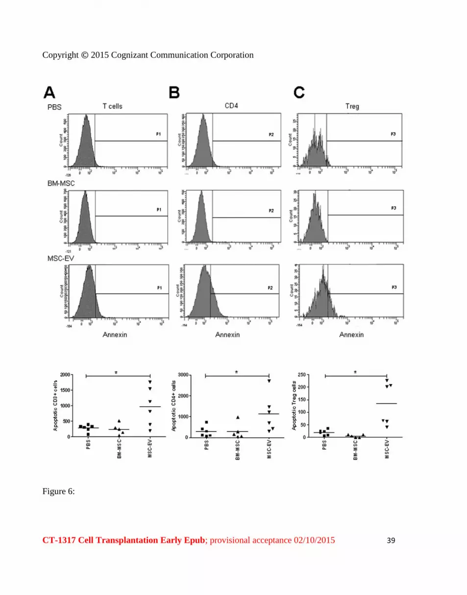
Copyright copy 2015 Cognizant Communication Corporation
CT-1317 Cell Transplantation Early Epub provisional acceptance 02102015 41
Figure 8
Copyright copy 2015 Cognizant Communication Corporation
CT-1317 Cell Transplantation Early Epub provisional acceptance 02102015 42
Figure 9

Copyright copy 2015 Cognizant Communication Corporation
CT-1317 Cell Transplantation Early Epub provisional acceptance 02102015 42
Figure 9


