Embed Size (px)
Citation preview
Cells 2021, 10, 1287. https://doi.org/10.3390/cells10061287 www.mdpi.com/journal/cells
Review
Extracellular Vesicles from Mesenchymal Stem Cells as Poten-tial Treatments for Osteoarthritis Nur Azira Mohd Noor 1, Asma Abdullah Nurul 1, Muhammad Rajaei Ahmad Mohd Zain 2, Wan Khairunnisaa Wan Nor Aduni 1 and Maryam Azlan 1,*
1 School of Health Sciences, Universiti Sains Malaysia, Health Campus, Kubang Kerian 16150, Kelantan, Malaysia; [email protected] (N.A.M.N.); [email protected] (A.A.N.); [email protected] (W.K.W.N.A.)
2 Department of Orthopaedics, School of Medical Sciences, Universiti Sains Malaysia, Health Campus, Kubang Kerian 16150, Kelantan, Malaysia; [email protected]
* Correspondence: [email protected]; Tel.: +60-9-767-7836
Abstract: Osteoarthritis (OA) is a chronic degenerative disorder of the joint and its prevalence and severity is increasing owing to ageing of the population. Osteoarthritis is characterized by the deg-radation of articular cartilage and remodeling of the underlying bone. There is little understanding of the cellular and molecular processes involved in pathophysiology of OA. Currently the treatment for OA is limited to painkillers and anti-inflammatory drugs, which only treat the symptoms. Some patients may also undergo surgical procedures to replace the damaged joints. Extracellular vesicles (EV) play an important role in intercellular communications and their concentration is elevated in the joints of OA patients, although their mechanism is unclear. Extracellular vesicles are naturally released by cells and they carry their origin cell information to be delivered to target cells. On the other hand, mesenchymal stem cells (MSCs) are highly proliferative and have a great potential in cartilage regeneration. In this review, we provide an overview of the current OA treatments and their limitations. We also discuss the role of EV in OA pathophysiology. Finally, we highlight the therapeutic potential of MSC-derived EV in OA and their challenges.
Keywords: extracellular vesicles; exosomes; mesenchymal stem cells; osteoarthritis; chondrocytes
1. Introduction Osteoarthritis (OA) is a common degenerative disorder of the joints that affects the
knee, hands, hip, spine and feet. Osteoarthritis represents a large and increasing health burden, causing patients’ function deterioration as well as extending public health costs to deal with an increasing OA prevalence worldwide. According to data from the United States Center for Disease Control and Prevention, OA affected 52.5 million people in the US in 2012 and is expected to affect 78 million people by 2040 [1]. In 2017, OA accounts for approximately 7.1% of the musculoskeletal disorders burden and showed a statisti-cally significant increase in comparison to 31.4% in 2007 (95% CI:30.7,32.1) [2,3]. Mean-while, between 1990 to 2019, the number of people affected globally by OA increased by 48% [4].
The prevalence of OA is increasing due to obesity and lifestyle. Patients with OA have limited daily activities and a greater risk of mortality [5]. Age is the main factor for OA due to ageing, muscle weakness and thinning of cartilage. Meanwhile, obesity is strongly associated with knee OA [6]. Apart from that, genetics and diet also influence the risk of OA. To date, the treatment for OA involves pain management using non-steroid anti-inflammatory drugs (NSAIDs) and pain killer to relive the symptoms [7] and surgical therapy for end-stage OA patients [8]. However, surgical therapy is costly and may lead
Citation: Mohd Noor, N.A.;
Abdullah Nurul, A.; Ahmad Mohd
Zain, M.R.; Wan Nor Aduni, W.K.;
Azlan, M. Extracellular Vesicles
from Mesenchymal Stem Cells as
Potential Treatments for
Osteoarthritis. Cells 2021, 10, 1287.
https://doi.org/10.3390/cells10061287
Academic Editor: Tong-Chuan He
Received: 14 April 2021
Accepted: 20 May 2021
Published: 22 May 2021
Publisher’s Note: MDPI stays neu-
tral with regard to jurisdictional
claims in published maps and institu-
tional affiliations.
Copyright: © 2021 by the authors. Li-
censee MDPI, Basel, Switzerland.
This article is an open access article
distributed under the terms and con-
ditions of the Creative Commons At-
tribution (CC BY) license (http://crea-
tivecommons.org/licenses/by/4.0/).
Cells 2021, 10, 1287 2 of 23
to tissue hypertrophy. Therefore, appropriate therapeutic strategies are crucial to over-come existing problems. Recently, mesenchymal stem cells (MSCs)-derived extracellular vesicles (EV) therapy has been suggested as a potential therapeutic strategy for OA in an attempt to replace invasive treatments. MSC-derived EV is safe and a promising therapy due to their unique properties including a small size, stable culture, low immunogenicity, specific targeting and carrying biologically active components which make them a poten-tial natural therapeutic delivery agent [9]. The small size of MSC-derived EV permits them to pass through cell barriers easily, thus increasing their capability in delivering genetic materials directly into the cytoplasm of the recipient cells [10,11].
This review highlights information regarding OA, MSCs and EV. In particular, this review focuses on the recent findings of potential therapeutic effects of EV derived from MSCs for OA treatment.
2. Pathophysiology of OA Osteoarthritis is a progressive chronic condition that represents a pathological imbal-
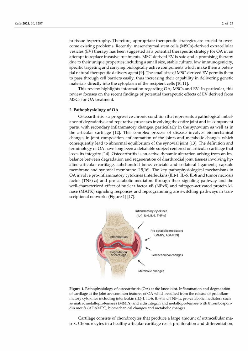
ance of degradative and reparative processes involving the entire joint and its component parts, with secondary inflammatory changes, particularly in the synovium as well as in the articular cartilage [12]. This complex process of disease involves biomechanical changes in joint composition, inflammation of the joints and metabolic changes which consequently lead to abnormal equilibrium of the synovial joint [13]. The definition and terminology of OA have long been a debatable subject centered on articular cartilage that loses its integrity [14]. Osteoarthritis is an active dynamic alteration arising from an im-balance between degradation and regeneration of diarthrodial joint tissues involving hy-aline articular cartilage, subchondral bone, cruciate and collateral ligaments, capsule membrane and synovial membrane [15,16]. The key pathophysiological mechanisms in OA involve pro-inflammatory cytokines (interleukins (IL)-1, IL-6, IL-8 and tumor necrosis factor (TNF)-α) and pro-catabolic mediators through their signaling pathway and the well-characterized effect of nuclear factor κB (NFκB) and mitogen-activated protein ki-nase (MAPK) signaling responses and reprogramming are switching pathways in tran-scriptional networks (Figure 1) [17].
Figure 1. Pathophysiology of osteoarthritis (OA) at the knee joint. Inflammation and degradation of cartilage at the joint are common features of OA which resulted from the release of proinflam-matory cytokines including interleukin (IL)-1, IL-6, IL-8 and TNF-α, pro-catabolic mediators such as matrix metalloproteinases (MMPs) and a disintegrin and metalloproteinase with thrombospon-din motifs (ADAMTS), biomechanical changes and metabolic changes.
Cartilage consists of chondrocytes that produce a large amount of extracellular ma-trix. Chondrocytes in a healthy articular cartilage resist proliferation and differentiation,
Cells 2021, 10, 1287 3 of 23
while chondrocytes in diseased cartilage proliferate and develop hypertrophy. The in-flammatory mediators, mechanical and oxidative stress compromise the function and vi-ability of chondrocytes, reprogramming them to undergo hypertrophic differentiation and early senescence, making them even more sensitive to the effects of pro-inflammatory and pro-catabolic mediators [18]. Products that are released from the cartilage matrix and chondrocytes in response to adverse mechanical forces and other factors induce the re-lease of products that deregulate chondrocyte function via paracrine and autocrine mech-anisms.
3. Treatment of OA 3.1. Osteoarthritis Management and Current Therapy
In view of the complexity of OA mechanism, a comprehensive plan for management of OA in different patients may include awareness in terms of educational plan, behav-ioral, psychosocial and physical interventions, as well as pharmacological treatment such as the use of topical, oral and intra-articular injection and also surgical treatment [19]. The principle of treatment should be addressed according to the degree of severity as well as the patient’s medical status to ensure that a personalized management strategy is tailored to their needs. The goals are to reduce symptoms and ultimately slow the disease progres-sion, which may in turn improve quality of life and consequently reduce the health cost burden.
3.2. Pharmacological and Non-Pharmacological Therapy A multidisciplinary, patient-centered combination of education, self-management,
exercise, weight loss with realistic goals, encouragement and regular reassessment is rec-ommended for individuals with OA [20]. Commonly, topical NSAIDs, oral NSAIDs and tramadol are used to treat OA. However, inconclusive evidence has been shown for acet-aminophen, non-tramadol opioids and intra articular injections of corticosteroids, hyalu-ronic acid (HA) and platelets rich plasma (PRP) [21]. Clinical trials of intra articular glu-cocorticoid injections have demonstrated a short-term efficacy in knee OA as well as for hand OA [19]. However, a recent report raised the possibility that specific steroid prepa-rations or a certain frequency of steroid injections may contribute to cartilage loss; how-ever, the clinical significance of this finding was uncertain, particularly since change in cartilage thickness was not associated with a worsening in pain, function or other radio-graphic features [22].
Surgical intervention is indicated in end stage OA when all previous treatment meth-ods have failed, a significant loss in quality of life and when the patient is severely symp-tomatic. Surgery is an effective treatment option; however, the long-term success rate is not clear and may fail depending on the longevity of the implant. Surgical techniques may include osteotomy around the knee, unicondylar arthroplasty (partial knee replacement) or total knee arthroplasty based on the patients’ condition and degree of deformity or joint destruction [23].
4. Mesenchymal Stem Cells 4.1. The Source of Mesenchymal Stem Cells, Isolation and Characterization
Mesenchymal stem cells (MSCs) were first discovered in 1966 from the bone marrow [24] and hold the concept from postnatal progenitor [25]. They are also known as multipo-tent mesenchymal stromal cells which possess two major features, including the ability to differentiate into multiple lineages and the capacity of self-renewal. According to the In-ternational Society for Cellular Therapy (ISCT), MSCs must meet three minimal criteria [26]. Firstly, MSCs must exhibit plastic-adherence when grown in vitro. Secondly, MSCs must express the surface antigens CD73, CD90 and CD105 while lacking expression of CD45, CD34, CD14 or CD11b, CD79α or CD19 and HLA-DR. Thirdly, MSCs must be able
Cells 2021, 10, 1287 4 of 23
to differentiate into mesodermal cell types (i.e., adipocytes, chondrocytes and osteoblasts) when cultured under specific conditions.
Mesenchymal stem cells can be isolated from various tissues and are not restricted to mesodermal origin-type of cells such as bone marrow, adipose, muscle or bone. They were originally found in the bone marrow [24], but later studies identified MSCs in other tissues such as in the peripheral blood, brain, spleen, liver, kidney, lung, thymus, placental, um-bilical cord and pancreas. Mesenchymal stem cells share a similar characteristic pheno-type [26] despite their presence in different tissue sources, albeit with some additional features that represent their tissue origin [27]. Although MSCs can be isolated from almost every type of connective tissues [28], studies have shown that bone marrow represents the major source of MSCs [29–31].
4.2. Mesenchymal Stem Cell-Based Therapy Mesenchymal stem cells have become a popular cell source for therapeutic purposes
due to their immunomodulation and regenerative properties [32–38]. Mesenchymal stem cells possess the capacity to migrate to injured sites in response to other cells and environ-mental signals and also promote tissue regeneration orchestrated by the paracrine secre-tion of a broad repertoire of growth factors, chemokines and cytokines [39]. Through in-teraction with the host niche, MSCs were able to secrete bioactive mediators, such as growth factors, cytokines and EV that exert immunosuppressive, anti-apoptotic, anti-fi-brotic, angiogenic and anti-inflammatory effects [40,41]. In addition, MSCs exhibited im-munomodulatory functions by preventing immune cells’ activation or proliferation [42]. The immunomodulatory function of MSCs make them a good option for cell-based ther-apy, as the possibility for cell rejection is reduced.
The ability of MSCs to differentiate into various cell types enables their use as a tool in regenerative medicine. Mesenchymal stem cells have been reported to be successfully used as a therapy against many diseases and clinical conditions. At present, there are over 950 clinical trials worldwide that have used MSCs to treat various diseases [43], including bone and cartilage repair, diabetes, cardiovascular diseases, liver disease, immune-re-lated, neurodegenerative diseases and spinal cord injuries. Mesenchymal stem cells have shown promising results in the clinical application of cell-based therapy. The efficacy of MSCs on bone regeneration in various orthopedic conditions has been widely demon-strated. Several clinical trials at different phases (I, II or III) have been performed for bone fracture repair using various sources of MSCs which were implanted either via direct in-jection or incorporated with osteogenic matrix or scaffolds which promoted bone repair and functions [44–46].
4.3. Mesenchymal Stem Cell-Based Therapy in Osteoarthritis Treatment Regenerative medicine has become increasingly popular as a promising new ap-
proach for OA since articular cartilage has a limited capacity for spontaneous intrinsic repair, and may progress to OA, owing to the sparse distribution of highly differentiated chondrocytes, the low supply of progenitor cells and the lack of vascular supply [47]. This is evidenced by wide availability in clinical practice and by publications of many case series and clinical trials.
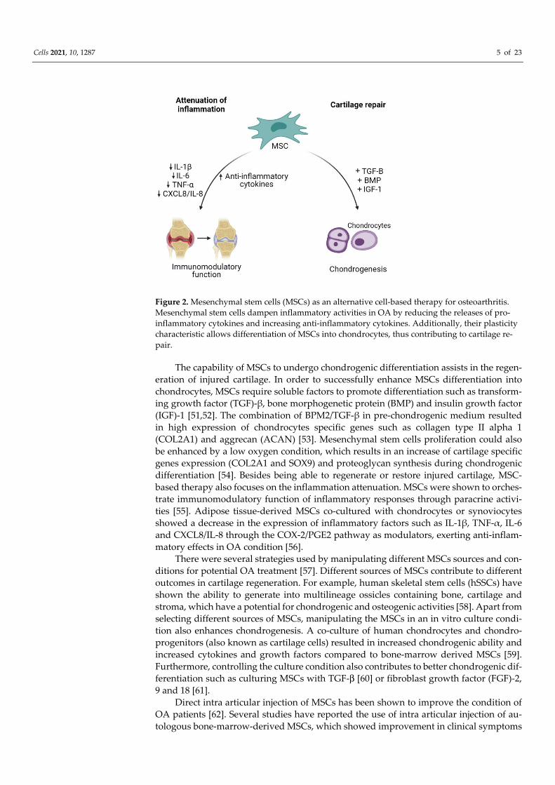
Cell-based therapy has been a promising option in OA because it is aimed at revers-ing the symptoms and pathophysiology of OA [48]. The ability of MSCs to differentiate between multiple lineages including musculoskeletal tissue supports the use of MSCs as an excellent source for degenerative musculoskeletal conditions such as in OA [49]. The therapeutic potential of MSCs in the treatment of OA is aimed at cartilage repair and res-toration (Figure 2) [50].
Cells 2021, 10, 1287 5 of 23
Figure 2. Mesenchymal stem cells (MSCs) as an alternative cell-based therapy for osteoarthritis. Mesenchymal stem cells dampen inflammatory activities in OA by reducing the releases of pro-inflammatory cytokines and increasing anti-inflammatory cytokines. Additionally, their plasticity characteristic allows differentiation of MSCs into chondrocytes, thus contributing to cartilage re-pair.
The capability of MSCs to undergo chondrogenic differentiation assists in the regen-eration of injured cartilage. In order to successfully enhance MSCs differentiation into chondrocytes, MSCs require soluble factors to promote differentiation such as transform-ing growth factor (TGF)-β, bone morphogenetic protein (BMP) and insulin growth factor (IGF)-1 [51,52]. The combination of BPM2/TGF-β in pre-chondrogenic medium resulted in high expression of chondrocytes specific genes such as collagen type II alpha 1 (COL2A1) and aggrecan (ACAN) [53]. Mesenchymal stem cells proliferation could also be enhanced by a low oxygen condition, which results in an increase of cartilage specific genes expression (COL2A1 and SOX9) and proteoglycan synthesis during chondrogenic differentiation [54]. Besides being able to regenerate or restore injured cartilage, MSC-based therapy also focuses on the inflammation attenuation. MSCs were shown to orches-trate immunomodulatory function of inflammatory responses through paracrine activi-ties [55]. Adipose tissue-derived MSCs co-cultured with chondrocytes or synoviocytes showed a decrease in the expression of inflammatory factors such as IL-1β, TNF-α, IL-6 and CXCL8/IL-8 through the COX-2/PGE2 pathway as modulators, exerting anti-inflam-matory effects in OA condition [56].
There were several strategies used by manipulating different MSCs sources and con-ditions for potential OA treatment [57]. Different sources of MSCs contribute to different outcomes in cartilage regeneration. For example, human skeletal stem cells (hSSCs) have shown the ability to generate into multilineage ossicles containing bone, cartilage and stroma, which have a potential for chondrogenic and osteogenic activities [58]. Apart from selecting different sources of MSCs, manipulating the MSCs in an in vitro culture condi-tion also enhances chondrogenesis. A co-culture of human chondrocytes and chondro-progenitors (also known as cartilage cells) resulted in increased chondrogenic ability and increased cytokines and growth factors compared to bone-marrow derived MSCs [59]. Furthermore, controlling the culture condition also contributes to better chondrogenic dif-ferentiation such as culturing MSCs with TGF-β [60] or fibroblast growth factor (FGF)-2, 9 and 18 [61].
Direct intra articular injection of MSCs has been shown to improve the condition of OA patients [62]. Several studies have reported the use of intra articular injection of au-tologous bone-marrow-derived MSCs, which showed improvement in clinical symptoms
Cells 2021, 10, 1287 6 of 23
and resulted in higher arthroscopic and histological grades than the control group [63,64]. In another study, an intra articular injection of autologous MSCs showed significant growth of cartilage and meniscus on magnetic resonance imaging (MRI), increased range of motion and modified visual analog scale (VAS) pain scores at 24-weeks post injection [65]. Intra articular injection of autologous bone marrow-derived MSCs showed signifi-cant improvement in terms of knee pain and quality of life during the 6-month follow-up [66].
Articular cartilage tissue engineering is another aspect that can be manipulated by using biomaterial constructs. Several biomaterials such as fibrin [67], biopolymer (chi-tosan) [68], synthetic polymers [69] and hydrogel [70] were widely used for their ability to degrade rapidly, their low immunogenicity and their ability to enhance MSCs prolifer-ation and chondrogenic differentiation.
Another strategy to achieve chondrogenic differentiation of MSCs is via extracellular matrix (ECM) application by using synthetic or natural scaffolds. MSCs cultured in vitro in serum free conditions or in hydrogel or scaffold materials such as polymers, alginate beads and collagen sponges will eventually promote the differentiation of MSCs into chondrocytes [71]. Early pre-clinical studies have showed promising chondrogenic differ-entiation properties when MSCs were seeded on scaffolds [72,73]. Autologous transplan-tation of rabbit MSCs with HA gel sponges showed a well-repaired cartilage tissue which resembles the articular cartilage of the surrounding structure [73]. Another study has also used intra-articular injection of autologous adipose MSCs combined with HA which sup-pressed the OA progression and promoted cartilage regeneration [74]. The first autolo-gous bone marrow-derived MSCs transplant embedded in vitro in a collagen gel was re-ported in 2004. A year later, the use of the matrix-induced autologous chondrocyte im-plantation (MACI) technique was reported by using a collagen bilayer seeded with chon-drocytes [75]. Since then, many studies reported promising findings by using the MACI technique with various types of scaffolds, especially collagen due to its natural presence in the ECM [76]. Articular cartilage regeneration using allogeneic human umbilical cord blood-derived MSCs which were expanded in HA hydrogel demonstrated production of hyaline-like cartilage after one year and the regenerated cartilage persisted after three years [77].
The use of MSCs for articular cartilage regeneration has several limitations. A tran-scriptome analyses of human neonatal articular cartilage (hNAC) and MSC-derived car-tilage has demonstrated that over 500 genes that are highly expressed in hNAC were not expressed in MSC chondrogenesis [78]. This suggests that in vitro MSC-derived cartilage may not represent the actual in vivo chondrocytes. In addition, in vitro cultured MSCs displayed a hypertrophic phenotype, thus limiting their application in articular cartilage tissue engineering [79]. Since MSCs are heterogenous, their numbers vary in tissues and different MSC sources may not be equivalent in terms of function [79,80].
5. Extracellular Vesicles 5.1. Extracellular Vesicles Biogenesis
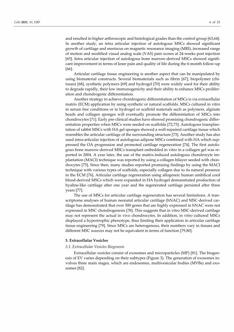
Extracellular vesicles consist of exosomes and microparticles (MP) [81]. The biogen-esis of EV varies depending on their subtypes (Figure 3). The generation of exosomes in-volves three main stages, which are endosomes, multivesicular bodies (MVBs) and exo-somes [82].
Cells 2021, 10, 1287 7 of 23
Figure 3. The formation of extracellular vesicles (EV). Exosmes are released from multivesicular bodies (MBVs) through exocytosis, while microparticles (MP) shed from cell membrane through budding.
During the endosomes stage, the formation of early endosomes is characterized by a tube-like shape, which occurs when endocytic vessels are transferred and located closer to outer edge of the cytoplasm. Subsequently, early endosomes further mature into spher-ical late endosomes and are located near the nucleus. The second stage of exosomes for-mation involves degradation of late endosomes (also known as MVBs) that carry intralu-minal vesicles upon fusion with lysosomes [83]. Exosomes are subsequently released from MVBs into the extracellular space through exocytosis of plasma membrane [84]. In con-trast, MP are generated from outward blebbing of the plasma membrane through two main steps, which are the rearrangement of cytoskeleton and externalization of phospha-tidylserine (PS) [85]. In response to cell activation, an increase of intracellular calcium ac-tivates calcium-dependent enzymes such as kinase, calpain and gelsolin and also inhibits phosphatase [86] by facilitating the cleavage of cytoskeleton proteins [87]. Calcium influx also leads to the activation of cytosolic enzymes such as flippase, floppase and scramblase [88] that mediate the externalization of PS and phosphatidylethanolamine, while internal-izing phosphatidylcholine and sphingomyelin [89]. Thus, cytoskeleton proteolysis and phospholipid imbalance favor the cellular blebbing, which ultimately leads to the shed-ding of MP [90]. In OA patients, EV may be generated from articular cartilage, which is known as articular cartilage vesicles due to the pathological process of OA [91], while EV in the synovial fluid is possibly released from chondrocytes and synoviocytes [92].
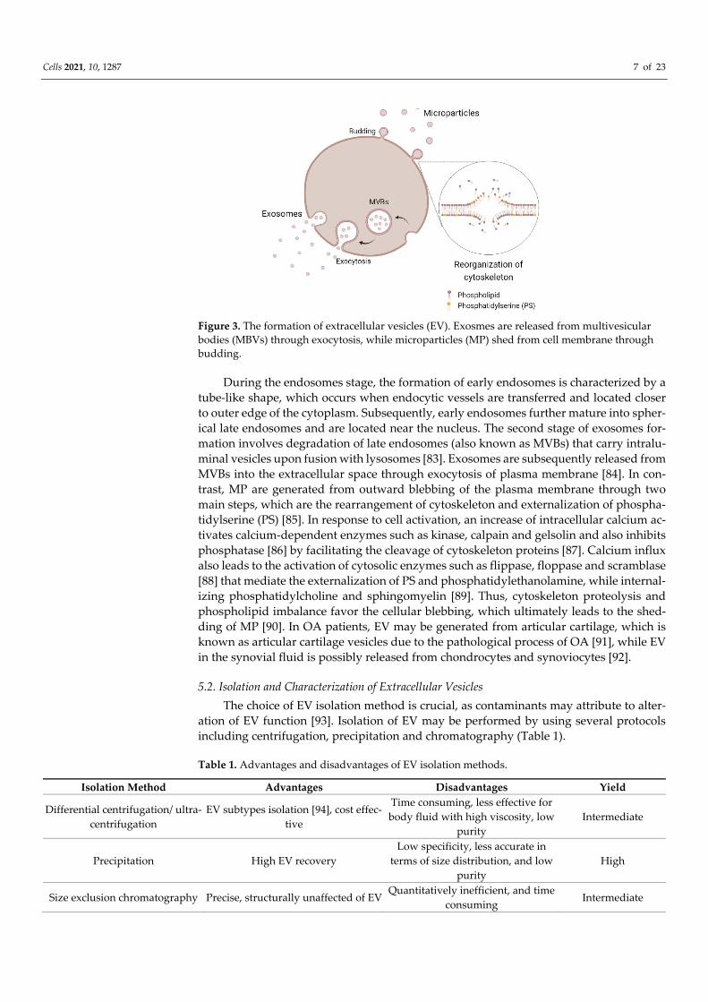
5.2. Isolation and Characterization of Extracellular Vesicles The choice of EV isolation method is crucial, as contaminants may attribute to alter-
ation of EV function [93]. Isolation of EV may be performed by using several protocols including centrifugation, precipitation and chromatography (Table 1).
Table 1. Advantages and disadvantages of EV isolation methods.
Isolation Method Advantages Disadvantages Yield
Differential centrifugation/ ultra-centrifugation
EV subtypes isolation [94], cost effec-tive
Time consuming, less effective for body fluid with high viscosity, low
purity Intermediate
Precipitation High EV recovery Low specificity, less accurate in
terms of size distribution, and low purity
High
Size exclusion chromatography Precise, structurally unaffected of EV Quantitatively inefficient, and time
consuming Intermediate
Cells 2021, 10, 1287 8 of 23
Ultrafiltration EV subtypes isolation based on size
[93], and cost effective Low specificity, and time consuming Low [95]
Field-flow fractionation (FFF) High specificity [96], accurate EV size
distribution and High EV integrity Small volume of sample Intermediate
Commercial kits (eg: ExoQuick, ExoMir kit)
High EV integrity, convenient proce-dure [97]
Costly, low purity and low repro-ducibility
Intermediate
Immunoprecipitation High purity [98], EV subtypes isola-
tion based on protein marker [99] Costly and time consuming [100] Intermediate
Immunoaffinity columns Fast and high reproducibility Low specificity [101] Intermediate
Differential centrifugation followed by ultracentrifugation is the most widely used protocol to isolate EV [102] from OA patients. This technique allows the removal of cell from the synovial fluids, since slow initial centrifugation results in cell precipitation. Meanwhile, ultracentrifugation at 20,000 xg permits the recovery of MP without exosomes contamination [103], while exosomes can be isolated from the synovial fluid with ultra-centrifugation at 100,000 to 200,000 xg [104]. However, the ultracentrifugation method re-quires a large volume of synovial fluid, is time-consuming and is only effective for body fluids with low viscosity [105]. Extracellular vesicles can also be isolated using the precip-itation method, in which synovial fluid is added into large polymers such as poly-thrilenglycol and polyethylene glycol (PEG), followed by EV precipitation [105,106]. Alt-hough the concentration of purified EV using the precipitation method was high, the pu-rity and accuracy in terms of size distribution of EV was low [107]. It has been shown that EV isolated by this method exerted similar effects in an ischemic stroke model and their corresponding cells [106,108]. Additionally, size-exclusion chromatography isolates EV from synovial fluid precisely based on size without affecting the structure of EV [105]. In fact, size-exclusion chromatography involves the penetration of particles through a col-umn, where larger particles will be eluted prior to the smaller particles [109].
The use of single isolation method usually results in low specificity; therefore, the combination of techniques in order to achieve better specificity of EV isolation is recom-mended. Apart from the techniques discussed, other techniques such as ultrafiltration, washing with EV-free buffer [110] and two-step centrifugation are commonly used. In addition, isolation of EV using combination methods of ultracentrifugation with ExoQuickTM precipitation resulted in high recovery of EV, while combination of ultracen-trifugation with density gradient centrifugation permits isolation of EV with intact mor-phology [111].
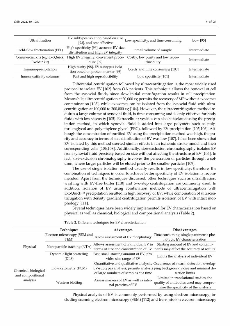
Several techniques have been widely implemented for EV characterization based on physical as well as chemical, biological and compositional analysis (Table 2).
Table 2. Different techniques for EV characterization.
Techniques Advantages Disadvantages
Physical
Electron microscopy (SEM and TEM)
Allow assessment of EV morphology Time consuming, single parametric phe-
notypic EV characterization
Nanoparticle tracking (NTA) Allows assessment of individual EV in terms of size and concentration of EV
Starting amount of EV and contami-nants may affect the accuracy of results
Dynamic light scattering (DLS)
Fast, small starting amount of EV, pro-vides size range of EV
Limits the analysis of individual EV
Chemical, biological and compositional
analysis
Flow cytometry (FCM) Quantitative and qualitative analysis,
EV-subtypes analysis, permits analysis of large numbers of samples at a time
Occurrence of swarm detection, overlap-ping background noise and minimal de-
tection limits
Western blotting Assess markers of EV as well as inter-
nal proteins of EV
Limited in translational studies, the quality of antibodies used may compro-
mise the specificity of the analysis
Physical analysis of EV is commonly performed by using electron microscopy, in-cluding scanning electron microscopy (SEM) [112] and transmission electron microscopy
Cells 2021, 10, 1287 9 of 23
(TEM). The use of electron microscopy assists in the visualization of EV morphology at a high resolution image [109]. However, this technique limits multi-parametric phenotypic EV characterization, and lengthy sample preparation. Apart from that, nanoparticle track-ing (NTA) is useful in EV characterization, as this technique provides qualitative analysis in terms of size and concentration of EV [109,113]. NTA also allows appropriate resolution in characterizing individual particles of EV. Another technique that allows physical char-acterization of EV is dynamic light scattering (DLS). In contrast to EM, DLS provides in-formation regarding the average of the size distribution of EV by determining collective mobility of EV instead of a single EV [114,115].
Furthermore, characterization of EV could be performed by implementing chemical, biological and compositional analysis. Flow cytometry (FCM) is the most widely used technique to characterize EV. This method permits the analysis of large number of sam-ples in a short time [88] and also provides high resolution of quantitative and qualitative data based on individual EV. Currently, the characterization of EV is ascertained by West-ern blotting, which mainly assesses the markers expressed on EV; thus, this technique appears as a conformational technique of EV [116]. Western blotting also enables the de-tection of both surface proteins and internal proteins of EV [109]. However, the use of Western blotting is limited in translational studies, as it requires EV in a large quantity [117]; as additionally, the specificity and reproducibility of EV analysis may interfere with the quality of the antibodies used [109].
5.3. Extracellular Vesicles in Osteoarthritis Cells in tissue and leukocytes that infiltrated the joints affected with arthritis may
release EV into the extracellular space such as in the synovial fluid. It has been previously reported that elevated levels of EV in synovial fluid from OA patients were capable of triggering synoviocytes to secrete cytokines and chemokines [118]; thus, EV could poten-tially act as a biomarker in OA. Previous study has shown the upregulation of microRNA (miR)-16-2-3p and downregulation of miR-26a-5p, miR-146a-5p and miR-6821-5p in syn-ovial fluid-derived EV from female patients with OA compared to non-OA female pa-tients [119]. In contrast, down regulation of miR-6878-3p and upregulation of miR-210-5p were observed in synovial fluid-derived EV from male OA patients compared to non-OA male patients. These results suggest that EV may be used as potential OA biomarkers in a gender-specific manner. However, further studies at the molecular level are necessary for a better understanding of EV. The use of EV as potential biomarkers in other arthritis-related diseases, particularly RA, has been previously reported. For instance, a significant increase of four-fold of Hotair expression was reported in exosomes from RA patients compared to non-RA patients [120]. Hotair is a long non coding RNA (IncRNA) that mod-ulates the migration of active macrophages to the site of inflammation. The expression of Hotair was downregulated in EV derived from blood of a non-RA patient with a high C-reactive protein (CRP). This suggests that EV may be used as a potential biomarker to diagnose RA [120].
As EV can be released by various cell types, EV also carry genetic and cytosolic com-ponents which are similar to their origin cells including cytosolic proteins, mRNA, miRNA and small non-coding RNAs such as long non-coding RNA (lncRNA) and circular RNA (circRNA) [121]. These cargos of EV modulate gene expression, altering the down-stream functions and behavior of recipient cells as well as exerting physiological and pathological effects [122]. In healthy individuals, EV derived from chondroblasts and os-teoblasts in the developmental phase were actively involved in the process of chondro-genesis by accumulating calcium and inorganic phosphate from ECM, which results in mineralization in the lumen and subsequently form hydroxyapatite crystals [91]. Several proteins and growth factors such as BMP and vascular endothelial growth factor (VEGF) have been reported to exist in EV [123]. This finding indicates that EV are also involved in angiogenesis, a process of blood vessel formation as well as in chondrocytes and oste-oblast differentiation in growth plate. In addition, EV derived from normal human AC or
Cells 2021, 10, 1287 10 of 23
AC vesicles were responsible for the neutralization of adenosine triphosphate (ATP), cal-cium and inhibition of phosphorylation, which may be deleterious to adjacent chondro-cytes [91].
To date, the role of EV in the pathogenesis of OA has been poorly understood. How-ever, previous study has reported that OA pathogenesis may be driven by interaction be-tween resident cells and immune cells, ECM of various tissues and also the synovial fluid [124]. Meanwhile, EV derived from cells within the joints may mediate the pathogenesis and progression of OA by assisting these cell-cell communications [125]. Previous study has suggested that in OA pathogenesis, EV mediates the activation of fibroblast-like syn-oviocytes by synovial macrophages and infiltrating leukocytes in the synovial membrane [92]. In response to activation, synoviocytes further release cytokines and enzymes, thus retaining joint inflammation. Additionally, EV may promote changes in subchondral bone and matrix degradation. It has been demonstrated that EV derived from chondrocytes of OA patients engage with secretion of atypical protein and enable the transfer of infor-mation between cells and pathological calcification in articular cartilage [126]. Treatment of EV from OA on macrophages resulted in secretion of proinflammatory cytokines and chemokines such as matrix metalloproteinases (MMP)-7, MMP-12, IL-1β, CXCL1, CCL8, CCL15 and CCL20, which initiate cartilage degradation and inflammation in the joints [107]. In articular chondrocytes, treatment of synovial fluid-derived EV from OA de-creases cell survival and downregulates anabolic genes expression including COL2A1 and ACAN, while increases catabolic and inflammatory gene expression including IL-6 and TNF-α [119]. The role of EV in communication between fibroblast-like synoviocytes and chondrocytes was previously investigated. It was found that exosomes derived from IL-1β-stimulated synovial fibroblasts resulted in OA-related gene expression in articular chondrocytes such as ACAN and MMP-13 [127], which further leads to degradation of ECM, thus promoting the progression of OA.
5.4. Therapeutic Potential of MSC-Drived EV in Osteoarthritis The capability of cartilage to regenerate or self-recover in OA is limited. Mesenchy-
mal stem cells-derived EV offer a new therapeutic strategy for OA. The mechanism regu-lated by MSC-derived EV in OA treatment may be mainly through their genetic compo-nents that promoting paracrine action [128,129]. Mesenchymal stem cells-derived EV also possess immunomodulatory properties. A previous report demonstrated that MSC-de-rived EV were responsible for suppressing pro-inflammatory cytokines and elevating anti-inflammatory cytokine secretions [130]. This suggests that MSC-derived EV could potentially be used in future OA treatment. The potential of MSC-derived EV in OA treatment has been extensively studied (Table 3). A previous study has demonstrated that MSC-derived EV protected a collagenase-in-duced OA mice model from joint damage [131]. Such protection includes prevention of bone and cartilage in OA, which could be due to upregulation of COL2A1 and ACAN, downregulation of MMP-13, a disintegrin and metalloproteinase with thrombospondin motifs (ADAMTS)-5 and inflammatory markers, as well as preventing apoptosis of chon-drocytes and macrophage activation. Additionally, the injection of intra articular of EV derived from human embryonic MSCs in femur resulted in regeneration and repair of osteochondral defect, subchondral bone and cartilage, as well as deposition of normal ECM in a rat OA model [132]. A comparativestudy between synovial membrane MSC-derived EV (SMMSC-EV) and EV secreted from induced-pluripotent stem cell-derived MSCs (iMSC-EV) found that both SMMSC-EV and iMSC-EV treatment stimulated the proliferation and migration of chondrocytes and attenuated OA in a collagenase-induced mouse model [133]. Similarly, treatment with EV from human bone marrow-derived MSCs (hBMSC-EV) promoted cartilage repair by triggering the production of ECM by OA chondrocytes and improving inflammatory response in vitro [134].
Cells 2021, 10, 1287 11 of 23
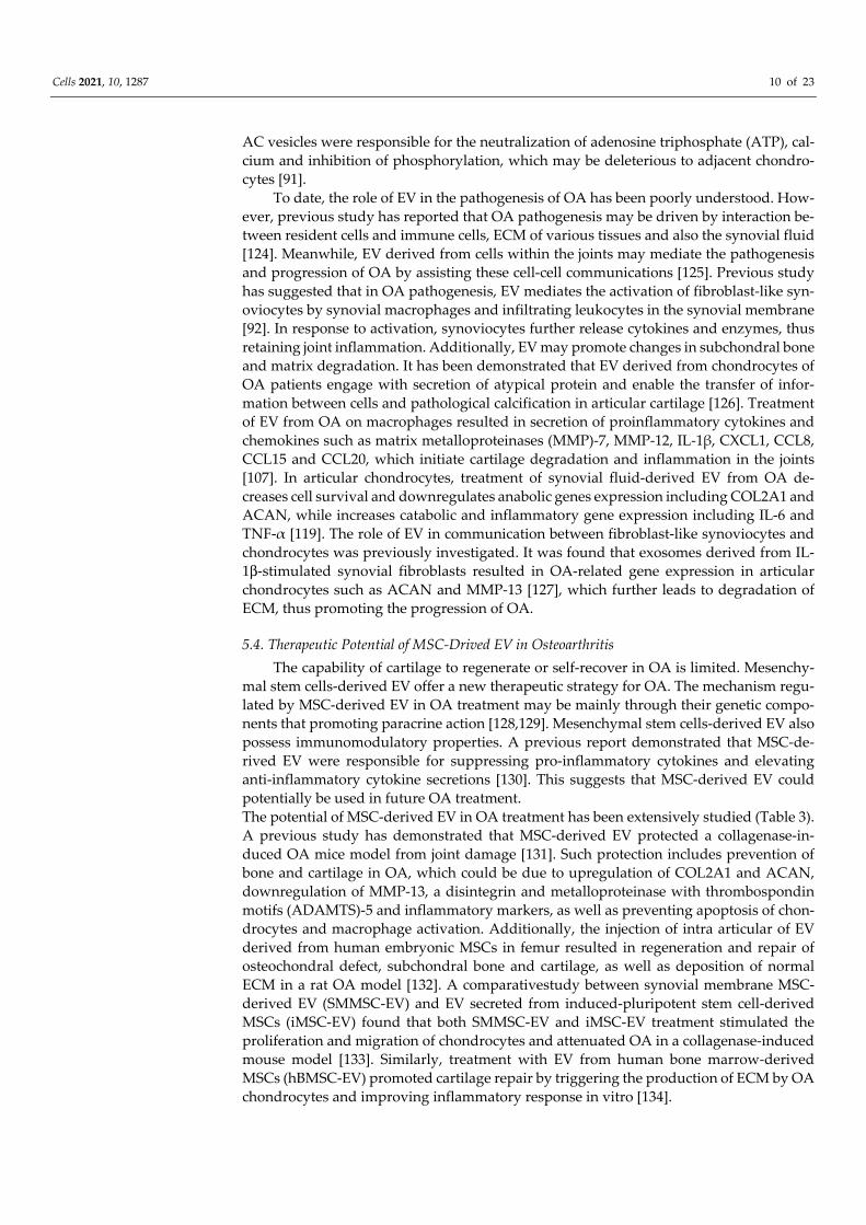
Table 3. Therapeutic evidence of MSC-derived EV in OA treatment.
Type of EV Model Marker Time Point of Assay Specific Character-
istic of In Vivo or In Vitro Studies
Findings References
Exosome derived from human bone marrow-derived
MSCs (MCS-Exos and MSC-miR92a-
3p-Exos)
i. Human bone mar-row MSCs (normal
and OA)
ii. Mouse (collagen-ese-induced OA)
i. MSCs: CD73, CD90, CD105
ii. MSC-ExoS: CD9, CD63, CD81, and
HSP70
i. Proliferation assay. MCS-Exos and MSC-
miR92a-3p-Exos were in-cubated with normal and OA chondrocytes for 0–5
days
ii. Transfection. MSCs were transfected with
miR-92a-3p mimic or in-hibitor for 48 h (qRT-PCR)
and 72 h (western blot)
iii. In vivo study. MCS-Exos and MSC-miR92a-3p-
Exos were injected into collagenase-induced OA
mice after 7, 14 and 21 days following OA induc-
tion
i. Proliferation as-say. 200 μg exo-somes/mL were
used
ii. Transfection. 50 nM of miR-92a-3p mimic or inhibitor
were used
iii. In vivo study. 500 μg/mL of MSC-Exos and MSC-miR-
92a-3p-Exos were used
i. Both MSC-Exos and MSC-miR92a-3p-Exos significantly en-hance chondocytes proliferation
compared to control group, where MSC-miR92a-3p-Exos ex-
ert a more potent effect com-pared to MSC-Exos.
ii. MSC-miR92a-3p-Exos signifi-
cantly upregulated ACAN, COL2A1, SOX9, while COL10A1,
RUNX2, MMP-13 and WNT5A were significantly downregu-
lated. This indicates the capabil-ity of MSC-miR92a-3p-Exos in enhancing cartilage develop-
ment.
iii. Both MSC-miR-92a-3p-Exos and MSC-Exos prevented carti-
lage matrix loss compared to the OA group, where the level of
COL2A1 and ACAN were signif-icantly better with the presence
of MSC-miR-92a-3p-Exos.
[135]
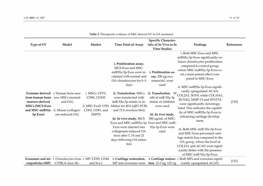
Exosomes and mi-croparticles (MP)
i. Chondocytes from C57BL/6 mice (IL-
i. MP: CD29, CD44 and Sca-1
i. Cartilage restoration. MP and exosomes were
i. Cartilage restora-tion. 12.5 ng; 125 ng
i. Both MPs and exosomes signif-icantly upregulated ACAN,
[131]
Cells 2021, 10, 1287 12 of 23
derived from mu-rine bone marrow-
derived MSCs
1β-induced OA-like phenotype)
ii. Mouse (collagen-
ese-induced OA)
ii. Exosomes: CD9,
CD81
incubated with chondro-cytes following OA induc-
tion for 24h
ii. Apoptosis induction. MPs or exosomes were
added into chondrocytes-MSCs cocultures for 6 h
iii. Monocytes activation.
MP and exosomes were incubated with murine spleen-derived macro-
phage for 3 days
iv. In vivo study. MPs and exosomes were injected
into mouse at day 7 after OA induction and har-
vested at day 42
or 1.25 μg of MP and exosomes were used
ii. Apoptosis induc-tion. 125 ng or 250 ng of MP and exo-somes were used
iii. Monocytes acti-vation. 50 ng of MPs
or exos were used
iv. In vivo study 500 ng/5 μL of MP or 250 ng/5 μL of exosomes
were used
COL1 and COL2B expression in a dose-dependent manner and
down-regulated MMP-13, ADAMTS-5 and inflammatory iNOS, thus indicating MP and
exosomes exhibited a condropro-tective effect
ii. MPs and exosomes prevented apoptosis and reduced the level of apoptotic chondrocytes with significantly lower doses than
MSCs, where Exos exert a more potent effect compared to MPs.
iii. MP and exosomes inhibiedt the expression of CD86, MHCII or CD40 as well as downregu-
lated TNF-α and upregulated IL-10, and thus possessed an immu-
nosuppresive effect
iv. MP and exosomes signifi-cantly improved volume, carti-lage degradation (surface/vol-
ume ratio) and thickness of artic-ular cartilage, indicating protec-
tion of cartilage degradation Exosomes derived from human em-bryonic stem cell (HuECS)-derived
MSCs
Rat (osteochondral defect)
Exosomes: CD81, TSG101
Intra-articular injections of exosomes or PBS were weekly administered at the site of osteochondral defect for 12 weeks and
100 μg exosomes was administered
i. At 12 weeks, exosomes pro-moted almost complete neotissue coverage with good surface regu-
larity and complete integration with the adjacent cartilage.
[132]
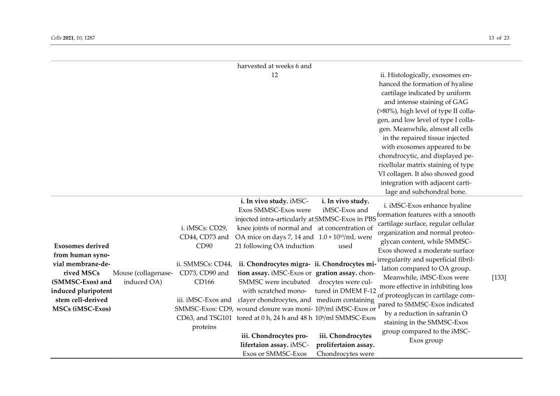
Cells 2021, 10, 1287 13 of 23
harvested at weeks 6 and 12
ii. Histologically, exosomes en-hanced the formation of hyaline cartilage indicated by uniform and intense staining of GAG
(>80%), high level of type II colla-gen, and low level of type I colla-gen. Meanwhile, almost all cells
in the repaired tissue injected with exosomes appeared to be
chondrocytic, and displayed pe-ricellular matrix staining of type VI collagen. It also showed good integration with adjacent carti-
lage and subchondral bone.
Exosomes derived from human syno-vial membrane-de-
rived MSCs (SMMSC-Exos) and induced pluripotent
stem cell-derived MSCs (iMSC-Exos)
Mouse (collagenase-induced OA)
i. iMSCs: CD29, CD44, CD73 and
CD90
ii. SMMSCs: CD44, CD73, CD90 and
CD166
iii. iMSC-Exos and SMMSC-Exos: CD9, CD63, and TSG101
proteins
i. In vivo study. iMSC-Exos SMMSC-Exos were
injected intra-articularly at knee joints of normal and
OA mice on days 7, 14 and 21 following OA induction
ii. Chondrocytes migra-tion assay. iMSC-Exos or SMMSC were incubated
with scratched mono-clayer chondrocytes, and wound closure was moni-tored at 0 h, 24 h and 48 h
iii. Chondrocytes pro-lifertaion assay. iMSC-Exos or SMMSC-Exos
i. In vivo study. iMSC-Exos and
SMMSC-Exos in PBS at concentration of 1.0 × 10¹⁰/mL were
used
ii. Chondrocytes mi-gration assay. chon-drocytes were cul-
tured in DMEM F-12 medium containing 108/ml iMSC-Exos or 108/ml SMMSC-Exos
iii. Chondrocytes
prolifertaion assay. Chondrocytes were
i. iMSC-Exos enhance hyaline formation features with a smooth cartilage surface, regular cellular organization and normal proteo-glycan content, while SMMSC-
Exos showed a moderate surface irregularity and superficial fibril-
lation compared to OA group. Meanwhile, iMSC-Exos were
more effective in inhibiting loss of proteoglycan in cartilage com-pared to SMMSC-Exos indicated
by a reduction in safranin O staining in the SMMSC-Exos
group compared to the iMSC-Exos group
[133]
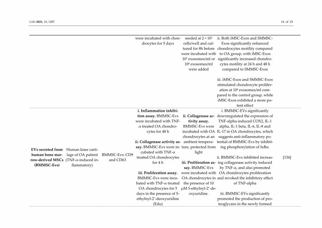
Cells 2021, 10, 1287 14 of 23
were incubated with chon-drocytes for 5 days
seeded at 2 × 10³ cells/well and cul-tured for 8h before
were incubated with 107 exosomes/ml or
108 exosomes/ml were added
ii. Both iMSC-Exos and SMMSC-Exos significantly enhanced
chondrocytes motility compared to OA group, with iMSC-Exos
significantly increased chondro-cytes motility at 24 h and 48 h
compared to SMMSC-Exos
iii. iMSC-Exos and SMMSC-Exos stimulated chondrocyte prolifer-
ation at 108 exosomes/ml com-pared to the control group, while iMSC-Exos exhibited a more po-
tent effect
EVs secreted from human bone mar-
row-derived MSCs (BMMSC-Evs)
Human knee carti-lage of OA patient
(TNF-α-induced in-flammatory)
BMMSC-Evs: CD9 and CD63
i. Inflammation inhibi-tion assay. BMMSC-Evs
were incubated with TNF-α treated OA chondro-
cytes for 48 h
ii. Collagenase activity as-say. BMMSC-Evs were in-
cubated with TNF-α treated OA chondrocytes
for 4 h
iii. Proliferation assay. BMMSC-Evs were incu-
bated with TNF-α treated OA chondrocytes for 5
days in the presence of 5-ethylnyl-2’-deoxyuridine
(Edu)
ii. Collagenase ac-tivity assay.
BMMSC-Evs were incubated with OA chondrocytes at an ambient tempera-
ture, protected from light
iii. Proliferation as-
say. BMMSC-Evs were incubated with OA chondrocytes in the presence of 10 μM 5-ethylnyl-2’-de-
oxyuridine.
i. BMMSC-EVs significantly downregulated the expression of TNF-alpha-induced COX2, IL-1 alpha, IL-1 beta, IL-6, IL-8 and
IL-17 in OA chondrocytes, which suggests anti-inflammatory po-
tential of BMMSC-Evs by inhibit-ing phosphorylation of IκBα.
ii. BMMSC-Evs inhibited increas-ing collagenase activity induced
by TNF-α, and also promoted OA chondrocytes proliferation
and revoked the inhibitory effect of TNF-alpha
iii. BMMSC-EVs significantly
promoted the production of pro-teoglycans in the newly formed
[134]
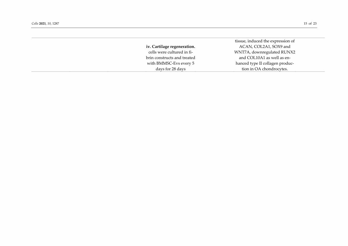
Cells 2021, 10, 1287 15 of 23
iv. Cartilage regeneration.
cells were cultured in fi-brin constructs and treated with BMMSC-Evs every 5
days for 28 days
tissue, induced the expression of ACAN, COL2A1, SOX9 and
WNT7A, downregulated RUNX2 and COL10A1 as well as en-
hanced type II collagen produc-tion in OA chondrocytes.
Cells 2021, 10, 1287 16 of 23
Osteoarthritis treatment with EV from adipose tissue-derived MSCs has shown a promis-ing alternative therapy. It was demonstrated that infrapatellar pat pad MSC-derived EV may prevent cartilage degradation and enhance the chondrocytes autophagy level by in-hibiting the rapamycin signaling pathway [136].
Since heterogenous cell entities of MSCs may give rise to different EVs with different functions, whether they exert similar therapeutic effects on OA is still questionable. In addition, different EV preparations may also influence their therapeutic activities. Fur-thermore, not all MSC sub-types are capable of mediating clinical impacts on OA, and neither are their EVs. Therefore, further study on MSC-derived EVs is needed for better understanding of their mechanism in OA treatments.
5.5. The Promise and Challenges of EV as a Therapeutic Delivery System Recently, MSC-derived EVs have obtained interest from regenerative medicine due
to their high therapeutic efficiency. They have been suggested as a clinical effective deliv-ery agent in OA treatment, as they meet the focus of clinical therapy by exerting two im-portant therapeutic effects including cartilage protection and regeneration as well as an anti-inflammatory effect. Additionally, MSC-derived EVs is a promising therapeutic agent with high sustainability, a non-invasive collection process and highly reproducible and safe characteristics [137], which is indicated by its low toxicity and low immunogen-icity [138]. The use of MSC-derived EVs also confers a few other benefits such as the ability to cross the biological barriers [139], as they are able to communicate directly with the target cells, thus offering rapid clearance, lowering the risks, and reducing toxicity [140]. Moreover, MSC-derived EVs may avoid immunogenic reactions such as immune rejection due to a lack of MHC class I/II [141].
Despite the therapeutic potential of MSC-derived EVs, a number of challenges re-main, including exosome molecular diversity, a lack of exosomal targeting properties [142], and excessive transfer of gene information [143]. A large-scale MSC-EV isolation process, for example, may pose technical and experimental challenges. The existing EV generation methods such as tissue culture methods in flask limits the large-scale produc-tion of EV, thus hindering the use of EV in clinical therapeutic applications [144]. Alt-hough sustainable quantities of EV may be harvested through the long-term passaging method, this method may lead to differentiation of MSC [145]. Apart from that, reliable EV characterization methods, rapid and precise methods for characterizing EV functional cargo, as well as the pharmacokinetics and transfer mechanism of MSC-EVs remains un-clear. MSC-EV has been suggested to have a therapeutic effect via miRNA, but cytoplas-mic and membrane proteins, mRNA and small non-coding RNA, all of which can be passed to recipient cells [146] but are not always evaluated.
6. Conclusions In conclusion, both MSCs and EVs play a significant role in immunomodulation of
OA. Therefore, MSC-derived EV-based therapy has a potential role in enhancing dam-aged articular tissue repair. As MSC-derived EV are secreted under physiological and pathological conditions, they may exert different effects via different pathways. The mem-brane as well as cytosolic protein and lipids and genetic components including mRNAs siRNA, miRNAs and ribosomal RNAs of EV are the main factors that allow them to facil-itate cell-to-cell interaction. MSC-derived EV therapy offers a potential safe approach for OA treatment. Studying the potency assay of MSC-derived EVs is challenging.
Although previous findings have demonstrated a promising therapeutic effect of MSC-derived EVs which was similar to MSC; however, several critical issues should be considered in designing MSC-derived EV therapy, including the source of EV as distinct MSC may give rise to different EV subtypes with different biological effects and standard EV preparation protocols, since independent MSC-derived EVs preparation may differ in
Cells 2021, 10, 1287 17 of 23
regards to their therapeutic potentials, a safe and effective administration route with proper targeted treatment sites as well as a sufficient dose and frequency of EV admin-istration to inflict an optimal therapeutic effect on OA or arthritis-related disease gener-ally. Therefore, further functional testing of MSC-derived EVs, particularly for OA treat-ment, is necessary.
Author Contributions: Conceptualization, A.A.N., M.R.A.M.Z. and M.A.; formal analysis, N.A.M.N., M.R.A.M.Z. and W.K.W.N.A.; writing—original draft preparation, N.A.M.N., M.R.A.M.Z. and W.K.W.N.A.; writing—review and editing, N.A.M.N., A.A.N., M.R.A.M.Z. and M.A.; supervision, A.A.N. and M.A. All authors have read and agreed to the published version of the manuscript.
Funding: This research was funded by the Fundamental Research Grant Scheme (FRGS) provided by the Ministry of Higher Education Malaysia (grant number: 203.PPSK.6171255).
Institutional Review Board Statement: Not applicable.
Informed Consent Statement: Not applicable.
Data Availability Statement: No new data were created or analyzed in this study. Data sharing is not applicable to this article.
Conflicts of Interest: The authors declare no conflict of interest.
References 1. Hootman, J.M.; Helmick, C.G.; Barbour, K.E.; Theis, K.A.; Boring, M.A.J.A. Rheumatology. Updated projected prevalence of
self-reported doctor-diagnosed arthritis and arthritis-attributable activity limitation among US adults, 2015–2040. Arthritis Rheu-matol. 2016, 68, 1582–1587.
2. Kloppenburg, M.; Berenbaum, F.J.O. Osteoarthritis year in review 2019: Epidemiology and therapy. Osteoarthr. Cartil. 2020, 28, 242–248.
3. Vos, T.; Allen, C.; Arora, M.; Barber, R.M.; Bhutta, Z.A.; Brown, A.; Carter, A.; Casey, D.C.; Charlson, F.J.; Chen, A.Z.J.T.l. Global, regional, and national incidence, prevalence, and years lived with disability for 310 diseases and injuries, 1990–2015: A system-atic analysis for the Global Burden of Disease Study 2015. Lancet 2016, 388, 1545–1602.
4. Hunter, D.J.; March, L.; Chew, M. Osteoarthritis in 2020 and beyond: A Lancet Commission. Lancet 2020, 396, 1711–1712, doi:10.1016/S0140-6736(20)32230-3.
5. Palazzo, C.; Nguyen, C.; Lefevre-Colau, M.M.; Rannou, F.; Poiraudeau, S. Risk factors and burden of osteoarthritis. Ann. Phys. Rehabil. Med. 2016, 59, 134–138, doi:10.1016/j.rehab.2016.01.006.
6. Silverwood, V.; Blagojevic-Bucknall, M.; Jinks, C.; Jordan, J.L.; Protheroe, J.; Jordan, K.P. Current evidence on risk factors for knee osteoarthritis in older adults: A systematic review and meta-analysis. Osteoarthr. Cartil. 2015, 23, 507–515, doi:10.1016/j.joca.2014.11.019.
7. Rannou, F.; Pelletier, J.-P.; Martel-Pelletier, J. Efficacy and safety of topical NSAIDs in the management of osteoarthritis: Evi-dence from real-life setting trials and surveys. Semin. Arthritis Rheum. 2016, 45, S18–S21, doi:10.1016/j.semarthrit.2015.11.007.
8. Okoro, T.; Morrison, V.; Maddison, P.; Lemmey, A.B.; Andrew, J.G. An assessment of the impact of behavioural cognitions on function in patients partaking in a trial of early home-based progressive resistance training after total hip replacement surgery. Disabil. Rehabil. 2013, 35, 2000–2007, doi:10.3109/09638288.2013.770082.
9. Li, J.J.; Hosseini-Beheshti, E.; Grau, G.E.; Zreiqat, H.; Little, C.B. Stem cell-derived extracellular vesicles for treating joint injury and osteoarthritis. Nanomaterials 2019, 9, 261, doi:10.3390/nano9020261.
10. Jiang, L.; Vader, P.; Schiffelers, R.M. Extracellular vesicles for nucleic acid delivery: Progress and prospects for safe RNA-based gene therapy. Gene Ther. 2017, 24, 157–166, doi:10.1038/gt.2017.8.
11. Mehrotra, N.; Tripathi, R.M. Short interfering RNA therapeutics: Nanocarriers, prospects and limitations. IET Nanobiotechnol. 2015, 9, 386–395, doi:10.1049/iet-nbt.2015.0018.
12. Martel-Pelletier, J.; Barr, A.J.; Cicuttini, F.M.; Conaghan, P.G.; Cooper, C.; Goldring, M.B.; Goldring, S.R.; Jones, G.; Teichtahl, A.J.; Pelletier, J.-P. Osteoarthritis. Nat. Rev. Dis. Primers 2016, 2, 16072, doi:10.1038/nrdp.2016.72.
13. Hunter, D.J.; Bierma-Zeinstra, S. Osteoarthritis. Lancet 2019, 393, 1745–1759, doi:10.1016/S0140-6736(19)30417-9. 14. Loeser, R.F.; Collins, J.A.; Diekman, B.O.J.N.R.R. Ageing and the pathogenesis of osteoarthritis. Nat. Rev. Rheumatol. 2016, 12,
412–420. 15. Fu, K.; Robbins, S.R.; McDougall, J.J.J.R. Osteoarthritis: The genesis of pain. Rheumatology 2018, 57, iv43–iv50. 16. Prieto-Alhambra, D.; Arden, N.; Hunter, D.J. Osteoarthritis: The Facts, 2nd ed.; OUP Oxford: Oxford, UK, 2014. 17. Liu-Bryan, R.; Terkeltaub, R.J.N.R.R. Emerging regulators of the inflammatory process in osteoarthritis. Nat. Rev. Rheumatol.
2015, 11, 35. 18. Mobasheri, A.; Batt, M. An update on the pathophysiology of osteoarthritis. Ann. Phys. Rehabil. Med. 2016, 59, 333–339.
Cells 2021, 10, 1287 18 of 23
19. Kolasinski, S.L.; Neogi, T.; Hochberg, M.C.; Oatis, C.; Guyatt, G.; Block, J.; Callahan, L.; Copenhaver, C.; Dodge, C.; Felson, D.J.A.; et al. 2019 American College of Rheumatology/Arthritis Foundation guideline for the management of osteoarthritis of the hand, hip, and knee. Arthritis Rheumatol. 2020, 72, 220–233.
20. Fernandes, L.; Hagen, K.B.; Bijlsma, J.W.; Andreassen, O.; Christensen, P.; Conaghan, P.G.; Doherty, M.; Geenen, R.; Hammond, A.; Kjeken, I.; et al. EULAR recommendations for the non-pharmacological core management of hip and knee osteoarthritis. Ann. Rheum. Dis. 2013, 72, 1125–1135.
21. Jevsevar, D.S. Treatment of osteoarthritis of the knee: Evidence-based guideline, 2nd edition. J. Am. Acad. Orthop. Surg. 2013, 21, 571–576, doi:10.5435/jaaos-21-09-571.
22. McAlindon, T.E.; LaValley, M.P.; Harvey, W.F.; Price, L.L.; Driban, J.B.; Zhang, M.; Ward, R.J.J.J. Effect of intra-articular tri-amcinolone vs saline on knee cartilage volume and pain in patients with knee osteoarthritis: A randomized clinical trial. JAMA 2017, 317, 1967–1975.
23. Liu, C.Y.; Li, C.D.; Wang, L.; Ren, S.; Yu, F.B.; Li, J.G.; Ma, J.X.; Ma, X.L. Function scores of different surgeries in the treatment of knee osteoarthritis: A PRISMA-compliant systematic review and network-meta analysis. Medicine 2018, 97, e10828, doi:10.1097/md.0000000000010828.
24. Friedenstein, A.J.; Chailakhjan, R.K.; Lalykina, K.S. The development of fibroblast colonies in monolayer cultures of guinea-pig bone marrow and spleen cells. Cell Tissue Kinet. 1970, 3, 393–403, doi:10.1111/j.1365-2184.1970.tb00347.x.
25. Bianco, P.; Robey, P.G.; Simmons, P.J. Mesenchymal stem cells: Revisiting history, concepts, and assays. Cell Stem Cell 2008, 2, 313–319, doi:10.1016/j.stem.2008.03.002.
26. Dominici, M.; Le Blanc, K.; Mueller, I.; Slaper-Cortenbach, I.; Marini, F.; Krause, D.; Deans, R.; Keating, A.; Prockop, D.; Horwitz, E. Minimal criteria for defining multipotent mesenchymal stromal cells. The International Society for Cellular Therapy position statement. Cytotherapy 2006, 8, 315–317, doi:10.1080/14653240600855905.
27. Hass, R.; Kasper, C.; Böhm, S.; Jacobs, R. Different populations and sources of human mesenchymal stem cells (MSC): A com-parison of adult and neonatal tissue-derived MSC. Cell Commun. Signal. 2011, 9, 12, doi:10.1186/1478-811x-9-12.
28. da Silva Meirelles, L.; Chagastelles, P.C.; Nardi, N.B. Mesenchymal stem cells reside in virtually all post-natal organs and tissues. J. Cell Sci. 2006, 119, 2204–2213, doi:10.1242/jcs.02932.
29. in ‘t Anker, P.S.; Noort, W.A.; Scherjon, S.A.; Kleijburg-van der Keur, C.; Kruisselbrink, A.B.; van Bezooijen, R.L.; Beekhuizen, W.; Willemze, R.; Kanhai, H.H.; Fibbe, W.E. Mesenchymal stem cells in human second-trimester bone marrow, liver, lung, and spleen exhibit a similar immunophenotype but a heterogeneous multilineage differentiation potential. Haematologica 2003, 88, 845–852.
30. Wexler, S.A.; Donaldson, C.; Denning-Kendall, P.; Rice, C.; Bradley, B.; Hows, J.M. Adult bone marrow is a rich source of human mesenchymal ‘stem’ cells but umbilical cord and mobilized adult blood are not. Br. J. Haematol. 2003, 121, 368–374, doi:10.1046/j.1365-2141.2003.04284.x.
31. Zeddou, M.; Briquet, A.; Relic, B.; Josse, C.; Malaise, M.G.; Gothot, A.; Lechanteur, C.; Beguin, Y. The umbilical cord matrix is a better source of mesenchymal stem cells (MSC) than the umbilical cord blood. Cell Biol. Int. 2010, 34, 693–701, doi:10.1042/cbi20090414.
32. Connick, P.; Kolappan, M.; Crawley, C.; Webber, D.J.; Patani, R.; Michell, A.W.; Du, M.-Q.; Luan, S.-L.; Altmann, D.R.; Thomp-son, A.J.; et al. Autologous mesenchymal stem cells for the treatment of secondary progressive multiple sclerosis: An open-label phase 2a proof-of-concept study. Lancet Neurol. 2012, 11, 150–156, doi:10.1016/S1474-4422(11)70305-2.
33. Götherström, C.; Westgren, M.; Shaw, S.W.; Aström, E.; Biswas, A.; Byers, P.H.; Mattar, C.N.; Graham, G.E.; Taslimi, J.; Ewald, U.; et al. Pre- and postnatal transplantation of fetal mesenchymal stem cells in osteogenesis imperfecta: A two-center experience. Stem Cells Transl. Med. 2014, 3, 255–264, doi:10.5966/sctm.2013-0090.
34. Karantalis, V.; DiFede, D.L.; Gerstenblith, G.; Pham, S.; Symes, J.; Zambrano, J.P.; Fishman, J.; Pattany, P.; McNiece, I.; Conte, J.; et al. Autologous mesenchymal stem cells produce concordant improvements in regional function, tissue perfusion, and fibrotic burden when administered to patients undergoing coronary artery bypass grafting: The Prospective Randomized Study of Mesenchymal Stem Cell Therapy in Patients Undergoing Cardiac Surgery (PROMETHEUS) trial. Circ. Res. 2014, 114, 1302–1310, doi:10.1161/circresaha.114.303180.
35. Rushkevich, Y.N.; Kosmacheva, S.M.; Zabrodets, G.V.; Ignatenko, S.I.; Goncharova, N.V.; Severin, I.N.; Likhachev, S.A.; Potap-nev, M.P. The use of autologous mesenchymal stem cells for cell therapy of patients with amyotrophic lateral sclerosis in belarus. Bull. Exp. Biol. Med. 2015, 159, 576–581, doi:10.1007/s10517-015-3017-3.
36. Thakkar, U.G.; Trivedi, H.L.; Vanikar, A.V.; Dave, S.D. Insulin-secreting adipose-derived mesenchymal stromal cells with bone marrow-derived hematopoietic stem cells from autologous and allogenic sources for type 1 diabetes mellitus. Cytotherapy 2015, 17, 940–947, doi:10.1016/j.jcyt.2015.03.608.
37. Vega, A.; Martín-Ferrero, M.; Canto, F.; Alberca, M.; Garcia, V.; Munar, A.; Orozco, L.; Soler, R.; Fuertes, J.; Huguet, M.; et al. Treatment of knee osteoarthritis with allogeneic bone marrow mesenchymal stem cells: A randomized controlled trial. Trans-plantation 2015, doi:10.1097/TP.0000000000000678.
38. Fernández, O.; Izquierdo, G.; Fernández, V.; Leyva, L.; Reyes, V.; Guerrero, M.; León, A.; Arnaiz, C.; Navarro, G.; Páramo, M.D.; et al. Adipose-derived mesenchymal stem cells (AdMSC) for the treatment of secondary-progressive multiple sclerosis: A triple blinded, placebo controlled, randomized phase I/II safety and feasibility study. PLoS ONE 2018, 13, e0195891, doi:10.1371/jour-nal.pone.0195891.
Cells 2021, 10, 1287 19 of 23
39. Yao, Y.; Huang, J.; Geng, Y.; Qian, H.; Wang, F.; Liu, X.; Shang, M.; Nie, S.; Liu, N.; Du, X.; et al. Paracrine action of mesenchymal stem cells revealed by single cell gene profiling in infarcted murine hearts. PLoS ONE 2015, 10, e0129164–e0129164, doi:10.1371/journal.pone.0129164.
40. Fan, X.L.; Zhang, Z.; Ma, C.Y.; Fu, Q.L. Mesenchymal stem cells for inflammatory airway disorders: Promises and challenges. Biosci. Rep. 2019, 39, doi:10.1042/bsr20182160.
41. Caplan, A.I.; Dennis, J.E. Mesenchymal stem cells as trophic mediators. J. Cell Biochem. 2006, 98, 1076–1084, doi:10.1002/jcb.20886. 42. Mundra, V.; Gerling, I.C.; Mahato, R.I.J.M.p. Mesenchymal stem cell-based therapy. Mol. Pharm. 2013, 10, 77–89. 43. National Institutes of Health. Available online: www.clinicaltrials.gov (accessed on 13 October 2020). 44. Ho-Shui-Ling, A.; Bolander, J.; Rustom, L.E.; Johnson, A.W.; Luyten, F.P.; Picart, C. Bone regeneration strategies: Engineered
scaffolds, bioactive molecules and stem cells current stage and future perspectives. Biomaterials 2018, 180, 143–162, doi:10.1016/j.biomaterials.2018.07.017.
45. Calabrese, G.; Giuffrida, R.; Forte, S.; Fabbi, C.; Figallo, E.; Salvatorelli, L.; Memeo, L.; Parenti, R.; Gulisano, M.; Gulino, R. Human adipose-derived mesenchymal stem cells seeded into a collagen-hydroxyapatite scaffold promote bone augmentation after implantation in the mouse. Sci. Rep. 2017, 7, 7110, doi:10.1038/s41598-017-07672-0.
46. Gómez-Barrena, E.; Rosset, P.; Lozano, D.; Stanovici, J.; Ermthaller, C.; Gerbhard, F. Bone fracture healing: Cell therapy in de-layed unions and nonunions. Bone 2015, 70, 93–101, doi:10.1016/j.bone.2014.07.033.
47. McIntyre, J.A.; Jones, I.A.; Han, B.; Vangsness, C.T., Jr. Intra-articular mesenchymal stem cell therapy for the human joint: A systematic review. Am. J. Sports Med. 2018, 46, 3550–3563, doi:10.1177/0363546517735844.
48. Kong, L.; Zheng, L.Z.; Qin, L.; Ho, K.K.W. Role of mesenchymal stem cells in osteoarthritis treatment. J. Orthop. Transl. 2017, 9, 89–103, doi:10.1016/j.jot.2017.03.006.
49. Wyles, C.C.; Houdek, M.T.; Behfar, A.; Sierra, R.J. Mesenchymal stem cell therapy for osteoarthritis: Current perspectives. Stem Cells Cloning 2015, 8, 117–124, doi:10.2147/SCCAA.S68073.
50. Nöth, U.; Steinert, A.F.; Tuan, R.S. Technology insight: Adult mesenchymal stem cells for osteoarthritis therapy. Nat. Clin. Pract. Rheumatol. 2008, 4, 371–380, doi:10.1038/ncprheum0816.
51. Yu, D.A.; Han, J.; Kim, B.S. Stimulation of chondrogenic differentiation of mesenchymal stem cells. Int. J. Stem Cells 2012, 5, 16–22, doi:10.15283/ijsc.2012.5.1.16.
52. Puetzer, J.L.; Petitte, J.N.; Loboa, E.G. Comparative review of growth factors for induction of three-dimensional in vitro chon-drogenesis in human mesenchymal stem cells isolated from bone marrow and adipose tissue. Tissue Eng. Part B Rev. 2010, 16, 435–444, doi:10.1089/ten.TEB.2009.0705.
53. Ronzière, M.C.; Perrier, E.; Mallein-Gerin, F.; Freyria, A.M. Chondrogenic potential of bone marrow- and adipose tissue-derived adult human mesenchymal stem cells. Bio-Med. Mater. Eng. 2010, 20, 145–158, doi:10.3233/BME-2010-0626.
54. Bae, H.C.; Park, H.J.; Wang, S.Y.; Yang, H.R.; Lee, M.C.; Han, H.-S. Hypoxic condition enhances chondrogenesis in synovium-derived mesenchymal stem cells. Biomater. Res. 2018, 22, 28, doi:10.1186/s40824-018-0134-x.
55. Glenn, J.D.; Whartenby, K.A. Mesenchymal stem cells: Emerging mechanisms of immunomodulation and therapy. World J. Stem Cells 2014, 6, 526–539, doi:10.4252/wjsc.v6.i5.526.
56. Manferdini, C.; Maumus, M.; Gabusi, E.; Piacentini, A.; Filardo, G.; Peyrafitte, J.A.; Jorgensen, C.; Bourin, P.; Fleury-Cappellesso, S.; Facchini, A.; et al. Adipose-derived mesenchymal stem cells exert antiinflammatory effects on chondrocytes and synovio-cytes from osteoarthritis patients through prostaglandin E2. Arthritis Rheum. 2013, 65, 1271–1281, doi:10.1002/art.37908.
57. Colombini, A.; Perucca Orfei, C.; Kouroupis, D.; Ragni, E.; De Luca, P.; ViganÒ, M.; Correa, D.; de Girolamo, L. Mesenchymal stem cells in the treatment of articular cartilage degeneration: New biological insights for an old-timer cell. Cytotherapy 2019, 21, 1179–1197, doi:10.1016/j.jcyt.2019.10.004.
58. Chan, C.K.F.; Gulati, G.S.; Sinha, R.; Tompkins, J.V.; Lopez, M.; Carter, A.C.; Ransom, R.C.; Reinisch, A.; Wearda, T.; Murphy, M.; et al. Identification of the human skeletal stem cell. Cell 2018, 175, 43–56.e21, doi:10.1016/j.cell.2018.07.029.
59. De Luca, P.; Kouroupis, D.; Viganò, M.; Perucca-Orfei, C.; Kaplan, L.; Zagra, L.; de Girolamo, L.; Correa, D.; Colombini, A. Human diseased articular cartilage contains a mesenchymal stem cell-like population of chondroprogenitors with strong im-munomodulatory responses. J. Clin. Med. 2019, 8, 423, doi:10.3390/jcm8040423.
60. Giuliani, N.; Lisignoli, G.; Magnani, M.; Racano, C.; Bolzoni, M.; Dalla Palma, B.; Spolzino, A.; Manferdini, C.; Abati, C.; Toscani, D.; et al. New insights into osteogenic and chondrogenic differentiation of human bone marrow mesenchymal stem cells and their potential clinical applications for bone regeneration in pediatric orthopaedics. Stem Cells Int. 2013, 2013, 312501, doi:10.1155/2013/312501.
61. Correa, D.; Somoza, R.A.; Lin, P.; Greenberg, S.; Rom, E.; Duesler, L.; Welter, J.F.; Yayon, A.; Caplan, A.I. Sequential exposure to fibroblast growth factors (FGF) 2, 9 and 18 enhances hMSC chondrogenic differentiation. Osteoarthr. Cartil. 2015, 23, 443–453, doi:10.1016/j.joca.2014.11.013.
62. Lopa, S.; Colombini, A.; Moretti, M.; de Girolamo, L. Injective mesenchymal stem cell-based treatments for knee osteoarthritis: From mechanisms of action to current clinical evidences. Knee Surg. Sports Traumatol. Arthrosc. Off. J. ESSKA 2019, 27, 2003–2020, doi:10.1007/s00167-018-5118-9.
63. Wakitani, S.; Nawata, M.; Tensho, K.; Okabe, T.; Machida, H.; Ohgushi, H. Repair of articular cartilage defects in the patello-femoral joint with autologous bone marrow mesenchymal cell transplantation: Three case reports involving nine defects in five knees. J. Tissue Eng. Regen. Med. 2007, 1, 74–79, doi:10.1002/term.8.
Cells 2021, 10, 1287 20 of 23
64. Wakitani, S.; Mitsuoka, T.; Nakamura, N.; Toritsuka, Y.; Nakamura, Y.; Horibe, S. Autologous bone marrow stromal cell trans-plantation for repair of full-thickness articular cartilage defects in human patellae: Two case reports. Cell Transplant. 2004, 13, 595–600, doi:10.3727/000000004783983747.
65. Centeno, C.J.; Busse, D.; Kisiday, J.; Keohan, C.; Freeman, M.; Karli, D. Increased knee cartilage volume in degenerative joint disease using percutaneously implanted, autologous mesenchymal stem cells. Pain Physician 2008, 11, 343–353.
66. Garay-Mendoza, D.; Villarreal-Martínez, L.; Garza-Bedolla, A.; Pérez-Garza, D.M.; Acosta-Olivo, C.; Vilchez-Cavazos, F.; Diaz-Hutchinson, C.; Gómez-Almaguer, D.; Jaime-Pérez, J.C.; Mancías-Guerra, C. The effect of intra-articular injection of autologous bone marrow stem cells on pain and knee function in patients with osteoarthritis. Int. J. Rheum. Dis. 2018, 21, 140–147, doi:10.1111/1756-185x.13139.
67. Bensaïd, W.; Triffitt, J.T.; Blanchat, C.; Oudina, K.; Sedel, L.; Petite, H. A biodegradable fibrin scaffold for mesenchymal stem cell transplantation. Biomaterials 2003, 24, 2497–2502, doi:10.1016/s0142-9612(02)00618-x.
68. Deng, J.; She, R.; Huang, W.; Dong, Z.; Mo, G.; Liu, B. A silk fibroin/chitosan scaffold in combination with bone marrow-derived mesenchymal stem cells to repair cartilage defects in the rabbit knee. J. Mater. Sci. Mater. Med. 2013, 24, 2037–2046, doi:10.1007/s10856-013-4944-z.
69. Cui, L.; Wu, Y.; Cen, L.; Zhou, H.; Yin, S.; Liu, G.; Liu, W.; Cao, Y. Repair of articular cartilage defect in non-weight bearing areas using adipose derived stem cells loaded polyglycolic acid mesh. Biomaterials 2009, 30, 2683–2693, doi:10.1016/j.biomateri-als.2009.01.045.
70. Varghese, S.; Hwang, N.S.; Canver, A.C.; Theprungsirikul, P.; Lin, D.W.; Elisseeff, J. Chondroitin sulfate based niches for chon-drogenic differentiation of mesenchymal stem cells. Matrix Biol. J. Int. Soc. Matrix Biol. 2008, 27, 12–21, doi:10.1016/j.mat-bio.2007.07.002.
71. Pittenger, M.F.; Mackay, A.M.; Beck, S.C.; Jaiswal, R.K.; Douglas, R.; Mosca, J.D.; Moorman, M.A.; Simonetti, D.W.; Craig, S.; Marshak, D.R. Multilineage potential of adult human mesenchymal stem cells. Science 1999, 284, 143–147, doi:10.1126/sci-ence.284.5411.143.
72. Lee, K.B.; Hui, J.H.; Song, I.C.; Ardany, L.; Lee, E.H. Injectable mesenchymal stem cell therapy for large cartilage defects--a porcine model. Stem Cells 2007, 25, 2964–2971, doi:10.1634/stemcells.2006-0311.
73. Kayakabe, M.; Tsutsumi, S.; Watanabe, H.; Kato, Y.; Takagishi, K. Transplantation of autologous rabbit BM-derived mesenchy-mal stromal cells embedded in hyaluronic acid gel sponge into osteochondral defects of the knee. Cytotherapy 2006, 8, 343–353, doi:10.1080/14653240600845070.
74. Lv, X.; He, J.; Zhang, X.; Luo, X.; He, N.; Sun, Z.; Xia, H.; Liu, V.; Zhang, L.; Lin, X.; et al. Comparative efficacy of autologous stromal vascular fraction and autologous adipose-derived mesenchymal stem cells combined with hyaluronic acid for the treat-ment of sheep osteoarthritis. Cell Transplant. 2018, 27, 1111–1125, doi:10.1177/0963689718773333.
75. Bartlett, W.; Skinner, J.A.; Gooding, C.R.; Carrington, R.W.; Flanagan, A.M.; Briggs, T.W.; Bentley, G. Autologous chondrocyte implantation versus matrix-induced autologous chondrocyte implantation for osteochondral defects of the knee: A prospective, randomised study. J. Bone Jt. Surg. Br. Vol. 2005, 87, 640–645, doi:10.1302/0301-620x.87b5.15905.
76. Davies, R.L.; Kuiper, N.J. Regenerative medicine: A review of the evolution of autologous chondrocyte implantation (ACI) therapy. Bioengineering 2019, 6, 22, doi:10.3390/bioengineering6010022.
77. Park, Y.B.; Ha, C.W.; Lee, C.H.; Yoon, Y.C.; Park, Y.G. Cartilage Regeneration in Osteoarthritic Patients by a Composite of Allogeneic Umbilical Cord Blood-Derived Mesenchymal Stem Cells and Hyaluronate Hydrogel: Results from a Clinical Trial for Safety and Proof-of-Concept with 7 Years of Extended Follow-Up. Stem Cells Transl. Med. 2017, 6, 613–621, doi:10.5966/sctm.2016-0157.
78. Somoza, R.A.; Correa, D.; Labat, I.; Sternberg, H.; Forrest, M.E.; Khalil, A.M.; West, M.D.; Tesar, P.; Caplan, A.I. Transcriptome-wide analyses of human neonatal articular cartilage and human mesenchymal stem cell-derived cartilage provide a new mo-lecular target for evaluating engineered cartilage. Tissue Eng. Part A 2018, 24, 335–350, doi:10.1089/ten.TEA.2016.0559.
79. Somoza, R.A.; Welter, J.F.; Correa, D.; Caplan, A.I. Chondrogenic differentiation of mesenchymal stem cells: Challenges and unfulfilled expectations. Tissue Eng. Part B Rev. 2014, 20, 596–608, doi:10.1089/ten.TEB.2013.0771.
80. Strioga, M.; Viswanathan, S.; Darinskas, A.; Slaby, O.; Michalek, J. Same or not the same? Comparison of adipose tissue-derived versus bone marrow-derived mesenchymal stem and stromal cells. Stem Cells Dev. 2012, 21, 2724–2752, doi:10.1089/scd.2011.0722.
81. Halim, A.T.A.; Ariffin, N.A.F.M.; Azlan, M. the multiple roles of monocytic microparticles. Inflammation 2016, 39, 1277–1284. 82. Colombo, M.; Raposo, G.; Théry, C. Biogenesis, secretion, and intercellular interactions of exosomes and other extracellular
vesicles. Annu. Rev. Cell Dev. Biol. 2014, 30, 255–289, doi:10.1146/annurev-cellbio-101512-122326. 83. Hessvik, N.P.; Llorente, A. Current knowledge on exosome biogenesis and release. Cell. Mol. Life Sci: 2018, 75, 193–208,
doi:10.1007/s00018-017-2595-9. 84. Ståhl, A.-L.; Johansson, K.; Mossberg, M.; Kahn, R.; Karpman, D. Exosomes and microvesicles in normal physiology, patho-
physiology, and renal diseases. Pediatr. Nephrol. 2019, 34, 11–30, doi:10.1007/s00467-017-3816-z. 85. Said, A.S.; Rogers, S.C.; Doctor, A. Physiologic impact of circulating RBC microparticles upon blood-vascular interactions. Front.
Physiol. 2018, 8, doi:10.3389/fphys.2017.01120. 86. Morel, O.; Jesel, L.; Freyssinet, J.-M.; Toti, F. Cellular mechanisms underlying the formation of circulating microparticles. Arte-
rioscler. Thromb. Vasc. Biol. 2011, 31, 15–26, doi:doi:10.1161/ATVBAHA.109.200956.
Cells 2021, 10, 1287 21 of 23
87. Cohen, Z.; Gonzales, R.F.; Davis-Gorman, G.F.; Copeland, J.G.; McDonagh, P.F. Thrombin activity and platelet microparticle formation are increased in type 2 diabetic platelets: A potential correlation with caspase activation. Thromb. Res. 2002, 107, 217–221, doi:10.1016/s0049-3848(02)00334-1.
88. Burger, D.; Schock, S.; Thompson, C.S.; Montezano, A.C.; Hakim, A.M.; Touyz, R.M. Microparticles: Biomarkers and beyond. Clin. Sci. 2013, 124, 423–441, doi:10.1042/cs20120309.
89. Bevers, E.M.; Williamson, P.L. Phospholipid scramblase: An update. FEBS Lett. 2010, 584, 2724–2730, doi:10.1016/j.feb-slet.2010.03.020.
90. Burnier, L.; Fontana, P.; Kwak, B.R.; Angelillo-Scherrer, A. Cell-derived microparticles in haemostasis and vascular medicine. Thromb. Haemost. 2009, 101, 439–451.
91. Gao, T.; Guo, W.; Chen, M.; Huang, J.; Yuan, Z.; Zhang, Y.; Wang, M.; Li, P.; Peng, J.; Wang, A.; et al. Extracellular vesicles and autophagy in osteoarthritis. Biomed. Res. Int. 2016, 2016, 2428915, doi:10.1155/2016/2428915.
92. Malda, J.; Boere, J.; van de Lest, C.H.; van Weeren, P.; Wauben, M.H. Extracellular vesicles—New tool for joint repair and regeneration. Nat. Rev. Rheumatol. 2016, 12, 243–249, doi:10.1038/nrrheum.2015.170.
93. Théry, C.; Witwer, K.W.; Aikawa, E.; Alcaraz, M.J.; Anderson, J.D.; Andriantsitohaina, R.; Antoniou, A.; Arab, T.; Archer, F.; Atkin-Smith, G.K.; et al. Minimal information for studies of extracellular vesicles 2018 (MISEV2018): A position statement of the International Society for Extracellular Vesicles and update of the MISEV2014 guidelines. J. Extracell. Vesicles 2018, 7, 1535750, doi:10.1080/20013078.2018.1535750.
94. Villa, F.; Quarto, R.; Tasso, R. Extracellular vesicles as natural, safe and efficient drug delivery systems. Pharmaceutics 2019, 11, 557, doi:10.3390/pharmaceutics11110557.
95. Lobb, R.J.; Becker, M.; Wen Wen, S.; Wong, C.S.F.; Wiegmans, A.P.; Leimgruber, A.; Möller, A. Optimized exosome isolation protocol for cell culture supernatant and human plasma. J. Extracell. Vesicles 2015, 4, 27031, doi:10.3402/jev.v4.27031.
96. Escudier, B.; Dorval, T.; Chaput, N.; André, F.; Caby, M.-P.; Novault, S.; Flament, C.; Leboulaire, C.; Borg, C.; Amigorena, S.J.J.o.t.m. Vaccination of metastatic melanoma patients with autologous dendritic cell (DC) derived-exosomes: Results of thefirst phase I clinical trial. J. Transl. Med. 2005, 3, 10.
97. Liu, F.; Vermesh, O.; Mani, V.; Ge, T.J.; Madsen, S.J.; Sabour, A.; Hsu, E.C.; Gowrishankar, G.; Kanada, M.; Jokerst, J.V.; et al. The exosome total isolation chip. ACS Nano 2017, 11, 10712–10723, doi:10.1021/acsnano.7b04878.
98. Tauro, B.J.; Greening, D.W.; Mathias, R.A.; Ji, H.; Mathivanan, S.; Scott, A.M.; Simpson, R.J.J.M. Comparison of ultracentrifuga-tion, density gradient separation, and immunoaffinity capture methods for isolating human colon cancer cell line LIM1863-derived exosomes. Methods 2012, 56, 293–304.
99. Carnino, J.M.; Lee, H.; Jin, Y. Isolation and characterization of extracellular vesicles from Broncho-alveolar lavage fluid: A re-view and comparison of different methods. Respir. Res. 2019, 20, 240, doi:10.1186/s12931-019-1210-z.
100. Heath, N.; Grant, L.; De Oliveira, T.M.; Rowlinson, R.; Osteikoetxea, X.; Dekker, N.; Overman, R. Rapid isolation and enrichment of extracellular vesicle preparations using anion exchange chromatography. Sci. Rep. 2018, 8, 5730, doi:10.1038/s41598-018-24163-y.
101. Reátegui, E.; van der Vos, K.E.; Lai, C.P.; Zeinali, M.; Atai, N.A.; Aldikacti, B.; Floyd, F.P.; Khankhel, A.H.; Thapar, V.; Hochberg, F.H.; et al. Engineered nanointerfaces for microfluidic isolation and molecular profiling of tumor-specific extracellular vesicles. Nat. Commun. 2018, 9, 175, doi:10.1038/s41467-017-02261-1.
102. Gardiner, C.; Di Vizio, D.; Sahoo, S.; Théry, C.; Witwer, K.W.; Wauben, M.; Hill, A.F. Techniques used for the isolation and characterization of extracellular vesicles: Results of a worldwide survey. J. Extracell. Vesicles 2016, 5, 32945, doi:10.3402/jev.v5.32945.
103. Menck, K.; Bleckmann, A.; Schulz, M.; Ries, L.; Binder, C. Isolation and characterization of microvesicles from peripheral blood. J. Vis. Exp: JoVE 2017, doi:10.3791/55057.
104. Lacroix, R.; Dignat-George, F. Microparticles as a circulating source of procoagulant and fibrinolytic activities in the circulation. Thromb. Res. 2012, 129 (Suppl. S2), S27–S29, doi:10.1016/j.thromres.2012.02.025.
105. Attur, M.; Mignatti, P.; Han, T.; Attur, M.G. Extracellular vesicles biology and its emerging role in osteoarthritis and related arthritides. Rheumatology 2019, 9, doi:10.35248/2161-1149.19.9.254.
106. Ludwig, A.K.; De Miroschedji, K.; Doeppner, T.R.; Börger, V.; Ruesing, J.; Rebmann, V.; Durst, S.; Jansen, S.; Bremer, M.; Beh-rmann, E.; et al. Precipitation with polyethylene glycol followed by washing and pelleting by ultracentrifugation enriches ex-tracellular vesicles from tissue culture supernatants in small and large scales. J. Extracell. Vesicles 2018, 7, 1528109, doi:10.1080/20013078.2018.1528109.
107. Domenis, R.; Zanutel, R.; Caponnetto, F.; Toffoletto, B.; Cifù, A.; Pistis, C.; Di Benedetto, P.; Causero, A.; Pozzi, M.; Bassini, F.; et al. Characterization of the Proinflammatory Profile of Synovial Fluid-Derived Exosomes of Patients with Osteoarthritis. Me-diat. Inflamm. 2017, 2017, 4814987, doi:10.1155/2017/4814987.
108. Witwer, K.W.; Van Balkom, B.W.M.; Bruno, S.; Choo, A.; Dominici, M.; Gimona, M.; Hill, A.F.; De Kleijn, D.; Koh, M.; Lai, R.C.; et al. Defining mesenchymal stromal cell (MSC)-derived small extracellular vesicles for therapeutic applications. J. Extracell. Vesicles 2019, 8, 1609206–1609206, doi:10.1080/20013078.2019.1609206.
109. Doyle, L.M.; Wang, M.Z. Overview of extracellular vesicles, their origin, composition, purpose, and methods for exosome iso-lation and analysis. Cells 2019, 8, 727, doi:10.3390/cells8070727.
110. Montis, C.; Zendrini, A.; Valle, F.; Busatto, S.; Paolini, L.; Radeghieri, A.; Salvatore, A.; Berti, D.; Bergese, P. Size distribution of extracellular vesicles by optical correlation techniques. Colloids Surf. B Biointerfaces 2017, 158, 331–338, doi:10.1016/j.colsurfb.2017.06.047.
Cells 2021, 10, 1287 22 of 23
111. Yamada, T.; Inoshima, Y.; Matsuda, T.; Ishiguro, N. Comparison of methods for isolating exosomes from bovine milk. J. Vet. Med. Sci. 2012, 74, 1523–1525, doi:10.1292/jvms.12-0032.
112. Mutalib, N.M.R.A.; Marhalim, S.Z.; Bakar, N.A.; Azlan, M. Monocytic Microparticles (mMP) derived from different monocyte subsets display similar morphology and size. Malays. J. Microsc. 2020, 16, 7.
113. Ramirez, M.I.; Amorim, M.G.; Gadelha, C.; Milic, I.; Welsh, J.A.; Freitas, V.M.; Nawaz, M.; Akbar, N.; Couch, Y.; Makin, L.; et al. Technical challenges of working with extracellular vesicles. Nanoscale 2018, 10, 881–906, doi:10.1039/C7NR08360B.
114. Szatanek, R.; Baj-Krzyworzeka, M.; Zimoch, J.; Lekka, M.; Siedlar, M.; Baran, J. The methods of choice for extracellular vesicles (EVs) characterization. Int. J. Mol. Sci. 2017, 18, 1153, doi:10.3390/ijms18061153.
115. Berne, B.J.; Pecora, R. Dynamic Light Scattering: With Applications to Chemistry, Biology, and Physics; Courier Dover Publications Mineola, New York. 2000.
116. Lässer, C.; Eldh, M.; Lötvall, J. Isolation and characterization of RNA-containing exosomes. J. Vis. Exp.: JoVE 2012, e3037, doi:10.3791/3037.
117. Street, J.M.; Barran, P.E.; Mackay, C.L.; Weidt, S.; Balmforth, C.; Walsh, T.S.; Chalmers, R.T.A.; Webb, D.J.; Dear, J.W. Identification and proteomic profiling of exosomes in human cerebrospinal fluid. J. Transl. Med. 2012, 10, 5, doi:10.1186/1479-5876-10-5.
118. Furuta, T.; Miyaki, S.; Ishitobi, H.; Ogura, T.; Kato, Y.; Kamei, N.; Miyado, K.; Higashi, Y.; Ochi, M. Mesenchymal stem cell-derived exosomes promote fracture healing in a mouse model. Stem Cells Transl. Med. 2016, 5, 1620–1630, doi:10.5966/sctm.2015-0285.
119. Kolhe, R.; Hunter, M.; Liu, S.; Jadeja, R.N.; Pundkar, C.; Mondal, A.K.; Mendhe, B.; Drewry, M.; Rojiani, M.V.; Liu, Y.; et al. Gender-specific differential expression of exosomal miRNA in synovial fluid of patients with osteoarthritis. Sci. Rep. 2017, 7, 2029, doi:10.1038/s41598-017-01905-y.
120. Song, J.; Kim, D.; Han, J.; Kim, Y.; Lee, M.; Jin, E.J. PBMC and exosome-derived Hotair is a critical regulator and potent marker for rheumatoid arthritis. Clin. Exp. Med. 2015, 15, 121–126, doi:10.1007/s10238-013-0271-4.
121. Li, Z.; Wang, Y.; Xiao, K.; Xiang, S.; Li, Z.; Weng, X. Emerging role of exosomes in the joint diseases. Cell. Physiol. Biochem. 2018, 47, 2008–2017, doi:10.1159/000491469.
122. Tkach, M.; Théry, C. Communication by extracellular vesicles: Where we are and where we need to go. Cell 2016, 164, 1226–1232, doi:10.1016/j.cell.2016.01.043.
123. Nahar, N.N.; Missana, L.R.; Garimella, R.; Tague, S.E.; Anderson, H.C. Matrix vesicles are carriers of bone morphogenetic pro-teins (BMPs), vascular endothelial growth factor (VEGF), and noncollagenous matrix proteins. J. Bone Miner. Metab. 2008, 26, 514–519, doi:10.1007/s00774-008-0859-z.
124. Loeser, R.F.; Goldring, S.R.; Scanzello, C.R.; Goldring, M.B. Osteoarthritis: A disease of the joint as an organ. Arthritis Rheum. 2012, 64, 1697–1707, doi:10.1002/art.34453.
125. Murphy, C.; Withrow, J.; Hunter, M.; Liu, Y.; Tang, Y.L.; Fulzele, S.; Hamrick, M.W. Emerging role of extracellular vesicles in musculoskeletal diseases. Mol. Asp. Med. 2018, 60, 123–128, doi:10.1016/j.mam.2017.09.006.
126. Anderson, H.C.; Garimella, R.; Tague, S.E. The role of matrix vesicles in growth plate development and biomineralization. Front. Biosci. J. Virtual Libr. 2005, 10, 822–837, doi:10.2741/1576.
127. Kato, T.; Miyaki, S.; Ishitobi, H.; Nakamura, Y.; Nakasa, T.; Lotz, M.K.; Ochi, M. Exosomes from IL-1β stimulated synovial fibroblasts induce osteoarthritic changes in articular chondrocytes. Arthritis Res. Ther. 2014, 16, R163, doi:10.1186/ar4679.
128. Han, C.; Sun, X.; Liu, L.; Jiang, H.; Shen, Y.; Xu, X.; Li, J.; Zhang, G.; Huang, J.; Lin, Z.; et al. Exosomes and their therapeutic potentials of stem cells. Stem Cells Int. 2016, 2016, 7653489, doi:10.1155/2016/7653489.
129. Linero, I.; Chaparro, O. Paracrine effect of mesenchymal stem cells derived from human adipose tissue in bone regeneration. PLoS ONE 2014, 9, e107001, doi:10.1371/journal.pone.0107001.
130. Chen, W.; Huang, Y.; Han, J.; Yu, L.; Li, Y.; Lu, Z.; Li, H.; Liu, Z.; Shi, C.; Duan, F.; et al. Immunomodulatory effects of mesen-chymal stromal cells-derived exosome. Immunol. Res. 2016, 64, 831–840, doi:10.1007/s12026-016-8798-6.
131. Cosenza, S.; Ruiz, M.; Toupet, K.; Jorgensen, C.; Noël, D. Mesenchymal stem cells derived exosomes and microparticles protect cartilage and bone from degradation in osteoarthritis. Sci. Rep. 2017, 7, 16214, doi:10.1038/s41598-017-15376-8.
132. Zhang, S.; Chu, W.C.; Lai, R.C.; Lim, S.K.; Hui, J.H.; Toh, W.S. Exosomes derived from human embryonic mesenchymal stem cells promote osteochondral regeneration. Osteoarthr. Cartil. 2016, 24, 2135–2140, doi:10.1016/j.joca.2016.06.022.
133. Zhu, Y.; Wang, Y.; Zhao, B.; Niu, X.; Hu, B.; Li, Q.; Zhang, J.; Ding, J.; Chen, Y.; Wang, Y. Comparison of exosomes secreted by induced pluripotent stem cell-derived mesenchymal stem cells and synovial membrane-derived mesenchymal stem cells for the treatment of osteoarthritis. Stem Cell Res. Ther. 2017, 8, 64–64, doi:10.1186/s13287-017-0510-9.
134. Vonk, L.A.; van Dooremalen, S.F.J.; Liv, N.; Klumperman, J.; Coffer, P.J.; Saris, D.B.F.; Lorenowicz, M.J. Mesenchymal stro-mal/stem cell-derived extracellular vesicles promote human cartilage regeneration in vitro. Theranostics 2018, 8, 906–920, doi:10.7150/thno.20746.
135. Mao, G.; Zhang, Z.; Hu, S.; Zhang, Z.; Chang, Z.; Huang, Z.; Liao, W.; Kang, Y. Exosomes derived from miR-92a-3p-overex-pressing human mesenchymal stem cells enhance chondrogenesis and suppress cartilage degradation via targeting WNT5A. Stem Cell Res. Ther. 2018, 9, 247–247, doi:10.1186/s13287-018-1004-0.
136. Wu, J.; Kuang, L.; Chen, C.; Yang, J.; Zeng, W.N.; Li, T.; Chen, H.; Huang, S.; Fu, Z.; Li, J.; et al. miR-100-5p-abundant exosomes derived from infrapatellar fat pad MSCs protect articular cartilage and ameliorate gait abnormalities via inhibition of mTOR in osteoarthritis. Biomaterials 2019, 206, 87–100, doi:10.1016/j.biomaterials.2019.03.022.
Cells 2021, 10, 1287 23 of 23
137. Yeo, R.W.; Lai, R.C.; Zhang, B.; Tan, S.S.; Yin, Y.; Teh, B.J.; Lim, S.K. Mesenchymal stem cell: An efficient mass producer of exosomes for drug delivery. Adv. Drug Deliv. Rev. 2013, 65, 336–341, doi:10.1016/j.addr.2012.07.001.
138. Fleury, A.; Martinez, M.C.; Le Lay, S.J.F.i.i. Extracellular vesicles as therapeutic tools in cardiovascular diseases. Front. Immunol. 2014, 5, 370.
139. Zhuang, X.; Xiang, X.; Grizzle, W.; Sun, D.; Zhang, S.; Axtell, R.C.; Ju, S.; Mu, J.; Zhang, L.; Steinman, L.; et al. Treatment of brain inflammatory diseases by delivering exosome encapsulated anti-inflammatory drugs from the nasal region to the brain. Mol. Ther. J. Am. Soc. Gene Ther. 2011, 19, 1769–1779, doi:10.1038/mt.2011.164.
140. Bellavia, D.; Raimondi, L.; Costa, V.; De Luca, A.; Carina, V.; Maglio, M.; Fini, M.; Alessandro, R.; Giavaresi, G.J.B.e.B.A.-G.S. Engineered exosomes: A new promise for the management of musculoskeletal diseases. Biochim. Biophys. Acta Gen. Subj. 2018, 1862, 1893–1901.
141. Qin, Y.; Sun, R.; Wu, C.; Wang, L.; Zhang, C.J.I.j.o.m.s. Exosome: A novel approach to stimulate bone regeneration through regulation of osteogenesis and angiogenesis. Int. J. Mol. Sci. 2016, 17, 712.
142. Jafari, D.; Malih, S.; Eslami, S.S.; Jafari, R.; Darzi, L.; Tarighi, P.; Samadikuchaksaraei, A.J.B. The relationship between molecular content of mesenchymal stem cells derived exosomes and their potentials: Opening the way for exosomes based therapeutics. Biochimie 2019, 165, 76–89.
143. Valadi, H.; Ekström, K.; Bossios, A.; Sjöstrand, M.; Lee, J.J.; Lötvall, J.O.J.N.c.b. Exosome-mediated transfer of mRNAs and microRNAs is a novel mechanism of genetic exchange between cells. Nat. Cell Biol. 2007, 9, 654–659.
144. Whitford, W.; Guterstam, P. Exosome manufacturing status. Future Med. Chem. 2019, 11, 1225–1236, doi:10.4155/fmc-2018-0417. 145. McKee, C.; Chaudhry, G.R. Advances and challenges in stem cell culture. Colloids Surf. B Biointerfaces 2017, 159, 62–77,
doi:10.1016/j.colsurfb.2017.07.051. 146. Gowen, A.; Shahjin, F.; Chand, S.; Odegaard, K.E.; Yelamanchili, S.V. Mesenchymal stem cell-derived extracellular vesicles:
Challenges in clinical applications. Front. Cell Dev. Biol. 2020, 8, 149–149, doi:10.3389/fcell.2020.00149.


