Embed Size (px)
Citation preview

ORIGINAL RESEARCH ARTICLEpublished: 06 January 2015
doi: 10.3389/fendo.2014.00232
Impact of maternal melatonin suppression on amount andfunctionality of brown adipose tissue (BAT) in the newbornsheepMaria Seron-Ferre1,2, Henry Reynolds1,2, Natalia Andrea Mendez 3, Mauricio Mondaca1,2,Francisco Valenzuela1,2, Renato Ebensperger 1,2, Guillermo J. Valenzuela4, Emilio A. Herrera2,Anibal J. Llanos2 and ClaudiaTorres-Farfan3*1 Facultad de Medicina, Laboratorio de Cronobiología, Instituto de Ciencias Biomédicas (ICBM), Universidad de Chile, Santiago, Chile2 Programa de Fisiopatología, Facultad de Medicina, Instituto de Ciencias Biomédicas (ICBM), Universidad de Chile, Santiago, Chile3 Facultad de Medicina, Laboratorio de Cronobiología del Desarrollo, Universidad Austral de Chile, Valdivia, Chile4 Department of Women’s Health, Arrowhead Regional Medical Center, San Bernardino, CA, USA
Edited by:James Olcese, Florida StateUniversity College of Medicine, USA
Reviewed by:Thomas Scherer, Medical Universityof Vienna, AustriaTamara Castañeda, German DiabetesCenter, Germany
*Correspondence:ClaudiaTorres-Farfan, Edificio CienciasBiomédicas, Facultad de Medicina,Universidad Austral de Chile, Isla TejaS/N, P.O. Box 567, Valdivia, Chilee-mail: [email protected]
In human and sheep newborns, brown adipose tissue (BAT) accrued during fetal develop-ment is used for newborn thermogenesis. Here, we explored the role of maternal melatoninduring gestation on the amount and functionality of BAT in the neonate. We studied BATfrom six lambs gestated by ewes exposed to constant light from 63% gestation until deliv-ery to suppress melatonin (LL), six lambs gestated by ewes exposed to LL but receivingdaily oral melatonin (12 mg at 1700 h, LL+Mel) and another six control lambs gestatedby ewes maintained in 12 h light:12 h dark (LD). Lambs were instrumented at 2 days ofage. At 4–6 days of age, they were exposed to 24°C (thermal neutrality conditions) for1 h, 4°C for 1 h, and 24°C for 1 h. Afterward, lambs were euthanized and BAT was dis-sected for mRNA measurement, histology, and ex vivo experiments. LL newborns hadlower central BAT and skin temperature under thermal neutrality and at 4°C, and higherplasma norepinephrine concentration than LD newborns. In response to 4°C, they hada pronounced decrease in skin temperature and did not increase plasma glycerol. BATweight in LL newborns was about half of that of LD newborns. Ex vivo, BAT from LL new-borns showed increased basal lipolysis and did not respond to NE. In addition, expressionof adipogenic/thermogenic genes (UCP1, ADBR3, PPARγ, PPARα, PGC1α, C/EBPβ, andperilipin) and of the clock genes Bmal1, Clock, and Per2 was increased. Remarkably, theeffects observed in LL newborns were absent in LL+Mel newborns. Thus, our resultssupport that maternal melatonin during gestation is important in determining amount andnormal functionality of BAT in the neonate.
Keywords: development, constant light, circadian, SCN, thermoregulation, newborn
INTRODUCTIONThe unusual profile of melatonin in fetal circulation, providedby the mother during fetal life – and its absence in the earlynewborn – suggests that maternal melatonin contributes to mod-ulate several functions of key importance in fetal physiology or inpreparation for extrauterine life. Along these lines, several stud-ies demonstrate a role of maternally derived melatonin in fetaladrenal function, fetal hippocampus, and fetal heart gene expres-sion, and importantly, its prenatal absence has long-term effectsin the offspring (1–5).
A recent observation by our group suggests that maternal mela-tonin during gestation may also play an important role in brownadipose tissue development and newborn thermoregulation. Wefound that capuchin monkey newborns from mothers chroni-cally exposed to constant light during pregnancy (treatment thatsuppress maternal melatonin), had a lower 24-h mean body tem-perature. This was normalized in newborns whose mothers equallyexposed to constant light, received a daily dose of melatonin (6).
Since the 24-h mean of the temperature rhythm represents thebalance between heat production and heat loss, we speculatedthat some of these mechanisms were altered by fetal melatonindeprivation.
At birth, the neonate needs to maintain central temperaturewhen facing the transition from the in utero environment (over39°C) to the cooler postnatal environment. Basic mechanisms areturned on at birth to increase heat production and heat conserva-tion. In precocious newborns like sheep, heat production by brownadipose tissue (BAT), accounts for about half of the heat needed bynewborns to maintain central temperature, the remaining beingproduced by muscle thermogenesis (7). BAT mitochondria possessuncoupling protein 1 (UCP1), which uncouples oxidative phos-phorylation of fatty acids to produce heat under noradrenergicstimulation (8). BAT is accrued during fetal life in human andsheep newborns (9, 10). The major deposit of BAT in fetal sheepis the perirenal adipose tissue, which is extensively innervated bythe sympathetic nervous system and expresses adrenoceptor beta
www.frontiersin.org January 2015 | Volume 5 | Article 232 | 1

Seron-Ferre et al. Maternal melatonin role in BAT
3 (ADBR3) (9, 11). However, in vivo, the response to norepineph-rine (NE) is absent during fetal life in BAT. This is attributed toplacental or maternal factors, incompletely known (12, 13). Insupport of maternal melatonin being one of them, we found thatfetal perirenal adipose tissue expresses functional melatonin recep-tors and that melatonin directly inhibits the lipolytic response toNE (14). At birth, when connection with the placenta/mother issevered, exposure to cold stimulates NE release leading to hydrol-ysis of the triglycerides (TAG), stored in BAT cells, to glyceroland fatty acids (8, 15). Heat production by BAT cells in responseto cold, concurrent vasomotor mechanisms for heat distribution,heat conservation, and dissipation (11) and the ability to sus-tain a circadian temperature rhythm entrained to environmentaltime cues (6, 16), allow the precocious newborn to keep centraltemperature in the postnatal environmental conditions.
In the present study, we explored the role of prenatal melatoninon newborn thermoregulation utilizing the lamb as a model. Thepregnant sheep and her fetus provide the opportunity to conductchronic studies in a diurnal species that, like humans, typicallyproduces one or two offspring, similar to the human in weightand several developmental milestones including a prenatal devel-opmental of brown adipose BAT (10). We utilized an integrativeapproach to study the role of maternal melatonin deprivationand replacement during gestation in the newborn lamb perirenaladipose tissue. We measured thermogenic and lipolytic response(glycerol) on exposure to cold in vivo; assessed the perirenal adi-pose tissue weight and the response to NE in perirenal adiposetissue explants ex vivo and determined the perirenal adipose tissuegene expression in vitro.
MATERIALS AND METHODSANIMALSAll experimental protocols were reviewed and approved by theCommittee for Animal Bioethics, Faculty of Medicine, Universityof Chile (CBA No. 01479) and FONDECYT (Project FONDECYT1060766). Animal care, maintenance, procedures, and experimen-tation were performed in accordance with the Guide for the Care
and Use of Laboratory Animals published by the US NationalInstitutes of Health (NIH Publication No. 85-23, revised 1996)and adheres to APS’s Guiding Principles in the Care and Use ofAnimals.
Eighteen newborns lambs aged 5–6 days were studied. Twelveof these newborns were gestated in ewes maintained in constantlight from 63% gestation until delivery (term 147± 5 days). Six ofthese mothers received daily 12 mg melatonin [General NutritionCorporation (GNC), Pittsburgh, PA, USA] at 1700 h until deliv-ery. Tablets were given orally mixed with grass and the other ewesreceived only grass. Chronic exposure to constant light suppressedthe maternal melatonin rhythm (Figure 1), preventing the increasemelatonin concentration seen at 2000 in ewes kept in LD. Dailyreplacement with 12 mg melatonin given at 1700 h resulted in con-centrations at 2000 h about threefold those found at 2000 h in LDmothers. Melatonin concentration decreased reaching values at0800 similar to those found in control ewes at 2000 h. Anothergroup of six dams were maintained in a 12 h light:12 h dark lightcycle (LD, lights on at 0700). Food and water were provided ad libi-tum during gestation and postpartum. All newborns were fed bytheir mothers and maintained in LD cycle after birth.
Newborns were weighed and clinically examined at birth anddaily until the day of the experiment by one of the DVM ofthe team. There were no effects of the maternal treatments uponweight or clinical conditions at birth and 5 days of age. Birth weight(means± SD) were: 3.85± 0.265 kg in newborns gestated in LD,3.68± 0.33 kg in newborns derived from dams whose melatoninwas suppressed by exposure to constant light (LL newborns) and4.3± 0.18 kg in newborns from dams exposed to constant lightreceiving a daily melatonin replacement (LL+Mel newborns).
EXPERIMENTAL PROTOCOLSThermogenic and endocrine response to coldAt 2 days of age, the lambs were premedicated with atropine(0.04 mg/kg im; Atropina Sulfato; Laboratorio Chile, Santiago,Chile). All surgical procedures were performed under gen-eral anesthesia with ketamine, 10 mg kg−1 im (Ketostop; Drag
FIGURE 1 | Means±SEM plasma melatonin concentration at 0800 and2000 hrs in pregnant ewes at 90% gestation. LD ewes (n=5) (A) werekept in photoperiod 12:12, lights on at 0700 hrs; LL ewes (n=6) (B) were kept
under continuous light from 60% gestation and LL+Mel ewes (n= 6)(C) were kept in continuous light but received an oral dose of 12 mgmelatonin at 1700 hrs. *Different to 0800 h, p < 0.05 (paired t -test).
Frontiers in Endocrinology | Systems and Translational Endocrinology January 2015 | Volume 5 | Article 232 | 2

Seron-Ferre et al. Maternal melatonin role in BAT
Pharma-Invectec, Santiago, Chile) and diazepam 0.1–0.5 mg kg−1
im (Laboratorio Biosano, Santiago, Chile) with additional localinfiltration of 2% lidocaine (Dimecaina; Laboratorio Beta, Santi-ago, Chile). Polyvinyl catheters (1.2 mm internal diameter) wereplaced into the descending aorta and inferior vena cava anda Swan-Ganz catheter (Edwards Swan-Ganz 5 French, BaxterHealthcare Corporation) was placed in the pulmonary artery. Athermistor (MLT-415, Powerlab, ADinstruments, Dunedin, NewZealand) was placed in perirenal adipose tissue. All cathetersand the thermistor were exteriorized and kept in a pouch sewnonto the skin. Ampicillin 10 mg kg−1 iv (Ampicilina, LaboratorioBest-Pharma), were given every 12 h while the animals were instru-mented (17). Once recovered from surgery, newborns returnedto their mother. At 4–6 days of age newborns were brought tothe laboratory, placed in a sling inside a clear acrylic box and askin thermistor (MLT-409A) was attached to a previously shavedspot in the rump skin. The box was connected through separatedoutlets to a heater and to a cooling system. In preliminary exper-iments, we verified thermoneutral temperature (temperature atwhich oxygen consumption was lowest) in a separated group ofcontrol newborns aged 4–11 days. In our experimental conditions,we found the lowest oxygen consumption occurred between 24.0and 26.5°C and that newborns appeared comfortable at this tem-perature range. In the present study, newborns were exposed for1 h at 24°C (basal period), followed by 1 h at 4°C (cold period) and1 h at 24°C (recovery period) to assess thermogenic and lipolyticresponse to cold exposure in vivo. During the experiment arter-ial aortic and pulmonary blood samples were taken every 15 minto measure cardiorespiratory variables to determine arterial pH,PO2, PCO2, hemoglobin concentration (Hb), and percentage sat-uration of hemoglobin (SaO2) [IL-Synthesis 25 (InstrumentationLaboratories, Lexington, MA, USA); measurements corrected to39°C]. The oxygen content was calculated from aortic and pul-monary arterial samples. Cardiac output was determined just afterthe blood sampling by the thermodilution method as the averageof three determinations after injection of 3 ml of chilled (0°C)NaCl 0.9% into the pulmonary artery (model COM-2 cardiacoutput computer, Baxter, Irvine, CA, USA. The neonatal oxygenconsumption (VO2) was calculated by the Fick’s method:
VO2 = O2 content (ascending aorta − pulmonary vein)
× cardiac output.
Systemic arterial pressure was measured continuously usinga pressure transducer and recorded by a data acquisition sys-tem (Powerlab/8SP System and Chart v4.1.2 Software; ADIn-struments, NSW, Australia) connected to a personal computer.Heart rate and mean systemic arterial blood pressure (MAP)were obtained from this record. Central temperature was mea-sured every 15 min (when measuring cardiac output), whilst skintemperature, perirenal fat temperature, and mean arterial bloodpressure was measured continuously.
When blood samples were taken for pH and blood gases,an additional amount was obtained to determine plasma glyc-erol, NE, glucose, thyroid hormones (T3 and T4), and cortisolconcentrations.
Experiments started at 1000 h. Upon completion of the previ-ous experimental protocol, newborns were euthanized with anoverdose of sodium thiopenthone 100 mg kg−1 iv (Tiopental;Laboratorio Biosano, Santiago, Chile). Perirenal adipose tissueand fetal organs were dissected and weighed and the position ofthe perirenal thermistor was confirmed. Pieces of perirenal adi-pose tissue were stored in TRIzol at 4°C for molecular biologymeasurements; others were fixed in paraformaldehyde for histol-ogy. In addition, pieces of fresh perirenal adipose tissue were cutin explants to perform the tissue culture experiments reportedbelow.
Perirenal adipose tissue lipolytic response to norepinephrinein vitroPerirenal adipose tissue dissected from individual newborns, wascut in small explants (about 25 mg), which were mixed and sus-pended in culture medium (D-MEM F12, Sigma-Aldrich, St.Louis, MO, USA). Triplicate explants were pre-incubated in cul-ture medium for 6 h at 37°C and aerated with 95% air CO2, thenexplants were incubated in triplicate for 12 h in 2 ml mediumalone (basal) or containing 0.01, 0.1, 1, and 10 µM of NE (Lev-ofed 1 mg/ml, Hospira Inc., McPherson, KS, USA). At the end ofincubation, the supernatant was collected, explants were weighedand lipolysis was determined measuring the glycerol present inthe supernatant fraction. Production of glycerol was calculated asmicrogram per milligram of tissue and expressed as percentageproduction by basal explants.
ASSAYSPlasma cortisol concentration was measured by RIA (1). Mater-nal melatonin concentration in plasma was measured mela-tonin antiserum (AB/S/02, Stockgrand Ltd, Guildford Surrey,UK) and [O-methyl-3H]melatonin (TRK798, Buckinghamshire,UK) as a tracer, following the manufacturer’s recommenda-tions. The inter and intra assay coefficients were <15%. Plasmaand supernatant glycerol was measured by the glycerol oxidasemethod as reported previously (14) using a working reagentprepared by Valtek Diagnostics (Santiago, Chile) containing glyc-erolkinase, glycerolphosphate oxidase, peroxidase, and ATP. Glu-cose was measured by the glucose oxidase method with a kit(Glucosa LS, Valtek Diagnostics, Stgo. Chile). T3/T4 was mea-sured by RIA using kits (T3: DSL-3100 ACTIVE; T4: DSL-3200ACTIVE, DSL Inc., Webster, TX, USA) following the manufac-turer’s recommendations. Plasma NE was measured by HPLC ina commercial laboratory (Barnafi Krause Diagnóstica, Santiago,Chile).
Gene expression was measured by qRT-PCR. Primers reportedin the literature were used to measure UCP1 (18), adrenergicreceptor β3 (ADBR3), PGC1α, PPARγ, PPARα (19), C/EBPβ (20),perilipin (21), and the clock genes Per1 (22), Bmal1, Clock, andCry2 (23). Per2 primers were designed by us (For: 5′-agc aag tgaaag cca gtg agg agt-3′, Rev: 5′-cag cgg cca caa aca tat cca cat-3′).18SrRNA (24) was used as housekeeping gene. Adipose tissue sam-ples (about 100 mg) were homogenized in TRIzol and the RNA wasextracted by the procedure of the SV Total RNA Isolation Kit Sys-tem (Promega), which includes a DNase treatment. RNA obtainedwas resuspended in nuclease-free water and the absorbance was
www.frontiersin.org January 2015 | Volume 5 | Article 232 | 3

Seron-Ferre et al. Maternal melatonin role in BAT
measured at 260 and 280 nm. The ratio 260/280 was 1.9–2.05. TheRNA was stored at −20°C. Reverse transcription was performedfrom approximately 0.3 µg of RNA with 100 ng of primers (Ran-dom primers) and 200 U M-MLV RT (200 U/µL) in a final volumeof 20 µL. Transcription conditions were 65°C for 5 min, 4°C for5 min, 37°C for 2 min, 24°C for 10 min, 37°C for 50 min, and 70°Cfor 15 min.
Quantitative real time PCR. Assays were performed in a StepOnethermal cycler from Applied Biosystems, CA, USA. The quantifica-tion of samples was performed using a standard curve constructedwith serial dilutions of known quantities of PCR products for eachgene. These products were prepared by conventional PCR, puri-fied, amplified quantified by densitometry against a 300 bp DNAmarker of known concentration and finally stored in aliquots at−20°C. Non-template controls were included in every PCR reac-tion and a cDNA pool was included to assess inter assay variability.The threshold cycle (Ct) of each sample and the internal con-trol was interpolated in the respective standard curve. The resultswere expressed as the ratio gene/18SrRNA. Values obtained werenormalized calculating the percentage change of each treatmentrespect to control.
DATA ANALYSISData are expressed as means± SEM. Data were analyzed byANOVA for repeated measures or ANOVA and the post hocNewman–Keuls or Tukey tests as appropriate. Statistical analyseswere performed using GraphPad Prism 4.00 (GraphPad software,Inc.). Differences were considered significant when P < 0.05.
RESULTSBASAL CARDIORESPIRATORY AND HORMONAL DATAMaternal exposure to constant light or constant light+melatoninreplacement did not affect newborn weight at birth nor at 5 daysof age. Newborns appeared clinically healthy. Cardiorespiratoryvariables, pH, cortisol, T3, and T4 measured under thermal neu-trality conditions (24°C), showed similar values in the three groupsof newborns (Table 1). These variables changed similarly dur-ing exposure to 4°C and again during recovery (re-exposure to24°C) in the three groups of newborns (data not shown). However,the maternal treatment resulted in differences in other variablesbetween newborns that became evident during thermal neutralityand cold exposure.
MELATONIN DEPRIVED NEWBORNS WERE COLDER THAN LDNEWBORNS UNDER THERMAL NEUTRALITY CONDITIONS ANDRESPONDED ABNORMALLY TO COLDWe measured central, skin, and perirenal fat temperature underthermal neutrality conditions (24°C), and under exposure to 4°Cto stimulate facultative thermogenesis. These three temperatureswere lower in LL newborns than in LD and LL+Mel newbornsunder thermal neutrality conditions, suggesting that melatonindeprivation during fetal life affected some mechanisms of ther-moregulation in the newborns (Figure 2). The three groups ofnewborns maintained central body temperature during the hourof exposure to 4°C at the level observed at 24°C. However, thedifference in skin temperature between LL and LD newborns
Table 1 | Means±SEM of cardiorespiratory variables and plasma
hormones in 5 days old lambs during thermoneutrality.
Group LD LL LL+M
pH 7.44±0.01 7.44±0.01 7.43±0.02
pCO2 (mmHg) 37.64±1.05 37.63±0.95 41.4±2.9
pO2 (mmHg) 79.67±1.47 80.48±2.27 77.33±1.89
Hb (g/100 ml) 11.03±0.62 10.54±0.69 9.648±0.61
%Sat Hb 90.56±0.71 87.48±0.71 88.70±1.94
VO2 (ml/kg/min) 17.44±0.10 18.25±0.92 15.29±1.14
Cortisol (ng/ml) 31.84±2.36 30.8±3.39 33.65±2.8
T3 (ng/ml) 2, 48±0.13 2.63±0.14 2.82±0.14
T4 (ng/ml) 63.26±3.05 68.68±5.81 71.07±3.71
LD, newborns gestated in ewes exposed photoperiod 12:12; LL, newborns ges-
tated in ewes kept in continuous light from 60% gestation to term and LL+Mel,
newborns gestated in ewes kept in continuous light but receiving an oral dose
of 12 mg melatonin at 1700 h. N=4 per group. VO2 =oxygen consumption.
Measurements were done at 30, 45, and 60 min of exposure to 24°C.
increased over twofold during cold exposure. Perirenal temper-ature increased during exposure to 4°C in the three groups ofnewborns, returning to basal values at the end of the cold exposureperiod. However, a difference was present in the duration of theresponse to cold. Whereas perirenal temperature was maintainedbetween 30 and 60 min of cold exposure in LD newborns, this tem-perature decreased during the second 30 min of exposure to 4°C, inLL newborns although temperatures values were still higher thanduring thermal neutrality. LL+Mel newborns showed an increasein perirenal temperature that lasted only 30 min (data not shown).
The newborn lamb maintains central temperature whenexposed to cold by producing heat and reducing heat loss. Over-all heat production is measured by oxygen consumption (VO2).Exposure for 1 h to 4°C induced a marked increase in VO2 inthe three groups of newborns, reaching a similar maximal valueand returning to basal values at the cessation of cold exposure(Figure 3).
Known responses to cold in the newborn lamb at the sys-temic level are increases in plasma NE concentration, in plasmaglycerol due to brown adipose tissue lipolysis and in plasma glu-cose concentration (15). As shown in Figure 3, LL newbornsdid not respond to cold with the expected increase in plasmaglycerol concentration, although such response was present inLD and LL+Mel newborns (Figure 3). Lambs neonates frommothers exposed to constant light plus melatonin had a lesserbut significant increase in plasma glycerol in response to cold.Glucose concentrations at 24°C were slightly lower in LL new-borns than in LD newborns whereas LL+Mel newborns showed aslightly higher glucose concentration (104.2± 2.5, 97.9± 2.2, and107.8± 0.98 mg/dl, LD, LL, and LL+Mel, respectively, P < 0.05,ANOVA and Tukey’s test). The response of glucose to exposure to4°C was similar in the three groups of newborns (Figure 3).
As shown in Figure 4, the absence of glycerol response to coldin LL newborns occurred in the presence of a marked increase inplasma NE. Plasma NE concentration, measured at 24°C, imme-diately before initiation of exposure to 4°C, was higher in LL
Frontiers in Endocrinology | Systems and Translational Endocrinology January 2015 | Volume 5 | Article 232 | 4

Seron-Ferre et al. Maternal melatonin role in BAT
FIGURE 2 | Response of central, skin, and perirenal temperature toexposure to 4°C in 5 days old lambs. Data are means±SEM. LD,newborns gestated in ewes exposed to photoperiod 12:12; LL, newbornsgestated in ewes kept in continuous light from 60% gestation to term andLL+Mel, newborns gestated in ewes kept in continuous light but receivingan oral dose of 12 mg melatonin at 1700 h. LD and LL n=6. LL+Mel n=4.*P < 0.05 vs. LD; &P < 0.05 vs. first values at 24°C. ANOVA andTukey’s test.
newborns than in LD newborns. Exposure to 4°C induced anincrease in plasma NE in the LD and LL newborns, which becamesignificant at 30 min of exposure, whereas no significant increasewas detected in LL+Mel newborns.
MELATONIN DEPRIVED NEWBORNS HAD LESS PERIRENAL ADIPOSETISSUE, WHICH DIFFERS FROM PERIRENAL FAT OF LD AND LL+MELNEWBORNS IN RESPONSE TO NE AND IN GENE EXPRESSIONUpon completion of the in vivo experiments, perirenal adipose tis-sue was dissected and weighed. A striking finding was that perire-nal adipose tissue weight was lower in LL newborns compared toLD and LL+Mel newborns (Figure 5).
Perirenal fat in the 5 days old newborn is composed of patchesof multilocular (brown) adipocytes intermingled with unilocu-lar (white) adipocytes (9). These two types of cells were presentin perirenal fat in the three experimental conditions tested in thepresent study (Figure 6) thus our measurements reflect both typesof cells.
Using adipose tissue explants, we detected differences betweenthe groups in the in vitro response of perirenal adipose tissueto NE. As shown in Figure 7, unstimulated glycerol produc-tion was higher in the perirenal adipose tissue explants obtainedfrom LL newborns than in those of LD and LL+Mel newborns.Moreover, adipose tissue from LL newborns did not show theincrease in glycerol production in response to increasing doses ofNE observed in perirenal adipose tissue explants from LD andLL+Mel newborns.
Additional differences were detected in gene expression(Figures 8 and 9). We measured genes related to thermogenesis(UCP1, ADBR3, and PGC1α) and adipogenesis (PPARγ, PPARα,and C/EBPβ) (9) and of perilipin associated with packing of lipiddroplets and lipolysis (25). In addition, we measured the expres-sion of clock genes Bmal1, Per1, Per2, Clock, and Cry2 knownto be expressed in brown and white adipose tissue (26–28). Asshown in Figures 8 and 9, all genes but Per1, Clock, and Cry2showed a higher expression in perirenal fat from LL newborns.The increases in UCP1, ADBR3, Bmal1, and Per2 were reversed inperirenal adipose tissue of LL+Mel newborns.
DISCUSSIONIn previous studies, we have investigated physiological roles servedby the daily passage of melatonin from the mother to the fetus inseveral species. Our general hypothesis has been that that mela-tonin may influence fetal functions critical for neonatal adaptationto extrauterine life. The present experiments investigated the roleof maternal melatonin during gestation on preparing the perire-nal adipose tissue of newborns lambs for postnatal thermogenesis.We studied newborn lambs chronically deprived of maternal mela-tonin during their last third of fetal life by exposing pregnant ewesto constant light. We found that at 5 days of age, these newbornswere colder than control newborns, had increased plasma NE lev-els and did not increase lipolysis in response to cold exposure.Moreover, the amount of perirenal adipose tissue was decreased tohalf, showed an increased basal lipolysis and did not respond to NEin vitro, confirming the in vivo findings. In addition, the expres-sion of genes related to both white and brown adipocytes, wasenhanced suggesting an overstimulated perirenal adipose tissue.The effects of maternal melatonin deprivation were minimizedin newborns whose mothers received melatonin during gestationwhile being exposed to constant light. Overall, these findings sub-stantiate a role of prenatal maternal melatonin in the preparationof postnatal perirenal adipose function.
www.frontiersin.org January 2015 | Volume 5 | Article 232 | 5

Seron-Ferre et al. Maternal melatonin role in BAT
FIGURE 3 | Response of O2 consumption and plasma glycerol andglucose to exposure to 4°C in 5 days old lambs. Data aremeans±SEM, LD, newborns gestated in ewes exposed photoperiod12:12; LL, newborns gestated in ewes kept in continuous light from
60% gestation to term and LL+Mel, newborns gestated in ewes keptin continuous light but receiving an oral dose of 12 mg melatonin at1700 h. N =6 per group. *P < 0.05 vs. values at 24°C. ANOVA forrepeated measures and Tukey’s test.
The increased levels of plasma NE and the decrease in perire-nal fat weight in LL newborns were unexpected. In the lamb andother newborns, plasma NE concentration increases markedly atbirth and remains elevated during the first 10 days of life (29). Innewborns and adults plasma NE, represents overall spillover ofNE, resulting from the balance between secretion and reuptakeof NE from peripheral sympathetic terminals in most newbornorgans and clearance from plasma (30, 31). Thus, the elevatedNE levels in LL newborns may represent the continuation ofincreased NE levels already present during fetal life. We cannotattribute this solely to melatonin as in our experiments plasmaNE in newborns of LL+Mel mothers show intermediate valuesto those of LL and LD newborns. Spontaneous and high alti-tude chronic hypoxia and growth retardation increase NE levelsin the fetus and newborn (32, 33). These factors were not present
in our experiments judging from the lack of effects of the treat-ments in body weight and cardiorespiratory variables found at5 days of age. Although our experiments were not designed tostudy the sympathetic nervous system, they suggest that mela-tonin exerted a restraining role for NE during fetal life, eitherat central or peripheral levels. Melatonin receptors are presentin several areas of the fetal brain including areas that may beinvolved in sympathetic nervous system control in human, sheep,rat, and in newborn pigs (34–37). In adult hamsters, MEL 1amRNA was co-localized with SNS outflow neurons that innervatewhite fat (38). Several studies in the adult human and rats showacute effects inhibitory of melatonin in the modulation of thesympathetic system ranging from decreases in plasma NE levelsand blood pressure (39, 40) to lowering the response to ortho-static stress and decreasing muscle and skin sympathetic nerve
Frontiers in Endocrinology | Systems and Translational Endocrinology January 2015 | Volume 5 | Article 232 | 6
Seron-Ferre et al. Maternal melatonin role in BAT
FIGURE 4 | Response of plasma norepinephrine (NE) concentration toexposure to 4°C in 5 days old lambs. Data are means±SEM. LD,newborns gestated in ewes exposed to photoperiod 12:12; LL, newbornsgestated in ewes kept in continuous light from 60% gestation to term andLL+Mel, newborns gestated in ewes kept in continuous light but receivingan oral dose of 12 mg melatonin at 1700 h. N = 4 per group. Samples weretaken at the end of 1 h exposure to 24°C, and at 15 and 30 min of exposureto 4°C. *P < 0.05 vs. LD; &P < 0.05 vs. values at 24°C. ANOVA and Tukey’stest.
FIGURE 5 | Perirenal adipose tissue weight in 5 days old lambs. Dataare means±SEM, LD, newborns gestated in ewes exposed photoperiod12:12; LL, newborns gestated in ewes kept in continuous light from 60%gestation to term and LL+Mel, newborns gestated in ewes kept incontinuous light but receiving an oral dose of 12 mg melatonin at 1700 h.N =6 per group. *P < 0.05 vs. LD; ANOVA and Tukey’s test.
response to several stimuli (41–43). Since the lamb perirenal adi-pose tissue is heavily innervated by the SNS, increased pre andpost natal sympathetic stimuli may impact adipose tissue amount,promoting lipolysis rather than lipogenesis. Indeed, overstimu-lation with a β adrenergic agonist, ritodrine but not with NE,
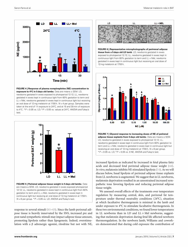
FIGURE 6 | Representative microphotographs of perirrenal adiposetissue from a 5 days old LD lamb. LD, newborns gestated in ewesexposed to photoperiod 12:12; LL, newborns gestated in ewes kept incontinuous light from 60% gestation to term and LL+Mel, newbornsgestated in ewes kept in continuous light but receiving an oral dose of12 mg melatonin at 1700 h.
FIGURE 7 | Glycerol response to increasing doses of NE of perirenaladipose tissue explants from 5 days old lambs. Data are means±SEM.LD, newborns gestated in ewes exposed to photoperiod 12:12; LL,newborns gestated in ewes kept in continuous light from 60% gestation toterm and LL+Mel, newborns gestated in ewes kept in continuous light butreceiving an oral dose of 12 mg melatonin at 1700 h. N =6 per group.*P < 0.05 vs. LD; &P < 0.05 vs. 0 NE. ANOVA and Tukey’s test.
increased lipolysis as indicated by increased in fetal plasma fattyacids and decreased fetal perirenal adipose tissue weight (44).In vitro, melatonin inhibits NE stimulated lipolysis (14). As we willdiscuss below, basal lipolysis of perirenal adipose tissue explantsfrom LL newborns is augmented. We suggest that in LL newborns,melatonin deprivation resulted in an unrestrained increased sym-pathetic tone favoring lipolysis and reducing perirenal adiposetissue weight.
We assessed overall effects of the treatments over temperatureregulation by measuring central, skin, and perirenal fat tem-perature under thermal neutrality conditions (24°C), situationat which facultative thermogenesis is minimal in the lamb andunder exposure to 4°C to stimulate facultative thermogenesis. Inthese two environmental conditions, we found lower temperaturesin LL newborns than in LD and LL+Mel newborns, suggest-ing that melatonin deprivation during fetal life affected newbornthermoregulation. In the newborn lamb, Williams and cowork-ers demonstrated that during cold exposure the contribution of
www.frontiersin.org January 2015 | Volume 5 | Article 232 | 7

Seron-Ferre et al. Maternal melatonin role in BAT
FIGURE 8 | Expression of adipose tissue genes (means±SE foldchanges vs. LD newborns) in 5 days old newborns. LD, newbornsgestated in ewes exposed to photoperiod 12:12; LL, newborns gestatedin ewes kept in continuous light from 60% gestation to term and
LL+Mel, newborns gestated in ewes kept in continuous light butreceiving an oral dose of 12 mg melatonin at 1700 h. LD and LL n=6.LL+Mel n=4. *P < 0.05 vs. LD; **P < 0.05 vs. LD; and LL+Mel,ANOVA, and Tukey’s test.
perirenal adipose tissue heat production, accounts for about 40%of the increase in oxygen consumption used to defend centraltemperature, the remaining heat being produced by muscle con-traction (shivering). In our experiments, oxygen consumption wassimilar in the three groups of newborns studied at 24°C andincreased similarly at 4°C, thus total heat production was simi-lar in each group. Perirenal adipose tissue temperature increasedin LL newborn exposed to cold, compared with the temperatureof this tissue at 24°C. However, total contribution to heat produc-tion of this tissue would be lower, considering its decrease in size,thus other mechanisms possibly shivering (not quantified in ourexperiments) accounted for the increased oxygen consumption.We assessed further the contribution of perirenal adipose tissueon heat production by measuring cold induced lipolysis in vivoand NE induced lipolysis in vitro. LL newborns responded to acold challenge with an increase in NE and plasma glucose but notwith an increase in plasma glycerol, suggesting limited inductionof lipolysis in vivo by NE. The in vitro studies, performed afterthe cold exposure experiment, confirm these findings and demon-strate almost a doubling of basal glycerol production suggestingthat adipose tissue was already maximally stimulated in absence ofadded NE. This increased basal glycerol production per milligramtissue may have little impact on plasma concentration given thereduction in adipose tissue weight. Exposure to cold, in addition
to stimulate heat production, triggers mechanisms that decreaseheat loss. One of them is skin vasoconstriction that reduces skintemperature. In our experiments, skin temperature was alreadyabout 1.6°C lower in LL than in LD newborns while at 24°C sug-gesting an important vasoconstriction. Of note, the difference inskin temperature between LL and LD newborns increased overtwofold during cold exposure, indicating that skin vasoconstric-tion plays an important role in maintenance of central temperaturein LL newborns. Whether the increased in NE levels discussedabove are involved it is not known. Overall, these results suggestthat in LL newborns, the reduced amount of perirenal adiposetissue, although competent to produce heat makes a minor con-tribution to temperature defense, the task being shifted to othermechanisms.
We next investigated possibly changes on gene expression on theadipose tissue that could relate to the above findings. We found thatthe expression of UCP1, ADBR3, PPARγ, C/EBPβ, and perilipinincreased about 10-fold, and PGC1α, PPARα, and the clock genesPer2 and Bmal1 increased about 4–6-fold in perirenal adiposetissue from LL newborns. PPARγ is central to brown and whiteadipose function and directly controls the expression of genesinvolved in lipid transport, lipid metabolism, insulin signaling,and adipokine production (45). In addition, it controls perilipinexpression (46). Of note, over expression of perilipin in mice white
Frontiers in Endocrinology | Systems and Translational Endocrinology January 2015 | Volume 5 | Article 232 | 8

Seron-Ferre et al. Maternal melatonin role in BAT
FIGURE 9 | Expression of clock genes (means±SE fold changes vs.LD newborns) in 5 days old newborns. LD, newborns gestated inewes exposed photoperiod 12:12; LL, newborns gestated in ewes keptin continuous light from 60% gestation to term; and LL+Mel,
newborns gestated in ewes kept in continuous light but receiving anoral dose of 12 mg melatonin at 1700 h. LD and LL n=6. LL+Meln=4. *P < 0.05 vs. LD, **P < 0.05 vs. LD, and LL+Mel, ANOVA, andTukey’s test.
adipose tissue results in increased basal lipolysis (25). Per2 proteinis a component of the negative branch of circadian clocks and hasbeen shown to interact with nuclear receptor proteins in the liver,providing a link between metabolic activity and circadian func-tion (47). Such interaction has been shown for Per2 and PPARgin white adipose tissue (27) and Per2 and PPARa in brown (26);functional results of these studies were an increase in UCP1 expres-sion, among other genes. Changes in UCP1, ADBR3, BMAL1, andPer2 gene expression were completely reversed, and PPARγ andperilipin partially reversed in LL+Mel newborns’ perirenal tis-sue. The increases in gene expression in adipose tissue from LLnewborns may relate to the increased NE stimulation discussedpreviously as shown for UCP1 and PGC1α in the perirenal adiposetissue of 5 days old lambs (24), and for Bmal1, UCP1, and PGC1α
in adult mice brown fat (28). Recent studies, also in adult micebrown fat, showed that cold exposure increases Per2 and Adbr3expression (26). On the other hand, direct effects of melatoninon clock gene expression have been demonstrated in the adrenalof several species (48–50) and in striatral neurons in vitro (51).Given that functional binding sites for melatonin are present infetal perirenal adipose tissue and that melatonin directly inhibitsNE stimulated lipolysis, we suggest that the combined effects of
increased NE stimulation plus prenatal melatonin deprivation mayexplain increased gene expression on perirenal adipose tissue.
Previous work from our laboratory and others showed theinvolvement of melatonin during gestation in perinatal adrenalfunction in primates and rats (1, 2), in rat hippocampal expres-sion of NMDA receptors, and fetal cardiac genomics (3, 4), withlong-term consequences for the offspring spatial memory (4) andenergy metabolism (5). In the present study, we extend mater-nal melatonin role to the adipose tissue and suggests a wider roleincluding the sympathetic nervous system. Altogether, these evi-dences highlight the importance of maternal melatonin duringpregnancy for the newborn.
Regarding the potential relevance of the present findings tohuman health, it must be kept in mind that exposure to light atnight (which effectively suppresses melatonin), most probably isa perinatal environmental risk factor imposed by a modern 24/7society. In this context, our data demonstrate negative effects ofmaternal melatonin suppression during pregnancy in the newbornlamb, a species very relevant for studies on perinatal physiology.Whether this might carry on into adulthood as abnormal physio-logical traits, is a possibility that needs to be seriously considered,since there is a body of evidence suggesting that a deleterious
www.frontiersin.org January 2015 | Volume 5 | Article 232 | 9

Seron-Ferre et al. Maternal melatonin role in BAT
maternal environment increases the risk of developing diseaseslike diabetes, hypertension, obesity, and metabolic syndrome thatappear in adult life (52).
ACKNOWLEDGMENTSWe are very grateful to Ms. Auristela Rojas for help with the exper-iments and biochemical measurements. This work was supportedby Grants 1060766 and 1090831 (Maria Seron-Ferre) from FondoNacional de Desarrollo Científico y Tecnológico, Chile (FONDE-CYT), a grant from the Department of Women’s Health, Arrow-head Regional Medical Center (CA, USA) and ANILLO ACT-1116(Claudia Torres-Farfan-Maria Seron-Ferre).
REFERENCES1. Torres-Farfan C, Richter HG, Germain AM, Valenzuela GJ, Campino C, Rojas-
Garcia P, et al. Maternal melatonin selectively inhibits cortisol production in theprimate fetal adrenal gland. J Physiol (2004) 554:841–56. doi:10.1113/jphysiol.2003.056465
2. Mendez N, Abarzua-Catalan L, Vilches N, Galdames HA, Spichiger C, RichterHG, et al. Timed maternal melatonin treatment reverses circadian disruption ofthe fetal adrenal clock imposed by exposure to constant light. PLoS One (2012)7:e42713. doi:10.1371/journal.pone.0042713
3. Galdames HA, Torres-Farfan C, Spichiger C, Mendez N, Abarzua-CatalanL, Alonso-Vazquez P, et al. Impact of gestational chronodisruption on fetalcardiac genomics. J Mol Cell Cardiol (2014) 66:1–11. doi:10.1016/j.yjmcc.2013.10.020
4. Vilches N, Spichiger C, Mendez N, Abarzua-Catalan L, Galdames HA, Haz-lerigg DG, et al. Gestational chronodisruption impairs hippocampal expressionof NMDA receptor subunits Grin1b/Grin3a and spatial memory in the adultoffspring. PLoS One (2014) 9:e91313. doi:10.1371/journal.pone.0091313
5. Ferreira DS, Amaral FG, Mesquita CC, Barbosa AP, Lellis-Santos C, Turati AO,et al. Maternal melatonin programs the daily pattern of energy metabolismin adult offspring. PLoS One (2012) 7:e38795. doi:10.1371/journal.pone.0038795
6. Seron-Ferre M, Luisa Forcelledo M, Torres-Farfan C, Valenzuela FJ, Rojas A,Vergara M, et al. Impact of chronodisruption during primate pregnancy onthe maternal and newborn temperature rhythms. PLoS One (2013) 8:e57710.doi:10.1371/journal.pone.0057710
7. Alexander G,Williams D. Shivering and non-shivering therogenesis during sum-mit metabolism in young lambs. J Physiol (1968) 198:251–76.
8. Cannon B, Nedergaard J. Brown adipose tissue: function and physiological sig-nificance. Physiol Rev (2004) 84:277–359. doi:10.1152/physrev.00015.2003
9. Pope M, Budge H, Symonds ME. The developmental transition of ovine adiposetissue through early life. Acta Physiol (Oxf) (2014) 210:20–30. doi:10.1111/apha.12053
10. Symonds ME. Brown adipose tissue growth and development. Scientifica (Cairo)(2013) 2013:305763. doi:10.1155/2013/305763
11. Alexander G, Bell AW, Hales JR. Effects of cold exposure on tissue blood flow inthe new-born lamb. J Physiol (1973) 234:65–77.
12. Asakura H. Fetal and neonatal thermoregulation. J Nippon Med Sch (2004)71:360–70. doi:10.1272/jnms.71.360
13. Power GG, Blood AB. Perinatal thermal physiology. In: Polin RA, Fox WW,Abman SH editors. Fetal and Neonatal Physiology. Philadelphia, PA: WB Saun-ders Co (2004). p. 541–8.
14. Torres-Farfan C, Valenzuela FJ, Mondaca M, Valenzuela GJ, Krause B, HerreraEA, et al. Evidence of a role for melatonin in fetal sheep physiology: direct actionsof melatonin on fetal cerebral artery, brown adipose tissue and adrenal gland. JPhysiol (2008) 586:4017–27. doi:10.1113/jphysiol.2008.154351
15. Alexander G,Mills SC,Scott TW. Changes in plasma glucose, lactate and free fattyacids in lambs during summit metabolism and treatment with catecholamines.J Physiol (1968) 198:277–89.
16. Recabarren SE, Vergara M, Llanos AJ, Seron-Ferre M. Circadian variation ofrectal temperature in newborn sheep. J Dev Physiol (1987) 9:399–408.
17. Herrera EA, Pulgar VM, Riquelme RA, Sanhueza EM, Reyes RV, Ebensperger G,et al. High-altitude chronic hypoxia during gestation and after birth modifiescardiovascular responses in newborn sheep. Am J Physiol Regul Integr CompPhysiol (2007) 292:R2234–40. doi:10.1152/ajpregu.00909.2006
18. Bispham J, Gardner DS, Gnanalingham MG, Stephenson T, Symonds ME, BudgeH. Maternal nutritional programming of fetal adipose tissue development: dif-ferential effects on messenger ribonucleic acid abundance for uncoupling pro-teins and peroxisome proliferator-activated and prolactin receptors. Endocrinol-ogy (2005) 146:3943–9. doi:10.1210/en.2005-0246
19. Myers DA, Hanson K, Mlynarczyk M, Kaushal KM, Ducsay CA. Long-termhypoxia modulates expression of key genes regulating adipose function inthe late-gestation ovine fetus. Am J Physiol Regul Integr Comp Physiol (2008)294:R1312–8. doi:10.1152/ajpregu.00004.2008
20. Frey E, Regenfelder F, Sussmann P, Zumstein M, Gerber C, Born W, et al. Adi-pogenic and myogenic gene expression in rotator cuff muscle of the sheep aftertendon tear. J Orthop Res (2009) 27:504–9. doi:10.1002/jor.20695
21. Sumner JM, McNamara JP. Expression of lipolytic genes in the adipose tissueof pregnant and lactating Holstein dairy cattle. J Dairy Sci (2007) 90:5237–46.doi:10.3168/jds.2007-0307
22. Cushman RA, Allan MF, Jones SA, Rupp GP, Echternkamp SE. Localization ofperiod 1 mRNA in the ruminant oocyte and investigations of its role in ovarianfunction. Anim Reprod Sci (2007) 99:93–105. doi:10.1016/j.anireprosci.2006.04.057
23. Varcoe TJ, Gatford KL, Voultsios A, Salkeld MD, Boden MJ, Rattanatray L, et al.Rapidly alternating photoperiods disrupt central and peripheral rhythmicityand decrease plasma glucose, but do not affect glucose tolerance or insulinsecretion in sheep. Exp Physiol (2014) 99:1214–28. doi:10.1113/expphysiol.2014.080630
24. Lomax MA, Sadiq F, Karamanlidis G, Karamitri A, Trayhurn P, Hazlerigg DG.Ontogenic loss of brown adipose tissue sensitivity to beta-adrenergic stimula-tion in the ovine. Endocrinology (2007) 148:461–8. doi:10.1210/en.2006-0918
25. Sawada T, Miyoshi H, Shimada K, Suzuki A, Okamatsu-Ogura Y, Perfield JW II,et al. Perilipin overexpression in white adipose tissue induces a brown fat-likephenotype. PLoS One (2010) 5:e14006. doi:10.1371/journal.pone.0014006
26. Chappuis S, Ripperger JA, Schnell A, Rando G, Jud C, Wahli W, et al. Role of thecircadian clock gene Per2 in adaptation to cold temperature. Mol Metab (2013)2:184–93. doi:10.1016/j.molmet.2013.05.002
27. Grimaldi B, Bellet MM, Katada S, Astarita G, Hirayama J, Amin RH, et al.PER2 controls lipid metabolism by direct regulation of PPARgamma. Cell Metab(2010) 12:509–20. doi:10.1016/j.cmet.2010.10.005
28. Li S, Yu Q, Wang GX, Lin JD. The biological clock is regulated by adrenergicsignaling in brown fat but is dispensable for cold-induced thermogenesis. PLoSOne (2013) 8:e70109. doi:10.1371/journal.pone.0070109
29. Martinez AM, Padbury JF, Humme JA, Evans CW, Shames L. Plasma cate-cholamines and their physiologic thresholds during the first ten days of lifein sheep. J Dev Physiol (1990) 13:141–6.
30. Smolich JJ, Esler MD. Total body catecholamine kinetics before and after birthin spontaneously hypoxemic fetal lambs. Am J Physiol (1999) 277:R1313–20.
31. Esler M. The sympathetic nervous system through the ages: from ThomasWillis to resistant hypertension. Exp Physiol (2011) 96:611–22. doi:10.1113/expphysiol.2011.052332
32. Gardner DS, Fletcher AJ, Bloomfield MR, Fowden AL, Giussani DA. Effects ofprevailing hypoxaemia, acidaemia or hypoglycaemia upon the cardiovascular,endocrine and metabolic responses to acute hypoxaemia in the ovine fetus. JPhysiol (2002) 540:351–66. doi:10.1113/jphysiol.2001.013434
33. Riquelme RA, Herrera EA, Sanhueza EA, Pulgar VM, Reyes VR, Parer JT, et al.Adrenocorticomedullar response to acute hypoxemia in chronic hypoxemicnewborn lambs. The Physiological Society, Annual Meeting, King College. London:(2004). C177 p.
34. Helliwell RJ, Williams LM. The development of melatonin-binding sites in theovine fetus. J Endocrinol (1994) 142:475–84. doi:10.1677/joe.0.1420475
35. Thomas L, Purvis CC, Drew JE, Abramovich DR, Williams LM. Melatoninreceptors in human fetal brain: 2-[(125)I]iodomelatonin binding and MT1gene expression. J Pineal Res (2002) 33:218–24. doi:10.1034/j.1600-079X.2002.02921.x
36. Williams LM, Martinoli MG, Titchener LT, Pelletier G. The ontogeny of cen-tral melatonin binding sites in the rat. Endocrinology (1991) 128:2083–90.doi:10.1210/endo-128-4-2083
37. Williams LM, Hannah LT, Bassett JM. Melatonin receptors in neonatal pig brainand pituitary gland. J Pineal Res (1999) 26:43–9. doi:10.1111/j.1600-079X.1999.tb00565.x
38. Song CK, Bartness TJ. CNS sympathetic outflow neurons to white fat that expressMEL receptors may mediate seasonal adiposity. Am J Physiol Regul Integr CompPhysiol (2001) 281:R666–72.
Frontiers in Endocrinology | Systems and Translational Endocrinology January 2015 | Volume 5 | Article 232 | 10

Seron-Ferre et al. Maternal melatonin role in BAT
39. Arangino S, Cagnacci A,Angiolucci M,Vacca AM, Longu G,Volpe A, et al. Effectsof melatonin on vascular reactivity, catecholamine levels, and blood pressurein healthy men. Am J Cardiol (1999) 83:1417–9. doi:10.1016/S0002-9149(99)00112-5
40. Laflamme AK-, Wu L, Foucart S, de Champlain J. Impaired basal sympathetictone and alpha1-adrenergic responsiveness in association with the hypotensiveeffect of melatonin in spontaneously hypertensive rats. Am J Hypertens (1998)11:219–29. doi:10.1016/S0895-7061(97)00401-9
41. Muller MD, Sauder CL,Ray CA. Melatonin attenuates the skin sympathetic nerveresponse to mental stress. Am J Physiol Heart Circ Physiol (2013) 305:H1382–6.doi:10.1152/ajpheart.00470.2013
42. Ray CA. Melatonin attenuates the sympathetic nerve responses to orthosta-tic stress in humans. J Physiol (2003) 551:1043–8. doi:10.1113/jphysiol.2003.043182
43. Grossman E, Laudon M, Zisapel N. Effect of melatonin on nocturnal bloodpressure: meta-analysis of randomized controlled trials. Vasc Health Risk Manag(2011) 7:577–84. doi:10.2147/VHRM.S24603
44. Bassett JM, Symonds ME. Beta2-agonist ritodrine, unlike natural cate-cholamines, activates thermogenesis prematurely in fetal sheep. Am J Physiol(1998) 275:R112–9.
45. Koppen A, Kalkhoven E. Brown vs white adipocytes: the PPARgamma coregu-lator story. FEBS Lett (2010) 584:3250–9. doi:10.1016/j.febslet.2010.06.035
46. Arimura N, Horiba T, Imagawa M, Shimizu M, Sato R. The peroxisomeproliferator-activated receptor gamma regulates expression of the perilipin genein adipocytes. J Biol Chem (2004) 279:10070–6. doi:10.1074/jbc.M308522200
47. Ripperger JA, Schmutz I, Albrecht U. PERsuading nuclear receptors to dance thecircadian rhythm. Cell Cycle (2010) 9:2515–21. doi:10.4161/cc.9.13.12075
48. Campino C, Valenzuela FJ, Torres-Farfan C, Reynolds HE, Abarzua-CatalanL, Arteaga E, et al. Melatonin exerts direct inhibitory actions on ACTHresponses in the human adrenal gland. Horm Metab Res (2011) 43:337–42.doi:10.1055/s-0031-1271693
49. Valenzuela FJ, Torres-Farfan C, Richter HG, Mendez N, Campino C, Torrealba F,et al. Clock gene expression in adult primate suprachiasmatic nuclei and adrenal:
is the adrenal a peripheral clock responsive to melatonin? Endocrinology (2008)149:1454–61. doi:10.1210/en.2007-1518
50. Torres-Farfan C, Mendez N,Abarzua-Catalan L,Vilches N,Valenzuela GJ, Seron-Ferre M. A circadian clock entrained by melatonin is ticking in the rat fetaladrenal. Endocrinology (2011) 152:1891–900. doi:10.1210/en.2010-1260
51. Imbesi M, Arslan AD, Yildiz S, Sharma R, Gavin D, Tun N, et al. The melatoninreceptor MT1 is required for the differential regulatory actions of melatonin onneuronal ‘clock’ gene expression in striatal neurons in vitro. J Pineal Res (2009)46:87–94. doi:10.1111/j.1600-079X.2008.00634.x
52. Barker DJ, Bagby SP, Hanson MA. Mechanisms of disease: in utero programmingin the pathogenesis of hypertension. Nat Clin Pract Nephrol (2006) 2:700–7.doi:10.1038/ncpneph0344
Conflict of Interest Statement: The authors declare that the research was conductedin the absence of any commercial or financial relationships that could be construedas a potential conflict of interest.
Received: 20 October 2014; accepted: 15 December 2014; published online: 06 January2015.Citation: Seron-Ferre M, Reynolds H, Mendez NA, Mondaca M, ValenzuelaF, Ebensperger R, Valenzuela GJ, Herrera EA, Llanos AJ and Torres-Farfan C(2015) Impact of maternal melatonin suppression on amount and functionality ofbrown adipose tissue (BAT) in the newborn sheep. Front. Endocrinol. 5:232. doi:10.3389/fendo.2014.00232This article was submitted to Systems and Translational Endocrinology, a section of thejournal Frontiers in Endocrinology.Copyright © 2015 Seron-Ferre, Reynolds, Mendez, Mondaca,Valenzuela, Ebensperger ,Valenzuela, Herrera, Llanos and Torres-Farfan. This is an open-access article distrib-uted under the terms of the Creative Commons Attribution License (CC BY). Theuse, distribution or reproduction in other forums is permitted, provided the originalauthor(s) or licensor are credited and that the original publication in this journal is cited,in accordance with accepted academic practice. No use, distribution or reproduction ispermitted which does not comply with these terms.
www.frontiersin.org January 2015 | Volume 5 | Article 232 | 11


