Embed Size (px)
Citation preview
J Korean Acad Pediatr Dent 39(1) 2012
36
IMPACTION OF PRIMARY TEETH ASSOCIATED WITH ODONTOMA: CASE REPORTS
Jung-Woo Kim, Teo-Jeon Shin, Hong-Keun Hyun, Young-Jae Kim,
Jung-Wook Kim, Ki-Taeg Jang, Chong-Chul Kim, Sang-Hoon Lee
Department of Pediatric Dentistry, School of Dentistry, Seoul National University
Odontomas generally appear as small, solitary, or multiple radio-opaque lesions found on routine radiographic
examinations. It is a comparatively common odontogenic tumor, and may lead to interfere with the eruption of
its associated tooth. In general, odontomas occur more often in permanent dentition and are very rarely associ-
ated with primary teeth.
This report deals with five rare cases of primary teeth impaction associated with odontomas, with spontaneous
eruption occurring in all five cases after simple surgical removal of the odontoma. Impacted primary teeth may
be associated with defects in development and eruption of their permanent successors, and thus long-term ob-
servation is necessary until the permanent successors erupt.
Key words : Odontoma, Primary teeth impaction, Spontaneous eruption
Abstract
Ⅰ. Introduction
Odontomas are benign tumors of odontogenic origin,
which exhibit complete dental tissue differentiation.
They consist of enamel, dentin, cementum, and pulpal
tissue and comprise 22% of all odontogenic tumors1).
Odontomas are considered to be developmental anom-
alies rather than true neoplasms2,3). The World Health
Organization has subdivided odontomas into compound
and complex types based on histologic and morphologic
criteria4). The calcified dental tissues in compound odon-
toma are arranged in an orderly pattern resembling mul-
tiple, small toothlike structures, whereas complex odon-
tomas have an amorphous and disorderly pattern.
Complex odontomas consist of a conglomerate mass of
enamel and dentin, and bear no anatomic resemblance
to a tooth. Compound odontomas are more frequently di-
agnosed than complex odontomas, and there is a distinct
difference in the location in which they proliferate1-4).
Specifically, compound odontomas are reported to occur
more commonly in the incisor-canine region of the maxil-
la, whereas complex odontomas are more frequently lo-
cated in the premolar and molar regions of the jaw
bones5). Odontomas are usually diagnosed during the
first two decades of life6-9), and in most children, these
tumors are associated with tooth eruption disturbances
such as delayed eruption of primary and permanent
teeth or overly retained primary teeth6,9,10). In general,
odontomas occur more often in the permanent dentition
and are very rarely associated with the primary
teeth5,11,12).
In this report, five rare cases of primary teeth im-
paction associated with odontoma are presented. The
treatment methods and prognoses are reported in detail,
교신저자 : 이 상 훈
서울특별시 종로구 창경궁로 166 / 서울 학교 치의학 학원 소아치과학교실 / 02-2072-2680 / [email protected]
원고접수일: 2011년 09월 01일 / 원고최종수정일: 2011년 12월 23일 / 원고채택일: 2012년 01월 05일
http://dx.doi.org/10.5933/JKAPD.2012.39.1.36
한소아치과학회지 39(1) 2012
37
and similar reports that have appeared in the literature
are discussed.
Ⅱ. Case Reports
Five cases were studied in the Department of Pediatric
Dentistry, Seoul National University Hospital. There
were no specific findings on the family, medical, or den-
tal history of the patients.
Case 1
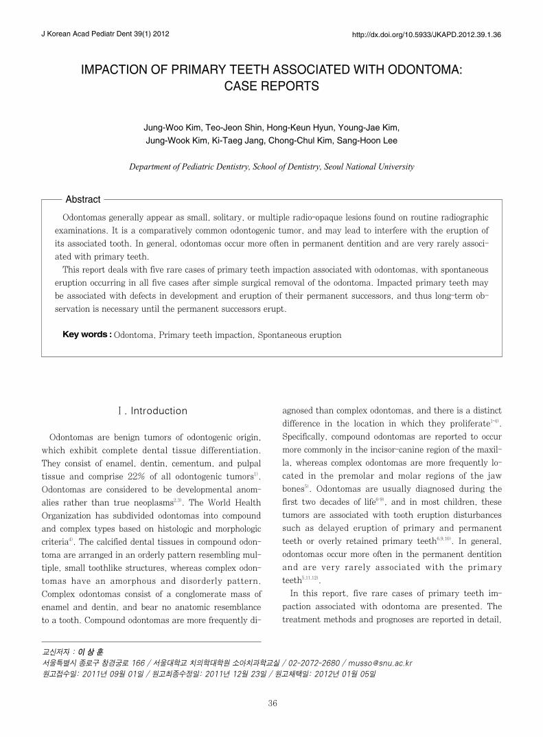
A 3-year-old boy was referred with a chief complaint of
an unerupted upper right primary central incisor.
Radiographic examination revealed the presence of a ra-
dio-opaque mass near the crown of the primary central
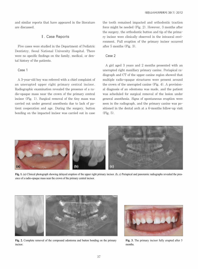
incisor (Fig. 1). Surgical removal of the tiny mass was
carried out under general anesthesia due to lack of pa-
tient cooperation and age. During the surgery, button
bonding on the impacted incisor was carried out in case
the tooth remained impacted and orthodontic traction
force might be needed (Fig. 2). However, 3 months after
the surgery, the orthodontic button and tip of the prima-
ry incisor were clinically observed in the intraoral envi-
ronment. Full eruption of the primary incisor occurred
after 5 months (Fig. 3).
Case 2
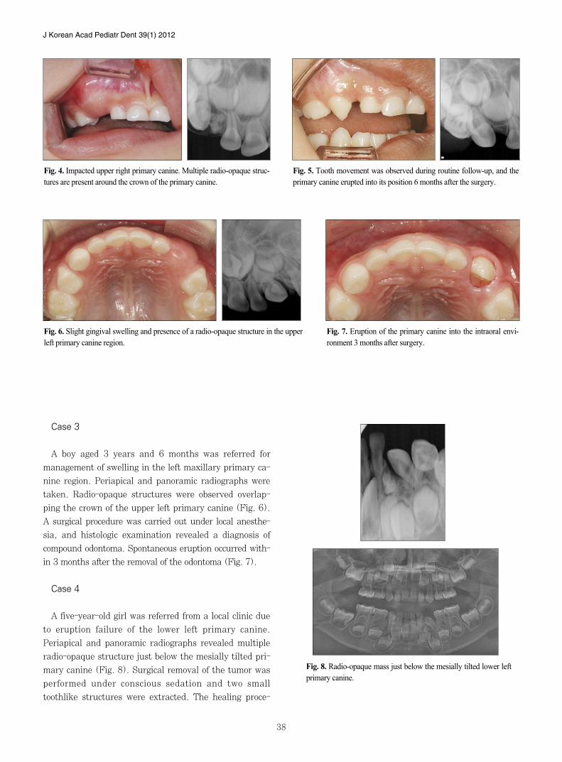
A girl aged 3 years and 2 months presented with an
unerupted right maxillary primary canine. Periapical ra-
diograph and CT of the upper canine region showed that
multiple radio-opaque structures were present around
the crown of the unerupted canine (Fig. 4). A provision-
al diagnosis of an odontoma was made, and the patient
was scheduled for surgical removal of the lesion under
general anesthesia. Signs of spontaneous eruption were
seen in the radiograph, and the primary canine was po-
sitioned in the dental arch at a 6-months follow-up visit
(Fig. 5).
Fig. 1. (a) Clinical photograph showing delayed eruption of the upper right primary incisor. (b, c) Periapical and panoramic radiographs revealed the pres-ence of a radio-opaque mass near the crown of the primary central incisor.
(a) (c)(b)
Fig. 2. Complete removal of the compound odontoma and button bonding on the primaryincisor.
Fig. 3. The primary incisor fully erupted after 5months.
J Korean Acad Pediatr Dent 39(1) 2012
38
Case 3
A boy aged 3 years and 6 months was referred for
management of swelling in the left maxillary primary ca-
nine region. Periapical and panoramic radiographs were
taken. Radio-opaque structures were observed overlap-
ping the crown of the upper left primary canine (Fig. 6).
A surgical procedure was carried out under local anesthe-
sia, and histologic examination revealed a diagnosis of
compound odontoma. Spontaneous eruption occurred with-
in 3 months after the removal of the odontoma (Fig. 7).
Case 4
A five-year-old girl was referred from a local clinic due
to eruption failure of the lower left primary canine.
Periapical and panoramic radiographs revealed multiple
radio-opaque structure just below the mesially tilted pri-
mary canine (Fig. 8). Surgical removal of the tumor was
performed under conscious sedation and two small
toothlike structures were extracted. The healing proce-
Fig. 4. Impacted upper right primary canine. Multiple radio-opaque struc-tures are present around the crown of the primary canine.
Fig. 5. Tooth movement was observed during routine follow-up, and theprimary canine erupted into its position 6 months after the surgery.
Fig. 6. Slight gingival swelling and presence of a radio-opaque structure in the upperleft primary canine region.
Fig. 7. Eruption of the primary canine into the intraoral envi-ronment 3 months after surgery.
Fig. 8. Radio-opaque mass just below the mesially tilted lower leftprimary canine.
한소아치과학회지 39(1) 2012
39
dure was uneventful and full eruption of the primary ca-
nine occurred within 3 months (Fig. 9). The gingiva was
very natural and healthy looking.
Case 5
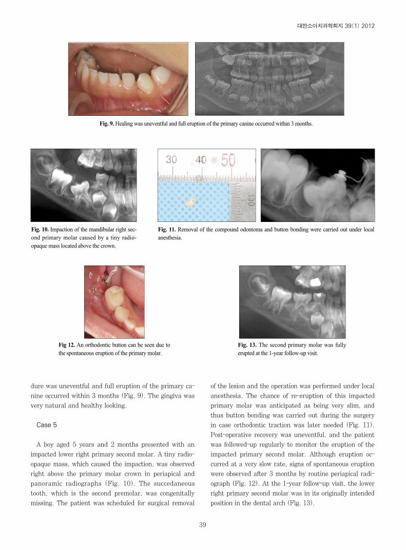
A boy aged 5 years and 2 months presented with an
impacted lower right primary second molar. A tiny radio-
opaque mass, which caused the impaction, was observed
right above the primary molar crown in periapical and
panoramic radiographs (Fig. 10). The succedaneous
tooth, which is the second premolar, was congenitally
missing. The patient was scheduled for surgical removal
of the lesion and the operation was performed under local
anesthesia. The chance of re-eruption of this impacted
primary molar was anticipated as being very slim, and
thus button bonding was carried out during the surgery
in case orthodontic traction was later needed (Fig. 11).
Post-operative recovery was uneventful, and the patient
was followed-up regularly to monitor the eruption of the
impacted primary second molar. Although eruption oc-
curred at a very slow rate, signs of spontaneous eruption
were observed after 3 months by routine periapical radi-
ograph (Fig. 12). At the 1-year follow-up visit, the lower
right primary second molar was in its originally intended
position in the dental arch (Fig. 13).
Fig. 9. Healing was uneventful and full eruption of the primary canine occurred within 3 months.
Fig. 10. Impaction of the mandibular right sec-ond primary molar caused by a tiny radio-opaque mass located above the crown.
Fig. 11. Removal of the compound odontoma and button bonding were carried out under localanesthesia.
Fig 12. An orthodontic button can be seen due tothe spontaneous eruption of the primary molar.
Fig. 13. The second primary molar was fullyerupted at the 1-year follow-up visit.
J Korean Acad Pediatr Dent 39(1) 2012
40
Summary of the five case reports are presented in
Table 1.
Ⅲ. Discussion
“Tooth impaction”is defined as a cessation of the
eruption of a tooth caused by a clinically or radiographi-
cally detectable physical barrier in the eruption path or
by an ectopic position of the tooth. Typical examples of
barriers are supernumerary teeth, odontomas, cysts,
crowded tooth germs and erupted teeth13,23). Teeth with
eruption problems are mainly permanent teeth and
rarely primary teeth. Moreover, most odontomas are as-
sociated with permanent teeth and rarely with primary
teeth5,8,11). Indeed, there are very few reports in the liter-
ature of odontomas associated with unerupted primary
teeth5,14-18,23,24). Katz7) found only 5 cases (1.26%) of com-
pound and complex odontomas in association with
unerupted primary teeth in his analysis of 396 cases of
odontoma. These figures reaffirm the rarity of the five
cases presented in this report.
The treatment advocated for odontomas in both prima-
ry and permanent dentition is surgical removal. If odon-
tomas are extirpated early without disturbing the under-
lying tooth germ, eruption of the impacted tooth is ex-
pected to occur spontaneously or after orthodontic trac-
tion10,12,14,15,18,19). Of the five cases presented, button bond-
ing was carried out in three primary teeth in case the
teeth remained impacted and orthodontic traction was
deemed necessary although spontaneous re-eruption of
all five teeth occurred within a few months after surgery.
Numerous theories of tooth eruption have been pro-
posed, most of which involve almost all of the tissues in
or near an erupting tooth. However, none of these theo-
ries alone can account for complete tooth movement. At
present, root elongation, alveolar bone remodeling, and
periodontal ligament formation provide the most con-
vincing model of tooth eruption13). Root formation of the
impacted teeth was complete in all five cases. It is diffi-
cult to propose a clear explanation with respect to how
the five primary teeth reported in this study, with fully-
grown roots, spontaneously erupted after removal of the
odontomas. Indeed, very little literature regarding spon-
taneous eruption of impacted primary teeth associated
with odontomas is available. Otsuka et al.20) observed
that 8 of 9 primary teeth re-erupted after surgery, while
Teruhisa et al.21) and Yeung et al.22) claimed that the
chance of spontaneous eruption is slim in all cases.
However, if we compare the prognoses of impacted per-
manent and primary teeth, primary teeth have a higher
chance of spontaneous eruption than permanent teeth,
which leads to a more positive prognosis20). A simple but
convincing explanation for this observation is that the
depth of impaction is shallower in primary teeth because
of the presence of a physical barrier, namely, the suc-
cedaneous tooth germ. Moreover, the alveolar bone that
surrounds permanent teeth is denser than the one cov-
ering the primary tooth13). Thus, when an obstacle that
is obstructing the eruption pathway is removed, the im-
pacted tooth can begin to move in the less pressurized
gingival direction.
Based on this study, we formulated a policy for the
treatment of impacted primary teeth. When impacted
primary teeth have sufficient space to erupt in the den-
tal arch, surgical exposure with removal of the overlying
gingiva or any overlying odontoma should be performed
and the impacted teeth kept under observation for three
months. Orthodontic traction should be applied when
the tooth fails to erupt. It is important to note that, in
the cases described here, impaction of the primary teeth
may be associated with disturbance in their permanent
successors, and thus long-term observation is necessary
through eruption of the permanent successors.
Ⅳ. Summary
Impaction of primary teeth is a rare condition that is
most often associated with the presence of supernumer-
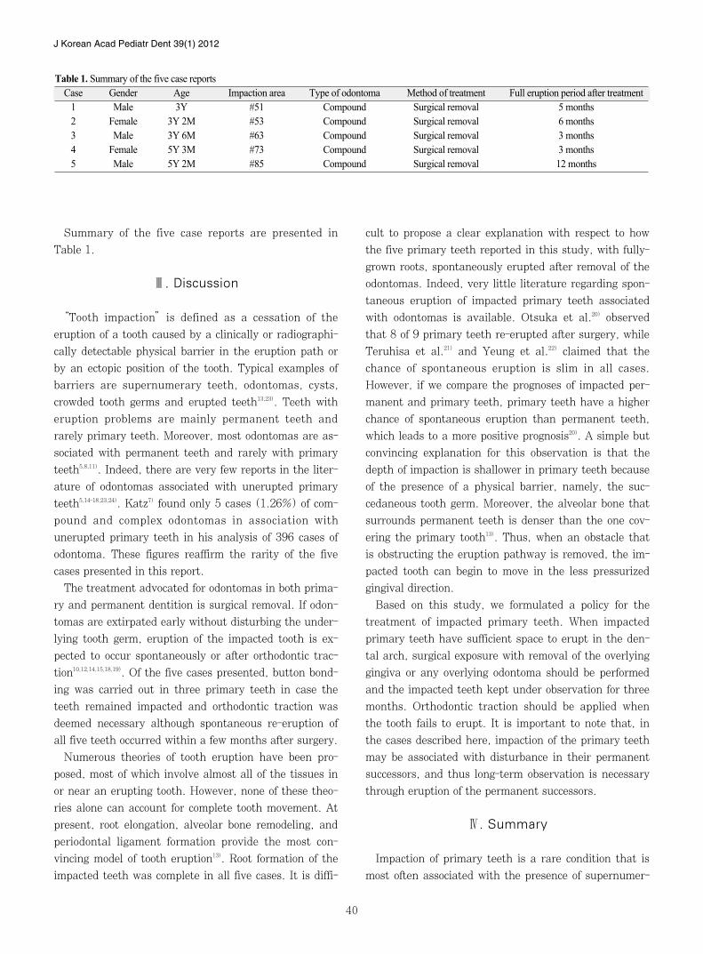
Table 1. Summary of the five case reports Case Gender Age Impaction area Type of odontoma Method of treatment Full eruption period after treatment
1 Male 3Y #51 Compound Surgical removal 5 months2 Female 3Y 2M #53 Compound Surgical removal 6 months3 Male 3Y 6M #63 Compound Surgical removal 3 months4 Female 5Y 3M #73 Compound Surgical removal 3 months5 Male 5Y 2M #85 Compound Surgical removal 12 months
한소아치과학회지 39(1) 2012
41
ary teeth or odontomas. All five cases presented in this
report involved impaction of primary teeth caused by
odontomas. Signs of spontaneous eruption of all primary
teeth were observed within 3 months of odontoma re-
moval. Although orthodontic traction is another useful
treatment option for impacted teeth, one can expect the
eruption of a primary tooth with a fully developed root
only after surgically removing the odontoma.
References
1. Neville BW, Damm DD, Allen CM, et al. : Oral and
Maxillofacial Pathology 2nd edn. Saunders,
Missouri, USA, 631-632, 2002.
2. Sheehy EC, Odell EW, Al-Jaddir G : Odontomas in
the primary dentition. J Dent Child, 71:73-76,
2004.
3. Hisatomi M, Asaumi J-I, Konouchi H, et al. : A case
of complex odontoma associated with an impacted
lower deciduous second molar and analysis of the
107 odontomas. Oral Dis, 8:100-105, 2002.
4. Kramar IRH, Pindborg JJ, Shear M : World Health
Organization International Histological Classification
of Tumors - Histological Typing of Odontogenic
Tumours, 2nd edn., Springer-Verlag, Berlin
Heidelberg, Germany, 11-42, 1992.
5. Stajcic ZZ : Odontoma associated with a primary
tooth. J Pedod, 12:415-420, 1988.
6. Owens BM, Schuman NJ, Mincer HH, et al. :
Dental odontomas: a retrospective study of 104 cas-
es. J Clin Pediatr Dent, 21:261-264, 1997.
7. Katz RW : An analysis of compound and complex
odontomas. J Dent Child, 56:445-449, 1989.
8. Regezi JA, Kerr DA, Courtney R : Odontogenic
tumors: analysis of 706 cases. J Oral Surg, 36:771-
778, 1978.
9. Sohma Y, Takagi R, Hoshina H, et al. :
Clinicostatistical evaluation of odontogenic tumors;
Report of 110 cases during the past 23 years.
Japanese J Oral Maxillofac Surg, 47:109-112, 2001.
10. Kaugars GE, Miller ME, Abbey LM : Odontomas.
Oral Surg Oral Med Oral Pathol, 67:172-176, 1989.
11. de Oliveira BH, Campos V, Marcal S : Compound
odontoma-diagnosis and treatment: three case
reports. Pediatr Dent, 23:151-157, 2001.
12. Noonan RG : A compound odontoma associated with
a deciduous tooth. Oral Surg Oral Med Oral Pathol,
32:740-742, 1971.
13. Andreasen JO, Petersen JK, Laskin DM : Textbook
and Color Atlas of Tooth Impactions. Mosby,
Missouri, USA, 23-26, 1997.
14. Motokawa W, Braham RL, Morris ME, et al. :
Surgical exposure and orthodontic alignment of an
unerupted primary maxillary second molar impacted
by an odontoma and a dentigerous cyst: a case
report. Quintessence Int, 21:159-162, 1990.
15. Brunetto AR, Turley PK, Brunetto AP, et al. :
Impaction of primary maxillary canine by an odon-
toma: surgical and orthodontic management. Pediatr
Dent, 13:301-302, 1991.
16. Haishima K, Haishima H, Yamada Y, et al. :
Compound odontomes associated with impacted
maxillary primary central incisors: report of two cas-
es. Int J Paediatr Dent, 4:251-256, 1994.
17. Kilpatrick NM, Hardman PJ, Welbury RR :
Dilaceration of a primary tooth. Int J Paediatr Dent,
1:151-153, 1991.
18. Yassin OM : Delayed eruption of maxillary primary
cuspid associated with compound odontoma. J Clin
Pediatr Dent, 23:147-149, 1999.
19. Tandon S, Radhika M : Compound composite odon-
toma in primary dentition - a case report. J Indian
Soc Pedod Prev Dent, 16:111-114, 1998.
20. Otsuka Y, Mitomi T, Tomizawa M, et al. : A review
of clinical features in 13 cases in impacted primary
teeth. Int J Paediatr Dent, 11:57-63, 2001.
21. Teruhisa U, Murakami J, Hisatomi M, et al. : A
case of unerupted lower primary second molar asso-
ciated with compound odontoma. Open Dent J, 3:
173-176, 2009.
22. Yeung KH, Cheung RC, Tsang MM: Compound
odontoma associated with an unerupted and dilacer-
ated maxillary primary central incisor in a young
patient. Int J Paediatr Dent, 13:208-212, 2003.
23. Han YB, Kim SO, Lee JH, et al. : Orthodontic trac-
tion of the lower deciduous second molar impacted
by an odontoma. J Korean Acad Pediatr Dent, 36:
84-88, 2009.
24. Ryu JR, Kim YJ, Kim HJ, et al. : Eruption distur-
bance associated with a developing odontoma. J
Korean Acad Pediatr Dent, 37:505-511, 2010.
J Korean Acad Pediatr Dent 39(1) 2012
42
치아종에 의한 유치의 매복: 증례 보고
김정우∙현홍근∙김 재∙김정욱∙장기택∙김종철∙한세현∙이상훈
서울 학교 치의학 학원 소아치과학교실
치아종은 유치나 구치배의 지속적인 치배 형성이나 법랑기 세포들의 비정상적인 증식의 결과로 발생하는 방사선 불투과
성 병소로, 비교적 흔한 치원성 종양이며 인접한 치아의 맹출을 방해 할 수 있다. 치아종은 구치열기에서 주로 관찰되고,
유치열기에서는 매우 드문 것으로 보고되고 있다.
본 증례 보고에서는 치아종에 의해 매복된 유치가 치아종의 외과적 발거 후 자연 맹출된 증례들을 보고하고자 한다. 매복
유치는 계승 구치의 발육과 맹출 문제를 야기할 수 있기 때문에 치아종이 제거된 후에도 계승 구치가 맹출할 때까지 장
기적인 검사 및 관찰이 필요하다.
주요어:치아종, 유치의 매복, 자발적 맹출
국문초록


