Embed Size (px)
Citation preview
Improved Antioxidant Effect ofIdebenone-LoadedPolyethyl-2-CyanoacrylateNanocapsules Tested onHuman Fibroblasts
Maddalena Palumbo,1 Alessandra Russo,1
Venera Cardile,2 Marcella Renis,1 Donatella Paolino,3
Giovanni Puglisi,3 and Massimo Fresta4,5
Received July 12, 2001; accepted October 8, 2001
Purpose. The protective antioxidant role of idebenone both as freedrug and drug-loaded Tween 80-coated polyethyl-2-cyanoacrylate(PECA) nanocapsules is reported. The relationship between oxida-tive damage and apoptotic or nonapoptotic cell death is evaluated invitro.Methods. Idebenone-loaded nanocapsules were prepared with theinterfacial polymerization method in the presence of Tween 80. Hu-man nonimmortalized fibroblasts, under different stress conditions,either 0.5 mM diethylmaleate (DEM) for 60 min or 0.1 mM H2O2 for30 min, were used as the experimental in vitro model. The productionof reactive oxygen species, the cell viability, and the nuclear DNAdamage were evaluated. The presence of apoptotic damage wasevaluated both by the determination of caspase-3-like protein activityand by Promega’s fluorescent apoptotic detection system.Results. DEM and H2O2 affected the cultured cells in different ways.DEM induced a moderate cellular insult, which was efficaciouslyantagonized by idebenone-loaded PECA nanocapsules. H2O2 elicitedsevere damage to nuclear DNA, which was reduced by idebenone-loaded PECA nanocapsules. The free drug was less effective thanidebenone-loaded nanocapsules.Conclusions. The findings reported here demonstrate that an im-proved antioxidant effect was obtained with a low idebenone con-centration (0.5 �M) when the drug was entrapped within Tween80-coated PECA nanocapsules.
KEY WORDS: antiapoptotic effect; COMET assay; free radical-induced damage; idebenone; PECA nanocapsules; oxidative stress.
INTRODUCTION
Different environmental, physical, and chemical stresseson cells may cause an oxidative wave, inducing either an over-
production of reactive oxygen species (ROS) or a deficiencyin antioxidant enzymes (1). Oxidative stress has been impli-cated in many physiologic and pathologic conditions occur-ring at the level of different tissues, i.e., the central nervoussystem, and the heart as well as in aging (2,3). The determi-nation of massive cellular damage by oxidative stresses, dueto necrotic cell death in the presence of lipid peroxidation andalterations of proteins and nucleic acids, has been extensivelydocumented (4,5). An emerging concept is also the involve-ment of ROS in physiologic death, such as programmed celldeath or apoptotic cell death (6).
The regular intake of antioxidant agents seems to pre-vent or limit the dangerous effects of chronic or transient-drastic oxidative stresses (7). A number of investigations haveshown the effectiveness of various molecules such as freeradical scavengers to reduce or to prevent oxidative cell dam-age (8,9).
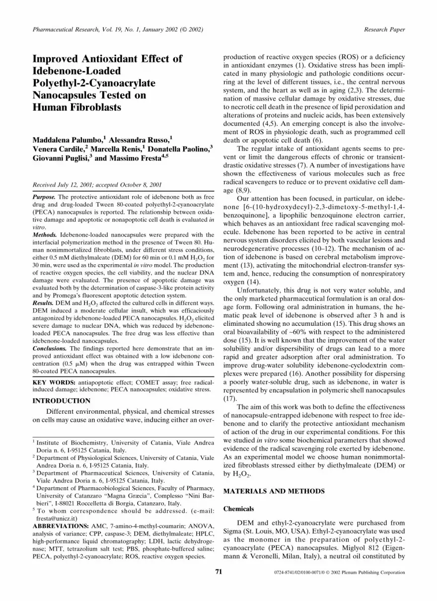
Our attention has been focused, in particular, on idebe-none [6-(10-hydroxydecyl)-2,3-dimetoxy-5-methyl-1,4-benzoquinone], a lipophilic benzoquinone electron carrier,which behaves as an antioxidant free radical scavenging mol-ecule. Idebenone has been reported to be active in centralnervous system disorders elicited by both vascular lesions andneurodegenerative processes (10–12). The mechanism of ac-tion of idebenone is based on cerebral metabolism improve-ment (13), activating the mitochondrial electron-transfer sys-tem and, hence, reducing the consumption of nonrespiratoryoxygen (14).
Unfortunately, this drug is not very water soluble, andthe only marketed pharmaceutical formulation is an oral dos-age form. Following oral administration in humans, the he-matic peak level of idebenone is observed after 3 h and iseliminated showing no accumulation (15). This drug shows anoral bioavailability of ∼60% with respect to the administereddose (15). It is well known that the improvement of the watersolubility and/or dispersibility of drugs can lead to a morerapid and greater adsorption after oral administration. Toimprove drug-water solubility idebenone-cyclodextrin com-plexes were prepared (16). Another possibility for dispersinga poorly water-soluble drug, such as idebenone, in water isrepresented by encapsulation in polymeric shell nanocapsules(17).
The aim of this work was both to define the effectivenessof nanocapsule-entrapped idebenone with respect to free ide-benone and to clarify the protective antioxidant mechanismof action of the drug in our experimental conditions. For thiswe studied in vitro some biochemical parameters that showedevidence of the radical scavenging role exerted by idebenone.As an experimental model we choose human nonimmortal-ized fibroblasts stressed either by diethylmaleate (DEM) orby H2O2.
MATERIALS AND METHODS
Chemicals
DEM and ethyl-2-cyanoacrylate were purchased fromSigma (St. Louis, MO, USA). Ethyl-2-cyanoacrylate was usedas the monomer in the preparation of polyethyl-2-cyanoacrylate (PECA) nanocapsules. Miglyol 812 (Eigen-mann & Veronelli, Milan, Italy), a neutral oil constituted by
1 Institute of Biochemistry, University of Catania, Viale AndreaDoria n. 6, I-95125 Catania, Italy.
2 Department of Physiological Sciences, University of Catania, VialeAndrea Doria n. 6, I-95125 Catania, Italy.
3 Department of Pharmaceutical Sciences, University of Catania,Viale Andrea Doria n. 6, I-95125 Catania, Italy.
4 Department of Pharmacobiological Sciences, Faculty of Pharmacy,University of Catanzaro “Magna Græcia”, Complesso “Nini Bar-bieri”, I-88021 Roccelletta di Borgia, Catanzaro, Italy.
5 To whom correspondence should be addressed. (e-mail:[email protected])
ABBREVIATIONS: AMC, 7-amino-4-methyl-coumarin; ANOVA,analysis of variance; CPP, caspase-3; DEM, diethylmaleate; HPLC,high-performance liquid chromatography; LDH, lactic dehydroge-nase; MTT, tetrazolium salt test; PBS, phosphate-buffered saline;PECA, polyethyl-2-cyanoacrylate; ROS, reactive oxygen species.
Pharmaceutical Research, Vol. 19, No. 1, January 2002 (© 2002) Research Paper
71 0724-8741/02/0100-0071/0 © 2002 Plenum Publishing Corporation
triglycerides with mixed acyl-chains of fractionated coconutfatty acids C8–C10, was used to achieve the oily nanocapsulecore. Tween 80 (Sigma), a nonionic surfactant, was used tostabilize the colloidal nanocapsule dispersion. Apoptosis wastested with a kit purchased from Promega (Madison, WI,USA). Dulbecco’s modified eagle’s medium and fetal calfserum are products from Life Technologies (Milan, Italy).Inorganic salts (purity >99.5%) NaCl, KH2PO4, and NaOHare products of BDH Laboratory Supplies (Poole, UK). Ide-benone was kindly provided by Takeda Italia FarmaceuticiS.p.A. (Rome, Italy) and was used without further purifica-tion (Fig. 1), although its purity was >99.5% as assayed byhigh-performance liquid chromatography (HPLC) analysis.Double-distilled pyrogen-free water was used. All other re-agents and solvents were of analytical grade (Carlo Erba,Milan, Italy).
PECA Nanocapsule Preparation
Lipophilic components, i.e., Miglyol 812 (1 ml), idebe-none (150 mg), and the monomer (300 �l), were dissolved in50 ml acetone. This organic solution was added to 100 mlaqueous phase (sterile double-distilled water, pH 7) contain-ing Tween 80 (at different concentrations) at a flow rate of 0.5ml/min. The presence of the nonionic surfactant allowed thepolymerization of ethyl-cyanoacrylate at the oil/water inter-face, thus encapsulating Miglyol 812 droplets (18,19). Theimmediate polymerization triggered the formation of drug-loaded nanocapsule colloidal suspensions. PECA nanocap-sule colloidal suspensions were concentrated under vacuumby evaporating off acetone up to the point when no trace ofthe organic solvent could be detected.
Drug-loaded PECA nanocapsules were centrifuged at20,000 × g for 1 h at 4°C to remove the untrapped idebenone.The floating nanocapsules were collected and suspended indouble-distilled water. This washing procedure was carriedout four times. After purification, no presence of both theTween 80 (20) and idebenone was observed. Final drug-loaded PECA nanocapsule colloidal systems, suspended insterile double-distilled water, were made isotonic with NaCl.
Drug Loading Determination
After purification, nanocapsule suspensions were lyoph-ilized overnight. Freeze-dried nanocapsules were solubilizedin an acetone-methanol (9:1 v/v) organic mixture and madeup to 25 ml. This solution was filtered through 0.45-�m poly-tetrafluorine-ethylene membranes (Sartorius, Gottingen,Germany) and were submitted to HPLC analysis to deter-
mine the amount of entrapped idebenone. A Hewlett-Packard (Milan, Italy) model 1050 liquid chromatographicsystem equipped with a 20-�l injection valve (model 7125,Rheodyne, Cotati, CA) was used. Substance detection wascarried out at 282 nm. Chromatogaphic analyses were per-formed at room temperature with a reverse-phase ODS Hy-persil C18 column (5 �m, 125 mm × 4 mm i.d.; Merck, Darm-stadt, Germany). The mobile phase was an acetonitrile/watermixture (70:30 v/v) with a flow rate of 1 ml/min. The eluentmixture was filtered through 0.22-�m poly-tetrafluorine-ethylene membranes (Sartorius). The amount of idebenonewas calculated using a calibration curve, reporting concentra-tion and peak area. No interference of the other nanocapsulecomponents was observed. Results are expressed as encapsu-lation yield (percentage of the starting drug that became en-capsulated). The drug release from PECA nanocapsules wasdetermined by incubating the colloidal suspension in the cellculture medium. At time intervals, the suspension was cen-trifuged and the supernatant was assayed by HPLC.
PECA Nanocapsule Colloidal Characterization
Dimensional analysis of PECA nanocapsule suspensionswas carried out by photon correlation spectroscopy with aZetamaster (Malvern Instruments Ltd, Sparing Lane South,Worcs, England). A solid-state laser was used as the lightsource. This laser diode had a nominal power of 4.5 mW witha maximum output of 5 mW at 670 nm. The photon correla-tion spectroscopy measurements were carried out at a scat-tering angle of 90°. As a correlation function, a third-ordercumulant fitting was applied to obtain mean particle diameterand polydispersity. Samples were suitably diluted with fil-tered water (0.22-�m membrane filters, Sartorius) and wereplaced in quartz cuvettes. Thirty measurements per samplewere carried out.
Electrophoretic mobility and zeta potential distributionwere measured with the Zetamaster particle electrophoresisanalyzer set-up (Malvern) equipped with a 5-mW HeNe laser(633 nm). Zeta limits ranged from −120 to 120 V. A Smo-luchowsky constant F(Ka) of 1.5 was used to achieve zetapotential values from electrophoretic mobility.
Morphologic characterization was carried out by freeze-fracture electron microscopy, as reported elsewhere (16).
Cell Cultures
Our study was carried out in vitro on human nonimmor-talized fibroblast cell lines that were kindly donated by Pro-fessor A. Simeone (Istituto Internazionale di Genetica Mole-colare, CNR Naples, Italy). The cells were cultured in humidi-fied atmosphere (5% CO2 at 37°C) in 35-mm dishescontaining a final volume of 2 ml made up of Dulbecco’smodified eagle’s medium plus 10% (v/v) fetal calf serum, 1mM glutamine, and 100 �l penicillin-streptomycin. Whenconfluence was reached, the cells were challenged by expo-sure to 0.5 mM DEM for 60 min or 0.1 mM H2O2 for 30 min.DEM was dissolved in dimethyl sulfoxide, which was assayedalone in the cultures. The employed concentrations of the twooxidants and the times of the treatments were selected on thebasis of our previous study (unpublished data). Free or en-capsulated idebenone was added to the cultures at 0.5 �M
Fig. 1. Chemical structure of idebenone, 6-(10-hydroxydecyl)-2,3-dimethoxy-5-methyl-1,4-benzo-quinone.
Palumbo et al.72

concentration at the same time as DEM or H2O2 and wasmaintained in the culture for 60 min. The total amount ofPECA added to the culture medium was 1 �g.
ROS Determination
Intracellular ROS was determined both directly in thedishes and in scraped and resuspended cells using 5,6-carboxy-2I,7I-dichlorodihydrofluorescine diacetate bis aceto-methyl ester (Molecular Probes) as a fluorescent probe. Thisprobe, diffusing through cell membranes, reaches the cytosol.At this level, the probe is hydrolyzed by esterases to 5,6-carboxy-2I,7I-dichlorodihydrofluorescine, which is oxidized tofluorescent 5,6-carboxy-2I,7I-dichlorofluorescine by ROS.The fluorescence intensity is proportional to the amount ofintracellular ROS (21). The 5,6-carboxy-2I,7I-dichlorodi-hydrofluorescine diacetate bis acetomethyl ester was added tothe cultures after DEM or H2O2 treatment and was incubatedfor 30 min. Thereafter, the medium was discharged, the disheswere washed with phosphate-buffered saline solution (PBS),and the fluorescence intensity was both qualitatively andquantitatively evaluated by fluorescence microscopy and byspectrofluorimetry at �ex 475 nm and �em 525 nm (F-2000,Hitachi, Tokyo, Japan), respectively. 5,6-carboxy-2I,7I-dichlorofluorescine-stained cells were not exposed to light be-fore measurement.
Lactate Dehydrogenase Release
Lactate dehydrogenase (LDH) activity was determinedas a kinetic reduction of NADH(H+) to the oxidized com-pound NAD+. The reduction of the amount of NADH(H+) isfollowed by ultraviolet adsorption at �max 340 nm (22). Theoxidation of NADH(H+) to NAD+ is due to the presence ofLDH, which catalyzes the reduction from piruvate to lactateusing NADH(H+) as a cofactor. The release of LDH wastested for each sample, both in the dish medium and in thecorresponding cells, following cell disruption by sonication.The percentage of LDH released in the medium (reflectingcytotoxicity) was calculated considering the total LDH activ-ity, namely, that measured in the medium plus that measuredin the sonicated cells.
Tetrazolium Salt Test
The tetrazolium salt test (MTT), a calorimetric assay,was used to indirectly monitor cell growth. The cells werecultured in 200-�l microplates for 12 h. Cells were thenwashed with PBS and incubated either with DEM or withH2O2. At the end of incubation, MTT (5 mg/ml) was added toeach well and incubation was continued for a further 24 h.The concentration of tetrazolium salts, following conversionto the colored product formazan, was determined at 570 nmwith a Titertek Multiskan microplate spectrophotometer(Flow Laboratories, Milan, Italy) (23). The amount of forma-zan is directly proportional to the number of living cells pre-sent in the culture.
Caspase-3 Activity
The caspase-3 (CPP) activity was quantitatively mea-sured by the Casp-ACE assay system according to Promega’sprotocol. This kit provides fluorometric reagents to quicklymeasure the activity of the CPP-32 enzyme. Cell extract en-
zymes were assayed. We used a tetrapeptide substrate la-belled with a fluorochrome, which is released upon cleavageby CPP-32-like enzymes. The yellow-green fluorescence wasproportional to the amount of CPP-32 activity present in thesample. Fluorescence intensity was determined at �em 460 nmby using a Hitachi F-2000 spectrofluorometer.
COMET Assay
DNA damage was evaluated using single-cell gel electro-phoresis (24). Briefly, the cells were scraped, washed, andresuspended in a low volume of PBS. 10 �l (about 10–20 × 104
cells) were embedded in 75 �l 0.5% (w/v) low-melting-pointagarose and spotted as a second layer on a first layer of nor-mal melting point agarose 1% (w/v) covering a microscopeslide. Finally a third layer was applied with 75 �l of low-melting-point agarose without cells. The slides were im-mersed first in pH 10 lysing buffer at 4°C for 1 h, and thenthey were placed in a horizontal electrophoresis tank (Bio-Rad, Burlington, Massachusetts). The tank was filled withalkaline buffer (300 mM NaOH and 1mM Na2EDTA at pH13), and an electrophoretic run was performed at 4°C for 20min applying a 300-mA current with a potential difference of25 V. The slides then were neutralized in 0.4 M Tris HCl pH7.5, stained with ethidium bromide, and examined under afluorescence microscope. During these experiments, yellowlight was used to prevent additional DNA damage. DNAdamage was measured on 100 counted DNA molecules (rep-resentative of 100 cells) with regard to tail length as follows:0, no tail; 1, short tails; 2, medium tails; 3, long tails; and 4,very long tails. The percentage of tailed DNA molecules(cells) was also determined.
Apoptosis Detection
The presence of apoptotic cell death was investigatedusing the TdT-mediated dUTP nick-end labeling assay (Pro-mega) in which terminal deoxynucleotidal transferase wasused to add fluorescein-12-dUTP to the 3’ OH ends of frag-mented DNA present in the cells. Before analysis, cells werescraped, fixed on slides, and labelled both with propidiumiodide and fluoresceine, according to Promega’s protocol.The apoptotic cells were visualized within a cell population byfluorescence microscopy. In particular, green nuclei con-tained fragmented apoptotic DNA, whereas red nuclei con-tained intact propidium iodide-stained DNA.
Statistical Analysis
Data are presented as means ± SD and were analyzed byStudent’s t test and one-way analysis of variance (ANOVA)with a posteriori Bonferroni test. Data with P < 0.05 are con-sidered to be significant.
RESULTS
PECA Nanocapsule Preparation and Characterization
In this investigation, nanocapsules (Fig. 2) were preparedby using acetone as the organic phase, as reported in a pre-vious article (17). The influence of the Tween 80 concentra-tion in the polymerization medium on the formulation param-eters of the colloidal carrier was evaluated. As reported inTable 1, the Tween 80 concentration noticeably influenced
Improved Antioxidant Effect of Idebenone-Nanocapsules 73

the mean size of PECA nanocapsules. The higher the Tween80 concentration, the smaller the colloidal suspension meansize up to a surfactant concentration of 3% (w/v). At Tween80 concentrations higher than 3% (w/v), no significant sizevariation of the nanocapsule colloidal suspension was ob-served. The effect of Tween 80 on the emulsion formation wasevaluated by determining the size distribution of the emulsiondroplets without monomer and with increasing concentra-tions of the surfactant. Tween 80 was able to reduce the hy-drodynamic size of the emulsion droplets as a function of itsconcentration (Table 1). At concentrations of Tween 80higher than 3% (w/v), the mean size of the emulsion dropletswas not influenced. Beyond a concentration of 3% (w/v), asecond colloidal population is clearly observed by photon cor-relation spectroscopy and is characterized by a very small size,showing the presence of micelles. Increasing Tween 80 con-centrations, a reduction of the polymeric wall thickness ofPECA nanocapsules was observed (data not reported), ac-cording to our previously reported observations (17). The
presence of different amounts (from 20 mg up to 200 mg) ofidebenone had no influence on the physicochemical charac-teristics of the colloidal suspensions (data not reported).
Surface properties of Tween 80-coated PECA nanocap-sules are characterized by a zeta potential of −13.1 ± 3.4 mV.A lower zeta potential value should be observed, but thepresence of the nonionic surfactant Tween 80 on the PECAnanocapsule surface behaved like a polyoxyethylene shieldaround the particles, which elicited a reduction of the zetapotential. This parameter was poorly influenced by the vari-ous preparation conditions.
Idebenone presented an encapsulation yield within nano-capsules of 97% with respect to the amount added duringpreparation. This drug, due to its highly lipophilic character,is solubilized within the oil core of PECA nanocapsules. Thepoor water solubility and the high affinity for the oil core ofPECA nanocapsules of idebenone also justify the poor re-lease from PECA nanocapsules in an aqueous environment.In fact, after 12 h only 2.7% of the entrapped amount ofidebenone is released into the cell culture medium. For invitro assays, idebenone-loaded PECA nanocapsules were pre-pared in the presence of 3% (w/v) Tween 80.
Experiments on Human Fibroblasts
For all the in vitro experiments, a control assay was car-ried out to measure the effects of unloaded PECA nanocap-sules. The results showed that, according to the literature(25–27), the addition of nanocapsules to the fibroblasts elic-ited no modification in the cell cultures during the differentassays (data not shown). The experiments were carried out byadministering in vitro free or encapsulated idebenone at aconcentration of 0.5 �M. To evaluate whether the presence ofthe carrier can modulate the action of the drug, a physicalmixture of the free drug and an unloaded nanocapsule werealso assayed. In this case, the effects on the various param-eters were similar to those obtained for the free drug (datanot reported).
ROS Determination
Our data (Fig. 3a) showed, with respect to the control(untreated cells), a more severe oxidative damage for H2O2-treated cells than for DEM-treated cells. Dimethyl sulfoxide,the solvent used for DEM, elicited no significant modificationunder our experimental conditions.
At the drug concentration investigated, no free idebe-none antioxidant effect was observed in the case of cellstreated with stress-inducing substances (DEM or H2O2).Conversely, idebenone-loaded PECA nanocapsules wereable to exert an effective antioxidant activity (Fig. 3a).
COMET Assay
The stress conditions are associated with nuclear DNAdamage as shown by the COMET assay (Fig. 3b). Similarly tothe ROS experiment, H2O2 treatment triggered a more se-vere damage of genomic DNA than that observed after DEMtreatment. DEM caused DNA damage ranging from 1 to 2 asregards tail length, whereas H2O2 damage was from 3 to 4.
Also in this case, the idebenone encapsulated in PECAnanocapsules was much more effective than the free drugregarding the reduction of DNA fragmentation (Fig. 3b), par-
Table I. Light Scattering Analysis of Colloidal Dispersions of PECANanocapsules and Emulsion Droplets Prepared in the Presence ofDifferent Concentrations of Tween 80 in a Polymerization Medium at
pH 7a
Tween 80(% w/v)
PECA nanocapsules Emulsion dropletsb
Size (nm) PIc Size (nm) PIc
0.5 312.3 ± 21.1 0.21 ± 0.04 289.8 ± 14.5 0.16 ± 0.051.0 247.8 ± 13.4 0.15 ± 0.02 217.2 ± 12.1 0.14 ± 0.033.0 185.1 ± 18.6 0.11 ± 0.02 179.2 ± 13.8 0.06 ± 0.016.0 183.5 ± 15.7 0.23 ± 0.03 175.4 ± 11.6 0.09 ± 0.029.0 176.9 ± 20.9 0.17 ± 0.04 177.3 ± 12.9 0.11 ± 0.03
a Each value is the average of four different experiments ± SD.b Emulsion droplets obtained without the monomer.c Polydispersity index. This value represents the dimensional homo-
geneity of the Tween 80-coated PECA nanocapsule colloidal sus-pensions.
Fig. 2. Freeze-fracture electron micrograph of idebenone-loadedTween 80-coated PECA nanocapsules. The black arrow shows theinternal oil (Mygliol 812) droplet surrounded by the polymeric shell.The bar is 100 nm.
Palumbo et al.74

ticularly in the case of DEM-treated cells. Further evidence ofthis came from LDH release experiments.
LDH release
This cytotoxicity assay measures the activity of LDH as astable cytosolic enzyme that is released into the culture me-dium following the disruption of the cellular cytoplasmaticmembrane. An increase of LDH leakage was observed in thecase of stress-inducing molecule treatment (Fig. 3c). Also inthis experiment, the damage was more severe after H2O2
treatment.Free idebenone was not able to significantly reduce the
LDH release. Conversely, idebenone-loaded nanocapsulesreduced the LDH release by ∼50% for both DEM-stressedand H2O2-stressed cells.
MTT Assay
The cell viability test (MTT) was also carried out toevaluate the effects of the stress-inducing substances and ofthe antioxidant agent on cell growth. In fact, this test deter-mined the number of viable cells. As shown in Fig. 4, a mod-erate decrease in cellular viability (−10%) was observed afterDEM treatment. The damage was clearly evident after H2O2
treatment (−60%). The antioxidant idebenone was able torepair the damage induced by DEM but only partially re-paired that induced by H2O2, when administered encapsu-lated in PECA nanocapsules.
Apoptosis Detection
Because oxidative damage causing single-strand ordouble-strand DNA breakage may be responsible for celldeath, apoptosis or necrosis, depending on the severity of theinduced stress, was evaluated by Promega’s apoptosis detec-tion system. This assay is designed for the specific detection ofapoptotic cells within a cell population by revealing DNAfragmentation at the level of the single cell.
The results concerning apoptotic experiments obtainedin the presence of free and encapsulated idebenone are pre-sented in Fig. 5. After DEM treatment, a diffused presence ofgreen apoptotic nuclei within cells, which seemed to haveintact plasmatic membranes, was observed. After H2O2 treat-ment, more severe DNA damage with different diffusedgreen domains and an alteration of cellular structure was ob-served, which is in agreement with the results obtained by theCOMET assay and LDH release. Idebenone showed a moreeffective antioxidant activity toward DEM-treated fibro-blasts, particularly when administered as idebenone-loadedPECA nanocapsules.
CPP Activity
Usually, two or more independent methods should beused to confirm apoptotic cell death. For this reason, CPP-
Fig. 3. Evaluation of ROS production (a), DNA damage (COMETassay) (b), and LDH release (c) of stress-induced, human, nonim-mortalized fibroblasts. C, untreated control cells; DEM, cells treatedwith DEM (0.5 mM) for 60 min; A, DEM-stressed cells treated withfree idebenone; B, DEM-stressed cells treated with idebenone-loaded PECA nanocapsules; H2O2, cells treated with H2O2 (0.1 mM)for 30 min; D, H2O2-stressed cells treated with free idebenone; E,H2O2-stressed cells treated with idebenone-loaded PECA nanocap-sules. Histograms show the mean value of five different experiments± SD. *Student’s t-test P < 0.001 compared with the respective stress-ing treatment (DEM or H2O2). All sample values were significant (P< 0.001) compared to the control. ANOVA is P < 0.001.
Fig. 4. Cell viability evaluation by MTT assay in human nonimmor-talized fibroblasts stressed with DEM (0.5 mM) for 60 min or H2O2
(0.1 mM) for 30 min. C, untreated control cells; A, DEM-stressedcells treated with free idebenone; B, DEM-stressed cells treated withidebenone-loaded PECA nanocapsules; D, H2O2-stressed cellstreated with free idebenone; E, H2O2-stressed cells treated with ide-benone-loaded PECA nanocapsules. Histograms show the meanvalue of five different experiments ± SD. *Student’s t test P< 0.001compared with the control. � Student’s t test P < 0.001 compared withthe respective stressing treatment (DEM or H2O2). ANOVA is P <0.001.
Improved Antioxidant Effect of Idebenone-Nanocapsules 75

like activity was measured. Caspase (interleukin-1�-converting enzyme ICE/CED-3) is a family of cysteine aspar-tic acid-specific proteases. These types of enzymes have beenshown to play fundamental roles in the inflammation andapoptosis processes of mammalian cells (28). The assay sys-tem used a specific tetrapeptide substrate that is labelled with7-amino-4-methyl-coumarin (AMC). AMC is released fromthe substrate upon cleavage of a CPP-like enzyme, thus pro-ducing fluorescence. The amount of fluorescence is propor-tional to the amount of CPP-like activity present in thesample. The results (Fig. 6), contrary to those obtained for theLDH release, showed a noticeable increase of CPP activityafter DEM treatment, whereas a moderate increase was ob-served after H2O2 treatment. The increase of caspase activitywas reduced by both the free drug (0.5 �M) and idebenone-loaded PECA nanocapsules (0.5 �M) in the case of bothDEM-treated and H2O2-treated cells. Idebenone antioxidantaction was more efficacious for DEM-stressed cells than forthose stressed with H2O2. In particular, idebenone-loadedPECA nanocapsules were more effective than the free drug.
DISCUSSION
The preparation procedure herein reported allowed theformation of nanocapsules in which Tween 80 is adsorbedinto the PECA polymeric shell, drastically reducing the pres-
Fig. 6. Fluorimetric evaluation of CPP-like protein activity of humannon immortalized fibroblasts stressed with DEM (0.5 mM) for 60 minor with H2O2 (0.1 mM) for 30 min. C, untreated control cells; A,DEM-stressed cells treated with free idebenone; B, DEM-stressedcells treated with idebenone-loaded PECA nanocapsules; D, H2O2-stressed cells treated with free idebenone; E, H2O2-stressed cellstreated with idebenone-loaded PECA nanocapsules. AMC fluores-cence intensity was determined at �em 460 nm. Histograms show themean value of five different experiments ± SD. *Student’s t test P <0.001 compared with the control. Student’s t test P < 0.001 comparedwith the respective stressing treatment (DEM or H2O2). ANOVA isP < 0.001.
Fig. 5. Fluorescence microscopy detection of double-stained (propidium iodide and fluorescein), human, nonimmortalized fibroblasts. Apop-totic nuclei (green) were detected by TdT-mediated dUTP nick-end labeling-like analysis carried out with Promega’s apoptosis detection kit.Fibroblasts were stressed with DEM (0.5 mM) for 60 min or H2O2 (0.1 mM) for 30 min. Control, untreated cells; A, DEM-stressed cells treatedwith free idebenone; B, DEM-stressed cells treated with idebenone-loaded PECA nanocapsules; D, H2O2-stressed cells treated with freeidebenone; E, H2O2-stressed cells treated with idebenone-loaded PECA nanocapsules.
Palumbo et al.76

ence of free surfactant in the aqueous suspension. Nanocap-sule colloidal systems, that is, an oil core surrounded by apolymeric shell, are more suitable colloidal carriers thannanoparticles for the delivery of lipophilic substances such asidebenone, which can be solubilized in the oil compartment.The size of this colloidal carrier can be controlled within acertain interval by regulating the Tween 80 concentration. Infact, a nanocapsule size reduction was achieved by increasingthe surfactant concentration. This finding is mainly due to twofactors: (1) the size reduction of the emulsion droplets wherepolymerization takes place; and (2) the increase of the num-ber of the droplets. This situation happens up to a 3% (w/v)concentration of Tween 80, because beyond this value Tween80 forms micelles and is not able to further reduce the dropletsize and increase the number of the Miglyol 812 emulsiondroplets.
We demonstrated the greater effectiveness of idebenone-loaded PECA nanoparticles toward human fibroblasts thanthe free drug by investigating the in vitro antioxidant effectunder different stress conditions (DEM or H2O2).
DEM is a depleting agent of the cellular GSH/GSSGratio, thus eliciting a moderate increase of ROS and somealterations in the electron transport chain (29). These alter-ations seem to be responsible for DNA damage and can bepartially antagonized by some free radical scavenging species,such as idebenone.
H2O2, by increasing the number of radical species incells, reduces the antioxidant cellular defences and triggers adepletion of ATP concentration (30), thus causing a severecellular insult in both the cytosol and the mitochondria (31).Under these conditions, cellular macromolecules and geno-mic DNA can be altered in a lethal way.
Our results show that idebenone-loaded nanocapsulesexert a greater protective antioxidant effect after DEM-induced stress than that after H2O2 treatment. This action isprobably due to two different contrasting factors: (1) a mas-sive involvement of mitochondria in DEM-induced damage;and (2) the electron carrier activity of idebenone. As regardsthe molecular mechanism of action of idebenone, its protec-tive effect, under our experimental conditions, seems to beexerted more on DNA damage and membrane breakdownthan on ROS production. This activity could be due to anidebenone-dependent activation of protective pathways (32)that is exerted more at the level of mitochondria than ofcytosolic ROS. Electron carrier and antioxidant activities ofidebenone maintain mitochondria membrane integrity, socontrasting the release from the organelles of dangerous pro-apoptotic factors (i.e., cit C, APO 1, and AIF), which, in turn,activate caspases and poly-A-ribosyl-polymerase, inducingapoptotic DNA fragmentation.
The difference in the biological action between the freedrug and idebenone-loaded PECA nanocapsules is probablydue to a mechanism based on the adherence of nanospheresto the cells, thus allowing the lipophilic drug, i.e., idebenone,to diffuse into cellular and subcellular structures. Our findingsshow that idebenone encapsulation within Tween 80-coatedPECA nanocapsules may be useful both to ensure a greaterdrug effectiveness by reducing the concentration to be used,by allowing an easier passage through biological barriers, andby drastically reducing the restrictive binding of the drug withserum proteins, and to avoid the useless accumulation of thedrug. In our experiments the dosage of idebenone-loaded
PECA nanocapsule was 0.5 �M, which is lower than plas-matic levels of patients submitted to oral drug treatments(33).
The increased CPP activity, which was particularly highafter DEM-induced stress, shows that (at the dosage used inour experiments) oxidative damage is prevalently of theapoptotic type, because caspase activation is required for de-velopment of the complete apoptotic phenotype in differentstress conditions. In the case of H2O2-induced stress, we hy-pothesize that, in an initial phase, an apoptotic cellular deathoccurred, followed by tardy necrotic damage (see LDH re-lease). This situation can explain the minor extent of CPPactivity achieved for H2O2-treated fibroblasts and the highestpercentage of LDH release in the medium. Because the oxi-dative damage is partially recovered by the idebenone-loadedPECA nanocapsules in the case of DEM-stressed fibroblastsand a good percentage of recovery is obtained after H2O2
treatment, idebenone can also be considered both as an an-tiapoptotic and an antinecrotic agent in some physiopatho-logic conditions.
In conclusion, our study provides further important evi-dence about the antioxidant effect of idebenone, which isexerted particularly at the mitochondrial level, and shows theimproved effect of this drug when encapsulated in Tween80-coated PECA nanocapsules. This colloidal drug deliverysystem can be efficaciously used in many conditions in whichoxidative damage occurs. In particular, these colloidal sys-tems may allow not only the oral administration of idebe-none, thus resulting in a greater drug bioavailability, but alsointravenous administration (a useful therapeutic approach inacute treatment). Colloidal nanocapsule suspensions mayprovide an easy passage of the drug through the blood-brainbarrier (34) and can be formulated in a such a way as to haveactive targeting properties. Furthermore, PECA nanocap-sules could also increase the life span of idebenone in thecirculation (35). To our knowledge, this is the first report inwhich idebenone is considered as a possible antiapoptoticagent. Our results open different and interesting ways bothfor the investigation of the antiapoptotic activity of idebenoneand for the possibility of developing new pharmacologic ap-proaches.
ACKNOWLEDGMENTS
This work was financially supported by Italian MURSTLOFIM 2001. The authors are very grateful to Dr. AntonyBridgewood for his revision of the language of this article.
REFERENCES
1. E.N. Ames, M.K. Shigenaga, and T.M. Hagen. Oxidants, antioxi-dants and degenerative diseases of ageing. Proc. Natl. Acad. Sci.USA 90:7915–7922 (1993).
2. P. Rustin, J.C. von Kleist Retzow, K. Chautrel Groussard, D.Sidi, A. Munnich, and A. Rotig. Effect of idebenone on cardio-myopathy in Friedveich’s ataxia: a preliminary study. Lancet 354:477–479 (1999).
3. P. Grieb, M.S. Ryba, G.S. Debicki, W. Gordon Krajcer, S. Ja-nuszewski, and S.J. Chrapusta. Change in oxidative stress in therat brain during post cardiac arrest reperfusion, and the effect oftreatment with the free radical scavenger idebenone. Resuscita-tion 39:107–113 (1998).
4. B. Halliwell and O.I. Aruoma. DNA damage by oxygen-derivedspecies. FEBS Lett. 281:9–19 (1991).
5. M. Renis, V. Calabrese, A. Russo, A. Calderone, M.L. Barcel-
Improved Antioxidant Effect of Idebenone-Nanocapsules 77

lona, and V. Rizza. Nuclear DNA strand breaks during ethanol-induced oxidative stress in rat brain. FEBS Lett. 390:153–156(1996).
6. G. Kroemer, P. Petit, N. Zamzami, J.L. Vayssiere, and B. Mi-gnotte. The biochemistry of programmed cell death. FASEB J.9:1277–1287 (1995).
7. Y. Morel and R. Barouki. Repression of gene expression by oxi-dative stress. Biochem. J. 342:481–496 (1999).
8. J.K. Callaway, P.M. Beart, and B. Janot. A reliable procedure forcomparison of antioxidants in rat brain homogenates. J. Pharma-col. Toxicol. Methods 39:155–162 (1998).
9. B. Halliwell, J.M.C. Gutteridge, and C.E. Cross. Free radicalsantioxidants and human disease: where are we now? J. Lab. Clin.Med. 119:598–620 (1992).
10. K. Yamada, T. Tanaka, D. Han, K. Senzaki, T. Kameyama, andT. Nabeshima. Protective effects of idebenone and �-tocopherolon �-amyloid-(1-42)-induced learning and memory deficits inrats: implication of oxidative stress in �-amyloid-induced neuro-toxicity in vivo. Eur. J. Neurosci. 11:83–90 (1999).
11. V. Bruno, G. Battaglia, A. Copani, M. A. Sortino, P. L. Canonico,and F. Nicoletti. Protective action of idebenone against excito-toxic degeneration in cultured cortical neurons. Neurosci. Lett.178:193–196 (1994).
12. J.C. Gullis, P. Benefield, and D. McTavish. Idebenone: a reviewof its pharmacodynamic and pharmacokinetic properties, andtherapeutic use in age-related cognitive disorders. Drugs Aging5:133–152 (1994).
13. H. Houchi, M. Yoshizumi, K. Minakuchi, M. Azuma, K. Morita,and M. Oka. Idebenone, an agent improving cerebral metabo-lism, stimulates [14C]tyrosine uptake and [14C]catecholamineformation by cultured bovine adrenal chromaffin cells. Biochem.Pharmacol. 42:951–954 (1993).
14. Y. Ikejiri, E. Mori, K. Ishii, K. Nishimoto, M. Yasuda, and M.Sasaki. Idebenone improves cerebral mitochondrial oxidativemetabolism in a patient with MELAS. Neurology 47:583–585(1996).
15. A. Nagaoka. Idebenone. In A. Scriabine (ed.), New Cardiovas-cular Drugs, Raven Press, New York, 1987 pp. 217–235.
16. G. Puglisi, C.A. Ventura, M. Fresta, M.A. Vandelli, G. Cavallaro,and M. Zappalà. Preparation and physico-chemical study of in-clusion complexes between idebenone and modified �-cyclodex-trins. J. Inclus. Phenom. 24:193–210 (1996).
17. M. Fresta, G. Cavallaro, G. Giammona, E. Wehrli, and G. Puglisi.Preparation and characterization of polyethyl-2-cyanoacrylatenanocapsules containing antiepileptic drugs. Biomaterials 17:751–758 (1996).
18. F. Chouinard, S. Buczkowski, and V. Lenaerts. Poly(alkylcyano-acrylate) nanocapsules: physicochemical characterization andmechanism of formation. Pharm. Res. 11:869–874 (1994).
19. M. Gallardo, G. Couarraze, B. Denizot, L. Treupel, P. Couvreur,and F. Puisieux. Study of the mechanisms of formation of nano-particles and nanocapsules of polyisobutyl-2-cyanoacrylate. Int. J.Pharm. 100:55–64 (1993).
20. N.B. Cucakovich. Determination of Tween 80 in tissue culturemedia, vaccines and related products. Anal. Biochem. 40:183–186(1971).
21. C. Montoliu, H. Sancho-Tello, I. Azorin, M. Burgal, S. Valles, J.Renau-Piqueras, and C. Guerri. Ethanol increases cythochrome
P-450 2E1 and induces oxidative stress in astrocytes. J. Neuro-chem. 65:2561–2570 (1995).
22. T.H. Murphy and J.B. Baraban. Glutamate toxicity in immaturecortical neurons precedes development of glutamate receptorcurrents. Dev. Brain Res. 57:146–150 (1990).
23. M.B. Hansen, S.E. Nielsen, and K. Berg. Re-examination andfurther development of a precise and rapid dye method for mea-suring cell growth/cell kill. J. Immunol. Methods 119:203–210(1989).
24. G. Frenzilli, C. Betti, T. Davini, M. Desideri, E. Fornai, L. Gian-nesi, F. Maggiarelli, P. Paoletti, and R. Barale. Evaluation ofDNA damage in leukocytes of ex-smokers by single cell electro-phoresis. Mut. Res. 375:117–123 (1997).
25. R. Fernandez-Urrusuno, E. Fattal, D. Parquet, J. Feger, and P.Couvreur. Evaluation of liver toxicological effects induced bypolyalkylcyanoacrylate nanoparticles. Toxicol. Appl. Pharmacol.130:272–279 (1995).
26. R. Fernandez-Urrusuno, E. Fattal, D. Parquet, J. Feger, and P.Couvreur. Influence of surface properties on the inflammatoryresponse to polymeric nanoparticles. Pharm. Res. 12:1385–1387(1995).
27. R. Fernandez-Urrusuno, E. Fattal, J. Feger, P. Couvreur, and P.Therond. Evaluation of hepatic antioxidant systems after intra-venous administration of polymeric nanoparticles. Biomaterials18:511–517 (1997).
28. W.C. Earnshaw, L.M. Martins, and S.H. Kaufmann. Mammaliancaspases: structure, activation, substrates and functions duringapoptosis. Annu. Rev. Biochem. 68:383–424 (1999).
29. C. Garcia-Ruiz, A. Colell, A. Morales, N. Kaplowitz, and J.C.Fernandez-Checa. Role of oxidative stress generated from themitochondrial electron transport chain and mitochondrial gluta-thione status in loss of mitochondrial function and activation oftranscription factor nuclear factor-kB: studies with isolated mito-chondria and rat hepatocytes. Mol. Pharmacol. 48:825–834(1995).
30. M. Panayiotidis, O. Isolas, and D. Galaris. Glucose oxidase pro-duced H2O2 induces Ca++-dependent DNA damage in humanperipheral blood lumphocytes. Free Rad. Biol. Med. 26:548–556(1999).
31. T. Ollikainen, K. Linnainmaa, and V.L. Kinnula. DNA singlestrand breaks induced by asbestos fibers in human pleural meso-thelial cells in vitro. Environ. Mol. Mutagen 33:153–160 (1999).
32. B.S. Polla, S. Kantegwa, D. Francois, S. Salvioli, C. Franceschi, C.Marsac, and A. Cosarizza. Mitochondria are selective targets forthe protective effects of heat shock against oxidative injury. Proc.Natl. Acad. Sci. USA 193:6458–6463 (1996).
33. A. Mordente, G. E. Martorana, G. Minotti, and B. Giardina.Antioxidant peptides of 2,3dimethoxy-5-methyl-6-(10-hydroxydecyl)-1,4-benzoquinone (idebenone). Chem. Res. Toxi-col. 11:54–63 (1998).
34. J. Kreuter, R.N. Alyautdin, D.A. Kharkevich, and A.A. Ianov.Passage of peptides through the blood-brain barrier with colloi-dal polymer particles (nanoparticles). Brain Res. 674:171–174(1995).
35. C. Damge, C. Michel, M. Aprahamian, and P. Couvreur. Newapproach for oral administration of insulin with polyalkylcyano-acrylate nanocapsules as drug carrier. Diabetes 37:246–251(1988).
Palumbo et al.78


