Embed Size (px)
Citation preview

BASIC SCIENCE
Nanomedicine: Nanotechnology, Biology, and Medicine8 (2012) 891–899
Research Article
Lapatinib/Paclitaxel polyelectrolyte nanocapsules for overcomingmultidrug resistance in ovarian cancer
Daniele Vergara, PhDa,b, Claudia Bellomo, MSc, Xingcai Zhang, MSd, Viviana Vergaro, MSc,Andrea Tinelli, MDe, Vito Lorusso, MDf, Ross Rinaldi, PhDg, Yuri M. Lvov, PhDd,
Stefano Leporatti, PhDc,⁎, Michele Maffia, PhDa,b
aLaboratory of General Physiology, Department of Biological and Environmental Sciences and Technologies, University of Salento, Lecce, ItalybLaboratory of Clinical Proteomic, “Giovanni Paolo II” Hospital, Lecce, Italy
cNNL–Istituto Nanoscienze, ItalydInstitute for Micromanufacturing, Louisiana Tech University, Ruston, Louisiana, USA
eDepartment of Obstetrics and Gynaecology, Vito Fazzi Hospital, Lecce, ItalyfDepartment of Medical Oncology, Vito Fazzi Hospital, Lecce, Italy
gNNL-Istituto Nanoscienze, CNR University of Salento, Department of Innovation Engineering, Lecce, Italy
Received 20 May 2011; accepted 30 October 2011
nanomedjournal.com
Abstract
The sonication-assisted layer-by-layer (SLBL) technology was developed to combine necessary factors for an efficient drug-deliverysystem: (i) control of nanocolloid size within 100 – 300 nm, (ii) high drug content (70% wt), (iii) shell biocompatibility and biodegradability,(iv) sustained controlled release, and (v) multidrug-loaded system. Stable nanocolloids of Paclitaxel (PTX) and lapatinib were prepared bythe SLBL method. In a multidrug-resistant (MDR) ovarian cancer cell line, OVCAR-3, lapatinib/PTX nanocolloids mediated an enhancedcell growth inhibition in comparison with the PTX-only treatment. A series of in vitro cell assays were used to test the efficacy of theseformulations. The small size and functional versatility of these nanoparticles, combined with their ability to incorporate various drugs,indicates that lapatinib/PTX nanocolloids may have in vivo therapeutic applications.
From the Clinical Editor: The efficacy of Lapatinib/Paclitaxel polyelectrolyte nanocapsules is described in this study in cell cultures ofmultidrug-resistant ovarian cancer. If in vivo studies also result in similar efficacy and low toxicity, this may represent a viable avenue toaddress such malignancies.© 2012 Elsevier Inc. All rights reserved.
Key words: Ovarian cancer; Lapatinib; Paclitaxel; Nanocapsules; Sonication-assisted Layer-by-Layer
Ovarian cancer is one of the most common gynecologicalmalignancies in women and the leading cause of gynecologicalcancer-related deaths in developing countries.1 Due to the
No conflict of interest was reported by the authors of this article.This work was funded by the Italian Ministry of University and Research
(MIUR) through the FIRB project no. RBLA03ER38, “Con il contributo delMinistero degli Affari Esteri (MAE), Direzione Generale per la promozione ela Cooperazione Sociale ” (Italy –USA Large Scale Bilateral Project “Nano-trasportatori per la terapia del cancro”). This work was also supported by thePS105 ARTI strategic project “Development and realization of bio-chip formolecular diagnostic and categorization of human pathogenic viruses (HPV,HCV)” of Apulia Region and by a contribution of “ANGELA SERRA”Foundation For Cancer Research, Parabita (Lecce, Italy). No support camefrom any commercial association.
⁎Corresponding author: NNL, Istituto Nanoscienze, CNR Via Arnesano16Lecce 73100, Italy.
E-mail address: [email protected] (S. Leporatti).
1549-9634/$ – see front matter © 2012 Elsevier Inc. All rights reserved.doi:10.1016/j.nano.2011.10.014
Please cite this article as: D., Vergara, et al, Lapatinib/Paclitaxel polyelectrolNanomedicine: NBM 2012;8:891-899, doi:10.1016/j.nano.2011.10.014
absence of early symptoms, the disease is often undiagnoseduntil the advanced stage, when it has already spread to distantsites. The picture is complicated by the failure of the currentlyavailable therapies to prove effective. Themajor obstacle consistsof a broad variety of acquired drug-resistance mechanisms, someof which have been well described for many of the chemother-apeutics currently used in clinical practice, including Paclitaxel(PTX).2 PTX is a drug of natural origin isolated from the bark ofTaxus brevifolia and used for ovarian cancer and breast cancerclinical treatment.3 PTX promotes microtubule assembly andstability, thus leading to the disruption of the normal microtubulenetwork required for mitosis.
In past years, many efforts have been made to overcome drugresistance associated with PTX treatment, and a number ofspecific factors affecting the drug response have been identifiedat many levels.4-8 However, this has not yet resulted in the cureof patients with recurrent disease. Furthermore, PTX shows poor
yte nanocapsules for overcoming multidrug resistance in ovarian cancer.

892 D. Vergara et al / Nanomedicine: Nanotechnology, Biology, and Medicine 8 (2012) 891–899
water solubility, and it is formulated for its current clinicaladministration in a mixture of Cremophor EL/absolute ethanol(50% v/v) that has been associated with several side effects,including nephrotoxicity and neurotoxicity.9
To overcome toxicity, to increase bioavailability, and tocontrol drug release, several approaches for packaging PTX areunder investigation with numerous advantages over conventionalmethods and early nanoparticle (NP) products.10-13 Among thewide variety of novel formulations, previous studies have shownthat sonication-assisted layer-by-layer (SLBL) nanoassemblytechnique can be used efficiently for the nanoencapsulation ofpoorly soluble anticancer drugs.14,15
Under powerful ultrasonication, the air bubble that wasdissolved in water underwent the formation and implosion of thecavity, followed by the jet flow. Thus an extreme physicochem-ical environment was created. Drug crystals were broken intosmaller and smaller particles, down to nanoscale. During thisprocess, polyelectrolyte with an opposite charge to that of thepharmaceuticals was added and absorbed onto the drug crystalsthrough electrostatic forces; this prevented any aggregation ofthe newly formed NPs. Because the ultrasonication is based onthe collapse of air bubbles, a bubbling agent such as NH4HCO3
was mixed with the polyelectrolyte to increase the intensity ofultrasonication.14 By using this method, drug NPs of approxi-mately 150 nm ± 30 nm diameter are obtained. In many cases,initial drug particles were negatively charged. After the first layercoating with polyelectrolyte (in this case, polycation), the surfaceelectrical potential become positive and of larger magnitude thanthe one of the initial particle. Then the second polyelectrolyte (inthis case, polyanion) layer was coated, and the surface chargereversed to negative. This regular surface charge alternationdemonstrated successful formation of multilayer shell withpredetermined architecture and the capability of controlledrelease by tailoring the number and chemical composition ofcapsule layers. The SLBL technique can also be applied tocombine two drugs in one nanocolloid system for synergisticmedical treatment.16
We hypothesized that PTX clinical efficacy can beincreased through a strategy that combines improvements inPTX cellular delivery and a combination of targeted therapiesto inhibit one or more signalling pathways involved in PTXresistance. In particular, ATP-binding cassette (ABC) trans-porters, such as the multiple drug-resistance transporter multi-drug resistance 1 (MDR1),17 or P-glycoprotein or ABCB1have been associated with a poor response to chemotherapy.18
Therefore, MDR1 impairment is likely to have a significantimpact on PTX clinical action. However, until now, many P-gpinhibitors failed to show efficacy during preclinical and clinicalstudies.19 Recently, lapatinib, an epidermal growth factorreceptor (EGFR) and Erb-2 dual tyrosine kinase inhibitor,has been shown to inhibit the function of ABC transporters,including P-gp.20
In this study, a multidrug-resistant ovarian cancer cell line,OVCAR-3, has been used to test the efficiency of PTX-loadednanocolloids in comparison with that of PTX given alone. Inaddition, we successfully combined PTX and lapatinib innanocolloids to overcome MDR. Such combination enhancedthe cytotoxic efficacy of PTX.
Overall, the results of this study show that PTX-nanocolloidsincreased in vitro antitumor efficacy of PTX and that thecombination with lapatinib can significantly overcome multidrugresistance in ovarian cancer cell lines. These results areencouraging for the development of multifunctional nanocol-loids that could be used in the clinical practice.
Methods
Reagents and cell culture
The source of antibodies and inhibitors is provided inSupplementary Materials, available online at http://www.nanomedjournal.com.
The ovarian cancer cell line OVCAR-3 and the breast cancercell line MCF-7 were maintained in Dulbecco's modifiedEagle's medium (DMEM) supplemented with 10% fetal bovinserum (FBS), 2mMglutamine, 100U/mL penicillin and 100μg/mLstreptomycin, and cultured at 37°C in a humidified atmosphereof 5% CO2. Cells were subcultured every 2–3 days.
Preparation of nanocolloids
To build up the nanocapsules for a simultaneous controlledrelease of two drugs, we used a SLBL technique. Biodegradablechitosan (polycation) and alginic acid (polyanion) were chosenfor a biocompatible and biodegradable coating on drug NPs.
PTX nanocolloids
PTX (40 mg), chitosan (6 mg) and NH4HCO3 (40 mg) wereadded to distilled water (30 mL), stirred for 5 minutes and thenultrasonicated in a water/ice bath for 45 minutes (to makePTX/chitosan nanocores). The following LBL self-assembly ofalginic acid and chitosan were performed using washless LBLmethod.21 After 45 minutes' processing, 4 mL of alginic acidsolution (1 mg/mL) was added and sonicated for 25 minutes.This made a second anionic layer in the capsule shell resulting inPTX/chitosan/alginic acid core-shell structure. Chitosan andalginic acid solutions were sequentially added to the PTXnanocolloids maintaining sonication. The coating of chitosan andalginic acid were repeated to make a three-bilayer capsule wall ofPTX/(chitosan/alginic acid)3 composition. Particle size andsurface electrical ξ-potential were measured after deposition ofeach next layer, allowing monitoring of the capsule assembly.For each test, the solution samples were centrifuged at 2500 rpmfor 20 minutes to remove TiO2 particle impurities from thesonotrode during powerful ultrasonication.
Dual drug nanocolloids: PTX/chitosan/alginic acid/chitosan/lapatinib/chitosan/alginic acid
The above procedure was used to obtain PTX(chitosan/algi-nic acid)3 nanocolloids, and 20 mL of lapatinib (0.5 mg/mL)were added followed by the addition of alginic acid (maintainingultrasonication) to make PTX/(chitosan/alginic acid)3/-lapatinib/alginic acid samples. With this complicated shellarchitecture, we located PTX in the capsule core and usedcationic lapatinib for assembly of the capsule shell. The samples

893D. Vergara et al / Nanomedicine: Nanotechnology, Biology, and Medicine 8 (2012) 891–899
were centrifuged at 10,000 rpm for 10 minutes to remove theupper liquid.
Nanocolloids characterization
The Brookhaven ZetaPlus Microelectrophoresis Instrumenthad been applied for monitoring the surface ξ potential and theparticle size of drug nanocolloids and nanocapsules. The changesin surface ξ potential allowed monitoring the successful coatingof oppositely charged polyelectrolytes.
Hitachi S 4800 field emission scanning electron microscope(FESEM) had been used for imaging morphology and particlesize of drug nanocolloids.
Cell viability assay
Cells were seeded at a density of 5 × 10³/well in a 96-wellplate containing 100 μL of full medium and allowed to adhere tothe plate overnight. For determining cell viability, the MTT assaywas used. After treatment with PTX or LBL-PTX nanocolloidsfor 24 hours, the culture medium was aspirated and 100 μL offresh medium containing 10 μL of MTT solution (stock 5 mg/mLin PBS) was added to each well. Cells were then incubated forfurther 2–3 hours. After removal of MTT solution, 100 μL ofDMSOwere added to the wells to dissolveMTT-formazan crystalsand maintained in agitation for 15 minutes. Absorbance of theconverted dye was measured at a wavelength of 570 nm withbackground subtraction at 690 nm. The relative cell viability wasexpressed as a percentage of the untreated control wells. Values aremean ± standard error of the mean (SEM) of three independentexperiments performed in triplicate.
Reverse transcription-PCR (RT-PCR)
Total cellular RNA was isolated by Illustra TriplePrepextraction kit following manufacturer's instruction and immedi-ately used. Purified DNA and protein pellet were stored at −80°Cfor further analysis.
Total RNA (1 μg) was reverse transcribed into cDNA usingthe high capacity RNA-to-cDNA Master Mix (Applied Biosys-tems, Monza, Italy). PCR was conducted on a MyCycler thermalcycler (Bio-Rad, Milan, Italy). The final volume of 25 μLincluded 1 μL of cDNA template, 12.5 μL of PCR Master Mix(Promega, Milan, Italy), and 1 μL of a mix containing primers.
Immunoblotting assay
Whole cell lysates were prepared in RIPA buffer (50 mMTris-base, 150 mM NaCl, 0.1% SDS, 1% Triton X-100, 0.5%sodium deoxycholate, 1 mM sodium orthovanadate, 10 mMsodium fluoride, 1% protease inhibitor cocktail) and clarified bycentrifugation at 13,000 g for 15 minutes at 4°C. Proteinconcentration was determined using the Bradford protein assay.Next, 50 μg of proteins were separated on 10% polyacrylammidegel and transferred to nitrocellulose membranes (Amersham-Biosciences, Milan, Italy). The membranes were blockedovernight in 5% nonfat milk in Tris Buffer Saline and 0.1 %Tween 20 (TBST) buffer at 4°C under agitation, andsubsequently probed by the appropriately diluted primaryantibodies in blocking buffer. The blots were then incubatedwith HRP-conjugated secondary antibody for 2 hours at room
temperature (20°–25°C). Target proteins were detected byenhanced chemiluminescence reagents and visualized onHyperfilm ECL films (Amersham-Biosciences).
Confocal microscopy
Exponentially growing ovarian cancer cells were seeded on25 mM square glass coverslips placed in 35 mM diameter culturedishes. After treatment, cells were fixed for 5 minutes with 3.7%formaldehyde in phosphate-buffered saline (PBS) solution,permeabilized with a 0.1% solution of Triton X-100 in PBS,and incubated for 30 minutes at room temperature withphalloidin-TRITC (Sigma, Milan, Italy). Preparations weremounted in 50% glycerol in PBS and observed under a TCSSP5 laser confocal microscope (Leica Microsystem GmbH,Mannheim, Germany).
Statistical analysis
Statistical analysis was performed using GraphPad Prism 4.1(GraphPad Software, San Diego, California). Differencesbetween group means were compared by Student's t-test. Dataare presented as mean ± SEM. A probability level of P b 0.05was considered statistically significant.
Results
The aim of this study is to develop new strategies based onsonication and LBL technique to overcome multiple drugresistance (MDR) associated with PTX in ovarian cancer.Preliminary experiments were performed to identify cellulartargets of PTX action. The identification of these targets will beuseful to use them as markers of LBL-nanocolloids actionrespect to PTX alone.
The ovarian cancer cell line OVCAR-3 was chosen for itsdrug-resistance phenotype and used as model system in this study.As evaluated by RT-PCR, OVCAR-3 cells constitutively expresshigh levels of TLR-4 (Toll-Like Receptor) and MDR1 (Figure 1,E), which were both associated with PTX chemoresistance.4,6,7
TLR-4 and MDR1 mRNA expression levels in OVCAR-3 werecompared with those of the PTX-sensitive breast cancer cell lineMCF-7. MDR1 mRNA is greatly expressed in OVCAR-3 withrespect to MCF-7 whereas no significant differences wereobserved for TLR-4, suggesting the involvement of P-gp as themajor determinant of PTX resistance in OVCAR-3.
Dose-response studies highlighted the resistant nature of thiscell line (Figure 1, B). Exponentially growing OVCAR-3 cellswere exposed to increasing concentrations of PTX (from 1.5 ng/mLto 5000 ng/mL) for 24 hours and MTT cell viability assay wasperformed. As shown in Figure 1, the cancer cell line exhibiteda characteristic dose-response curve; in fact, treatment with PTXat concentrations above 10 ng/mL did not induce a proportionalreduction of cell viability. Comparable findings were obtainedin a similar experiment, in which the drug concentration wasfixed and the number of cells seeded on the plate was changed(Figure 1, C). Ovarian cancer cells seeded at four differentconcentrations and treated with PTX at 5 μg/mL for 24 hoursshowed a similar survival ratio for both control and treated cells.

Figure 1. PTX decreased OVCAR-3 cell viability. (A) Effects of PTX on OVCAR-3 cell morphology evaluated by phase-contrast microscopy after 24 h ofincubation (magnification 10×). Cell death is observed after PTX treatment as evidenced by the increasing number of floating cells. (B) PTX reduces cellviability. OVCAR-3 were seeded overnight into 96-well plates and incubated with PTX at the indicated concentrations. After 24 h, cell viability was detected byMTT test. OVCAR-3 demonstrated a plateau in survival at concentrations of PTX above 5 ng/mL. (C) PTX reduces cell viability. OVCAR-3 were seededovernight into 96-well plates at the indicated concentrations and incubated with PTX for 24 h at 5 μg/mL. The response of OVCAR-3 to PTX is independent ofthe number of cells seeded. (D) PTX reduces Akt phosphorylation at ser 473. OVCAR-3 cells were cultured in DMEM supplemented with 10% FBS for 24 hfollowed by starvation for 12 h in serum-free media. Cells were switched to the media in the absence (CTR) or presence (treated) of PTX 5 μg/mL for 15 min, 1 h,4 h, and 24 h, respectively. The whole cell lysates were prepared and western blotting was performed as described in the Methods section. Blots were strippedand re-probed with total Akt isoforms. The level of tubulin was used to indicate relative amounts of protein loaded. Experiments were performed three times, anda representative experiment is shown in this figure. Akt1 and Akt2 mRNA expression levels after PTX treatment were detected by RT-PCR. The expression ofGAPDH is shown as internal control. Akt primer sets were from Wang J et al.22 (E) Expression of MDR1 and TLR-4 measured by RT-PCR in untreatedOVCAR-3 and MCF-7 cells. The expression of GAPDH is shown as internal control. MDR1 primer set was from Wang Y et al.23
894 D. Vergara et al / Nanomedicine: Nanotechnology, Biology, and Medicine 8 (2012) 891–899
This finding means that cytotoxicity due to PTX is lessdependent on the concentration of the drug at concentrationsabove 10 ng/mL. In the case of OVCAR-3, the IC50 were notdetermined because even at concentrations of PTX greater than20 μg/mL, more than 50% of the cells remained viable (data notshown). In contrast, the IC50 of PTX was 100–200 ng/mL forthe drug-sensitive cell line MCF-7. This suggests that the pooreffectiveness of PTX in OVCAR-3 is associated with theexpression of the multidrug resistance transporter MDR1.
In recent years, the phosphatidylinositol 3-kinase (PI3K)/Aktpathway has risen to prominence as a regulator of cell survival andgrowth in many different cell types. In ovarian cancer, constitutiveAkt activity or gene amplification was frequently detected intumor samples and associated with chemoresistance and poorprognosis.24,25 Akt promotes cell survival and growth through avariety of mechanisms, including the regulation of proapoptoticproteins Bad and caspase-9 and cyclin D1 expression.26
To determine the effects of PTX on this signalingpathway, we evaluated by western blot the phosphorylationstatus of Akt (p-Akt) at ser 473 after PTX treatment.Ovarian cancer cells were grown under normal conditionsand were deprived of serum overnight. Cells were thentreated or not (control) with PTX in the presence of serum.Western blot analysis showed that levels of p-Akt ser 473were reduced in comparison with those in control cells that
otherwise were increased when stimulated with serum at theindicated times (Figure 1, D).
No differences were noted in the levels of total Akt1 and Akt2isoforms at both mRNA and protein levels suggesting that PTXregulation of this pathway results largely from post-transcrip-tional regulation.
To investigate whether the encapsulation of PTX innanocolloids leads to a general improvement of the drugefficacy, we assayed the cytotoxicity of the LBL-PTX towardthe OVCAR-3 cell line and compared the results with that ofPTX free. To assess this point, two types of LBL-nanocolloidswere fabricated and tested: PTX-chitosan and PTX-chitosan-alginic acid. In particular, chitosan is a positively charged naturalcarbohydrate polymer with minimal toxicity largely used as abiomaterial due to its physicochemical and biological properties(e.g., biodegradability). Chitosan shows strong electrostaticinteraction with the negatively charged mucosal surface,27 aproperty that can further provide a rationale for using chitosan asdelivery system in ovarian tumors considering the higherexpression of mucin and other heavily glycosylated extracellularproteins in ovarian tumors in comparison with the surroundingnormal tissue.28
Both preparations were tested on ovarian cancer cells for theirability to increase PTX efficacy. The results obtained by MTTtest were comparable for the two types of nanocolloids and for

Figure 2. Comparison of cell viability in OVCAR-3 cells after treatment with free PTX and LBL-PTX nanocolloids. Citotoxic effects of PTX alone or LBL-PTXnanocolloids at the indicated concentrations were measured by the MTT assay as described in the Methods section. The cell viability is related to control wellstreated with vehicle (DMSO) or empty LBL-nanocolloids (drug free). Effects of PTX and LBL-PTX nanocolloids on the expression of phospho-Akt and cyclinD1. Ovarian cancer cells were treated with PTX or LBL-PTX nanocolloids at the concentration of 1.5 ng/mL (A) or 5 μg/mL (B) for 24 h. The whole cell lysates(50 μg in each lane) were prepared and subjected to western blotting analysis by using specific antibodies against p-Akt ser 473 and cyclin D1. The Cyclin D1mRNA level remains unaffected after treatment, suggesting the regulation at the protein level.
895D. Vergara et al / Nanomedicine: Nanotechnology, Biology, and Medicine 8 (2012) 891–899
this reason, we use the general name of LBL-PTX nanocolloidsto designate both formulations. As shown in Figure 2, PTX andLBL-PTX have a similar trend of cytotoxicity in ovarian cellswith a higher cytotoxicity for LBL-PTX than for PTX alone. Itshould be noted that, because cells incubated for 24 hours withempty LBL-nanocolloids (drug free) did not show anysignificant difference in cell viability in comparison with controlcells (data not shown), we exclude the role of emptynanocolloids to explain the difference between PTX and LBL-PTX. Moreover, we observed a significant inhibition of cellgrowth after treatment of ovarian cancer cells with LBL-PTX atthe concentration of 1.5 ng/mL, whereas no change was observedafter PTX-free treatment at the same concentration. We thendetermined whether p-Akt ser 473 and cyclin D1 levels, adownstream target of the Akt signaling pathway,29 were affectedat this concentration of drug. As shown in Figure 2, western blotanalysis revealed a downregulation of pAKT ser 473 and cyclinD1 only for LBL-PTX treated cells, further suggestingthe sensitivity of OVCAR-3 to LBL-PTX but not PTX at thisdrug concentration.
We next determined whether the increased cytotoxicity ofLBL-PTX nanocolloids with respect to PTX free could beexplained by a higher downregulation of the Akt pathway.Western blot analysis of p-Akt ser 473 from ovarian cells treatedwith PTX and LBL-PTX nanocolloids at 5 μg/mL showed asignificant difference in the phosphoprotein level in the controlwith respect to treated cells with a further downregulation inLBL-PTX treated samples (Figure 2, B), a result that correlateswith the increased cytotoxicity of nanocolloids in comparison
with PTX free. This finding could be explained considering thedifferent mechanisms of drug-nanocolloids uptake in compari-son with PTX free. Several mechanisms have been describedincluding the increased accumulation of the NPs in the cells andtheir entrapment in the endosomes/lysosomes, rendering the druginaccessible for P-gp and enhancing in this way the sensitivity ofcancer cells to the drug.30
A possible link between the activation of PI3K/Akt pathwayand actin remodeling has been described.31,32 It has been shownthat the PI3K/Akt pathway induced cell migration through theremodeling of actin filaments and that actin is a cellular target ofthis kinase.33,34 It is possible that the involvement of thispathway in PTX action could have consequences in theorganization of actin cytoskeleton after PTX treatment.TRITC-phalloidin staining of F-actin followed by confocalmicroscopy analysis revealed some differences in the cell shapeand organization of actin filaments for PTX free and LBL-PTXcells in comparison with the control (Figure 3, A). Non-treatedcells show regularly shaped bodies, with a readily visible actinstaining in the periphery, actin protrusions (yellow arrows,Figure 3, A), and the absence of bundles of actin filaments.Rounded cells appeared after PTX treatment together with theformation of blebs (green arrows, Figure 3, A). No actinprotrusions are visible in treated cells. Furthermore, a moredistinct net of actin filaments is visible in the cytoplasm ofOVCAR-3 treated with LBL-PTX nanocolloids.
The organization of actin filaments is governed by a plethoraof proteins that regulate the rate of actin polymerization. One ofthe key proteins in this scenario is cofilin, which can regulate the
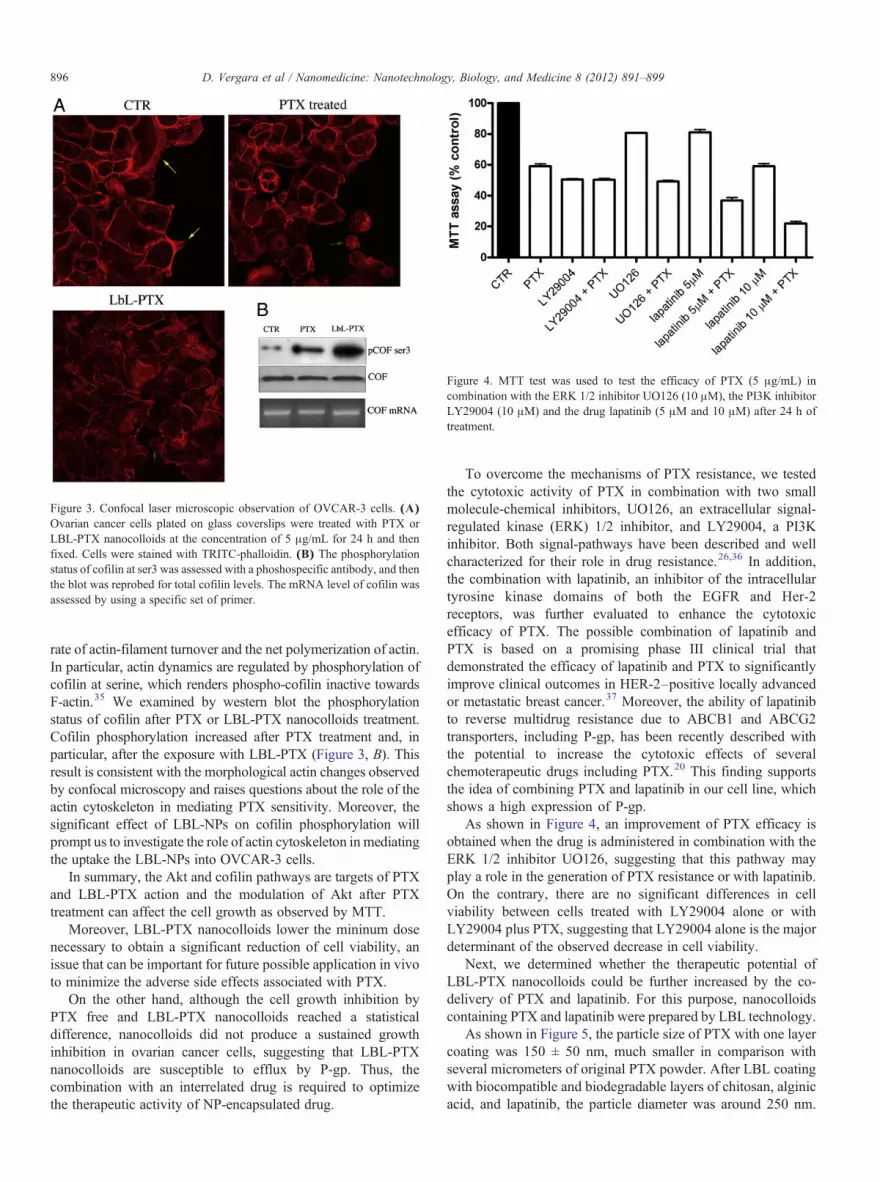
Figure 3. Confocal laser microscopic observation of OVCAR-3 cells. (A)Ovarian cancer cells plated on glass coverslips were treated with PTX orLBL-PTX nanocolloids at the concentration of 5 μg/mL for 24 h and thenfixed. Cells were stained with TRITC-phalloidin. (B) The phosphorylationstatus of cofilin at ser3 was assessed with a phoshospecific antibody, and thenthe blot was reprobed for total cofilin levels. The mRNA level of cofilin wasassessed by using a specific set of primer.
Figure 4. MTT test was used to test the efficacy of PTX (5 μg/mL) incombination with the ERK 1/2 inhibitor UO126 (10 μM), the PI3K inhibitorLY29004 (10 μM) and the drug lapatinib (5 μM and 10 μM) after 24 h oftreatment.
896 D. Vergara et al / Nanomedicine: Nanotechnology, Biology, and Medicine 8 (2012) 891–899
rate of actin-filament turnover and the net polymerization of actin.In particular, actin dynamics are regulated by phosphorylation ofcofilin at serine, which renders phospho-cofilin inactive towardsF-actin.35 We examined by western blot the phosphorylationstatus of cofilin after PTX or LBL-PTX nanocolloids treatment.Cofilin phosphorylation increased after PTX treatment and, inparticular, after the exposure with LBL-PTX (Figure 3, B). Thisresult is consistent with the morphological actin changes observedby confocal microscopy and raises questions about the role of theactin cytoskeleton in mediating PTX sensitivity. Moreover, thesignificant effect of LBL-NPs on cofilin phosphorylation willprompt us to investigate the role of actin cytoskeleton in mediatingthe uptake the LBL-NPs into OVCAR-3 cells.
In summary, the Akt and cofilin pathways are targets of PTXand LBL-PTX action and the modulation of Akt after PTXtreatment can affect the cell growth as observed by MTT.
Moreover, LBL-PTX nanocolloids lower the mininum dosenecessary to obtain a significant reduction of cell viability, anissue that can be important for future possible application in vivoto minimize the adverse side effects associated with PTX.
On the other hand, although the cell growth inhibition byPTX free and LBL-PTX nanocolloids reached a statisticaldifference, nanocolloids did not produce a sustained growthinhibition in ovarian cancer cells, suggesting that LBL-PTXnanocolloids are susceptible to efflux by P-gp. Thus, thecombination with an interrelated drug is required to optimizethe therapeutic activity of NP-encapsulated drug.
To overcome the mechanisms of PTX resistance, we testedthe cytotoxic activity of PTX in combination with two smallmolecule-chemical inhibitors, UO126, an extracellular signal-regulated kinase (ERK) 1/2 inhibitor, and LY29004, a PI3Kinhibitor. Both signal-pathways have been described and wellcharacterized for their role in drug resistance.26,36 In addition,the combination with lapatinib, an inhibitor of the intracellulartyrosine kinase domains of both the EGFR and Her-2receptors, was further evaluated to enhance the cytotoxicefficacy of PTX. The possible combination of lapatinib andPTX is based on a promising phase III clinical trial thatdemonstrated the efficacy of lapatinib and PTX to significantlyimprove clinical outcomes in HER-2–positive locally advancedor metastatic breast cancer.37 Moreover, the ability of lapatinibto reverse multidrug resistance due to ABCB1 and ABCG2transporters, including P-gp, has been recently described withthe potential to increase the cytotoxic effects of severalchemoterapeutic drugs including PTX.20 This finding supportsthe idea of combining PTX and lapatinib in our cell line, whichshows a high expression of P-gp.
As shown in Figure 4, an improvement of PTX efficacy isobtained when the drug is administered in combination with theERK 1/2 inhibitor UO126, suggesting that this pathway mayplay a role in the generation of PTX resistance or with lapatinib.On the contrary, there are no significant differences in cellviability between cells treated with LY29004 alone or withLY29004 plus PTX, suggesting that LY29004 alone is the majordeterminant of the observed decrease in cell viability.
Next, we determined whether the therapeutic potential ofLBL-PTX nanocolloids could be further increased by the co-delivery of PTX and lapatinib. For this purpose, nanocolloidscontaining PTX and lapatinib were prepared by LBL technology.
As shown in Figure 5, the particle size of PTX with one layercoating was 150 ± 50 nm, much smaller in comparison withseveral micrometers of original PTX powder. After LBL coatingwith biocompatible and biodegradable layers of chitosan, alginicacid, and lapatinib, the particle diameter was around 250 nm.

Figure 5. SEM image of PTX (A) and lapatinib (B) before treatment. Ultrasonication-assisted coating of first layer on PTX: (C) sonicator, (D) SEM image, (E)light scattering result.
897D. Vergara et al / Nanomedicine: Nanotechnology, Biology, and Medicine 8 (2012) 891–899
Such architectural nanocapsules allowed dual delivery of thesetwo drugs. Results obtained by MTT test confirmed theenhanced cytotoxic activity of these nanopreparations incomparison with free PTX and LBL-PTX (Figure 6).
Discussion
Ovarian cancer still remains one of the most letal malignan-cies among women. Despite enormous progress in understandingovarian cancer biology to date, there are no examples of therapiesleading to cures. Therefore, improving the efficacy of currenttherapeutics will have a great impact in the management of thedisease. PTX is widely used for the treatment of patients withovarian cancer, but despite substantial clinical efficacy, theoptimal administration regimen remains elusive. Many questionsremain concerning the way to administer the drug and themolecular mechanisms at the basis of chemoresistance.
Among the proteins related to the chemoresistance process,the overexpression of P-gp has profound implications in clinicalpractice. In fact, the presence of drug-efflux pumps that mediatethe active efflux of chemotherapeutics is one of the most
extensively described mechanisms of drug resistance, andstrategies to modulate or block this process have beeninvestigated actively in oncology.38 In ovarian cancer, theexpression of P-gp has been implicated in chemoresistance,correlated inversely with patient survival and associated withresistance to PTX.29-41
These observations set the stage for the development ofefficacious instruments to increase PTX efficacy by limitingadverse side effects and increasing its cytotoxic action. In thisregard, nanotechnology has been recognized as a fundamentaltool in cancer research,42 and the potential of nanocarriers toincrease drug efficacy is well described.43-45
Here, we describe a SLBL method to efficiently convert PTXinto drug NPs. It allows clinicians to combine many necessaryfactors for an efficient drug-delivery system: i) control ofnanocolloid size within 100 – 300 nm, ii) high drug content ofapproximately 70% wt, iii) shell biocompatibility and biode-gradability, and iv) sustained controlled release. Overall, thesecharacteristics, including the small size and the net negativecharge (see Supplementary Materials for details), that can beadvantageous for their penetration to and within tumors, makeNPs attractive candidates for possible in vivo applications.

Figure 6. LBL-lapatinib/PTX nanocolloids demonstrate significant cytotoxicactivity in P-gp overexpressing ovarian cancer cells as determined by MTTtest (P b 0.05 ⁎; P b 0.01 ⁎⁎; P b 0.001 ⁎⁎⁎).
898 D. Vergara et al / Nanomedicine: Nanotechnology, Biology, and Medicine 8 (2012) 891–899
In addition, in this research we elaborated nanoformulationof two drugs in one nanocapsule locating PTX in the core andlapatinib on the shell periphery. The rationale for consideringcombination therapy is to overcome major problems associatedwith PTX administration, such as the counteraction of PTXresistance and, in combination with dose-escalation, thepotential reduction of systemic toxicities. Moreover, with thisstrategy both drugs can be temporally co-localized in the tumorcells for optimal synergy, limiting possible differences in thepharmacokinetics and tumor accumulation of the two differentagents. Given the molecular complexity of cancer, drugcombinations are most likely to translate into a significantclinical benefit.
To further increase the therapeutic potential of nanocapsules,a research objective that remains to be explored regards therealization of a target delivery system. Surface functionalizationby targeting ligands or antibodies is an attractive opportunity todirect NPs toward cancer-specific cells or tumor-specific cloneswith substantially greater selectivity in tumor killing versustoxicity to normal host tissues. Several types of targeting ligandsshould be used for this purpose, including peptides andantibodies. These ligands enable NPs to bind specific receptorsand to be internalized by endocytosis, enhancing the intracellularaccumulation of drugs. The feasibility of the LBL method makeseasy the realization of functionalized NPs by using polymerswith free reactive groups for the outer layer of LBL NPs. On thecontrary, a relevant concern is the identification of reliableligands to impart a precise biological function to NPs. Significantresearch efforts have been made in a recent study from theNational Cancer Institute Pilot Project for the acceleration oftranslational research, where 75 possible tumor antigens wererecognized.46 Some of these tumor-associated antigens, includ-ing MUC1, CA 125, NY-ESO-1, and human epidermal GFR 2(HER2)/neu are potential targets in ovarian cancer. In particular,due to its role in cellular transformation and tumorigenicity,MUC1 received great attention in those years. Recently, a
monoclonal antibody anti-MUC1 has been utilized alone or incombination with docetaxel (DTX) in preclinical models ofovarian cancer, leading to a significant increase in survival.Furthermore, a MUC1 aptamer-guided nanoscale drug-deliverysystem was developed to enhance the PTX delivery to MUC1-overexpressing MCF-7 cells in vitro.47,48
To characterize the clinical potential of nanocolloids loadedwith PTX and lapatinib, preclinical studies in animal tumormodels are necessary, including a detailed evaluation ofpharmacokinetics and pharmacodynamics and active intracellu-lar delivery of LBL nanocolloids after intravenous or intraper-itoneal administration. Extensive future research is warranted.
Because many women experience recurrences during ovariancancer therapy due to drug-resistance mechanisms, we postulatethat our approach aiming at limiting this problem may serve thepurpose of improving the treatment of ovarian tumors.
Appendix A. Supplementary data
Supplementary data to this article can be found online atdoi:10.1016/j.nano.2011.10.014.
References
1. Jemal A, Siegel R, Xu J, Ward E. Cancer statistics. CA Cancer J Clin2010;60:277-300.
2. Vasey PA. Resistance to chemotherapy in advanced ovarian cancer:mechanisms and current strategies. Br J Cancer 2003;89:S23-8.
3. Wani MC, Taylor HL, Wall ME, Coggon P, McPhail AT. Plantantitumor agents. VI. The isolation and structure of taxol, a novelantileukemic and antitumor agent from Taxus brevifolia. J Am ChemSoc 1971;93:2325-7.
4. Sandor V, Fojo T, Bates SE. Future perspectives for the development ofP-glycoprotein modulatore. Drug Resist Updat 1998;1:190-200.
5. Ferrandina G, Zannoni GF, Martinelli E, Paglia A, Gallotta V, MozzettiS, et al. Class III beta-tubulin overexpression is a marker of poor clinicaloutcome in advanced ovarian cancer patients. Clin Cancer Res 2006;12:2774-9.
6. Szajnik M, Szczepanski MJ, Czystowska M, Elishaev E, MandapathilM, Nowak-Markwitz E, et al. TLR4 signaling induced by lipopolysac-charide or paclitaxel regulates tumor survival and chemoresistance inovarian cancer. Oncogene 2009;28:4353-63.
7. Kelly MG, Alvero AB, Chen R, Silasi DA, Abrahams VM, Chan S, et al.TLR-4 signaling promotes tumor growth and paclitaxel chemoresistancein ovarian cancer. Cancer Res 2006;66:3859-68.
8. Huang L, Ao Q, Zhang Q, Yang X, Xing H, Li F, et al. Hypoxia inducedpaclitaxel resistance in human ovarian cancers via hypoxia-induciblefactor 1alpha. J Cancer Res Clin Oncol 2010;136:447-56.
9. Van Zuylen L, Verweij J, Sparreboom A. Role of formulation vehicles intaxane pharmacology. Invest New Drugs 2001;19:125-41.
10. Zahr AS, Pishko MV. Encapsulation of paclitaxel in macromolecularnanoshells. Biomacromolecules 2007;8:2004-10.
11. Li X, Li P, Zhang Y, Zhou Y, Chen X, Huang Y, Liu Y. Novel mixedpolymeric micelles for enhancing delivery of anticancer drug andovercoming multidrug resistance in tumor cell lines simultaneously.Pharm Res 2010;27:1498-511.
12. Ooya T, Lee J, Park K. Hydrotropic dendrimers of generations 4 and 5:synthesis, characterization, and hydrotropic solubilization of paclitaxel.Bioconjug Chem 2004;15:1221-9.
13. Zhao H, Wang JC, Sun QS, Luo CL, Zhang Q. RGD-based strategies forimproving antitumor activity of paclitaxel-loaded liposomes in nude

899D. Vergara et al / Nanomedicine: Nanotechnology, Biology, and Medicine 8 (2012) 891–899
mice xenografted with human ovarian cancer. J Drug Target 2009;17:10-8.
14. Lvov YM, Pattekari P, Zhang X, Torchilin V. Converting poorly solublematerials into stable aqueous nanocolloids. Langmuir 2011;27:1212-7.
15. Pattekari P, Zheng Z, Zhang X, Levchenko T, Torchilin V, Lvov Y. Top-down and bottom-up approach in production aqueous nanocolloids ofpaclitaxel. Phys Chem Chem Phys 2011;13:9014-9.
16. Zhao J, Cui Y, Wang A, Fei J, Yang Y, Junbai L. Side effect reduction ofencapsulated hydrocortisone crystals by insulin/alginate shells. Lang-muir 2011;27:1499-504.
17. Linton KJ. Structure and function of ABC transporters. Physiology(Bethesda) 2007;22:122-30.
18. Simşek T, Ozbilim G, Gülkesen H, Kaya H, Sargin F, Karaveli S. Drugresistance in epithelial ovarian cancer: P-glycoprotein and glutationS-transferase: can they play an important role in detecting response toplatinum-based chemotherapy as a first-line therapy. Eur J GynaecolOncol 2001;22:436-8.
19. Krishna R, Mayer LD. Multidrug resistance (MDR) in cancer:mechanisms, reversal using modulators of MDR and the role of MDRmodulators in influencing the pharmacokinetics of anticancer drugs. EurJ Pharm Sci 2000;11:265-83.
20. Dai CL, Tiwari AK, Wu CP, Su XD, Wang SR, Liu DG, et al. Lapatinib(Tykerb, GW572016) reverses multidrug resistance in cancer cells byinhibiting the activity of ATP-binding cassette subfamily B member 1and G member 2. Cancer Res 2008;68:7905-14.
21. Bantchev G, Lu Z, Lvov Y. Layer-by-layer nanoshell assembly oncolloide through simplified washless process. J Nanosci Nanotechnol2009;9:396-403.
22. Wang J, Wan W, Sun R, Liu Y, Sun X, Ma D, et al. Reduction of Akt2expression inhibits chemotaxis signal transduction in human breastcancer cells. Cell Signal 2008;20:1025-34.
23. Wang Y, Niu XL, Qu Y, Wu J, Zhu YQ, Sun WJ, et al. Autocrineproduction of interleukin-6 confers cisplatin and paclitaxel resistance inovarian cancer cells. Cancer Lett 2010;295:110-23.
24. Altomare DA, Wang HQ, Skele KL, De Rienzo A, Klein-Szanto AJ,Godwin AK, et al. Akt and mTOR phosphorylation is frequentlydetected in ovarian cancer and can be targeted to distrupt ovarian tumorcell growth. Oncogene 2004;23:5853-7.
25. Yuan ZQ, Sun M, Feldman RI, Wang G, Ma X, Jiang C, et al. Frequentactivation of AKT2 and induction of apoptosis by inhibition ofphosphoinositide-3-OH kinase/Akt pathway in human ovarian cancer.Oncogene 2000;19:2324-30.
26. Hennessy BT, Smith DL, Ram PT, Lu Y, Mills GB. Exploiting thePI3K/AKT pathway for cancer drug discovery. Nat Rev Drug Discov2005;4:988-1004.
27. Felt O, Buri P, Gurny R. Chitosan: a unique polysaccharide for drugdelivery. Drug Dev Ind Pharm 1998;24:979-93.
28. Chauhan SC, Kumar D, Jaggi M. Mucins in ovarian cancer diagnosis andtherapy. J Ovarian Res 2009;2:21.
29. Gao N, Flynn DC, Zhang Z, Zhong XS, Walker V, Liu KJ, et al. G1 cellcycle progression and the expression of G1 cyclins are regulated byPI3K/AKT/mTOR/p70S6K1 signaling in human ovarian cancer cells.Am J Physiol Cell Physiol 2004;287:281-91.
30. Onyüksel H, Jeon E, Rubinstein I. Nanomicellar paclitaxel increasescytotoxicity of multidrug resistant breast cancer cells. Cancer Lett2009;274:327-30.
31. Qian Y, Corum L, Meng Q, Blenis J, Zheng JZ, Shi X, et al. PI3Kinduced actin filament remodeling through Akt and p70S6K1:
implication of essential role in cell migration. Am J Physiol CellPhysiol 2004;286:153-63.
32. Amiri A, Noei F, Jeganathan S, Kulkarni G, Pinke DE, Lee JM. eEF1A2activates Akt and stimulates Akt-dependent actin remodeling, invasionand migration. Oncogene 2007;26:3027-40.
33. Vandermoere F, El Yazidi-Belkoura I, Demont Y, Slomianny C, Antol J,Lemoine J, et al. Proteomics exploration reveals that actin is a signalingtarget of the kinase Akt. Mol Cell Proteomics 2007;6:114-24.
34. Cenni V, Sirri A, Riccio M, Lattanzi G, Santi S, de Pol A, et al. Targetingof the Akt/PKB kinase to the actin skeleton. Cell Mol Life Sci 2003;60:2710-20.
35. van Rheenen J, Condeelis J, Glogauer M. A common cofilin activitycycle in invasive tumor cells and inflammatory cells. J Cell Sci 2009;122:305-11.
36. Abrams SL, Steelman LS, Shelton JG, Wong EW, Chappell WH,Bäsecke J, et al. The Raf/MEK/ERK pathway can govern drugresistance, apoptosis and sensitivity to targeted therapy. Cell Cycle2010;9:1781-91.
37. Di Leo A, Gomez HL, Aziz Z, Zvirbule Z, Bines J, Arbushites MC, et al.Phase III, double-blind, randomized study comparing lapatinib pluspaclitaxel with placebo plus paclitaxel as first-line treatment formetastatic breast cancer. J Clin Oncol 2008;26:5544-52.
38. Leonard GD, Polgar O, Bates SE. ABC transporters and inhibitors: newtargets, new agents. Curr Opin Investig Drugs 2002;3:1652-9.
39. Yakirevich E, Sabo E, Naroditsky I, Sova Y, Lavie O, Resnick MB.Multidrug resistance-related phenotype and apoptosis-related proteinexpression in ovarian serous carcinomas. Gynecol Oncol 2006;100:152-9.
40. Surowiak P, Materna V, Denkert C, Kaplenko I, Spaczyński M,Dietel M, et al. Significance of cyclooxygenase 2 and MDR1/P-glycoprotein coexpression in ovarian cancers. Cancer Lett 2006;235:272-80.
41. van der Zee AG, Hollema H, Suurmeijer AJ, Krans M, Sluiter WJ,Willemse PH, et al. Value of P-glycoprotein, glutathione S-transferasepi, c-erbB-2, and p53 as prognostic factors in ovarian carcinomas. J ClinOncol 1995;13:70-8.
42. Ferrari M. Cancer nanotechnology: opportunities and challenges. NatRev Cancer 2005;5:161-71.
43. Heath JR, Davis ME. Nanotechnology and cancer. Annu Rev Med2008;59:251-65.
44. Vergaro V, Scarlino F, Bellomo C, Rinaldi R, Vergara D, Maffia M,et al. Drug-loaded polyelectrolyte microcapsules for sustainedtargeting of cancer cells. Adv Drug Deliv Rev 2011;63:847-64.
45. Palamà IE, Leporatti S, de Luca E, Di Renzo N, Maffia M, Gambacorti-Passerini C, et al. Imatinib-loaded polyelectrolyte microcapsules forsustained targeting of BCR-ABL+ leukemia stem cells. Nanomedicine(Lond) 2010;5:419-31.
46. Cheever MA, Allison JP, Ferris AS, Finn OJ, Hastings BM, Hecht TT,et al. The prioritization of cancer antigens: a national cancer institutepilot project for the acceleration of translational research. Clin CancerRes 2009;15:5323-37.
47. Wang L, Chen H, Pourgholami MH, Beretov J, Hao J, Chao H, et al.Anti-MUC1 monoclonal zntibody (C595) and docetaxel markedlyreduce tumor burden and ascites, and prolong survival in an in vivoovarian cancer model. PLoS One 2011;6:e24405.
48. Yu C, Hu Y, Duan J, Yuan W, Wang C, Xu H, Yang XD. Novelaptamer-nanoparticle bioconjugates enhances delivery of anticancerdrug to muc1-positive cancer cells in vitro. PLoS One 2011;6:e24077.


