Embed Size (px)
Citation preview
ORIGINAL ARTICLE
Improving detection of foraminifera by cathodoluminescence
Bogusław Kołodziej • Agata Jurkowska •
Michał Banas • Daria Ivanova
Received: 1 July 2010 / Accepted: 21 December 2010 / Published online: 9 February 2011
� The Author(s) 2011. This article is published with open access at Springerlink.com
Abstract Cathodoluminescence (CL) studies of Lower–
Middle Oxfordian marls and limestones, as well as clasts
from the uppermost Turonian–?Early Coniacian conglom-
erates of the Cracow Upland (southern Poland), reveal that
the CL view of foraminifers from some lithologies differs
from that in transmitted light. In particular, the CL tech-
nique revealed abundant tests of planktonic species Glob-
uligerina oxfordiana in the Middle Oxfordian glauconitic
marls, which under transmitted light are either poorly
visible or remain completely undetected. Bright red–orange
luminescence characterizes originally hyaline aragonitic
tests of G. oxfordiana, but also several calcitic benthic
species, in spite of their different taxonomic position and
original test structure and mineralogy. In sponge microbial
boundstones, foraminifers generally do not show the CL
emission, or show a weak luminescence. Similarly, Late
Cretaceous foraminifera represented mostly by planktonic
taxa were detected or their view was clearly improved
under CL only in some clasts from the uppermost Turo-
nian–?Early Coniacian conglomerates filling karstic cavi-
ties. In other clasts, foraminifera are clearly visible only
under normal transmitted light, therefore the luminescence
signature is highly spatially variable. These results indicate
a strong influence of lithology and diagenesis and rather
minor effects of shell structure on luminescence of mi-
crofossils. The CL technique can be a useful tool in the
detection and documentation of abundance patterns of
foraminifers that are poorly preserved under transmitted
light.
Keywords Cathodoluminescence microscopy �Foraminifera � Oxfordian � Late Cretaceous �Cracow Upland � Southern Poland
Introduction
Cathodoluminescence or CL is a kind of luminescence in
which longer wavelength infra-red emissions become vis-
ible by reflecting properties of the studied material. In
carbonates, the main activator elements, usually incorpo-
rated during diagenesis, are Mn2? and REEs resulting in
red–orange luminescence, whereas Fe2? is the most
important quencher element. Cathodoluminescence is a
significant tool in sedimentary petrology (for reviews, see
Pagel et al. 2000; Richter et al. 2003).
Although this method has been successfully employed
in the study of carbonate and non-carbonate fossils, it is
still not widely used by palaeontologists. Outlines and
internal structures of skeletal organisms, invisible in light
microscope, can be enhanced under CL, which thus helps
in the identification of poorly preserved fossils (e.g.,
Amieux 1987; Martini et al. 1987; Rittel and Stanley
1993). In order to get better insights into the preservation
and variations in the abundance of foraminifers from Upper
Jurassic and Upper Cretaceous deposits, the present study
applies the simple cold CL technique. More complex
techniques, including CL spectral analysis and high
B. Kołodziej (&) � A. Jurkowska
Institute of Geological Sciences, Jagiellonian University,
Oleandry 2a Str., 30-063 Krakow, Poland
e-mail: [email protected]
M. Banas
Institute of Geological Sciences, Polish Academy of Sciences,
Senacka 3 Str., 31-002 Krakow, Poland
D. Ivanova
Geological Institute, Bulgarian Academy of Sciences,
Acad. G. Bonchev Str., Bl. 24, 1113 Sofia, Bulgaria
123
Facies (2011) 57:571–578
DOI 10.1007/s10347-010-0256-7
sensitivity (hot CL), can provide more detailed data on
biomineralization, skeletal microstructures, environmental
parameters and diagenetic history (e.g., Barbin et al. 1991,
1995; Barbin 2000; Kershaw 1994; Tomasovych and
Farkas 2005; England et al. 2006).
The objectives of the present paper are twofold: (1) to
test whether cathodoluminescence affects the detectability
of foraminifers in the Oxfordian and the uppermost Turo-
nian–?Lower Coniacian deposits of the Krakow region, and
(2) to encourage the application of this technique in fora-
miniferal studies.
Material and geological context
The studied material was derived from Oxfordian and the
uppermost Turonian–?Lower Coniacian sediments of the
Krakow (Cracow) Upland, which is a southern part of the
Polish Jura Chain. The studied area is characterized by
horsts and grabens resulting from Miocene tectonic activity
at the front of the Outer Carpathians thrust.
Oxfordian limestones and marls, including well-exposed
sponge-microbial buildups, were formed on the northern
Tethyan shelf. They usually cover Callovian siliciclastic
rocks, marls and limestones. The oldest Oxfordian rocks
are developed as marls and marly limestones. Middle–
Upper Oxfordian facies are dominated by bedded lime-
stones and sponge-microbial bioherms (e.g., D _zułynski
1952; Matyszkiewicz 1997; Matyja 2006). Foraminifera
from Oxfordian rocks from the Cracow Upland have been
the subject of a few studies (Olszewska and Wieczorek
1988; Barwicz-Piskorz 1989). Localities (Podłe _ze, Młynka,
Zalas) selected for this study expose Early–Middle Ox-
fordian marls and limestones (Fig. 1).
Podłe _ze The studied foraminifera from Podłe _ze come
from a 40-cm-thick interval of Middle Oxfordian (Plicatilis
Zone) glauconitic marls that have recently been poorly
exposed. Thin sections were made mainly from sediment
filling ammonite shells that had been collected and studied
by Hoffmann (1983).
Młynka (inactive quarry) The Lower Oxfordian interval
(Cordatum Zone) is represented by grey marls with abun-
dant ammonites, as well as by marls and marly limestones
with sponges. The Middle Oxfordian is developed as
peloidal platy and mid- and thick bedded wackestone
limestones (Plicatilis Zone), covered by sponge-microbial
bioherms and related detritical limestones (Transversarium
Zone) (Hoffmann and Matyszkiewicz 1989; for detailed
biostratigraphy, see Głowniak 2006; Głowniak and Matyja
2006). In the upper part of the bedded platy limestones, a
thin horizon of glauconitic marls has been recognized
(Jurkowska 2008).
Zalas quarry In a well-known Jurassic section in the
Zalas quarry, Oxfordian strata lie above Callovian silici-
clastic and carbonates sediments or locally even directly on
Permian rhyodacites. Callovian–Oxfordian deposits have
been the subject of sedimentological and palaeontological
papers, most of which were focused on biostratigraphy
(e.g., D _zułynski 1952; Gi _zejewska and Wieczorek 1977;
Matyja and Tarkowski 1981; Matyja 2006). The Lower
Oxfordian interval (up to 2 m thick) is condensed and
consists of marls representing the Mariae Zone and the
Cordatum Zone. Above this, occurs a complex of sponge-
microbial bioherms interbedded with thin-bedded lime-
stones and marls (ca. 10 m) representing the Middle Ox-
fordian Plicatilis Zone (Gi _zejewska and Wieczorek 1977;
Matyja and Tarkowski 1981; Matyja 2006).
The Late Cretaceous foraminifers we studied come from
carbonate clasts within conglomerate filling karstic cavities
developed on the surface of an abrasion platform cutting
Oxfordian limestones in Cracow (Fig. 1). They were
exposed in temporary building pits at the Pychowicka
Street, Zakrzowek Horst. Preliminary studies of the car-
bonate clasts (mostly foraminiferal-calcisphere wacke-
stone) and matrix within these conglomerates revealed
foraminifers and nannoplankton indicating the latest Tu-
ronian–?Early Coniacian age (Kołodziej et al. 2010). In
Cracow, Upper Cretaceous sediments are preserved as
discontinuous cover (up to 25 m thick). The Coniacian
stage used to be considered to be absent or not documented
palaeontologically, although it was recognized ca. 30 km
north of Cracow (Walaszczyk 1992; Olszewska-Nejbert
and Swierczewska-Gładysz 2009). In the section studied,
Santonian sediments are absent; Campanian marls and
limestones, with abundant well-preserved, mostly planktonicFig. 1 General geographic and geological position of the studied
localities. Simplified geology based on Gradzinski (1993)
572 Facies (2011) 57:571–578
123
foraminifera, occur directly above Oxfordian strata or fill
injection dykes within the Oxfordian basement (Kołodziej
et al. 2010).
Methods
CL analyses were carried out on polished, uncovered thin
sections with a Carl Zeiss Jena Long Distance petro-
graphical microscope (JENAPOL 1.4/910) with a Cam-
bridge Image Technology (CITL) 8,300 Mk III cold
cathode instrument (Institute of Geological Sciences, Pol-
ish Academy of Sciences, Cracow). This equipment oper-
ated at a beam voltage of 15–20 kV and current of
400–500 mA. All figures were performed under compara-
ble exposure times. Around 20 thin sections from the Ox-
fordian and six from the uppermost Turonian–?Lower
Coniacian rocks were observed under CL.
In the present study, only polished thin sections were
analysed. CL studies of polished thin sections give better
results for studies of small-scale skeletal structures in
comparison with unpolished thin sections (Rittel and
Stanley 1993).
Results
Observations of foraminifers from some Oxfordian and the
uppermost Turonian–?Lower Coniacian deposits reveal
that their CL view is radically different to that in light
microscope view. CL observations of the Middle Oxfor-
dian glauconitic marls show that foraminifers are abundant
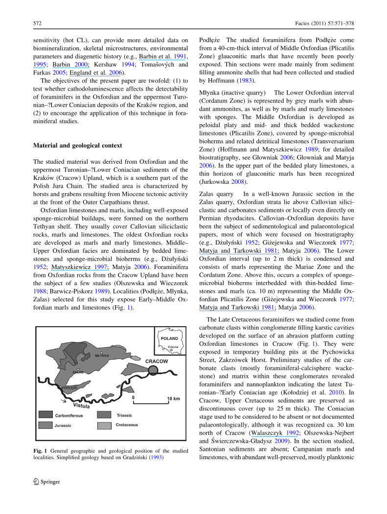
(Fig. 2b, d, f, i, k). When thin sections are observed in
transmitted light, foraminifers are poorly visible or even
indistinguishable (Fig. 2a, c, e, h, j), whereas they show
bright red–orange luminescence under CL. Foraminiferal
assemblages are pauspecific, consisting mainly of abundant
planktonic species, possibly Globuligerina oxfordiana
(Grigelis) (see also Olszewska and Wieczorek 1988; Bar-
wicz-Piskorz 1989), although random test sections do not
allow for precise determination of all specimens. The
morphotypes are equivalent to medium and large forms of
G. oxfordiana (Grigelis) according to the classification of
Wernli and Gorog (2000). Other determined foraminifers
recognized in thin sections are rare. Benthic species,
Nodosaria sp. and Lenticulina sp. (Fig. 2g, i), show similar
luminescence as G. oxfordiana. Undetermined benthic
foraminifers with tests of dark, ‘‘micritic’’ appearance
show poor luminescence, and they are much more visible
in transmitted light.
Associated fossils observed in thin section include
ammonite shells (Fig. 2f) and sponge spicules (Fig. 2d)
show bright red–orange luminescence. The fossils are
clearly visible within micritic matrix, which is weakly
luminescent. The matrix, composed of microsparite or
sparite, is luminescent, but foraminifers are still visible
under CL (Fig. 2i). Green–brown grains of glauconite
occur commonly within the chambers of foraminiferal
tests. Glauconite is non-luminescent (Fig. 2b, d).
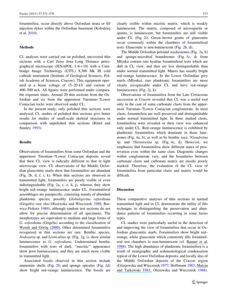
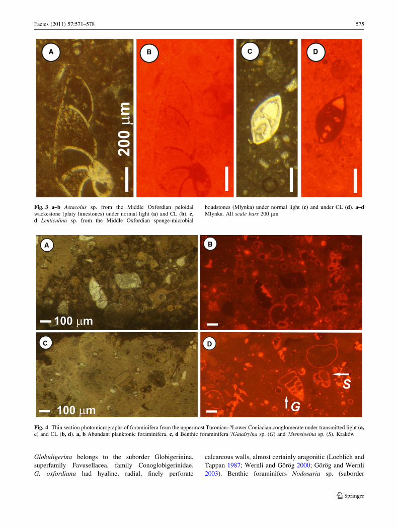
The Middle Oxfordian peloidal wackestones (Fig. 3a, b)
and sponge-microbial boundstones (Fig. 3c, d) from
Młynka contain rare hyaline foraminiferal tests which are
dull in CL view, and thus are less distinguishable than
under normal transmitted light. Matrix has usually bright
red–orange luminescence. In the Lower Oxfordian grey
marls (Młynka), rare planktonic foraminifers are more
clearly recognizable under CL and have red–orange
luminescence (Fig. 2j, k).
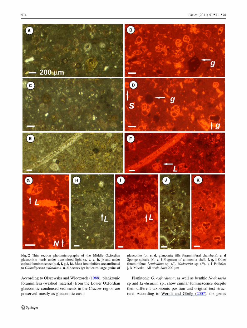
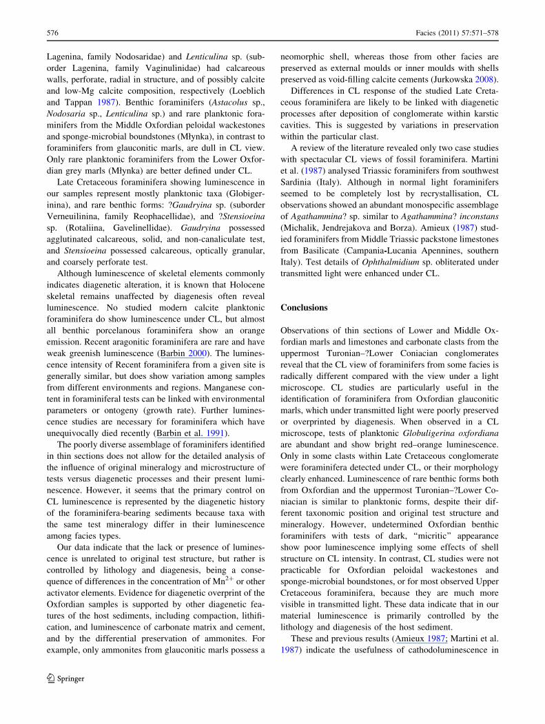
Observations of foraminifera from the Late Cretaceous
succession at Cracow revealed that CL was a useful tool
only in the case of some carbonate clasts from the upper-
most Turonian–?Lower Coniacian conglomerate. In most
clasts, foraminifera are well preserved and distinguishable
under normal transmitted light. In three studied clasts,
foraminifera were revealed or their view was enhanced
only under CL. Red–orange luminescence is exhibited by
planktonic foraminifera which dominate in those lime-
stones (Fig. 4a, b), as well as by benthic taxa: ?Gaudryina
sp. and ?Stensioeina sp. (Fig. 4c, d). However, we
emphasize that foraminifera show different states of pres-
ervation even within the same clast. Diagenetic changes
within conglomerate vary, and the boundaries between
carbonate clasts and carbonate matrix are mostly poorly
marked. Therefore, the evaluation of the CL view of
foraminifera from particular clasts and matrix would be
difficult.
Discussion
These comparative analyses of thin sections in normal
transmitted light and in CL demonstrate the utility of this
technique in distinguishing the preservation and abun-
dance patterns of foraminifers occurring in some facies
types.
CL studies were particularly useful in the detection of
and improving the view of foraminifera that occur in Ox-
fordian glauconitic marls. Foraminifers show bright red–
orange, while glauconite which commonly fills foraminif-
eral test chambers is non-luminescent (cf. Banner et al.
1988). The high abundance of planktonic foraminifera is a
result of stratigraphic and sedimentological condensation
typical of the Lower Oxfordian deposits, and locally also of
the Middle Oxfordian deposits of the Cracow region
(Gi _zejewska and Wieczorek 1977; Hoffmann 1983; Matyja
and Tarkowski 1981; Olszewska and Wieczorek 1988).
Facies (2011) 57:571–578 573
123
According to Olszewska and Wieczorek (1988), planktonic
foraminifera (washed material) from the Lower Oxfordian
glauconitic condensed sediments in the Cracow region are
preserved mostly as glauconitic casts.
Planktonic G. oxfordiana, as well as benthic Nodosaria
sp and Lenticulina sp., show similar luminescence despite
their different taxonomic position and original test struc-
ture. According to Wernli and Gorog (2007), the genus
Fig. 2 Thin section photomicrographs of the Middle Oxfordian
glauconitic marls under transmitted light (a, c, e, h, j) and under
cathodoluminescence (b, d, f, g, i, k). Most foraminifera are attributed
to Globuligerina oxfordiana. a–d Arrows (g) indicates large grains of
glauconite (on c, d, glauconite fills foraminiferal chambers). c, dSponge spicule (s). e, f Fragment of ammonite shell. f, g, i Other
foraminifera: Lenticulina sp. (L), Nodosaria sp. (N). a–i Podłe _ze;
j, k Młynka. All scale bars 200 lm
574 Facies (2011) 57:571–578
123
Globuligerina belongs to the suborder Globigerinina,
superfamily Favusellacea, family Conoglobigerinidae.
G. oxfordiana had hyaline, radial, finely perforate
calcareous walls, almost certainly aragonitic (Loeblich and
Tappan 1987; Wernli and Gorog 2000; Gorog and Wernli
2003). Benthic foraminifers Nodosaria sp. (suborder
Fig. 3 a–b Astacolus sp. from the Middle Oxfordian peloidal
wackestone (platy limestones) under normal light (a) and CL (b). c,d Lenticulina sp. from the Middle Oxfordian sponge-microbial
boudstones (Młynka) under normal light (c) and under CL (d). a–dMłynka. All scale bars 200 lm
Fig. 4 Thin section photomicrographs of foraminifera from the uppermost Turonian–?Lower Coniacian conglomerate under transmitted light (a,c) and CL (b, d). a, b Abundant planktonic foraminifera. c, d Benthic foraminifera ?Gaudryina sp. (G) and ?Stensioeina sp. (S). Krakow
Facies (2011) 57:571–578 575
123
Lagenina, family Nodosaridae) and Lenticulina sp. (sub-
order Lagenina, family Vaginulinidae) had calcareous
walls, perforate, radial in structure, and of possibly calcite
and low-Mg calcite composition, respectively (Loeblich
and Tappan 1987). Benthic foraminifers (Astacolus sp.,
Nodosaria sp., Lenticulina sp.) and rare planktonic fora-
minifers from the Middle Oxfordian peloidal wackestones
and sponge-microbial boundstones (Młynka), in contrast to
foraminifers from glauconitic marls, are dull in CL view.
Only rare planktonic foraminifers from the Lower Oxfor-
dian grey marls (Młynka) are better defined under CL.
Late Cretaceous foraminifera showing luminescence in
our samples represent mostly planktonic taxa (Globiger-
inina), and rare benthic forms: ?Gaudryina sp. (suborder
Verneuilinina, family Reophacellidae), and ?Stensioeina
sp. (Rotaliina, Gavelinellidae). Gaudryina possessed
agglutinated calcareous, solid, and non-canaliculate test,
and Stensioeina possessed calcareous, optically granular,
and coarsely perforate test.
Although luminescence of skeletal elements commonly
indicates diagenetic alteration, it is known that Holocene
skeletal remains unaffected by diagenesis often reveal
luminescence. No studied modern calcite planktonic
foraminifera do show luminescence under CL, but almost
all benthic porcelanous foraminifera show an orange
emission. Recent aragonitic foraminifera are rare and have
weak greenish luminescence (Barbin 2000). The lumines-
cence intensity of Recent foraminifera from a given site is
generally similar, but does show variation among samples
from different environments and regions. Manganese con-
tent in foraminiferal tests can be linked with environmental
parameters or ontogeny (growth rate). Further lumines-
cence studies are necessary for foraminifera which have
unequivocally died recently (Barbin et al. 1991).
The poorly diverse assemblage of foraminifers identified
in thin sections does not allow for the detailed analysis of
the influence of original mineralogy and microstructure of
tests versus diagenetic processes and their present lumi-
nescence. However, it seems that the primary control on
CL luminescence is represented by the diagenetic history
of the foraminifera-bearing sediments because taxa with
the same test mineralogy differ in their luminescence
among facies types.
Our data indicate that the lack or presence of lumines-
cence is unrelated to original test structure, but rather is
controlled by lithology and diagenesis, being a conse-
quence of differences in the concentration of Mn2? or other
activator elements. Evidence for diagenetic overprint of the
Oxfordian samples is supported by other diagenetic fea-
tures of the host sediments, including compaction, lithifi-
cation, and luminescence of carbonate matrix and cement,
and by the differential preservation of ammonites. For
example, only ammonites from glauconitic marls possess a
neomorphic shell, whereas those from other facies are
preserved as external moulds or inner moulds with shells
preserved as void-filling calcite cements (Jurkowska 2008).
Differences in CL response of the studied Late Creta-
ceous foraminifera are likely to be linked with diagenetic
processes after deposition of conglomerate within karstic
cavities. This is suggested by variations in preservation
within the particular clast.
A review of the literature revealed only two case studies
with spectacular CL views of fossil foraminifera. Martini
et al. (1987) analysed Triassic foraminifers from southwest
Sardinia (Italy). Although in normal light foraminifers
seemed to be completely lost by recrystallisation, CL
observations showed an abundant monospecific assemblage
of Agathammina? sp. similar to Agathammina? inconstans
(Michalik, Jendrejakova and Borza). Amieux (1987) stud-
ied foraminifers from Middle Triassic packstone limestones
from Basilicate (Campania-Lucania Apennines, southern
Italy). Test details of Ophthalmidium sp. obliterated under
transmitted light were enhanced under CL.
Conclusions
Observations of thin sections of Lower and Middle Ox-
fordian marls and limestones and carbonate clasts from the
uppermost Turonian–?Lower Coniacian conglomerates
reveal that the CL view of foraminifers from some facies is
radically different compared with the view under a light
microscope. CL studies are particularly useful in the
identification of foraminifera from Oxfordian glauconitic
marls, which under transmitted light were poorly preserved
or overprinted by diagenesis. When observed in a CL
microscope, tests of planktonic Globuligerina oxfordiana
are abundant and show bright red–orange luminescence.
Only in some clasts within Late Cretaceous conglomerate
were foraminifera detected under CL, or their morphology
clearly enhanced. Luminescence of rare benthic forms both
from Oxfordian and the uppermost Turonian–?Lower Co-
niacian is similar to planktonic forms, despite their dif-
ferent taxonomic position and original test structure and
mineralogy. However, undetermined Oxfordian benthic
foraminifers with tests of dark, ‘‘micritic’’ appearance
show poor luminescence implying some effects of shell
structure on CL intensity. In contrast, CL studies were not
practicable for Oxfordian peloidal wackestones and
sponge-microbial boundstones, or for most observed Upper
Cretaceous foraminifera, because they are much more
visible in transmitted light. These data indicate that in our
material luminescence is primarily controlled by the
lithology and diagenesis of the host sediment.
These and previous results (Amieux 1987; Martini et al.
1987) indicate the usefulness of cathodoluminescence in
576 Facies (2011) 57:571–578
123
studies of foraminifera from rocks with different deposi-
tional and diagenetic histories, particularly when they can
only be studied in thin sections. If the CL response of
cement or host sediment strongly contrasts the skeleton
material under CL, this technique can also be a useful tool
in the better documentation of the studied fossils.
Acknowledgments We thank Dr. Adam Tomasovych (Chicago)
and an anonymous reviewer for valuable comments and critical
remarks on the manuscript, and Dr. Stephen Vincent (Cambridge) for
linguistic help.
Open Access This article is distributed under the terms of the
Creative Commons Attribution Noncommercial License which per-
mits any noncommercial use, distribution, and reproduction in any
medium, provided the original author(s) and source are credited.
References
Amieux P (1987) Description petrographique de foraminiferes par
combinaison d’images en lumiere naturelle et en cathodolumi-
nescence. C R Acad Sci Paris (II) 304:741–744
Banner JL, Hanson GN, Meyers WJ (1988) Determination of initial Sr
isotopic compositions of dolostones from the Burlington-Keokuk
Formation (Mississippian); constraints from cathodolumines-
cence, glauconite paragenesis and analytical methods. J Sediment
Res 58:673–687
Barbin V (2000) Cathodoluminescence of carbonate shells: biochem-
ical vs diagenetic process. In: Pagel M, Barbin V, Blanc P,
Ohnenstetter D (eds) Cathodoluminescence in geosciences.
Springer, Berlin, pp 303–329
Barbin V, Ramseyer K, Debenay JP, Schein E, Roux M, Decrouez D
(1991) Cathodoluminescence of Recent biogenic carbonates: an
environmental and ontogenetic fingerprint. Geol Mag 128:19–26
Barbin V, Brand U, Hewitt RA, Ramseyer K (1995) Similarity in
cephalopod shell biogeochemistry since Carboniferous: evidence
from cathodoluminescence. Geobios 28:701–710. doi:
10.1016/373S0016-6995(95)80064-6
Barwicz-Piskorz W (1989) Microfauna of Lower Malm deposits at
Zalas, South Poland. Kwart AGH Geologia 15:6–27 (in Polish
with English summary)
D _zułynski S (1952) The origin of the Upper Jurassic limestones in the
Cracow area. Rocz Pol Tow Geol 21:125–180 (in Polish with
English summary)
England J, Cusack M, Paterson NW, Edwards P, Lee M, Martin R
(2006) Hyperspectral cathodoluminescence imaging of modern
and fossil carbonate shells. J Geophys Res 111(G03001):1–8.
doi:10.1029/2005JG000144
Gi _zejewska M, Wieczorek J (1977) Remarks on the Callovian and
Lower Oxfordian of the Zalas area (Cracow Upland, Southern
Poland). Bull Acad Pol Sci Ser Sci Terre 24:167–175
Głowniak E (2006) The Platysphinctes immigration event: biostrati-
graphic and palaeoblogeographic implications for the Middle
Oxfordian (Late Jurassic) seas of Central Europe (NW Germany
and Poland). N Jb Geol Palaont Abh 241:155–201
Głowniak E, Matyja BA (2006) Młynka Quarry. Lower to lower
Middle Oxfordian. In: Wierzbowski A, Aubrecht R, Golonka J,
Gutowski J, Krobicki M, Matyja BA, Pienkowski G, Uchman A
(eds) Jurassic of Poland and adjacent Slovakian Carpathians.
Field trip Guidebook of 7th international congress on the Jurassic
system. Krakow, 6–18 Sept 2006, pp 138–141
Gorog A, Wernli R (2003) Palaeobiogeography of the Middle Jurassic
protoglobigerinids (Foraminifera) in thin sections. Eclog Geol
Helvet 96:237–248
Gradzinski R (1993) Geological map of Cracow region without
Quaternary and terrestrial Tertiary deposits. Muzeum Geologi-
czne, Instytut Nauk Geologicznych PAN, Krakow
Hoffmann M (1983) Stratygrafia jury okolic Mirowa i Podłe _za. M.Sc.
thesis, Jagiellonian University, Krakow (in Polish)
Hoffmann M, Matyszkiewicz J (1989) Wykształcenie litologiczne i
sedymentacja osadow jury w kamieniołomie Młynka. In: Rut-
kowski J (ed) Przewodnik LX Zjazdu Polskiego Towarzystwa
Geologicznego, Krakow14–16 wrzesnia 1989, pp 78–83 (in
Polish)
Jurkowska A (2008) Tafonomia i paleoekologia amonitow z osadow
oksfordu okolic Krakowa. M.Sc. Thesis, Jagiellonian University,
Krakow (in Polish)
Kershaw S (1994) Cathodoluminescence of Silurian stromatoporoids
from Gotland, Sweden. Cour Forsch Inst Senckenberg
172:307–318
Kołodziej B, Szulc J, Machaniec M, Kedzierski M, Duda M (2010)
Injection dykes as evidence of Campanian synsedimentary
tectonics on the Cracow Swell, southern Poland. Ann Soc Geol
Polon 80:285–301
Loeblich AR, Tappan H (1987) Foraminiferal Genera and their
classification. Van Nostrand Reinhold, New York
Martini R, Amieux P, Gandin A, Zaninetti L (1987) Triassic
foraminifers from Punta Tonnara (SW Sardinia) observed in
cathodoluminescence. Rev Paleobiol 6:3–27
Matyja BA (2006) Stop A17–Zalas Quarry–Callovian transgressive to
condensed pelagic deposits, Lower to lowermost Middle Oxfor-
dian deposits of sponge megafacies. In: Wierzbowski A,
Aubrecht R, Golonka J, Gutowski J, Krobicki M, Matyja BA,
Pienkowski G, Uchman A (eds) Jurassic of Poland and adjacent
Slovakian Carpathians. Field trip guidebook of 7th international
congress on the Jurassic system, Krakow, 6–18 Sept 2006,
pp 70–72
Matyja BA, Tarkowski R (1981) Lower and Middle Oxfordian
ammonite biostratigraphy at Zalas in the Cracow Upland. Acta
Geol Pol 31:1–14
Matyszkiewicz J (1997) Microfacies, sedimentation and some aspects
of diagenesis of Upper Jurassic sediments from the elevated part
of the Northern peri-Tethyan Shelf: a comparative study on the
Lochen area (Schwabische Alb) and the Cracow area (Cracow-
Wielun Upland, Poland). Berlin Geowiss Abh E 21:1–111
Olszewska B, Wieczorek J (1988) Callovian–Oxfordian foraminif-
era of the northern Tethyan shelf: an example from the
Cracow Upland (Southern Poland). Rev Paleobiol Spec Vol
2:191–196
Olszewska-Nejbert D, Swierczewska-Gładysz E (2009) The phos-
phatized sponges from the Santonian (Upper Cretaceous) of
the Wielkanoc Quarry (southern Poland) as a tool in
stratigraphical and environmental studies. Acta Geol Pol
59:483–504
Pagel M, Barbin V, Blanc P, Ohnenstetter D (eds) (2000) Cathodo-
luminescence in geosciences. Springer, Berlin
Richter DK, Gotte T, Gotze J, Neuser RD (2003) Progress in
application of cathodoluminescence (CL) in sedimentary petrol-
ogy. Miner Petrol 79:127–166. doi:10.1007/s00710-003-0237-4
Rittel JF, Stanley GD Jr (1993) Enhanced skeletal details and
diagenetic processes of Triassic corals revealed by cathodolu-
minescence. Cour Forsch Inst Sencken 164:339–346
Tomasovych A, Farkas J (2005) Cathodoluminescence of Late
Triassic terebratulid brachiopods: implications for growth pat-
terns. Palaeogeogr Palaeoclimatol Palaeoecol 216:215–233. doi:
10.1016/j.palaeo.2006.06.028
Facies (2011) 57:571–578 577
123
Walaszczyk I (1992) Turonian through Santonian deposits of the
Central Polish Uplands; their facies development, inoceramid
paleontology and stratigraphy. Acta Geol Pol 42:1–122
Wernli R, Gorog A (2000) Determination of Bajocian protoglobige-
rinids of Gyenespuszta (Bakony Mts., Hungary). Rev Paleobiol
19:399–407
Wernli R, Gorog A (2007) Protoglobigerines et Oberhauserellidae
(Foraminiferes) du Bajocien-Bathonien du Jura meridional,
France. Rev Micropal 50:185–205
578 Facies (2011) 57:571–578
123


