Embed Size (px)
Citation preview

THE MIC,ROSCOPIC ANATOT'ûY OF HI]IUA}T ENDOCRINE ORG¿'NS
A Thesis
Presented to
the Faculty of Medici-ne
Ilniversity of Manitoba
In Partial- Fulfillnent
the Requirements for the Degree
Mastor of Scienoe
by
Jack Edwin Newell
UIaroh, 1951
of
L- | E: í:.; ,t7:..,::;;_\f

ACIO{OiI'ILEDGMENTS
No project, regardless of its size, is often accomplished wíthout
the aid of others and this modest piece of work ís no oxception. There
are many to thank without whose helpr so wiLlingly givenr the present
work v¡ould have been hoPeLess.
The project and study was conducted. under the guidance of
Professor D. J. Bowie of the Department of .Anatomy. Professor Bowie has
offered constructive critioism throughout the wholo worlc and proparation
of the manuscript. The vwiter has been fortunate to have the opportunity
to examine many soctions of Professor. Bowiets collsction. Professor
De¡1iel Nicholson of the Department of Pathology gave permíssion to carry
out the work and during its course offered nany helpful suggestions a:ld
criticlsms. Professor J. M. Led.errnan gave helpfì:I criticism of portions
of the manuscript. I am indebted to Dr. D. T[. Penner v¡ho assisted
greatly in the collection of fresh human material. Dr. A. .4.'Earn kíndly
loaned his oollection of placental preparations and, also was very help-
fut in his critioism of the chapter on the pLacenta and. of the chapter
on the hormones, of reproduction. During the oourse of the study¡ it
beca¡ne apparent that at least somo Im.owledge of photography was r.eoessêrfr
.An able teacher was found in lvtlss L' Nason, who assisted also with the
e:qposingr developing and printing of many photomicrographs. Miss M.
Melvill-e provlded the najority of the excellent hematoxylin and eosin
preparations. Most of the speoial stain preparations'are due to the
efforts of [[iss Melvi]Ie. Miss 1. Toupin assisted by preparing those

iii
ACICSOIU.LEDGIIH{T S ( aont inu ed )
soctions stainod by the Masson trichrome and simple stain techniques.
Miss Toupin labelled many of the d.rawings, nrhile Miss E. Green Iabelled
some of the remainder. Miss Monk and the library staff have been most
oooperative.
tho writer wishes to aclarowledge with appreciation the vrritten
permissions obtained. from the following publishers¡ The TIlstar Institute
of Anatony and. Biology for permission to reproduoo Figuro 1, ofnThe Meningeal reLations of the hypophysi" ""."O"rtt by H. G. Schwartz,
appearing in the .{natomioal Record, Volumo 67¡ pp' 35-443 The publishers
of The Bulletin of tho Johns Hopkíns l{ospital for pormission to reprod.uce
Figure 1, of n0n the glandular elements in the posterior lobe of the
human hypophysistt by Ðean Lewis and. F. C. Lee, appearíng in the Bulletin
of the Johns Hopkins Hospital' Volurne 41, pp. 241-277; The I1[. B. Saund.ers
Companyr Phila.¡ publishers of .A Textbook of Histology, by A. A. Ma¡ci¡row
and ü&n. Bloom, Fifth ed.ition, tg¿g, pp. 700, for perrnlssion.to reprodu-ce
Figure 503, page 572 (îrcm Hertig and Rock, Carnegie Contrib. to Ernbryol.¡
VoI. 29, 1941, Fig. 15).
The pen and ink drarvings were done by the author and any shorL-
comíngs or orrors in them are due to him.
Last but certainly not least, appreciation is exprossed to the
t¡pists. Mrs. J. E. Morrison assisted with rnuoh of the first draft of
the thesis and gave considerabl-e assistance in that phase of the work,
The final drafb and settíng-up was ty¡:ed by Mrs. T[. Jn Bellr whose
excellent and metÍoulous work in the pages to follow speak for themsolves.
::;.t Ìltit.i;.1-,..,,"j

T.A3LE OF CONTENTS
CIfAptER P.AGE
1, Tffi.A-DRHIA.LGLAIüDS . t." " o " " " .. ' o " t I
Tntroduction'. . . . . ' ' ' ' t ' ' t ' ' ' ' ' ' ' t t 1
....tIThe0ortex . . . .. . t " " " t "'' t "'
thg oapsüIe . . . . . . . . " t " t t " " t o' I
The blood vascular sfstêIn . o o''' o o''''''' I
Zonaglomerulosa ....... .. r " t " e ' o ' 5
Zonaîasaiculata ..... t ' ' ' ' o ' ' t ' ' ' ' ' 9
Zonareticularis .... t. t " " " " t t " 9
The coftical lipids .' . . . . .''''''''''' 15
Development oftho cortex. o . . e ' ' ' ' ' o ' " ' 15
. Lifehistoryoftheoorticaloells r.. o. '.. t. L7
variatíon of cortical norphology . . . o . . i . . . . 18
variation of cortical thickness o I . . . . . . . . . 18
R¡nctions of the ad.renaL oortox . o . . . . . . . . . 18
Thg oortical hormoflês r ¡ r . . . . .''''' t''' 20
Crystalline fraction . o . o''' t' t'''' t t' 20
. Amorphousfraction .... r .. r .. " " ' ! " 23
cholesterol and asoorbÍc acid. in coftox o .' . . . . . 23
The functÍons of the hortoll€s r . . . . . . . . . . . . 25
TheMedul]-8,.... o...... t..'t' o' t o' t 26
The ahromaffin systelll r r t r o . o . . . o . t o t'' 27
DevelopmentofthenreduLla .. t... ! o t. r. " 27
Thgb]-ood suPply o . . Ò . t . . o t . . . ' o ' ' ' t 2'î

v
CHAPTm. PAGE
Thgngrvg supply . t'''' o t''' t i''' t'' 28
Adreni-rrg . . . t ' t I ' ' t ' ' o ' ' ' ' ' ' ' t t ' 39
BIBLIOGRAP}IY o''''''' t t''' o''''''''' o 32
II. TIfi MALE REPRODUCTM SYSTEM (tgn fnSfnS) . . . . . . o o . 56
Introduction . o ' o t ' t .' t t t ' ' ' ' ' ' ' ' t ' t 86
The connooti.ve tisstlo frameulork . . . . . . . . . . . . ¿ 37
.Theartorialsupplyof thetestis " ' ' I ' ' ' ' ' ' I 37
Arterial supply of the lobe, o o' "''''''''' 39
Venousdrainage.... ' ' ' ' ' ' ' ' ' ' ' t ' t ' t o 39
Lynrphatio d.rainage of thetestis .. .. . . o .. . . . 40
Nervesupplyof,thetestis .... ' ' ' ' ' ' ' ' ' ' ' 4I
ThgTubu]-arsystem ... t t o. " .. " t "'' 42'
.General pla.n of the tubular system . o' : " "' Ò 42
The seminiferous tubules . . t t t'''''''' ! t 44
. The colls of sertoli . ., . . . . . . . . . . . . . . 44
' The spermatogenic oêlls . . . .''' t'' o' t' t I 47
" Sperrnatocytogenesis i ¡''' i t o' o'' t'' o'' 47
Spermiogengsis ? . . . . t' t'''' t'' t t t t I 48
Ûlature spermatozoa o . . . . . " "' o " " " 50
Abnormal forms of spermatoZo& r . . . . I . . . . . . . 53
Spermatogenioïvave ...... t ' ' ' ' ' ' ' ' ' ' ' 54
Theoryof d.eter¡ninationof sex .I... o ' ' ' ' ' r 54
Sgmen.. ! ô.t.....t" t"tt" "'.' 57
Thephysiologyofthetubular syste4 t.. ' ' ' ' ' ' 58

vi
CEAPTM, PAGE
TheÏnterstitialTi-ssue ... o. !... ' o " " t ' 59
Introduotion . . . . I . . t . . . . . .'' i'' t t I 59
The interstitial ceLls of Leydig . . . . . . . . . . .' 60
I!I! _.urstrrbution and amount of interstitial cells . o o . ' 60
Histological details . . . . . r . . . . . .' .. . t' t 65
Relationship between nervos and the Leydig cells . . . o 66
Variationsofthetesteswithago ' ' o o..... '. 69
Note on d.evolopment of the testes . . . . . . . . . . . 72
Thehormonesofthetestos o... o........ ¿ 73
BIBLIOGRAPIIY . . . c ¡' . . . . . o . . . t . t . .' 74
rrÏ" TirE FEMALE REPRODUCTTVE SYSTEM (rnE OV¡ny) . . . . . . . . . 78
Introduotion . t . . . . o . . . . t . . . . . . . . . . . 78
General description of the ovaries . . . r . r . . . . .' ?B
The bLood supply of tho ovary . . . . r . . . o . . o . . 79
Vessels within the organ t . r . o . . t o . . o . . . t . B0
Thg nerve suppl)r r e . . . o . . o . . . . . o . . . . . o 81
Note on embryotogy of ovaries' . . o . ) . . . . . t . . BL
A summary of the fate of the priuiary foi.liclos . . . . . r Bz
The gern-rinal epithelium . . . . . .' . . o . . . . . . . 84
Cortexand.mgdulla. r . t t.... t .. . ..... . , BT
Originofova r o. r o................. BB
Primaryfo].licles .. o o. . .. ... r.. .. . ... Bg
Growingfollicles . t...... t. t... t..... 92
Maturefolliolgs . r o..-............... 99
. vii
CEAPTER' PAGE
0vum.... o......t o o........ " "t 100
ttoo......IOzOogenesis . . . . . . . ... . . t . . . o t o t'
tr\¡rther d.evelopment of the Graafian follicles' . . . . . I04
AtresiaofthefollioLes ............ ' ' ' o 104
ThgCorpusLutgum ... r........ o.... ' ' ' 109
Introduction. . . . . . . . . . . o . . o . .'. t t ' ' 109
TheCorpusLutoumof Menstru.ation ........ '. o t Ll-O
Hypereaic stage . . . . . . . . . . . . . o . . . . . o 110
Stage of vascularization . . . . . . . . . o . r . . . . 110
Stageofnaturity ............ o r.. o.. lL4
Stagoofregression ............. o. o.. 116
tho Corpus Luter¡m of Pregnanoy . r . . .' r . . . .' . . !2L
The granulosa-lutein layer . . o . .. .' r . o . . . . o I24
The theca-lutein layer . . t . . o . .' . . . . . . . o L25
TheTestosalr'dOvariesCompared ..... o... '.. . !26
Thelìortonesof theOgarios o.. o. ' o o....... I27
BIBLIOGR-APIIÏ ...... o r... o..... .. r.... I2B
IV. THE PLd'CEt(fA . . . . . . . . . . . . t . . . . . . . . . t' 131
fntroduction . . . . . . . . . . . . . t . . . . . . . o 1õ1
Dovelopmont before implantation . . . . . o . . . . o'. . I32
Implantation .. r. .... o t t... o o ¡..... ]-34
P]-acgntation . . . . r . t . . t . o . t . . . . . t . . 155
Growbhandmaturityoftheplacenta '.. r. o. o... 156
Changes inthe endometrlum ' . . . ¡ . r ' . . o . . . . . L37

CEAFIM,
.Angiogenesis . t............ o t ' o " l '
Villiat12TI@êks ¡ . . . . . . . . . . . . r' t t o t'
Vi1liat20T\rooks. o . . . . . . . . . . . . . .'' t'
ViLli at SOwggkS ¡ o o . . . . . t . . . .. ' ' ' ' ' '
ViLli at 40 wegks (term) . . . r . t . . . . . . r'' ' '
Expulsion of the placenta . . . . . . . . . . . . . o' I
Grou¡bh Chenges in Placenta during Gestation . . . . ¡ i, .
ChangesintheVil1i............ I ' ' ' ' ' '
Hofbauer oells . . . . . . . . . . . . . . o . . o | .
The sync¡rbium . . . . . . . . . t . . o . . ' t . ' '
the oytotrophoblast (Langhanst layer) . . o' . . . . .
Thergticulum ... o... . r...........
Degeneratlvechangeg ........ t..... o. .
ThePlaoentaatTerm ... o.. t....... t.. o
Placental Circulation . . . . . . . . . . . . o .' o o
Bumrols theory . . . . . . . . . . . t . . . o . . . . .
Spannerts theory . . . . . . . . . | . . . o . . . . .
Fgta]- Circulation. r... ..... . . ... o. ...
BIBLIOGRAPIIÏ o............... ' ' ' ' ' ' '
Vr TI#EORIIONES0FREPR'ODüCTION ] I o r. !.... ' t...
Introduction . . . . . . . . . . . . . . . . . . . ù. o
Thghormongs r r i r . . . .. . .. . . . . ! . . . . .
Fol-licuLar stimLtlating hormone (FSH) . . . . . r . . ! .
Luteinizinghormone (frn) ........ o..... t o
vlii
På.GE
138
139
isg
L43
14.õ
143
]'49
149
150
L5l_
156
l_57
L57
160
160
t6L
161
165
166
169
L69
171
171
t73
.!r':.".\':t:i
i:.¡;:,ìril

ix
OHAPTER' P¿'GE
Chorionic gonadotropfrin (.APL) . . . . o . . . . . .'. . . L76
Prolactin. . o . . . t . . . . t . .. . o . . . . . . . L74
.Aadrogens.. r. . o t .. .. ... u.t... .... 174
Oestrogons ... .. o..... ......... . t. L75
Progosterone . o . . . . . . . . . . t . . . o . . . . . !t5
Mgthod.s of stu-dy . . . | . . o . . . . . . . o . o . . . I75
' The humanma].e oastfat€ r . . . . t . . . . . . . . . . . 176
EarlyouiluchisIlLo.......... c.... ' t '., !76
Latg eunuohism . . o . . . . . I t . . . t . . . . . . . 177
The pre-pubertal female castrate . r . . . . . . . . . o 178
The post-pubertal femal-e castrate . r . . . . . . . . . . 178
Pre-puberta]- fgmale . . . . . . . . . . . . . r . . . . . 179
I/lgnstruation r . . . t . . . . . . . . t . . . . . t t . 179
Thehormoneg ...... e.... r t.... o. o. lBO
Ne¡'vr¡erconceptsof menstruation .... r... '. o. l-Ba
Tineof ovulation. o . . . . . o t . r . . . . . 185
Characteri.stios of the n'or:nal menstrual Gycl€, o r . o . 184
Cyolic changes in the accessory organs of roproduction 184
Pregnanoy.. . . ... . . .. .. r . t... .. . t. 187
, Menopauso and climagterio . . . . . . . . . . . o . . . . lBB
Hormone integration in'the male . . . . . . . . . . . . . 190
The male climacteric . . . . . . . o . . . . . . o t . . 191
BIBLIOGRAPHÏ . . . . t t . . . . . . . t . . . t . . . . . 192
VI . TEE PåR¿.THffi.OID GIrAIüDS . . o . . . . . . . . . . . . . . . I95

x
CEAPTM, PAGE
fntroduction . . . . o . r . . . . r o . . . . . . . . . ].95
Gross desoription . r . . . . . . . . ¡ . . . r . . . . . 195
SizgandlÍeight ...... o r..... r. r.... 195
Position ....r r.r ... .r...... o.... L96
Nr¡mberof glands ..... o.............. L97
Blood supply . r ¡ . . . . t . . . . ) . . . . r r . . . L97
Nervg supp].y . . . . . . . o o . . . . . . . . r . . . r 197
L¡nnphatic drainage . . . . .. t | . o . . . . . . . . . . 198
Note on embryology . . . . . . . . . . . . . . . . . r t 198
Histology . . . . . ... . . . . . . . . . . . . . . . . 199
Capsule ... ....o........ oo. o... | 199
RetiGülültr. r c r.. ... ... .. r r.. . ... . 199
Fat0gllS¡ o r.. ... . ...r .. .... ... o 199
Blood vessels within tho organ . . . . . . . . . . . . 199
Pigments . o.. .... ..... r. ..... o. . ZAL
Lipoid.s .. ... .. o. r . r. r'. ... o... o zOL
General arrangement ofparenchyna. . . . . . o . .. , e01
. Comp&Gtr...o .................. 2O3
Coarsgtrabgoular. r....... .. o ... .. , ZO3
Lobular... o.r. o... t rr.... .... . ZOs
Largeacinar ..... o. . o. r....... . . 203
The Cells of the Parench¡ana . . o o .' . . . .' . ! . 2O3
Prinoipal oells . . . . . | . . . . . . . o . . . . . 2O4
Darkpríncipal cells . .. . . . . . ' ... .. o 204
xi
CEAPTER' PAGE
Pale principal oelLs' . .' . . . . . . . . . .' . 2O5
'ffater-cLear (ï$asserheLle) oeIls ..... o r. o , ZO5
0r¡¡phil ce1ls . . r . t t . . . . . . . . . . . . . . 2O7
Dark orr¡rphil cells . o o . . . . . . . . . . . . t . 2OT
PaIe ox¡rphil ceLls . . ... . . . . . . . . . . . . . 2O7
fnoonstant Structures t o. ... ' . r .. . r. ... ZOg
Columnar-cgl"led alvooli o . o . . . r' . . . .' o . ZOg
Colloldvesiclgs.. r...... ....... .. . 2Og
Cystic vesioles . r .. . . . . . r o . . . . . . r r . ?Ll
Othgr cysts o . . . . . . . . . . . . . . . . . . . . ?J-L
The Parathyroid Hormono . o . . . .' o o . o . ¡ . . . 2!Z
CaLciummotaboLism. ... o . . ' o. . . . . . . t . 2].]2
Phosphorous motabo].ism . . . . . . . . . . . . . . . . 213
llagnesium . o . o | . . o . t . o t . . . . o . . . . ?Ls
ûther faotors influenoing calciummetabolism. . . . . 2L3
Present theorios regardíng the function of the homone 2L4
H¡poparathyroidisn . q o . . '' . . . . . r . o' . . 2I5
H¡rperparathyroidisn . . . . . r' . . .' . . . . r . 216
BIBLIOGRåPEÍ .' . . r . . . r o, t o . . . t .' I t t . ZIB
VIf . THE ÏEYROID GL.A¡TD t . . . . . . . . . . . . ' . . . . o . . ZZL
Introduotion . r . . . . . . . . . . r . . t . . . . . . . ZZL
Gross description . . o . o . . . t . . . . . . . . I . I 222
BloodsupPly.. .. ... .. .t .. :.... .. .. . 223
Vonous drainage g . . . . . . . . . . . . . . . . o . . . 224

CIIAPTER.
Lyrnphatics . . r . . . . . . . o o . . . o . . . . . . .
Ngrvgs a. o r r. ... ... . a .r a a . o ... . .
Ernbryologioal note . . . o r r . r o . . . . ! . . . o .
Tho microscopio a,nâ.torn[¡ . . . . . . . . . . . . o . . . .
Thgfo].licles r o .. . . .. . . ... .. r r .. r.
Size . . r a a . o a a . a . . a a o . . r . . a . r a
Shape . . o . . . i . . r . r. . . . . . . . . l . . . .
Co11oid... ... ... r .. r .. .. . .. ... .
Epitheliun
Thevariations o r . o. . r r r.. r. t. .... o
The funation of the thyroid . r . . . . . . r . ! .' . .
Introduation r . . . . t . . . . . . . . . o . . . . .
Thohormone. i... ... .. .... ..... . . t
Theaotionsofthehormone '... r r....... r
Ioding o.. r r ... . . .! .. o. .. .. ....
BTBLI0GBAPHY . . r . . . . . . . . . . . . . . . . o t . r
VIII. TTIE FAI{CRE¿'S . . . . . . . . t . . . . . . . . . . . . . .
Introduation .... r.. r. ¡ r......... o.
The exoorlne panoreatio tissuo . . . r . . . . . . . . .
Thgacini o l .. ... . .. r . . . l. .. o . t o ..
Thoduot systeû. o r o . . .. . o .. . o . . . o . .
Tubulgs . o.... . .. l. r. . r. .t. .o . r
The blood vascular supplS r o . . . . . . . . .' . . .
Ngrvg supply . r . . r . r . . . r . . . . . .'. . . .
:(ii
PAGE
224
225
225
226
227
227
227
227
228
23L
2,35
235
235
238
240
242
245
245
246
246
249
252
255
257
i.
i: .
,:.ijj

CITAPTER
xiii
PAGE
The Lynphatio drainage . . . . . . o . . . . o' . . . 257.
The physiology.of the êlroofin€ r r . o . . . . . . . o 258
The socretingrreohanism o . o. r .. .. .. .. . . 259
Pancreatic juice . r r . r . . . . . o . . o . . . r . 260
Changeswithage.. r... .. r.. o. .. r. . r 261
The endocrine pancreatic tíssue . . . . . . . . . .' . 264
-Anountanddistributionof islettissue ... t.. . 264
Variation of amount of acinar and islet tissue with
age ..r...... t.............. 268
Thgislgts. r... ... ... ..... ..r o. . 269
The origin of the islets . . . . o . . . . . . . . r, 274
The relation of ducts to islets r' . . . . . . . . . 274
The rslation of acini to islets . . . . | . . . . t . 276
Stainlng of the islets . . . . . .' . . r . o . . . . 277
Insular blood suppL¡¡.. r . . . . . . . . . . . o . . . 279
The nerve supply of the islets . r . . i . . r . . . . : ?BL
The neuro-ínsular complexes of Simard . . . r . r r . ZBz
The lynrphatic drainage of the islots . . . . . . . .' 2Bz
The physiology of the islots . .' . i . . l . . . . . r ZBz
Introductio¡. . o . . . . . . ? . . . o . . . . . . . . 2Bz
Sourceof insu]-in . r o. r.... r....... . ZBz
Pancrgatectomy . . . . . . . . . . . . . . . . . | . . 285
Injectionof insulj.tl .r ¡ . .. ... .. .. .. . . . .. 286
H¡lorinsulinisn .. o. r... r......... r 2,BT

CHAPTER
Note on carbohydrate metabolism . . . . . . . ., . t
Diabgtgs " ! c r . r . . . r ..... . r.. ... .
Tho history of the panereas . . . . . . . . . . . . o
Note on embryology . . . . r . . . r . . . . . . . . .
BIBLIOGR.APHY . . . . . . . . . . . . . . t . t r . . . . .
ïX. TIIE HYPOPEYSIS CB.EBRI . . . . . . r . . . ., . . . . . .
Tntroduction. . o . . o . . . r .. . . .. . . . . . r
TonuÍnology . r t . .! . ... . .. r r. .. . t r .
Parsanterior . . . . r. . . . . . . .. r .. . . .
Parstuberalis.. . r... .. . o . r ... .. . .
Pars intgrmedía . o . . . . . ..r . . .' o . . . . .
InfundibuLum r . . . . . . . . o . . . . . . . . t . .
Parsngrvosa. . . t .. . . . . . . o . o . .. . r .
BlrbryologicalDote r... t. o... .... I o...
IVleningoal relations . . o . . . . . . o r . . . o . . .
Weight of tho Hy¡lophysis . . ' . ' ... . . . ' . . . . .
The arteríal zuppIy aJrd venous drainage . . . . r . . .
Innervation e . . . . . . . . t . . . r .. . . .. r o
L¡fmphatios . . . . o . . . . . . . . . . . . . . . . . r
Pars Nervosa (The posterior Lobe, Neuroh¡4gophysìs) . . r
Pituicybes r o . . . . . .. . r . . .. . :. . . . .
Pignont . . . o . . . . . . r . . . r . . . . . .. . .
The connective tissug of the pars Ttervosa . . . . r .
Epithelial cel].s . t . . . . . . . . . . . o . o.o r .
xiv
PAGE
zBB
289
290
294
295
500
600
302
302
503
õ03
505
504
304
505
510
gLL
3r4
315
515
5L5
3I7
317
518

xv
C}TAF]ER. PAGE
Pars intofilgdig. ¡ . . . . . . . . . . . . . . . . . . . . . 32O
Surunary of the epithelial oeLls of the pars intermedia . 324
Theaoolloidnbod.iesofthoh¡4gophysis . r.. i ¡ .. ' 325
The pars antoriof . r . . . . . . o . . . . . ! r i . . . . 3Zg
Co3,1 t¡rpes .... r ?.. r t............ I 329
Distribution . . . . . . . . . . . . . . r . . . . o r o 550
Peroontago of oelI t¡rpes . .' . o' . . . . . . . . . t 530
llistoLogyoftheceLls .... o... '..... t. . 333
Thg eosinophils . . . . . . . . . . . . . . . . o . . . 535
Thebasophils. .... o . o. .... ... . f.. o 336
Pars tuberalis . . . . . . . . . r r . . . . . . . . . . . 556
The hypophyseal sbalk and. modian eminence r . . . r . . . . 337
Concretions and. extraneous måterial . . . r o . . . . . r . 84O
Thehonnonesoftheh¡¡pophysis ...... '. r. '... 34L
The;hormones of the pars anterior . . ! . . . r . . . ? . 34L
Follicle-sti¡ntrlating hormone . .' . r' . . . r . . o 342
Luteinizing hormons . . . o r . . . . r . t . r ! r . . 942
LutootrophÍc hormone . o . . . r r . . . . . . . o r . 342
Corticotrophic hormone r' . . . . . . . . t . . . o . 343
Thyrotrophiahormono .. o. o.. . r. ...... . 34g
Somatotrophio hormorte . . . r . . . . . . . . . . . . o 343
Qther tactionsB of arrterior lobe oxtraots . o . . . . . 343
The horrnones of tho pars nervosa .' o . . . . . . . . . 344
Vasoprossin .... r t r o..... o.. . .. .. 345

CHAPTM,
xvi
PAGE
Ox¡rboein . . . . . r . . . . . . . . . . ! . . . . . o 345
The pars interrnedia . . r' . . . . . o . . . . . . . ¡ 345
BIBITOGRAPIiY. o....... r.... r. r '.. '. r 3+6

LIST OF FIGURES
FTGT]RE
The Adrenal Gland.s
1. Retículum of the capsule, zona glomerulosa and zona
fasoiculata of the adrenal gland . . . . . . . . . . . .
2. The oapsuLe of the adrenaS. gLand o . . . . . r' . . . . r
3. Blood vascular pattern of the adrenal gland' after Flint r
4. Ad.renaloorbexand junctionof themedulia ....... '5. ItTomenclature of zoïÌes of the adrenal Goftêx . r . . . . . .
6. Zona glomerulosa snd aapsule of the adrenal gland . . . . .
'l . Zona fasciculata . . o . . . . . . . r . . . . . . . . . .
B. Spongioc¡rûos as seen with the oil immersíon lens . ' o . .
9. Zona retícularis and medulLg . . . . ¡'¡ . . . . : . . . .
10. Distribution of the lipids in the oortex of the adrenal
g1a,Iid." t . r o. . . .. .. ... . o. e.. . . .. t
l-1 . Lipid droplets, as seen wíth the oil. inunersion lens . . . .
L2. Rotioulumof thonredul]-a .. . r r. o... .......
Tho Testis
Schenatic represontation of the tubular system of the
tgsticlg . o . . r . r . . ' . . . . . r . . . . . . . .
The seminifgrous tubule r . . . . . . . . . . . . . r . . i
Diagrammatio representation of the dovelopment of the
SpgfmAtOZOA. . . l . . . . . . o .. . . . . . . . o o .
Mevosl concopt of tho spernatozootl . . . . . . r . . . . .
The oapsule and tunica vagina}is of the testis of a twelve-
yeaf-O1dbOy .. .. o... ... o.. . . .. t .. t
PAGE
3
4
þ
?
I
10
11
IZ
L4
15
16
50
1õ.
14.
15.
16.
17.
43
45
49
5I
52

xvíii
FTGURE PAGE
18. Development of the sex cells . . . o . ., . . o . . . . . o 55
19. fnterstitia]. cells . . . . . . . . . . . . . . . . . o . ., 6I
20. Interstitial ce1ls . . . . . . . . . . . o . . . . o . . . . 62
2I. Intorstitial cells . . . . . . . . . . . . . . . . . . . r . 63
22. Interstitial cells .' . . i . . . . . . . . . . o . . o, . 64
22a. lnterstitial oells containiug crystalloids and pigment
$fâEUlgSr o.. f . r... ri ... .. r .... .. . 64a
23. Relationship of the intorstitial cells to nerves of the
tgstos ......................... 68
24. Sgstes of the infant' . . . . . . . . . . . . . . . . . . . 70
25. Tubules and interstitial tissue of a tv¡elve-year-o1d. boy . . TL
The Ovary.
26. Ðevelopnent of the Graaflan folLiale and its fate . . . . . 84
27. Gerrninal epithelÍr:n of the ovary . .. . â o ô . . . . . . . . 85
28, GerminaL epithelium and the basement membrene of the ovary . 86
29. Gemlinal epithelium of the ovary of an infant . . . . . ., gO
5O' Primary follicle of adult ovary . . . . . r . . . . i . . . 91
3It A growing follic1e . . . . . . . . . . . . . . . . . . . r . 93
32, A growing foLlicle . . _. . . . . . o . , . . . o . . . . a . 94
53. A growÍng follicle, high nagnj.fioation . . . . . . . . . . . 95
34. Developing follicle wíth oarly vesicle formatíon . . . . . . 97
55. Call-Erurer body o ¡ . .. . . . . . . . . . . . . o . . . . r gB
36. "4. growing follÍglo . . o . . . . . . . l . . . . . . . . . . IO1,
37. Díagrammatic representation of the d.everopnent of the ova . log

FTGURE
38. Forrning corpus fibrosum r . o . . . . . . . . . . . . . | . .
39. Corpusfibrosum ' ô......... o
44. Corpus luteum of rnenstruation o . . . . . o . . . . . . . . .
41. Corpus lutoum of menstruation' high power field . . o . . . .
42. Corpus luteum of menstruation . . . . . . . . . . . . . . . o
43. Degenerating corpus futeuia of menstruâtion ., . . . . . . . .
44. Macrophages contaÍning lipid material, in neighborhood of a
degenerating corpus luteum t . . . . . . . . . . . . . .. . t
45. Corpusalbicans ..... r r.............. r.
46. Corpus luteum of pregnsnoy at three months, low power
xix
PAGE
105
L06
LL2
115
115
117
1L8
120
L22
The Placenta
47. 'Pïacental villi of 6 weeks gestation . . . . o . . . . . . . . L4O r
I
48. Villus of 6 weeks gestation . r r . . . . . . . . o . . . .' 3AL
49. Langhants oell-s and synoyLium of a villus of sixTueeks
gestation . . . . . . .... . o . . .o .. o. . . . o . L42:.
50. Villi of twenty-nino îreeks gestation . . . . . . . o . . o . o 144',l'
51. Villi of twenty-nine lr¡eeks gestation . . . . . . . . . . . . . 145 l.''
52. Vitli of twenty-nine weeks gestation . . . . . . . . . . . . . 146
53. VÍlli at fuII term . . . o o.. . . . o . . . . . . . . . . . . L47
54. Villi at fult te¡m. . . . . . . . . . . . ' . . . . . . o . . 148 :.. .t'
55. Nuclear hot of a fuIl term vil-lus . . . o' . . o o . . . . . 152
56. Cybotrophoblast and sync¡rbium of six weeks gestation . . . . . L54
57. Villus at full term . . r . . . . . . . ' . . . . . . . ' . . 155
58. Reticulum of the vi}lus at six lvêoks . . . . . . . . . . o r . 158t:l;:
FTGURE
59. Reticulum of the villus at full term . o . . . . .
60. Bummts ooncept of the pLacental circuLation . . .
61. Spannerts concept of the placental circulation . ..
Tho Parathyroíd Gland
lcç
P¿.GE
159
r62
163
229
230
232
233
62,
63.
64.
65,
66.
The capsule of the parathyroid glsrrd . . . . . . . ., . . . 200
General arrangement of the parenoh¡naa of the parathyroid
gLand . . . o o r . . . . . . . . . . . . . . . . . . 2Oz
NormaL parathyroid. tissue . . . . . . . . . . . . ., . . . 206
Orqfphil cglls . . . . . . . . . . . . ' . . . . . . . . . . 2OB
Colloid filled vesicles of the parathyroid gIand. . . . o . . ?LO
The Thyrroid GLarld.
67. Norrnal thyroid
68. Norrnal thyroid
69. thyroid tissue
70. lhyroid tissue
tissue, low powor . . .
tissue, high power . .
in hyperthyroidisn . .
in hypotlryroidisra . .
a a a a a. a a
aaaaoaa
aaaaaaa
aaa.aaa
aaaa
aati
aaaa
the Pancreas
72. Relationships of the ducts, acinar oel-ls and centro-acinar
ce]-ls . . . . . . . . . . . or . l . ? . . . . . . . . . . 247
73. The z¡rmogen granules of the acinaf GoLLs . ¡ . ., ¡, r . . 248
74. VarÍations of the coraon bile duct and the duct of TVirsung . 25L
75. The centro-acinar cells of the pancres.s . . . r . . . . c . 259
76. Centro-acinar cells of the pancreas . . . . . . . . ., . . 254
77. The pancroas in old age . . . . . . | . . . . . . . . . . . 262
7B', Lowpower field of the pancreas . . . . . r . , . . . . o r 263

trxi
FIGURE PAGE
79. Islet of Langerhans . . . . . . . . . . . . . . . . . . . . 27I
BO. Tho relation of the islets of Langerhans to the ducts .', . 2'15
BL. The blood vascular supply of the islets . . . . . . o . . . 28O
82. Ganglion aells and nerves inthe pancreas ' . . ' . ! . . . 283
fn" nyp"ptty"is C"""¡*
85. The meningeal relationships of the hypophysis, accord.ing to
.Atwe].L . r . o . . o t . . . . . . . . . t . . . . . . . . 506
84. The meningeal relationships of the hypophysis, according to
Bailey.. ....o...t.......... .o!. . 3O7
85. The meningeal relationships of the hypophysis, aecording to
Schwartz . . . .' . . . . . o . . t o . . . . . . . 5OB
85a. The capsul-e of the pars anteriof . . . ! .. . .' . . . . . . 30Ba
86. The conneclive tissue trabeouLae of the pars anterior ' . . 3!2
87. Relationshíps of the pars anterior and pars nervosa . . r . 316
BB. Parsintermedia, accordingtoLewisandLee ... '. r... 519
89. thehypophyseal cleft of atv'¡elve-year-oldnale ...... 3ZI
90. Ciliated""ff"oftheparsintermed.ia ..... o.... . 323
91. Colloidbodyof parsanterior . ¡ '... o........ r 326
92. A lanellated calcified. body of the tryrpophysis . . . . . . o 327
95. Chromophil and ohromophobe oells of pars anterior . . o . . 334
94. Groups of squ.amous cells in the stalk of the hypophysis . ' 535
95. Transverse section of the stalk .' . . . . .' . r . . . . r 338
96. The neurovascular zone of the stalk . . . . . . . . . o . . 539

r-.ìi::!ì'i+ttii.:i.'-.i1:it::Ì:íI:"t?it:ïi!{*f:iìtil11rììiii4i"{j'"ti'xr.t:**::17i,!t1-Ì.ïiì?.'i::',:}i:;?:i.#É'..ê
TNTRODUCTTON
One difficulty encounterod in the histological study of any human
organ is the oontinued reference in terbbooks to animal material. 1o
the student of human histology and pathologyr this practice is most
d.iscouraging. tho histology of one species is often simÍlar to that of
a¡rother species, but the finer details seldom coincide. The'details of
enimal histology are important to the j.nvestigator in pure researoh,
providing the histology of cats is used ¡¡hen he is working with cats,
rats when working with rats, humans when working vríth humans and. so on.
In the early phases of this stu.d¡r, it soom bocame apparent that the
authors of some tertbooks v,¡ere in many instances describing the histology
of lower animals¡ the details of whibh were not strictly applicable to
the humair. except in a general way only. furthor, statements made and
remade cane to assume reality and finality but¡ in the light of more
recent investigations¡ did not hold. For some reason, merry papers
dealing with huu,an material have been neglected and are not included in
tho usual te:tbooks. An atternpt has been made to list some of these,
but the lists admittedly fa1l short. The earLier oxperiments s:lrd studios
of pioneer investigators using animal material have sontributed to the
Later study of human material by others and in this particular study
only hrrman material was utilized.
Ifuman material suitable for study is erbremely difficult to obtai:r.
Most of tho material used and studied was obtained at autopsy within half
an hour of death. To collect the material for the few organs described,
over one and one-half years were required,. ûltrafresh material is seld.om

rr]ciii
seen by the pathologist. A parallel series of orga^rrs vrhich have under-
gone the usual postmortem dogenerations should be incfuAeA but this
work is confined to a moro or less aoademic study until time and other
factors permit further v,¡ork.
The variations of most organs at different age groups are well
recognized, but there are aatual.ly few comprehensive reports available.
Such organs as the ovary and the uterus are well studied, but only
recently has a report on the variations of the testes boen pubLished.
Not atl the organs knorrurr to produce hormones have been included.,
but rather those organs whose major or secondary function is hormone
production. Thus, the thyroid, parathyroid¡ pancreas, ad.renal, testis,
ovârlr placenta and h¡rpophysis cerebri are describod. Each ohapter
contains a brief introduction, the nerve supply, blood supply, ryrnphatic
drainage' gross description and embryological note followed by a more or
less dotailed description of the histology. ït is impossible to attempt
any sun¡nary of tho endocrinology l-iterature. the monographs of Professor
A. T. Ca¡neron and Professor Hans Se1ye are excollent source books and
have been utilized. for this aspoct of the rrorko
Many problems are stiIl unsolved. Meny of these problems have
more than academic importance. The problems have aIl been appreciated by
former investigators¡ indeed, some form the basis of strong controversy
in the literature. Many of the problems do not lend themselves to present
methods of investigation. Others have apparently given way to the most
recent means of ínvostigation such as ultramicroscopes, enzy-ne stainingn
etc. The nerve supply of the testis, and. especially the d.istribution of'j,:1ìt
xxiv
tho nervos v¡ithin the organ, a.wait confirmatíon. The distribution and
the origin of the lymphatics within the testis is still to be determined
satisfactorily. The I¡nrphatic drainago and the distribution of the
islets of the pancreas awaÍt further work and erploration. the blood
vascular pattern of the human ovary and the bl-ood supply within the
human adrenal apparently requá-re re-investigation. Spermatogenesis of
the human has not been worked. out in final dotail. Ornrlation in the
human female has never been obsorved.. A ¡rethod. of 'quantitative
estimation of the islets ín the pancreas and the interstitial cel1s of
the testes is still wanting. Fbrther confj-rmation of Spannerts work is
being offered only in part. The l.r¡mphatic drainage of the parathyroids
is stiLl unknolvn. Days 3 to 6 inalusive are still unknowt in the life
cycle of the human zygote. The microscoþic structure of the thyroid
glared is far from being settled and there are still some who include
interfollicular epitheliuni in their disoussion of the organ' despite the
fact that Reinhoff and othor ablo investígators have disproved its
existence. Lastl-y, the dural relatj.ons of the pituitary body are still
ill defined.
Despite all those hiatuses in our }crowledge of hume"n histologyr
the respect for early investigators grows steadily when one studíes
their papersr

CEAPÏER ONE
ÎEE .AÐRH{¿"L GLANDS
(Suprarenal BottÍes)
i:.:¡:u,

SHÀFTM, I
TEE .AÐR3N4J, GI.A}TDS
lntrod.uotion. Tho paired adrenal glands, one at the upper polo
of either kidney, are oomposed of two d.istinot types of tÍssue, eaoh of
¡¡hioh has a dlfferent origin, produces dífferent hortones, and has díf-
ferent stainlng reaotions. 0n cross sootion, the fat laden pale yellow
oortex ls seen st¡rror¡nding the dark red and grey vascuLar msdulla.
Each normal glandl weighs frm. 5 to 5 grams, and measures from
4O to 60 nrn in length, 20 to 50 nm in rridth, and 2 to B run i4 thiclmsssr
The size and woight vary in d.lfferent ind.ividuals, in sex, in health
and disease.
i.:' ' i,
T. TËE CORTEX
Íhe capEuLe. The well def,inod. capsule eonsists of dense connoo-
,'tive tissue embedded in uhioh are artories, arteríol.es, nerves, and
eollootlons of s¡mpathetio ganglionío oells' Fine retiouLar fÍbres i,':',i,:'l. l-:,t,'.,','
arÍse at ríght angl-es from the oapsule, travorse the eortex and are ,',,,,,,,.: : :.: :.:
for¡nct in olose assooiation with the radial oortieal oapilS.aries. Those """retioular fibres are well seen ín silver, periodio aold, 3fa11ory aniLino
blue, and trtasson triehrome preparations.
Thê blood vasouLar system. Three arteries usuaLly supply the i+i
adrenal gland, the superior suprarenal artery, the niddle d.iroot from
the aorta, and the inferlor from the ronal arberyz. The smallest

. 2 !.'ê"::_.¡.r.1:/ rr4i;.-i ._¡._ r.j...-.. r-_-j.1.=;.1.::.::.j:¡ll:i¡..:l::)j-.r:,:-:.:::.,:,:i.:.i.
ì: ::-
FIGURE 1
Reticuh¡m of the oapsuler zona glonerulosa and. zonafasoioulata. The fibres arisa at right ang1os fromthe oapsule supporting the endothellal cel.ls and' theparenchJrmåtous oells. Fo¡malin flxation. HenatoryIinand eoslnr plus perlodíc aoid. 2OO x.
I
i..I I.l;:i::: ji: i -l.l"'
'I
:

ARTERþLEGANCLION
CELL
VEIN
ELSPSULE
RETICULUM
<Í1.
FIGÎIRE 2
Composite drawing of the adrenal oapsule.Different fields rrere used from variouspreparationsn .[pproxinately 550 x.
i,',':F1'I:: ,::

5
arterioles of the oapsule ompty directly into the capiLlaries of the
oortex, while some of the ì.arger arterioLes pi-eree the cortex to enpty
into the med.ul-Iary slnusos. The medullary slnuses, which orÍginate from
the oontinuatlons of the oorbieal oapíllaries and from the arterloles
passíng d^lrectly from the oapsule, ovontuaLly drain Ínto central.veins
whiah Join to fom a sing}e oontral voln that leavos the g1end. at the
bll-us. lhe d.iagram of flIintõ sorves wolL to i}lustrate tho vaseul.ar
systera of the adrenal gland (Cf. post). $o lSrrophatios hevs beon domon-
stratad in the substanoe of tho gland oxcept those arow¡d the larger
veÍns of the híIus4. hrd.otheLial oell-s antt fíxed maorophages Line the
oapillarios and tho sinusoids. lhe macrophages are part of the rotioulo-
endothe}íal eystem.
The cortex. three vaguo!.y defíned layers of the cortox are
rooognizod. in the adult g1and, an outer zona glomerulosa, a mi.ddle zona
fascioulata, and an Ínnor zona retlcularis. fho transition from one
to another Ís gradual. Soveral n&mes for the varíous layors havo
proposed by differont autbors. The tenns acoeptabl€ are Lísted on
right-hand. side of Figure 5 (0f. post), and aro taken from 0owdry5.
The narroïr zona glojlerulosg ís sitr¡atoct just below the capsuLe,
small cells boíng arranged iu olusters or Í:r closely packed ovoid
groups. Thers fs close assooiatÍon with the eapÍllarios whioh, beÍng
the first iir cowdryrs vascular gradlontS, receive the arteriole suppry
from the oapsular vossels. lfost of the oo].ls havo an outer freo border
adjoinÍng a eapillary. The nuolei stein deepLy, and in man irregular
zone
been
tho
the

ARTERIOLE,DIRECT TOMEDULLAZONAGLOM E RULOS A
ZONAFASCICULATA
ZONARETICUL,ARIS
C E NTR,{L VE I N
FIGIIRE 3
Diagran to show the arrangement of the lntrinsiobLood vesgels ln the oortex and the medulla of thed.ogts adrenal. (Redrawn and nodified frorr Fliut¡ J.M.)Contrlbutíon So" Med. .... Rtpils TI[" N. Ïllelohr BaIt.¡1900. 153-228.
l:l,Ì;-
i.......r::..::.:,.i..i.I : I r:: . :: . : !:: I ::
Iiü¡,rì,:r:il: . .'.
lir;:+:.1:'
l.:.:...Ì.'',.'Ì'i.I
ì

: :..
il::.i::::L
:
i.r.t'.
FIGITRE 4
.Adrona1 cortex and Junotion of nedullar Speoimon from a24 year old male who dled of self lnflloted gun wound.Death 8 hours after iajury. Merourio chlorids forrnallnfixatioa. Masso¡ts trlchrome stain' 50 x. No. wGH A 7025.
i
t,,,i -
':,:.

GERMINAT ZONÉ-NEOGENOUS ZONE
TRANSITION BAND
SPONGY ZONE
DEEP ZONEOF F.ASCICUL.AT
CENTRAL BODYTE MPOR.ARY cORrE{
FETAT CORTEXBOUNDARY ZONE
FIGURE 5
Ðrawing of a portLon of the oortex. thevarious zones given by different authorsthe lefb-hand side of the figure. ThoseCowdry are Ìisted on tho right-hand eide.same preparation as Fig. 4"
ZON.AGLOMERULOSÀ
Z ONAFASC ICU LATA
Z ONARETICUT.ARIS
JUXT,{ IMEDUTLÂRy zoNE I
r NTERMEDTA zorve IL
t'?l'
names of theare listed onaooepted by
From the
li.ir.._I .: ..

Iclr.mps of rnaterial i:r the soanty o¡rbopLasm take on a ohrouatin stain.
Tho oells of the layor oontain little fat when stainod wíth Sudan IV,
0i1 Rett rrgil o" osmlc acld. lMren presont, the droplets are often seen
on one slde of tbe aucleus next to the oapÍL1ary; however, this rela-
tionship is not constantly for¡nd. Tfiany investigators fi:rd t]rat the
gror,vbh of the oortsx occurs in this rog,ion6, ?, th" glomerulosa cells
arísing from iadifferent oeLls resønbl1ng oonneottve tissue (sptanohnlo
mesoderrr) and transfonn to tho speoifio f.ipid rioh spongioo¡rbes.
' In tho wider zoÃa fasoioulata, the ooll-s are polyhedraL, larger,
oontain more fat\r substaneer â:rd are arranged i.n oolums which are mor€ ì
iobvious.CapiI1aríesandfi¡rorotigu1unsoparatetheco].r.¡unsandare
well iltustratotl in uateriaL fixed in merouric ohlorÍd.e forralin sineo :
the red bLood aells are weLl proservod and take on a brilliant red stqin. i
i
In paraffin seotions" the lipíds of the oeJ.Is being díssolvod out, the i
oells havs a vaouolated. appearanae and are oelLed, ttepongioo¡rtesrt. The I
nuoLeí a¡e centraLty placed and oogasionally there are two in the one I
'
oelI. Groups of oelLs, having less lipid. oontont, take on a deeper red i
.stain in triohrome prep€ratÍons, and aré usually for.ud. in the deeper t,
Layers. Zwemerts work6 e:rplains tho reqson for this dífferenoe Ín lÍpid
oontent of the aells. Thís work le dÍsoussed in oonneotion with the
Lífe oyoi.e of the oorbioal cells on page 17.
In the ínneruost zone of the oorbex, the zona retidularÍs, the
cel-Ls form a network, the coLumns proJeotíng Ínto the msdulla for vary-
lng distanoes. A close relationshíp to tho oapillarios still exists.
In the outer parb of the zone, sf-nitarity of the cells to those.of t¡e
1..!,..
l : ..r.L..l'-a::
I ;i; i,iil:.;;:: ::'::1..,. r.,:.:.1

10
l:' -'' ,'
t...'i i':. '::
FIGIIRE 6
Zona glomenrloea and the capsuler The vascularoongestedr and appear as dark ilBSSoso Merouricformalln fíxation. Magsonts trlohrome. õ50 x.as Flg. 4c
sPacos afsshlorlde
Same caseI i.- ..l1.....!.:_:.:
i::::: :-
; '- :::.
i,,.t '-:: : ,.rI . ... ..:I''"ii';irr¡l'
11
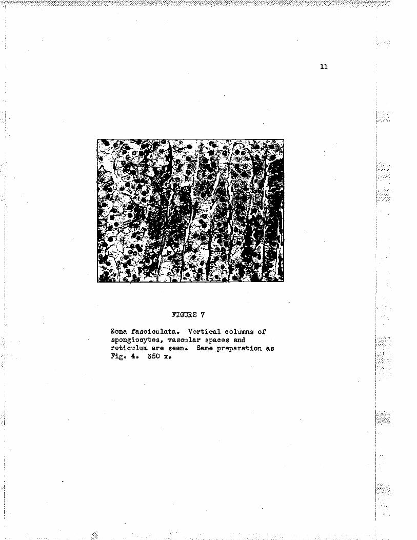
FÏGÜRE 7
Zona fascíoulata. Vortioal ooluwre ofspongiooyfeg, vasqllar spaces and.reticulum âro seêDr Same preparation asFlg. 4. 550 xo

L2
FIGURE 8
Appoaranoe of the spongiocytes under oilirunerelon Leng" 1200 x. Same tissue asFig. 7.
l-. ',.',:I i.ìi.'::rir..

1õ
fasciculata is noted, with one oxception: thoro is lsss f-ipid naterial
in the ceLls. Near the moduLla, howevor, two distinat coll types are
soons a larger ttlÍqhtt¡ celI, with granular oytopl.asm and pale vesioular ,,,,,,
nuoleus, and a smaller ttd.arkt! oel-L w?rich has doeply stainÍng e¡rtoplasrn
and d.ark shn¡nkon hyperohronatlo nuoleue, Theso dark oells are rÍch in
lipid droplots and oontain yerrowieh or bro¡uaish pÍgnent granules r, ,
(espooÍeL1y i¡¡ older indivlduals). :.t:':'
j',1,,',
Ílho- oortícal lipÍds. Tho lipid droplets staÍned. wíth conventional . i:ii':;'
toohniques (Suaan IV, Oit Red ttOrr, and osmlo acid) are smal-L and nr¡merous
ingomeoo}Is,feworand1.argerj¡others.hanygivencg].1thesizeísI
approxi:nately the same..
flre fat-ljlco meterial 1s apparontly a. nÍxbure of doubry refrao- I
tiveoho1estero1estorsandisotropicfatty1ng].usionsofneutra]'andI
ofatty aoids".
DevolotrEnent of the oorüex. The adrenaL oorüox develops from
oplanohniu *uuodunrll. ïn enbryos of 5 to 6 rur, the mesothelírm at the
levol of the upp€r thÍrd of the mesonephros prol,iforatos and ssnds buds
ínto the mesonc?qmo at eaoh side of the root of the dorsal mesentoryl.
Sonor¡uhat Leter in dovelopmont, at the lz rnm stage, Iateral migration of
prinitive oelLs of the nedulLa (s¡rmpathogonía) tor¡¡ards the oorticaL
anlago begins. Migration is oonplete and the medurla foruod at ths
85 rnnr (to too ur) stagell.
In fetal. Iife the adrenaL ís almost all oortox, and at the thirdmonth is actually larger than the kidaey. Von Giorke52 glouu the following
'.'i-it¡i,: jttif l
Ì.::ra,.;.:l
i-:,;'¡,

15
FTGIIRE ].0
The distribution of lipids in the adrenal cortex of a 50
year old malo who diecl suddenly of unsuspeoted brainabscess. Tissue obtaiaed three quarters of an hour post-mortem. FornsLin fixatlon. 0i1 Rod tOt. Ths stainappears bLack in the photograph. 50 x"
'-t ..:':.
l:.::1_--_-...t..-.-
1,,...,t,: .,
1tr,...,t'
lr.ii:|.1
i1..
I

L6
the Eame tissue as1200 x,
FIGIiRE ].I
Flg. 10" seen ¡r¡lth oil innerslon lens.
f
i
:

L7
size oomparísons. Dr:ring the first half of fetal lífe the adrenals are
Larger than tho kidnoys, at six months porhaps 1e2, at birth 1:5, and
Ín adults the kidneys are larger than the adrenals in the ratio of 1:28.
.The oells of tho fetal cortex contain no lipid natori.a!. and are q¡¡qngod
Ín sheets instead of tho oolums desoribed i¡ the adult type of oottox.
At bÍrth there is a thin layer of aortex of ths adult type around the
medui.la. the entire fetaL oorbox dogenerates after birth, and is slowly
replaoed by oel.i.s of the atlult t¡rpe, the ohange being oompleto about the
end of the first year of lifelz.
tífe historT of the corticaL gg]Þ. Znemoro in agreement with
othor ínvestigatsrs finde that tho corbex grorvs from without inward,
the now oolls being fomod at tho periphery and d.estroyed in the rotic-
uLar zone. This investigator finds aLso thet the glomerular oelts ariso
from lndifferent eonneotive tfssue-like oeLLs in tho oapsule.
These capsu!.ar cells lose thelr J.ong prooesses, booome shortoval.s, and take up lipoid clroplots. å, further increase in theamouut of oytoplasm and markod increase in eeII fats nark thetransitíou to the spongioqrtes. Thoso retain and emulsifþ theirfat oontent as thoy are graduaLly pushed inv¡ard þ the foruatíonof new cells. . . .
As the oeLLs seorete, the ratio of oybopla'sn to nualeus isgreatly d.f-nÍnÍshed., so that the inneruost regions of the oeLLeords consÍst of rows of nucleÍ ur:ith very smaLL re¡nnants ofq¡toplasrn stíLl surrounding them. In thã end stago the ooLl isrepresonte$ by a pyhotic nuoleus which is finally phagoo¡rtosed.(cf Zwener)
Ingle8 bas rs¡uarked that the throe zonos of the aduLt adrenal
cortex. reprosent difforent phases in the life history of the corbioal
celIs. Thus, oue nay say that the retÍoularis is the graveyard of the
cortex.

18
Variation in oorticaL morpholory. å,s in nany otber of the
specíalizod organs, the adrenal suffere early fn post mortem ohaages.
For thís reason it is dÍffiautt to obtain striotly nortal hr¡nan natorial..
Contlitions exÍstlng in the organísm i-mmediately prior to death have pro-
found influenco upon the cytologr and morphol-ory. of the oortex. Thus
Zvromer6 desoríbes at least eight variations of the cortex, d,ependíng on
tho d.onand of the body for the corËi.costeroid hor¡iones before death.
VariatLon in oortical thichnese. lhere Ís üârkod varíatisn in
thg thicloess of the cortex and of the fat oontent of the oslLs. Thus
Tr*riteheadl0 duron"tratod wide variations i-n the adnenal sortex of noiuaL
guinea pígs, There i-s orbenslvo hyportroptry drrring fetal develo¡ment,
puberty, prognancy¡ êrrd in scurvyS. The effect of other ho¡:srones also
influencoc the thiclsrese of the cortex, belng e factor in some of those
condítions mentionod, Slight en}argement follov¡s castratloa and the
administratíon of adrenalotropío homoo"rlzo Ïtre admÍnistration of
oeetrogens 1s fol.lowed by aortical hypertrophy, whfl.e androgens rna¡r
cause regreseS.on iu sizeI5, In most, iJ not aLl, mamals, tho adronal
oortex of the fernelo is larger than that of the ralel4.
F\rnetions of tho adrsnal cortex. l,{aqy of the funotions attri-bubed to the adrenar oo¡tex are baeed upon observations nade on the
adrenaLectomÍzed anlmal" the. reeponso of suoh anlmals to the inJeotion
of, eorbicaL exbraots, and on patients suffering from Add.isonrs dj.sease.
rhe hÍstory of the physiology of the adrenals dates back to tho
r:: -.: 'r":
i _:.
l..r
original alinÍoal and pathotogioal observations reported by AddisoulS

19
l¡ his 1866 paper. ïn this paper, ho repor*ed his observations upon
eLeven patlents sr:ffering from the disease whioh now bears his nauo.
IÌ[ve of these patients had tuborculosiE of the adrenal glands, one bad
atropÌ5r, and five had metastatio nw growth i¡volvlng the glande.
ålldíson obeerred the plgnentation, the weab¡,esa, anorexia, nausea and
vonitlng, the oonstipation, aqd the enaoietlon that these trntientd suf-
f,ered. Líttle haa been added to the slÍnfoal ploture sinoe hts tine,
antl the weah,ess, pignentation, gastroLlntestinal.: s¡m¡Êons, the wasting
and the feeble pulse, together wlth tho fataL outoome, still fo¡m the
basLs of the'diagnosis at the present timel.
In L856, Brouna-Sequard e:rtfrpated. the adrenal.s i"n experi.roental.
anfmals, and rapidly fatal- outosme oor,rinoecl hi.u that the glands were
indispeneabLe tô 1iJeI6.
In L895, Ol.furer aad Sohafu"eg pt"pared an aEueous extract of
adrenal gland, whioh vrhen injeoted. had profound pressor effeots. Other
workers, from L86? to 19Oõ, attempted organotherapy for treatnent of
Addisonfs diseaee in the fom of whoLe desf.coated gLand or extraots, by
nouth and inJeotionr Some of the resulto r¡ere enoouraglng. For the
ne:ct few y€ars treatment using adrenlne ruas attempted for Addisonts
disease, and as expeoted, with lÍttLe euscess.
In the early twentÍes, interest rias again aroused 1n obtainiag
a oortioal e¡rtraot, bub it rpae not r¡ntil tho Late twentios that Hartn¿n
anct his assooíate"l9 "ooo"edod
fn obtainíng extraots from the oo¡tex
shioh had profor¡nd effeots upoa the adrenaleotomized animl. Ihis was
thougþt at first to be the horsoue of the cortex aad was naned ioor.birln.
..:

zo
Later Ëarbnan hínseLf was ablo to Éhow that there existed more thau one
substaaoe. fn 19õ4 orystalline aompounds wero s€paratod uhich eouLd
naíntai¡r the lifs of adronaleotomÍzed rats and dogs and. by ISSB eeveraL
crystallin€ oompounds had. boen separated2l.
lhe oortíoal hor=cones. Ropeated fraotional extraotions using
bonzone, uator, isopropyl. aloohoL, and ohLorofom result,in two types
of ooanpountis¡ the orystalliae fraotiou whioh oontai.ns sevsral, steroÍds
a¡rd the rramorphoue fraotLonll which is vory aotive rnaterlal.
WhoIe
adronal
aortioaL
erctraot
Crysta).Line
fraot:i.ons
Ánorphouo
fraotion
The follogriag table is foted in Sofferr s nonographl and that
whioh foLl.ows has been paráphrassd from this lrork.
Dosoqyoorfioosterone
Corbioosteron€
Dehydroo ort i eost eroue
I 7-tqrdro¡yc ori Íoo st e roÐ,o
1?-þdrory-1 l--debydrooort io o storone(ttEn of, KendaLl, $Ftr of Pfiffher &
'$Ii:rtersteiner)
1?-hydrory-L1-dosoxyaortLeoetorone ( nSn )

2L
DESOXYCORTTCOSÎERONE
' Thie horsone causes narked retontion of soditm,ahlorido and water, a[d jncreasos the r¡rinary oxoretion ofpotassÍrm. and phosphorous. It hae no effeot upon oarbolqr-drate metabofÍsm or the pigrnentetion of âddisours d.isease.T.u' ad.ronal Lnsuffioienoy, it r,:il1 restore the eLeetrolybepattem of the bloscl to olosely nornal pÍoture, a¡d will.el.evato the blootl pressure. Its oontinued use nay prod.uoea temporary hype*ension, eder¡a ancl oardiao failure. Tlhonthore is lryperfimotion of the adronal- oortex, adminietra-tion of the ho¡mone produo€s an Ínoreased exofôtion ofsodírm and. ohloride Í¡ the urlne.
CORTTSOSTERONE
-0
ïtrotC=0
ï**
lii-i,:;: '.'
t -'. . .a':;LI :'..i -ii::
i ':lI ::

22
ÐEÏTTÐROCORT IgOSTMONE
17 -HYDRONT. 11-DETITDROCORT TCOSTERONE (rtEtt of Kenda3.L, rtFtr of Pfiffbor, eto.)
ï*o'C:0
thess three eompoulds, (oortioosterone, delrydrooortioosterono,17-hydroxy-1L-deþdroçorbioosterono), exeroide a marked effeet onoarbohyclrate metabolism and oorrest defoots in these substanoos 1ninsuffioienoy. TÍhen theEe horrones ar€ injeoted into the eubjeot,glyoogon ís stored i¡ the l.1vor, the blood sugar levels are raised"and h¡rpoglyoemia is prevontede 17-Ìrydrory-1t-deþdrooorbisosteronehas thÞ most proaormoed effeot upon oarboþdrate metabolism, belngthe only one of these th¡:eo to forrn gluoooe from Laotia and p¡rruvioaoíds¡ it protluo€s a n€gatÍve sodiw balanoe rith increased urinaryoutput of this ion, ur?rÍLe corbÍoosterone aad tlehydrseortioosteroneheve llttlo effect on electrol¡rbos, oausing ninJ-al retentLon ofblood sodir¡m.

23
17-ffÐRorffCoRÎ TCoSTERoNE
r"*C-0
I*ríe compormd exeroíses a narked oontrol over oarbohydratametabolism and also oauses an Íncreesed urÍnary e$crotton sfsodile, índuciag a negative balaaoe. (gf. Sofforr)
.Aqorphous fraetjsn. this higþIy aotive resídue fE Left afber
the removal of the orystallino fraotions, and although it exerts no
effect upon carbohydrate metabollsm, it is exoeodÍ.ngIts potent iu tts
iufluenoe upon the distríbutj-on of eLectrol¡Èos. Acaording to KendalLzl
only one or two míorograms per ki!.o are required. to ûsintain the eleo-
trol¡rbe pattem in adrenaleotomfzed dogs. Relatively lerge anounts
of desoxycor-bioosterone are required to produoe tho sane effeets.
ChoLesterol and ascorbic aoid in the oortex. Cho1estoro].
(geZE4e0) is oJ.osoly related Etruotrrrally to tho ohslis acids of biLe, i
i:'
vitanÍnD5¡audtothehomonesandotherstero1dsoftheadrena1
oortex and gonadse¿
i..,.r:: -- ì. -:,: :... -:
t : - ._.ì': t:_::
substanoo of theso
. Thore is evidenoe that oholeÈteroL is tho parent
oonpormds and its fo¡mula ls fnol.uded here.

24
Structural fo¡rula of oholgqterol (çZZFry*AO)(aooording to Carnoronz?)
Trlngz3 in a reoent paper rovlewe some of the líterature aad
reports new urork when he rrrltes of the aondltions assooiated with the
sooretion of the adrsnal cortex. The foLlow'ing is paraphrased from
this papor;
The ohemioal oharacteristioe of the adrenaL oortex areits hlgh oontsnt of sholesteroL.and asoorbío aoid. No othertissue of the botl¡r oontains sueh a high quantit¡r of thesesubstances. Tho rol,e of ohoLesteroL with respoot to oortioalsterold horrnones has been a natter of speoulatLon for sonoti¡er 0n injeoting adrenooorbicotropfo hsmone of ths anteriorpituitery (*Cfn¡, lt ruas for¡nd that ihere is a decroase in thea¡nor¡nt of aholestersl. in the adrenal glandl, while this deoreasowas not noted ia other.orgâ[sr Thus this responso is regardedby long to be a speoifle response to the trophic hom,one¡ Theaeoorbie acíd oontent also d,oor.eases in the adrenal gland fol-S.owlng the inJeotion of ÂC18. Ðireet evídeu.oe, howeior, islacking for the convorsion of the adrenaJ- oortisal steroidEfrsm oholestorol, and the relatlonship of asoorbio àoitl is nothror¡s¡r Ï{owever, the deoreased a¡nor¡nts of oholesterol. aadascorbio aciô ln the eorbex fo1low:lng the Ínjeotion of ÂCTE 1s

ZE
assooiat€d w'Íth increasod rate of socretlon of the oortiaaLsteroid horrrones, and it is probable that oholesterol ís adlreot preoursor of tho corticaL storolds. StjmuLation ofthe autononio nervous system, w'ith oonsomitant reloase ofadrenÍne appearE to be a najor factor in stimulating tbeproduction of ACTfi from tho anterior lobe, The manner inwhåoh the adrenine produces thls aetivation is not hlorn:aothe arrbhor deal.e also wÍth othor faotors suoh as the bLeoclLevel of tho hormono of the target gLand. (fongeg)
Ilre fi¡nstions of the homones. å,coordíag to Oa¡noro&z i,Jru
various oomporrnds whose aotions are associetecl with the holtn f,unotioneI -;'r _ i ::_,1' of the adrenaLs nay be dívided iato two groupsr
Group I oompor¡nds have a regulatory astion of the blood elsotro-t- l¡Èos. Inolud.etl here are deso:içroorbieostorone and a oompormd preEentl
I in the anorphous fraotion.l
I
I those homonss rvhioh havo an oxrygon atom attaahod to
' The exaot number of horsones produaed by tho adronal oortex ie
' rxot brouta at the present tims. Frobably somo hormones whose predom-
Ínating aotivity is assooi¿tecl w:ith sex firnction are intemediate pro-j
.duots of metabolism. Ganeron points out in his mouograph that tho two
' d.istinot t¡4pos of aottuiþ of the oompounds of Groups I and II suggest
that at least two speoÍfic ho¡monos ar€ produeod., and that Lt seems
unlikel-y that four disti.¡aot Group II ho¡uones should be produoed.
i Caaeron, in agreenent with Earbman, bel.ieves that the four erystallÍne
oompounds so far isoLated are the stable derivatlves of a less stebLe
oompormd," the true ho¡mone.
j:ì.r:r:.r:,i

26
lhree groups of fr.motions ar€ llstsdl by Lo\82
l. Control of oarbohydrate motabolism, aud proteinmetabolism.
Z. tontrol of eleotrol¡rbes and wator netabol-Ísm.
5. Provision of a meohanism of resietanco to variousstrossos such as thoee oausod by beoterialtoxíns, histamiqe, shock, water, intoxioationtlow terÍrporature, and low oxygen pressür€r
The L?-keto steroids. The neutral lZ-koto steroids aro the ::i:ì:l
urinary excretory produots of androgsnlc metabollsm and arise from the ir"..i:ijri:l
1substanoes produced by the adrenal glancls and the nalo gonad.s'.
ì rr. lIlE lfEÐUl,r{a
Tlre line of demaroation betTueen the zona retícularis of the
: projoot i.nto the medulla for varying d.istanoss':
: lno types of eells are desoríbed ín the aclult medulla. These
are arrau,gotl lrreguLarLy arrd seemingly withoub pattom. Ganglíonlo
, 'oolls are formd in groupe or síngly. They are not freguont in nrmber.
, .{¡ones fron these gangLionís oelle end in oLose assooiatÍon with the ',chronaffin ceLls whioh they lnvrorvate aad stfmulate to produce adrsnine.
ür" o¡rouaffln oells nake up the ,*""lof the medulla. Thoy are
irregular 1n shape, and have usuall¡¡ abr¡ndant o¡rboplasn. The tondenoyII to shrink is msrked even in well preserved tÍssuo. F?equentl.y a steL-
lats shape Ís obser:ved.. Theso ool-ts ars aharacterízed by the pressnoe
of fine brown granules in the e¡rbopLasm when the. tissr¡e is fíxed in

z7
ohromie acid or its saLts. It 1s from thls reaotion, whieh is shared
with other tissues of lthe Ch.r'onaffir Systen that the eells derÍve thelr
nânê¡ The reaction 1s thoughf to be due to tbe pol¡rmerization or the ,,,.,:
oxidation of the adrenine (or its precursor). lïre oells are green when
stained with either Sohormlrs or WeiseLts.urethod.. Cramer26 uslng osm:io
aoid vapour rÍês able ts demonstrate adrenine graaules ín the ohronaffin ,,,,,r,,
oeLls and actusl-ly formd the granules Í-n the neighbouring sfnuses. i'."'
-A thírdt Wpe of cell, is oecasionsLly seen in the medulla. These 1,,,,.,
are arranged Ín srna1l groups, ar€ snaller i.n size, and take a deop
staín. lfiany workers fee] that these are i.mnture s¡mpathetlo oe!.Is,
whlLe others believo thet they are possibly lynphooytesz.
llre êevelotrment- of the meduLLa. Ïhe embryolory has been presented
by nabinll.' The meduLla and the ohromophile bodies develop from the
primltive oells of the s¡rnpathetio gangl-1a or tho s¡mpathogonia, whioh
in turtr develop from the neu¡al crest. In later stages of fetal develop- i
nent, the sympathogonia differentfate ínto two oelL üypes, the gangLlon
oells and the pheoohromoo¡rbes. The migration of tho medullary tlssues
has been mentíoned in conneotisn ¡¡"ith the tlevel.o¡meat of th.e csrtox on
¡nge 14r Ðuring thls migration, accordlng to Rabin, portlons of the
embryonlo tiesue nay become spt ít off qnd these deveLop fnto separate
organs at värying distanoes from tho aorta to forr the organs of
ZuokerkandL.
The blood S3pglt. Ihis has been discussed on page 4. llhe
vascular spaoes of the meduLLa are uider than those of the oortex, and.

28
nay properl.y be temred sinusoids. Sharpey-Sohafer2S h"" renarked that
the medulLa.consísts of te solíd. cell-mass pemeated þ sÍnus-like
blood vesseJ.s ïrith the oelLs oompaotly arranged between and around
them. tr The same i¡timate relation wÍth the reticuhm. exists as in the
cortex, although tho retiouLrm, Ís more irregular and not as healy.
Tho ry
supply. The nerve eupply of the.adrenal ié best
dísoussed in oonneotion mith the rredulla, for there ís uo horrn nervous
eontrol. of tho fi¡notional aotivity of the adronal oortexS.
the twenty to thirty nerves to eaah gland oors mostLy from the
ooeliao plexus (s¡r:npatbetio), in part from the greater splanchnio
nerves and possibly from the vagus a1so2. Tho i¡nervetion of the
moduLLa is pre-ganglloni-o. The fibres pass wlthout s¡maptio interrup-
tion to the seLLs they innervate. lhe greater splanchnlo norvo oonduots
most of the fibres to the gl.and where they fora a netrvork ín the aapsule,
penetrato the oortex without inrrorvating Ít, and end ín the nedulla in
olose relatíonshíp with the oells there, each fibro being closoly asso-
oiated. wíth a definite nrmber of oello?7, 0ther s¡rmpathetio fibres
enter the gland througþ the hilus2.
Stimulation of the splanchnic nerves leads to the i.lberation of
an inoreased amor¡nt of adrenine fron tho meduLla. Cramerts illustra-
tions show the granules 1n tÌ¡e ahronaffÍn oelle and in tho neighbouring
slnusoids af'ber the fresh gland u¡as treated with osnic aoid vapo*z8.
The methocl of forration of adreni:re is at present unhrown, although
theoretically, tyrosiue 1s possibly the parent substanoe (Camerone8).
i.:ìinr:';

z9
AdronLne. As early as f894, OLtver and Schaf""Z9 d,"ronstrated
the remarkable rÍse in blood pressure folLorning an inJectíon of an
exbract of the edrenal meduLla. Í:r 190L, Aldrfoh and Taka¡rine50, tl,
independently, lsoLatsd tho compormd adrenÍne CgH1gNOg. AbetIB reporteô
a method for produolng a orystaLline substanoo whieh he consíderod the
aotÍve prlnoipLe and whioh he aaLled epinephrJne. SubsequentS,y it was
shovm that the hormone has the oonstítution¡
iTC)--rHoH
oH2 NHCH3
CONSTIÎITIONAL FORMUI,A OF AÐRET{TNE
(S¡¡n. - Epínephrfne, S.dro4qliue)(aocording to Caneronzz)
lghen injeoted, adreaine produoee oLosely the effeots obtalued^
upon stimulati.ng tho s¡rmpathetic nerves, and for this reason has been
caIIed. a s¡mpathomi¡netio drug. The most striking offeot is the ooa-
strlotion of arterioLes of tho skía, muoou¡¡ membranes and cerebnm,
resulting in'an fnaroased p'lood pressure which, rro*Jo"", Lasts fsr a
short tÍne, the brood pressure rapiaty returning to no¡aa!.. Adrenino
aooeleratos the enz¡nnatio aonversion of Liver glyaogen to glucose,
HO
I
H

30
FIGURE 12
RetiouLunr of the modullan tho sinus-I1ke spaces whíchpermeate the tissue ara wider than those of the cortexnA tributary oentral vein is partly shown. lhe junctÍonof the zona retieularis and the medulla crosses theupper third of the figure. Same section as Fig. 1" 200 lCs

3t
and musele glyoogen to laetio aoid, thus erpLaining the iucreased blood
values of those two substanoes after the injeotion??. AS.though the
effeot of adrenine ís usually the oonstrlction of smooth muscle fibres,
suoh i-e uot always the oase. Tho eoronary arteries are dilatetl, heart
rate ie insreased, rúth inoroased oardiao output¡ auriouloveatrioular
condustion tÍmo is deoreased¡ the arterioLes of striated musclee are
d.iLated whlle those musoLes are oontraoted.. The smooth musoles of the
bronohi end bronohioles are relaxed. There is decreasoit motiLity of the
musole of the stomaoh and the remainder of the gastro-intestinaL traot.
ExporlnentaLly, Bome an͡aLs survivo the loss of the rnedu1Lary
tÍssue. Some writers foel that the horanone is rmnecessRry, and thís isprobably true fn Pad, providlng the experinontal ani¡ra1s are kept ia a
nortal envÍronment. tr4nn with diseased adrenels does not require replaoe-
mont thera¡:gr. Eowever, it is diffloult to believe that suah a ¡ntent
pbyeiologioal agont serves no usefr¡L firnctioa in body eoonory, simply
because we are r¡nablo to attributo to it a fimction absolutoi.y noo€ssary
for 1!Pe. Iongrs reoent paper, to wtrioh reforenoe has beon made, sheds
some líght on the probJ.em.
As aa emergonoy moohanism, seoretion of inoroasod amouubs of
ad.renine has obvious advant"gà"ez. Such a statement is based on Ërossobservatioae suoh as the riso of blood pressure, and thsse furotions
whioh are usually attributed to this hornoneo Perhaps sdrenfne plays
a more useful functlon in the msintonanco of the race, and perhaps
booause of it, men are men, and. mlqe aro mioe, a dÍfferenoe too often
forgotten þr men of soieaoe,

32
BTBLIOffi.APtr
L. Soffer, Ipuis Jo, Díseases of the .ådrenals. Seaond edition¡Phil-e. ¡ Lea & Febiger, 1948. 520 pp.
2. trrfaxinow, A. å,.r and Tlfur. Bloom, Textbook of Eístolory. For¡rthediition; Fhila.l Tt B. Saunders tompqny, 1944. 695 pp.
3o Fl-lrrt, J. M,, The Bl.ood, Vessels, ÁngÍogenesis, Organogenesis,RetiouLrm and Histology of the .åd.renal. Contrlbution So. Med.ò ¡ . firpf1s'l[. E. WeLoh, Balt.r 1900. L53-228t B pp.
4. Maxi"mow, Â. å., and16n. Bl.oom, 0p. oit.
5r 'Cowd.ry, E. V., á. lerbbook of Histolory. Third edition¡?híLa.: Lpa & Febíger, L944.
6. Zwemer, R. L.¡ A Study of Adronal Co¡tex nf,orphology, .åner. J. Path.,1956, þtLO7.
'î. Hoerr, N;, The Cel1s of tho Supraronal. Cortex ín the Guínoa-pig.theÍr Reaotlon to Injury and lheår Replaoemênt. Amor. J. ÂRat.,
.195I, 48¡159.
8. Ingle, Ð. J., hoblems Retating to the Adrenal Cor*sx.brdoorinology, l-942, 91r4J.9.
9. Soffer, Louis J., 0p. oit.LO. Tdhítehead, Ro, lhe Sex Differonoo 1n the ProportÍon of the Suprarenal
Cortex Oooupiotl by Lipoid in Guinea Pigs over One Year Ol.d.J. åJtat., 1995-36, ?O¡L2õo
11. Rabin, CoLeman 8., Chromaffin CelI ü¡nor of the Suprarenat MedulLa(Fhooohronooylorna). {rch. lath., 1929, 7 &28.
12. Boyd, T[n., Â Terrtbook of ?athology. Fifth ettition3Philq.s Lea & Febiger¡ 1947r
15r Sohacher, J.¡ Je S. L. Browao and. Hr Selye, fffeot of TariousSteroLs on Th¡rmus 1n Adrenaloctomized Rat. Proo. Soo.Expor. Biol. & I[ed. , L937, 56¡488.
14. Trrgle, Ð. J., 0p. oit.

33
I5r .åddisono T.¡ 0n ÐÍsease of tho Supranenâl Capsules. London Med.Gaz.2 teSS, 43¿5l-7. Entiro p*p"" is reprintstt inrnonographby Rowntreer-E Gr¡ erd A. M. Snoll, ttA glinical Study of
i dd.isonts Diseaser$ Phi1a.: 1T'. B, Saunders Courpargr¡ 195L.
: :: ::, L6. Brovun-Soquard, 8." Ia Physiologie ei la Pathol.ogie des Capsules : "
Surrenalos. Aroh. Gen. de Med.¡ 1856, 8rã85. Quotecl bySoffer (I).
18. .A,beL, John tI., 0n a 5inpLe Method of Preparing Epfnephrin and ítsCompound.s. Bull-. Johns Hopkins Hosp., 190¿1 13¡29. ...i:
- 19. Har.tman, F.4., et al, The Horrono of the Adrenal Cortex. l:t:"'l
I å¡ner. J¡ Plrysíolo, L928, 86¡655..i 1.. -::::i , . i..:...'...""..' 2O. Itrartmau, F. 4., et al, Q,uoted. by Carroro!.¡ A. T. (2,2). : : ::
2L. Kendall, E. C., tho fr:nction of the AdronaL Cortex, Proo, StaffMeet. }'[ayo CIin.¡ 194O¡ L52297,
22, Caneron, Â. 1., Reoent Âdvanoee in Endocrínology. SÍ:cth edition¡I¡ndon¡ J. &.ê.. ChurohÍIL Ltd., L947. 44õ pp.
28.Ipng,c.}I.N.¡TheConditions.A.ssociatedwiththeSecret1onofthe .A.drenaL 0ortex. Federatíon Proc. , L947, 61461. .
;abo].io Fr¡notíons of tho araoo"il" Glands. i24. Long, C. H. N., Metabo3.io Fr¡notíonsI âot. Rev. Ptrysiol. , J.942, 41465-502.
' 26, Sharpoy-Shafer, Tho Þ¡rdocrj¡e Glands. Seoond edition, ¡nrt 1¡ l
I-ondon¡ Greon & Co. , 1924. pr 95.:
, eU. Cranor, W., Furblìer Obsorvetions on the Ttryroid Adronal Apparatus. i,:,,,,¡,,.,;.
,å Elstochemioal Methocl for the Demonstration of .å,ctrenalin , ,'
, Granulos in the Suprarenal Gland. 1,,'-1,",':,' ,f. Plrysíol..¡ L9L8¡ S.r viif-x (Proo.)' 1,,',."''';,
27. Yor.m.g, J. 2., Partial Dogoneratíon of the Nervo Supp1y of the.ådrenal. å, Study in Âutonomi o Inr¡ervatlon..I. ånat., 1959, 732540.
I e8. Cra:ner, TlI., Fever, Eeat, Regulatfon, CUmate and the thyrold-adrenal ,:,r: Apparatus. Longmans, Groen & Co., Ltd. Quotod. and reproduoed :'1
in Cowdryrs Terrtbook of Eistology, Seoond odition, p. 126.
29. 01Í.vor, Gr¡ arld Schaefer, E.4., The Physiological Effeots of Þrbraetsof the Suprarenal Cortex, J. PlgrsioL.¡ L895e lB¡250. :
Quoted by Soffer, ttDiseasos of the å,drenalsrr-Seoond ed.ition;PhiLa.t Loa & Feblger¡ 1948. p' 153.

30. Takamine (190L). Quoted by A. T. Camoron, rrReoent ld.vances inEhdoorinoloryrrr Síxth odition¡ Iondon¡ J. &.å,. ChurahÍl}ttd., LgåT.
51. .A,Ldrioh (fgof). Suoted by Ao T¡ Cameron (so)
32. Gierke, Er volr¡ Ðrllsen nit innerer Solcetion. Cha¡rter lfiV inPathologÍsohe *qnatonie, 6 Ar¡fl.r e Bd., Spez, TeíI, Edit.
' LuclwÍg .A,sohoff. Guetav Fisoher, LgZ9, Joaa, LO50 pp.
34
iì1',¡,ri .rji:iì. ii i:l:.:'...1

CH.A.PTER TTFO
THE MALE REPRODUCTIVE SYSTHû
!.: , :
I a:::a::
l.:-.t. 1
l"r::'

CHAPTM. IT
TI{E MALE RETRODU'CTIVE SYSTEM
Introduction. The.naLe reproduotive system aonsists of the
testes, Trith a eompl.ete system of excretory duots (rete testes,
epididyrnÍs a¡rd vasa deferentia); w.ith auxilLary glands (seninaL
vesioles a^ad prostate), and the ponis. In this disoussion the testes
only will be oonsidered j.n detail, oinco these organs are the only
part of the male reproduative system hnown to produce hormones.
Eaoh testis is an oval- body wlth flattenod sides' varying
oonsiderabLy in size. The aduLt organ averages 58 rm 1n lengthr 25 rmr
in the antoro-posterior dia.neter, and somewhat Less fron side to sidsl.
Each organ is Looated in a serous sas whÍsh normalLy contains
only a thin ffl¡o of serous fl-uid to separate the parietal a:rd vlsoeral
layors of the tunioa vaginalis. Ihe visoeral layer of the tunica
covers the anteríor su.rfaoe and the sides of the testis.
Two t¡res of specialized tÍssue are found in tho teotis. . ',.',',,
Highly spoeializod stratified epithelium Lines the serniniferous '.' ".,,..,.,' -: :
tubules. From this epithelium are produced the speruatozoa required
for the propagation of the speoles. Produotion of spermatozoa is
oonsidered to be the basio function of the testis. Tho seoond tissue l:,:,,,:.=,ii.:i:' '..
:';-:'- -
lis found scatterod irregularly throughout the tostis as singly
oacurring or grouped Leydig ce}ls, which ars considered by most
invostigators to prod.uoe one or more of the malo sex hormoflos¡ â
sooondary function of the testis. ;,r.,,i.:.-:,,

37
The oo¡rneotlve tissuo framework. f¡unodiateLy beneath the
tunioa vaginalis thero is found the tunica albuginea' which is a
thiok, tough, white fibro-elastic aapsule rneasuring from 0.4 to Q.g nrn
in thiolmsss. The d.oep surface of the tunioa is very vascular,
eepeoially in the youngr and has bosn called the tunica vasculosa.
the tunioa albuginea forrns the basis for the entlre fibrous frame¡rcrk
of tho orgâRr Posteriorly the aapsulo is thickened to produco a
vortioal rid,ge' the mediastinum testis. The mediastinum oonsists of
dsnse fibrous tissue, a few strands of smooth muscls, æd a few el-astic
tissue fibros. From the modiastinum rad.iate fÍbrous ribbons of tho
septulao testis. These pass forward and lateraL1y to booome attaohed
to the inner surface of the tunioa albuginoa. In this manner the
organ is sì"rb-dividod into L00 to 200 conioal oompartments¡ thet
lobu3.os', oaoh with the apex toward the modiastj-num. The septulae
are incomplete in placeo, espooiall-y toward. the poriphoryr where the
lobules oomr¡unisato. Connective tissus ertends from the septulao to
support tho oontsnts of tho lobules' ELastio tissuo fibrss i.ncrease
with advancing age in tho tunioa aLbuglnea.
The artorial supply of the testis. ElLLZB, a student of MaL1,
glves an exaellont aooount sf the blood supply of the hurnanr testis.
Oklsels and Sand.5z io r recent paper, agree essentially with HiII.
The testiaular artory arisos from tho antorior aspect of the
abdoninal aorta at a level stightLy inferior to the origin of tho
ronal arterles' Each slend.er artery passes obliquoly d.ownward,s,

58
retroperitonoally on the psoas m;scLo, to reach the doep inguinal ringr
From here the artery follows the spormatic oord to the tostis. The
tosticular af,tory gives rise to a bra¡ech (errbernal sperrnatio arbory)
high in tho cord., shortly after that struoture loaves the errbernal
abdominal ringo The externaL spormatic artery dívides ínto srnalLor
branches as it desoend.s in the cord., and supplioè tfre mombranes of
tho testis.
The main trunk of the testiouLar artery ends 1n one or more
terminal bra¡rches whioh become vory tortuous just bofore reaohíng the
mediastinum. The terninal arteries further divlde noar the media-
stinum and sond a groat numbor of snall arteries into the glanit. From
the mediastinum, yossels (ascending) foltow the septulae bstrreen the
lobules¡ radiating like tspokes of a whoel.f The ascondlng artories
give off finor branches to the tubulesr
Near tho globus major, one large vessel frour the terminal
branohes of the testicular artery goos to supply tho tunica albuginoa
and encircLos the testis, while on tho innor or deop surface of the
tunica¡ desoending branohes from this capsuLar vessel enter the gland
substanoe and anastomose with the ascending arteries given off at ths
modíastinum,
A small branch from the terminal arterios d,osoends to ths
globus minor and, passes under the tunlca albuginea to run under the
oapsule and a¡rastomose with the capsular vossel arising at the loveL
of the globus major. These oapsular vessol-s send out raany snrall
arteries which aro tortuous and whioh encirolo the gland in the deep
g9
surfaoo of the tunica albuginea, termod tho tunioa vasonlosa by Astley
Coopor' Brenohes of those a"nasto¡nose with vessels from the mediastinum.
(ct. nrttz8)
ArtorlaL sJrpply of the fobule. Eaoh Lobule reoeives an arterial
supply from two or moro arteries arising at the mediastinum (asoending)
and an equal number from the eapsular vessels (d.esoend.ing), and anongst
aLl those thare are rioh anastomoses. SnaII artorioLes arise from
these vesgels. These arterioles enoircle the tubules and. end as
plexuses about thom.
Eille8 points out that the arteries and veins w?iiah suppLy eaoh
lobule are in the septulae and, beoause of the presonce of these vessels'
the septulao are moro oonspiouous whioh give the testis a definite
Lobular appearanoer
Venous d.rg,inagg' The voins foLlow the general oourse of the
arteries.. SeveraL Large capsular vessels oncirale the gland while on
the d.eep or inner surface of the tuniaa albuglnea. Theso enpty into
the parnpinif,orm ploxus. Capsular veins receivo blood from the venules
ar¡d veins on the inner surface of the tunioa albuginea' from tho tunioa
vaginalis a¡rd from anastomes with the ascending veins of the lobu1es.
Blood is also returned, to tho pa:npinifonn plexus by the desoendiig
veins which foLLow the courso of the desoending artories' These enpty
into the vonous plexus at the mod,iastinum. (Cf. uitt28)
The oonvolutsd panpiniform pLexus eventuaLly gives rise to a
single spermatlo vein on oithor slde. Tho right spermatio vein enters'triirìlliì:;

40
the lnforior vena cava at an acuts angle. 0n the Left eide the
spernatio vein enters ths renal vein at a right ahgle. Acoordiag to
Rivington?9, tlr" testicular voins are suppLied with valves.
Ll¡nphatio drainage of the $[þ30, 51. Within the tostis' the peritestioular, poritubular and, porllobuLar llmph oapiltarles forrn,
a ríoh notrnork, Aocord.ing to Rouvie""gl ty-phatic oapillaries from
the interstitial tissuo pass into the soptulae aad then into the media-
stinuni; or more frequontly to the tunioa albuginea to follow the
Eurfaoo of the testis, AII the tributarios eventually arrive at the
postero-superior border of the org&tro From hore 4 to B oolloctilg
vsssels ascsnd. in the csrd i:a the oonparry of the vei-ns60.
Above the loveL of the inguinal riag, the asoont is retro-
peritoneal on tho antorior surface of the psoas rmsole stil.l in the
aompaay of the spormatia vossels. At the leveL where tho spermatio
vessels oross the ureter, the lynph vessels leave the oompany of the
veins and as wo1l part from eaoh othor. In the upper part of the
abdonÍnal oourse the vessolE bra¡roh, aad also oo¡nmunioate with ono
ønother, Eaoh testÍs has its olvn group of nodos whioh conmunicate with
eaoh other' and reoeive as weLl lfmph from other org&Bso
On the right sid.e, one to throe nodos lle in the groove betwee¿
the aorta and the vona oava, and one node is deeply placed between the
two vasoular trunks. In many cases nod.es are found at a lower level
arrangod in a more or less írregular üsltnorr
On tho Lefb síde the nodss lie in the tissue to the lefb of the
l-r.l

aorba, generally groupod behind
artery. Oocasionally a node is
end as Low as the angle between
artery.
4l
the origin of the inferior mesonterie
found as high as the left renal vein,
the aorta and, the left connon ilias
The secondary nodes are scattered. irregularly arnongst the
para-aortic groupr a¡rd, reoeive l¡mph from tho primary nodes, as well
as from nodes abeve and below the level of the renal voins, and fron
stÍLl others Ín the ohain of nod,es along the outer side of tho oormon
illac artery. (cr. Jamioson and Do¡son5o)
Norve supply of the testis. The testioular plenls is derived
from the renal ploxus (forrced from the ooeLi.ao pLexus, the aortioo-
reaal pLe:nrs and the aortic plexus, and also from the lowest spla:aohnies
a¡d bra"nohes from tho vagus). The nerves aocompany the testiouLar
vossols aJxd. are d.erived from tho tenth thoracíc sogment of the spinal
oord through the renal a:ad the aortio plexus. Fibres from the pelvio
plexus are carried by the artery to the vas doferens to supply the
testes a¡rd the epidtidyn:is (E11, 12 and t1)34' 35. Tho distribution of
the nervss wlthín the testes a¡ad the relationship of these nerves to
the interstitial ae}ls are d,isoussod on pago 66.

42
I. TITE TUBUT,AR STSTETI
General plan of the tubular -W$9. Eaoh lobule contains one
to three, and sometimes more greatly oonvoluted seniniferous tubules
(tututi contorbi) which are supported, by delioato oonneotive tissue.
The seminiferous tubulos are very long slender loops that are ooiLed:
r or bent in sharpr short ourves' eaoh tubule forming soveral small
i "ompaot oonvoluted masses2. The tubuLos measure from 80 to 7O
oentinetres in length and from 150 to 250 nicra in diameterg. The
oo¡nblned tength is estimated to be 250 metros by Maxirnow and Bloon3,
' antl by John"ool6 as BO0 feet for eash testls.
Aooording to Johnsool6, th" conroonest tJEe of tubule found are
the anastomosing Loops conneoted, ln a conpleto sories. Single
amastomosing loops and singLe unbrenohing tubules are found, but are
, râro¡ Snall diverticuLae are found here and. there along the tubules,
and are more frequent near the modlastinal end of the tubules.
I The tubuLos foIlow thoír tortuous course, oonverge on tho apex
: of the lobule, pass abruptly into tho straight tubulos (tubutl reoti)
that enter the mediastlnum and, joint to form a raoemose notwork sf
epitheliun. The duot system emerges from the testis proper at thls
point as tho ductuli efferentes. These pieroe the tunioa along the
posterior border of the testis near the upper pole, forning the coni
vasoulosi¡ ând oonnest tho reto testis ts the highLy oonvoluted oa¡nal.

:
l.l:f:I
i
I
i.t::
I'
FIGT]RE 1g
Sch,enatic- tÊpresêlrtuùi'o¡¡ -of- -the tubularsystsm of the teetiole. Redrar¡a andmodifled after EermanS?, a¡d Johneonl6.
43
Poradidymis (upPer)
Vas Deferens
Porodldvmis(lowár)
Vas A berrctnsSuperior
Appendlx Epididymís(Wolffian)
Appendtx Testls(Mullers Duct)
Appendtxof Rele
Vos Aberranslnferlor

44
of the opididyrnis. (Cf. Pier"ol4)
the epididynis has about 26 feet of tubules of inconsta¡.t
volums and, d.iarnetu"25. Finally from tho epididyriris, there enorges
the vas d.eferens to oonduot the products to the oxtorior. (Cf. post)
Tho seminiforous tubulos. A highly spociallzed. stratlfied
opithellun lines the seminifsrous tubulos' This epitheliun rests on a
wolL d.eveloped. flneLy fibriLlar basement membrano. A thin layer of
cond.ensed oonneotivo tissuo is applied to the outer surfase of ths
tubul-es.
Two kinds of celle are found in the complox opitholiun; tho
sustentacular eolls (of Sertoli) have a supporting and nutrient
funation, and, secondly the germ or spermatogenic-(sex) ceLls whiob
after a series of proliforations a¡rd transforulstions give rise to
n¿ture spermatozoa. The histotogy of the sominiforous epÍtheliun
varies with age. Tn tho testis showing active spornatogonosis'
spermatogonia, primary ar¡d sooondary sperrnatoc¡rtes' spermatid.s'
irnmature and mature spermatozoa aro present.
The oells gj! Sertoli. Numorous names havo beon apptiod to tho
Sertoli cells from tine to tinrel9i among these are the ternrs
sustentaouLar ceLls, folliolo oolls' a¡rd the more desoriptive t sperm.-
nourishing oells. I
In the testis of the adult' during the period of aotive
sporrnatogenesis, the Sertoli oeLls are slender, elongated.' irregularly
pyranidal cells, perpondioular to tho basement membraïr.e on whioh they

45
t...i..i- iì
|.:::.-:L-. .,
l'..r,r.::.r
FTGTIRE 14
Seniniferous tubule shoïring the fibrillar struotureof the waII, the seminiforous opithe).ium and theoontents of the interstitial space. Stainod withperlod.ic acid, Sohiffrs reagent a¡d henatoxylinr Â300 x.
7088.

46
f,ost. IFíde d.ivergonoe of opinion elrists as to the nature of these
oel}s, whether or not they form a s¡mo¡rbÍum5. Eanesl9 points out that
Regaud in 1910 desoribed the Sertoli aells as forming a sy:aoyblum,
dospite tho fact that von Sbnor in 1902 was able to teaso the ceLls
apart- in PhYsÍologiaa1 saliner
Although the colL outlines are indofinite, identification of
the Sertoll ceLls ls usually possible by reoognition of the charaoter-
istio nuoleus, The nueLeus is found. in the ex¡randod basal portion of
the eell, usually some distanca from the basemont membraae. The
nuoleus as a rule is oval in shape, with the long axis radlally dirsotod. ,
:
The average size is 9 by 12 níara5. À compound nuoleolus¡ oonsisting '
of an aoiciophil granulo a¡d. one to throe basophil granules¡ furthor ,
:
oharaoterizes the vesicular nuclouso Mltotio dlvision is rare. i
Tho Serboli eeLls have several kinds of inolusions. Granulos '
staining rrlth lrsu heuatoxylÌn¡ wavy fibrits¡ Lipoid granuLos and. a i
spindle ehaped crystalloid (Charoot-¡Sttsohor) may bo found in the
CSftOplaSno ;,,,,,,,,,,, ,,,,ì.::: -:r¡' .'..::,
The SsrtoLi oelI e are presumed to have at least two funotiong. ,::',',,::,:, :',:
Or¡e funotlon is to support the germlnal oolls. The seoond ls nutrient.
Spernatlds and unrlpo sPermatozoa afe attaohed to the distal end of
the oelIs. Thq spermatozoa Líe with the heads attachod to the eybo-
plasn of the aeLL as figured' on pago 49.
In the straight tubulee, the Sertoli oelts are more eolurnrxar
in shapo, in oontrast to the pyranidal shape of those ln the
sominiferous tubulos, Trhere they errbend, out to tho lumen¡ the distanoeI ii.:f ,..ÍI¡:Ii:í.:il.:r

4T
varying nrith the stage of the oell they aro nursioglT.
The aells are highly resistant to noxious agents sueh as
Roentgen rays' toxins of d.ifferent sorts, and. high temperatures, all , ,,,:.,:,:
of ¡rliioh are injurious to the more sensitive spermatogenic oolls.
Tho sp-ermatogenig oells. Spermatogenesis may be divided into : :-.
two phases or stages. In ths first phaso, hrorvn'as sperrnatoo¡rbogenesis, .:
the spermatogonia undergo ropeated divisÍons; primary sper-matoc¡rtos i.,
are formed from sperrnatogonia¡ seoondary spermatooybes are formod, frort
primary spermatocytes; spermatlds are formed from seoondary spermato-
o¡rtes. No further oe}l division oocurs after the spermatids are
formed. In tho seoond phaso, hovrn as spormiogenesis, mature ,.
,
sperinatozoa are formed from spernatid.s after a sories of oomplex I
struotural transformations. The struetural details of the two phases
are not completoly eluoídated for t".n3' l7 ' 2'1.
Sperratocybogonesis. Spermatoo¡rbogonesis begins with the
nitotic division of the spernatogonia, which aro found, situated gloso
to the basoment membrano. The prinary gonorations of the spernatogonia
are relative}y large ool1sr eaoh with a spherioal nucleus which oontains
dust-l1ke particles and. a rou:ad body that stains similar to ahromatin.
The oyboplasm oontains granular mitoohondria and. near the nuoleus a
pair of oentriolos5. Oceasionally there is a small rod-llke
crystalloid (of Lubarsch) whioh is smâller than those found in tho
Sertoli oel-Is17.
Tho spornatogonia of the flrst genorations are pushod toward
it:::!1:i-i

48
tho lunon by suoaeed,ing generations, Spermatogonia of the Lator
generations are smaller, and tend to havo a smaLlor nucleus¡ although
the d.ifferences between earlier and laten genorations of the spennato-
gonia are not marked.
Aftor an undeterminod numbsr of divisions, the spernatogonia
develop into primary spermatoo¡rbes, lhese prlinary spernatoo¡Ées are
moved, towards the lumon of ths tubule. .A.fber rnany gonerations¡ thore
is considerablo variation ln cell size and nucLear morphology. The
fully developed oeLl is large, either spherioal or oval, with the Long
axis of the oeII perpendicular to the basement menbrene. The nuoleus
is large and shows at1 the variations enoountered in meiotio division.
the sooondary spernatoo¡rtes develop into spermatids.
Spormatlds aro relatively small oells having a small darkLy
staÍning sphoricaL nuoleus eaoh about 5 to 6 miara in dianeter. At the
stage of sperrnatid. formatiou, tho phase of spomatoe¡rbogenesie is
oonpletod. (Cf. page )
Spermiogenesig. The phaso of spormlogenesis consists of the
prod.uction of mature spernatozoa fron the spermatidsr a process which
reguires a serios of complioated changos ín the Latter. No coll
division occurs in this phase.
lÏhíle the detaiLs of spermiogenesis are not vrithin the scope of
this work, the paper of Gattonby and B"a.ms1? is mentloned as an
exoeLLent 'reforonce.

lñtorst¡tial
einke C rystalloid
Lubarsch CrystCha¡cot -Bottsche¡
Crystel
F¿t in Se¡
i in SpermetocyteG ol9
Mitochondria
olgi in Spormatid
NearlyRipe
SÞerm
nch e tte
lgi Remnant
FIGTTRE 15
Diagran to shor the development of thespermatozoa in the hunan testis. Redrawnand simpLifled after Gattenby and Beame.
49
{
I
i
1....,:. ,:l.:: 'l: .
I..'.i :i: :.'.1.,.'l.:. l| ..r:l.:
I
l
lI
l
I
l
ìII
i.
l: r-,lr:ìÍ..ii
I
lI

50
Mature spormatozoa, Leeuwenhoek roported the disoovery of
sperriatozoa to the Royal Sooiety of London ln 16?7' the Fliving
a¡rimaloulesn were first seen by lTann who direoted the attention of
Leeuwenhoek to thom in a speoimon of human seIIlgII. teeuwenhoek
continued the study a¡rd found spermatozoa in enormous nr¡mbers in the
sqmen of fishos, birds, insoats and quad.raped.s' (Cf. 8""*ur2)
A mature spermatozoa consists of a head and a tailr the latter
being divided into three Parts.
The fl-attened head is ovoíd and when 6een or1 edgo is p¡rriform
ln shape. The anteríor nargin is sharp and is supposed by some to be
a outting edge when the spermatozoon enters the ovum. The head has
three subdivlsions. The anterj-or two thirds ís the galea capitis
wfrion stalns less intonsoly with henatoxylin than tho posterior one
third. The galea is said to be covered with a thín¡ alosely adherent
structureless memb"urr"S which originates as the aorosome from Go}gl
appatatusl?. The head moasures 4 to 5 mÍora ia length and 2.5 to 3'5
micra Ín witlth¡ 1t is aotuaLly the condensed nualear material of the
spormatid and. aontains the chromosomes of that haploid oo11. The
chromatin d.oes not appear to be arranged in threads or networks.
Tho nook is aetuaLly the point of attaohrnent between the head
a¡rd the tail. No marked constriotion is found in human spermatozoa.
Separation of the head from the tail occurs easiJ-y at this point.
The tail has three partsr the oonnecting pieoe (nidd.Ie piece
sf some authors)¡ tho ohief pieco; a¡d the end piooo' The cornecting
pieee measures 5 to 6 miora in }ength and is approximately I micra in
r:r:;:
i.;i
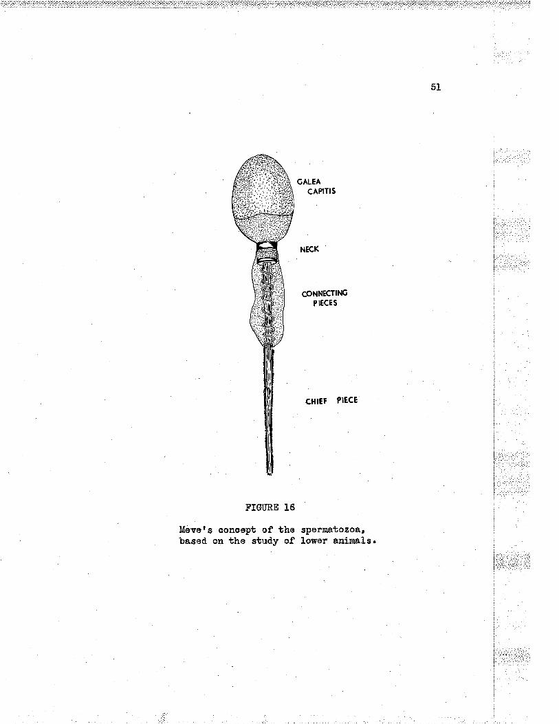
5L
GALEA
CAPITIS
FIGTRE 16
Mevets conoept of thebased on the study of
CONNECTINGP IECE S
CHIEF PIE,CE
spermatozoa,lower animals. ]
;
lì:']:jr:'.Ì. t..ì; ::::.,:l. :t ..1. til1 r:_ :ì t: -:t_I
I
i.I
i.!i
i
t.
¡ . -:l' ::r.:.i: .: t .
j: j
I
I

52
I ,,,','1: :'l .
1..:.
i.,'iil
if.:f,hft_
:.,_ :.: :
FIGURE 17
the capsule and tunlea vaginal.is of ths testisof a twelve-year-o1d, boy" Henatorylin and eosinstainn 150 x' i,riGE A 7911,

5g
d.iameter. It consists of oytoplasm, an naxiaLn element a¡d two parts
of the posterior aorosorno¡ The spiral element, whioh is often figured
I in textsr does not exist in human matoriaL aooording to Gattenby and
B"*"17 or Bowun27. The ohief piooe meaoures from 40 to 60 mícra in
length and graduaLly tapers as the tip is reached. An axial filanent
r surround.od by a membrane makes up the chiof pieoe. The end piece is
sinply the last 10 miora of the tail and is the naked prolongation of
: the axial filanent. In ordinary preparations, the head and the tail
are easily identified.. Special nethods of staining and special lenses
are required to study many of the above dotails which are usually
, figured in te:* books of c¡rboIogy.
Spermatozoa are produced in great numbers, the average count
I being 6OTOOO per cubia niLlimetro of semen. During the aotive
reproduotive poriod approximately 54O billion spormatozoa are producod,
by an indívtduaIz.
Abnormal forms. gl spermatozoa. Abnornal. forms of spermatozoa
aro vory conxlorrr Moenchl5 after the study of a small series coneludes
that nor:na} somen may contaÍn up to 20 per cent of abnormal sperm heads.
When tho sperm head abnorrnalíties exceed ?5 per eent' cLinical
sterility oan be âssürned.o ånong the fifty or more abnor-nal forms this
author descrÍbes are miorosperm, megalospermr narro1r irregularly
staining aplastio head, coil-ed tail, abnormal attaehment of the taílr
no tailr multiple tails, thiokened body. the most important varíaut
from the normalr fron the standpoint of fertility, is the frequent
L-: ..:
1 ':i

finding of tapering narrorr headsr or thoso that aro slaklo shaped'
Motíl1ty of the sperm is not as important as their morphology from
the sta¡dpoint of fertillzing po*""14.
soermatoeenio wave. A marked differenco in tb.e stages of
spermatogenesis is notod when examining'one tubule afber another in
the same cross section. The proooss is not s¡mchronous along tho
tubule. Spermatogenosis at one point is different in the stage
reachod at another point. The study of a longitudinal section of a
tubuLe makos the diffsrenoe more evid.ent. Sorne J-aboratory eJrimaLs
have a definite wave pattern whioh is moasureabLe and oonsta¡t for
the spooies. Such a oondition d,oes not exist for mâIr' Ihe reason for
the spermatogenic wave is not brovun at the presontr
Theory of d,eterrnlnatioq € sg. Forty-eight chronosomes are
found in the somatic oolLs of nan18. These chvomosomos consist of
twenty-three paÍrs of varying shapes and sizss (autosornes) and one
pair of sex chromosomes' which oarry the genes control-}in$ soxo In
the fomale, the two sex ohromosomes aro simíIar and differ littl-e
fron the autosomes. They are the X chromosollleso In the mal-et howevert
the sex chromosomes aro unlike' one being the X chromosone of materrral
origin and the other the Y chromosome of paternal origin. The latter
is small and. atypicaL53.
During the divísion of the spermatogonia of mer. tho chromosomes
spLit in the usual way (nitotic division)' Forty-eight ohromosomes
are found. in each of the daughter ceL}s. Primary spermatoo¡rbes
i...:..ì..: : 1l
54

t:I
l
: i:, , ':,i
S PE RMATOGONIA
OOGO NI A L '',.". -a
OOCYTES S PER MATOCYTESll
55
O NE OVUI.,{ SPERM ATI DS
SPERN/ATOZOA
FIGIIRE 18
Dlagran to lllustrate the developnent of themature sex oells of the naLe and fenale.Redrawa after Bremerr slightl.y nodified.

.56
develop from spermatogonia.
Through a process of re-arrangement of the chromosomese prlmary
spormatoc¡rtes develop from the spermatogonia. The ðhromosomes arrange
themselvos in pairs and fuso. In this oonjugation' there is simply a
reduotion in the number of ohromosomos and no roduotion in the emount
of the ohrornosome natsrj.al' Eaoh prímary spermatoayte contains twenty-
throe paired autosomos and the sex ehronosomes f,lY.
Secondary spermatoc¡rbes arise from the prinary spernatoc¡rbes
through a process of meiosis in which the amount of chromosome material
is reduced.. Each paired. ohromosome splits longitudinaLlyr resulting
in 28 autosomes and the .f, ohrornosomo going to one soaondary spormatoa¡rte,
and 25 autosomos a¡rd the Y chromosome going to the other' Thus in the
j hìtma¡r, the secondary spermatocarbe and all- cells subsoquent to that stage i
have twenty-three ordinary ohromosomes (autosomos of Painterl8) and
elther an ll or a Y chromosome to make up the twenty-fourth'
Secondary spermatoo¡rbes dlvide to forn the spemiatlds. The
ohroúosomos spllt longitudinally so that from each haploid seoondary
spornatoa¡rte, two haploíd spermatids aro formod, each yrrlth twenty-tþ¡es
plus X ohromosomes or twonty-three pLus ï ohromosomes' depending upon
the ehromosome makeup of the mother oelI.
No divielon occurs in the transformation of the spermatid.s into
spermatozoa. In thoory there aro two t¡rpes of "p""t
t ozo&¡ One half
of the spermatozoa contain 23 ord.lnary ahromosomes plus an J[ ohromosorne,
the other helf aontaining 23 ord.inary ehromosomos plus a Y ohromosome.
i ., Every oruum contains 25 chromosomes plus an X ahromosomo alwayslS--never
I
57
a Y chromosoro. Thus the sex of tho organism depends entirely upon the
t¡rpe of the spernatouoa whÍoh unites with the ovr:m d.uring fertilization.
Dtring fertilization, the total and original nunber of chromosomes
numbering forty-eight is restored.. If by union' there resuLts an X-Y
combination of the sex chromosomos, the new individual is a male. A
femalo rosults if the oombination is X-](.
The theory of determination of sex as outlined above is not.
unlversaLLy accepted. It has not been proven in the oase of humans.
Two t¡¡pes of spermatozoa oontaining either an Ï or a Y ohromosome have
not been desoríbed. rnorphologloally for marì thus far3.
Semon. I{hile on}y a snaLl part of semon ís actu-ally a produot
of the testis, this disoussion 1s inoluded to give a short sumnary of
'.5Klllianr s paper' .
Semen is the fluld modium 1n whioh tho spermatozoa are
transritted during ooitus. It is tho prod.uct of the conbination of
many seoretions from the auxil-lary sex glands such as the prostate and
the seminal vesiolos. It is thiok, viscid, opalescent' slightly
aLkaline fluid,, of 2 to 5 ocrs volume. Fow reports of tho metaboLisur
of tho huma¡ spernratozoa aro available. Kil-lianl5 ht" reportod some
sf his studies. this worker finds that the conoentration of sugar in
semen is from 4 to 6 tines higher than that of the blood. Ehen semon
is inoubated at 5Bo C, the sugar content falls off progresslvely for
the flrst 6 to t hours, with a corrosponding increase in Lactio acid.
The pE is slightly higher tha.n of blood. plasna, an average of 7,6.
The uree content ie about twioe that of blood being about 60 nilligra.ns
I r _ r.:
. ...:..l.a,:.,c

58
per lOO mI. Addition of soLutlons having a pH between pE 5 and
pE 8 have no influenoo upon the ¡notility of tho spernatozoâ.c Solutions
with a pE Lower than pH 5 or higher'than pH I d.epressed the notilityr
the maxi-mim depression being at pH of 5 and lL respeotively. Also ths
lower the viooosityr the greater the notiLÍty. The ad.dition of uroa
solutions increases the notility of the spermatozoa, and afso deoreases
the visoosity of sêrtrBEr (cr. rittru^nlS)
the physiology of the tubular systerr. Not all speraatogonla
beoome spermatozoa, Degenerating spermatoc¡rbes and spermatid.s nay be
seen in the lunen of normal tubuLes. Present also in the lunen is a
colloid material' tho source of whioh appears to be the autolysis of
degenerating germ ceLls and apparently also from tho Sertoli ce11s25.:
Physiologically speaking¡ the tubuLes form s closed system of i
tubules¡ open onLy at the straight tubules. The oontinuous fornation:
i..:.i
:of coLloidal material, the d.ogeneration of germ cells¡ a¡d the
proliferatlon of cells prod,uces a head of pressure whioh foroos itsetf 1,..i.._1.:,.,i....:_'.:ir..
along the tubules. This head of pressure can be oasily demonstrated,. , .'.,'.:,.,
.A.fter rigation of the vasa offerontia, the testÍs bscomes tonse a¡rd :'¡"""''"
swollon, so ruuch so that the tunica will on occasioo r,rpt.rrez5.
Tho movemont of tho secretions has been graphically described.
25by Oslund t
This material movos slowly but steadily through theseminiferous ùubules to the collecting tubules. Its movement,brushing along the sides of the tubules' causes the spermatio
,
eLements to slant muoh I1ko grass or weed,s in a slowly movingstrea¡n. (... )
i.,,i.,....r:r..:
ì- r' ,, :.1 .: -
i::.ì:r..'',.r'ì:, l

59
The oelLular elements inoluding the spermatozoa are therofore
oarried passively along tho tubules. this conoept was put forward by
Gríffiths in his 1895 series of Leetures24. Spormatozoa are inaotive
i¡r the tubuleszSr probably a fortunato circumstanoo considoring the
maze of tubular arohlteoture. The column sf fluid foroes its way from
the seminiferous tubules¡ through the straight tubuLes, the rete, the
ducts of the epididy,nis and to the vas doferens.
Thore is no appreciable storago of spormatozoa in tb.e geniteL
traotz5. Spermatozoa are not stored in the seminal vesioalsl4' 25.
lndeod, rather than reservolrs, these appendages are consid.ered as
their g"avey"rdsl4. Os1und points out that the epididynis is suppllod
with notor rrorvesr Moenoh believes that most storage of spermatozoa
occurs in the epidid¡nnis, where in the tail- they are kept inactive by
laok of oxygen and ßinhíbiting secretions of the epíthelium.n
II. TIIE INTER.SITTI.ATJ TTSSUE
IntroduotÍon. Filling the angular spaces between the tubules
are thin col.lagenous fibres, blood and l¡ncryh vesgels, nerves and
conneotive tissue aells. Anong these latter oells are fibrobLasts,
fixed and wanderíng macrophages, fat ool1s rarely, perivascular embry-
onio elements and the interstitial cells of Loydig. The intertubular
connective tissue reprosonts from I3.5 to 50 per cent (average 22 por
oent) of the tissue of the testis6. About 66 per cent of tho testis
is tubular tissue. (Cf. Fig. r page )
li,i,1 i::t-
ir:
In this discussionn the ter.rn ninterstitial tíssuen wiLl be

60
used to include all those tissues filling the intertubular spâcosr
Tho term nintorstitial celIs[ rl'iIl- be reserved for those co].ls
doscribed by Leydig.
The interstitia!. cells of teydig. LoydigS íIr 1g5O noted the
rnå.sses of oel1s whioh eoatained either colourless or yelLowish fatty 1,:.;...;,:,
globules and which are eonstantly in the interstitial tiesu€o ¡,:;,,:,:;':t
i:,: ',1't;¡
SeveraL nârnes have boen assigned to thoso cells. Bouin and
rr :' ''t, .,
.AnoeL'r |n 1903 designated then as the ninterstitial glandr of the
tostis. The undesirabls term of spuborty gIandtr was introduoed. by I
i
SteinbachS in 1911. I
Distribution and a¡nount of interstitial cell-s. Botweon I and
:28 por oont (average L2 por cent) of tho testis is oaoupied by i
fnterstitial oe1Ls6. Such a high percentage is not apparent from the )
ìcasual study of stained sections.
GeneraS.ty the cells are found. in rather dense ilâsses¡ although
slngle cells oooqre They aro found saattered throughout the tostis, i,u;|,:'.
. ,.:;ì:i,_j.:.
ever¡rwhere excopt within the tubules. :: :r:::'j:-'.:i:.1:l
ìãthen the interstitial oells fill the intertubular spaces a
olose relatlonship to blood. vessels naturally resuLts. This arrange-
nent is probably secondary, for if a deorease of the amount of
interstitial tissue occurs, the oLose reLatlonshlp to bLood vossels
d.oes not appear to existg. The aeI} nosts are not espocially
vascular and. are considerably less vasoular than nost of the other
endoarinesro.

FIGIIRE 19
Interstitial oeLls almosb oonpletoly surroundingthe seminiferous tubule at the plano of section.EernatoryIÍn and oosin stains, A ZOBB. IZ5 xn
61
t....!-:
t-.,
| '-::' íi: .-.'
I ...
i:'i 1..
| :.t'l:.,ir::1..,,'
l ::
'- ":rr: ;i::'
!:rr+'::::,r;
l,-,'..I.;i.:
Ì:.'..'i'II

62
FIGI]RE ?O
Interstitial cells situated just beneaththe tuniaa vasoulosa. Henatorylin a.ndeosin stain" A 7088, 260 x.
i;.,.:a:ri.ll:.?:: ::::¡ -l

ll;:rií*T¡li*Edl'iå:¡:Sri tiil::iiiiîiì{i'r..-¡_îi älfifìlí¡..j:t¿,ij
FIGURE 2I.
InterstitÍal oolls situated just beneaththe tunloa vasouloga. IIømatorylin andeosin stain. A 7088. 260 x.
63
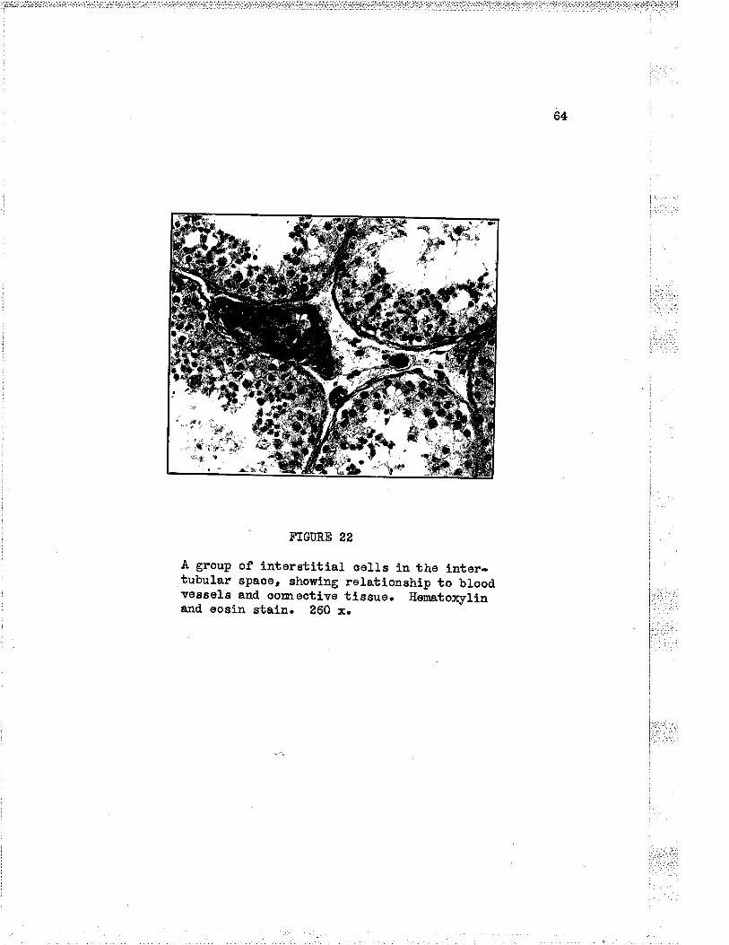
64
FTG¡ÛRE 22
A group of interstitÍal solls in the inter-tubular spaoe, ehowing relatlonship to bloodvesseLs and oomoctive tissue. Eonatoxylinand eosin stain. 260 x.
t'l:'t.l;'l
i.....,1....:t....,L.I.

64a
FIGIÍRE 22 A
The interstitial tissue and oeLLs ofeLderLy man, showing the pigment andE & E. 350 x. (WCU ¿tgS-50).
the testis ofcrysta1loids.

65
The interstitial celLs appear first in the L9.5 mn hunan
enbryog8. Most authors agree that they probably arise from
mesenoh¡m,al oolls. The eells are abundant at bir.bh¡ howover, they ,., .,:,::
subsequenbly disappear. At the time of puberty they reappee.ro
Spermatogenesis conmenoes at the tine of their reappearanoer
.. - . - ..i,..
Histologleal details. T¡r,pically the interstitial cells are l,.lir.l.¿-':large, írrogular polygonal colIs, measuring from L5 to 20 micra=. . ...,
1,,t¡..iri ''.,Numerous inoLusionÊ make the abundant oyboplasm granular.
Thenuc1eusmeasuresfrom6to]-0rricrarnost1y7'6micra11.
Usually there is one nucleus, aLthough some authors have reported the
rare finding of binueleated, cells. Mitosis i" rarelo. One¡ or
occasionally more nucleoli shoïu distínotly after most staining
reactions, The epherioal nuoLeus ís vesicular a¿d has a typical.
ar_rangement of chrornatin and snall granules.
A large idiosome is present in tho ondopLasm near the nuoleus.
Two rod-shaped oentrioles are usuaLLy prosent. the nitochond,ria are
in the form of filanents and. granules. A typioaL Golgi net is found.
in the interstitial cell-s. Various round. granules whloh are Larger
than the nitoehondrÍa have been desoribed. by some authors. No agree-
mont has been reaohed as to the nature or significance of these
graaures. (cf. Rasnu"u"og)
Abundant fatty gtoUrrt"s are found in the futly doveloped
interstitial aell-, lhere is considerable variation in the slzo of
the globules. These fatty substances aonsÍst of oholesterol¡
jrì::.:!lr;lii

66
oholesterol- estors, and smaller anounts of cerebrosidos and phosphatideslz.
'The role of the lipoÍd. material ís not h.own9.
Bror¡u¿ plgment in the forn of granules is usually not found i¡
Èhe human interstitial oells until the adult stage is reached9, aftor
which time the amount of the pignrent i.ncreases. In old ago, numeroug
oells may be packed with granules of pigment.
The first desoription of elongated arystalloids in human inter-
stitial oelle was given by Reinke in 1896. these insl.usions are i:
generally regarded as charaoteristic of humans, although they have
beendosoribedinafewotherspecies.Tbecrysta11oidsdonotappeari
until aftor puberty¡ afbor which time they persistg.
Four oharacteristlos of the interstitial oeIls in humans may be ,
listed here. Comparod with the lnterstitial oells of other species¡ ,
those of the hunan have reLativel-y large am.ounts of f.ipoid matorial.
The crystallold of Roinko ls absent or rare in othor spocies. No I
seasonal variation in the number or appearaace of the Leydig cells isi'.'' i¡,
found in ma.n. Tho coLls form largo groups or colleotions whiah aro , ,,,.:..:: -
oasiLy id,ontified. Tn many of tho lower animals, the Leydig qells are .':.ri,-': ':|:.
found singly placod, and are often diffioult to iaoniify. ïn other
animals tho interstitial cells are very [r.lrnerousr
Relationship between nerves and tho Leydig 99lf.g, Dotails of
the rslationship of the nerves supplylng the testls and tn-e Loydig
ce1ls are due nostly to the work of 0kkets and Se¡rd.5?, whose paper is
sumnarized i¡ the paragraphs to follou¡.
i'i,i'j.ì:: lni:l' 'irr: i

67
32Aooording to Oklcels and Sand , tho nerves of tbe human testis
are more numorous than has been previously assumed. The nerves on
,.1 reaching the organ in company with the blood vesseLsr oontÍnuo to ,..,,.::.
follow the courso of the latter into the interior of the gLand¡ some
paesing dlrectly through to the modiastinum¡ but tho najority and the
; larger aervos following the arteries which run in the deep Iayers of r,.,..:.r j:....:.;.:r.j the tunloa albuglnea. From the doeper layers of the albuginea, they : :
: tr'
,i n""u. *:o the interLobular septao. i,ì,;,i
Three t¡rpes of oon¡reetions are doscribed by Okkels and Sa¿d¡ ,
(a) perineural Leydig cells are found. in the region of the hilun or at l
\
a point just after the nerves have entered the tunica yasculosa. The l
I "ontact is essentialLy superfieial¡ (b) whíle trr-o nerves are still in l
i tho albuginea, and are about to enter the septulae, occasionally are it-
, raisins in a bunt; (c) when the large masaes of Leydig oells aro' j
reached., the nerve spLits,up and blends with the oells¡ where the¡..-,t..
' neurofibrils sbop at the surface of the ceI1¡ but do not penetrate ¡',',',.'
.
, into tho o¡rtoplasrr. These lnvestigators have not been able to :,:,;,,,,,
demonstrate an intinate relationship betweon the norves a¡rd the Leydtg
seLls before puberty.
ì Tho larger nerves folLow a poriarterial oourso until the -a-- ---- ij1:::,ri':r:l i i,ìi;a:ii
parench¡maoftheg1andisreached.Duringthiseourse,itisprobab1e
thatvasomotorn.ervesaregivenoff.Thenervesapparent1yhavea
I fìrnctional significanoe e¡rd. Okkets and Sand suggest that this function
., is secretory. (cr. oklcols and san¿õZ)
i t:.:,
'

68
PERINEURAL
-l
I NÏRANEU RAL
INTERTUBULARMASSES
FIG.URE 25
Diagram iLlustratlng the r¡arying reLationship ofinterstitial cells to a nervo as it courses fromthe oapsule of the testis into the septum. Re-drawn after Okkels and Sand.
lr.
F

69
Variation of the testes ìmith .3gg. FulI size or funstlon of the
testis is not attained untiL puberty or afterwards. In a boy of ten
yoars of age, the organ is alnost round¡ and is firm and compact. '' ,,',":-.t
Tho organ measures 10 by 10 by 15 mro in size. The seminiferous
tubules aro almost indistinguishablo from one another. Tho tubules
are not greatly convoluted and are closely packed, togother. There is ,., ,,,
I ..a.t
little interstitial tissuo, most of which is fibroooltular connootivi:t,.t,.''tiszuo. Eaoh tubulo consists of a so1ld ooLumn of oells. ÂLl tho j.,;ì.''
ceLls are in a quiescent stats.
In the adult, the organ is softer¡ larger and moro plunp. 'Hlhen i
the..organ is out in the fresh stato, tho yel-low tissue bulges. From
thesurfaoethetubu1esmaybepiokedupwithfineforgopsand'are
seen to resemble fine coatorted threads. The tubules are larger than i
l
thosooftheyoungmale.Thetunicapropriaisthín.;
The prooess of aging leaves its effoct upon tho male genital i
'
system but the changes are not so uniform when compared to similar , ::.
ohaieges in the fenalo genital system. Nor are the ehanges found in t;"'-..;.:. :r¡'I i.l -:
old ago so weII lstos¡n. There is a Laok of published data. Spermato- ',.rj.:,
genesis oeases at a perÍod varying greatly w'ith the indívldual.
Reports found in tho literatr¡re vary greatly with respeot to the uppor :
linlt of this period. In nany indlviduaLs there occurs with failing i,,Èii -. i' j::
spernatogenosis a thiokening of the basement membrane of the tubulo.
The corureative tissue of tho tunica propria inereases in thiekress, ,
usually assooiated with a deoroase in the size of the tubule. As i
well thero is an inoroase in pignont found in the intorstitial cells ,,,,i,,,

70
l't.....i-'.--'
FIGURE 24
the testis of an infant naLe of one úonth. thetubules have no lumen. The lntertubular tlssuels vory oellular, but shows no interstitlal cellsrCE.48-2067. Eenato4ylin and €osirtr tow power.
[ .:.:..l.':.1!i,,.,f .: i:.' .,. -
f:J

7L
I
!: .
l1
FIGURE E5
The tubules and. interstitial tissue of tho testisof a nonnal twelve-yoar-old boy who died ofaccidental electrooution' Autopsy twelve hourepost-nortem" Eenatorylín and oosin stainn 2'î5 x,wGH A 7911.

.72
of many ind.ividuals. (cr. nngleã6)
Ng!" on development of tho testis. The dovelopment of the
testos and tho ovaries is closely assooiated with the devolopmont of
tho urinary systom. the devolopment of the genital system is oomplex
owing to the fact that íts diffsrent porbions arise from diverse
primordla. The aontributions from theso primordia vary qualitatively
and quantitatively ,in the two sexes'
lffhiIe the sex of tho future embryo is dotsrnined at ths moment
of fertiLization by the prosonoe or absence of tho Ï-chromosomo, the
identifioation of the sex of the embryo is not possible until the 17
mn stage.
The primordia of the sex glands appear in embryos of 4 to 5 rm
(CR) lengtfr. The genital ridgos appear as thickenings of the coelomio
epithelium on the mod.ial aspoct of the mosonephros' The basement
membrane separating the ooelomic epithelium from the underlying
mesenoh¡rme disappoars. Cords of colls proliferate fron tho epitheliun
and project into the mesonah¡rme. Until tho 17 mm ,stage, these develop-
mental ohanges in these ridges aro the same in either sex.
*{,t about the 15 rrnn stage, the gonadal blastena becomes divided
into cords by the davelopment of fibrous tissuo bundlos. the sex
oords are at first Joined to the germinal epithoLium. *{t about the
25 nn stago, the dense tunioa albuginoa forms and separates tho oords
from the epithelium. Lator, the cords orbend into the region of the
mesorchiun whore they fornr a.network, the rete testos. Some of the
l
ì.iil

78
oords beoome oanalized. to form the sominiforous tubulos" the oells of
urhloh are either the sustantacular coJls or the primorclial germ cells.
Some of the shorter oords do not becorne aanal-ized, &nd persíst as small
groups to bocome the interstitial oo11s.
The ovaries and the testes in late fetal life oooupy a different
position from that in whioh they are found in the enbryo. The change
in position is most inarked in the case of the tostes, sinoe theso
organs pass from tho abdominal- oavity through tho abdominal into the
sorotal sac.
By the third month of fetat fife (ZO nn), the testis lios
rotroperitoneally in the farse pelvis. rt renains at the abdominal
end of the inguinal oanal until the seventh nonth. During the soventh
month it passos through the inguinal oanar. Normally the testis iswithin the scrotun by the end of the elthth fotal month. (Cf. Harniltor¡¡
Boyd and Mo"sman53)
The
in Chaptor
hormones sf the testes. The maLe sex hormon,os are dÍscusssd
Vr'together with the other hormonos of reproduction.
I .:::.::.:: ]

74
BIBLIOGRAPHT
1. Cunningha.nts Manual of Praotical .Anatomy¡ VoI. II, Revisod byA. Robinson. Eighth edition¡ London: Humphrey MilfordOxford University Press, Ì9õ0. 524 pp.
2. Bremer, J. L. (Rerr'ritten by if. L.'cteathorford), A Textbook ofHistology. Phj-Ia.¡ The Blakiston Companyr 1944. 723 pp.
3. Maxlmowr A. 4., and lf,Ãn. Bloomr A Toxübook of Histology. Fifthedition¡ Phila.r Tf. B. Saunders Conpany, 1948. 700 pp.
4. Piersol.rs Normal llistology (ro-edited by I$.E'F. Ad.dison).Thirtoenth edition¡ Phila.¡ J. B. LippÍncott Conpany, 1926,
5. Leydig, F., Zur Anatoqie aer nårurtichen Gesohectsorgene und,.AnaLdruson der Sängotiere, Ztschr. f. wissonoh. ZooL.1850, ii, I' (quoted by lianes- 19)
6. Slotopols$, B. åJrd. H. R. Sohinz, Histologisshe Hodenìefundeboi Sexualverbreohern. Aroh. f. path. .Anat. u. Physiol.u, f, klin. Med., 1925, 25?¿294. (Quotett by Rasmussen - 9)
1. Anoel and Bouin (Quoted by Sharpey-Schafor - 10)
B. Steinbach, E', Umstirurung d.es GesohLeahtsoharalcbers bsiSäugetíeren durch Austausoh der Pubertät¿rüsen.Zentrabl. f. Physiol., 1911, 25t723. (quotoa by Rasmussen)
9. Rasmussen, A. T., Interstitial Cells of the Testis. Section XüII,Special Cytologyr VoI, III, pp. L675-1.726, Ed.itod byE. V. Cowdry. Neur York¡ Paul B. Hoeber fncorporated., L932.
10. Sharpey-Schafer, E, 4., The Endocrine Organs. Second edition¡part If¡ Londsn¡ Longmans, Green and Co. Ltd.., L926. 418 pp.
1I. Cl-ara, M., Untersuohungen an menschlichen HodenzwisohenzeLlenZugloich ein Beitrag zur Kenntnis dos rhybhnischenlffaohstu¡rs der Zellen durch Verdopelung thres Volumsns.Ztsoh. f. mikrr âBat. Forsoh.r 1928, L3zT2.
. (q¡rotea by Rasmusson -. 9)
L2. Lotz, A.¡ &nd R. Jaffe, Dio Eoden bei Ai-lgemeinkrankungenoZtsoh. f. Konstit., L924, 1O:99. (Quotod. by Rasuusson - 9)
15. Moonch, G. L', and. E, Holt, Sperm Morphology i,n Relation toFertility. .Aner. J. Obstet. and G¡mec., 1951, !21199.

75
14. Moench, G. L.r.A. Consid.eration of So¡ne of the.Aspeots of Sterility..A¡rer. J. 0bstet. and G¡mec., 1956, 32t4O6.
15. Killiarr, J. 4., Metabolisn of Spermatozoa in Semen. .Am. J. Surg. t1953' 19¡ 76. , ,, ,
16. Johnson, F' P., Dissoctions of Human Seminiferous TubuLes.Anat. Roo., L934, 59¡L87.
L7. Gatenbyr J. B.¡ end E. Tl. Beams, The Cyboplasmic IacLusions inthe Spernratogenesis of Man¡ Q¡larl . J. Mioro. So.r L955, 78¡1. ,.,:,,,,,
1:,,
'_:r'::;. :
18, Painter, T. S., Studios in Ina¡nnalian Spormatogenosis. II. The : ir:':'
Sperrnatogenesis of l¡lan. J. Errper. ZooL., L923, 372291. .,t.,
.
1: r:¡; .:'l':
19. Ilanesr F. M., The Relations of tho Interstitial CeLLs of 'Leydigto the Produotion of an fnternal Secretion by SrlararnallanTestis. J. Exper. Med., 1911, L5r538.
20. Kolliker, Mikroskopisohe .Anatomie oder Gowebelehre d.os Mensohen, i
Leipzig¡ 1854, pt. ii. (quoted by Hanes - 19) l
n2L. Reinke, F., Beitrågu "o"
I{istotogie dos Mensohen. f . üUsrKrystalloidbíld.ungen in den interstiellen Zellon dosmonschlichen Hodons, Arch. f. mikr. 3nat., 1896, 47¿34.
i(quoteA by Rasmusserl - 9, and Hanes - 19) '
?2, Bouin et Anool, Rocherohes sur Ia signification physiologiquede].agIand'einterstitie].1edutestiouIedesme.urmife.res,Jour. de physlol. of de path. g6n.,1904, 61L012.(Quotea by Hanos - t9)
., ',.'.,,,,,,.
23. Bouin et .Anoel¡ Recherohes sur les oeLlulss interstitielles ¿tu "'1"'''';testicule d.es ma¡nmiforesr Aroh. d.o zoo1. explr.r 190g, , ,,,,,,
Sories 4, L¿457. (Quoted by Hanes - 19) ,l,li.,' ,,
24, Griffiths, J., Threo Leotures upon the Tsstes. Lancet, 1895, l
l¡791, 795, 916.
25. Oslund, R. M., The Physiology of the Male Reproduotive Systern.J.A.M,A., lgZB, 9O¡ 829,
i.rì.'':.,i¡:,:i:.!::;
26. Brannan, 0., Tþe Synpathicotropie CeLLs of the Ovary and Testis'Aner. J. Path., L9ZT, 3¡543.
27, Areyr L. 8., New DÍagrams Tllustrating the Transfomration Periodof the Human Sporrnatid, as Propared by the Late ProfessorBowen. ånat. Reo,¡ 1950, 47t51.

76
28. 8i11, E. C.¡ tho Vasoularization of the Human Testis.Imer. J. 3nat.¡ 1909, 9¿463.
Zg. Rivington, Quotod in Grayr6 .Anatonyg4.
90. Jamleson, J' K.¡ and J. F. Dobson, lho tynphatlos of tha Testicle.Iancet, 1910, 1¡495.
91. Rouviere, 8., .Anatomie dos lynphatiques de lrhomno.Paris¡ Masson et Cie., 1932. 489 pp.
32, Okkels, IÍr¡ Bnd, K. Sand.¡ Morphological Relationshíp bstween thoTestÍou1ar Norvos arid tho Leydig Cel1s in l[en. J. Endoorin.,1940, !t39.
33. Hanllton, 1lF. J.¡ J. D. Boyd¡ a¡rd E. Tf. Mossman, Hun^e.n Embryology.Ca,:rbridger ïf. Ileffor and Sons, Ltd.¡ 1945. 566 pp.
34. Grayrs ânatomy¡ edit. by T. B. Johnston and J. WhllLis,Twonty-niath edition¡ London: Longmans, Green and. Co.¡' L947,
597 pp.
55. Johnston, T,-8." A Slmopsis of Regional Snatorqy. Sixbh edition.Phila.¡ Lea and Febígor, 1948. 436 pp.
g6. Engle¡ Earl T., MaIe Reproduatlve Systom, Chaptor 15, inProblens of Agoing, E, V. Cowdry, Editor.Bal-t.¡ The Tf11líans & Titilkins Companyr 1939. Z5B pp,
37. Eerman, L.¡ The Praotioo of Urology. Phila.r Iìf. B. Saund.ersCompany, 1958, 925 pp.
58. Selye, Eans, Tertbook of Endocrinologyr Lg47. IlniversitJ deMont.real, Montroal. 914 pp.

CEå.PTER THREE
FruAI,E REPRODUCTIVE SYSTMû
Trm 0v4Rv
' .::f5')ir-,.:-;:ìì

CEAPTER, ITI
Tffi FM{ATE REFR,ODUCTIVE SYSTMI
Introduotion. the female reproduotive systean oonsísts of the
ovaries, ovlduots, uterus¡ vagina, mamoery glands, and ths externaL
genitalla. 0f these, the ovarles produoe hormones and dísoussion tlrill
be restricted to them. Disoussion of tho plaoonta is found iu Chaptor
Four.
Þnbryology¡ anatorqrr and funotion of tho ovaries and testes have
neny símÍLaritleE and, as may be expeoted,, nany d.ifferêncese A conparison
of the ovaries ar¡d testes is mado on page
GeaeraL desorlpt,lon of the gg. lexÈbook deseriptions of
t-he ¡ormaL ovary are often too restrioted and stereot¡rped. to aLlow for
the great variations whiah nay fall- lcithin the normal range sf the organr
Measuroments generalLy glve range from 2.5 cm to 5 cm in tength, 1..5 om
to 5 om in vuldth, a¡d 0.6 om to 1..5 om in thlebaess. It is fairLy
comnon for the normaL ovary to attain larger pToportÍonsi All too
often theee fflargen ovaries are regarded as pathologioal and are sent
to tho surgical laboratory for exanlnation.
there are marxy qulte aormal variations of tho surface appes,rar.oeo
3, pale mooth or slightly pitted surface is oonsidered by maay as the
noryral appearance, while a vigorous and healthy organ with its bulging
foLlicles, deep 6o8.f's¡ hemorrhaglo and non-hemorrhagie oorpora lutea is
offen íaoorrecti.y oonsidered. ebnormal.

79
Cross sections of adult organs show variations from speoinen to
spooimen. The thín¡ pa1e, genninal eplthelíum on the surface oeJalxot bo
dístinguished by the naked eye. The tunica albuginea in¡nediately
beneath the epitholium is nroh thinner than in the testes and is not
prominent ' .An ilI-defined rather firni corte:( surround.s tho sofèer
more vascuLar medulla. Near tho hiLus a few gaplng sLLts, the rete
overli¡ aro presont. Follic1es which are actively growing may reach
the size of a feur miLlimetres and are thus visible to the naked eye.
Corpora lutea are usually easy to identiff,r espeoially lator in thoir
development whon thoy assumo a fairly oharacteristic shape and yelLow
oolour. Corpora albfoa¡tes and the smaller corpora fibrosa are whíte
in oolour becauso of tho Large esrount of fibrous tÍssue present ín
tlieso struoturos. Atrotio follíoles nay attain great sizo, frequentLy
reaohing the dlameter of 'a centinotre or lnorer These follioles are
filLed with aloar fluid wtrich Ís ofbon under oonsiderable tension.
lffhen tho fo1llole is out aorossr the fluid esoapes and the wall of
tho oystic struoture above the surfaoo of the ovary oolLapsesr lrrhile
the wa1l boneath the surfaae ís held in shape by the stf,ouå,.
Thg blood supply of the .9yg8. The ovarian artaries arise from
the aorta in a mannor similar to the testioular arterios in the maLeo
The arteries arise from the antero-lateral aspeot of tho abdominal
aorta, near the origin of the renal arterles. From hore they descsnd
retroperitonoally, croõs tho exbemal iliao arteries approximately an
lnoh below the level of their origin¡ and. then desoond into the
..: : rr.. i:+",r?"::.1r)
::'.:::'

80
infundibuLo-pelvic ligameat. Afber r.eaohi"ng the mesosalpinx, eaoh
ovarían artery a.nastomoses with the aorresponding uterine artery, and
after giving off some branohes to the orriduct, traversos the mosovarium I ,1.., ,,1 ,:,:)-....:.-::-
to onter the hilun of the oo*y7.
The vei¡s ernerge from the hilun to fom a parnpiniforn píexus
whlch oomrnicates with the ploxrs of the utorlne vosgols. Two veins .,;:;:,-, .;:
1ssuefromthisp1exrsarrd'asoendretroper1tonoa11yinamanì1or
similar to the testioular velns?. ',,..,:';',;;,;,i':,',
L¡mrphatic vessols from tho ovarles are assoolated with the
lyln¡rhatios of the body end fundus of the utorus¡ They drain ínto tho',
latoral and pro-aortic g"orpu7.
Vessele within the. organ, Reoent studies of the vasoularI
patterns of the human ovary show that sta.ndard d.esoriptions found in
text books are no longer teriablo' Delson¡ Lubinr and. Ro¡mold.s8 report :
thelr stud,ies and the red.isoovsry of Farrets d.osariptlon of 1858.
Themainarteryasítooufsesinthehi1umoftheovary1sj_:._--..:.::,:i...
. .i:..i..unduLa¡t, tort¿ous and is somewhat flattened,. I'rom the naÍn artory .t-.,,-,¡,¡.,.'
r*^ -::--::::':':
ariso the prinary arteries whioh are mssbly torbuous aüd undularrt.
Secondary arteries ariso from the prÍmary vossol.s' As these beoomo
snaller thoy show a tendenoy to spiralling. The smalLest veesels that:r-...._.., Ì.:. t,. .:.;:j.
'':'
:::'I
these lnvestigators were ablo to injeot with plastlc wero the tertÍary r'tìii:1.i:,rrf'r'::ii
vessels. lheso showed rogularly the greatesb spiral.!.ing. The spirals
wore in the form of a oountor-olookrrise helix of gradually diminishÍng
dianetor' ' ,'.'
_:ti:l:.::;-:.,.:..:.:: j.'

81
The veins show no spiralllng and are larger and irregularly
flattened. Anastomoses are freguont. Tho veins drain into the
pampiniform ple:ms in the hil.ar region. (Cf. Delson¡ Lubin and
ReynotasB)
The nerve suBpLy. Fibros from the cooliao, renaL and suporior ,,,.,;
mesonterie ple:nrses supply the ovary. Ihe origln of the ovarian ple:nrs ,;,',.
ls often dlffioult to determÍno by dlsssctlon. The fibros are vory .,,1,
fíne and surround the vessol-s to the ovaries in the form of a netrrork. 't:'
Following the vesseLs¡ they rsaeh the hllus whoro twigs aro glven off
to suppLy the ovid.uot and othors oontinué with the vessols ín the
'. dlt L""9).ovary a¡od branoh wlth thern, (Cf
Afbsr reaohing tho ovarian stroma¡ only fine non-medullated,
nervo fibres are found,. l,¡,T11u"9 oould find. ao ganglloníc oolLe. Fine
non-modulLated, nerves roaoh the follloles' The negg layern (oorbox) is
devoid of nervos. (cf. u$I].u*g)
Note on embryology of ovarL.es. The early developmont of the
gonad,s ín either sox is símilar until the L? m. stage (sevoa wooks) is
reaohed.. The eseential features of the oarly stages woro glvon
briefly ln oonneotíon rrÍth the development of the malo gonads at tho
end of Chapter II.
About the tlno the 13 m stage (slx weeks) is reaohod, the
gonadaL bLaste¡ra beoomes d.Ívltled into cord,s by the fornation of
fibrous tlssue bundLes. The oords are at first ettaohed to the
"gennlnal epitheLium¡ but soon bscone.broken ir¡to ieolatod eell me,sôêsr

82
The snarl groups of oells rosurting from the fragmontation becomo
grouped to form tho primordiaL folLioLos. (Cf. Herniltonr Boyd and
Mossnanzo)
A sumoary of the fate of th_e primary_ foLlic.les¡ i. er¡
d,evelopment of the Graafía¡r follioLes¡ Corpora lutea, Gorpora albicantos¡
and atrotio follioles. Probably no other organ has so many variabl.e
oomponents. The folliolos whioh have beea deslgnatod as¡ (a) prinordial,
(U) prinaryr and (o) vesiaular (maturing and Graafían), are not statio
but aro ohanging from d.ay to day. Tho corpora Lutoa as welr undergo
changes rapidly. A definite pattern is foLlovred in the oase of eaoh
structurer but beforo considering tho detailed rni.crosoopio picturo of
oaah a¡ed the various phases of its exístenoo, a briof sumnary of the
deveLopment of, en ovum qrid its subsequent possiblo fates is give,n.
FYom the germ oelLE there develop -the primary fo1Lic1es, eaoh
of whioh is essentially an ovum gurround.ed by a slngLo 1ayer of
flattened fol.licular ooIls. .as the forlíole grorrs, the foLlisuLar
ool.ls become more numerousr and. eventually are separated from the ornrm
by tho developnent of a orear, gLaesy membrane bro¡vn as tho zona
polluoida. Fluid, aolleots 1n ths interspaoes of the follioul,ar ce}Is.
As thls foLliouLar fluid. oolleots, lt enlarges the laterspaces until a
oontinuous follioular cavity is fomed. Eventually the ovr¡m wlth its
zona pellucida and surround.ing follicuLar colls is situated. as a
roundod mass of oe}ls (cumulue o8phorus) on the inner surface of the
lining of the vesícle. lhe folliole (now mature a¡d oalled the
Graafia¡r fol,lioLe ln this dleoussion) oontains fluid whÍoh is und,er il:i..: -,r.,:
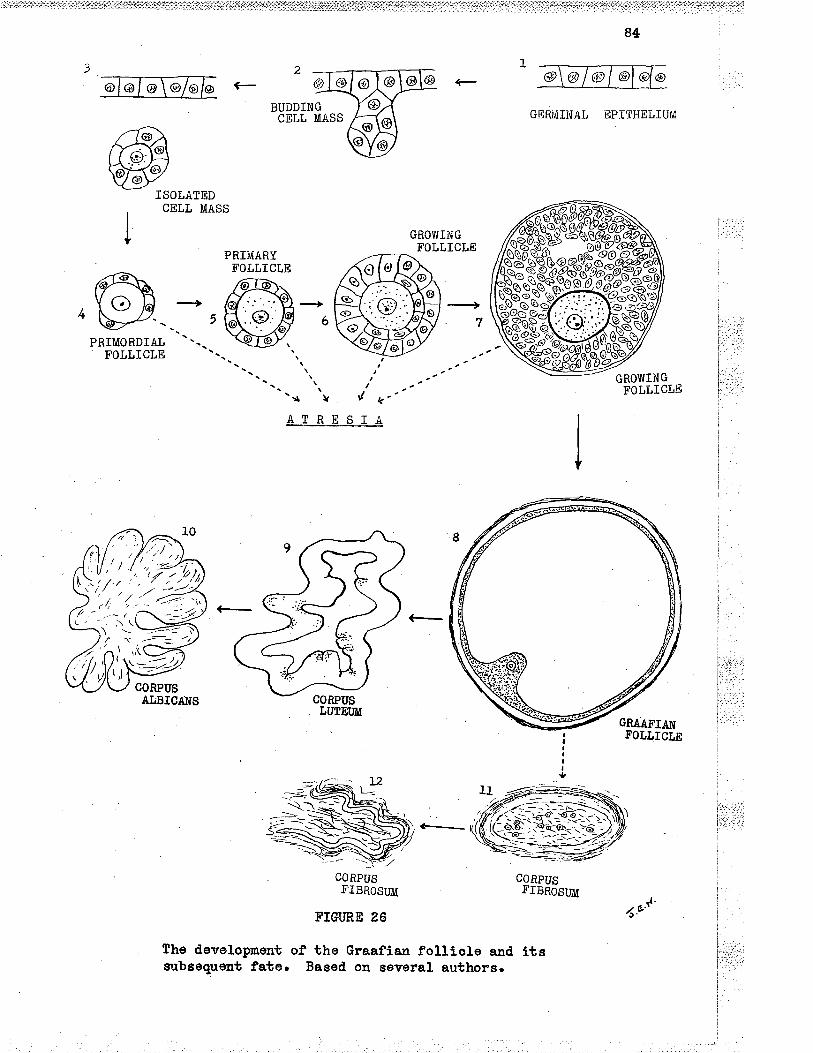
84
ISOLATEDCELL MASS
BUDDINGCELL MASS GERMTIÙAL EPITHELIUM
GROWINGFOLLICTE
PRIMORDIATFOLLICTE
PRIii{ARYFOLLÏCLE
The developmentoubsequent fate.
'/ k"ATRESIA
CORPUS CORPUSFIBROSUff FIBROSW
¡'TGURE 26
of the Graafian folliole and iteBaeed on several authors.
GROWINGFOLtICtE\
AL.BICAI{S
1.u'4'

84
oonsiderable tension. The follicle ruptures to releaso the on¡n and
some of the liquor folLioull. FolLowing the rupture, the u¡all
collapses and ohanges soorx oocur in the lining follicutar oolls to
become the celL's of the corpus Lpteun. The subsoquont history of the
oorpus lutoun dopends upon the presenoe or absence of conceptioa. If
conception has oocurred., the corpus Lutoum oontir¡.ros to grow in size
and inoreasos in ftrnation until the period between 50 and, 60 days of
tho gestati,on¡ after whloh it undorgoes re€rossion. In the absence of
oonceptlon, regresslon of the oorpus luteum ooours forbh¡'¡ithr ¡rlthout
oontinuatíon of growth. Finally the corpus luteum is repLaoed by
oollagenous oonneotive tissue of the oorpus albioans.
Not al.l prinordia develop along the same plan as gíven above.
countLoss numbers becomo vesiouLar follioles¡ but as a rule only one,
fnfreguontry more tha:r one, reaches the obago of maturity to discharge
&R ovulll¡ When ovrrlation ooGUrEr for some unlmown reason, the other
follioles Braoing to naturityn undergo an arrest of d.eveJ.opment and
booome atretia foLlicles.
The gerninaå epi.tholiun, Except at the hiLun¡ tho ovary is
covered with a sírrgle layer of cuboidal or oolunnar ngernrinal
epíthelirrn.n Îhis layer iE desorlbed as nri¿ified peritoneun or
modified meeothelÍum. .As age increasôs¡ the oeLts tend to becone
flattoned,. No basement membraae is found bonoath the 3.ayor. A s3.lghb
cond.ensation of oollagenous fibres, termed. the tunioa aLbuglnea, íg
direotì.y beneath the gerrrrlnal epithelium. Compared to the struoture of
.ß:''i:.
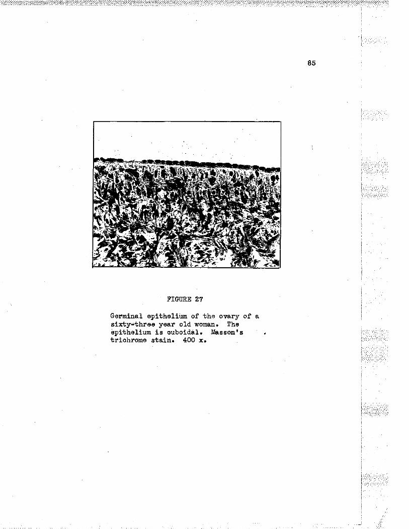
85
FIGIIRE 27
Ger¡ninal epttheliun of the ovary of asirtY;f,þ¡ee Yea? o1d womaa" Thsepitheliun is ouboidal. tllassonts rtrichrone stain. 400 x.
t,-'i r'lt..i-
t-:
ì:,:.:
l:: r-.i.,
l.r.i- i. . .: ...i :.
:
I
ìj
ì
I
i,.r. . .:i .. :

86
F]GURE 28
Germinal. epithelium. Higher nagnifieation of the semesection as Fig. 85. Tho basemsrt monbrane is sesn wherethe'opithulir..T is missing (artefact produoed in handlingof tho tissue). Massontã triahrcme stai.n. 800 x.

87
the same name in tho testis¡ this tunica is thin and delioate,
There is general agreemont among various workers that the
getminar epitheliun arieos from coelomic epitheliun in enbryonio
developnent4.
Throughout tho fi¡actional period of the ovary, the gernitlal
epitholíum show's marketl aotivity of groruth. Groupa of celLe from the
epltheliun nigrate into the oortex end flnally reach the deeper oortex
and Lose thelr attashmont. whether these oelLs devolop into ova or
d.egenorate is not d.efinitely lm,own. (Cf, SoÌærørz et al.5)
Cortgî and. meduLla. Ðivision into eortsx a¡rd, med.u1la is poorly
defined in the ove.rJi,r I'ollioles and their derivatlveo are found in e
oharaoteristio, stroma of oompaot spindle-oellod oouneotive elements of
the aortex. The oortex is wide d.uring the reprod,uotive period. nakiug
approxlmately one half to threo guarbors of tho sross-soetlon,o The
medulla has a loose oonneot!.ve tissue stroma and nurnerous large
oontorted blood vossels. .At the hilusr tho meduLla is not covsred by
tho oorbox. Smooth rnueoLe fibres erbend la from the mesovariun at the
hilus¡ olsev¡hsre a ferr¡ may be found¡ chiefly around the blood veesels.
A few sllt-like spaoes are found at tho hilue, .lhese are the
rete ov¿-ree a¡d are lined with a sÍmpIo low eplthsJ.ium.
In the senile o'\rary, the oortex is a rolatively thin soae of
oonnective tissue. The vessele of the medulla show endarteritis
obliterans and in the r¡r¡alle of many of them there ie ofben extensive
oaloífioation' Corpora lutea ars not present. Primery folllcleE a¡ad

B8
atretio vesioular fol}lolos are found¡ although they â,re f,aro¡
Orlgln of ovg. I$hile aonsideration- of the differont phasoe of
maturation of the developlng ovum is not mithln the soope of this
dieoussion, bríef reforonce ls nade to the work of Swezy and. Eva¡rel.
á.coording to these authors, the lurman ombryonlo ova arise by
, proliferation from the gernlnal epithelium. During tho period. between
: sevon woeks to th¡ee months of intrauterine development only grorrrbh
en¿I nuLtiplioation take plaoe. Maturatlon phases of the ova are forrnd
during tho period from three moaths to five montbs, From five months
to geven months rnaturation phases which are si¡nilar to thoso found in
I tho nsle germ cells are found.
i fn the ovary of the ad.ult, ømbryonie ova havo disappeared.i
Definitlve germ oeLls arise then in oue of two methods¡ first by
i prollferation'from the ger:mínal epithellum of the surfaoe of the organ,l
and seoond.ly by divieion of gern cells already exlsting in tho orgâtro
(cf. Svrezy and gva¡s1)
Sohrvarz et a1.5 eupport the vierry that neru¡ ova aro fornod, after .. ,'',:..-.: --
birth.TheorigíniEmainl.yfromtrregàrtina1ep1the1iumaooorclingto
these'workers. Following is a quotation from their paper¡
(...) The conoept that in the maÍimals the ova are aLL fornedbefore blrth and remâin quiescent r¡ntil cyolo naturity has no ,.,-.foundation in fact. On the contrary¡ all of the ova in aduLt l.i¡:r::ii
Life are now forrnations and are being oonstantly produced andare oonstantly being destroyeil. (.,.)

89
Pri-mary follisles. Sinklnu6 d"fio"s ths primordial folticles as
an ovun whioh is inoonetantLy and inoomplotely surround.ed by flat and
ellipsoid,al oells. The averago diameter of the primordial follicLes is
30 mlcra. Pri.mary follioLes aro completely surrounded by spherioal
osl}s and-havs en avorage diameter of 50 nlorao In the ovary of the
nevrborn, the primordial folLioles greatly outnumber the prlmary
foLlieles¡ rrhile at 14 yoarsr their numbers are about eEral6.
the singLe layer of ElherÍoal to cuboidal epitheLium rests oa a
basomont merabrane and. surrounds tho orrum or germ oell. The nucleus of
the ovr¡m is Large and vosioular y¡ith a looso aetwork of Linln throads¡
emall ohronatin granules and a large chromatin nuoleolus.
Surror¡ndlng the folLioler the stromal sells are 1ÍghtLy stained
and glve the lmpresslon of a capurlo. This is not a true oapsule. The
appoaranco ls mentloned here beoause of the subsequent fate of theee
seIls. lhey booome the theoal oells to be discusesd later.
Counts of the folliolos vary oonsiderablyr probably due to
dÍfferent teohnlqueeS snd are subjeot to suoh wid.e variations thet
estimatss have no sígnifícanoe6. Maxi-mow and, Bloomz nention the figure
of 4O0rO00 ova in the ovaries of a ner¡vborn. v'Ea¡nsem¿nng glves the
number of ova ín a singlo ovary between 40rO0O aad 8O,OO0 in the
newborn. v¡Ea¡nserna¡rn found only 20rO0O ova at the tenth year of lifo
and by the beginniag of puberty L6ro00 i.n oaoh ovêrlro While the
numbers aro very variable, the tend.enoy for a pf,ogresslve decreaso in.
the total number with age ls establishedz.
j. r: r.:.:.

90
FIGURE 29
The germlnal epithelium and cortex of tho o'ùaryfrom a femals lnfant, aged 3 months¡ showingcolumnar gominal epithellum and nany prlnaryfollicles. Eernatoxylln and eosln staln"cH 48-4191.

9I
FIGT]RE õO
Prinary folllcle of an adult ova,f,fr The nucleus showssome degenefationn Massonfq triohroms stain. 800 x.
:.: _.: r;:::.-
92
GroJrlng follioles. .Ag development progresses in tho foLlio1es,
the nuoLeus of the ovr¡m incroasos in siae_, and the c¡rtopl.asm íncreasos
in arnounto Yolk granuLes of two t¡rpes appear in the o¡rboplasn at a
later d.ate, sim¡ltaneouslye ohangos also ocour in the foLtloular
coll,E ard the adjaoent oounootlve tissúe. It is believod that ten to
fourbesrx days are regulred for a mature forricl-e to develop fron a
prirnary fot1iolez.
By the time the ornrm is 60 to BO mtara in diameter, a hi.ghly
rsfraotile menbrane, the zona pelluoida, surrounds itZ. The ohemioal
nature of this thin layer of orear-substa¡oe Í's'not boown, although
from ite stainlng reaction it m¡st oontaÍn mostly proteinlo. rt
stains deopl.y¡ is highly refractile, ar,d increeses in thiclnress as the
orn¡m and. foLliole:nature. Striations and layers have been desoribed
by older writors. Theso striations aro nover soen in fresh up"oi*"oulo.
(cf. Corner)
fulargement' gf the folt lole ln the earry stages is dué mostly
to the rapÍd protiferation of ths forlioular ceLls. lthire still a
single Layer¡ the follioular oelIs inorease in size and number, their
form ohanging from flattenod to solunnar.oeLlsll. proriferation
oontj-nues until the ovum ls surrounded by a solld, nantlo of darkly
sbaining oslls many S.ayers thick' Grorrb.h of the follioular oolls ísecoentrio being more rapid at the polo containing the otrm with the
result that the folliele is oval i-o shape2. Iilo blood vessols are found
anong the folriourar.oellslz, whioh are row also oaLled the granul.osa
oeL1s.

93
FIGT}RE
A groring folliole.staln. 400 ¡rr
31
Massonts trichrone
i
ii.:rì':.,'l.:j,j:.,jitr' ... ..:
Ì,''I
lÌ
;

94
FIGURE g2
A gronlng folliole whiah has prooeeded in develop-ment sligÉLy further than the folllole shoum inFig. 31. MassontE triohrome staln. 400 r"
::1,:i

95
tho growing follicle.thres and four Layersstain. 8O0 r.
FIGIIRE 35
The f,ol.l.icular celle arethick. Massonts triohrome

96
ïlhen the fo].liclo has reaohed a diameter of 0.2 mr, irregular
E)aoes filled with liguor fotrliouli appoar betwoen the graaulosa oelLe.
.As moro fl-uid. aocumulates, the ooLiecbions of fluld coalescer and as a
resul-t, the foLliols increases in size and, ís now tormed a Graafian
foIticIe2.
The Graafia¡r foLliole ís d veslole filled wlth fLutd. The
granulosa colls havs darkly staining nucLel and c¡rboplasa. Tho oells
are arrangecl in sheets, densely paoked together¡ a¡rd aesume a
polyhedral shape. At one síde of the vosiole, there is a mass of
oe!.Ls whioh form tho orrmulus oäphorus (disous proligerus), derivlng
its narne from the faat that tho olr¡m is errbedded in this mouad of
colls.
The granulosa coLls adjaoent to the ovrrm form tho coroaa
radiata, whoro the coLls are arranged radiatly to the zona pelJ.uoida.
Next to tho bodies of Call-&srer¡ and aerct to the thooa intorna, the
granulosa oelLs tend. to be rad.1a1Ly arranged. and. to be eolurmar in
shape, (cf. corn""lo)
Ca1.L-&mer bodies rnay bo present ia the growing or mature
foLLioLes. These spherioal bodies measure from a feur mlcra to as
3.arge as 100 mlora ia d,iameter. They are not found. in atl follioleeo
Âbout them the granulosa cell6 aro arranged in a radial fashíonr a.nd
the oells tend. to be ool-urmar. The Ca.]'l-Þcner bodies staln faíntly
wlth eosln and sometimes have basophll grarrulos 1n thoir contres.
(cf. corn"rlo) very little is loolrm of tholr origin, oomposition, or
.: .-ì. - .-::l':.::. . ::-: :.
,."1¡;.;::: ;i 'j
funotional oignifioatro ee
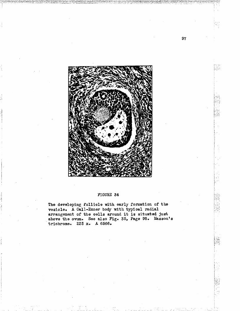
I
¡t .'.
FIGÜRE 54
The devsloping folLicle with earLy fomation of thevosicl.e. A Ca1l-E:çner body wlth ty¡rical radialarrangement of tho oells arouad it is situated justabove the orn¡n. See al-so Ftg. 35r Page 98. Massonrstrichrome " 225 x. .A 6566.
:ir.:i:
97

98
FIGM,E 95
The CaLl-Fxner body shown in Fig. 54.Massonts triohrorne stain. 500 x.
I
ì.i..,..I ,.r-. -.t:-:::-:
I
I
l
I
[:t:!]::ij::ì:l
ï: r. L',ì.i
i.I{-5
i. -:
l':- :,': .
Il
i
i
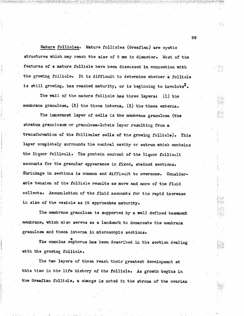
99
}úature foLliolos. tr[ature fot].iotes (Graafian) are oysblo
sbruotures whioh may reaoh the sise of B nm in d,j.ametor. Most of the
features of a mature folllole have been disoussed ln conneoti.on with :i,.,
the growing fo1Lic1o. It is diffioult to determine whether a folliolois sbilr growingr has reaohod naturity, or is beginning to involutez.
The wall- of the mature folriole has three rayersr (r) tire ,.,r,
membrana granulose , (z) tho theoa Í-aterna, (s) the theoa exborna. "'r'il,::..:
Tho irnermost layer of celLe is the mernbrana granulosa (the i:.,':
sbratum granulosum or granulosa-lutein.Layer resulting from a
transformatLon of the fol.lioular oells of tho growing fol.liole). thie
layer oonpleteLy surrounds the oentraL oavity or antrum whioh aontains
the ltguor foLliouLi. The proteiu oontent of.the Liquor folLicurlI
aooor¡nts for the granular appearanee ia fixed, statned, sectionsr ;
:.
Shrinkage in sections ie ooÍ¡rnon and. diffíoult to ovêroottrer Conslder- iab1etensionofthefo11io1eresu1tsasmoreandmoreofthef1uidI
collaote. Aocumrlation of the fluid aooounts for the rapid inoreasel tl;'t.ín size of the veelcle as it approaohes maturlty. i'.:::r:i
. .-:_.;'1.: .:
The menbra.n'a granulosa ie supported by a welL cleflned baesneûb ,,..,,,
rtertbra^u.o, whloh also serves as a lendnark to demaroate the menbraJaa
granulosa and theaa laterna ln mlorosooplo seotlons.
The or¡nuLus o8phorr¡s has been dosoribed in the seotion deaLing i;r.:,lj:;:.:.1
with the growlng fol}Íole.
The tlro layors of theoa reaoh their groatest doveropmont at
thls time in the life history of tho folLiole. As grorvbh begins in
theGraafianfo11io1e,aohengeiEnotedinthestrouagftheovarian

1-O0
cortex 1n the imnod.iate vioínity of the foLtiole. I¡r the earLiost
' etages of developmont of the follic1e tho oonneetive tissue sürona
surroundiag the'folliole is simiLar to the stroma found elsewhere. Ae
d,evelopmoat prooeods¡ two I-ayers €rr,ef,gêo When the foLlÌole reaches
tlne 2 to 5 m stage, the innormost Layer beoomos very vasoular (sthe
porigranulosa vasoular wreathn). tho oelIs of the innermost theoa
- layer aro lsosely ananged. a¡rd form tho theoa Ínterna. Drring the
dovelopment of the folliole those oelLs are very sensitlve to hormonal
gtimrLation a¡d take the appearaûoe of nepithelold.n oells whi.oh are
polyhed.ral in shape and whioir havs oval nuolei a¡rd fino llpoid droplets
1n the o¡rtopl-asnla.
The theoa erterna, howover¡ retains Íts appeara¡.ee of oond.enseê¡
laminated, oonoentrÍoaIly arraqged oonneotive tissue oeLLs whioh aro
fusiform in shapea. The layer has many large blood. vesseLs. ae far as
is hno¡m, the rather oondensed ovaria¡ Ebroma of the theaa e¡lterna
plays no inportant part in the life history of the follicl.el?.
lhe ornrm. The orrum in tho mature folliole reaohes a dia¡netor of
ii :tì-:i...-..i..i-r
120 miora o" rorr2. A thiok structureless mombrane, termed the ' 'ì'-'
zona peLluoida or oõlemar srìrroìlnds the ovun and is closely applled to
tho surfaoe. The large eooentrio nucleus measuros up to ?5 rriora ín
dianetor, has a thiok nuolear membrane, slightly grarrular línin i,.¡,,,.
network, and a large nuoleolus (the nacula germÍ-nativa)Z, Yolk
granulos of dlfferent sizes a¡d of different ooLorrr aro found
soattered throughout the o¡rtopLasm when the ovr¡m is soen in tho fresh
Ebate?. ' i,,.:,..

101
l: : l .':.t. .l ..: ' :-ì
j._.
ï-..-
FIGURE 36
Graafian foLlicle showing signs of atresian Thespace between tho granulosa lutein ceLls and thetheca Lutein ceLls is an artefacto The growingfol-licle in ths upper right hand corner is shownin Fig. 322 page 94. Massonrs trichromo stain'1?5 x.
,Ìi:!r:1.'à
t.t..ia.
I

í
102
Oägenesis. Tho devoLopment of the mature ova from the primord.iaL
sex oells is similar in narry respects to the devolopmøtt of sparmatozoa
. in the testes. Spernatogenesis ls discussed. ln Chaptor 1I.
The primitlvo fenale'sex cslls are the o8gonia, whioh oorrespond.
nto spernatogonia in the nale gonad. 4n oögonium divldes a¡r ind,efinite
number of tineso Eaoh daughter oelI resulting from these nitotic
divielons has 48 ohromosomes, consistÍng of 46 ordinary chromosomes
and a pair of Ï ohromosotoer
EllOögonia givo rise to pri-nary o8oytos by the process of conJugation,
fn drich the ohromosomes arrange thomselves in pairs ar¡d fuse. In the
prooess, the number of ohromosomes i.s reduoed to one half' but here
there ís no reduotion in tho eaount of ohro¡natin material. When
reduoed to ono halfr the nurnber of ahronosomes 1s said to bo haplold¡
tho fr¡II number being dipLoid.. Tho ohromosomes of tho primary o8.oytesIt
divid.e by nrelosls ts prod.uce seoond,ary ooo¡rbeor Fron the divieion of
eaoh prirnary o8oyte, there results t¡m oe1ls of unequal size. These
oeLls have haplold nuolel. By far the greater naes of the o¡rtoplam
go6s to the seoond.ary o3"ytt. The other oeLL is ve4¡ mroh smal.Ler,
ahas the sano a¡nor¡nt of chrourosome material as the ooo¡rbe and nuoh lese
oybopLaon, and, 1s lcxoïm as tho first polar body or polar oelL. The
polar body nay dlvÍde, giving rise to two polar bodies or it utsy
degenerate urithout tho dívision taking p1aoe. All haptoid fo¡nale sex
oells oostain 25 ord.inary chromosomes a¡rd. oae X ohrsmosome.
ItA seoondary oöa¡rte dÍvid.es by neiosis to produoo oira and. smalLer
polar bodles. Durlng the divisions from primary o8oyUe to seeondary
f-.':.

t-
105
FIGT'RE 37
Diagram to ilLustrate the devoloprnent of thenaturs sex ooLls of the nale and fenale.Rsdramo afbsr Bremerr slightty modified.
It.
i:.,:,'', '
S PE RMATOGONIA
OOGONIA
S PERMATOCYTESil
ONE OVUM SPERM ATI DS
SPERMATOZOA7l | *u.,,,
MALEZYGOTEZYGOTE

104
n__ltoöoyte to olumr thers results onl.y one ottrm from one prirnary ooc¡rLe,
and not four Eex oells as in the oase of spornatogenesis.
As in the oase of the male sox eell-sr n¡rch of the process above ,,,,,.,''':_
outlj-ned Ís still theorotiaal and has not besn observed in'man. Many
obsorvati.ons of similar nature havo besn made in lower asimalsr and it
is assume¿l that tne ste cell divisíons are present in human o8genesis. ,,,;,,;1¡,': : .: -::
(cf. Brem"rl9)
tr\rrt_her development of the Graafian follicLes. Follo¡ring the
stage of maturity, the further devolopment of the Graaflan follioles i
foLlows elther of two oourses¡ First¡ tho folliole may mature¡ il
disoharge its ovr¡tr and then booome a corpus lutei¡n. Tho oorpus luteum i
l
is further di.soussed on page . Secondlyr atrosia of the folLiole rnay i
l
ocoufr .the d.etails of wtrioh are givon in the seotion below.
Atresta of the foLlieles. At the time of ovtrlation from a
follicle, oountless numbers sf other Graafia¡ foLlial-es are approaohing ..,::r:i:i"ttt""naturity simultaneousl,y but only rarely does ovulation ocour from rnore i..,, "t..,i-,
than one folLíole. T[hy ono fo!.Iicle s]rould be so aelocted. is not knoumr ',i'i','...
The remainder undergo involution and regresslon, oonnonLy terted
atrosia folLlouLí.
King (quoted by Gardne"ll) lísbs varisus forms of regressíon¡
(a) absorption, (u) atreeia with oystio foruation, (o) atresia r¡ith
proliferation of the sbratum graaulosum and oyetio fornationr and
(d) atresla with proliferation of the stratum granulosum aad no oystÍc
formationr t.:;:;:;,,;::l
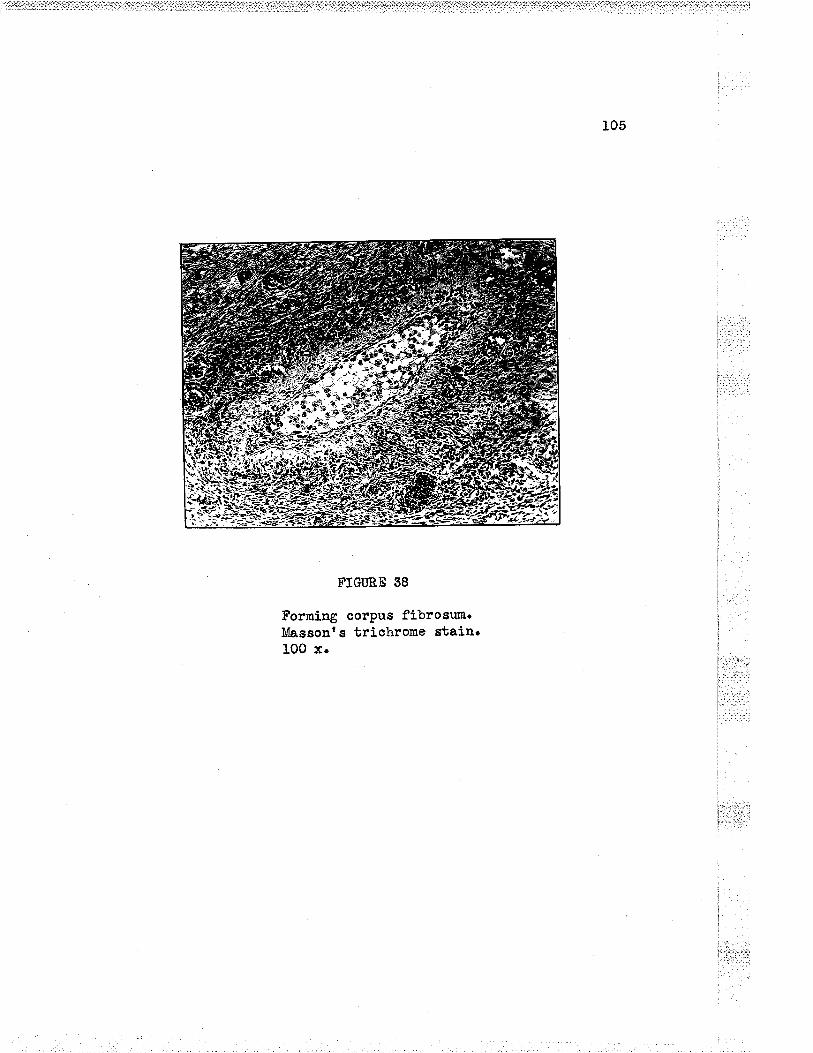
105
FTGURE g8
Forning corpus fibrosum.Massont s triohrome stain.IO0 x"

106
i, . :t.
I:] :
l.:: i,::'
Ì
I
l. :: --.
I
| .'.-rI
FIGURE 69
fibrosun. Massonrs triohrome stainrCorpus100 x. I
¡
t,.I .':¡'lr't:ì: .'

107
The oe1.Ie lining the atreùio folliolos are granulosa oelle.
They are oonsiderabLy snaller than the luteln oel]s a.nd. are somøwh¿t
snaller than tho grenulosa oells of the folliole from ¡rhioh they
originated.. 0ooasionally if a seotion in tbo correct plaae happens to
bo obtained, ths oumulus oäphorus and the degenerating ovurr is seen.
Lining the rssb of the follicle the oells are for¡nd ln layers whioh
vary ln thiohßess from four to eight ooLls thiok. In others, the cells
aro thinnod out to layers of one to two oolle in thiolaoess. The shapo
of the ourmluE o8phorus in older oysts varies greatly a¡d is ofton
illstorted in shape. (cf. caran"rll)
AtreÈic foll-icles are found. in the ovarlog of all normal women
d,uring theÍr aetive reproductivo period of }ife. Sueh follio1es are
found aLso in the ovaries of youqg girls and of womon afber merrop&usoo
In these as woll as in active ovaries, they nust be considered as
arrestod developing foLllcLes. These foLlicles are usually less than
one aentimetre i-n d.ianetãr, but occasionalLy and. probably quite
normally may roaoh larger dimensions, In tine they disappear, loavÍng
as their mark a snall mass of fibrous tissue ¡¡hioh differs in pattern
outllne from the corpus albíoans¡ To thís mass of fibrous ti.ssue is
given the nano corpus fibrosr¡nIz or co"pos candioansl5.

T. TIÍE CORPIIS LTITEI}M
Introductisn. The transitory nature of the oorpus luteum adds
to the diffieulty of the study of this struoture. It is not stati.c,
and ariy desoription must take into account lts continuous and,
reJ-atively rapid transformation from one stage to anotherl3. The
whole pioture is pieced together fron tho study of many ovaries
together with a correlation of olinioal and operative findings.
Roberb Moyer (quoted by NovaÉ4) "r.u
the ffrst to call to
attention that the corpus luteum passes through certain stages
corresponding to phases in the endomotrlum. The corpus luteum under-
:.:-:: _:,
goes ohanges fron day to day14.
The corpus Luteum arlses frori the granulosa cells of the ì
Graafia¡r folIlclo afber the ovnm is discharged from the folLicl-e. lhe ' '
ohanges begin immediatel.y after ovulation. Ïnrnediately after the l
rupturo of the Graafian follicle of course there is no d.emonstrabLe
ahango of the granulosa celLs. Soon howover, the waLls aoLlapse and. ,.,,t,,,;,,-:r ::;r
the earLy corpus luteum is a thin wallod oollapsed struoture¡ Tho ,.r..i::.:
thin maLi-s are moderatery undulating and havo a grayish yellovr oolour ":":'1':"'
instead of the brilliant yel.Iow of the lator stages' At thÍs stage j
thestructureiseasi1yoverIooked;itísinconspiguousanditsI'1 i'-i 'ri;:"
discovery is largely accídentaII4. The cormon descríption of the '': '"
bríght yellow thick layer with a wavJr outline is tho structure of the
cofpusluteumseenon1yatoertainofthe1aterstages.
Reforence to the influence of conception upon the development
!.1 ..:. 1
109
a¡d history of the corpus luterua has been made on page . If
oonceptlon hae not ooourredr the corpus luteum begins forthwith to
show degenerative and regressÍve stages prior to the onset of the
menstrual flor¡v' rf oonception has ocourred, thsn the corpus luteum
oontinues to grow and funotion until the pregnancy is defínitely
eetablished. .ê.fbor 5o to 60 days it then begins to show signs of
regression. ïn either case, conceptioïl or no conceptíon, ths
ultinate outcomo is fibrosís to form tho Gorpus albicans whioh is a
f¡¡notionless ma6s of fibrous tissue. rn time, the corpus arbioa¡rs
itself disappears.
That the oorpus. luteum is indíspensable to monses was pointed
out by lrTovatl4 in 1916. During the mmstruaL life of a norrraL woman,
LuteÍn tiesue in some stage or another is.always to be found in the
ovarios. 0ß the other hand, in non-menstruating women, eorpora lutea
are absent. Nor are oorpora lutea found in the ovaríes of the fetus¡
aw¡ born, or girl before puberty. For a nuuiber of months af.ter the
last menstruaL flovr at monopause, regressive corpora lutea are found
in different stages. The amonnorrhoea of lactation is explained by
other hormonal factors suppressing the aetion of the corpora Lutea.
(cr. tqov*<l4)
Four stages in the history of the oorpus l-uteum of meastruation
have been d.escribed. The terms are mostly due to Meyer (Aroh. f.llclmät., 19L1, 93rõ54--quoted, by ivovaÉ4)¡
L. Stage of proliferatÍon or h¡rperemia
2, Stage of vascr¡larization
i::ì.è11Ii::: l:: r.
i:,'i.ir.
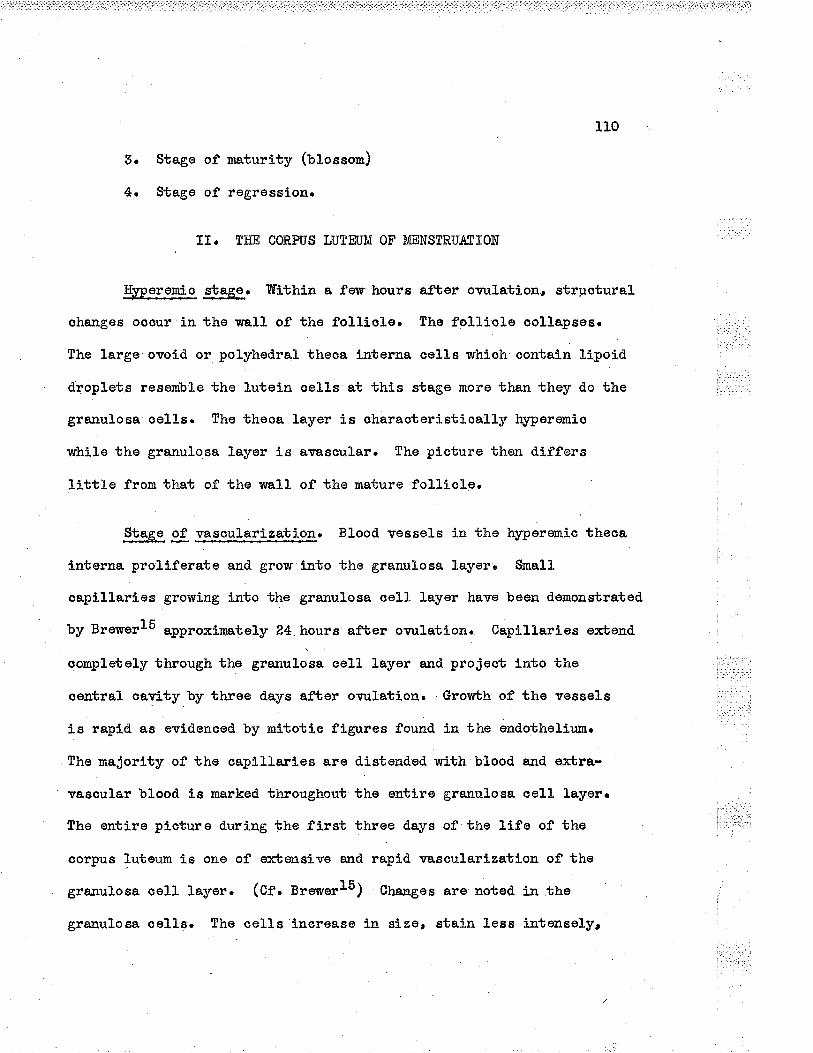
11.0
3. Stage of rnaturity (uJ.osson)
4. Stage of regressiorl.
1I. THE CORPIIS LÜÎEUM OF ME"NSTRTIATION
Hlfpere-nic stage. Trfithln a fer¡r- hours after ormlation, strpoturaS.
ehanges ocour in the wal1 of the folliole. the folliale oollapses.
The trarge ovoid or polyhedrat theoa interna celIs whioh oontain lipoid
droplets resemble the lutein oells at thÍs stage more than they do the
granulosa ceLls. The theoa layer is oharacteristicall-y Ìr¡rperemio
while the granulosa layer j.s avascular. The pioture thon differs
little from that of the uralL of the mature folliale.
Stage of v.ascularization. Bl.ood vessels in the h¡4geremic theca
interna proliferate and grow into the granulooa layer. Snall
oaplllarios growíng into the grarrulosa oell layer have been demonstrated
by BrewertS "pp"o*inately
24 hours after ormlation. Capillarios exbond.
oonplertely through the granuLosa celt layer and projeot lnto the
oentraL cavity by throe days af'ter ovrrlation. Grov'¡th of the vessel-s
i.s rapld. as evidenaed by mitotÍe figures found in the endothelÍr¡m.
The nrajority of the oapillaries are distonded nrith blood a¡d e¡rbra-
vascular blood is marked. throughout the entire granulosa oeII layoro
The entire picturo during the first throe days of the Life of the
oorpus luteum is one of e¡rbensÍve and rapid vascularization of the
grarrulosa ceIl- layer. (Cf. Breru""l5) Changes are noted, in the
granulosa oelIs. The cells íncrease in size, stain less j:etenselyt

tlL
have a reLatÍvoly snall nuoleus and ofben a vaouolated appearârroeo
Flve days after ovulation' tho oapillaries are tortuous and.
arranged so that nost of the granulosa cells are i-a contact with a
vessel. thero ie an ingrorrubh of sonnectlve tlssue along with the
oapillaries v¡ùrich project into the central cavity. Eemorrhage into
the cavity ooours from these vesseLs' Drring the noxt-few days there
forms a sort of lining separating the granulosa oells from the centraL
cavity.
ffihile tho lutoin cells are d.eveloping from the granulosa cells¡
the cells of tho theca interna are rogrossing. With a loss of l-ipoid.s
a¡d a d.ecrease in size, theSr take the appears;noe of ordinary oonneative
tj.ssus oell-s. Trabecul.ae, whioh are wed.ge shaped, grow into the
granulosa-lutein leyer to produce the femÍ liar r.¡nduLating *pp*""*r"á
of the corpus luteum. The corpus luteum now has a bright ye1J.ow
colour, wavy outline and. høorrhagio centre.
Acoording to Brerlrerl5, during the period of eight to ten days
after ovr¡Làtionr there is gradual dlminution of the h¡rperemia in the
granulosa-lutein layor, the oapillarios beeoning straighter a¡d
narrofl'er' with litt1e or no erbravasated blood. fn the oonneotive
tissue oore, howeverr the vessels are dilated. and filred rvlth blood.
Âocording to the sa.me author, afber ten days, there is little or no
blood. ín the granulosa-lutoin Layer. The vessels are mostLy collapsed
a¡rd the conneotive tissue around them increâsoso (cf. Bru*"rl5)
the graamLosa cells beoome larger 'and. acquire a more functional
a¡rd active appearance as the vasoularity of the granulosa-lutein layer
i -.'. :...
1.....,,
': j : i|l-l:-:.:: '
I
i::::.',1

tlz
i'i:;.
i"
I.:I'l.:
FTGIIRE 40
Corpus luteum of menses in the ovary of a younglÍ'onârrc the tissue has boon tora in the left-hand side. Eomatoxylin and eosin stain. G 4566'10 x.

113
l::
i
I.t..i,t,r
FIGURE 41
Eigher power field of the sane oorpusluteum as seen in Fig. 40. 100 x.
!'.,.'i:_:t-:.i
i...
ì:::-:.'ì:ì
l
1
I
I.'.ir -.-r, .i. .r.
¡_t-.
i
¡._.: l:I ::; .::'t:.- ...r1:: l1 -:ii

114
inoreases. AlL cells do not develop or rogross simultanoously' so that
the prooesses are patchy. "At sevon to eight days after ornrlationr
somo aelLs contain small lipoid droplets. The greatest inorease in
LÌpoid content is observed in the cells along the bord.er of the cavity.
(cf. trr"*"rl5)
At the end of ths vascularization stage at eight to nine d.ays
after ovulation, some of the granulosa-lutoin oells begin to degenerate.
ths oe!.Is showi-ng dogeneraåive ohanges have a large anount of cLear
o¡rtopS.asur and pyhrotia nucLei. At the end of elevon days the f-ipoid
d.roplots are larger, and there are extensive zonos of necrosis.
'Stage óf naturity. tr[ature corpora lutea nay be large structures
rreasuriag up to L.5 om 1n d,ianoter and. l-arger. The oentral coro is ,
:
¡
delinited by the surround.i:rg lutein tiszue. Fl-uid which is either i
ì
cloar or b3.ood.¡i' may be found in the central space' which is also very
variable in size. The cavity may be small due to the presence of a :
I
great doal of connectivo tissue. The folded bright yoL1ow Lrltein ia',
tissue varles groatly i:r thiohness and. may measuro up to 5 mm in ,,.,
thiobness. The oorpus lutoum is an snormous body when its size is 't''
oornpared. to ths.t of the prlnary folliole.
0ffshoots from the theca interna invade tho band of grenulosa- ,,,,,
lutein oe!.ls aad carry with thør nerry blood vessels. The theca oells i¡ Ììi.
the septa often becomo epitheloid Í-n appearance and. often shov'¡ an
alveolar """r"r*.ruo*. lhese are the paralutein aeLls of Finto (fgOS).
Thelr ondoorine funotion, if anyr is not klov,mr
' !.: :.:
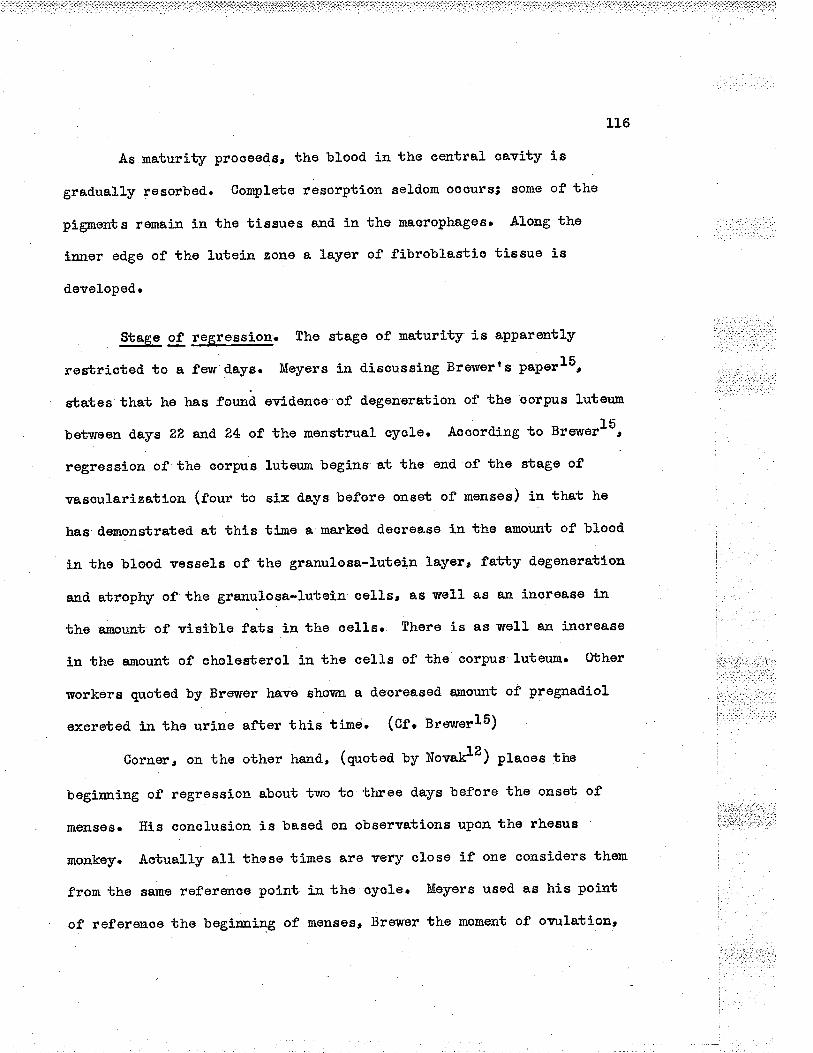
L16
As maturity proceeds, the bl-ood in the eentral oavity is
gradually resorbed.. Completo resorption seld.om occufsi eome of the
pignents remain in the tissues and in the macrophages. Along the
inner edge of tho Luteilr. zone a layer of fibroblastia tÍssue is
d.eveloped.
Stage of regression. the stage of maturity is apparontLy
restriated to a fevr days. Meyers in diseuseing Brewerts paperls,
sbates that he has for.æä ovidence of dogeueratÍon of the corpus Luterrm
betwoen ð.ays 22 and 24 of the monstrual eycle. Aocord'ing to BrsvuerlS,
regrossion of the cofpus luteum begins at the ond of the stage of
vasaularization (four to six d,ays before onset of menses) in ttat tre
has demonstrated at this tÍ-me a marked decrease in tho amor¡nt of blood
in the bloo¿ vessels of the granulosa-lutein layer, fatty degeneration
a,nd. atrophy of the grarruLosa-Lutoin eollsr as weLL as an inorease ín
the arnount of visÍble fats in the oel-ls. There is as weLL an increase
ln the e¡rount of cholesterol in the ceLls of the aorpus 1uteum. Ûbher
workere quoted by Brevrer have shovu¡¡. a d.eoreasod anount of pregnadiol.
exeroted. ln tho urine afber this time. (Cf. ¡reu¡erl5)
Cornerr on the other haxd, (quoted ty NovaÈz) places the
beginning of regression about two to three d.ays bofore the onsst of
tllgltses¡ His conclusion is based on observations upon the rhesuE
monkey. Aotually aL1 these times are very close lf one considers them
from the sa¡ne referenco polnt in the oyole. Meyers usod as his point
of referenoe the beginning of menses, Brewer the moment of ornrlation,

tr"7
FTGÜRE 4õ
Dogenerating eorpus luteum. The granulosalutoin layer shotvs some hyalinization*l,lasscnes trichrome" 50 x'

liB
FTGURE 44
Maerophagee fiLled with lipoid materiaL ln edegenerating oorpus Lu.teumu Stained rvithSudan III The dyo appoars blaek in the photo-graph" 400 x.

119
ând Corner aJ-so the begirrning of rûerr.sêsa If a.II the s'catecL tirnes are
e4pressed in the se:ne terms, they fal1 within two to three days of each
other "
The degeneration of the granulosa*Lr.rtein la;rer is a slow and
. gradual p"o""""17" îhe transfornation noed. not progres,s uniforrnly
at all points of tho ïratll8" Tn tìe early phases of regrossion, the
process is revorsible, should pregnancy oocurn Regression is not
sudden in onset and begins at the onset of the menstrual flov¡,
T¡4pically not all cells are involved,. fmmodiately prior to the onset
of mensesr lllanlr histological variations are encountered in the granulosa-
lutein layer. Uniforn d.egeneration rnay occasionalL;v involve all the
ceL1s. Degoneration ma¡r occut' in tocalized groups of celLs, or
d.egeneration rnay be minimal and scarcely evidont" Variations withín
the sa¡ne corpus luteum may ooour, (Cfo Brewor and Joneslz)
Ïn rogression, there is an increase in the anount of lipoids to
be found in the celLs" This increase of lipoids is the basj-s of
conrparison with the adrenal cortex" Tho comparison is applicable only
to the older corpora lutea and is not applicable to the early Gorporaa
Only old corpora lutea undergoing regression have a docided yellow
coloorl8o Not a}l species ha.ve a yellow corpus luteurn" Tho yellow
colour has been tra.ced. to carotín found. in the dietl0'
Fi'brosis increases and. oon'binues until the r¡rho1e structure is
sonverted into a mass of co¡rnective tissue, the lutein layer shows
also sone hyalinization so that the oontour of the layer is easily
reoognized" The ye).low colour may persist for so¡ne tiine but after raany

L20
FTGURE 45
Corpus albÍcans. Only part of the structure ísineluded' I{assonçs triohrome stain, 50 x,

123
rreekse the familiar white corpus abl-icans is rçell established, Tn
time, the corpus a.lbicans disappears to leave no tracell'
TIÏ. THE CORP-IJS LUTEUI{ OF PREGNAI\CY
Material for the study of the corpus luteum in all phases of
pregnancy is difficult to obtainu Feur roports are availableo The
account which foltovrs is mostly frorn the paper of Gillman and Steinl6.
Although thore are v'ríde indiviclual varíations, a grovrblr pattern
is foll.owed" -{fter conception" t}re corpus lutoum increases slowly in
si,ze" The oarly increase in size is due mostly to the formatíon of a
relativeLy large cavity oontaining flui<i within the centre of the
structuro" .åccording to Gillman and Steinl6, th" cawity increaseÊ. ,
slowly in size until the 50th d,ay of gestation, after which it enlarges
rapidly until the 60th day, and from then on it decreases rapidJ.y in
sizeu the decreaso in size is due to a decrease in the size of the
cavity which oontinues to docrease until about the f,ifth month when
obliteration is oompletou The amount of lutein tissue remÊ.ins fairly
oonstant throughout the pregnanolo
Septa fron the theca continuo to produce a folded appearanae of
the granu.losa-lutein layer. Betv¡een the septa are reticular fibres
which support the glandular cells and the fine capill-aries, The thecal
layer, so prorninent in the ear15. months of pregnanayo gradually
dininishes to completely disappear by the fifth month,
Dilated bloocì vesseLs and lynrphatiss are proroinent in the
capsule of the early oorpus lutoumu Fibrous tissue lines the central

129
FTGURE 46
The corpus luteum of pregnancy a.t three months.From the ovary of a t-r,venty-nine-¡rear-oLd vronÐ:rl
lvho died following air embolisn, resuliing frorna va.ginal douche with soapy solution*

IZ3
cavity and. consists of wídely separated yoìrng fibroblasts in a faintly
staining matríx'
The cells of both the granulosa-Lutein and theca-lutoin layers
have incLusions, some of which are colunon to both t¡rpes of oel1s,
¡¡hile other incl-usions are formd in onl¡' the granulosa-lu-tein sellsu
0f these ínolusions¡ the lipoid.s have received much attention'
The tipoids were first described by I'ieyer ín 1911" Gillman and. Steinl6
find that the number of fat containing oelIs is greater at the
beginning of pregnanoy than at term, although fat is found in some
cells throughout the whole of tho gestation. Generally the lipoid. is
for¡nd in the form of small globulos" Some may fu,se to form larger
ïtâssoso CholesteroL is fou:rd in relatively smalI amounts and is
characteristically scarce in the corpus luteum of pregnanay"
Searetory granules which stain red after Malloryr s stain are
found. in approxinately 50% of cslls in the early months" The granules
are not speoific for pregnancy and are also found in the corpus luteum
just before the onset ,of mqtseso
Gotloid droplets staining v¡tth MaLlor;rt s stain are oeIl
inolusions which are Iabile ln natttro and disappear quickly after
death" They are seldom seen in autopsy material unless they have
undergone degeneration and have become calcified" These bodies are
found. only in the _corpus luteum of pregnanoy" Somo of the oolloid
bodies have vacu.ol-os"'
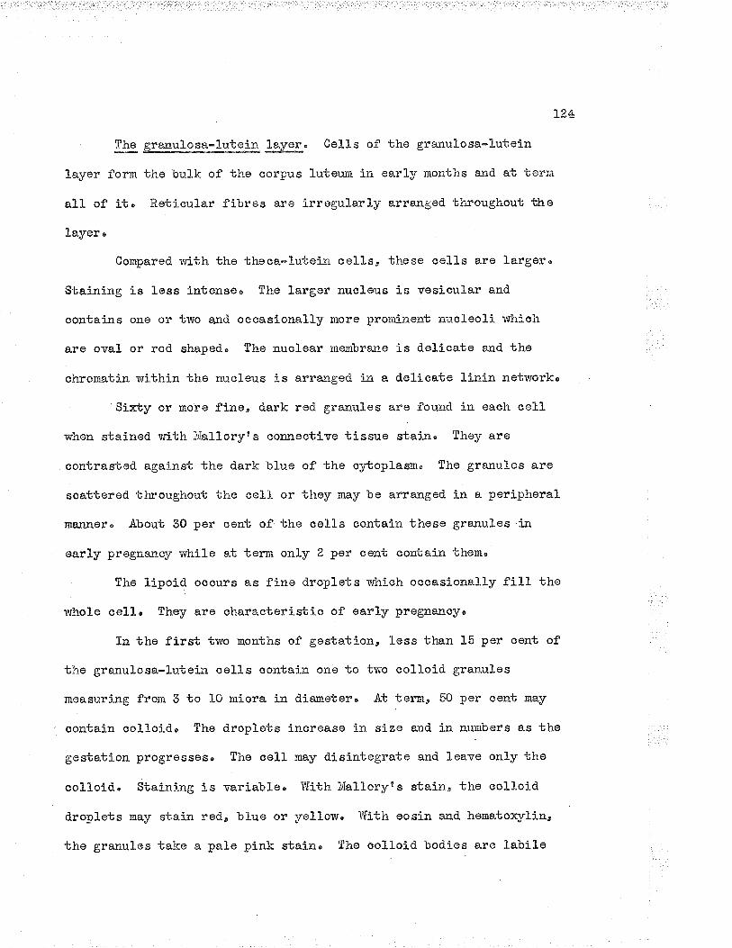
]"'24
Thg granulo1a_*_Lutein leyer' 0o11s of the granulosa*l-utein
Layer form the bulk of the corpus luteum in earl;r months and at i;errn
all of it' Reticular fibres aro irregularly arranged throughout tho
Layer è
Compared with ths theca-lutein cel1s, these cel-ls are largeru
Staining is less intensoø The largor nucleus is vosicul-ar and
contai¡rs one or two and ocoasionally moro prominent nucLeolí v'rhich
are oval- or rod shaped" The nuclear membrane is delicate and the
chromatin within the nucleus is arranged in a delicate linin networko
Sixby or more fine, dark rod granules are fou-nd in each celL
rrhen stained v,rith l{alloryt s connective tissue staj:r. They e.re
contrasted against the dark blue of the c¡rtoplasm" The granules are
soattered throughout the cell or they nay be arra.:aged in a peripheral
maJxnor. About 3O per cont of the oolls contaín those granules i:e
earLy pregnarlqf lrhile at ternr only 2 per cont contaj-n them'
The lipoid ocours as fine droplets which occasi.onally fill the
v¡hole cell' They are characteristic of early pr€gnanoyo
In the first two months of gestation, less than L5 per cent of
the granuLosa-lu.tei¡ oells contain one to tr¡rc coll-oid granules
measuring from 3 to 1O miora in dia¡retero At termo 5O per oont may
contain oolloid, The dropl-ets increase in size end in numbers as the
gestation progresseso The cej.l rnay disintegrate and leave only the
colloiri. Staioirrg is variableo T¡ith ivfalloryts stain, tho aolloÍd
droplets may stain red., blue or yeIlow' TJith eosin and henatorylin¿
the granules take a pale pink stain" Tho oolloid bodies are labile

L25
a¡1d. are seldom found. in au.topsy material" 1[o co]-l-oid bodies are fou.nd
in the theoa cells'
VacuoLes in the form of round colourless spaces in the granulosa-
lutsin cells are most numerous in early stages, especially ln the cells
bordering the central oavity' The vaouoles graduai.ly onlarge to fill
the whole ceII' Specimens from the latter half of pregnancy have
fewer vaouoles" The contents of the vacuoles become progressively more
stainable and form the oolloid droplets" Vaonoles are not found in the
theca-lutein cells'
Chromictial substance in the form of filanents, triangular or
club shaped boclies in the c¡rtoplasnr are found in the cells of both tho
granulosa-lutein anci theca-lutein layers' It is most abunclant in the
early months of gestation. ,{ progressive decrease in the anount of
ohromidial substa.nce is sesn throughout prognancyo Much of this work
of Gil]nra¡a and Stein awaits confirmation' (Cf' Cittmen arrd Steinl6")
The theca-l_utein layero The reticular fibres of the theca-
Lutein form a regular fine network and support regular groups of ceLls'
The colls are closely asSociated. with bl-ood vesselso The cells are
smaLlu polyhedral and measure 1.5 miora in dia-neter, 'Tho round nucleus
averages 7 mícra in d.iameter" l¡Iithin the nucLeuss a single large round
nucleolug is found" Conpared with the nucleus of the granulosa-lutein
cells¡ the nueleus is darker" The granules found in the theca-lutein
layer are slightl). larger than those of the granulosa-luteín layer and
average 2 micra in diameter" 0n1y a few are found in oaoh ceLl" The

126
1-ipoid granules are reLatively large and few are fou.:ad in each cell"
The colioíd bodies are absento
IV" TFIE TÐSTES ¡ND OVARTES CO¡'{P.ARED
Until the 17 nrn sbago (seven weeks) of development is reaohed.,
the ovaries and tostes have similar ernbryotogical histories. In each
oase the gonads arise as'thickenings of coelomic opitheliumn Later
ln deveLopmente the germ cells arise as part of the sox cords which
are developed from the ccelon,ic epithelium" Both the male and female
gonad.s descend. during d.evelopment, the d.escent being most marked. in
the case of the ilaleE The male gonads must be in an enwiron¡tent
slightly cooler than body temperature in order tha"t sperrnatozoe- may be
produ-ced, During development a complete excretory system is developed.
to convey the countless numbers of spermatozoa to ths extorior" 0n
the other hando tho fenale has what seerns to be a rather procarious
and incomplete duct system'
Development of the germ cells in the testes and ovaries is
essentiafly the sB.IIIêo Both ova and spermat.ozoa are haploid cells as
a result of nitotic and later the meiotic cel1 divisíon" One half of
all spermatozoa have 23 ordinary chromosomes prus an X chromosome3
the other half have ?3 ordinary chromosomes plus a Y chromosome' All
ova have 25 orclinar¡r chromosomes plus an X chromosome (never a Y
ohromosome), From one primary spermatoc¡rbe, four spernatozoa develop.
From one prinrary o8oyte, only one ovum develops (the one half of the
chromosonre materiat going to the polar bodies)o 0n the one hand,

L?,7
sperma.tozoa are produced by the thousands and with no apparent eoonomya
whÍle on the other hand ova are produced one at a tine" Several ova
may be developingr of coursou at the seme time, but only one i.s
erLru.ded as a ruLe'
Structurally the ovar)¡ is more complex than the testis. fn the
ovâr¡rs there are more variable c.omponents v¡hich unclergo cyclic changes
that may occur from day to day. The testis has interstitial oells
v¡hioh are not found in the ovaryo Senile changes occur in both malo
srd female gonads, the changes being more pronounced in the case of
the ovaries and occurring at aÌ1 earlier age as a ru-Le.
The testes produco more androgens than oestrogens, while tho
ovaries produce moro oestrogens than androgeîso The difference is
quantitative rather than qualitative' It ís interesting to note that
the ovaries, t.ostes and adrenal-s all begin their development in
approximatel.y the same part of the body and all produce in varying
amounts the steroici compounds which influonce the sex pattern of the
individual. The ovaríes havo one funetion for vrhlch the testes have
no counterpart in the produ.otion of progesteronen Both organs are
influenced by the hor¡nones of the anterior ]obe of pituitary as weLl
as by hormones of other endocrine organ.sø
V. THE HORIIOI{ES OF 1}1E OVARÎES
The hormones of the female and male gonads are discu-ssed in
Chapter V" in v¡hich the hormones of reproduction are considered,

128
B]BL]OGR¿PTiY
1" Svrezyr 0", and H" iú' Ee6Lns, the Euman Ovarian Germ CeIIs"J" iúonph" and Physiol', 1950, 42,t543"
?" tlaxinovr, A. Au,. errd 1,¡&n" Bloom, A Texbboolr of i{istolog¡" Fifthedition; Phila"¿ Tf" B' Saunders Compaay, 1948" 700 pp.
3u v' Hanulsemann, Quoted by L" Aschoff ' Chapter XT" t-feibl-icherGeschleotsapparat, Pathologische ånatomie" Edíted by Ludwig.Asohoff . Sechste "{ufLage" Zweite Bd., Spez" Tei1e, 1923"Gu-stav Fischer, Jenao¡ ppo 1050.
4u Pratt¡ J. P"s In Discussion of the Paper of Sch'¡rarz of aI"
5" Sohwarzo O' II'e C' C. Youngr an<i J" C. Orouse, Ovogenesj-s i¡r theÄdult Hunran Ovary. -Amero J' Obst' & G¡m. t I949t 58-t521'
6, Simkins, C" S"e Development of tÌro I{uman Ovary fro¡n Birth toSoxual Ma.turity' "Amor" J. Anat", L932u 514465"
7, Grayrs Anatomy, editod by T" B" Johnston end J. i\ihilLi.s"Twenty-ninth odition; Londone Longmans, Greon and Co", L947 "L597 pp,
B" Ðelson,8., S' Lubin, and S" Ru ilin Re¡molds, Vascr-iLar Patterns inthe Hurrtan Ovary" Amer. J" 0bstn & G¡zr", 1949, 5'7s842"
TE9' Ivluller" L. R;, Lobensnorven und Lebonstreibe" Dbritts "Auflage"1931" Julius Springer" BerLin. 99i. pp"
10" Corner" G. ï'tr,, Gybology of the Ovum, Ovary and Fallopian TubenSection ,trilX, Speoial Cybology, Vol, III, pp, 1838,Edited by E, V. Cowdry, Seoond Edition, Nevr YorkePauI B" Eoeber fncorporated., 1952"
11 " Gard.ner, Wn S,o i\Tornal and Pathological Developments from thoCeLl-s Lining the Graafian FoLlicle, So G. 0u, 1941, 7?¿129"
12, Novak" Eu, G¡mecological and 0bstotrical Pathology, Second ed.ition,Phila.¡ 1"f. B" Saunders Co"u L947. 570 pp,
L5" Pratt, Ju Pu" The Iluma¡r Corpus Lutor:m" ArcÌr.
]4, Novak¡ 8", The Corpus Lutsumn Ïts Life C)rcloI4enstrual Disorders, Ju Au M. -4,, 1916,
Path"e 1935,19¡580,
and its Role in6. r1285"

t29
15" Brorner, J" f ", Stud.ies of the Eunan Corpus Luteum' iìvidence forEarfy Onset of Regression of the Corpus Lu.ter.im. ofMenstruation' Aner" J. Obst" & G¡m" s I942t 44-ILO4B.
16o Gil-lmen, Ju¡ a.nd II. Bo Stoin¡ The Hurnan Corpus Luteun of Pregnancy"S. G. Oue L94Lt 72-tI29"
l7o Breïtrers Ju I , and Ho O. Jones, Studies on the Humau Corpus Lutoum'I{rstologic Variations in Corpora Lutea and in Corpus Luteum"Endonotrial Relationships at Onset of lüor¡aal lfenstruation.A¡ner" J. Obst" & G¡m" ' !947, !å:561'
]B' Allen 8", J' P" Pratt, 0,' U. Nevrol1, and L' J" Bland., Human Tuba1Ova; Related Early Corpora Lutea and Uterine TubesuContrib' Enbryol" Carnegie Inst', No. L27E Yo7-' 22' 1950'
19" Brener, J" L,, Textbook of i{isbologys Si:rbh edition, re-trrittea byH" L, ûtreatherford, Phila. ¡ The Blakision Company, r.944,723 pp@
ZOu Harniltonr'i\fo J.¿ J. Do Boyd, and H" itrf" lliossrnan, I{uman Enrbryo}ogyeCanrbridgel irü. Heffer and Sons Limi'bedo L944"

CHAPTER FOUR
1IÐ PLACENT"&

CTTAPTM. IV
Tm PLACET\TTA
'lThile nutrition of the fetus and remova] of its motabolitos are
functions of tho plaaenta which have been recognizod for a long tine,
the production of hormones has been proved only within reoont goe.TSo
The, presonce of oostrogenio substanoes in tho placenta was domonstratod
oarlyl8; howovor, their presence was attributed to storage of the
hornones by tho placenta and not to the production of them in the
orga,Bo In the early 1930?s" Collip produced crude activo erbracts
which he believed. contained. hormones prod.uced by the plaoenta"
Subsequently other investigators produced crystalline compounds and
within reoent years hormonos have been produced by placental tissuo
growing in tissuo cultureszLÐ 22.
Material forLhe stud.y of inrplantation and nidation of the
enbryo is diffioult to obtainu Early studies of these prooosses were
made on l-aboratory animals" Grosser8 revie$rs sorno of the oarly history
and points out that fowl and rabbits were the first animals to be
studied, Theso animals have features which are siinilar to eaoh othor
and whicÌr aro not found in other speci.es. The deductions based on ths
studies of these and. other a¡rimals rvere used to bridge the wide gaps
in our beowledge of early human and prinate development, Graduali.y the
gaps havo been narrowed by further studies of human matorial"
The oarl-y development of the plaoenta is íntimatel¡r associated
with the development of the ornrm, although ovulation, fertilization and

L32,
migrâtion of the ovlrm occur before implantation which is the iniiiel
stage of placontation' The entire life history of the pla.centa may be
divided as follows:
(") Inplantation (nide.tion) "
{¡) Placentation"
(") Grorirbh and. maturity of the placenta"
(¿) Expulsion of the Placentan
Rock and llertigl have tracsd hunan d.evelo¡rnent during the first
two weeks" The first part of the discussíon in this Chapter is found
mostly in their roporte
å" zygote results from the union of a.n ovum and. a sperntatozooÍ!.o
The term zygote has been used very little in recent literature and the
term ovr-rm is used to desoribe the newly forned organism up to the
fourieenth day' The tern enbryo denotes the organisia up to the end. of
the seconcl month, and from this tirne on, the torn fetus is used' fn
thi-s diseussion, the teriu ovum is used instead of zygote" /
l-"gl"pqr4t before iqgle+t.a!i_o+" The mature ov'¿rn is fertilized,
traverses the ovidu.cts and undergoes segmontation into at least t-r¡¡o
blastomeres duriug the first twenty-fo'¿r houl's after ovulation,
according to the deductions of Rock ancl Hertigl' By the third d.ay,
ths ovum is in the uterine cawityu The eight oo11 stage is reached by
the fourth day"
Developrnent bet'ween days 5 and 7 has not
human and deduotions are based on observations
beon observed in the
.1on the monkey*" In this

l'timiriuemesoblast
Embruonic Sur[aceEntoderm diíc eþith:elium Amnion
Cellulurtr oþltoblast
Endometrial gland Lacuna uìlh nlaternol Slrontablood c¿r1ls
Coiledarl crt!
Photomicro,qrâph of a section through the j.¡.,rplantation site Õf the el_even-da.-r¡ -4st¿i,{-Rock ovl¡m, masnified 140 dismeter,s" The bull< of the orruniconsists of irreSlr,la-r me.,sses of trophobla.st (s¡mcytium) -,l,,hicir is invadingthe endomet::ium. within. the sync¡ltiel t-r:ophoblast is the cel-lLÌlâr tr.opho-b-}"cst., thc ee1l s of whrch âre arrs.nged as sinple epithe)-ium except a.t C.The cel-lu.la:: trophobla,st Ím':lediately su.:.rou-r.as tne pr"initive chorionicmesobl-ast in rvhich the embryo is suspen.ded, (¡fter I{ertig and Rock,carnegie cnnt:'ib. to Emb:'vor,¡ v" zgå l-g<!1, Fì-g. 14) Reproduced f::om 't.fi.Tertbook of Histolo¡,-¡rr'! by A" ./i. ir{e.xirnoï.ï r,nd,,,'.1*" Bloo*" ¡,irtr, edition,p" 572, with uritten Ðermission from'che publ-i.sherse The W* B" Sa.under.sCompe.nye Phila. ô

L34
animal , during the periocl botweon days 3 and tíe a ¡m:ltisollular morula
develops and it acquires a contra] sogmentation cavity and so becomes
a blastocyst " At one pole, a small ma6s of colls ronaÍns grouped,
forming the irurer aell mass which gives rise to the errbryo proporn The
other cells spread out to form the oyst wall and give rise to ths
trophoblast and atlied. structuros'
lnplan-tatio¡tr" Tho cawity of the blastocysb is probably fornned
d,uring the fourth or fifth dayr following which there is differentiation
of three kinds of cells; the trophoblast on the wall of the blastooyst,
the ectoderrn erld endoderra of the embryo, Contact with the endometrium
occurs about the sirbh day' The segmentation cawity oollapses as it
does in the monkey" rlThe newly clifferontiated syacybial trophoblast
at i;he einbryonic pole engrossos some of the superficial opithelitrm and
Streeter puts it rrforagesnt its way into tiris tissuo.tt (Rook and't
I{ertig')
The ovum becomes embodded in the end.ometrial stroma and. this
prooess is laaown as interstitial implantation. In the monkey, at
day 9' tho bl-astocyst attaches itself to the endometrium and bogins to
inplant" The development in the monkey is not so rapid as in the
hrunann There is .intense edema e-:rd congestion of the q:.dómetrium Ín
the neighbourhood. of the orr*2u
At 11 days¡ the ovu¡r is a slightly raised pale ar€âe a little
less than 1 run iu dianetoro ft is translucent and is surrounded by
co:rgested endometrium2u By this time the ovum is buried in the
ondometriumlu

Placentation. Implantation
155
represents the initiai stage of the
process of pS.acentationz o
In the nine-d.ay-old specimen doscribed by Roclc and Hertigf,
it is illustrated as being armost entirely surrounded by endometrium"
Tho segmentation cavity is redistended and the ovllrre consisting of
ectoderm arrd endodenn, is found within it" the enlarging amniotic
cavity is bounded dorsally by the early a¡nnion ùhich is a product of
the adjacenb c¡rtotrophoblast' The mass of synoyLiu.m has i.ncreasod six
times in amounto Lacunae havo forned in the syoctial masse most of
¡¡hiah, if not all" conmunicate to form a ríetwork of canalsn In thelr
specitnen, one lacuna contains maternal bIood, forrning the first stage
of tho placental ci.rculation" (Cf, Rook and Hert:.gl)
By tvrelve dayse tho ovum is 1 ¡nm in diameter and is entirely
embedded in the endometriun although the site of entry is nôt quite
€overedo The embryo itself measures Ool r¡n in dia:netern It consists
of a pyriform plate of pseucLostratified ectoderrnal celLs and a thin
i-ayer of ondodermal cells" The mesodorra has not yet apÞeared, Tho
anniotÍc saa is a thin-¡,mIIed oyst on the dorsal surface bounded by
amnion and eotod.erm" The ombryo lies in a cavity which is five tjmes
its size and which is filled with fluid' å. Lacewor.k of mesoblasts
srrbdivíd.es tho erbraembryonio cavity into numerous spaces, one large
and many sma.lI'
Trophoblast surrou.nds the whole oentral space that contains the
enibryo, The trophoblast exhibits two layers, an inner o¡rtotropho-
bLastic layer and an outer s¡mcybiotrophoblastio layoru The

136
cytotrophoblast surrounds and gi-øes rise to the prirnitivo mssoblastic
tissue. The s¡neoybiotrophoblast hae made contact r,vith tho endometrium
and has t8engrossed and digested. the materiral stroma in v'¡hich it grows.&
A portion of a thin--waLled sinus has been dissolved" ivlaternal blood
has filled the fused Lacunae vrithln tho s¡¡ncybium. At this stage,
the ratio of syncytiotrophoblast to cybotrophoblast is about three to
one, a relationship which is soon lost" (Cf, Rock ana Hertigl)
By the fourteonth day, -the csrtotrophob1ast has grovrn rapidly
and by this time is arran¡ged in massos of diserete aells that projeot
into the synaybiuml" The-se are the primary villiz which have no
mesodermaf core' Exoept at the zone of contact wiih the endometrial
stroma, the s¡¡ncyLium forms now only a thin layer lining tho Laounaeu
The greater a¡nount of trophobLast is made up of aybotrophoblast and
a corrospondingly Less amount of foraging syncybiotrophoblast"
Exûensions from the mesobl-ast project into the primary villi forming
tho supporting structuro of the villi whioh are haonrn as socondary in
t¡pe' Maternal blood is found in the laeunar spâcoso The wholo ovr:m
now measures from 2 to õ ¡rm in diamoter in the fixed state, (Cf . Rock
and Hertigl)
Grgv¡Þh and maturíty of the placoglg. By the fifteenth or
sixbeenth daya secondary vilti have appeared and these oontain a cors
of erLraembryonic mesoderm in which a plexu-s of enrbryoni c vessels soon
d.evelops. The core of mesoderm is iruned.iately surrounded. by a layer of
cybotrophoblast which in turn is covered with a layer of

L37
srncytiotrophobLaste Tho tips of the secondary villi continue to
ertend a-s colui:ns of cybotrophobla.st" lviesoderrn gradually erbend.s into
these tips" The ends of the villi fuso at tho periphery of the
grov'dng ovum to form a trophoblastic shell which is couposed mainly of
cellular trophoblast " ( Ct, ï'li stoctlz )
Chanses in the endor_retrir+9" The endometrium adjacent to the
growing ovum undergoes a number of cira:gesu ln the zono where the
trophoblastic shell and endometriun meet and interrningle, tho maternal
tissue undergoes degeneration" This aroa has boen oalled the
ttjunotional"w Fcompositere or" the t$penetrationi8 zone" The end.ometriurn
is divided into three layers. . The superficlal layer is the stratrun
conpactun¡ in whish the stromal oells are transforrned into a zone of
Iarge, characteristic polygonal deoidual- ceLIs" The nid.dle zone is the
stratum spongiosum v¡hich is characterized by very much dilated actívely
seoreting glaads. The thin stratuil basilis is adjacent to the
myometriumo
The docidua is divided. into three regions: basaLis, capsularis,
and parietalis" Eash term indioates the relationsh:ip of the decidua
to the ovurau The dooidua basalis (serotina) is irn:lediatoly benoath the
ovum and for¡ns the maternal part of the p).acenta" The deoidua
capsularis (refloxa) is over tÌ:e ovuin and. separates it froin tho uterine
oavityu The decidua pa.rietalis (vera) lines the remainder of the
uterine cavity. Á11 tire decidua is cast off with the fetal membranes
during the third s-bage of labour. å.fter parturition" the endomotriu¡n

138
j-s reneived by subsequent growbh ,of those residual portions of the
stratun basalis which remain intact,
åt an early stageu the chorionic vil1i are present over the
ontire chorionic vesic.r-e" The villi become longa branched, and grow
more profusel¡r where the trophoblast of the ovum is in contaat ï¡ith
the deeper e¡.d more vascular endometríumu Those constitute the chorion
frondosum, and eventually develop into the placenta as we haow it at
termu Over the remainder of the o.rume the vil1i are much shorter a¡:d
their development is minimaL' By the third month of gestation, tb.e
vilIi, together with the deciclua capsulari-s, Cwind.le and l-eave a
relativel¡r snooth membrane termed the chori.on laeve" During l_ater
development, lr.ith enlargement of the fetus and the amnionic sac, the
chorion laeve eventualL¡r fuses v,¡ith the decidua parietalis of the
opposi.te vla}l, thereby obliterating the uterine cavity until the end.
of gestation. (Cf. Wisro"i.i2)
.Angiogeneslso Tolvards the ond of the first month of gestation
blood begins to circulate in the capiliaries of the chorionic viIli,
The red oelLs are nucleated" The secondar¡r villi continue to grovr and
branchu They project into tho intervillous spåces. "A certain number
of viili attach to the decidu.a and l:ecome enchoring vil1i"
(cf. wisl-ockiZ)
,A,ocording to llertig9, thore is morphological evidence that
angíoblasts and primar'¡r mesoderm originate simu]-taneously by
deLamination and differentiation fro¡r the chorionic tropì:o-olast"

159
Further origin of angiobl-asts and primary mesoderm is usual-J-y associated.
with the metamorphosi-s of trophoblastis coLu,mns into prirriar¡r vill-i and
continues only as long as the primary villi are'being forned" The
isolated vascular prirnordia thus formed soon possess the power of
independent growbh and lunon fo¡tnation, resulting not only in the
vascul-arÍzation of the chorion and primary vil1i bu.'b a-l-so of the
secondary vitli as wel-I" (cf ' Irertigg) rt is important to note that
some other authors do not support llertigls theoryu
Iill_i at 12 wgek_s. The vill-i at the twellth week of gestation
are large and have both cytotrophoblast and syncybiotrophoblast. The
outer sync¡rtiotrophoblast layer stains -þluish gray with hematoxylin e¡d,
the cybopla.sm is ver;r finely granular'" The nuclei stain darkly and are
somer¡¡hat irregular in shape' The cytotrophoblast (Langhanst layer) is
made up of large, cl-ear cells, v¡hich are discrete and have largo palo
staining nucleio The stroma of the viIli consÍsts of loosely icrit,
embr¡ronic connectivo tissue" våth scattered nucloi-o Blood vessels are
fow" s¡nall in catibreu and contain nucl-eated red bLood ceLLsu The
endothelial cells are large and embryonic" Hofbauer ceLls are.present"
Those contain lipoids, and appear vacuoLated.S,
Ui1l'i. el twq4ly qeels, The vill"i become progressivoly smaller
as the gestation advances" Such a change is evid.ent at this time if
the villi are compa.reo with those of an earLier gestation" The oybo-
trophoblast has begun to disappear by this tírneo There are more blood
vessels present and the stroma has bocome stightly more dense. iVuclear

1,40
FTGTTRE 47
FlacentaL villi of e six weeks gestation'Heniatorylin and eosin stain, T{GH i.208-48"75 x"

t41
FIGIIRE 4B
Víllus of six u¡eeks gestation, showingeytotrophoblast, synctiotrophoblast,l"oose stromo and felrr blood vesselsuTIGII 1208*48' Heruatoxy}in and eosinêtain. 175 x'

| 4/,
FTGURE 49
Villus of a six weeks gestation" The Langhanslayer and syneyLium are welt differentiated" Tho
loose strorna has fev¡ blood vesseis. Ilofbauor" cellsare prosent" Hematoxylin and eosin stainu 400 x"

I43
icrots are present, a.ithough they are not as numeroLis as at a- later date"
(cf, infra)
villi at thirtJ r¡eg4g" the vil-li are stitL smailer. Here and
there on the sy'n.c¡/cÍat layer the nuclei are grouped together, leaving
adjacent cilear areas in the s¡moybium devoid of nuclei" The groups of
nuclei llave been termed nuclear !:rots¡ and these are regularly present
in the plaoental villi after níneteen weeks" By thirty weeks the
cybotrophoblastic celLs âre râreo The stroma is stilr slightly more
compact and has stil1 more capil-laries" The red corpuscles in the
capillaries are w-ithout nuclei. Tho endothelial- cells lining the
capillarios are mature in that the cerrs are more compact and the
nucloi ere flattenedo
IiålJ e.t' forlrr w'eekg (term), At term the viLii are smallest and
are more numerous in a unit area than at any other time of the
gestationn The syncytiun at terrn is thin and acidophilio with rnany
nucLear lcnots attaehed to the sido of the viIlus" Cybotrophoblast isaharacteristically absent' The capillaries make up two third.s of the
connective tissue cor€o Hofbatrer ce1ls are not presentS,
Erpql sjgn_ oi tE placentg. Eqgulsion of the placenta oocurs
during the third stago of labou-ru Normallyn separation occurs'along
the spongy layer of the decid.ua basalis"

\44
Lovr power fieldrseoks gestation"trichro¡ne staín"
FTGIIRE 50
of the villi ofvfGH 4062-48.75 x"
a tv'renty-¡l¡6j,fe.sson! s

¡ ll)r
FTGUÎ'E 5].
Vii.lí at tv'renty-nine rre€kso Stroma is donser,and there are more bLood vosselso ldassonùstrichrome stain. l"?5 x,

1"46
FIGURE 52
VíÌ-Li at twenty-uine rs'eek6" Laaghans layer Ís beginningto dise.ppear" The red cell-s in the bLoad veesels åppearnucleatod due to the intensity of tho staining" iviaesonsstrichrome etain' 400 x"

L4?
FTGIIRE 5õ
trrill,i at ful-i term" HematoxyS-ín and eosin. 75 x*

1"d,8
FTGURE 54
Vil-li at f,ul"l term, showing nuel-ear knots andoxbensive hyalinizatlon" llematoxylin and eosinstain" 75 xu

'tloL=¿
Ï. GRO,¡íTI.I CAA.}IGES Ï1,( PI,ACENTA DUT.]NG GESTÁTION
Chql$Sç in thq IifJ!. ÞiorphologicaL changes occur in the villi
from the time of their earliest formation until parturition. The
aLtered structu-re is not a wearing out process but in aLl probability
a change v¡ith a definite purpose benefiting the fetusS" 'lllhile tho more
obviou-s details of morphology are well hrov,¡n, the finer details of
oyLology and physiology are still to be d.eterminedSn The follou,{ng
featuros should be noted when studying a series of placentae at
different ages;
(u) The number and. size of the viIlí.
(U) Change in the nature of the mesobl-astic core,
(c) fncrease in the size and nurnber of capiliaries.
(¿) Change in the endothelial lining of the capi3.laries"
(u) .{ppearance and clisappearance of the Hofbauer cel.lsn
(f) The presence of nucleated red cel-ls ín the capill.ariee
in the early months and the graduai change until
nostly non-nucleated at term"
(g) The disappearance of the cybotrophoblast (Langhanst l-ayer)"
(it) The appearance of nuclear knots,
(:.) tne persistence and thinning of the sync¡rtial layer,
(¡) Degenerative changes, the deposition of fibrin, and
caicificatíon n
lfanlr of the features l-isted above have been discussed and of those,
a brief suÌnmary wilf suffice" The Hofbauer celIs, the aytotrophoblast,

150
the syrrcytiu.m, and the degenerative changes will be d.iscussed. more
fu1Iy"
The number of vill-i in the early stages is naturai.ly small"
These early vilLi are relatively large" As the gesta-tíon proceed.s¡
the number of viLli seen in a unit area of section increaseso and at
the same time the average size progrossivoly d.ecreaseso The early
mosobLastic core of the villi is embryonic in natnren '
151
Hofbauer celis are phagocybic in nature, theSr exhibit amoeboid
rnovemqcts in fresh specimens, and take up neutral red dyes. Their
number present is erratio" Lew'is6 whon studying the materiar of the
Carnegie collectione was unabLe to fincl these cells in the 2n tne 3u
or the 5 u'un stago of embryo" They were present in the 5"2 and the
5'5 r¡n stages. Frorn six to tv'renty weeks the cells are abundant and
easily found. At term they are again rel-atively fevr" (Cf" Lewis6)
Hofbauer cells are found to be increased in some pathotogical
conditíons" They are associated with ciegenerative processes of the
nresench¡rme (Cf" Meyer). The origin of the Hofbauer cells is not hrown
at the present6"
The syncyb-iurru. TìIislooici and Bennett4 have rocently given a ful,l
description of the trophoblastic layersu According to these authorss
there is more or less general agreement that the srncytial trophoblast
is derived from the cyLotrophoblast layer. Cells in transitionaL
stages have been found" lúitotic figures are frequentl¡r seen in tho
c¡rbotrophoblast, v'Cnile they have never been desoribed in the s¡mcybiun,
The syncytium has more darlcly staining nuclel which are srnali end
irregular in outlíne as compared v¡ith those of the cybotrophoblast"
This latter observation is used as Brl inforence that the nuclei of the
syncybiun are olcier tle.n those of the c¡rbotrophoblast' Furthermoreå
tho cybotrophoblast disappea.rs as the gestation progf,ossêsE
(Cf . VUistocki and nennett4)
The outer surfaco of the s)mc;rbiun iS variabJe' Fixation plays

't trÐ
FIGIIRE 55
NucLear lcrot-*fu1l. term viLlus" Hema.torl¡i"inand eosin stain. 700 x.
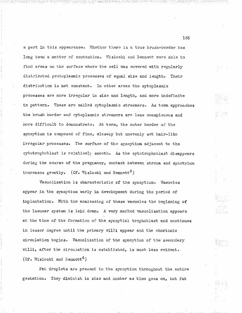
153
e. part in this appeârâJrcoo ir'Jhether tl:ere is a true brush-border has
long boen a matter of contention" T{is}oclc- and Beru:ett v¡ere able to
find areas on the surface v¡here the oell r¿as covered with regularly
distributed protopLasmic processes of equaL size and. Ìength, theír
distribution is not constant. fn other areas the cytoplasmio
processes are more irregular in size and length, and more indefinite
in pa.ttern" These are called cyboplasrn"ic streâtrrers, As term approaohee
ti:e 'brush border anrÌ cytoplasnic stresJoers are l-ess conspicu.ou.s and
more d.ifficult to d.emonstraten At term, the outer. border of the
syncyLiurn is composed of fine, olosely but unevenly set hair-like
irregular processesu The surface of the s¡mcSrbium adjacent to the
cybotrophoblast is relatively smoothu "A.s the cybotrophobLast d.isappears
du.ring the course of the pregnancyu contact between stroma and s¡rnctyium
increases greatly. (Cf " tffislocki a¡d nennett4)
Vacuolization is characteristic of the s¡maytiurro Vaouoles
appear in the s¡mcytiu.rn early in d.ovelopment during the period. of
implantation' Ifíth the coai-escing of these vacuoles the begínning of
the lacunar system is laid dortrno A ver¡r marked vasuolization appoars
at the tinre of the formation of the s¡mcytial trophoblast and. continues
in lesser degree until the primary villi appear anci tho chorionic
circulation begins" Vacuolization of the slmcytium of the second.ary
villi" after the circutati.on is establi.shed, is much less evidont"
(Cf " iUistocki and. Ber:nett4)
Fat dropLets are present in the syncybium throughout the entire
gestationu They diminish in size and number as tirne goes on, but fat

1trt
0yLetrophoblastweeks gestation,Hernetoxyi.in and
FIGURE 56
and s¡mcytiotrophoblast of a sixThe ltbru.sh-bordere is also s€êno
eosin stain. 800 x"

155
FÏGUF.E 5?
Villus at fulL term" Trcphoblast is nolv almost allsyna¡rbium" Stroma is comparatively dense and hasrnany large blood t'esselso Hematoxylin and eosinstain, 7OO x"

156
is preseni at term in the form of very fine droplets" (Cf. TrrisLoci<i
and Bennett4)
The developmen'c of nuclear i¿nots in the s¡mcybium is character-
istic and becomes more marked as the prognancy advances (cf, post)"
Tfith the dovelopment of nuelear lsrots¡ the s¡mcytium is irregularty
thinned over the surface of the villi"
The _cylctrophoblast (La¡rghqn_s1 lryrl), The villi of the very
early brastocyst are made up of oybotrcjphobl-ast al-onen After the
appearance of tho mesench¡rme and during the first fov¡ woeksa the
mesench¡rme is everywhere separated from the sync¡rbium by cybotropho-
blastn Gradually the cybotrophoblast disappoars, apparently transform-
ing ínto syno¡rbiumu There is no agreenent when the c}-botrophoblast
cornpletel]¡ disappoarsn The fifbh and sirbh months are mentioned most
frequently, Wisl-ocki a¡d Berrnett4 Oelieve that they find ver¡r
occasionall¡r ss11* at terrn which probabl¡r represent oybotrophoblasto
The cell-s of the cybotrophoblast are spherical" ova1, or
somewhat polyhedral" Thoir si¿o varies considerably, probably because
they are dividing. It{itoses are rather frequent. The faintly granular
c¡rLoplasrn is cfearer, and te.kes less stain than that of the s¡mo¡rti.um"
In many ceLls there are faint vacuoles"
No fat is found nornr.ally in the cytotrophoblast. The Golgi
apparatus is of the usua] net t¡rpo, vrhile that of the syncl.tium is of
the dispersed tvpe. (Cf " lllislocki and Bennett4)

157
bythe roticulum' Reti oular and argyrophil fibres demonstrated
silver impregnation metho¿s (¡ietschowslçy) are fine and hair-like
during the early v'roeks of gestationo During the later months and at
term these fibres aïe hoavier, longer, and more rru-rrlorou.so This'point
is demonstrated in Figures and
Degen_er?tiv9- c"hgllgeÊ" Degenerative ehanges occur in the
placenta, the most froquent of which are fibrin deposition and oal-
cification, but not a]I placentae shovr these changesu the degree varíes
from specimen to specinen"
The earliest and most frequent change noted is the deposítion
of fibrinu Subchorionic deposition of fibrin is seen j-n nearly evory
placenta after the sirbh month3" This fibrin is in the form of flat
white pla-ques of 2 to 5 uun in di-ameter, aird occasionaS-ly may be largor
in size. Fibrin is deposited. also on the villi and under the
syncybiun in nearly all placentae after the sixbh inonth3u The
deposition is associated with the degenerative changes in the s¡mc¡rbiumu
During the last haLf or third of the gestation when nuclear lff.ots are
forming, the thinned s¡mcybir-rm frequently becomes the site of fibrin
deposítion. If sufficient fibrin is deposited to encase the villus,
ischemic necrosis of that structure may result" Intervillus deposition
of fibrin may also occrlro
No placenta is entirely free of calcifioation at terrn" Tts
presence is of no signifi-cance, other than perhaps a sign of senility
of the orgÐrrø Catcium deposits are most frequentLy found along tho
stems of the main villi or in tho placental septao5. (Cf" Helfunan)

158
P 4rç,
FIGURE 58
ReticuLum of the placental ril"l-i at sixweeks" Compare with Fig, 59u page i.59'Bielschowsky sil-ver impregnation' 500 x"

i.59
FIGURE 59
Reticulun of plaeental- v-illus at term'Bielschowslcy silvor inpregnationuS00 x" T,IGII 4062-48"

160
The= pl-qcent-a at term._ The placenta at term is a disc-liice organ
measu.ring 15 to 20 cm in diameter a.nd up to 3 cm in thichless, Ït
vreighs approximately 500 grarrs or one seventh of the weight of the
fetus, The fetal surfaco is smooth and is covered by vascular ohorion
oontaining fetal vesseLs" The amniotic sac is closely applied to the
ohorion on its fetal siden At the placental margin the membranes are
cormonly fused. (cr" n*ro16)
The maternal surface is dark ín appearance and is divioed into
cotyledons which appear as mounds when the expelled placenta is examinedo
Separating the cotylodons are septae, trhich are d.ocid,ual and tropho-
blastic in origin' These septae do not reach the ohorionic platel7"
The cord vuhich carries one rein and two arteries is usuo'lly
attached eccontrically to the fetat surface, although central attach-
ment is found. in many speoimenso Its outer surface is covered with
a:rtniotic epithelium and the vessefs aro supported in a mucous
connective tissue icnov¡n as Ttrhartonr s jelly'
tr\-rrther descriptions of tho placen'ca at term are found in the
recent review by Earn16, and in the papers of Dodds (¿natu Rec, IgZZs
23_eZB7') and Dees*Mattingly (Arner" J" Anatoe I936e 59-¡a85)e
Placental circulation' Two systems of circuLation exist in the
placenta, the fetal circulation in the ohorion and viLlig and the
maternaL ciroulation in the intervillous spaces and the subchorj-onic
spâcoo The systems are separate and normâlly do not mixo
There are two main theories regarding the oiroulatíon of blood

16t
through the placenta' The earllost is that of Burrn, which has been
reproduced in te:rbs and has held univeréal sway sinco the end of the
last century. In 1935, Rudolf Spanner a¡¡ounced his concept of the
anatomy and circul-ation of the placonta' "t{hile there is not universaL
acceptance of Spanner I s thoorya it appears to be tho rnore correct of
the two" The essential features of the two iheories are shown in
Figu-re and Figuro
Bummi s theor.y, Idaternal blood enters the placenta by small
arteries of the decidua, r'¡hich are contínu-ous with the spiral arteries
of the uterine wal-ls. After the blood circu.lates among the vil1i a¡d
bathes them" it leaves the placenta by veins which drain into those of
the uterlne niuscl-e'
The villi, in vrhose stroma are the vessels carrying circulating
fetal blood, hang in tho in'cerviLlous spacos, tulike brenches of a tree"t?
Interchange of rnetabolic substa:rces oocurs across the plaoental barrier
v¡hich here consists of trophobla.sti-c aeLl-sa strornao and endothelial
cel-Ls of the vessels in the vÍlIiu (Cf" JohnstonelT)
Spanner-ts- t-4gpgy" Falkinerl5'15 has su¡¡rarized the work of
Sparueeru the acoount which follov,¡s is mostly frorn Falkinorts papersu
The structural unit of the placenta is the cotyledon¡ of v¡hich
thore are a varying number in each placenta, ranging from fourteen to
thirty, Each cotyledon is separated fron others by septa, which are
complete at the base of the placenta but do not reach the chorionic
plate" }{aternal bl-ood is fed into the base of tho ootyledon by the

ICAL CORD
ROPHOBLAST CHORION
L62
MARGINALSINUS
;!!:...i;::
' /' '' I'/l,'/tLz
.:/ .,
t-!. ri;:i.
/i/t':'',
:-;::,.'7
'¿l:
t/t
NCY LAYER
ACT LAYER
UTERINE ARTERY VEIN
FIGURE 60
Burune s coneept of_plecental circuLetion.F-ed.:'a¡m fron Áreyl9n

16"3
RECURRENT ARTERY ¿ VEIN
MATERNAL
\e2=
^an
122
)
MARGINAL SINUS
PÄRIETAL DECIDUA
PLACENTAL VESSELS
INTERVILLOUS SPACE
STEM VILLUS
CHORIONIC PLATE
FÏGII8"E 6].
Spannerîs concept of-pl"acental circulation"F-edrsYrn afto: Bre¡nerlZ "

]-64
spiral arteries" The blood wells up over the septa to for¡n a }ake
under the chorion (tl:e subchorionic space) and from here enters the
marginal sinu.s whÍch drains directly into the uterine veins,
Eaoh ootylodon has at least one main villous trunk" These
large villi are the only structu-res to cross the subchorionic .pr."u13
whiah measures fro¡r 0'5 cm to more in depth, The spaoe is not seen in
the separated and collapsed placenta because of the loss of bl-ood but
is woll d.emonstrated in the specimen whj-ch has been injected in situ16"
During early development, while the implantation cavity is
enlarging, the decidua is split and many vascular units consÍsting of
artorioles a:rd venuLes are opened lrpl5" Gra-dually hundred.s of the
spiral arteries described by Bartelmez open into the implantation1Êcavity'"o The coiled arterioles of the endometriurn gradually become
tho coil-ed utero-placentar artoriesu spannerl4 counted as many as 4BB
openings in one specimen. The distribution of the arteries in the base
of the cotytedon is irrogular, and they do not pass a.cross the septa.
(cf, natrio""l5¡
Maternal bLood flows from tho subchorionic spa.ce into the
marginal sinusu The marginal sinus is cirauÌar, following the outer
edge of the organ, and measuros frorn 2 to õ am in diameter in p1a.ces,
while at. sorue points the sinus ma)¡ be as narror¡ as 0o5 cm in v¡idth"
fnconplete septa often drrride the sinus, (Cf , Falt<iner15)
Spanner holds tlnt the return of venous bLood of the placental
circulation is entiroly liurited to the periphery of the organ via the
aircul-ar sinuslS and not by veins from the intorvillous spa.ceso Blood.

165
from the circu. l-ar sinus drains into a fine netvrork of venou.s channels
situated jusb under the decidua' This system has no diroct connection
vrith the intervillous space in the central portion of the placentan
and is drained into a more su.perficial networlc r¡rhich in turn is
drained by the tributaries of the ovarian and uterino veins"
(cr, ralrinerI3"15)
Fetal circul_ation. The feta-L vessels running from the chorion
into the viLlous trunlc pass right across the chorio-decid.ue.I space and.
enter superficially into the substance of the decidrulTu The vessels
then turn back towards the chorionic plate and re-enter tir.e chorio-
decidual space' still within the vilIi" Thus, instead of appearing
like the branches of an ordinary tr-ee, such a.s the spruce, they
resemble nors tire â.ppearance of the fueeping will-owr? or Ran oId
fashioned cha¡rdelier"E (cf" JotmstonulT, Falkinerl3r15¡

166
BTBLTOCIR^Á3HY
1" Rock, Jee ard J-" To Eertiga The Human Conceptus During the þ'irstTwo l4ieeks of Gesta.tion, Ámer" J" 0bst. & G¡nr;, 1948, 5536.
2" trr{islocki¡ G"n The Placenta in Textbooic of Histology by J. Lo Bremer,(re-written by E" L" Weatherford) Sixbh Edition,Phita"s The Blakiston Company, 1944" 723 pp'
3, Hollman, L' Mo, AbnormaLities and Diseases of tho PLacenta andAppendages (Ottrer than llydatídiform Ìúole and.Chorionepithelioma), Chapter ÐOCIV in GSmecological andObstetrical Pathol-ogy by E, ltTovak, Soconci edition,phila,: 1¡tr. B " Saunders Companye L947
"
4, Tiislockie G" Be g.nd. H" S' Berrnett, The Histology and CyLology ofthe Human and Monkey Placentae with Special Reference to theTrophoblast. .Ameru Ju Anat"¿ L943o !!a335,
5u Ìt[eyer, A, Tlf.n l{ofba"trer Cells in Norna]. end Pathological Conceptuses"Contrib " Embryo3.. Ce"rnegie ïnst o l'ria"sh. e l-921, 1år 301'
6' Lewis, trlFo Eu, I{ofbauer Cells (ClasniatocyLes) of t}re Human ChorionicVillus" 8u11" Johns Hopkins Hosp" e I9Z4s 35e18õ"
7, Hofbauer, cited by Levris (O) a¡r¿ lr,teyers (5).
L Grosser, 0o" Human and Comparatíve Placentatione Including theEarly Stages of Humerr Developmont. Lancet, 1935, 22421053"
9u Hortigs A" T', Angiogenesis in the Early Ïluman Chorion and in thePrirnary Placenta of the lfacaqu-e'ivlonkey" Contribu Embryol-,Carnegie Instit" trTash"e 1935, 25t,37 "
10' Dod.ds¡ G" S", The Area of the Chorionio Vil-li in the Fbll TernPlacenta" Anat" Rec" 19220 24a287'
11. Dees-Mattinglya ivi." "Absorptivo á.rea and Volu.me of Chorionio Villiin Circumvallate Placentas" j,rnen J" Anat"e 1956, 59:485.
3,Z. Bremer, J' L", TerLbook of Histology, Re-'w-ritten by H" Luïrieatherford" Phila"¡ lhe Blakiston Company, 1944"Sirçbh edition,
13' Falkiner, ItT., Circulation of the Maternal Blood through thePLacenta" Iro J" 1liedu Sc,u 1999, Silrbh serca po 59'

I6T
!4" Spanner, R', i'¿,Ttt"rlicher und Kinclerticher Kreislar-rf deri'{enschliohen Placenta und seine Strombahnen.Zeitschrift ftìr Anatomie und. Entwicklungsgeschichto, 1935,:L65. 6"uoted by Falkin"r (fS) and Johnstone (12).
J.5, Falkiner, N,, Placenta] Circulatlon" Proc. Roy' Soc" I,[ed." s !g44,Q7_t4I7 "
16o Earn¡ A" Au, lJaster of Science Thesis, 1949, University of ÌÍanitoba,
L7" Johnstonee R, T[o, A Terbbook of i,Íídvriferyu Eleventh edition.Londona Adam a¡rd Charles Black, L942"
18' Ca:neron, .Au T", Recent "Advances in Endocrinologyo Si:rbh edition"London¡ Jn & A, Churchill Ltd"" 1947" 443 pp,
19' Àreys L" Bo, Developmental J'iratomys lhird odition'Phila'¿ ïf. B" Saund.ers Compa:ryr 1956" 593 pp'
20, Baker, B" Lne Su Jn Eook, and A, E" Severinghause Cy'cologicalStructuro of Huma:r Chorionic Villus and Decidua Parietafis,Amer, J" Anat", L944þ 74a29!"
2L, Stevrart, Ho L,e ÌVI" E. Sa:ro, and T" t" l,{ontgomeffa Eormone Secretionby Human Placenta groïm in Tissue Cultureo J' Clin"Endocrinol", 1948, B¡175"
22" Jones" Seegaro Go En, G" 0n Gey, and lvi" K' Gey, IlormoneProcluctíon by Placental Cells Xfaintained in ContinuousCuliure. The Joh¡rs llopkins llosp" BuJlu, :-.943, 72a26'

CHÄPTER. FTVE
TriE HORll0NlJS 0F REPROÐUCÏION

CE¡,PTøR Y
THE HORhÍON!]S OF REPRODUOTIOi{
Introduation, The differeÌLce bet'r,veen rnaleness e.nd fe¡raleneSS
is qqantita.tive rather than qualitativeo Thus the rnale has feinale
potentialities and sirnilarly the female has mal,: potentialiiiesn Ïn
early d.evelopment, unless one set of characters olearly predominatos,
the individual becomes an intersox or hermaphrodíte (Cf. lúoort16,
t.Camerono). Ilorlnones acting upon a specific genetio oons'citu.tion
result in those characieristics associated with maleness or fomale-
ïtos$o Cameroa2 states¡
l\foro than one ond.ocrine compound is ossontially (but notneoessarily exclusively) associated with t?malenoss,tE e:nd morethan one ondocrine oompound. is essentially (¡ut not necessarilysxclusive]-y) associated o'rith nfomalonesstr .,
"
The testes are the prinaipal source of androgens, r'rhile the
ovaries are the pr"incipal source of oostrogeÐ.Sø Tho adrenal cortex
produces weakly androgenic and oestrogenic substances, while ihe
placenta procluces strongl¡' oostrogenic substao"uazo
Androgenio substances are general3-y assrtmed to be produced. by
the Le)¡dig ceLls of the testes56" The androgens are roguired for the
d.evelopment, maintenance and control of the male reproductive systom"
The socond.ary sex orgalls (penis, prostaten sominal vesicles) as woll
as sucir fa.ctors as body contour' pi'boh of voice, hair distributiona
and. in lower animals at least, the sex drive are infLuenced by the
androgons "

l-70
Oestrogenic substancos control a:ed me"intain the seoondary se:r
orga"ns (uterus, vø-gina, clitorise mãJûtrary glands) and as wolI those
charaateristics usually assooiated with femaieness sueh as bod.y
oontour, hair distribution, and pitch of voioe'
Neither the testee]6 oor the ovaries are automatio but in turu
are controLled by other influences in the body' The chief of these
influences are those hormones of the anterior lobe of tho piiuitary
hrown as tho gonadotrophins, of which there are at least trn¡o" i{or is
the pituitary tho only sourae of tho gonadotrophins' The placonta
producos a gonadotrophin whioh j-s aotive only in the presenee of a¡
intaot pituitaryzsL4 "
Close associstion and interplay betwoen the hormones of the
a¡terior lobe of the pituítary and those of the gonads exist.
Ilormones of the adrenal cortex and the thyroid- gland also exert an
influonce' Xlith these factors, together with the fact tì,rat the male
produces oestrogens as welL as androgens, and the female produces
and.rogons as weLl as oestrogense the piétu.re becomes one of great
oomplexity" The effect of all these factors on tho individual organ
and in turn upon the wholo body is thus dependent upon tho sumnation
of rüany factorsoI
Sel-ye' in his monograph oa ondocrinology" notes that
approximately 5000 papers a year are published in the field of
endocrinology" The task of tabulating and evaluating these publications
can be irnraed.iately appreciated," The discussion in this chapter is
basod mostly on standard monographs by authorities in the field^ of
T7L
endocrinol-ogys such as Selyel, Cumeron?u Af1on, ¡'ngJ-e, Mocre16,
Pe.paniculeau et a1 9, as well as Fluhma.nr.l3 ar,d NovakS, Very little
reference ha.s been made to original contributions,
The.. ho{4loneq. The hormones ø.ssociated vrith sex and reproduction
form two groups of cornpoundsn Tho gonadotrophins and prolactinu r,vhich
o.re proteins, make up the fírst groupa while the and:'ogensa oestrogens,
and progesterone, dnich are steroidso forrn the seoond group. The
eonpounds are listed on page l7Z in tabu.lar forrn,
Ig.Ulglg qli$ulatiqå 4qrryqq-e_ (¡'SE) " This hormone can be
obtained from pituitarS' glancls a.nd fr"om the u.rine of normal¡ Íì€l1o-
pe.usal and castrated humanso ft is protein in naturen The horrnono
câuses the gro'mth of follicles in the ovary and the development of
tÌre seminiferous tubules in the testis" Tts action alone does not
sti¡rn:.late the production of sex hormonesn For this a smalL proportion
of luteini"zi.ng hormone (m) is neededu

CIffi'{rC/{,t I{A.TURE
PROTETl\T
GROIIP
GOI{.ADOTROPHINS
TEE HORAIONES OF REPRODÜCTTON
slmroÏD
Follicul"ar st irnulating hormono(rss)
LUTEOTROPHTN
NAJ],[ES
Inters-,,itia1 ceLl stimulatinghor:none (rscn)
cR
Lut einizing-Tormone (tH)
âNDROGENS
,A¡rterior pituitarv-lilce (APt)
(Synon¡nns - prolactin, lactogenichornono, mammotrophin,luteotropirin (f,tn)
OESTl).OGENS
TestosteroneAndrostoroneDehydroandro sterono
SOURCE
r'lntorior lobe ofpituitarv
PROGESTER.ONE
-oestradiolostroneestriol
Anterior lobe ofpituifary
Placenta
progesterone
Anterior lobe ofpituitary
Mostly from thointerstitial cellsof testos ' S"lsofrom tho adrenaloortex"
Mostly from thefollicular cellsof the ovarfo alsofroia the Corpusluteum ancl theadrenal cortex.
Oorpus luteumU{tü

L73
-I,1¿!-=g::g åo,r¡LoJlg (1,.U_).ø LH is a gLvcoprotein vrhich au.gments
the a-otion of FSH, the combined. action lea.ding to the production of
the sex hormones of the ovar¡¡ and ovr-rlation" Luteal tissue develop*
¡nent and devel-opmøri of corpora. lntea are stimu.lateo, Undsr the
influence of LI{ the corpus Luteunr is formed- frorrr the pre-formed
graafian follicLe" LH is presumably the ovulatj.on prodr-',cing hormone,
but appa.rently does not regulate the corpus luteurn once it has formed
(Cf , Colaznierl4)" Tn the testes, LH stim.rlates the interstitial
cells to produce androg"rr*2,14"
The moleoular weight of LH (ICSH) shows a species variation"
ïn sheepr the molecular weight is estimated to be 40r000 and in hogs
100p000, while in the human the molecular weight is not hrorlmu
Chorionic Sonqdotrophiq f4lÐ. The ple.cental gonadotrophin is
chenicaliy a¿d biologically distinct from the pituitarSr þqr.mon€se It
is a glycoprotein of a nolecular weight bet'çveen 601000 and 80r000"
Garactose is part of the moreculel4' Erperimentally, .APL is v¡ithout
effect in the h¡ryophysectomized animal, but is effective in the
@.A.ccording to Selyel, the Luteinizing hormone (i,U) is identicaL withthe interstitial cell stimulating hormone (fCSn). Since the humanovary does not^irave interstitial cells, clinicians objeot to tho termICSH" Camerona on tiro other hand, uses the term, preferring LFi as asynonlmo In this discussion, the term luteinizing horuone is usedwith the understa.nding that IC$H s:rd LH a.re identicaL,

LV4
presence of ar. intact pituitaryz'
Eu-man trophoblastic cells grorm in continuous aulture produ-ce
chorionic gonadotrophin. The ty¡re of eeIl responsible is the
T,anghans ""1L17'19,
P_roLaetil" Prolaotin augment s the acticn of TJ{ (f CSU) being
required before the oorpus luteum producos progesteronen a.nd is
essentia] for the actual seoretion of n1lk in the iïìaïmary gland"
Evidence suggests that the horrnone is responsible in part for
matoraal behavlourZ" further evidense suggests that luteotrophin
(ftn) and prolactin are identioal" (Cf" Cotaziniexl4 quoting Evans
et a.1") (¿tso cf " Selyel)
4gffigu åndrogens control the <ieveloprnent and maintenance
of the seeondary sex charaoters assosiated with malenessu
Androsterone, dehydroandrosterone and testosterone are the three
major and.rogens" Thoso isolated from the adrenals are relativety
weak in their androgenio properties" 0f the androgens, testosterone
is the most potent, and has beon isol-ated frora the bul-L testes only"
ït is assumod to bo tho chief androgenic substance of other species.. o-(0f" Canerono)
The androgens are steroid in nature and are olosely associated
in their cirenicaL constitution to the horroones of the adrenal cortex
a¡.d to cholesterolZ' contemporary opiníon is greatly in favouv of
considoring tho Leydig oel]s as tho produ.cors of the androgens of
testioular originl"
175
Oestrogens" Oestrogens controL the devei-opment and
maintenance of the secondar¡r characteristics usua-1.ly aseociated v¡ith
femaleness " Estrone, estriol and estradÍol are the most important
of this groupo -oêstradj-ol is the most potent natural oocurring
oestrogen and has been isolated from the ovaries of soï¡s, hu_man
placenta and staLlion testesz" Oestrogens exert their greatest
effeot on tissues of Ir¡riltterian origin such as the ovíducts, uteruse
arrd upper portion of the vagina3*
P-Lo-gest-e{one, Progesterone produces changes in the uterus and
vagina follov,ring oestrogenic sti¡rulati.on. The secretory changes of
the endornetrium, deposi-tion of gJ-ycogen in the epithelium, and increase
of vascula.rity are dtre to progesterone" Tnhibition of ovulation is
said to be another effect" The hormone is required for the maintenance
of pregnancyo should pregnanc]¡ occur" (Cf " Ca.meron2)
4"!ftgQs_ € study. Castratj.on of nan and domesticated anímal-s
has been practised for centuries, afford.ing ample opporttinit], to
show the effects of l-oss of gonadal tissue. Experirnental investig-
ation has played an irnportant role" Early experi.mentation consisted.
of the removal of the gonads" Removal of the testis¡ and grafbing
was nerb performed' Ertracts of gonads v¡ere found to corect some of
the defects produced by cestrationu
y'"t the present tíme, maxy concepts are based on e:cperimental
ovi<ienceu Conclu,sions drav::n frorn experirnentation of one speoiss do
not necessa.rily apply to another species"
176
Clinicat observations of functioning tumors in r¡¡hich an excees
of a hormone is produoed has gíven another approach to the function
of some of the hornones"
castration of pre-pubertal females is rare' Examples of
hy¡rogonadisn and congenitally absent ovaries have been observed" The
post-pu'bertal fe¡nale oastrate is oommon and the s;nadrome produood oy
10ss of ovarian tissuo in the female adult is well i<nov¡l'
lhe hutngq trgle cqe-tratg" Castratíon effeats vary greatly
d.epending upon the period at which the testicular tissue is lostn
Pre-pubertal castration (early eunuchism) nas a moro profound. offeot
upon the bod.y thel post-pubertal castration (tate eunuchism) ' Tn
this discussion, the'most severe forn of h¡rpogonadisrn is considered"
since this state gives us an indication of the functions of the mafe
hornones" The graphic description of Selyel is eraellentn
Ea-rly eunqcFism" Aplasia of the gonad.s, surgioal removal for"
disoaso, or destruction of the gonads for eny reason before puberty
leads to marked. deviations from the state of malenessn The
tosticu}ar deficiency occurs before the morphogenesis of organs and
structures has proceeded far enough to give the individual definite
male charaoteristics'
By the timo the third decade is reached, early eunuchs present
usually a fairly typical picture" Excessive grov'rtlr of tho long bones
of the exbremities gives tho patient an awl<ttrarci appearâr.Geo The
Lirnbs are disproportionately long cornpared to the body' Tho gait is

!77
affectedu Poor musoular development together with la,ck of muscul-ar
strength, Iaslc of initiative and drive add to the plight of the
intraverted., shy and frequently sullen earl¡r euitucho the boyish
voice is in koeping with the infantile face which lacks beard and
which lacks the bone differentiation of the adult, but which is in
contrast with the senile tirod.ness and fine rrrinktes, Sebaceous
secretions are dininished, the hair and the skin are dry. The
patients do not become bald" Ðistribution of hair overbhe rest of
tho bod¡' is femaie in t¡rye" The pubic, axillary and lanugo hair is
spÐ"rseo The tall patients a.re thin, The short patíents tend to 'be
fat, the adipose tissue ìreing found in the breasts, the hips and the
abdomen" The general body configuration is feuele in ty¡re, the
broad hips aocentuating the picturoa The testes aro absent" The
penis and other aocessory sex orgar.s are siaall and infantilo,
Libido and potentia are markedll' diminished or absent"
Excretion of androgens, 17-i{S and oestrogens is decreased
since the main €oLrrce of such compounds is removedo The continuod
oxcretions of smalL amounts of these substanoes is probably
attributable to the adrenal cortex. Gonadotrophin oxcretion is
increased since the inhibiting influence of the testicular tiszue is
lacking" (cf, setyol)
Late eunuohismo Post-pubertal l-oss of testicul-ar tiselre does
not affect the organism to the erbreme
castrationo The appearaJlce of the l-ate
degree soen in pre-puberbal
eunuch differs but little

178
from the norrnal male adult. There may be atrophy of the male
accessory organs, bu-t these organs tond to rercain fairJ-y norila.l in
size" Grov¡"Lh of bone ar'd growth itself is not disturbed. The
beard and pubio hair are nor:nal throughout life" Potentia and libido
are not marked as a rule but are not necessarily absent. Psychia
disturbances related probably more to the trauma than to endocrinology
may be present, Vasomotor disturbanoes such as Ehot flushesrer
tachycardia, otc.e siinil-ar to those of the fenale nenopause, are not
unoorunon in sorne individuals" (Ct" s*ty"l)
The pre-pubertaL fq¡aie- oas_Llqle" Very few cases of castration
of the female before puberty are reported in tÌro literature"
Agonesis of the ovaries is also r&r@o True ovarian agenesis is
u-sualJ.y accompanied by shortness of stature, Ínfantile development
of the maÍffrary glands, uterus and vagina and diffuse osteoporosis'
Ilrinary exoretion of gonadotrophins is increased" Often otlrer eon-
genital anomalies exist" (cf" Selyel)
The pos_L-pube.Ltg.i temale clstratg" Castration of wonen after
puberty is oommon, and the picture rr:sulting from such an operation
is well lcnown" Usually: the younger the patient the more severe are
the subjective syrnptoms such as !?hot fru-shesrrû tachycarclia, vertigo,
nervousness, excitability, irritabilityr and headaehe' Gradual
regression of the development of the aocessory selç organs follows"
Ilenstruation coasesn S¡rnptoms may reappee"r as the patient enters the
fifth decade" at the time when climacteric r¡ould have baen expected

L79
to oocur"
Pre_-pub_ortal lqryqlj, Maturity must reach e. certain stage
before the gonads become responsive to the gonadotrophins of the
antorior lobe of the pituitary. FSH and LH stinuli gradually increase
causing enlargement and. grovrbh of more and more Graafian follicles,
v¡hioh produce moro and more oestrogensu Bet.¡¡een the ages of three
to seven yearse both boys and giris excrete srnaLl and constant
amounts of oestrogens and keto-steroids in the urinee From eight to
eleven years the exoretion of oestrogens by girls increasesn
Pituitary FSH has been dotocted in the urine of girls as early as
eLeven years of age" ,{bout one and one-ha}f years bofore the
menarche, the exorotion of oestrogons becomes oyclic" eaoh cyclo
increasing in intensity" The stimu.lation is eventually sufficient
to prodr-tce rapid growLh of a sufficient number of foLlicles which are
able to produce an effective quantity of oestrogens' One of theso
follictes ruptures ano a corpus luteum is formod." (Cf, Caoreron¿)
1{lit}r the first ovulatione the wholo system is faunched into
a ner¡r era in which reproduction is possible. The waxing and waninga
growbh and rogression, contro] and reciprocal control, augmenting
and depressing continues in a fairJ-y definite cyclic pattern once
esiablished. Anovulatory cycles are apparently cotrlrron in the teen-
age gir1"
Menstruation" nThile bleeding is the most.stritcing feature of
it is but only a phase of a serj.es of eventstire menstrual cycle,

1.8CI
'¡,¡hich affect the bod¡r as a v'¡holeu It may be consi-cl-ered the end of
one short chapter of the reproduct j-ve hi story" *{ co-ordj.nation of
the v¡hole cycle is briefly given"
The hor¡nones. fmrnediatel;r foli-owing menstruation, FSH of tho
enierior lobe of the pituitar¡r, stj.mulates a large number of follicles
to devefop in each ovar),'6 FSI{ stiniulates tho morphological responsee
but not the prod.uction of hormonesl¡ for this a sma1l amou:rt of LH is
requiredo. -As the fol-Iiales devolop they produce incroasing amounts
aof oestrogens'n The oestrogenic hor¡nones of the follicles have their
greatest effect u.pon tissues of iUüllerian origin suoh as the oviducts,
uterus, and upper part of the vagina" In al] these ti.ssues, the
response is grov,rth, the greatest change being noted in the
errd.ometrirm3 'As a" rule, onl_v one foiliclo reaches maturity end dischargos
elt ov¡¡Ilo The unruptured follicles continue to produce hormones, but
soon und.ergo atresia and as they do so, produco less and. loss
oestrogens 'After rupture of tho fol.Licle and erbrusion of the ovr-¡,m, the
corpus Luteum forms from the granulosa cel.[s under the influenoe of
LH of the anterior pituitary,z" Prod.uction of LT{ from the anterior
pituitary is initiated by FSHl. The corpus }uteum continues to
prodtr.ce oestrogens in snaller guantities and, as well, begins to
produce a second hormone, progesterone' Production of progesterone
from the corpus lnteum is under control of T,H and prolactinz"

181
Progesterone is produced only after ovulationóe since corpus luteum
forl:iatj.on forlor-us ovulation" under the infLuence of proges,cerone,
tite endometriu.n enters the secretory phase and ovul-ation Ís inhibited3.
/'fter ovulation numerou.s grovring fol-licres undergo atresia
resulting in a decreased production of oestrogens" The corpLls luteum
continues to produco oestrogens in lesser announts and after rrraturity
when regression begins, produces l-ess and loss oestrogens as tj¡re
goes ollo Less progesterone is produ-ced as regression of the oorpusD
luteum proceedso" Degeneratíon of the granul-osa-lutein cells of the
corpus l-u.teum begins from nine to eleven days after ovuLatiorr9 o"
from four to six days before the onset of menstruation4" The clegener-
a.tion is slov¡ and gradual, and is reversible shourd. pregnancy o"oorSn
The gradual v¡ithdralval of progesterone and oestrogens has a tv¡o-fold
effect" onset of the menstruai flow is the most striking effect"
The ovarian hormones causing grov,"th are no longer sufficient to
maintain the endometriun in a growing state" The second. effect is the
removal of the reciprocal action of the ovarian hormones deprossing
the production of FSH in the anterior lobe of the pítuitary" l\'ith
inhibiting influence removedn the pituitar.¡r producos increasing
amounts of FSH to sti¡nulate more folIÍclss in the ove.r)r to grour, errd.
thus a new cycle is initiated, (Cf, Ca:aeron?)
WithdrawaL of oestrogens is the prime cau.se of the endometriaf
regression, although there are probably other factorsZ, progesterone
administratíon will delay the onset of menses" e¡rd the removal of a
recently formed corpus luteum vrill result in menstrual bleeding within
tB2
beforty-eight hours' The onset of þleeding after such removaL ca"n
dela-yed by use of progesterone, while oestrogens do not have this
eff ect " ( cf . celreronz )
,4n anow"latory clrcle differs little from the ovu.latory cycle,
and yet no corpu-s luteum has forrned" Thero mu.st be occa,sior:s v'¡hen a
functloning corpus luteum has not forme<ì afte:' ow.lation6" Stii.l the
corpus luteum ís indispensable to menstruation, for there are none
fou.nd in the ovaries of individuals who do not menstr'rrate?u
Ner,ver. gggpl3. of i[gns-!-rua.t$n, Daronll emphasised that the
vasculattrre of tho endometriu.m undorgoes cyclic changes as v¡eIl- as
the glands and. stroma" l,[arkeelz obs"r.ring endometrial implants in
the a:rterior cha¡nber of a monkeyr s e)¡er described the bulk of the
hemorrhage as arterioLar in origin and therefore the coiled arterioles
con'brol the amount of the menstruaL hemorrhage' The explanation of
monstruation as a oonseguenoe of vascular changes is based prima-rii-y
on these observations6o
Kaiserrs description6 of the vasculatu.re changes of the ond.o-
metrium are excellent" This author outlines the two main concepts
of menstruation as fou.nd. in the Literature¡ (a) tne coiLed
a.rterioles increase rapidly and exLend as the secretory phe.se of tho
cyc}e progresses and this increasing complexity is supposed to reach
a poini r¡¡Ìrere it impedes the fLow of blooa to produce ischemia r'.¡hich
ín turn sets off the chain reaction of menstruation; (¡) the
pharrnaco-d;mamic concept vrhioh srlpposes the prolonged periods of vaso-

tB5
oonstriction observed prior to menses to be due to vasomotor
substances produ-ced and the se-c¡-rence of vaso-constriction, ischemia
and nenstf,uation' (cf" r"isu"6)
Aacording to Kaiuu"6, both of these concepts assume that the
complex coiled arterioles are necessary precursors of menstruation.
The hypotheses by¡iass and actually contradict earlier v¡ork on
hormonal withdrawaf as a conmon precursor of menstruaL flov¡" Nor do
the conoepts expla.in menses in the anorrulatory cycle in which the
coiled arterioles a.re considerabl-y' Iess complex' Tn some experirnental
animals which menstrriateu there are no coiled arterioles" Certainly
thore rr¡rsb on occasion, be a failuro to form a functioning corpus
luteum, and if such should happen, this lessens the conplexity of the
arterioLes. (cf" Kai*u"6)
Time of ovulation' V'¡ith inevítab1e variations¡ the mass of
evidence indicates that ovulation in the human female oocurs 14 to
15 days prior to the onset of menstru.al flow" in a c¡rsle v¡hich is
28 to õ0 days in length" Ilumerous methods of invostigation have been
fol-l-owed to d.eternine this tfme of ovulation' During the cycle"
should ovulation occur, a slight rise in body tenperature corresponds
to the time of ovulation' Temperature recordings over a number of
oycles thus gives a good indication if and vrhen orml-ation has
oocurred" Studies of corpora lu.tea and ovaries removed at operation
have given some infornration" The recover¡r of ova from the ovidtr.ct
e:rd endometrial- studies âre u"sed.ø During the menstru.al cyc1e, there

184
are variåtÍons of electrical potentials, differences in the notilit¡r
of the uterus and ovicìu-cts. These have been used in determination
of tirnes of ovuiLation" Pregnanc;i occu.rring from a single coitus in
cases are hrov¡n' The sudden rise in the amount of pituitary gonado-
trophiris is said to be a sensitive indicator, the rise oorresponding
closel;r to the ovulation tj:ne.
Chefaglerislicg oJ. !¡o ngrügl menstrual cy-cle. Goldziehor
et al.B hu.or recently reported. data of 524 cycles of 109 selected
normal worïlêï1.ø Of these 524 c¡rclsg, 500 u,ere ovurlatory" No cycle r,rras
less than 19 days when the anovulatory cycles were exclu.dod" Over
hal"f of the normal cycles were from 26 to 29 days in length and
92,8 per cent of thern fell- within tlne 23 to 56 days interva"L" The
oestrogenic phase in the niajority of cases was from 10 to 16 da.ys in
length" Of 490 cyclos, 69.5 per cent were 11 to 14 days during the
progestoronal period, and 94 per cent feLl within the 10 to 16 day
periodo The dnration of bleedíng in 95"4 per cent v¡as fronr 3 to 7
days, end in 68"5 per cent from 5 to 5 days, (Cf, Golazieher et at8)
ïrregularities frorn established cyoles are associated v,¡ith
minor disturbances in many caseso Iviinor illnesses, changes in
envirorunent, travell-ingr fear or desire of pregnanoy may all influonce
the ti¡re of onset of menstruation, as well as many other erternal
envirorunental facto rs'
CyoLiq chanß_g_q in the qcaeqssly_ o_qflanq o_! {-epqqluo_t_igg"
Cyelic changes in the epithelia of the orgs.ns of reproduction occur

185
in response to hormonal- stilnuli throu.ghou.t the mørstrual cycleu
Papanicolaou, Traut and i\iiarchotti9 h*lr" recently reported. their
stu-dies in the form of an excelLent monograph' A brief sumna.ry of the
cycl-ic changes is included here for the sake of completeness" L{uch
of the materiaL is taken frorn their :ironograph. lhe epithelium
covering the surface of the ondornetrium, and lining its glands, es
weLl as the epithelium of the endosalpinx and endocerwix have the
samo embrlrological derivation froro the epithelium of ttre t'¡iiïtlerian
du.ct which in turn is dorir¡ed from coelomic epitheliumn ft is
reasonable to expect that these tissues exhibit sinrilar responsos to
tho same homronal stimulí
The endometriu:n shows cyclic morphological- variations more
pronounced than a:ry other pa.rt of the reproductivo systemn Th.ree
È¡4pes of cells are found in the endometrium, namely the secrotory,
the ciliated and the rsrodl? celIs. During the foLlicular phase, the
glandular epithel ium is low and columnary there are fov¡ ciliated
cells, and rod celis âr€ re.f,eo The glandrr.lar epithelium increases in
height, and at the time of ovrlation reaches its maximu-m height' The
nucloi tend to become ovoid or oblong and assume a central position
in the cell. The ciliated colls on the surface are more rrülrroror-rsø
The transition between follioular arrd secretory pha.ses is not
striatly slmchronized 'with ovulation. tho interval betwoen ovulation
and secretory activity varies greatlyn A regional variation from ono
area to another is also present" Soon after ovulation, changes
characteristio of the luteal phase are noted" The nuclei of the

186
secretory ceLls become more basal in position and sma.l-I blebs of
secretion projeot beyond the lin:its of tLLe terrninal barsn Glycogon
becomes abundant" As the lutoal pha.se advanoes the cytopla-sm becomes
pal-er staining, the nuclei of the cell-s more basala a.n<i the secretion
more abundan-Lu The glands show an increased. tortuosity and the
stroroa has a looser texture" Increased tortuosity of the blood
vessels and h¡ryertrophy of the stroma ceLls is also noted' The
piotu.re is most marked at a perio<i 7 to 10 da.¡rs after ovul-ation'
Regressive cha:rges in the endo¡netrium mirror the regreseion changes
in the corpus luteu¡nu The glands oollapse and show even greater
tortuosity" Disintegratj-on of the mucosae degeneration of mucosal
cel1s, resorption of the edema, infiltration v¡ith leucocSrtes,
erbravasation of red blood ceLlso especially in the compact layer,
congestion, engorgement of the vascu-lar bed, and a greater tortuosity
of the spiral vessels oomplete tho picture of the late futeaL phaseu
During rnenses¡ the necrosis of the nn-icosa u-sually erbends to the
basalis' The nocrotio portion is cast off in cell olusters and
fragments" The epitheliuru of the basal glandular rer¡urants groï¡s
along the su,rface to meet proli-forating epithelium from adja.cent
glandso the endometrium is now roady to respond to further oestro-
genic stimuli from the nov,¡ increasing a:rd growing foJ.licles of the
nexb cycle' (Cf . Papanicolaouu Traut and I','b,rohetti9)
Snyc.erlo demonstrated that the tu.bal epitheliu,m shov,¡ed
varíations during the normaL menstruaL cycle" The most character-
istic morphological variation in the epithelial ceLl-s of the muoos&

187
is the change in their height" The cells are ta]Lest during the
foLlicu.lar phase, In the lutea1 phase, secrotion and the nuclei of the
non-citiated celis protrude into the tubal lumen boyond the li¡rits
of the ceIlso tho nuoleÍ of the ciliated cells remain practícalIy
unohanged during the lu.tea1 phase" In pregns.nclr, tho features of
the tubal mucosa are essentially the same as during the luteal pha-se"
Ðuring tho menstrual pha.se the epithelium is at its minimal height"
The ailiated colls are low oolumnar cells with large round or ovaf
vesj.cul-ar nuclei which are centrall¡r plaoed' The relatively
abundant cybople.srn is pale staining' The cilia are easily identifiod.
The non-ciliated oeIls Lre more compe.c-b, with less distinct celI
boundaries. Their nu.clei are oblong and the cytoplasn of the cells
is relativoly red.uced." (Cf . Papanícolaou, Traut and i\{archetti9)
Cyclic modifications of tho endocervix are most distinctly
ex¡rressed in tho glandular epithelirm. The most distinctive ohange
is found in the late follieular phase when the cells lini.ng tho
endocervical glands are talI and columnar, e.nd show increased
secretory activity. The nuclei are elongated and the papillary folds
protruding into the glands grow largor du.ri.nþ the late follicular
phe.se' (Cf " Papinicolaor:.a Traut and ii.farchetti9)
Prognanc¡¡" During the normal menstrual cycle, there are
qu.antitative and qu-alitative variations in the amount of gonado-
trophins €xcreted, The greatest increase of gonad.otrophins excreted,
lasts from the tenth to the fifteenth day of the cycle, with the poak

189
emounts of oestrogefiso
The subjective slmptons of the climaoteric are vari-ed in
nature and in severity from case to case, and the onsot and cessatlon
of these synptoms are insid.uous and. 111 defined. ivienopause or tho
cessation of menses" howovero is fairly weLl defined, and accordin.g
to most authors occurs du.ring the fifth decade of Life, with many
excoptions "
"Afber tho menopause, the reproductive organs undergo retro-
gressive changes" The ovaries decrease in vreight due to atrophy
and. non-formation of new struoturosu The irregu.lar pitted surfa.ce
gradually becomes relativeLg smooth" FoLlieLes in atresia are found
for a var¡ripg period of time after the menopausee but soon these and
the primordial fol-licles dlsappear" A corpus 1uteum may or may not
be found in one of the ovaries for a few months following the last
menstrual periodT" The ovarian vessels sholv a marked degeneration¡g
of the irtionckeberg type,
The accessory organs share in the atrophic chenges' The
uterr.rs becomes smal ler duo to atrophy of the myoraetrium' The thin
endometriu.m nieasures onLy 0"2 to 2"0 rnn in thichree s" No different-
iation into basal and functional layers is possible" The glands
docrease gradually in number and rnany becorne cystic with a Low f.ining
epithelium' The cervix shov¡s a dininution in size at a later age,
a¡.d. eventu.alJ-y has f er,^¡er gland s which prodr:.ce onl¡r a scanty secreticn.
(cf , Fluhniarìt 1õ)
Atrophic changes of the oviducts, va"gina and vulva are notod
lBB
a.t about the trTelfth and thirteenth d.a¡,. This p.eak occurs after the
oestrogens have showro their peaku FSII predomina-tes during the first
half of the oyole" Increased amounts of lu.teinizing hormone is
associated v,'ith the time of ovulation' At the peak of gonadotrophin
oxcretion abou-t 15 RUU (rat uterine units) a-re found, in each litre of
urine e
ïtiith conception, the gone.dotrophin level remains elevated and
oontinues to rise. '[Ù]ren excretion of 400 to 60O RIIU per litre of
urine is presenta pregnancv tests are positiveu This additional
amount of gonadotrophins is due to the presenoe of largo qr-rantities
of chorionic gonad.otrophins" (cf, cotaznierl4)
A peak phencmenon occurs in aII normal pregnanciesu About the
sirbh week thore is a sudden rise, the maximu.m is quickly reachod,
and by the twelf'th i¡¡eek a deoline. Tho daily output at the peak ís
varíab3-e, ranging from 75r00O to 1003000 RUU" After the peak, the
rate of excretion is from 1500 to 5000 RIIII per litro of urlneo
¿\fber parturition and expulsion of the plaoental tissue, there
is a precipitous drop in the curve of excrotion' On1y 52 per cent
of v¡omen have a positive pregnancy test 24 hours after delivery, and
none have a positive pregna^rìcy test afLer 4 days" (cf " coldztrierl4)
ivleÊopause gnd- olimaateric" Growblr. of follieles in the ovarios
'wanes narkedly as the fortieth year of life is reached and in some
cases evon before this tinel8" The climacteric and monopause aro
assooiated with the fewer follia1es and the gradually Lessenod
190
as $rell. There is diminu.tion of elastic fibres, e.n increase of
fibrous tissue" and a decrease in the size of muscle fibres, Tho
epithelium beoomes thin a¡rd the blood vessels show degenerative
cha:rges" The amount of glandular tíssue in tl:e breast decreases.
(ct" nlunri*onls¡
Tho menopa.u,se a¡rd clj.mactoric are fu.ndamentally endocrine
manífestationsl5, although the factors which brlng about these
phenornena are not olearly establÌshed" Control does not appear to be
r+dthin the gonads for the s¡rmptoms of the clima.cteric are freo,uently
present before menopau.se¡ r¡¡hile there is stil1 functioning ovarian
tissu-e" Exhaustion of suitable .ovarlan iissue may weLl be the câ.u.se
of the phenomena (Zondek--quoted by Cameronz)" '!"fith cessation of
ovarian response to pituitary stimulation, there is rapid oessation
of oestrogens duo to lack of follicLes and with this laok of
oestrogens there is atrophy of the s€x orga.ilso The regression of tho
ovaries removes the inhibition they norma.lly exert on the anterior
lobo of the pituita-ry, (Vtazer and f srael--guoted by Fluhmaorlg)
Hormgne_ integf'+_tlon iq tLq mqle_" Ðuring the early years of
life there is little difference in the excretion of oestrogens and
androgens by either sex" Later, girls excroto more oestrogens than
boys, and conversely boys oxcrete more keto-steroids than girls"
The increa"sed excretion of keto-steroids becomes marked about the
eleventh yoar wh.en the secondar¡' sslc characteristics begin to appear,
The endocrines leading to sexual development beao¡re evident s-bout

1"91
fíve years beforo sexual maturity.
There is no evidenoe of cyclic fertility in man" FSH controls
the sper:natogeni c elements and ICSH (LH) oontrols the deveLopment of
the interstitial colls. Secondary sex aharacteristics aro dopendent
upon the for¡''ation of the androgens which are the secretions of the
interstitial cells. (Cf. Cameronz)
the relationship between the interstitial ceIIs and the
seminiferous tubules is rrnlcro,,',¡n15"
The malo alimacteric" Some males experionce su.bjective
s¡rmptoms such as nervousness, depressione docline in memory and povror
of concentration, fe.tiguabilityc disturbed s1eep, and other s¡rmptoms
si¡nilar to those of the femaLe climacterio. The number of individuals
having sueh slmptoms is considerably less than in the case of
females and. much more severo in intensity. Ás a rule, the onset of
s¡nnptoras is later in life in the ease of ma"les' As in the case of
tho female, the cfimacteric syndrome in maLes is attributed to
hormone imbalance' (cf. cameron2)

192
B]BLlOGRJ,PHY
1, Selye, Hans, Terbbook of Endocrínologyr L947" Àcta Endocrinologica,Universite de fulontreal, Nfontreal' pp n 9L4,
2, Camerons A. T'" Recent Adva-nces in Endocrinologyo Sixth ed.itionuLondon: J' & An Churchill Ltd", 1947' pp' 443.
3, Novak¡ E", G¡mecologica.l and Qbstetrical Pathology, Secondedition, Phila's n-'f. B. Saunders Company' L947. PP" 570"
4, Brevrer, Jo fo, Studies of the Human Corpus Luteum' Evidence ofEar3-y Onset of Regression of the Corpus tuteurn ofMenstruation" .A¡ner, J. Obst. & Gyn" e 1942' 44':lO4B'
5o Brewer, J. 1 and H. 0. Jones, Studies on the Human Corpus Luteum"Histologic Variations in Corpora Lu.tea and in Corpus LuteumuEnd.ometrial Relationships at the 0nset of Normal lúenstruati"on".Amer' J" Obstn & Gyn", 1947, _53 1561-"
6, Kaiser, f " I[,, Newer Concepts of Menstru-ation" Ameru J" Obst' &
Gyn,¿ 19481 56;10õ7"
'1, Novak¡ E"n The Corlpus Luteum. Its Lifo Cycle and its Role inIvienstrual Disorders" Ju .Í{" I/Io }.n, 1916, 97*e tZB5.
B" Goldziehere J" trllu, A" E" lÌen]cins and En Cu Hamblen,characteri.stics of the Normal I'lenstrual Cycle, .Amer. J'Obst' & Gyn. s lg47e 54-:668'
.
I' Papanicolaou, G" N', I1' Fu Traut, and Ä" À" lt{archettis TheEpithelia of T{omant s Reproduotive Organs, 1948" TheCommom"¡ealth F\rnd, New York.
10, Snyder, F, F,u Changos in Eurcan Oviduct during the MenstrualCycle and Pregnau.cf,o Johns Eopkins Hospu Bu1l' ' !924sõ5¡141"
11., Daron¡ G' I[", The "Arterial Pattern of the Tunica ]úlucosa of thouterus in Macacus Rhesus, 3mer" Jn Anat'e 195ti, 58¡549"
!2, lviarkee, J' E., I[enstruation in Oci-r]ar Endonretrial TranspLants(Austract)" .Anat' Rec.o 1933, !5 (sunpl"):É;6.
13, F}uhmann, C" F", iúonstrual Disorders, Phila"å I'I, Bn SaundersCompany, l-939' PP' 5?9"

195
!4" Golclzhier, J" T'í", Gonadotropins Ìn tr"dóman. tevels in Hee.lth andDiseasec Therapetitic *{pplications" Â Review"Qu-ar"r" Revu Obst" & Gyn" n L947 ' 5¡5Éj7.
15" lhoi'rrpson, Wn O"e â-nd N" J. Heckele }[a]-e Sox llormone'J' Á.n jvi" åue 1959, 4þ.e2I24.
L6. T,,{oor"eu C" R', The Physiology of the Testis and Àpplica.tion ofMale Sex Horrrrone" J" Urol-" ' L942s !7e31 ø
L7t Jones, Seeg,ar G" Eue Gu 0n Gey" and ji,l" K" Gey, Eorinor'Le Produationby Placental Õells iifaintained in Continuous Cu.lturo"The Johns ÌJopkins Hosp" Bull' s 1943' 7L226"
18" Edger, Al1eno Female Reproduotive System. Problems of AgeingsChapter 14, Editor En V" Cowdry" Baltimore¡ The "liTiLliams
and ''ilIilkins Co ", 1939 " pp " 758 "
19' Stevrart, H. L., Mu E. Sano, ancl T, L" iflontgomerse HorlroneSecretion by Hurnan Placenta Grovn: in Tisflre Cultlì.re".J" Clin" Endocrin. â L94Bo 84175"

CHAPTER STX
THE PJRATHYROTD GLåNDS

OE/.FTM. VI
Tiß PåR]i.T]IYR.OTD GLA}TD,S
Introduction. The parathyroid glands .¡¡hioh are probably the
smalL,:st encLocrine organs of the bodyl, escaped attentÍon until IBBO,
'when Sand.strånr recognized then and considered. them as embryonic
vestiges of thyroid tissuoz, Kohn in 1899 reoognized. the parathyroids
as bodies independent of the thyroid" It was not untiL the begiming
of the present contury that removel of the gtands was lceolrm to cause
cieathz" Hansorr3 prod.uced a wealcly active erbract of the parathyroid.
glands in 1925' Cotlip4 simul-Laneously and independently procluced.
a mu.ch more a.ctive e¡rbract which, when injected into a parathyrold-
eotomized aniinal,'¡rould prevent the po st-parathyroidectomy s)¡ndrome u
Groså *gry{åE!ågå" The parathyroicl glands are snal-l, soft,
flabby, round or oval bodies" The srrrface is sinooth and covered with
a thin oapsule. Á. hiLus has been described, but is poorly defined
and difficul-t to identify" The col-our varies 'mith tlre age of the
subjeot. ïn infanc¡r, the gla.nds have a yellovri.sh-red colour, '¡rhich
gradually darkens to yellov'rrsh-brovm or redcì.ish-brov'm' Tho da::kening
of the colo..rr correaponds to an increasing deposition of interstitial
and intraceLlular pigrnent assooiated with increasing ageo
(cf" i,lorganS)
qLuS ql¡g fglgl!" The sí¿e of the glands varies to sorne erbent
accorcli.ng to the number presentS, with agel, -'arheth.er or not there is

t96
d.i sease in one or ïrlrre of the giandsz, v¡itn 'che prosonce or absence
of kid.ne;rr disease6" l{ith so many nor¡naL and. abnorr,raL faciors
influencing the size and weights of the parathyroicisn tne definite
figures frequently quoted mean very little'
Normally there are no great variations in size" The glands
measure from 4 to 9 mm in ton¡;th and 2 to 4 mm in diameterS" The
inferior pair of glands are usually larger than the su-porior pairu
The size of the glands is aotually little indicai;ion of the amount of
functioning parenchyma present, si.nce adipcse iissue is a normal
variable componentn
,Selye' gives tho range of 20 to 50 mgms as the average weight
of the glands, while $hellingl gives his figures as 35 to 55 "mgms"
The factors influencing the siae also influence the weights" In thek
ferûale", the glands weigh apÌrroxiinatel;,r 20 per cent heavier than those
of the male, After the menopau,se, the clifferen.oo in weights is less
apÞaront in correspondiirg age grou;os of mal-es and femalesu The
inferior pair of organs is apÞroximately one fifth hea-vier iha¡r the
superior pair" (Cf " :r,iorgatrS)
Pjå!!g, The glancls are situated.u as a rule, between the
postorior borders of the lobes of the thyroid gland and its capsule"
The parathyroicÌs are designated according to their situa.tion as
inferior and superior pairs"
The superior glands are usually tv¡o in number and are more¡
constant. in their position than the inferiorpair. The superior

L97
gl-ancls are $ituated one on either sider at the leve1 of the lower
border of the cricoid cartilage, behincì the junction of the pha"rynx
and the oesophagrrslS.
The inferior pair of pa-ra.thyroicì. glands are very variable in
their position" fnd.eedo Crottil6 states that the concept of two
suporior and two inferj.or pa-rath¡rr6ids is far from being correotn
Number of _g]q¡4qu Four or more glands were found in over
99 per cent of over one thousand bodies examined by Pepere (quoted in
Gralrls AnatornylS and try Crottil6)" Reports of as fe'w as one and t'wo
gla.nds and as many as eight are found in the literature according to5
iforgan"e who lists five possibilÌtíes for such ve"riations" The glands
rnay be congenitally absent, or buried in the thyroid- or thycruso
Abnormal positions are eomrnon" Finally asyrretrical distribution may
add to the difficulty of finding the glandso The number found at
autopsy or operation varies directly with the experience of the
operator. (cf, uorganS)
Blgod s,Upply" The inferior pa.rathyroid arteries are branches
of the inferior thyroid arteries which have rich ana.stonoses with the
arteries of the larynx, trachea" oesophagus, and also ana.stomoses v,rith
the su.perior thyroid arteries" Interruption of the thyroid arteries
at thyroidectoin¡r does not necessarily irnperil the parathyroids"
(cf. curtiul4)
Nerve qupplI" According to Rhinehartl3, the nerves of the

198
jrarathyroids ha-¡e the same origin as those of the thyroidu Fibres
from ihe cervicaL s-rrli;oathetic ganglia acconpeny the branches of the
thyroid arteries v¿hicir supply tho para-bhyroids, The nerves forro a
perivascular plexus, and they branch with the vosselse. so that the
s-raallor arteriole twigs are usi;ally accoinpani,ed by a síngLe nerve
fibre' He states that no nerves are found around the veins or
capill-aries" Theye are fewer ]ferves in the para'thyroid gland. than
are present in the thyroid" No ganglion cells are found in the para-
thvroids" Unfortu-nately, Rhinehart does not give the source of his
material, although his work is subsequently quoted in the Literature
as being on hu.man mater"ial, (cf " Rtrinehartl3)
!:Etp_þ?.tj* dJgigåßg" The lymphatics of the par:athyroids aro not
wel1 hrown, although Ít is fairly cortain-bhat they are associated
vrith those of the thyroid glandl"
{-9.!g on embryology. The developinent of the inferior pair of
parathyroid glands is intimatel¡r'associated with the development of
the th¡rmus" The third phar¡mgeaL pouoh grovrs out fron the pharSmx as
a holLow pear-shaped structuree the stalk of which is attached to the
phar¡rnx' The pouch becomes solid by proliferation of the endodernal
cells of the'r,va11" At the B rnn stage, a smal1 group of cells on tÌre
dorsal aspect bocone differentiated and later de¡¡efop as parathyroid
ITf v¡hich is the inferior pa.rathyroid of adult anatomy' The remaini.ng
and larger part of the staIlc separa'bes fron the parathyroid and
evontually fori'ns approxirnately one half of the th¡rrnus glandu

199
The fourth;oharyngeal pouch a.lso separa-'bes frour the wall of the
pharynx" Tt becornes soLid by a proliferation of the endodermar lvalL
and its dorsal portion shovus a- differentiation sj.nila.r tc the case of
the parathyroid III" This dorsa.l portion shows the differentiation
about the B mm stage also, and gives rise to parath;rroids IV or the
superior parathyroids of the adult anatorny" (Cf. Hamilton, Boyd and
i{o s snranlT )
T " H]STOLOC+Y
9?pq.u_f_q" Each gland has a very thin capsulo consisting of
collagenous connective tissue in r¡¡hich a fevr snall blood vessels may
be soen" Bands of con¡ective ti""l,* p*"" froLa the capsule into the
gland and separate the par"ench¡rmatous cel-l-s into lrregu.lar groupse
The capsule is frequently torn ar,rray fro;n the gland dr,rring disseotion"
Reticl*¡¡*" The retioulu,rn of
meshwork which sripporis tho groups
demonstrated by silver irnpregnation
tho parathyroids forros a fine
of cells" The fibres are weLl
or periodio acid methods"
Iat cel_ls' Adipose tissue cells are present at a.11 ages and
ín al-nost all glands, In general, the adipose tissue increaseJ from
infancy onwardÊ, although the amount is not marked l:efore the age of
thirty fea.rs o
Blood vessels within the 9J.fi*" The parathyroids are classed
body" The si.nus-likeamongst the most vs.scr-llar organs of the

200
FTGTIRE 62
the thin coS.Lagenous capstrle of thoparathyroid gland" Tho generalarrangeüent of the parenchytna isoompact in ty¡re" A 657?' H & E" 250 x"

capillari ee aTe in close relationship lrrith the epitheLial cel1sB
the reticul'¿m. The vessels traverse the interstiiì al con:rective
r¡hich forns bands arising frcm tlr.e capsule"
Elg4qnls_. snral-] i:.regular granulcs of pale yelLow to broanish
pigraenb a-re found in many cells of all- glandsn The granules of
pigment vary in size from 0"5 to 4 micra in diameter, and are most
conspicuor:,s in the oryphil celLs, In the younger age groups they aro
very fine, pale in colour and difficul-b to d.enonstrate" Although tire
exact nature of the pigment is not Ìcaotrn, it is not lipochromeu
. |¡.(Cf" Gihuour')
Lip-ol4s," Lipoids can be denonstrated with os'inic. acid metirods
and are found in variable amounts in the foru of minute granule or
globu.les vrhich may mes.sul'e up to ô"5 micra in diameter" Vaeuoles are
found in sorae of the globules" There is a close association between
the lipoids and pigmeirts, although the nature of ihis a.ssociation is
not heoi¡¡n . (cf " Gilurour7)
Ge3gfal- .Ar-ranEement of the P-are-noh)nnq. "According to i\'[arineg,
lTelsh in tEgB first suggested three types of gross s-bructure of the
parathyroid glands, Gilnour7 *.,ggusts a si¡nil-ar classificationu
adding a fou,rth group, The four gr.oups, i,9, ¡ compact, coarsely
trabeculare lobular and parbly large acinare a.ccording to Gihuour',
are illustrabed on page 202" I{o fl-rnctional significance has boen
attached to the general arrantement of the paronch¡nna of the
zat
and
tls sue

Conpact grossstructure.
za2
Coarsely trabeeulargross structure'
Loþular grossstructure. Partly
gros slarge acinir¡structure "
FIGTIRE 6õ
The generaX- arrangenent of the parenoh¡rna of theparathyroidse redrawn after Gilmourr, 1939" The
bl.ack sre&s represent tho pareneh¡rma of the gland.

2A3
pâT'ã.thyroid s "
9.oqp_qq-!_" The gland, or part of it, oonsists of a sof id ¡no.ss
of cells" Tho microsection-has a uniforn appearance and sho¡¡s sheets
of oe] ls" Re1a.tive to the othor types, there is little vascularityo
perivasou.l-ar corrnective tissuo, or ad.ipose tissr-re in the gland.
Coarse trabecularn This type oorrssponds to li'trelshrs
anastomosing strand or colunrr t¡rpe' The parench¡rma is arranged in
trabecul-ae which branch and. anastoÍroses Compared with the conpact typ",
tho perivascular connective tissue and adipose tissLre are more abundantu
Lobular' The parenoh¡rma of the v¡hole gland or parts of it is
areolar tissueosplit into angular masses by loose
targ_e- 4ciFaq"' This arra¡rgement is uncorsûorx' The arrangement
of the parench¡ma resembles that of the lobular type, except that the
masses are rounded instead of being angular in shape"
The gross structure of the parathyroids varies in different
age groupso Frorn birbh until ten years of age, the arrangement is
usually trabecular or corapactu Between the years of ten and twenty
the struoture is Iûoro variable" the majority of glands still being
trabecuta.r or compact" Âfter twenty years' there are extreme
variations, ïn the glands of tnost adultsgp more than one type of
arrangelnent may be seen in the sane glancl" (Cf . Cihnor.t7)
The oells of !hg- palenchlnnau Thero are two nain t¡4pes of

zo4
parenchSnae-I ae11e7'l1e10, the principal and the oxyphil, each '¡rith
vari€tiese iu€", d.ark, pa1e, and waterclear principal cellsa and dark
and pa.le oxyphil ceIIs"
Son:.e authorssel0 b"li".re that the different t¡rpes of cellÊ are
to be regarded as stages in the life cycle of a single lcind of
secretory cell-" The differences of opinion rogarding this concept
have lead to considerabLe confusion in te:rbs and pa.pers in the
literature, especía}ly with respect to nonenclaturo of the cel1s"
Covrdrylo states that lrthe oxyr:hil cellse particularLy the dark ones,
are senile.tt \,to::gan5 supports the concept that the stages of trans-
forna.tion go from pale principal to dark principal to daril oxyphil
'bo pale o4yphil' The theories that the princípa1 ce}ls can be
transforned into oayphi3- cells and that the two rnorphological types
merely represent different stages of activity lack proofz. The
physloJ.ogical significanoe of the various morphologicel cell- types ís
not htouno
Princ:Lpal e_el,ls: The greater portion of the parench¡nna cQnsists
of principal oells (chief oelts)' The majority of theso cells are
polygonal, however, they may be spherical, cuboidal, or cylindrical
'"n¡hen found in special formations" After fixation in wator¡r fixatives,
three varieties of principal cell-s may be rocognized, i"o'e dark, pale,
and waterclear (wasserhelle)7"
¡SfE mp"¡fgl cells-' The dark prínoipal cells have darkly
staining homogenous oyboplasm whioh filts the cel-lu The depth of

205
staining, althou.gh dark and basophil, varies in intensity" The ce]ls
rrar"y in diameter fron 5 to 12 inicra (average B micra)" The spherica.l
or.oval nuclei have distinct nuclear membranes" The nìtclgi of the
darlc prineipal cells stain darker. than those of the pale princípal
cells" One or twou occasionally three or four nucLeoli are present'
Dark principal cel]s are found in most glands, but aro not abundant'
lal? ry=*-*t oell-rq" Pale principal cells are call-ed the
transitional type of principal celts by some authors' The ceIl body
has a clear zone adjacent to the nucleusu The peripheraf c¡rLoplasn
is dark and basophilic and indistinctly granuJ"ar" In general this
t¡rpe of ceLl is the pr.edorninant type, and togother vrith the dark
principal cells make up the bulk of the gland " '{'iith advancing y"ttu5,
there is a slight relative decreaso in the number of pale principal
celL s "
The pale prineipal cells neasure fron 7 to 15 micra in diameteru
The nucleus which is sliglrbly larger than that of the dark varietyr
measures frorn 5 to 6 micra in diameter, and is irregularly spheriaal
in shape. Sone of the cells show vacuolization of the c¡rboplasm,
but this is variableo
\'tla!ercl"ear (wasserLglle.) pqiqoip.* cells-" These cel-Ls have a
clear zone vrhich ertends from the nuci-eus to the vrell defined oeIl
membraÌÌe" The dark staining spherical nucleus appears to bo situated
in an otherwise empty spâceo This Lppeararlce is due to the loss of
glycogon from the ceLl in the preparation of the section' The

246
FÍG-I]RE 64
Nornal parathyroid tåssue' the najoríty ofthe coLls in the fierd are-transitiona3.prinoipal oells and davk principa} ceLLs.Some watercLoar principal eelLs aro seenscattered throughout the fíeld" A 6597'E&Eu 450x,

207
diameter of the cells varios fxom 7 to 15 micra ro¡ith an average of
10 micra"
_O_*ypþil- 9e1- lå" Oryphit cells ms.y be identified froin princípaI
oolis in that they are larger, take an eosinophific stain, and har¡e
largor granules which may be packed in the cellu It is custonary to
describe tlro types of oq¡phil cells, i,ou¡ the dark oq¡phil and- pale
oxyphil ceIIs,
D*r5 ggqþ!! ce¿Lq. Dark ox¡¡phil cells are difficu.tt to find
in somo specimens, Rarely, if evere are these ceLls present in the
glands of infe¡rts' The oells may be large, measuring frorn 6 to 14
micra in diameter" The cyboplasm is alnost or entirely filled. with
granules of fairly unj.fotxr size" These granu-Ies, stained with eosinu
are dark red in soLour" The c¡rboplasn of the ael-1 is pink in
hematoxylin and eosin preparations. The nuclei are silnilar to those
of the dark principal cells and measure from 4 to 5 micra in dj-ametoru
Fal" oqrphÀl ce}ls. Pale ox¡rphil cel1s are more numerous than
the dark varietyu They are large, irregularly polygonal cells with
distinct oell outlines" The cells neasuro orì. an average from 1õ to ]5
micra in diametor" The cytoplasm stains reddish-pink and shows a
varying amount of va.ouolization. Acoording to Gilmourz these vacuoles
are fil-led wlth lipoid material in the fresh state, Granules v¡hose
number and staining intensity vary considerably" almost completely
fill the cytoplasm. In some cells the granules a.re abundantu densely

208
FÏGIIRE 65
Normal parathyroid tissuoø Nunorous largepale orqrphiL ceLls with finely granularc¡rboplasra and weLl defined oeLL membraneocoupy ono corner of the field, A 6597"H&8" 500x'
packed ald brightly
smallere sparse, arrd
PaIe o44:hi1
irreguJ-ar groups as
ara numerorìss Sorne
one gland, and very
2A9
stained" v,,hile in other cells the gralulos are
Less deepLy stained"
cel1s nay be f,ound as single cells or ín.
shown in Figure " fn sorne gJ-ands, the ce].Is
subjects may havo ser¡eraL pale o:c¡phil cel-ls in
fevr in the remaining glands.
Inconstant structurosu Under this headiirg, GiInoo*7 d"".ori'bes
the cotu-mnar-ceLl-ed. alveoli, oolloid vesicles (true eolloid. and cystic
vesicles) an.l K$rsteinerfs canars. This author gives an excelrent
detaiLed account of these structures"
Colun:tar,-ggtl_?* al_veoli-" A colurnnar-oeLled alveol-us has a
lumen whiah varies frorn 5 to 40 micra in diarneter and which is lined
with shori or tall coluÌnnar oo11s" usu.alIy of the transitional t¡rye"
The lumen is enpty or contains a littlo pale staining eosinophilic
granular coagulum" The structure resenbles an alveolus of a seoretingng1and. Gilmour' found them in random sections in 24 per cent of
normal subjects ocourring as single or mul',,ipIe groups. This author
considers that thqT are transforned into true co1loÍd vesicLosu.a?(cf, cilmour')
Colloid vesioles, Irue soll-oid vosioLes are present in almost
all adult glands and are found at an early age in lifeu TheSr aps
srnall, spherical or slightly oval, and are lined with a regular row
of sharpLy defined ceLls, a¡hi-ch are usually 'bransitional in t¡re" The
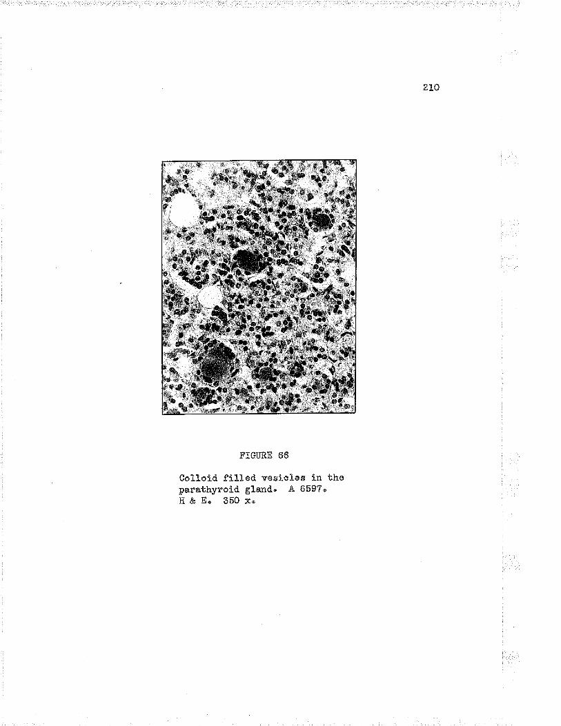
91rl
FIGURE 66
Col1otd fiLled vesiclee ín thoparathyroid gLand' .4, 659?"H&E' 550x'

211
luroen contains a deeply staining honogonous eosi.nophilie coagu.lum
Erhioh has the appearance of colloid material' The diameter of the
vesi cles varies from 50 to 40 nicra as a ruleg but there are many
oxoeptions. They a.re oonsiderod to be normal structures" The
vesicles may be scattered throughout thr: i¡/nole gland, but tend to be
arra-nged in groups at the surface or at one pole of the gland"
(cf" cit*or."7¡
_Cystic y_e_q¿gl?q. Cystic vesicles differ frorn true aolloid
vesicles in tnany features. The cystic variety are usually nuch
largern although their diameter seldorn exceeds 300 micra; their she"pe
is irregular and their lining oells show greater vaniations" A
coo.gulum, v¡hicÌr may have a reticular, globular, finety or coarsoly
vacuolated, or a homogonous appoarancee may or may not oornpletely fill
tho vosicle" The coagulum, conpared with tnat of the true coLloid
vesicleu stains less intensely with eosin' Cystic vesicles are found
at all ages and are more coünon in the inferior parathyroids tha¡r
in the superior pair" Their poeition within the glands is very
variable' The nature of these structures Ís obscure" There is no
evidence that they have a physiologioat significance' (Cf" Gilrno.tt?)
Obher -gÊ!s-" GilmourT d^escri'bes vestigal structuros a.nd. o],sts
arising from the parathyroids themseives, the thynic cords and the
th¡nnusn Col-lectivety these structures are called KrIrstej.nerrs canals"
Àpparentty the structu-res ars not connon and are seldon of clinioaj.
inportance "

2t2
Tè"_ pefg!¡y_{o'r"_q. þp{_ryg_qq," The hormone of the parathyroid gia_nds
has never been isoLated in the pu:.e state, but it ís almost ce:.ta.inly
a simple proteinz. The preparations at presont a.vailable behave as a
protein" These preparations are hydrotyzed in boiling acids and.
alkalis, and are renderod inert by trypsin and pepsins explaining why
they cannot bo ad¡ninistered by mouthlz. There is no conolusive
evidence that the parathyroids produce more than one hormone,
according to Selyez¡ who also states that ltthe meohanism through whioh
the parathyroid hormone exerts its diverse actions is stil1 not fu-lly
understood.'it Proi:abty most but not all of the aotions of the hormone
are socondary to its effect upon calcium and phosphorous metabolism2,ô
(Cf" Selye')
Caloium metabolisn" Normal values of blood calcium range
between 9"5 io 11 mgms per L0O cu'þic centimetres of whole b1ood"
Practically all of the calcium is found in the plasma, since little or
none is found. in the red blood ""11s2'12, only one harf of the blood
oal-cium is in the forn of Ca ions and thus diffusible through the
tissue menbranes" The renainder of the calcium is loosery combined
with serum' proteinslz"
The daily requirements of calcium in nian ranges fro¡n O,? to 1"0
grâ.ilrse Most of the caLoium is a'bsorbed in the upper intestino"
3.bout 70 to 90 per cent of the ingested calcium is eliminated. in the
feces, partly by excretion in the lov¡er gastro-intestinal tract and.
partly by incomplete absorption" Only small quantitios of calcium are

elçcreted in the urine, and if the blood calciu.m is less tha:r 6 mgms
per cent urinary e;rcz'etion cêB.sasø
Approxinatei-y 2 per cent of the adutt body is calciumo of v¡hich
99 per cent is found in the osseous skeletal system. The main
calcium reserve of the body is thus found in the bones, (cf. setyez)
Pþq+fhq_fqUç ¡rglgþ_olism. Phosphorous is present in the blood
pa-rtl-y as an acid soLublo (inorganic) forrn and partly as organic
phosphoric acid esters. The ino:'ganic phosphate of the seruin of
adults ranges from 2"5 to 3.5 mgms per cent, a:rd in chj-rdren from 4"5
to 6'0 mgms per cent' .A.bout 1"25 grams of phosphorous are ingested
dai1y, of vrhich about 75 per cent is excreted in the urine as acid
phosphates" (ct" su1y"2)
lúaånegigg" The metabolism
eal-oium" ,{bout 50 to B0 per cent
via the gastro-intestinal tractz.
magnesium is similar to that of
the ingested ma"gnesium is excreted
of
of
Whe:.feqlqfå i4lu_qgci!å qtrc-igg metabolism" ï,iJrile it is not
the purpose of this dissussion to inciude all those factors influencing
calcium and phosphorous metabolism, vìre ma¡r ¡n61¿ion the following to
indicate the cornplexity of tho subject, Besides the parathyroid
horrnone, other fa.ctors srhich play a part ares (r) tire calcium
requiremerts of the sketetal systemn (z) tire pH of the bloodn (s) the
proportion of calcium to phosphorous in the blood, (+) presence of
vitamin D in the food., (s) tire absorption of calcium and. phosphorous

214
froro the intestine, and (e;) tne rate of excretion of these substanceÊ"
Present theories. qg$e{qi_ru" !4q tqågtlg3 gf !þ9. hon_n-o,ngn
.)Solyoo, in his monogra-ph, reviews the theories rega.rding the function
of the parathyróid horrnone, One theory su,ggests that the hormone
lol'¡ers the renal thresitold for phosphates" ï,Iith the increased. urinary
exaretion of phos¡rhates" there fol-Iows a mobilization into the blood
of inorganio phosphates from the bones, vrhere it is present as calcíum
phosphate" "An i-ncrease of calciurm in the blood reslil-ts v¡hen the
phosphate ions a"re excretedn -{ccording to Selye, experinental
evicìence d.oes not support this theory. A seconcl theory proposes a
calcirr-m compou.nd in union wi'bh the hormone, resulting ln oa]cinm ã"
/" third theory proposes that the horrnone acts pr:imaril¡' upon the
osteoblasts, If the hormone is present in large quanti.ties the osteo-
blasts a.re tra-nsforned into osteoclasts, resulting in bone destru.ction"
lf the hornone is present in sma.lL quantíties the osteoblasts are
tstimulated to proliferate and form netu bone" Finally Se1ye" suggests
that tirere is a possibility that the hormone acts both on the
skeletal system and the kidn.or¡s"
The generaL effocts of the pa.:'a.thyro1d hormone car. be studied
following the injection of excessive doses of the extract of the
glands, or indirectl¡r by removaL of ihe glandsu
Foll-owing excessive doses of tho para.thyroictr hormone, there is
an afmost inmodiate increase in phosphate content of the urine, a
decrease in the blood phosphatese e.nd a rise in the bLood calcium
level- s "(cr' setye2)

21"5
FoJ-lovring erLirpation of the gla.nds in a"nirnaLs, or removar- of
too rauch pa.:'athyroj-d tissue in hunan thyroidectomyo there is a rapid
depression of blood plasma calciu.m, elevation of plasma inorganic
phosphates, and a dilnínu-tion of urinar¡r excreticn of phosphates,
Teta.ny slrpervenes v¡hich leads to death, if not treated, (Cf . Came"orrlz)
Hypopare.thryoidisg" Ex¡perimentally, h¡rpoparathyroidism isaccomplished usually by the excising of the lvhole thyroicì-pe.re.th¡r¡o!d
apÞaratus. ClinicalLy rnost cases develop after operative proced.u,res
on the neck" r¡.,here pe.r'athyroid tissu-e is rerrroved accidenta.lly" 0n
rare occasions, the condition deveJ-ops for some unl<no*rr,. ""*uor118,
The s1':nptoms usuali-y appear r¡¡ithin soven days after the
thyroideetomya bui may be delayed as long as two to three monthsu The
signs a:id sSrrnptorns of hypopa.rathyroiclism na.y be active or latent, and
are simifar in both the post-operative and idi,opathic caseso The
acute s¡nnptoms and signs may be sudden in onset a¡rd be manifest in. a
number of wa¡rs such as general-ized convulsions simrrlating idÍopathic
epilepsys acute psychotic s¡rrnptons, generalized rigiclit¡re or striclor
a.nd d¡rgpnss€t" Such erbreme phenomena are uu.colrunofio lviore commonly,
the attaclc is less su.dden v¡ith earl_)¡ signs of numbness, tingLing,
sensatíon of stiffness abou,t the face and moutir, and painfu-L
sontra.ctj.ons of the muscles of the extromities" (Cf , nosul8)
Foltr ovr"ing parathyroidectomys the blood calcÍum level falts to
as loi.¡¡ as 5 mgms per cent and the inorganic phosphates rise to 1eveLs
as high aÊ 5.5 mgnrs per cont in a.duLts ancl to z mgms per cent in
4crla-Ldlen o
216
There is decreased. r:.rinar¡r excretion of phospha.te, and a
clecreased urinarr. output of cafciu.rnIZ"
The distu.rbances follolving parathyroidectomy are due to
cl.istu.rbances in oaJcium and phosphorus meta,bolis*2 t" a rezuft of
whj.ch there is an increased irrita'bility of the neuromuscular system,
the slightest stimu-lus producing painful, tonic spasms or tetany"
.Tlyperpare.t-lUf.oid3Sg, Tn hyperpa.rathyroidism, there is an
inoreased. efimination of phosphate throu.gh the lcidne;,, o. lovrering of
plasma inorganic phosphøte, mobi-lizati.on of bone mineral, a.nd incroase
of caloium in the blood" There is an exaggerate<i action of the
hormone" (cf, cameronl2)
itTeoplasia and h¡rperplasia of tkre pa-rath¡rroids are listed by
10Copo'" a.s the chief causes of h¡rperparathyroidi-smu fn discussing his
series of 7O cases from the lliassachusetts General Hospi.tal, Cop"19
states that 65 per cent of 70 cases of hyperpara.thyroidism vrere due
to benign adenoma of the pa:'athyroíd' The parathyroi<ì tumor is
almost alvrays a benign adenoma. The fev¡ ma.lignant tumors reported in
the literature are mainL)¡ in patients v¡ho diil not have h¡lperpara-
T2th¡rroidism*"" In the primary tyne of' h¡rperpla-sia, the cells of the
glands are mostLy of tho v,raterclear ty¡rel9" Secondar¡, h),perplasia
resu.lts from the physiological need for greater a:nounts of the horrcone
and nay'be present a.lso in cases of vitamin D deficieneies and in
some oases of nephrj-ticl9.
Depen<iing on vrhether the urinar¡r systein or the skeletaL system
2L7
involvement predornínates and the degree of change present in each
s)¡stem, it is possible to describe severa,l types of cliseaseo The
s;imptornatoJ-ogy can be divided into three groups& i"e.e th.ose
s¡rmptoms due to (a) trypercalcenia, (¡) osteoporosisu er.d
(c) frypercalcinuria and hyoerÞhosphatnria" Hy¡rotonia, lassitude,
constipation, fatigu.abilit¡z, vreight loss" and anorexia ma.y be
attributed to the hypercalcemia. Bone pa.in, tenderness, joint pain,
fracti-:.res and deformities rnay be associated r,rith the osteoporosi Ê,
oysts and gia.nt cell tumors accornpanying the depletion of minerals
from the skeletal- systemu Pol-¡ru.ría,u polydipsiau nocturia, calculi
for¡ration, colic and hernaturia âre related to the h¡percalcinu.:'ia and
h¡4perphosphatu,ria, (cf, Âl¡right et u.I,20)

218
BÏBLTOGRAP}IY
1" Shellings D' H", The Parathyroids i.n Health and Disease"St" Louis¡ The C, '\f" T',,losby Company, 1935" pp. 355'
2" Se1ye, H'u Tertbook of End.ocrinology, L947. Acta EndocrinologicaeUniversite de iç[ontreale ]vlontreal" pp, 914,
õ. Hanson, Cited by Camerone A" T"12"
4" Co1lip, J. 8., The Parathyroid Gla¡ds" It{edicine, 1926, 911"
5o iviorgane J. R' E", The Parathyroid GLandso I" A Study of theNorrnal Gland" -A.rch" Path", 1956, 2!e10
6u Gilmour, Ju R." The Parathyrold Glands and Skeleton in RenalDisease, Londonc Oxford tniversity Press, !947.
7n Gilmouru J, R", The Normal Histoi-ogy of the Parathyroid Glands"Ju Path, & Bactue 1959, tB-:187;
B" Sharpey-Schafer, 8", Tho Endocrine Organs, Second edition, Part f"Londonc Longmansu Green and Con, 1924. pÞ' 175'
9u l"{arine, Davide The ThyroicÌ, Parathyroids arld Th¡rmus" Section XXII,in Special Cytology, SeconcÌ edition, VoI" fI, edited byE, V" Cowdry" New York¡ Paul B. Hoeber Inc.e 1952" pp, 1210"
10" towdrys E, Vn, Terbbook of I{istologys Third edition.Phila. ¡ Lea and Febigor, 1944"
11" Iúaximovr, An .Au and 1',1m" Bloom, Textbook of Histology, Fifth edition.Phila" t ïf. B " Saunders Compa.nye 1948 " ppo 700"
ir}. Co:noron, A" T", Recent Advancos in Endocrinologys Sirbh edition"London; J. & A" Churchill Ltd.s 1947' pp, 443'
1õ" Rhinehartn D" A"u The Nerves of the Thyroid, and Parathyroid.Bodies" Ameru Ju Anat"s 191-2, 15¡91.
L4, Curtiss G" l¿", The Blood Supply of the Hurnan Parathyroids,S, Gu 0u3 L950, 5Le805,
15. G::ays "Anatomyc Twenty-ninth edition, edited by T" B" Johnston andJ" 1,{hi11is. London¡ Longmans, Green and Cou, !947 " pp, 1597.
L6" trotti, 4., Diseases of tho Thyroid, Parathyroid." and Th¡rmus, Thirdedit j-on" PhiLa.. ¡ Lea & Febiger, 1968, pp . L229ø
2]-9
17' Hamiltono i;!'. J"u J, D" Boyd, and IL T'i" lriossmanu llurnan Embryology"Cambridge å tr¡,¡" lleff er & Sons, Ltd " , !945, pp, 566.
18_* Rose, E,, Ilypopare.thyroidism" Clinics, 1945, 1;L179"
L9' Cope, 0", Il¡4perparathyroidism¡ The Significance of GeneralizedHyperplasia" Clinics, L94Se !r1168.
ZO' Albright, F", Jn C. "Aub, and T,i" Bauer, Hy¡rerparathyroidism,J" Ao X[u 4., 1934, LO2zl-276"

CHAPTER SEVEN
THE TJIYRO]D GLAND

CT]APTER VÏT
THE TITTROTI] GI,AIüD
fntroduction, The thyroid gland accounts for only 0.5 per cont
of the body weightz and yet its hormone exerts ar: influ.ence over all
other tissues" IIo organ or tissue in the body is able to function
optinrally in the toi;al absence of tho thyroid. horrrtone, although not
all organs are impaired to the same d.egreel.
The norrnal histological picture varies with factors zuch as the
age of the patient, sex, food, siate of nutritiono goographical
locationo clima.ten season, and psychic stirmli" RienhoffS bolieves
that there is 'rperhaps no other organ in the human body which presents
such functional and histotogical lability as the thyroi-d"î¡ lfiarine and
Lenhardtll remarked in 19Ll that it was difficult to define the norrnaL
picture of the thyroid gland, and zuch is truo at the present because
of ihe wide normal variationsn IViost texbbook descriptions follow a
rather standardizod style but the figures given for tire norraal ranges
vary according to the observations of the au.thor in his oì¡m locality"
The nornal adult human thyroid vreighs between 2O and 25 granrs
which is not more than 0,õ5 grams per kilo of bocÌy i¡,rsig6¿' The gland
is slightly larger in ths fe¡na1e and is relatively larger for infa¡ts
(approxirnateJ-¡' 1:B5O for infants and Iz225O for adults)' The arnount
of iodine availabte in the diet is the most irnportant fastor influenc-
ing tho size of the gland" (Cf . ivlarine4)

222
Gross descrjiptioir. The thyroid gland is named frora the fact
that it superficially rese'qbles a shield (thyreos a shield.)" The
gland ís shaped rather more like the Letter nII$ than a shield, the two
latoral lobes representing the vertical olements of the let'ber, and
the isthrnus the transvarse portion"
The latoral lobes lie anteriorly'ooward the right a¡rd. loft of
tho trachoa and larynx, each resembling a three-sided pyrarnid" The
lower tip or pole reaches to within tv¡o cm of the suprasternal notch
(sirth tracheal ring)" The isthmus is very variabl-,e in size and. shape,
rn size it ma;r alnost equar a lateral lcbe or it may be a thin narroïr
band and ín so¡ne cases may be absent " Corrnected v,rith the upper border
of the isthmus is the pyramidal lobe, which is a very variable
elongated process rvith íts base below and apex extend.ed superiorJ-y for
a varia'ole rÌistance" (Cf . ¡ott5)
The thyroid g1and. has two capsules, i.ê.e a false and. a true
capsule. The false capsu-Ie is derived fron the pretracheal_ layer of
the deep cervical fascia" It is not uniform in thickness, nor does it
cornpletely surround the thyroide being strong and thiclc superiorly and
laterally, thin and imperfect inferiorly. Fine fibrous strands run
between the faLse and true capsule forrning the main support of the
gland. The true capsule is thin and. transparont and caruot be detached.
frorn the gland without injury to the palench¡rma, Thicl¡ened areas of
the capsul-e are in continuityl,vith the connective tissue found. botr,voen
the irrogula.r masses of parenclr¡nna, (Cf . Jol15)
RienhoffS'10 claims that distinct and true lobules do not existo

4,6.5
This investigator used wax plate reconstruction and. microdissectíon
methods and found that the mass of each lcbe of the thyroici is divided
and subdivided Itno" into rnany cor¡aecting or annectant bars, band.s and
plates of parench¡rna nnoorl The entire lobe is irregulari-y traversed
by septa of comaeotive tissue which carry btood vesselse nerves ar.d.
lyinphatics' The irr"eguLar rnassos of parench¡¡ma are joined to each
otìrer at one or more points" rn no instance dicl Rienhoff find a
pot"tion of the parench¡nna oompletely surrounded and separated fron the
rest of the gland" 0n the surface ti:e areas are a.rranged in a leaf-
like nanner, whilo deeper in the tobo the arrangement is more cornplex
than at the períphery. Rienhoff sr.rppor.ts his descriptions v,rith
oxcellent drarrings and photographs" (Cf, RientroffSrl0)
Thyroid tissue is translucentr pale amber red. to bright ambor
red in colour, and is firrn and elastic in consistenoy. On cross
section, the larger follicIes can be seon with the naked oye. The
follicles contain a clear, viscid, amber-coloured. protein su-bstanco or
co11oid.
B]ood supply" The thyroid is ono of the nost vasoular organs
of the body4r6r7. A volume of blood eqr-ral to tirat of the whole body
passes through the thyroid onae an hour9" the gIand, in proporbion
to size, receivos nearly fourbimes as much blood as the J<idneyB"
Four main ar*teries supply the thyroid gland¡ the paired.
superio-r: thyroid arteries arising.usu-ally fron the erLernal carotid
trunk¡ a¡ld the paired inferior thyroid arteries arising usually fron

224
the thyroid axis'rvhich is a branch of the subctavian artery. .& fifth
arterye ihe thyroideaima, is inconstant and nal¡ al'ise froin a number of
SOurces. BeSides the above mentioned there ato nunerous unnalned,
irregular arteries which are srnall- in the noriual gland but vrhich may
attain great size in some goiterso (Cf. .lott3)
Venoug drainqge-" Tho veins begin as a perifollicuLar plexus"
They follow the smaller arteries as far as the periphery of the gland
l.rhere tirey forn a conspiouous plexus on the surface which covers the
whole gland but is more obviou,s on the anterior" From this plerus a
variable set of trunlcs which may be divided into throe main groups;
(u) superior, (¡) lateral or rnid.d}e, an¿ (c) inferior' these veins
drain mostly into the internal iugular veins although there are lnany
variations. (cr" .rotr3)
Lymp-hgt:Lcs. The I5r:nphatics of the thyroid originate as a closed.
sys'cem in the for"¡r of an anastonosing plexus of capillaries in the
interfollicular spaces of the gland. The rich plexus consists of
co¡nnunicating capillar¡¡ 1¡¡nph channels r¡rhi-ch form rather dilated
endothelial sacs or srnalf pockets which vary in sizo" According to
ReinhoffS, there are three nain plelcqsese ioeoe in'uerfollicuJ-ar,
intraglandularn and extraglandlrlar" The interfol]ieular plexus,
mentioned above, which unlike the blood capillary system is not devoted
to separate and distinct foIlícles but rather to spaces bet'¡,¡een the
groups of follicles' The second division or intraglandular plexus is
composed of rather large connecting trunks situ.ated along the course

/.áÐ
of the septa and for¡ning freguent anastonoses with each other and v¡ith
the interfollicular plexus" The extraglandular vesseLs on the outer
su.rface of the gland lie external- to the bLood vessels er.d internal to
the fibrous true capsule enclosing the thyroid.. From the latter. plexus
originate the still larger collecting trunlcs which acconpany the
superior thyroi<l vessel-s to the cer"vicaL lynph nodes alßd a1 so to the
trunks v¡hich form a plexus in the pre-tracheal fascia oraining into the
mediastinal lrvmph nodes. (Cf " RienhoffS)
i{re, The nervo supply to the thyroid is chiefly s¡n'npathetic.
Fibres fro¡TT1 and T2 pass through the inferior cervical ganglion,
'rvhere some are interrupted, others passing on tc the middle and.
superÍor ganglia where thry are interrupted, Accornpanying the vessels
to the gland and in the glend, the nerves form a rich perivascular and,
perifolÌicular plexus. (Cf. Joll3)
Vagus fibres reach the gland via the superior and the recurrent
laryngeaLl""ou"3.
Embryological note. The thyroid begins at the 1"32 m¡n stagez
in the endodern of the floor of the pharynx as a thickening whioh
soon evaginates and by the 5 mn stage has the shape of two flask-Iike
lobes attached to a common holIow stalk which connects with the
pharyrex and is known as the thyroglossal ductf9. The thyroid primor.d.ia
has an intinato reLation with the aortic sac, The thyrogrossal d.uct
is tho most important part of the thyroici primordia, small contrib-
utions being received from the ventral components of the fourth

226
pharlrngeal pouoh, v¡hich Later fuse with the rnain prirnordia" rilith the
development of the neck, the heart and great vessels become vridely
separated frorn the thyroid, although remnants of thyroid rnay be fou_nd
attached to the arch of the aorta in adult life, Norraally the thyro-
glossal du.ct is reabsorbedo Rarely it nray persist, in whole or in part,
cau.dal- or cranially, a.s a. cyst or a duct. Occasj-onal-Iy the cau.dal
portion persists as the pyranidal lobe"
Initially the g1a::dular tissu.e consists of a solid mass of
cells. Branching plates one or two celis thick soon develop and these
aro fu,rtirer separa.ted by an ingrov,;tir of oonnective tissue and blood
vessels, The formation of the folliclos is a complicatod series of
clranges' By tlne 24 m-.4 stage the definitive follicles are su-rror:nded by
mesencLr¡rme7" Colloid appears in 60 mm embryoslg, although the colLoíd
is not abundant until after birth2" (Cf " Eamilton, Boyd and },fossmanl9)
thu *jolgq"gÈ= a:1qtomy. The u.ltinate histological unit of the
thyroid gland is the foLlicle, grolrps of v¡hich make up the mass of
parench¡runa" The fol]icles exe enbirely discrete separate individual
unitsl0" The follicles are bound together closely by interfoLlicular
connective tissue uhich carries arterioles, capillaries, venules,
nerves and lymphatics"
The tissu.e of the thyroid may be divided for descriptive
purposes into the parench¡rma (tollicles) and the stroma (interfollicular
tissue). Occasionally sma1l portions of thynu-s or parathyroid may be
founcì in a section" fn certain pathological conditionsg l¡rmphatic

ôôø
tissue, either diffuse or in the forrn of lyrnphatic nodules nÐ.y be found
ihr"oughout the gla.nd"
The vascular bed about the follicl-e is veÍv richl0" The small-er
capil-laries a,rising frorn the arterioLes j-n the inter"follicular
connective tissue forre an inti¡late netv,¡ork abor.lt each follicfe similar
to a basket"v'¡ork aror-rnd a ba117" The L;anphatics do not form a similar
netv¡orlc and are external to the blood vesselsS. Details of the l¡n-nphatic
system rvere given on page 224.
I O T}TE FOLL] CLES
Size" l,{an5r of the foll icles var-rr in size frorn 0.2 r¡rn to L mm
in diarneter, the great inajorit;r being small and in lnanv scarcely any
lumen is presentlo. Llost nornaL follicles ranqe from O'5 to O"B mm in
d.iameterlf " There is great irregu.lari.'b-v of size of the folI ic1es.
Larg;e and srnall may be indiscrilninately mixeci throughorr.t the gland.
9Lgpg. The shape of the follictes varies as mrr.ch aò the size.
No two follicles a.re sirnilar" Their shape ranges from aLmost a perfect
sphere to al-most a perfect cube" l{o psuedopodial outpou,c}rings of the
wall rn¡ere noted by Rienhoff, v¡ho founci that all major irregularities
in the epithelial v,¡aLl occur insicle the follicle" No true indication
of the shape of the foll-icl-e ce.n be gained from the studj-es of siirgle
sections" (Ct" Rienhoff)
Colloid' AlL the follicles .are f iIled rn¡ith clear, amber yelloi''i
viscid material, termed colloid, v¡hich is essentially insolubLe in

228
water, aLcohol- or etirer and ivhen coagr,rla.ted is easj-ly stainable with
eosin or acict d¡res6"
In the fixed state the colloid is frequentl.v separated frorn the
epitheliu.m by clear spaces, giving the appearance of a spiny border in
the most extrgme cases" The clear spaces are artefacts because '\¡re
icnow that the lumen of the follicle is completely filì.ed du,ring lifelz.
Vacr,-olation does, however, vary yfith the degree of activity of the
gland, being most marked in hyperthyroidism, and less marked in h¡po-
thyroidism or in the resting gland, Thus it appears that the artefacts
are more easily procluced in the colloid of the hyperactive gland.
Tho colloid is chiefly acidophilic but inay exhibit patchy or
diffuse basophilial2. the d.ifference in staining reactions of the
colloio in the follictes varies v¡itl: the degree of fr''.nctional activity
at the momentl6. There may be d.ifforences of staining in the solloicl
of the same follicleo The most acidophilic staining is fonnd in states
of hypoactivity.
The amount of colloid accumulating in tho vesicles at one time
d.enotes the degree of activity of the glandlT" During hyperactivity of
the gland, there is clecreased anolrnt of colloidr increased peripheral
vacuolation a¡d basophilia, v¡hil-e during hypoactivity there is
accumulation of colloid, decreased vacuolation and acidophilíal8. It
i.s generall.y agreed that the oolloid is a storage product and contains
large quantities of thyroic hormonez,
Epithelium. The epithelial cells form a continuous lining in

229
FTGURE É'?
Normal thyroid tissuea shotring the nor"rnLirr"egula.rit;r of size and sha.pe of thefol-licl-es, E & E" l.5O x" (so28-ø"a).

,3r)
ffi."&.W$,ø$#
FTGURE 68
Sa¡ne tíssue as Fig. 67 to sho'¡r more detnilof the errithelium" 400 x' H & E'

4,) I
the follicle arranged in a single 1ayer" irlo true base¡nent membrane ispresent6rT. The epithelir¡.m rests on a fine layer of condensed
cor¡rective tissue fi-brils,
Numerous types of cel1s have been doscribed, most of which
require special, rnethods for theír demonstration" Not all investigators
are in agreement regarding the t¡4pes of oells. i[iost authors think
that the different cells represent variations of a single cell t¡rye,
Others state that some of them are definitely degenerating.
Iiost of the cells have large vesicular nuclei" prominent
nucleoli and pale staining cytoplasm v'¡hich often contains granu,les
r,^¡hich stain liice coLloid and which are located in the pole norL to the
lumen" Occasionally a ceLl shows a dark, shrunken nucleus and stains
homogonously with acid dyos, sinilar to the col1oidl3.
The forlicular cells vary in height, depending on the degree
of physiological activity. uader ilor.-nrÊ.r conditions, the cells are
cuboidal or low corumnarl4. During hypoactivity the cells become
flattened and during hyperactivity they becone tall a-nd columnaru
The variqlions. From the foregoinê remarks, it is seen that
the picture is extrernely vari-ed., The size of the follicIeo the
arnount of its colloid, the staining reaction of the colloid, the
amount of vaouolation, the height of the epitheliu.m and the amou.nt of
interfollicular tissues especiall]' the lymphoict tissue, all varSr si!þthe a.ctivity of the gland"

/, q) ("
The500
FIGURII 69
thyroid in hypertlryroidism, H & E,xo (rzos-+p).

,í ).1
TheH&
FTGURE 7O
thyroíd gLand in coJ.loid goítre'E' 75 x' Jl 6043'

the folloruing table, given in more detail ly Cor*.dry18
summarizes the variations of the histological conponents of
gland. in states of hypoactivity and. h¡reractivity"
Z3a"
t
the thyroid
Hyperactivit¡r Hypoactivity
Colloidamount -donsity -
hor:rogeneitycells in -
d.ecreases (absorption)d.ecreases (nurnerou s
vacuoles appear)markedosqasional
increases (retention)inoreases (few
vacu.oles)absentquite numerous
Folliclessize -shiape -
smallvery irregular
very -largemore unifornr
Follicular cellssize - increased decreasedshape - columnar greatl¡r flattenednrultiplication - rnitoses occasional mitoses rare
Interfollicular ti ssueblood vessels - engorged less dilated than
llnnphaties - distend.ed r"Tl"ütt"e thannormal
cel1s - histioc¡rtes more histiocytes lessnoticeable' l¡rrnpho- noticeable' lympho-c¡rbes considerably cytes slightlyinareased" increased.

te.Ê.
ff . THE FUi.JCTION OF TiÌE TiJYROTD
fntroduction" I[ost theories of thyroid fu.nction recognize that
the secretion of the gland is linked with the oxiciative processes of
all tissue coll-s" Hovrever, it is not possible to exple.in all the
ma¡rifestations of hypothyroictism and h¡4perthyroiclism on this assLlmptionl6"
The hormone (or horrnones) prod.uced under physiologica.l conditions and
its node of action are not krrorrnz, More than one compound which is
physiologically active can be obtained by extraction of thyroid tissue,
and although the number is considerably smaller than that obtained in
exbractions of the adrenal cortox, there exists here a somewhat
parallel situation. ltr¡hether these conlgou-nds represont the hormone, a
part of it, or its precursor has become a moot cluestion. The already
voluninous literature on all the aspects of thyroio physiology
continues to increase rapidlyr and ín the disaussi-on to follov¡, the
monographs of Came"orrl6 and Selye? nuou boen used almost exclusivelyo
The horrnone. By a series of fractional extractions of thyroid
tissu.e¡ three iodine containing compounds are obtained¡ diiodotyrosine
and thyroxine which are iodized amino acids, and tieyroglobu.lin which is
a psuedoglobulin"
Diiodotyrosine contains 58'7 per cent iodine and has been
isolated fron thyroid tissue, several 6ources othor than thyroid
tissue, aï.d from enzymic hydrolysis of th;rroglobulin. It has been
isolated in crystalline foriri and has been found to have only a trace
of hormone activitSa if any.

t,50
Thyroxine contains 63"5 per cent iodine and v¡as isolated first
by Kendall in 1919, and in L927 Harrington and Barger established its
formula. Thyroxine, which is in the for¡n of colou.rless crystals, has
tvro isomers" 0n1y the I-thyroxine has physiological activity,
l-thyroxine closely imitates all the actions of functional thyroid but
it probably does not represent the form of the hormono v'.hich is
normally secreted by the g1and" 0f the substances no¡,v available in
pure form, thyroxíne possesses the greatest biologioal activityl6"
.&ny change in the structure of the thyroxine molecule rednces its
physiologicai activityg"
Thyroglobulin is a pseudoglobulinl6 and is the only protein in
the colloio9" It has a molecular weightl6 of appror-irnately 70O"000"
The substa.rroe possesses greater physiological activity than its
thyroxine content vrould indicate.
A number of compounds are inportant in the synthesis of
thyroxine. Thyronine is a compounci which results when the iodine is
removed frorn thyroxine" ït has no physiological activity. Tetrabrom-
thyronine is similar to thyroxine, except that bromine replaces the
iodino of thyroxine. It ha,s only slight physiological activitys
suggesting the specificit)' of the iodine fraction" Tyrosine, the
mother substance of thyroxine, is inert, Diiodotyrosine, resulting
from the substitution of io<Ìine at 5r and 5t for hydrogen, has only
a very slight trace of activity, so sliglrt tìrat some cìeny any"
The foll-ov,ring structural formulae are given by Se1yez;

23v
liHr
Ho-{----\ | """UCH2-c-c00H
H
TTROSINE
NH^t:-cHz-Ç -c00H
I
H
DÏIODOTYROSTI{E
T}I]TRONTNE
NH^ltI
-CHr - C- COOHI
I
tÇ.Fl
FzI
-CHZ- C -C00HI
H
THYROXIXiE

z38
Cameron revier¡¡s the problem of the nature of the true honnone,
Accordi.ng to thls revievu,er, some a'-rthors consicìer that the hornone is
neither thyroxine nor thyroglobulin but an iodized albumin, Another
group, Cameron states, consider that the th¡r¡ei¿ horrnone circu.lates as
an integro-I part of the plasma proteins in nuch the same'r/vay as
antibodies" The highest concentration of iodine is found with the
alpha and beta-globulinsu while the major part of it is present in the
plasma aLbumin fraction, (cr' cu.*""onl6)
There is littIe evidence that the thyroid produces normally
several hornonesZ" The exact na'brire of the hormone in the blood is
not lanov¡n, It is estimated that the human thyroid produces daily
anounts of the hormone equivalent to one-third mgm of thyroxine, the
dai.ly intake of iodine requirod for this production being betv¡een 0"1
to 0"2 mgm. (Salter - quoted by Cemeron)
The actions gf the hormone" The results of thyroicioctomy and
feeding dessicated thyroid tissue have given an indication of somo of
the functj-ons of the thyroid hormone. The rate of response to
thyroidectomyr substitution therapy after thyroidectomy.r or feeding of
thyroi<i to excess, is not ra.pid v,¡hen the action of thyroid is cornpared
wj-th the action of insulj.n or adrenalin" If an organizn is deprived
of all th¡'vei¿ tissue, the basal metabolic rate fa1ls frorn 40 to 45
per cent beloi¡¡ normal in 60 to B0 dayso The decline in the basal
metabolic rate follovrs a predictable curve, In the normal adult,
1 grain of desiccated thyroi<i by mouth, or 1 mgm thyroxine

239
intravenously causes a rise of 2.8 per cent in the basaL netabolic
tate" Upon the ad¡rinistration of a singLe dose, the rise in the basal
metabolic rate does not begin u.ntil seven hours lator and this rise
reaches its maxi¡num aftor one or two weeks" the single d,ose iaay
continue to exert influenco for a period up to two months af'ter
adrninistrationn (cr" s"lyuz)
The effects of thyroidectomy and the administration of thyroid.
exLract are listed iir the following table v¡hich is based on material
fron Selyez and carneronl6.
Organ orSystem
Effeots ofTìryroidectonry
Effects of FeedingDessicatod Thyroid
Body ternporature
B "l[nR,
Gluooso tolerance
Total blood lipidespeciallycholestorol-
Urinary excretion iV2
Blood iodine leve1
Calcium-pho sphorusexcretion
Growbh in young
Diuresi s
lowered
drops to -40%
Lncreas9s
rr- se
decreases
decreases
decreasod
retarded
tendency toaccumllate i{OH
slightly raised
sl-ightly raisedexcessive dose rise
up to plus 10Ol
decreases
deoreasod
incroases
inareases
increasod
if toxic - retarded
increases

240
Organ orSystem
Effects ofThyroidectomy
Effects of FeedingDessicated thyroid
Card.i o-va scuL arheart sizepr.ilse ratesystolic BPperipheral
Lytaphoid systoml¡"nph nodesspleenmarrowth¡rmu s
lvluscle systenr
Central nervoussystem
Accessory sex organsbotìr sexos
Menstruation
decreaseddecreaseddecreasedvasoconstricti on
show moderateinvolution
decreased strength
early opera'uion -inipedes devolop "of intelligence
late opera'bion -decreased activity
involution
amenorrhoea
increasedincreasedincreasedvasodil-ation
small doses - sti¡nulate
toxic do ses-involution
loss of strength
increased activity
involution
arnenorrhoea
((((
((((
Iodine" The olement iodine is vridel¡' distributed in nature,
although few materials are rich in it. The iodine content of plant
life, animals existing on it, and their products var)¡ with the iodino
content of the soil of a district" (Ct, Caneron)
Ìviost endocrine glands contain relatively mot'e iodine than non-
end.ocrine organs, The thyroid is the richest source in the body16.
The total iodine content of the hu,man body is approxirnateLy 50 rngmss
and of this the thyroid contains 10-15 mgms and the nuscles 25 nrgrnsZ"

z4I
Iod.ine is present in both tl:e cells ancl the colloid of the thyroidl6"
Forty per cent of the thyroid iodine is thyroxine iodine, the other
60 per cent corresponds to diiodotyrosinez,
Iodine is absorbed mainlv fro¡n the intestine and is excreted
nrainly by the kidney. 0n1y a part of the iodj-ne liberated frori the
decomposition of the hormono is excreted, the retnainder of it isD
fe-Uïl-I1Z€O o
The normal blood iodine level is froro 2"4 to 5"5 per cent and
in the ple.sma from 4 to 10 per centz.

.)Àt
BIBLIOGR,q,PHY
I, Carlson, .A' J., The Thyroid, Pancreatic Islets, Parathyroids,Adrenals, Th¡rmus and Pituitary. Chapter 13, in Problemsof Ageing" edited by E" V. Cor',idry. Baltimore¡ The Y'iilliamsand nÉilkins Company, 1959" pp. 758"
2u Se1yeo Hans, Terbbook of .Endocrinologys 1947" Acta Endocrinolo¿;ica,Université de llontréal. pp" 914.
3" JoIl, Cecil 4", Diseases of the Thyroid Gland. London: t''l-illianHeinemann (tøeaicat Booles) Ltd"' 1952. pp" 682"
4, i',[arine, David, The Th¡rroid, Parathyroids and Th¡rmus" Section ]CilI,in Special Cytology, Second edition, Vol' II' edited byEo Vo Corvdry. i'iew York: Paul B. floeber fncn, 1932. pp" 1210.
5u Rienhoff, I'iin' Fo¡ Jro, A i\*er;u Conception of some l"'loi"bid Changes0ccurring in Diseases of the Thyroid. Glend Based onExperimental Studies of the l\Jormal Thyroid Gl-and and theThyroid in Exophthal¡nic Goitre. li'ledicine, 1931, 19t257 "
6o Sharpey-Schafer, E., The Endocrine Organs, Second edition, Part I.Londonl Longnans, Green and Co", 1924. pp" 175.
7" Àiaximow, A" A' and -!'"Im' Bloom, Textbook of Histology, Fifth edition.Phila,! W, B" Saunders Cornpanyr 1948" pp" 700"
B o Burton-Opitz and Jldvra.rds, Amer o J" Physiol " , I9I7 ' xlii. Quotedìry Sharpey-Scìrafer (O)
"
9o Lermal, J"¡ The Physi-clogy of the Thyroid Gland" J'AolfuÄ's 1941,. L]-7 t549.
10, Rienhoff, liîm, F', Jrn, Gross and i'Íicroscopic Structu-re of theThyroid Gl-and. in i'rian. .{rch. Surg. , lgzg, 19(Pt "1):986"
1I. ll{arine, Do, and C" I{. Lenhart, lhe Pathological .Anatorny of theHrr,man Thyroid Gl-and" "Archo Intu lvled., 1911, 7:506.
1'2" Co.rdryr .8. V', TexLboolc of ilistolcgyn Third edition.Phila": Lea and Febiger, L944"
15" Bremer, J' L", Terbbook of Histology, Sirbh edition, rer¡¡rittonby H. L, {ieatherford" Phile.¡ Tho Blakiston Conpanyo L944'pp.723ø '

2 L'z'
L4, Baileya Fo R., Textbook of ilistologyr Tin¡elfth edition, revised byPu E. Snith ancL v'¡. lri" Copenhaver. Baltimorea The 'lTilliamsand T'Iilkins Co'¡ 1948" pp. 781"
15. Best, Cn H" and i{, B, Taylor, T}re Physiological Basis of ivledicalPractice, Fifth edii;ion' Baltimore¡ The Williams a:rdtrTilkins Co.' 1950" pp. 1330.
l¿j. Cameron, Ao Tu, Recent Advances i-n Endocrínology" Sixth edition"London¿ J" & A" Churchill Ltd", !947" pp. 445"
17, Sharpey-Schafer, 8", Essentials of Histology, Îv¡elfth edition'London¡ Longmans, Green and Co', 1929"
LB, Cowdrys E" V"o "4. Te:rtboolc of l-IistoJ-ogy, Second edítion.Phil-a.: Lea a.nd tr'ebi¿,er, 1938. pp' 600'
19" HamiJ-tonr'tì¡. J,¡ J. D, Boyde and ä"'vV" li,iossman, Human Ernbryology.Cambridge; l¡i" Heffer 3c Sons Lirnited, 1945'
20" ir,ioeÌrlig, R' C', I{¡4perth¡rroidisrn" Clinios, L943' 1¡11-l-5"
2L, Stein, H" È", the Ìvlorphology of the Fol-licle of the iformal AdultHunan (Aantu) Thyroid Gland v'rith Special Refereilco to theIn'crafol l-icuf ar Epi-bhelium and ir,ior;ohogene si s of Follic.l-es.S, Afr" J, Ivled. Sc", l"958o 3;15"

CHAPTER EIGHT
THE PANCREAS

CIIAPTLE. VIT]
T}TE P¿\iIICREÀS
ïntroduction. Nerû to the liver the pancreas is the largest
consists ofgland connected with the gastro-intestinal tract and
exocrine and endoorine tissue,
The glancl lies in the retroperitoneal space at approxirnatoly
the level- of the second and third lumbar vertebrae. Tt is subdivided.
into three parts, the head, body and taiI. the right erbrenity of the
gland, being thicker ancÌ wider, forms an enlarged bul-bous mass wllicÌr
is called the heado The micl portion is the body, a.nd the left
extremity which e:rtends alnost to the spleen is called the tail, The
divisions are arbitfarf: no sharp lines of demarcation being presento
In the aclult human, the gland measures frcm tln¡ent)r to twent¡'-
five centirnetres in length, and vreighs from 65 to 160 g"*.1 "
tOgilvie' gives the average figure of 2.6 grarns at birth, and at
twenty-one years it is stabilized at approximately 66 grams. There is
a r¡ide range of normal values"
The gland is soft in consistenc¡ro and on cross section is pale
pink in colour due to the vascularity. The lobules vary in sizen the
larger lobules being visible to tire naked eye" .A thin layer of
connectivo tissue covers the gla:rd, but does not, ho'wever, fo¡m a true
cap sul e o
ó+o
I o TIIE EXOCRTÌ{E PÁI'ICREATIC ÎISSUE
The pancreas i-E a corrlpound acinous gland vrhose lobules are
bound together by loose connective tissue through vrhich ru.n excretory
ducts, blood ¡resselse nerves and lynphaticsu The acini var;' from
sphericaL structures to short tubules, and are made up of a single
layer of pyrarnida.l ce1ls resting on a deLicate reticular mernbranel"
Tho secretions ernpty into a duct s¡rstem v¡hich penetrates the whole
gland, (Cf" post)
The acini. The acinar cell has trn¡o zones when seen in the
stained section" the apex of the cell next to the lumen is closely
paclced with coarse, round, highl¡' refractile bodios or granules"
These are the zJrmogen granules which stain brilliantlrv r,vith acid d¡reg
such as eosin, phloxine and fuschin" Their size is fairly u.niform,
although some varia"tion is fonnd" The number of granules varies vrith
secretory activity of the gland, During fasting thoy accumulate to
fill more and more of the apex of the cel1; while du.ring secretion of
pancreatic juice, their number decreases" This observation v,ras first
described. by Heidenhain9 in 1875, and observed in the living a:rirnal by
cove1110,-whose v¡ork is d.iseussed. in connection v¡-ith tho physiology
of the acinar ce11s on page 260" There is a variation in the number
of granules found in the celrs of different acini¡ since the rvhole
gland does not secrete at one tirne, some acini producing pancreatic
juice lr¡hile others are in a rosting statell,

24Y
FTGURE 7U
Dre-wing to shor'¡ the rej"etion of tho duets,acinar ce1lsu s.nd the centro*acinar cells"Redrar,¡r e-nd modified after Bensle¡r, Lglt"

ö,4 n
FÏGURE 73
The zymogen granules in the acinu-s celis, Oilimmersion* L000 x" lviesson Triahrone Stain.

/,+Y
ItIo granules are found in the basal portion of the cell v¡hich isoccupied by the cl:roni<iial substance, This substance stains intensely
blue v¡itir the basic stains sr-rch as hematoxylin and totuidine blue,
The stai-ning is most intense nerb to the reticular nembrane and
gradually fades in intensity as the apox is approachod, The chromiclial
substance is homogenous in the living statelo and after certain
fixatives. After fixatj.on in fluicis containing osmic or chromic acids,
no striations are de¡nonstrable. "A,fter fixation in fluids containing
acetic acici, such as Bouints or Zenkerts fluidso fíbriL1ary structures
are seen and it can be shor,',rr by speoial rrethocìs that they correspond.
to the large fil-arnentous mitochonclria and are d.ue to thenI2. The
striations are vertically placed. on either side of the nuereus, and
horizontal below it13.
The nucleus of acinus cetl is characterj.zed by the richness inchrornatin v¿hich forms a fine netv¡ork of threads and granuleso Granules
are in contact w'ith the inner surface of the nuclear membrane, One or
more large spherical, strongly acidophilio bodies are found vrithin tho
nucleus end are lirrovrn as the plasnosomes or nuc1eoli" Applied to the
surface of these bodies are granules of chromatin, celrs with two
nuclei are rare in man15"
the 9uct_ system. T¡rpicar pencreatic ducts are accompanied by a
system of tubules. The two pancreatic d.ucts" main (T{irsungrs) and.
accessory (santorinirs) carry pancreatic juices to the d.uodenu.m. The
tubures rnrhich have no rumon, according to cov*dryl4, consist of a cord

250
of celIs,
The duct of jifirsi-'.ng traverses the pancreas from the tail to the
head.. It begins in the tail as a fino delicate structr-rre and gradually
becomes greater in diarnetor as it approaches the head and receives in
its course nuaerous tributa.ries which open into it practically at
right angles' The duct is situated closer to the anterior su.rface'than
the posterior surface' It opens into the duodenurn b)' obliquely
penetrating the coats of the d.uod.enum side by side with the common bile
duct" They end in a pa.pilla-like elevation of the mucou.s membrane,
the short common cavity of r,vhich is the a:npui.Ia of Vaterll.
fn the head of the rnajority of glands there is a shorter duct"
the accessory pancreatic duct or the duct of Santorini, the relations
of which are,very variable" It usually opens by a separate orifice
into the descending portion of the duodenum and is always con¡ected
with the main d.uct by a transverse branch. iríore rarely it empties
into the main duct or malr be entirely absentf5,
The ducts at their proximal end aro lined rvith epithelium
closely resenbling that of the du.odenumn The celIs are tall and
columnar vnth distally placed elongated nuclei-. True goblet ceLls
occur vrith varying frequency and are well demonstrated by staining
rvith periodic acid. Near the duodenurnn the epithelium and the basement
membrane are thro'nn into folds. This appearance disappears as the
ducts becoine finer' The epithelium has a striking simiLarity to that
of the erbrahepatic biliary system, The ducts are su.pported by
relatively thiclc ba¡rds of eonnective tissueo chiefly oollagen fibres

Common Bile Duct
Wirsun g
F]GURE 74
Diagra.m shor¡'¡ing the çcrønen vÐ-rís.tions ofthe relation* betv¡een the corsnon bile du"ctend the duet of rili-rsultEo

252
vrith a fev,¡ elastic fibres' The epithelium changes from the columnar
t¡rpe to the cuboidal type as the du.cts becor,le snaller in calibre, and
the nucous secretion becomes less and. Ioss.
As the ducts enter the acinuso there is noted a type of cell
which is distinct from either the acinous ce1l or the duct cell" These
are the centro-acinous cells, so caIIed by Langerhans. Their shape isfusiforrnu often flat, and in some instances have short projections
which penetrate between the secreting celIs. lfany authors feel that
they represent ths terrninal portion of the duct as it meets the acinus,
The cento-acinar ceLls have pale staining acidophilic cyboplasm a.nd
possess no zymogen or other specific granules and no chromidial
substance. The nucLeus is usually oval, rich in chromatin and simiLar
to that of the acinus cell" The acidophile nucleolus, so prominent in
the acinus ce}l, is not conspicuor."l3.
Tubgles,. i'Ituch less obvious in a stained section is that system
of tubules described by Bens1eyl7. By injecting vital dyes, such as
pyronin and neu.tral red, .into the aorta of an aninal kil1ed by bleedingo
Bensley was able to demonstrate these tubules v¡hich escape detection
by ordinary injection methods. This system of tubules forms a
tortuous, intricate series of anastomoses which eonnect the duct and
branches of the duct. The tubures vary in thichaess from 12 to 27
micra in the guinea pig, and are sometimes thicker rn¡here small isletsare attachod, Bensley described the lumen as continuous throughout,
although it rnay bo as narrow as lo5 micra in diameter in placeso . .

FÎGURE 75
A group of centro-s.einer ce3ls in the pa,ncreas cfa.n p.dult me"le, 800 x" Henatox¡¡lin and eosin staine

, ÉA
FÏGTJF"E 76
-A¡rrother two groups of ceniro-s.einar ceLlsin the same tis*ue as Figure 75. 800 x,

t Ãtr
single islet ce1ls are sôen here and there, Also attached to the
tubules, although less frequently tl.ran the attachrnent of islets, are
srnall aoini, v¡hich vary frorn a singre acinou.s to a group of acini. ïn
some cases these acini arise from the ducts which lead to the islets.(cf . BensleylT¡ .ê,ccording to covrdryl4, tl,.u"" tubules do not carry
secretion" A further discussion of the tu.br:les is given in corrnection
vnth the islets on page ZZ4.
The blood vasc-ular supp-]y" Pierson3 gives an excellont account
of the arterial supply of the pancreas" The following tabre and
guotation are from his paper"
The arterial supply' of the pancreass as found by Fierson5 in the
study of fifty câsoso
Antorior superior pancreaticoduodenalin L00 per cent of cases
form the anteriorAnterior inferior pancreaticoduodenal arcado
in 100 per cent
Postorior superior pancreaticoduodenalin 96 per cent
fom the posteriorPosterior inferior pancreaticod.uocÌena1 arcade
in 100 per oent
ïnferior pancreatic in tCO per cent (t¡narton)
Superior pa-ncreatic in 54 per cent
Pancreatic branches of the splenic in 1OO por cent
Pansreatic branches of the gastrod.uodenar in 62 per cent
Pancreatic branches of the hepatic in 10 per cent

256
Typically the pancreas is supplied by two arteries from thegastroduodenal, tlvo from ihe superior mesenteric, and one frorneither the spl-enic, hepatic, or cel-iac arterieso fn addition tothese a variable number of snall arteries arise from the splenic,gastroduodena-l, and hepatic. ÎÌre larger vessels arising fron thegastroduodena] artery are the su,perior p?ncreat-icoduodenalarteries (right pancreaticodu.odenar or ffise eretv¡o in nurnbero an anterior and a pqgterior,. They anastomose v¡ithc or r e sp onai ng ant-effiãã p o st erñîffi;io r p en c r eat i c o d uo d.enalartqries (reft pancreaticodu-odenal) from the superior mesenteric.These vessels forin tv,¡o arterial arcades, one on the posteriorsurfece of the head of the pancreas, the othor, except for itslorvermost part, on the anterior surfe.ce, They are respectivelythe posterigr_ and the antqrior argades of ths pancreas. The twoinferior pancreaticodu.odenal- artories usuarly arise in a connontrunk from the superior mesentoric, the comÌîon infe{-ior pancrea!iog-duodgnal altery"
-The slrpe¡,Lor pancreatiffterFffi i€ast-constant of any of the large arteries to the pancreas. lÏhenpresent, it arises from the splenic, hepatic, or d.irectly from theceliac arteriesu rn addition to the arteries mentioned, aninferior pancreatic arteJJ¡, which passes arong the inferior marginof the body to the tail, nay take origin fron the su.periormesenterico the anterior superior pancreaticoduodenal" the inferiorpancreaticodu.odenal, or the suporior pancreatic arteryo
The relationship of the arterj-es to the veins is fairlyconstanto Every artery here described has a concomitant vei-n,Accorcling to the r¡r¡'iterr s observations, as werl as Petránt s, theveins usually lio nearer the surface of the glancl then the arterios.The arrteri,or superior pancreaticoduodenal vein drains constantlyinto the right gastroepiploic vein, which is a tributary of thesuperior ntesenteric. The posterior superior pancreaticodtrodenalvein almost alrvays drains directl¡' into the porbal voi.ït. The lov,rerveins empty directry into the superior mesenteric or one of itstributaries' The posterior inferior pancreaticoduodenal vein mayempty into the inferior nesenterj.c, The splenic vein, which issometimes imbedded in the. gland, receives several veins from thebody and tail" (Piersonó)
The intralobular pancreatic circulation has been reported by
Oppenheimer and hiann4, ruho used neoprene injection e¡td corrosion
methodsn TÌrese investigatcrs fouucl thatc
1u .Anastomos<¡s e::ist betrveen i.ntraLobular arterioles ancL alsobetween acinar capillary fields fed by different arterioJes¡,vithin any one lobule.

257
2o Bl-ood rnay reach acj.nar capillaries via islets of Langerhansor directl;r withorrt passing throu6h islet tissueu
3" The potential intralobular capillary ciroulation is verylarge '
The blooci suppl¡r of the islets is ciiscussed on'page 279n in
connection lvith the islets of Laagerhans.
Uglgi supply' S¡anpathetic preganglioníc fibros arise fron cells
in the interrnedi-olateral column in the çínal oorcl ancl :.each the celiac
plexus via the splanchnicso . Post ganglionic sympathetj.c fibres frorn
the celiac ganglia reach the pancreas via the hepatic" superior:
inesenteric and splenic plexu.ses" Stimulation of these fibres has no
¡rarlced effect upon pancreatic secretions. Tlre pa.rasFopatheti.c
preganglionic fibres arise j-n the dorsal motor nucleus of i;he vaêus
and roach the pancr:eas via the vagus rlerve" The postganglionic fibres
arise fro¡n celIs, probably in the pancreas or closely associated with
it. The fLlnction of augnentiirg both the external and internal
secretory activity of the pencreas iras been ascribed to these fibres(Cf. KuntzS) " The fibres accolnpanli the arteries into the gland. and
end aboui the acini in fine terürinal-sl" Tire islets have an abund.ant
supply of ner.ve, both nryelinated and unmyolinated6¡ however, it is
difficult to a.ssess the effects of this nerve control, since at the
present tine there is no nethod for estiinating the amount of insulin
in tno bLoodTo
The_ Iy¡rphatic draínag-e. The lynrph d.rainage of the pancreas is

adequ.atel;r described to d.ate21' caLlander8 descriì:ed the rynphatio
drainage as being extensive, the vessels draining into the pancreatico-
splenic nodes at tire hilrrs of the spleen, and into the pancreatico-
duodenaL and preaortic nodes near the origin of the srrperior mesenter.io
artery n
The Phys.i.ology "{ !g exocriÍre- p_ancrerus.. Yrfithin a fer,'¡ minutes
afber the ingestion of food, the flow of pancreatic juice increases"
reaches a maximun in two to three hours, decreases slow1y to reach
a basal- rate in five to nine hoursl8" Both nervotrs and. l:.umoral
mechani-sms control the flow of pancreatic juice" Secretory fibres
from the vagus and synpatnetio innorvate the aciniu The vagal
secretion is small i-n amorrnt and ìras a hi_gh content of organic
substances and fernentsl9" Pilocarpin ¡rrocluces the samo effect when
ínjected" Stimulation of the sy'npathetiò fibres causes the produotion
of a scanty secretion'r,vhich is similar to that causecl by vagal
stinuLatiorr2O "
];'ühi1e reflex nervous control of pancreatic secretion occursr
more important are the cheinical hu.moral inechanisms. These apparently
play the chief rore in the production of the pancreatic digestive
juicesl8,
Bayliss and Starl iog22 o in 1902, shorved. that if an acid erbract
of the epithelium of the duodenun ancì the upper portion of the s¡nall
intestine were injected intravenously into an animal, there resulted.
a copious flow of pancreatic secretionu Injection of the acid. alone

9troa¿¿
did not produ,ce the sarle resul-ûs" They post'uLated the presence of an
inactive su.bstanoe v¡hich they caLled pr_qqecr.etin in the intestinalmu'cosal eells" This inotirer substance l,vas changed into soc:.etin in the
presence of acid, anci this absorbed into the blooct streem acted as a
ohemical stilnu-lan'r, to the panoreaso fn this connection, they proposed
the terrn homrone which was used for the first ti,ne" secretin2S has
nove beon prepared in crystalline for¡i and has a molecule.r l'¡eight of
5000 "
 second horurone of the duoclenal- inucosa and upper snall intestinemucosa has been isolated and calLed pancreonyzin by Harper ancì ltaper24
in l-945' This horrnone stiinr-¡-l-ates .the socretion of the enzymes trypsin,
a¡nylase and lipase, and does so without increasing the volume of the
pancroatic juice. These authors su6¿;est that this hormone isresponsi.ble for the enz¡¡uic content of the pancreatic juice.
TL? sec_re'bing _mechaJri.gn depencì.s la.rgely on the s'birnulus from
the acid chyo'Le as j-t enters the first part of the dr-rcclenumo À certain
amount of secretin is quickly carried by the bloocl to the acinar cel_ls
of the pancreas and there excites tho secretion of a strongly alka1Íne
pancreatic juice' .A.s soon as enough of the juice is present to
neutralize the acid ch¡rme, secretin for.ination and hence fur.tirer
pancreatic socretion sto.os' -As long as the du-odenal- contents are acid
the pylorus remains closed.: therefore as soon as these contents are
nentralì zed the p}'lorns relaxes and allor',¡s more acid. ch¡'rne to enter"
Thus the fornta'bion of secretin proceeds afresh and the rvhole c¡ain
goes on until the stomach is em;cty" (cf . I'fccaughan and purcell2S)

260
The secretion of tÌre zymogen granules has been ol¡served in the
living animal by Cove1110* Itdice v,¡ere given intravenous injections of
neu.tral red and then an intraperitonear injection of pilocar¡lin or
secretin" seven minutes after the piì-ocarpin, large vacuoles were
noted in the distal pole of tho acinu.s celI. These vacuoles increased.
in size and passed ont of the cell into the lumen of the ac.inus.
Several zÍmogen granules 1trere a.I so extrud.ed, The vacuoles irad the
appearance of being pinched off after the¡r r¡¡e¡s extruried, tlirough the
cell me¡nbrârreo The intracellular vacuoles appeared to be forned
through the liquifaction of the zlnûogen grairules, (cf, covelr)
r""g"gq!-t.". j-uice. pancreatic ju-ice18 i" * clear arkarine
secr'¡tion having a pi{ from 8,7 to BngB" The daily quantity isestimated from 510 to 860 ccts by Babkin, and. between looo - 15oo by
ïvy" Trypsin, cb¡rmotrypsin, amylase and lipase are among the enzrrmes
in the seeretion' The enzirmes are not secreted. in their active fornrs,
but as protein proenzJ¡nes such as tr¡rpsinogen and ch¡rmotrypsinogen"
rnterokinu""lgo s.n enzlnne of the intestinal mucosa, has an optirnun pH
between 5"2 - Éj.0 and. in the presence of the acid ch¡rme, changes the
inactive tr¡psinogen into the active trypsin, and ch¡rmotr¡rpsi.nogen
into cìryn,otrypsirr, v,¡hich irave an optinrum pr{ of about B" Trypsinogen
may arso be activated by tr¡psin o:. by a slightly acid rnedium" 1,he
onzlanes are mainli¡ responsible for the pr.oteol;,tic activity of the
pancreatic juice. The final breal<d.ovm is brought about by other
enz)¡mes" /dmylase is a starch splittor, active in a neutral, slightly

FTGURE ??
The pancreas in ol-d age, showing thelooules, some fat and 'ruíde trabu.lae"A 7263"" Hem.atoxyl"in and eosin stsin,125 x"

Lov,r power fieldlobuJ-es, acini,,þ. 7261-. 50 x"
FTGURE ?B
of adu-It pancrees showingislets, du-ets and se¡tae'Hemaioxylin and eosin sta,in"

acid or slightry alkaline medium" Lipase is a fat splitterothe fat nolecule irrto fatty acids and glycerine19"
Ilocaughan and Pu.rcelr25 give the following anat¡rses of
fluid obtained from pancreatic fistulae:
pH 8"2 to 8"5rifater- (per cent) sg.45 to 98"86Total solids (/") I.15 to l"86Nitrogen (l) O.Oz5 to o.OgBAlbumen (,4) 0,099 to o"IZ4Ash (%) o"5BO to 0.950Specific Gravity I"005 to I"014Enz¡rmes - tipase, a:ny1ase, tryirsin
26L
breaking
pancreatic
Clianges- *it4 -Ug" ProLiferation of the ceI1s of the interlobularand intrelobular ducts is the firs-b and. most conspicuous sign of the
procoss of ageing, accord.ing to 1,'farrenZ3, Expansion of the ducts
distal to this proliferation leads to flatteniag of the epithetium and.
finally the fo-¡mation of cavity" These spaces so formecL" Tlarrenz3
tems locules, which are cavities l-inod r,l,ith verv flat epÍtheLium an¿
rvhicir may be quite 1arge, at tines involving a vrhoi.e lobule, They are
prosent in most subjects after the third docade, and becorne more
numerous in the senile group" Degenerative chan3es also ocour in the
alveoLi, in the forin of loss of basophil substance, Í-ncrease or d.eorease
in the size of the nucrei, and multipre nuclei in a cerr., The isretsfreq'ently atrophys periraps seconclary to some of the above changes"
True adipose tissue may be present in excess" (cf, -,/,tarrun23)

áo+
lï. TIIE EiriDOClìIi'IE Pi,i\lC'l.il*,TIC TISSUit
Scattered ihroughou'b the pancreas in an irreguLar nanner are
the islets of Langerhans" They are pale in a section stained with
heinatoxylin and eosin¡ for the islet celLs contain no ztloogen granules
anci no chromidial substanceo After good fixation and staining they
are sontrasted against the darker background of acinar tissue.
Tir.e structures l,vere discovered by Langorhans in 1869. fn the
sane year, subsequent to his announcem.ent, the nârne rI,es itâts ¿e
Langerhansr l'¡as applj-ed to then by Laguesse" They have been calred
rinterlcbular cell clumpsrr rsecondary cell groupst and rpointes
follicul-airesr by various authors, and by Anrerican anatornists, the
Islands of Langerhans" (cf, l*ne26)
The size of the islets varies consid.erably, ranging fro::n a
single ce]l to a conpact group of cells neasuring up to BOO micra in
dÌameter, the average being fron 70 - IT5 roi""az7r16.
.Amount and distribution of islet tissue" Iiøny estimations of
the number, total weight and distribution of the islets have beon made"
Authors agree on only a few points; that there is a v¡ide variation in
the total weight and number of islets; that there is a rvido variation
in these factors in sections from the same block, from area to area in
the sane specitnen, fron individuat to ind.iviclual in the same specios,
and frorn species to species; that the count depends mucl: upon the
method used in its deterr,r-ination.
opiel1 counted. the islets in stained. sections fro¡n the head.,

265
tail and bodlr e¡ ten specirLerÌso Since he r¡¡as st';dying on3-y one plane
he squared the counts and concludecl that the islets wel.e more numerous
in the tail portion of the gland than elseÌlùere ín the gla¡rd" ïiriting
in 1935, Opiul3 rvas still of the sane opinion" Bensle¡r28a using the
guinea pig and intravital staining founcl tirat the isl-ets were tnore
numerous in the tail than in the renainder of the gland, His figures
obtained for the dog pancreas are less convincing, Shields lYarrenl6
states that the islets are fron two to four times inol'e numerous in the
tail than in the otb.er parts of the g1and.. Tl-laters and. BestSB point out
that the nuilber of units of insulin obtained per unit rveight of tissue
is greater in the tail than elsewhere. Further, mâ¡)¡ of the isletadenomas arise in the tail portion, an argument used by some writers
to i"nfer an unequal dÍstribution of the islets. Gonori?T cloes not
believe that the notion that the islets are more nunerous in the tailis surbsta.ntiated. susi:ran3o agreos that the distribution is fairlyuniform"
Clartrcez9 found in seven subjects ranging from .bhe age of
one-haLf yoar to 45 years in age, the islets varied from lzooooo to
11760'000 in the orgaï1. ogilviez gives the figures of u84ro0o
average at birtho 9601000 average up to the third yeare and. from the
ages of 5 to 64 years the range of 618,644 to 2"50 million, srrsman3O,
using planinetric nethods expresses the isrets in percentages of the
total orgarr, and gives the norrnar range as 0,9 - z.z per cent foradults, and 0.9 - 3o6 per- cent for infants, Maximow and Bloomf give
the range of 2081369 - !17601000 as normal, Gomorj.Z? gives the range

266
of 250r00O - 2u 5001000e rv-iì;h an average norlla1 around 5001000.
Shields t¡'{arrenl6 gives the average figu.re of around ITOOO,OOO" These
figures are best suilmarized in the table given bolorv,
SLU].,Ii\íAR.Y OF THÌ¡ A].IOUI\T OF ISLET T]SSTJIÌ,A.IITD TilE ¡ÍU],lts]IR CF
]SLEI'S AS CÂLCIJLá,TED BY DTF.I-Uì.11\JT ÂUTIIORS
opietl head - 1B islets per 0n5 sguare centimetre(rsoo) body - 18
tail -
Bensleyl7 (g figures for the guinea pig(rgn)
c1arke29 lzo,ooo - lez6o,oo0 in the whol-e gland.(rgrs)
-2Ugilvre' birth average, 2841000 in or.gan(fs¡z) by third year,. 960,000 in or.gan
range - 1l8rl10 to 2.55 rnillion5 to ö4 yearse range - 6181644 to 2,3 mil-lion
Sus*an3o Average normal percentage of rveight of whole gland -{ts+z) in the infant - 0.9 to 8.6%
in ttre adult - 0"9 to 2"7/"
Gornori27 normal range - 2SO,OOO to 2,5O0,OOO islets(fS+¡) percentage (weight) of totaL gland - I to I.|i/,
average normal nulrrber of islets - 5O0rO00
úfarrenl6 average total nui-,rber of islets 1,O0O,OO0( rgsa )
l,{axirnow & normal range - 208,369 to 117601000Bloomf

267
Brief menticn of the methods of counting a.nd vreighing the
islets should be given at this point" the first method, used by 0pie11
and early investi6ators, consists of taking several blocks of tissue
ancl counting the isLets found in areas of stained sections" The number
of isLets counted in a unit area was sqr-rared or tho thichess of the
section was taken into accou.nt and the number estimated" This method.
assunos a mol'e or less even cì.istribution of islets, and is inferior
tc the method used by Bensley17, in r¡rhich an animal was killed by
bleeding and the aorta injected rn¡ith a solution of vital dye such as
neutral rsd 1 in 15u000 or janus green 1 in 151000, Both the islets
and acinar tissue take up the dye, which holvever is reduced more
slowIy in tho islets, Afber an interval the acinar tissue ís colcur-
less or almost so, aied the islets renain stained and well demaroated"
. /- . -^^^\ .Pyronin (1 in 1000) injected in a similar iaanner siains the duet
systeni, enabling Bensley to study the relation of the duets tc the
islets. By these methods, Bensley was abLe to count smal1 islets
which escape detection by ordinary methods of staining" Unfortuirately,
the method is not as successful with human material as in so¡ne
animals' The planirnetric nethod is a third technique, used by Susman3o
and al-so others, to obtain the relative rveiqhts of the isl-ets to the
weights of whole glandso fn this method, the stained sections are
projected upon a sheet of paper and the isLets s.re then pencilled in
outli.ne" The areas are then cut out of tìre sheet and weighed' The
relative weight of islets to acinar tissue is thus estimated"

Veriatjoq of a¡nount of acina_! and isle! tissue ¡,vith agg, The
isl-ets appear to be ulore Ílumerous per unit area in a stained section of
the pancreas of an infant than in the sa¡ne unit area of adult g1and"
- ?1J,aguesso"* considered that the islets $rere irìore numerous during fetallife than at birth, and afber l¡irth thev undergo fr-rrther d.ininution innumber" 0pie11(rsoo) thougtrt that after birth the islets rernain
constant in ntr.rrber ancÌ nerely beoame sepo.rated fron each other by
grov'rbh of aciuus tissue' Ogilvi"Z h** studiod the grov".bh of the acinus
and islet tissue. Accorcling to 0gi1vio2, both acinar and islet tissuo
increase after birth and exhibit a curve sinilar to that of the body
wei-ght except that there is no increase after the sirby-fourth yearø
There is a rapid rise in the total nurirber of islets in the period. after
birth, this increase being maintained ciuring the first tr,',ro years of
life' During the third yeare the rate becomes less marked ancL rapidly
becomes stabil-ized at 9601000, Thu¡ the total number of isrets
increases after birth, being 3"4 times during the first three $oarso
Ex,aressed in ter¡ns of weight, the islet tissue increases fro¡n the
average of 0"12 grarns at birth to the average of 1.oZ grarûs at the age
of tvrenty-one iears" The curve of islet tissue weight plotted against
áge is similar to the woight of the body and. pancreatic a.cinar tissue,
the incree.se being l-ess marked during cirildhood than in tho case of the
body and the acinar tissr¡o. (ct, ogitvie) susnan3O agrees vu-ith
Ogilvie that there is an increase in the total number of islets ancl as
well an increase in the absolute v,reight of then after birth"

tao
T!"_ ¡ELu!s. The islets form u¡el-I ci:rcunscribed grouiJs of ceIls,
consisting of anastomosing short corcLs only one or tlvo cel]s thiclc27"
There is no capsule in the strict sense of tlr.e word, the basement
meiûbra¡res of tho acini and d.ucts, ancì the interacinar corurective tissue
deli¡nitinq' the islets frorn adjacent tissue. Sorne are in direct contaat
qrith acini. rslet cells embedded in the duct epitheJ-ium are often
overlooked unless specific stains are einployed." Frequently islet
cells are seen wedged in atnongst the acinar celIs" Some of these are
probably peripheral processes of larger groupsr 'rvhile in other areas,
the individual islet cells are obviousiy outsj-de al1 insular relation-
ship. Ertrainsular islet cells are found. in varying numbers in arl
s,necies (cf . Gomori?T) "
The islet cel-Is are more or less characterristic in appearance
with almost any staining techliquo. Their shape is cu,boidal , sornetimes
coLumnar or wedge-fiko. Tüith routine staining, the c¡rtoplasm is
homogenous' although with gooci fixation of fresh tissue faint differ-ences in staining qualities can be detected, The most distinctive
feature, ho,,vever, is the presenco of specific granuleso
The granules v¡ere first detected in the islet co1ls by Laguesse
and Diamere indopendently in the rabbit pancreas. Schulze described.
si¡nilar cells in the guinea pig pancreas, (Cf . i,*ru26)
Lane26 using differential fixation founcL two types of cells in
the islets of the guinea, acccrding to the fixing properties of the
granules" To those cells whose granules -were precipitated by 50 - zo
per oent alcohol, he applied the term t-A.t cells. Those whoso granules

270
v,rere fixed in clrrorne-subl-i:liate solutions, he terrned ,B ce]-i.s. The
torns al-pha, d,, and /. cells are norrv rrsed intercha:rgeably for Lanef s rr{!
cell, and beta,.ß , B lor hísp t¡4pe of ceLl' The gronules of the tv,¡o
t¡,1pes of cell-s have different cirenical properbies, an<i at the same
time differ frorn the granules of the aoinar ce1Ls26. The d.ifferential
staininE is due to the ox¡rphilia of the A ceLls, and the basophilia of
the B cell-s52.
Bensley used differential staining instead of differential
fixaticn and v'¡as able to domonstrate the presence of the three t¡¡pes
of cell-s in the same proparationu He used a tneutralI stain in lvhich
a basic dye such as genetian violet is completely precipitated by an
acid dye such as orange G" L weak solution of the neutral preoipitate
is used. for staining, the differentiation done v.rith clove oil" After
fixation in chrome-sublirnate sorution, to v¡hiah is added a minu.te
quantity of acetic acid, the granules of the A ceIl stain bIu.e, the
granules of the B cells stain red. The C cells do not have grarru.les,
Other combinations to produce neutral stains are possible33r34 and are
discussod on page 277 o together vrith so¡ne of the difficurties
encoun-bered j.n islet staining"
B1oon35 has described a thlrd t¡'pe of cell containing very small
blue granu.los when stained lvith maIlor¡r stain, v,rhich. he call-s the D
celI" These âre reported to be universal, although Gomor"iz7 has found
ihem to be defini-tely present only in r¡ran and guinea pig.
ïn most specios, the B cells greatJ-y outnumber the alpha cellso
rn normal hnman. islets between 60 to go per cent of arl cells are

FTGI'RE 79
l¡r islet of te.ngerhnnsa ste"ined withand eosin" The pancreas is congestedvascul-s.:' si¡ruses are di lated* ï!GI{ A
henatoxylinso that the
7174" 200 :ç.

B cells, 2 to B per cent are D cel]s, e.ncì the rerläinder are À cells'
There is consiclerable variation frorn islet to islet" The orbrainsltlar
cr:lls are mostly J- celIs" (cf" Gomoriz7)
There is consid.erable variaticn in the granulariW of ii'r" i"1"t
cells" Opiel3 suggests that B cell-s are the parent cells for the
A cel1s on the basis of the shape of the nucleuso GomorizT consiclers
that the process is one of degranulation and notes that the finding is
con-ûnon in the B cells in appa:'entl.y normal httman materÍal' This
ar.¡.thor d-escribes three types of granulation; (a) aiff''se"
(¡) aiscontinuouso exd (c) margina.tion" The significa.nce of this
arrangoment is not knol,unu
Besides the differences in staining of the granu"les of the
islet ce11s, there are certai-n rnorphological differences"
The A cells are corrpare.tively larger tiran the B ceIl , and the
nucleus usually elliptica.l, although ma¡r be frequently circu.l-ar in
outline. The nucf eus is large, vesicu. lar a¡1d with very little
chronatin vrhicìr is distributed throughout in srnall spirerical masses"
The granules are packed throughout the entire cytoplasm in sorne cel1s¡
ín others they are gathered at one end, the remainder of the cytoplasn
being devoid of granulesu The cells are pol;rgonal and stald out
against the baekground of B ce}ls"
The B cells aro smaller in appearance, the nucleus is smaller
that that of the A cell, has more chrornatin in the forn of heav¡r
masses and is circular in outline, The B cells B.re nlore rullnerous than
the -4. cells, errd. appear to be arranged in continuorìs cords,

273
lruninterrupted b)' ì;he presence of A celt"tr26, Actuall¡r the cellsoccur v¡ithout any definite arrangement, aJ.thou6h the A cells do occur
a little more frequently about the peri.phery of the is1etl6.
The D cells v¡ere demonstratod by BloomSS ir, 1gBI, u.sing the
ûiatlory Azan method (Heicienhainrs modification of Lralroryrs anilinebl-ue stain) a.fter Zenker-fornol fixation" iTith this stain the isLets
are p,redominately yellovr-orange contrested against the deeper recls
ancl blues of the acinar tissu.e' the majority of the cells are filledwith very minute gra¡rfsh yeJ-Ior,v granules; tirese are the B cells.The À cells are scattered tl:roughoirt the islet and arouncl the periphery
and are filled with lar.ger, brilJ_iantty stained. recL granures" These
grann1es are much smaller than the u)rno6en granu. les of the acinus
cellsu .A.s with other stains, the granules in sorne of the À celLs e.re
seen occasionaS-ly throug,hout the c¡rbopla.srn, and in other cerls
accumulated at one pole' The D cells have pale blue cytoplasm, and
are filled v¡ith closely packed fine blu.e granuleso fn some the
c¡/uoplasm is ìronr.ogenous and no granules are ss€ïlo
Gomor"i4l believes that the  cerr-s erise from the duct
epitheliur' lie presents evicience that the D cells are actually aged Â
ceLls" Both á. and D cel1s are found in the acini, and in the isl_ets
have much the same distribution and predilection for the periphery
and around vessels. There is a 1ow D¡"A, ratio in the young islets and
in the lining of the ducts, while in older islets the D¡A ratio ismuch higher"
The alpha cells arise fro¡n duct epithelium, the D ceLrs are

274
probably aged -{ cel-lss and the origin of the B cells is unkno.wn,
(Gomor141 )
lhe _origin o{ the isLgts. ir{ost histologists agree that the
acini and the islets are forrned from the dr-rcts?? " T]n, duct connections
are vestiges shor,uing the origin of the islets. The positiorr of the
isret is determined by its developmental history" Tf the islot has
arisen frorn the main duct¡. ono of its branches, or frorn snalI tubules
in irnmediate vicinity of the main duct, the position of the islet wil1
be interlobu-lar and v,'ilf have the appearance of having a capsule" rf,however" the islet originated from several- intral-obular ductules, the
islet v¡i11 ever¡rv,rhere be surrounded by acini (Cf , Bens1"y12). f slet
cells, r,r,'hich are mostl¡¡ A ce1ls, are found scattered throughout the
epithelium lining the ducts" BensreylT int""prots these as hawing
differentiated from the epithelial lining of the tubules ancl also from
the columnar epithelirzn of the larger ducts,
The isLets forrn from ducts dr-rring adu-lt life, although there is
very little formatÍon of new islets in the adult huma¡ pancroas" The
epitheliu.m of the srnall ducts becones multilayered, to form bud.s which
beco¡ne detached after enlarging" (Cf" Gonori2T)
Tþe relati_o¡ of ducts !o i*S!-"-, By vital staining, Bensloy
has shoi¡,rr that in the guinea pig there exists an intricate,"""i*" or
tubules, uûrich are not cletected in ordinary stains" This au.-bhor has
shov.m that with fet'r exceptions the islets are all connected. at some
place v,rith the duct system" There nai¡ be moro than one connection,

2"7 fr
lnterlobulor
The relation ofdlr.c'ç s ' Redrar.nm
FÏGURE BO
the isSets of Langerhans to thea:rd sirnplifisd after Bensleyo 1911-
"

276
especially in the larger islets vrhich appear to have arisen fron more
than one du-ct, and later coa'ì escedu
Bensì-eyl7 has groupecl the islets into four classes, according to
their relation v'¡ith the ducts¿
(a) fstets in the interstitial tissue, connected. lrith the ductor its primary bra¡rches directly by short ducts or bytubules. These isfets are aL1 sizes, varying from a singleoell to the largest in the pancreas, and have no connectionv,¡ith acinu-s tissue.
(¡) f st"ts in tho pa.ncreatic lobules, connected with the inter-lobular system by tu,bules or branches. They have apparentlybeco¡ne secondaril¡r surrounded by acinu.s tissu-e.
/\(c) Islets r¡ithin the lobules in continuity luith the aeini orducts or both. Lriost of the islets are of this type, endare simil-ar to those described in man by Laguesse. thereis no connectirre tissue between the islet a¡d the aci-nus"
(¿) fsLets connectod with neither du.cts nor acini, and found inthe interstitial tissue or in the substance of the lobulos'These are few in number, (cf. Bensleyl7)
The
islets ere
relations of acini to islets" The question of whet?ier
forrned from acini, a,nd v,¡hether acini are formed from islets
has been the grou.nds of bitter contention
his Etheorie de balancemeot36ntt and. there
consisting of Lane26, Bensleyl-7'l.2" Ukai,
that once forned the islets and the acini
changeableo and each tissue is capable of
since Laguesse put forvrard
are two campsu One groupe
Allen, charnpion the theory
are specific, non inter-
grorvth by division of its
or¡ne eells and b¡r nev¡ formation 1'rorn the ducts. The othor group,
consisting of Saguchi, Vincent, 0tani and Sergeyeva sa)r that the
relation is d¡mamic rather than static and that one tissu.e can change
one to the other as the d.ernands to various stirnuli arise" Gomori2T

277
has never seen a celi in-bermediate betweon an acinus and islet cel-l-"
e-ard56 has had the same experience. The second of the a.bove theories
is contrary to l-alvs of the growbh e1 sev¡here in the body" lihile it is
true -that glands such as the thyroid change their norphology in
response to various stilnuli, the basj-c cel1 ty¡re of the epithelium does
not change. One t¡:e of epithelium rnay change to another type in some
mLr.cous su.rfaces as from colurnnar to squarnous, but this metaplasia has
d.efinite limits. Other exanplos may be given, but the nrore highly
specialized glanclular epithelia appear to be incapablo of true
rnetaplasiaz8" l3oth acinus ce1ls and. islet cells are highly specialized
t¡zpes of cell-s, each r,vith specific granules and cornplex secretions
(exocrine and onclocrine respectivel¡r) a:ed it would. appear unlikely that
transitions from one type to another should occur" This is a
dangerous type of reasoning and falls into the category of those whose
v¡orlc is based on too lax criteria of v¡hat constitutes islet tissueu
and their failure or refusal to use specific stains" Gomori2T iras
stated the situation by sayi.ng much the same as Bensley who wroto sono
thirty-three years before him, tulf this criteria (specific stains) ís
accepted, the overv¿helming majority of papers charapioning acinar-
insular transformation must be dismissed because the authors failed
to demonstreto sLlch celf s"re Gomori27, howev"r, does feel that there
is no doubt that the islets are forr'red in einbrS'onic life by the
budding of acini
Ë!C!tE"H. of the isletg. l\Ieutral stains of the type descrj.bed

27B
b)'Bensl-ey have forrned the basis of most investigations of islet tissue
sincr¡ the method I'¡as announced" The pri.nci-pal of neutral sta.ining has
been outlined. previously on page 270, ùÍartin33 has given a cÌetailed
account on the preparation of neutral dyes and the various combinations
of d¡res which may be used.. Bowieõ4 preferred Biebrich scarlet and.
ethyl violet whon vrorking on the principal islots of the teolost,
ldeomaonis griseus. ï{ith this stain he "lvas able to domonstrate the
presence of a third type of ce1l in the islet tissue of the teolost.
Unfortunately, the neutraL stains of' the t¡4pe described by
Bensley and Lane v¡ork woll for the pancreas of tho guinea pigo the
tissu.o for r,'hich the stain vras perfected, but for tissues of other
animals, similar results are difficu.lt to obtain, It is possibLe to
obtain reversal of staining reactions and rnuch of the coniroversy
found. in the l-iterature probably fincìs its origin in this fact4l,
Idany factors enter into the staining processe such as pI{ of fixation,
piI of the stain itsolf, and the obtainin.g of ultrafresh naterial"
Frequently investigators have used lots of dyes whích are impossible
for other r¡¡orkers to obtaino
Bloom in t9õ1 recommended the use of Ì,/a1 lory-Heiclenhain azan
stain for the differential staining of the islet tissüe. Different-
iation is good for the D cells and. A cells, but the B cells are not
brilliantl¡i stained 'Tn 1939, Gonori32 noting that in well fixed material a good
hematoxylin and eosin stain shorn¡s a difference in the alpha and beta
ce11s, suggested a modified hematoxylin and eosin stain, designed to

279
bri"ng out these staining differences. The method ma¡' be used. after
a'ny fixativo' a-lthough a ¡nodifiod Bouints fluid is recorunend.ed.. Best
results a-re obtained with tissues obtained v¡ithin four hours after
death, although sone good preparations are possible r¡¡ith tissue fixed
as long as ten hours after death. ¡e1142 confirms this lattor point"
Ciomori5z rooorrunencLs refixing of the sections in the modif,ied Bouinrs
f1uid" after the paraffin has beon removed and the sections taken to
water. Af'ber refixingr the sections are then treated with potassir.m
perrr.ìangana.te and potassium metabistr.lphite for oxiclation" Staining isdone with a ripened chromium hematoxylin ald either phloxine or poncee-u
de xylidone" the alpha granures are blue, the beta g,ranu.les red and
the D ce1l granules are not domonstratecl. This method. has been tried.
successfully in this study and although tho resurts are not as
brilliant as those depioted by different au.thors r,vorking with ultra-fresh animal material and other stains, nevertheless tho granules are
to be seêflø
the insular blood lupply. The vascularitlr of the islets of
Langerhans vras early recognised, and becauso of this featr-rro, early
investigators suggested that the isl-ets had so¡ne internal secretion.
Several ertensive studies have been madeu in which injection method.s
and congestion methods are the most popularu
Beci< and Berg43 came to the conclusion that the vascular
pattorn of the islots of the guinea pig, wh.ite rat, monkey and man
were similar to that of the rvhite mouse v¡hich they used. in their

lnterlobulctrA rtery
lnterlobulc¡.rVei
Ð Ôar
lnf rolobulq.rVessels
ln sularVessels
F'ÏGURE 81"
The bl,ood vascul-ar supply ef the ictets ofLangerhans" aeeording to Beck and Berg"Redra.l,rn after Beck and Berg, l_gßL,

ZBL
investigation, lhe afferent arterioles are short, slightly vricer in
diameter than capil]aries and arise in most cases directly fron the
intralobu.lar a.rteries to supply the islets in the central zone of the
prinary lobules. The islets situated in the periphery of the lobules
and in the interlobular connective tissue receive short branches from
the interlobular arberies, fslets of the larger order, over 0,15 nrn,
receive two or more arterioles¡ those smaller than 0"f5 mm in diameter
receiving usu.a1ly only one afferent vessel, Upon entering the islet"
the arterioles divj-de into a tortuous, anastornosiirg plexus of
capillaries. Anasto¡noses between the capillaries of the islets, and
those in the interacj-nar rete are cotnmone Usu.ally more than one
efferent venule is presontr deponding on the size of the isfeto These
venules are short, enptying directly into the intralobular veinso
0ccasionally the venules join the Ínterlobular and larger pancreatic
veins. After lea.ving the islet, the venules often course over its
surface and often recei-ve tributaries along the vray. Tributaries are
also received from the adjacent interaoinar capillaries" (Cf" Beck
and Borg45)
trltharton44s using single injection methods, agrees essentially
with Beck a:rd Berg43' IIis illustrations v,¡hich do not show the venous
system, are sinilar in their arterial pattern.
1¡üarrenl6 points out that tire islet capillarios are thin-walled
and lr"ithout the usual pe:'ivascular fibrou.s tissue,
The nerve supg_ly of the islets. The nerve supply of the islets

DÔT)
a-nd acini has been given on page 257.
The neuro-insglar complexes of Siloar_d. Intinate association of
nervous and epithelial- elements are found to be constant struqtures in
the interrobular and intralobular septa by siniard4S. The complexes
have both insular and acinar ceIIs, intimately mixed with the nerve
tissue, Simard suggests that the complexes malr se¡f,¡ibute to chemical
transmission of nerve in:pulses.
the lynphatic dr.ainage of !ÌLe
of the islets is not looon''r.r2f ,
islets. The lymphatic drainage
If T. T}IE PEYSIOLOGY OF TI{E TSLETS
ïntrodlrction, Since the exact llechanism of the production of
insulin and its mode of action are not krovrn, any complete discussion
of experimental work and the theories concerning such mechanis¡ns is
too long to be included in this brief slurmaryo W-aters ancl Best57,
Selye4o" and. Carterorr59 h*ou discussed. many of the details. OnIy a
brief suüünary of some of the aspects is attempted hero, although it is
necessary to include somo experinrental v,¡ork v,¡hich forms some of the
basis of the present theories concerning the physiology of the isLets,
Source of _insu,lin" The pancreas influences the metabolism of
carbohydrates through the medj-um of a hormono, sinse pancroatectorny
caLÌses dia.betes in the experimental anims.I. A transplantatíon by
vascu.lar anastornosis to the neck of an anirr.al which has been

28.\
FIGURE BA
 group of ganglion ceLls and a bundle ofunmerÌulLated nerves in close proxÍrnity'Jvie.sson Trichrome stain" 400 xn

pancreatectomized, rui11 prevent the development of d.iabetes40"
It is generally agreed ti:at insu¡.lin is produced only by the
islets of the pan"reas39, IIistologically the islets are glancir-r1ar
strrr.cturos 'u¡hich have the features of an endocrS.ne gla:ld" Relatively
large amoun'Ls of tho hormone are found in the principal islets of
teolost fishes in which few enz¡rme producing celIs are present, The
hormone is present in those glands where the ducts have been ligated
and. the exogenous gland tissue has unclergono atrophy leaving the
islets intact" Aftor erbensive partial pancreatectonr¡r (nine-tenths)
has been done, the islets in the remainder of the gland prevent the
sudden onset of diabetes. Soon, however, diabetes develops in
proportion to the progressive degeneration of the islets, Islet cel1
adenornata and even soüe carcinomas of islet ce1ls are ri-ch in insulin.
After removal of the adenonata, the s¡rmptoärs of hyperinsulism
disappear. (cf" setye4o)
It is generally accepted that the islet function is not related
to that of the acinar tissuo and the sole function of the isl-ets is
the production of insulin" (Cf" Came"otrS9)
There is considerable evidonce that the beta cells are the
producers of insulin" the development of diabetes after partial
pancreatectomy is proportional to the degenerative changes in the
remaining islet tissue, particr"rlarly in the beta ce11s" Diabetogenic
anterior-pituitary substances exert specifia damaging effects upon the
beta cells and at the same time decrease the amount of insu.lin which
is present in the pa.n.cr€e.so The beta granules, like insulin, are

285
soluble in al-coho1" A high carbohydrate diet cau-ses degranulation
of the beta cefls. Insulin cau.ses degranulation and involution of the
beta cefls, a process which is considered to be conpensatory atrophy.
Alloxan is a drug which has a rather selective action upon the beta
ceIls resulting in die-betes whiqh rather parallels th.e destruction of
that type of cell. (cf " 'i¡'Iaters and Best57, and Selye4o)
I{hile the beta cells are thought to be the producers of insulin
and the evidence presented above supports this, the function of the
other t¡rpes of cells in the islets is not ]clol.¡n'
l{erve endings of the vagu.s have been focated in the beta ce}ls
of the is1ets57" Vagus control is not essential- but air additional
mochanisin, .äyperglycenia stimulates the islets to produce insnLin,
IIo adequate evicìence is presen'ced tha.t the islets are under control of
an¡r pituitary horrnone although the pituitary horrnone producing
hyperglyceinia stinu.lates the islets inctirectly. (C¡. Cameron39)
The insu.lin is secreted into the circulation, the vascu-Iarity
of the islets facilitating this mechanism. Under tho action of
physiological- stirnuli, the islets secrete insulin in rninute amounts
either continuously or spasnoclically3g"
Pa!,createctony_. Removal of a dogrs pancreas is soon followed
by glycosnria and ketonuria. The serios of inetabolic distu¡:bances
that follov'¡ the ¡:ancreatectoiriy are sirnilar to those in a severe ease
of diabetes nellitus in the hu:nan srrbjecto The dog is rrnlikel)r to
survivo more than tr',¡o weelcs follovring the operation, even rvitn the

greatest care, unless insulj.n is adninistered in the forn of injecti-ons.
(cr, ,'iaters and. gest57)
Pancreatectomy decreases the anount of glycogen j-n the liver
while there is less effect upon tho glycogen of the muscle' Ïnsulin
repl-enishes the depleted glycogen storas in both the ntuscles and
liver4o "
Pancreatectomy disturbs lipid ¡¡.etabolism" There i s a transient
marked hyperli.pemia and hypercholesterofeinia r¡¡ith fat deposi.tion in
the liver. Part of this cii-sturbance may be due to the foss of the
exocrine secretion of the pa*""uus40.
Äcetone bodies (acetone, acetoacetic acidu and bstahydroxybutyric
acid) are nornally nresent in the blood in s¡raI1 quanti'bies and
originate fron the metabol-j.sm of anino acids and fatty acids in the
liver, Hepatectomy causes thej-r disappearânc@o Acetone bodies are
utilized to sonle extent in the bod.y nornal ly" fn the absencç of insulin,
glucose is not utilized, a.nd acetone bodies accurnulate in concentre.tions
too large for norrnal- metabolism, resr-rlting in the c1i.nical condition
knovm as ketosis. Presurnably the increase of the a-cetone bodies in
the blood is a reflection of the rate of increased u.tilization of
depot fat and proteins for energy requirements of the body, The ol-d
theory that fat burns in the fire carbohydrates must be abandoned.
(Cf" selye4o and lTaters and Best57)
14iu"!Ll+
is a lolrering of
of Insul-in. I-o1lowing the injection of insu.lin, there
the concentraÌ:ion of blood glucose, and if the dose ls

287
large enough, hypoglycenia ancl convulsions t'"sult39. In the intaot
animal there is considerable deposition of glycogen in the muscles
without any very definibe change in liver glycogen. In 'che pancrea-
tectoinized animal, depLeted glycogen stores are replenislied in both the
liver anci mu.sclu40. The precise chemj-cal actions of insulin have still
to be deterrninod and the fate of insr-ilin is not lcnorn¡n39'
onl¡r the liver participates approciably in the forrnation of
glucose. The liver can make blood glucose fro¡t its own glycogen stores
ar:d from floil.-s..lgâFS e The process is ter¡tted gluconeogenesis¡ the
mechanisns of v,¡hich are not fully unders¡ood.' Insulin inhibits gltr.co-
noogenesis ïtrhilo pancreatectony auginents it' Large quantities of
glucose ar-^ e>;creted in the nrine of fe"sting pancreatectomized animals,
the gluconeogenesis not being controlled, (Cf" Setye4o)
AI1 exper.ir,rental observations incticate ihat the most iinportant
meclìanisins through vrhich inzuIin influences carbohydrate metabolis¡rl
are by: (a) aecreasi.ng the rate of sugar forrriation from noncarbohydrate
sources (gluccneogenosi.s), (¡) increasing the rate ef sugar mobiliza'cion
and (c) increasing the rate of su.gar stcrage in the forr,r of muscle and
liver glycogen' (Cf. selye4O)
Hliperinsul-igisnr. The con<iition of hyperinsrrlinism vúas first
recognized by i{arris (Caneron). .Approxirnate}y one hundred cases are
, .40 ---!rr- ^,-..--!^--- ^t r^--.^^-1---nolu repolted'"u A group of conditi<>ns vrith s¡a'rrptons of hypogl¡rcenis
are not all associated vri.th hyperinsulinis,l . Cameron l-ists those as¡
(a) ttrose eases of hypoglycemia clue to hSloerinsulinisn and caused by a

28B
fu.nctioning tu:nor of islet tíssr-re, either a benign adeno¡rra or rnalignant'
nooplasrrr, (¡) ttrose no"u assooiated with h¡rperinsulinisin, and (c) those
cases ',shere the cause of tlro hypoglycernia is not hto'¡'n'
The syrnpto¡ns of hy¡roglycemia vary at different blood concentr-
ations of gluoose' Fatigue, lassitude, restlessness and nalais are
conüron conplaints, The accornpanying coropensa'cor)¡ secretion of adrenine
prcduces sr,rch s¡.mptorns as pa1lor' cold clarn-rn¡r perspira'bion, palpatationn
trelnor and often hunger or 'chirsto fn h¡rpoglycemia the senses are
clouded, and frequently the behaviour of the patient resetnbles thet of
alcoholic intoxication, .bravado, negativisrn and hallucinations.
Finally there ma¡r be convlifsions and paralysis l¡ith loss of rrernor¡rr
cona and even d.eath in e:rbrelne cases. (Cf . Cu*""orr.3g)
Note on carboh.]rdrat-e ¡relgþglis+" The nonnal blood sugar i s an
eguilibriu.n mirbure of tl'¡o isomers, alpha and beta glucoso, both dextro-
rotatory4o.
Absorption of carbohydrates is nostl;r froin the snall intestine,
tracos only being absorbed froro the stomach srr.d large intestine"
Complex carbohydrates are broken dor,vn into monosaccacharides or sitnple
sugars during digestion processe Follov,ring the absorption of large
quantities of glucose, sone of ít is oxidized and the re¡nainder is
depositecl in the liver or muscles as glycogen' (Cf' setyt4o)
The storage of carboh;vd.rates is almost exclusively as glycogen
in the liver and mu-scles' The decidi-ra and the placen-ba are the onll¡
other tissues knovm to have significant arnou¡:ts. liuscle glyoogen cones

289
chiefly fron blood sugar, v¡hile liver gllrcogen may coüe froln blood
glucose, l-aotic acici and îoilsügarso
In the post absorptive state, the sole source of blood glucose
i-s ihe 1iver57'
The rîCorit¡ cycle or lactic acid cycle appears to be most
important during exercise and is given together with a discu.ssion of
the various chenical steps by se1ye40.
There is a norlnal balance of regula.ti.ng endocrine faotors 'rvhich
oontrol carbohydrate rnetabolism" Tnsulin from the islets is counter-
acted. by the hormones of the pituitary, thyroid and adrenal cortex'
Tf, for oxample, there is an increase of pituitary horr'rone effect
without the corresponding increase of insulln output, a. relative
deficienc¡r of insulin is prosent which ina;¡ lead to the production of
the diabetic state" (cf' cameronõ9)
Tirat the pancreg.s, liver and pituitary are anonS those orgeils
playing a rnajor role is ciemonstrated by the Staub-Traugott phenotnonon'
If tr,'ro doses of glucose are given to an anintal in rapid suocession,
the second causes a less prollounced h¡ryerglycemiao The islets have
been alertecL.. The Staul¡-Trar-rgott phenonenon is absent in tire pancrea-
tectomized. animal a,nd as well in the hepatectornized or lrypophysoctoinized
animal" (cf" selye4o)
Diabetos. Ce:neronrs excellent short description of the seguence
of events occurring in tÌre unireatecl diabetic patient is quoted belovr,
directly fron his nonograPh.

290
In the untreated diabetic 'che course cf the abnor¡,ral- netaboLicchanges is (i-) try?ergl¡rceinia, increasj.ng until it is accoinpaniedly (ii) glycosuria, (.vhi.ch at first may occur onlv after rneals, arrdlater may be contimrous). As the glyoosrrria increases, sinceexcretion of glucose must be accolnpanled by excretion of extraluater, thj-s causes (i:.i) pol¡ru.ria, which i.ndnces (iv) pofydipsia.Distorted carbohydrate metabolism is follorved by distorted proteinmetabolism, so that an undue a¡nount of protein is catabolized toglucose and excretory nitrogenous coilDouncls arrcl so causes (v) anincreased azoturia" The carbohydrate and protei.n rrastage ma¡r þesufficient to load to (vi) loss of weight ãnd (vii) polynhagia,Ultimately fat metabolisr¡r becoines distorted, acetoacetic acicl isformed in too large an amount for its correct catabolism, and so(viii) ttie tîacetone boclies, tB acetoacetic aci.d., beta-hydroxybutyr.icacid, and acetone, appear in the urine, and. acetone may appear onthe breath. Finallyr through the large efflux of these tvro aciclsinto the blood, its mineral balance is disturbed, an¿ (ix) theplasma base becones inadequate to carry the nornal anount of carbondioxide fror¡ tissues to lungs, so that the accr-rmulation of carbond.ioxide in the tissues stiiru,rlates the respiratory centre tollair-hu.ngerrrB coma erLsu.es, and the patieni dies, (Ca:ner6¡39)
The h:is.to_ry. of lhe ruIeag" The older anatornísts belioved that
the pancreas existed to support and protect moro inportant organs in
contact with the g1and. 'i'Iirsung in 1643 described the main duct v,¡hich
traverses the length of the gland" with recognition of the dtict,
a ph¡rslslogical significance vras then possible. Santorini described
accuratelir the sr¡raIl duct which now bears his narne. His drarnr1ngs were
published in I775, some thirty-eight years efter his death,
(ct. opiell)
claude Bernard in 1856 recognized the zirmogon granu.les in the
aoinar cerrs" Heidenhain, in 1875, showed the chan¡;es in number of
zlmogen granules during the process of socretion of the gland.
Langerhans, in 1869, gave the first good. d.escription of the
microscopíc anatomy of the pârcrea.so i{e described the hístology of the

29L
acinar ce]1s, of the centro-acinar cells v¡hich he namedu ile aLso
recognizeci that vrhen pancreatic tissue v¡as stored in i,'iullerts for tv¡o
or three d.ays, sinalL groups of cells appearecL under lov¡ magnification
as intensely yellov¡ fleclcs. Under higher magnification, these groups
consistod of irregularLy placed polygenal cel1s. He al-so recognized
the vascularity of theso a.rêâso 0f the nature of these â.f,eâs¡
Langerhans claimed total ignorance, Subsequent to Langerhants announco-
rnente the name Rles ilots de Langorhanstr vlas applied to the structures
by Laguesse. (cf. Lunu26)
Twenty years after the discovery of the isl-ets, von lt[ering and
liiinkovu-ski found. that total pancreatectomy resul'bed in severe and fatal
diabetes46" The hypothesis that the pancreas possessod an anti-
diabetic function l,vhich was dependent upon an in'ternaL secretion was
accepted by .bhe majori'by of physiolcgists after these classieal
experinents+'/" In the decade that follovred, it was shol'm that the
pancreases of all vertobrate classes had isl"ts12. fn 1893, Laguesse
str.ggestod that the searcir in cases of human diabetes v'¡ould reveal
changes involving the islets of Langerhanslz' Schaf'e"48, in 1895"
suggested that on the basis of such experirnonts as those of Schiff and
ThiroLoix, and. on anatoniceil grounrÌs, the islets furnished an iniernal
secretion vrhioh had some control over carbohlrfl¡¿¡" metabolis¡rr'
Diarnere4g mad-e a siinilar suggestion" Ssobo1ewSo reported that after
ligating the duct of 'o!'irsung in dogs, the islets r¡rere not involved in
the initiaL sclerosi s¡ and believed that this explained the absense of
glycosur.ia after the ligation. ssobolev,rrs 1902 paper, quoted by

292
But"orrSl, using the pancreas of dogss cats and rabbits reports similar
findings. In a period fro¡a 30 to 120 days after ligation, the islets
did become involved in a sclerotic processr and with this glycosuria
mado its appearanceo
Opie5z in 1901, reported. a series of cases of diabetics the chief
or only lesion found in'which was in the islets' Reports'{;o the
contrary lvere not long in appearing, howev,er, for other investigators
were unable to find such a high percentage of diabetics with lesions
in the isletsf 2" According to lriacloed4T, several investigators after
the turn of the century succeoded. occasionally in dernonstrating
substa¡rces in extracts of the pancreas capable of dininishing one or
other of the s¡rmptorns of clj-abetic laboratory animals. CarlsonS3
reported that pregnant bitches near terni did not develop severc glyco-
su.r'ia after complete pancreatectorny until the young r¡ere borno
BensIeylz, in 1915, described. in detail the pathologlcal changes that
followed the ligation of 'bhe duots' Sharpey-Schafer6, in 1916,
introd.uced the ter¡r insulin for the then h¡rpothetical- homlone' unar,Iare
that de ii[ever had also suggested the same tern in 1909 (Arch, di flsiolo
vii, 1909) "
B""ron5f discussed in a paper the pathology of the rare condition
of pancreatic lithiasis, and the changes that follov¡ed the obstruction
of the ducts, either spontanoously or experimentally" He revierved the
v'rorl¡ of man¡r othersu and was in agreement that the islets secreted a
hor¡none into the blood or lynph stream and this hormone had some
control ovor carbohydrate metabolism"

4Jö
telhero could be little doubt of the existence of the hornone,
the problen ï{as to obtain it in erLracts of the glancl'! - iiÍac1eocl47'
After Barr.on! s paper, it occurred to Bantings4 that the failure
to obtain active exbracts of the whole gland may depend upon the fa-ct
that the horrnone vra.s destroyed b¡r the activation of the pancreatic
enzJ¡rrtes, also presont in the extracts" Ttith Best as an assistant,
Banting took ad.vantage of the acinar degeneration of the ligation of
the ducts55. In d.ogs, seven to ten l¡eeks after the duct ligation,
they found an abund.ance of healthy islet tissue and cornplete replace-
nont of the acinar tissue by fibrous tissr,r.eu The animals $Iere thert
given a lethal dose of chforoforra, the fibrous pancreas pla.ced in a
clrill-ed mortor v,rith Ringer!s solution, pari;iat1y lrozen, macerated and
filtered." The filtrate rrhen injected into diabetic dogs lowered the
level of the blood sugar" The diabetic animal v¡as able to retain a
much greater percenta6e of injected sugar than it couLd otherwise"
Tnteresting enough, these investigators cal-Ied the new hormone insulin,
entirely ignorant of Sharpe¡r-Schaferts earlier suggestion. In another
paper56, the sarne year, Banting end Best concluded from Carlsonrs
reportÐó, that the fetal ani¡nals provided the de-pancreatized pregnant
bitches with the necessary hormone to prevent a severe diabetos'
Erbracts of the pancreas of fetal calves of five rnonths gestation or
less con'tained Do @nz\¡meso The filtrate produced the saine effect as
the ertract of degenerated pancreas" There reinained only the
pur.ificaiion and large scale production of insulin for clinical
purposes, mu.ch of ¡,vhich is due to Collip and Best (Cf. nantingS4¡'

294
Unfortuna-tel-ys ihe discovery of insufin and the isoLation of the
hor.inone eventua]ly in crystalline forrn by Abel in 1926 did not settle
ma.ny contentions" Papers such as that of VincentST continu"ed. to appear'
Tn !924, Vincent did not beJieve bhat the islets llIere separate and
distinct organs, representing ins'bead teinporarily modified portions of
the secre'cory tubules. OtaniSB in !927 n ad¡nítted. that there was
evidence that the isLets had an internal secretory activityr but could
not be accepted, however, as eonstituting an organ srri gene:'is"
tf,o_tg op, _ÐþIyology-" The pancreas develops fron two separate
priinordia v,¡hich arise fro¡n the ouooenal endoderrn. The firsi primordia
to appear is the dorsal pancreatic bud (at the 3 mm siage)" The
ventral bud appears a little later" The t,.'to pancreatic primor<lia fuse
to forrn a single organ at about tÌ:e 12 nun stage' The lor,ver part of the
head represen'cs the ventral bud, whiLe the upper part of the head"
the body and tail represent the dorsal bud"
Bo'r;h the duct errd the acinar arise frorn the hollov'¡ pancreatic
buds" .A-s the buds increase in length" they ez-tend into -the snrrounding
mesenchymec Epithelial t$sproutstr arise fron their solid tips, becone
canalized and forrn the collecting ducts. Tho acini arise fro:n the tips
of the col-lecting ducts" Tire islets develop in mr-rch the same manner
and in the third month of fetal life sone of them begin to separate off
fro¡r the coll-ecting tubr-:,les. Secretion gra:ru1es appear later in the
islet cellsn Pancreatic enzymes are produ,ced by the acini af-ber the
fifth monthu (Cf ' llanilton, Boyd ancl iriossna¡rs8)

90q
BTBLIOGRAPJ{Y
1, I{aximov¡, Ao À"e B¡.d trTm" Bl-oom, Tertbool< of llistology, Fourthed.ition' PhiLa. s -1',1'u B' Saund.ers Conpanyr 1944" pp' 695"
2" Ogilvie, R. F", A Quantitative Estination of the PancreaticIslet lissue. Quart o J. iliied' e 1937 ' 9tZB7
"
3, Pierson, J, I'f., The Ârterial Blood supply of the Pancreas"Surg " , G)tn" and Obst ' , l-943 o 77 t426 '
4o Oppenheirner, ir,i" J,, and. F, C. Manrt, 'Ihe fntralobular PancreaticCirculation" GastroenteroÌogy, 1944' 3¿2L8.
5o Kuntz, 4", Terbbook of lier-'.roanatomy, Third edition'Phila'c Lea and Febiger, 1942" pp" 518.
6" Sharpey-schafer, 8., The Endocrine or6ansn second edition,Part ffo London; Longmans, Green and Co.e Ltd' " 1926'pp.41B.
7n Grol1man, Äo, Essentials of Endocrinology. Phil-a";Jo B. Lippincott Companye 1941" PP" 4B0n
Bo Callancler, C. Ln, Surqical Anatomy, Second edition"Phila' s 'Jf . B. SauncÌers Cornpany, 1939' pp. B5B.
9. Heidenhain
10o Covel1, l¡fo Poo A llicroscopic Stu.dy of Pancreatic Secretion inthe Living .Animal " Anat n Rec. , I?ZB ' 40t?l-3.
11. Opie" Eo Lo, On the Histology of the Tslands of Langerhans ofthe Pancreas. Johns Hopkins Hosp" Bull', 1900, 11s205'
I2" Bensloyr R' Ro, Structu.re and Relationships of the IsLets ofLan6erhans. Criteria of ilistological Control in Experimentson the Fancreas, The Harvey Lectures, 1914-15' Series Xs
p. 251"
13" opie, E" L", The CyLology of the Pancreas. Colvdr¡'rs SpecialCybolog¡¡, Vol. f , Second edition. Nern¡ York:Paul Bo Hoeber Inc., 1932"
!4" Corvdryr Eo V., A Textbook of Histology, Third edition.Phila' e Lea and Febigero I944o

4Jõ
15, Sobotta, Ju and J, P" llcivfurrich, $-tlas and Textboolc of äumanAnatomyr VoI. 2. Phila": T'i" B" Saunders Conrpany, 1906"pp.194'
16. ï{arreno Shields, The Pathology of Diabetes jrÍellitus, Secondedition" Phila.s Lea and Febiger, 1938" pp. 246"
17" Bensley, R. R., Stndies on the Pancreas of the Guinea Pi-gr.Amer n J, Aaat . , 1911, 12¿297 .
18. '[Iiggers, C" J., Physiology in iJealth and Diseaso, Fourth ociition.PhiLa"c Lea and !-ebÍger, 1944" pp" 1176"
L9. Bestn Co I{", and IV. B" Taylor, The Physiologica} Basis of iviedica}Practico, Fourth edition. tsaLt"; The lì'fiIlia¡ns and TtilkinsCompanyr 1945" pp" 1169.
ZOu Babkin, B" P,, et a1"e the Parasynpathetic-like Effect ofSplanchnic Stimulation on Pancreatic Secretion".ê,rner. J, Physiol. (proc) 1938, 28:5.
2L" Drinker, C, Kur and J, ìú. Yoffey, Lymphatics, Lymph and LyrnphoidTissue. Cambridgel Harvard University Press¡ 1941. pp. 406.
22" Bavliss,ìlt, !i., and E" Ho Starlingr The Mechanisrn of PancreaticSecretion" Jo Physiol.e 1902, 28t325.
23. Warren, 4., Senile Changes in the Pancreas of V\ristar InstituteRats and of iúan with Spocial Regard to the Similarity ofLocul-e and Cavity Formation, Amer, J, A¡at"o !944,7!297.
24" Harper" A" An, a:td H" S" Raper" Pansreoz¡¡min, a StimulaJit of theSecretion of the Pancreatic Enz¡rmes in Erbracts of the SmallIntestine. J. Physiol., L943, 1O?:I15"
25" iVic0aughan¡ J' iii., erìd Ho Ko Purcell, Pancreatic Fistula.. Arch' Surg. , 1-941.,- .32269"
26" Lane, it[, -4,., The Cybological Characters of the Á.reas of Langerha.ns"Amer" J" .Anatoe l9O7e 7s409'
27o Gomori, G", The Histology of the Norrnal and Diseased Pancroes"8u11" Nev¡ York Acad. IJed", 1945, 21;99.
28u Boyd, T&n., A Textbook of Pathology, Fif'Lh edition, Phil-a.;Lea & Febiger, 1947" pp" 1049,
29" Clarke, 8", Anato Anzo, Jena, 1913, 4õ:81 - quoted by Ogilviez.

297
õ0, Susman, -i¡rhru, The Ö,ua-ntitative Variations of Pancreatic IsletTissue in a ivlixed series of cases o Ju cl-in. Endocrinuo19420 2t97 "
5L' Laguesse, Quoied by Bensleylz and Lu.nu26,
32. Gomori, G', Ä Differential Stain for Cell Types in the Pancreaticïsl-ets. Ám,¡r. J" Path" " 1939, 15¡497 '
55, jr,lartinu rif. Bu, ÀTeutral stains as Àpplied to the Granu.les of thePancreatic Islet Cel1s. "{nato Rec"" 19151 9.2475'
3+" Bor¡ie¡ Do J', Cybological Studies of the lslets of Langerhans ina leleost, Neornaenis Griseusu -Anat. lìec.e L924' 29¿57"
35' Bloom,'lj{m"¡ A ]{ev'r Type of Gra-nular Cell- in the fslets ofLangerhans of I\,ian" /mat" R.ec,, 1931i t?l 363.
56" Hard, T\¡u L,, The origin and Differentiation of the /llpha andBeta Cells in the Pancreatic Islets of the iìat.Amer" J" A.natu, 1944' 7þ¿3e9.
B'7, Vincent, Svrale, The Rel-ationship betv¡een the f slets of Langerhansand the Z¡rmogenou.s Tissuo in the Pancroas" Lancet ' !924sl;947"
38, Otani, Sun Studj-es on the Islands of Langerhans in the Human
Pa,ncreaso Amer' J. Path', 1927' É2123"
39, Cameronu Ao T., Recent Advances in Enclocrinologyr SilçLh edition"London; J" 3': A" Churchill Ltd', L947" pp' 445.
40, Selye, i{ans, Textbook of llndocrinology' l.[ontrde.]-¡ Universitd O.e
ltlontrãal, Lg47 . pp. 9I4 "
4L, Gomori, G", Observations vd-th Differential Stain on .lfuman f sletsof Langerhans' .Amer, J" Path. e I94I 3 17:395"
42. Be1l, E' Tn, The Beta Granules in the Islets of Langerhans inÐiaì:etes lr{ellituso Arner" J. Path' (proc), 1946, LZ-a63L"
43" Beckn J" So P.e alld B, i{" Bergr The Circu.latory Pattern in theïslands of Langerhansn Amer' J. Path", f931" 7z3l'
44. Xl-hartono G" i{., The BJood Supply of the Pancreas, r,vith Specia}Reference to that of 'che Isl-ands of Langerhans'Anat, Rec'e L932, 52a55"

298
45" Simard-, Lo C", Le Complexe iiieuro-insulaire dLr. PancrJas chez lesi'Xaramif dres Adr,r,ltesu Revn Can' Bio1", !942, !t2"
46" von iviering Ðrr.d lúinkor¡rski, Diabetes nach Pankreasextirpation.Arch, f. exp' Pathn u" Pharm', 1889, XXVI, 37L'Quoted by Bàrron5l, Sharpey-Schafer6, cameron59 ancl others.
47, l¡lacLoed, J. J, R" o Insulin" Physioln Rev" s I924o 4a2!,
48" Schafer, E. Äno Internal Secretions" Lancet' 1895, 2232I.
49" Diamere" Quoted by Schafer"
50' Ssobolew, Zur Norniafen und Pathol-ischen L,iorphologie der ïnnernSekretion der Bau-chspeicheldruesen Arch, f" Path. ånat,eetc,e Berl-" o lgOZo Cl,XVltt, 9L. Suoted by Barron5l.
51 o Barron" Ivi", The Relation of the Ts1ets of Larigerhans to Diabeteswith Special Roference to Cases of Pancreatic LithiasisoSu.rg", G¡mec,, and Obstet.e I92O, -1I 2437'
52, Opie, E. L., Disease of the Pancroas" fts Cause and LTature,Second ed-ition. Phila.; J" B" Lippincott Companyu 1910,pp" 387.
53u' Carlsono Au Jo, 3mer. _{" Physio1", 191io 4:591" Quoted byBanting and BestÐo"
54" Bantingr F. Go, The History of Insulin" ,!din. Ì',{edu Jour,, 1929 ,
å9r1'
55" Bantirrgn F' G', and C" If' Best¡ The Internal Secretion of the. Pancreasu J" La.b" & Cl-in" i"ied.o 1922, 7,¿25I"
56" Bantingr -8-u G., ancì C. Ho Best" Pa¡creatic Extracts"J, Lab. & tlin, Ì,1ed., 1922, 7e464.
57" 1¡iaters: En Tn, and C. Ho Best, The Pancreas as an 0rgan ofInteryral Secretion. J"A'l{uå.'e L94L, 111:852,
58' Harlilton, !i,f' J"e Jo Dn Bo¡rd, and II' lJ" I:iossman¡ Human EmbryologyuCaubridges l¡T' i{effer and Sons Ltd"e 1945'

CHAPTER NINE
T]IE IIYPOP}TTS]S Cffi.EBRT
(rHu rrrurranv)

C}IAPTER IX
TFrn I{YPOP]IYSIS CB.EBRI
Introdu.ction" Îì:e hypophysis cerebri has tho most complex
structure of any of the endocrine organs. Its origin from two anlagen
e.ccounts for the composite structure of epithel-ial, glial, nerve fibre
eLements and connective tissue" Changes occur in the organ vrith
relation to age, btr,t these are not narked" Thus, concrr:ti-ons r¡¡hich
vary consiclerably in size and. shape are found in the pars anterior of
'çhe nel',rborno r¡iithin the first fer¡¿ months of postnatal- life these
concretions disappear. i'riany ciranges that occu.r v¡i'uh aåe are associated
v,¡ith the partial or con¡rlete oblitera'Lion of the h¡rpophyseal oleft,
the r.emnants of luhich are variablen Occasionally extraneous naterial,
even bone ancl. cartilage rnay be found in the gland' The siae and rn¡eight
of the different lobes vary accorciing to age and sex, a.nd in the fernale
the size ancì woi¿lht of the pars anterior are influenced, at Least in
solne, by pregnancy.
The hypophysis is a.n ovoid.-" so¡rev¡hat flattened appendage of the
hypothal-amus and i-s attached to tire ttber cinereum in the floor of the
third ventricle by tÌre neural portion of the stall<. It closely rosoinbles
a smal1 oherry to which the stem is attachecl' The stal-l< extends through
the apertqre of the oiaphlagrn of the sella tursica in a dot'l't1i,'¡aro and
foruv'ard. direction" Tìr.e bufbous portion of the gland is situated' ii:'
the sella tursica of the sphenoid bone and is separated fron bone by
the periosteu¡r of the bone, the d.ura. and the capsu.le of the gland" all

õ01
of l"¡hich are blended tcgether inrrerce"otibJ-y" The norrnal size and
.¡¡eight are discussed on page 510, vrhere it v¡ilL be seen that there is
a i¡ride renge of nornal- variation.
Several functions have been ascribed to the gland. These
functions are perforned through the elaboraiion of severaL hormones
vrhich exert influences over other ti ssr-res and endocrine organs to
earn for the h¡rpophysis the title of t¡iriaster Gland.m "At presentr six
hormones have been prepared in eiiher pure or alrnost Ìrure state frorn
extra.cts of tire anterior lobe, The process of purification i s
di.fficult since the hormorles are all protein substarcese Several
otirer'tactionst! have been ascribed to crude and purified ex-bracts of
the pars anterior of the h¡4gophysis, fn most instances these actions
are appa.ren'cly not due to specific Ìrornonr:so I{ormones have also been
erbracted. from the pars nervosa ancl pars intennedia.
i,{any questions rega.rding the nature of the glancl, its structure
and its functions are still to be ansvrered and these o¡estions form
the basis of considerable research at the preseir.t" Up to 1956 more
than five hundred sJ-gnificant histological- str-rdies had. been published54
and there have been nany nor"o si.nce that time" -,¡'Jh.en the nurnber of
papors dealing l''rith physiological, experimental and biocireinical lvork
are incl"urled, the literature is voluminous"

302
T " TB.IÍ]}IOLOGY
The terminology used in the d.eso::iptions of the h¡rpophysis
varios greatly. The nonenclatttre of Baileyz and Bucyl has been
adoptecl in this discussion. The follovring tabl-e is taken from Bucyla
-A' Pars Bucoalis;1' pars anterior I -.r -.-.2 " pars tuberalis I *Ïerlor ro'e
3" pars intermedia \)B, Pars nervosa: I posterior lobe
l" processus infundibuli I2 ' infundi-bulurn or stalk ,f
Brief reference to the rrran)¡ sjmon¡nns u.sed in discussions of the
h¡¡pophysis is includod hersn together v¡ith a brief description of the
Location and extent of tho lobes, r¡¡hich are illu.strated in Figure 83"
Pars antorioro The pars anterior is also called the anterior
lobe, glandular lobe and pars distalis" The lobe represents
approximately threo-fourths of tÌre mass of the gland and is the
largest part of the pars buccalis, represonting practically all of the
sto¡nod.eaL por'-r,ion. The lobe is situa.ted a little farther frore the
brain, hence the narne distalis. ft cons:lsts of epithelial ceLls rvhich
are arranged in cords or al-veoli, accounting for the tern glandular
lobe" The pars anterior is bounded su.periorl;r by pars tuberalis and
posteriorly by the hypophyseal cleft or its remnants. Infe:'iorly,
anteriorly and lateralLy it is bouncled only by its capsule r¡¡hich is
continuous rvith dura of oella"

Ó\JÓ
Pars tuberalis" The pars tuberalis der:ives its na.me fron its
rather inco;rsta,nt assocìatioir with the tuber cinereLÌmn The part is
snall and insígnificant in man" The extent of the pars tuberalis has
been v¡el1 describecl by GreenS2'¡'k'o found it constantl;r ¿sseciated' with
ihe anterior surface of the stalk, where it rnay extend sr-r1:eriorly up
to the gptic chiasna" Pcsteriorly, the relations r{ere variable,
except in the angle betrveen the pars nervosa and the stal-k" The pars
tuberalis ma¡r be founcl as a thin layer over the superior surface of
the pars anterior, pars internedla and the pars flervosa.o
Pars interittedia. The pars interrnedia represents another very
small part of tire hypo.olrysis (Z per cent of the epithelium at birth
and.1.5 per cent in adults), The structure is ver;r i¡¡egular and nay
be represenied. by a single la¡re¡ of cells, or it ma'¡¡ be many cells in
thichess, The lobe is bounded anteriorly by the cleft ol'relülants of
it anct posteriorllf by the capsttle of pars posterior' except at the
fenestrations of the capsule. Lateral and inferior bor¡ndaries are
the capsr,rfe of the hypophysis' The pars interlnedia may become
irnportant ori. account of its epithetial cel-ls thich invade the pars
nervosa- and because of cysts and locules that may develop in later
fêàI S e
Infuncl.ibulurn. The infundibulurn is corrunonl¡r
stalk of the hypoph¡rsl5" Vessels and non-rnedul]ated
connecting the hypothalamus and the different parts
are present in the infundibulum.
referred to as the
nerve fíbres
of the hypophysis

óu+
,lgs *Iyo!3" The pars nervosa ha.s been referrecì. to as the
neurohypoph)rsis, the ne,,tral lobeu the posterio:: l-obe and the pars
posterior' The Lobe is bounded every'r,rhere by its capsule exce¡rt
anterior"ly at the fenestrations and r,vhere it is continuous with the
stalk. Epithelial elements are present in the neural 1obe, bu,t these
originate from the pars inter'¡nedia, The pars ner"vosa represents about
one fifth of the hypophysis"
EmbIyologicaI noteo Rathke3 first stated that the hypophysis
arisos from two anlagens" The oral octoder¡n through the forrnation of
Rathket s pouch gives rise to the anterior or glandular lobe of the
h¡4pophysis3l" The neural ectoderrii forning tho infundibular region of
the third ventricle gives rise to the posterior or neural 1obe31" the
evaginatioir of Ra'uhkets pouch begins in the hu.man ernbryo at the 5 rn-m
Á
stage= and gror,,,rs u¡:warcls tolvards tire r.rentral surface of the neu.ral- lobel .
The anterior surface of the pouch gives rise to the pars anteriorl.
Growbh of cor¡aective tissue, trabeculae and blood vessels into the
central core and peripheral zone of the pars antericr occurs at a later
tine" The posterior waLl of the pouclr comes into contact with the
neural lobe at an eariy stage and remains relatively thin and epithel,ium-
like to givo rise to the pars interrnedia6" The fate of the clefb of
Rathket s pouch has been given in the d.iscussion of the pars intermedian
According to Atwe1l6, Titn"y first showed that the pars tuberal-is
¡,vas derived from a pair of lateral lobes¡ al'bhough this origin had been
suggested by others l¡or.kinij vrith reptiles. The eariiest appeara:rce of

505
the paired lateral lobeslvhicit for¡¡ the pars tuberalis lras observed by
Atvrell6 in a 10,5 mm embryon when the lobes appear as riclges. By the
45 m¡r stage, the pars tuberalis ËrorIs for'¡,¡ards a.nd ba,clru¡ards surrounding
the infr;.ndibulurn and spreading out under the tuber cinerenn
(cr" Âtwett6)
I,fe]-chionna and ],,ioore4O in a sbu.dy of 54 subjects at au-topsy
found. that 51 of these unseLected cases had a. s¡nall mass of t¡¡pical or
aty¡rical pituitary tissue in the pharyngeal wa1lu these nassos lvere
essentiall]' of undj.fferentiated epithelial cells and differentiated
cells similar to those of the pars anterior' In 25 por cent of the
câsos¡ chromophilic cells r,vere absentu .A.cidopirils and basophils vrhen
present were few in number and made up less than 1 per cent of the
celLs present" lr{elshionna and. juloore4o rvho review the earlier work of
Haberfiet-d (1909) and Christeller (fgf+) feel that because of the sizo,
growth and. histology these pha.ryngeal pituitary glands coirtri'bute litt1e
physiological activityo and explain their pre,sence by the ombryological
origin of the pars anterior. (Cf . it'tetchionna and i''{oore4o)
iileningeal reta!_io¡".. Until recentl¡', the meningeal relations to
the hy¡rophysis ro¡ere not definitelv established.o A.twetl6 in his texL-
figure does not attempt to represent the arachnoid pia. Baileyz
modified,A.tv¡ellts drawing and illustrated the arachnoid surrounding the
l,¡hole glanC except a-L the exbrene posterior pole lvhere the blood
vessels enterbhe posterior lobe" General textboolis seldom make
defini'be stateraentsl2 and nost illu,str¿.tions and discu-ssions e.re based

The moningealRedrav,,n from
relations ofAtvreì-lu lner"
FTGITRE 85
the hypophysisJ" Ana-t"s 7926â
ã06
accorcling to -àtwe1L6',37
"L59,

FIGURE 84
Diag:"amine"t ic r epre sents.ti onreLations of the hvpophysisBai1eyz, slightly nãaifíea,
of the menin¡1ea1according to

508
Pio-arochnoid
Subdqrolspaca.
ctinoid
Duro ---
pr0c¿ss---Pio-
qrqchnoid
- SubduroIspdcø
- Dqrc
Dcìra qndcqpsulø
F]GURE 85
The meningeal relations of the hypophysis of the dog*Reprocluced fron Schwartzr.Anat" Rec." 1936, 67""35,vri¿h ï,ryitten perrnission frorn the pu.blisherså-lfhelliistar Ins'citu.te of Anatoury n"nd Biology'

308 a.
FIG'IIRE 85 a
The eapsule of the pars anterior of the hypophysisof a tweLT€--v'ee.r-o1d boy' In this soction, onl¡rpart of the capsule is adherent to the gland, ther"enainder being l"eft ir¡ the sel-la tursica uÉth theperiosteum. HematoxryLin a.nd eosinu Ï'rGH A 7911.200 x"

309
upon the studies of ani:nal ma.teriallo" Schv¡artzlz pu.blished an
excellent ariicle in 1936 on the meningea.l relationships of the dog and
the fo]lolving year trl|islocic-11 pubh-shed his studies of the neningeal
reLations based on the str:dy of human ernbryos. fn 1945" Sunderland.lO
described the meningeat relations in hLraex infants and adul-ts and gives
a goocl review of the problem"
From his studies of human embryos, Wislooicill concluded that
the epithelial hypophysis loses its connection vrith the mouth and comes
to lie in diffuse mesencÌr¡nne at the be.se of the brain before the
meninges in this region are differentiated" ÍIe furtÌr.er concluded that
the ara-cÌrnoid space never surrounds the bod¡r of the hy¡lophysis within
the sella tursj.ca, and the space -rvhen cornpletely differentiated forrrrs
a cisbern r¡¡hich encLoses the stalk of the hypophysis in the forn of a
oolLar. The dura develops around the body of the h¡r.oophysis and
forning the sell-a diaphragm, f\rses ir¡ittr the superior srlrface of the pars
anterior a.nd the pars posterior, preventing the devolopment of the pia
arachnoid or subciural space around the bod¡r. (cf . wrsloctill)
Sunderland.rs descriptíonlo is similar in most respects to that
of lÏisloclcill in connection v,¡ith infants an<Ì children, although
variations occur in the adult, .A,ccor<iing to Sundorla¡dlo, the shalLow
sella of infancy and childhood is lined with dura v,¡hich also provides
the sella diaphragnr. The aperture of the diaphragm is uzuaIly 1ar6er
than the infundibulum and the pars tubera is passing through it. If
the anterior rin of the aperture is not closely applied to the
infurrdibulum and the pars tuberalis, the su-perior surfe-ce of the pe-rs

310
anterior is covered by the pia whicÌr descends a.Iong the stafk onto the
surface of the pars anterior and is refLected forwards to blend with
the margin of the aperture. The saJne arrangement holds if the posterior
rim of the apertu,re is not applied tc the stalk. NorrnaLly no arachnoid
is found bel-ow the l-ovel of the ciural aperture in infants and children.
In the adult, tho sane arrangement may exist, although it is
ofien modified. in some measltre by disproportionate growLh between the
sella and the gland. In adult life, especially in advancing fearso
the gland sinks into the fossa' This has th-'effeet of enabling the
pia, arachnoi,l ancl the subarachnoid space to descend through the
aperture and ul.tinately erbend over the entire.superior surface of the
gland. and. even further should the factors responsible for the altcred
relationships continue to operate" (cf" Sunderlandlo)
gg$j!. of lgg hypopþysi-q' The average vreiglrt of the main body of
the lrypopirysis is approxirnate}y 570 mitligre.ms with ertrene val-ues from
4OO to 855 milligralns in nornal adultsf4" the size anct vreight of the
h¡rpophysis is influ.enced not only by pathological processes but also
b)'such factor.s as agee pregnancy, castrationr body length anci the
influences of other endocrine organs" Sirunon.ts15, in a str:d.y of BOO
glands, concluded that the weight of the hy¡rophysis increased i;P to the
sixtieth year and then gradually decreased" The average v.'eight of the
glald of rn¡omen'*¡ho have born children is somewhat higher than those of
ma.les of a corresponding agt gtonpls" The size and,"voight of the o1'8arÌ
has a fairly wide range"

511
Ra.smussenls fo'ond that 'when age 1¡,¡as plotted against ',,',"eigþt for
122 specinens obtained frorn subjects bet'lveen the ages of birth to
nineteen years, the growbh curve was approximateLy a straight line"
Groi,vLh is faste:: in the feraale gla-nd than in the nale during the teens'
the sex difference being due to t}:e gror,rth of the anterior lobe rr,¡hich
represents three fourths of the gland" the individual variations are
nodera'ce anci the saine as for a,d.u1ts15"
1'he epithelial portion of the pars inten'nedia increases
relativeLlr less ra.pidly than the rest of the glanci¡ since at birth the
interrneciia repl'esents 2 per cent of the gland and at age nineteen years
slightly less than 1,5 por cent " The vreiglrt of this lobe i.s less
variable than the rest of the gland. The processLts infundibuli
represents one fifth of the gland and its grolubh is represented also by
t^^ .. t5\a srra,"gnE rlrfeo (cf. Rasnir"""n15)
Th" "fþIirl_ ggplrly gqq veågus drl4nage" The arterial supply of
the hypophysis arises from two sources, the internal carotid arteries
as they course through the cavernou.s si:nus and froin the circle of
T¡rillisl" The two h¡rpopþseal arteries forrn the main arterial snpply,
They are branches of the internal carotid arteries, arising: fron the
lefb e.nd right sides respectivel¡r" Occasionally there are more than
t-uro h¡rpophyseaÌ arterieslu the arteries tu,rn media11y, leave the
cavetnous sinuses, and reach the sialk. À ttrig to the pars nervosa is
given off as the arteries approach the gland. -rn"
arteries enter the
gland in a plane betv¡een the pars in'cerrnedia and the pars nervosa, and

FIGURE 86
The vascular connective tissue trabeculun of thepars anterior of the hypophysis of a tr,uelve-Sear-old boyo .,vho died of accidenta.l electrocution.Hernatoxylin and eosi.n stain' ÏfGH A 79i-l-" 50 x"

315
then turn anteriorfy to enter ihe pars an-berior folfowing tÌre rather
la.rge connective tissne trabecuLae v'rhich occupy the centra,l portion of
each lateral half of the l-obe" Nu-merous bralches are given off in
every direction to the many sinusoids u¡ithin the lobe'
The second ancl lesser arterial supply is forined by the nLune-rous
arteries from the ;oosterior and the anterÌor comnunicating arteries of
the circfe of Lr¿il-lisI' ,A.ccortling to Nickolskaia8p these vesseLs nuinber
fro¡n 20 t,o 25 in the human and go eith-er to the tuber cinereum or the
stalk" The arterieo converge o11 the staLk and pars tu,beralisl'
The vasculari-ty of the pars anterior, pars intermedia and pa.rs
nervosa is not the same" 0f the three lobes, the pars anterior lras the
greatest v¿sçal¿¡ity" the pars j.nterinedia has the least vascularityo
receiving its blood srrpply from the capillary netv,¡ork a-t the line of
separation between the pars intermedia and the pars n"rto"ul"
The venous drainage of ihe hypophysis consists of tr,¡o systens¡
tiro s;rsternic systen lvhich follolvs the arteries that supply the gland,
and the lwpopìryseo-portal system described. by Popa and I'ieldingT'30,
There is still considerable specul-ation regarding the direction of bLood
fIol,v in the h¡rophysi.s9, especially in connection ri.th the hy¡rophyseal-
portal system, According to Popa a¡d FielclingZ"30n who first described
the portal systern, veins r"u'hich arise in the different lobes follow the
course of the arterial tvngs and drain into the eaverlous situses,
The hypophyseo-portal veins a.rise from the sinusoids of the pars
anterior, the capilLaries of the pars nervosa a¡rd. a fevir bra-ircÌres fro¡n
the pa.rs internedia, The veins proceed u,pv,rards in the sta.lk
3r4
independently of each other, giving off no brancÌres or having no
conilunication v¡ith each other, After a short cou.rse in the stallc, the
veins acquiro a thick neu,r'oglial sireath" Beneath the infundibula-r
process, the veins l-ose their neuroglial- sheaths and open into a
network of fine cþannels v,¡hj-ch forms a secondary distributing net'
.{,cf" Popa and Fielding7,30¡ TËs1ocl.:r56, G"""r,.52 and Rioch5S are among
others who contend that the direction of the blood flov¡ is froin the
hypothalamic region to the anterio:: lobe in the portal s;r5lsn, that iss
the system is afferent a¿rd not efferentu lYislocki36 gives a good.
review of the problen"
fnnervation. ,{ large tract of non-nedul1ated nerve fibres
arising in the supra-opti-c nucleus in the floor of the third ventrícle
d.escends through the tuber cineroum, dor"'n the stall< and into the
Ínf\-rncl.ibular process. Rasmussen22 estimates that there are from 54rOO0
to 7O,0OO nerve oells in the supra-optic nucleus lvhich is about 5 mm
longr and that these cel1s give rise to at least 5O"0OO fibres" The
innervation appears to be disproportionate to the number of cells in
the pars n""ooru?Z' il'Tany of the fibres pass through the posterior lobe
to terminate on the con¡rective tissue capsule' Sone enter the pars
intermedia through fenestrations of the anterior l',¡alf of the capsule.
Stlll- others s,:pply the posterior lobel"
The fibres are grouped together in dense br.rnrlfes in the
infundibulum and are l,'Iell der,onstrated by Cajalts silver-pyrid.ine
method of impregnation" J,s the fibres reach the infr-r.ndibular process,

315
they spread out in a diverging marrner" AccorcÌing to Buc¡r1 , inclividu.at
fibres separate from the otirers and form a netr,vork abou,t the cells of
the pars ner:vosao Frequently the fibres terrnína¡'e in v,.hat ha.ve been
called reend-bulbs"rt Some authors, including Bucyl , feeL that the end-
bulbs are degenerated. h¡ra-line bodies cf Ïlerring" Rasmussen22 states
that they are nothing more than the close association of the nerve
fibres to nuclei and the appoarance i,s cÌue to heawy deposition of
silver "
Llmphaticso .As mav be expectod, no lyrr.phatics have been
demonstrated in the h¡rpophysis cerebrio
rT, P,{RS td'm.vos.a (TriE posT¡R.rciì LOBE, IüE'JRor-ryp0pHtrsrs)
The pars nervosa represents approximatelv 25 per cent of the
bulk of the h¡pophysis cerebri. Tt is ectod.ermal in origin and. arises
--fro,n the evagination of the neural tul,¡eo The ce11s inaking ul¡ tho pars
nervosa then have mor"ohology r.,rhioir in manlr'respeots resemble thc¡se of
other cells of tlre cen'bral nervous system" The blood supply he,s lleen
discu.sseci on page 311, "uhere it v¡a.s noted that the vascrr.laritr¡ r'as
considerably less than that of the pars anterior"
Pituicytes' The ti ssue of the pars nervosa consists of a netv'rork
of specia1 cel-ls and their processes6 i{enatorylin and eosin sta.ins
give little incìication of the true histology of these cel-ls v.'hich are
ro¡el-1 demonstrated by Penfieldr s modification of tiorte6ats siLver-
carbonate inethocl of irnpreg;nationl" Their" norphology differs fro¡t other

.1 i. t:!
FIGURE 87
Horizontal section of the hypophysis of a t'r,¡e1ve-year-oldboy kilLed accidentalty by electroeution" The lighterstaining pars nervo,sa is almost cornpletely surrounded bypars anterior in this fieId. The vascularity of bothlobes is well shown, H & E stain. 50 x" ,4. 7911."

317
lcel-Is of the central nerrrous systen in ma.ny respocts and Bu-cy-'has
named tirern pituic¡ri:es*
Each pitr-ticyte has one or more processes vrhich arise from the
ce1l bo<Ìy arrd exbend for long distances into the tissue of the pars
nêrvosâo The processes are l-onger than those of the glial cells seen
in the central nervous system" The cells are irregular in shape, almost
no two are alikeu The processes vary in length and nunrber" The ceLl
may have one or tv'ro long processes v¡ith several short processes or only
numer"ous short oreso lfnmerous processes divide dichoto¡rrously, but mo:'e
ofben short branches are given off, The processes usually terminate on
the con¡.ective tissu.e of blood vessels, the septa or the capsulo of the
pâfs f'€ï\ros3-o (cf" err";r1 )
lig{,gl!" Yell.ov¡ to yeliowish*broi¡,n pigment is usualLy founcl in
the pars r'r.er"vosa.o Characteri.stically, this pigment is not abundant but
ii rnal' be found in sufficient quantity to give the fobe a brov'nish
colourl3" In ordinary preparations, the pigment apllears in the form of
coarso granules found in the cyboplasn of both pituicybes er.d elongated
ovoid or polygona.l cells along the connective tissue septau The nature
of the pigment is not lc:.o'r'url "
The connective tissue of Lþe pqrq ILervgqao The pa.rs nervose. is
surrou.nded by a dense connective tissue la.yer ¡;vhich is j-ntact at the
junction betv,'een the pars nervosa and the pars interrnedia" i'Ierve
fibres pass fron tlie pars nervosa to epithelial tissue a.nd the epithelial
tissues invade the pars nervosa through small fenestrations in the

?l oÐIU
câpsu.lel, Posteriorl¡. tìre capstr.le is intact and is continuous r,vith the
duraf 11 12, L0. A dense netv,rork of connective tissue fibrils is sca.ttered
tilrolr.ghout the Lobe. The fibril-s are mostly reticula¡ ri¡ith some
collagen, The netvuorl: is cLensest alon¿i the posterior v"all- of the lobe,
rn¡hile e:lteriorl-v in the f obe less connective tissue is present"
(cr. nrr"yl )
Epithelial_ -ge]lÊ. Sinple or branched tubular glands continuous
i,uitli the pars interraed.ia e:cbend posteriorly into the pars ,."roo.af7
tìrrou.gh fenestrations of the connective tissne capsule separating the
tr,vo lobesl" The glands are usually founcl in subjeots less than four
years of age" They vary fr.om compound glarLds to relatively short
tubu-l-es17. The compou.ncl glands have numerou.s alveolar poclcets
communicating b¡r means of a narrow duct with. the cleft of the pars
interrnedial6u In generaL, the glands are situated in th.e anterior or
later.al- portion.s of the posterior 1obe16 and a.re relativefy short' llo
trr.bu,Lar glands extend entirely across the posterior lobe ancl none
penetrate the anterior 1obe, Aiter the fou.rth yqar the typical glands
vrere not found in the series stu.died. by Lov¡is a¡rd Leel6' Rasnus"urll?
found the glands more frequent in females (approximately 78 per cent)
than nales (approxinla.tely 49 per cent). They a.re linec1 with cylindrj-ca.L
epitheliaL eells, the nuclei of v,¡hich are usuall-)'round, basa] and
li.ghtly staining since on1¡r a moclerate amou.nt of chromatin is present"
Fine granules are scattered thror:.ghout the cytoplasrn which is stained
light blu.e j-n henatoryli.n and eosin preparations" The terminal portion

.1 l.v
Cell substahce'ì"emovedfrom postevior [obe,
SReletonizing glandsalemming frðrn-watl
Trarrsveroe section¿okt rî17
FTGTIR.E BB
Drat¡¡ings fro¡n the Ìrypophysis of a th.ree-r¡.nd-a:half:r¡ea-r-oid Eirl, Lef'bhend figuree magnified 29 tiines-, wa.s made l:artl-1' from a wax modelreconstrueti on to shov¡ the extent of the projection, into one poster"i-or.iobe of the tr:bulo-yacemose sfands. The ner;rogl-iar" tissue 'r;'¡as remor¡eclfron the posterior 1oìre, leaving the glandular tissue projectinE ¡s twol-argle r4la,nds a.ncì several smaller: elerrati ons " the position of the seglands is c.ui.te typical, The clefi is practicall¡¡ intact"
Figure on the right, ma6nifieci 185 times, reoresents an inoividual glandfrom the s&rne specimen,- The duct em¡-rties into the hypoph;rseo"l clef'cn
iì.eproduced f rorn Levris, D', and tee, Fn c", tuor the glanduLar elements inthe postr;ricv" .!-obe of the human hypophysisrts Bu.l 1, of the Johns i{opkinsIIosp' n I92T' fl:241p r¡dth the v¡ritten per"rnission of the publishers,
a)
fu\a_
(ù
\
bo$(J?
I
,\

ô/,v
of the duct is s¡n4.11 and lined r,vith sinaLl- cuboidaL cells" (Cf. Lewis
and l,ee16)
ÏTT. PJ!.S INTffi.J,'mDIA
Although the pars intermedia develops from Rathker s pouch, it is
u.suaL1y described in corurection vrith the pars nervosa, since most of the
epitirelial ele¡nents of the pars.nervosa are derived from evaginations of
the pars interrnedia" The pars intermedia represents su.ch a snall
Þroportion of the adu.It human h¡rpophysis that soroe authors deny its
existence in the human. Rasmu.ssen2S hu.. estimated the sj-ze and vreight
of the pars inter¡nedia in man" fn the human nev,rbornu this part represents
approxirnately 2.õ% of the epithelíal port1on of the h¡pophysis. There j.s
considerable variation. The parench¡rma of the part averages 0.9 per oent
of the hy¡roplrysis (range from 0"13 to 3"6 per cent), in the adult the
ratio being about Z/S of that found in the ner,vborno Tn the adult, the
I'reight is about tv,¡ice that found in the nervborn, the average being
4"6 rnilligrarns (range frorn O'5 to 20,1 mil-ligranis)n The rninimal r,vei6ht
of 0.5 milligra¡ns is the oqu.ivalent of a mass not more than 0"8 cubic
millimetres, whi ie the average vreiglrt of 4,5 milligra¡is is equivalent
to a nass sIíghtly less than 1"7 cubic millj:netres" (Cf" Rasmu"""t25)
The pa.rs intermedia of the fetns, after the h¡rpophysis has
assumed its general form, consists of a fairly uniform epithelial layer
severaL cells in thicl¡eess" The laSrer is situ.ated betv,¡een the cleft and
the pars rt€rvosÐ.ø This arran.qement v¡hich u.sually porsists until- the
tiute of birth is sinilar to the arrangement fou.nci in the usual laboratory

zÐ3
FIGURE 89
Remnant of the hypophyseal eleftinternedie, of the hypophysis ofyea-r*old boy, il & E. 175 x.
and pe-rse twelve-

,zt t
a¡inalsZ5. During childhood, consiclerable irregularit¡r sç..tts dr¿e to
the follnation of dir.rerticulae a.nd glald-Iike outgror,ths from the pars
interrnedia into the pars nervosa' The cleft may persist or be finally
obliterated. A varia'l¡l-e arûount of colloid accumulates in the cleft and
the diverticu.]ae. Becau.se of 1ts sroaLLness and i;he great variability
rnany au.thors do not regard the pars interrnedia as an Í-raportant functioning
lobe, at least in the hunano The for¡nation of the cystso trabecu.lae,
racernose glands and colloid rnasses fro¡n the epithelial elements of the
pars intermeclia, ma-ke Lø.olvledge of the lobe irnportant to the student of
histology and pathology" The pars i¡rtermedia rnay be represented by only
a single la1'e¡ of cells. The luinen of the cloft may be obliterated and
represented by only a fer,,r sma1l c]¡stic spaces" At the otirer extreme,
the abundant epithelial ceLls may forrrr a prominent Ia¡¡er, usually very
i-rregu.lar in thicl¡reess" The colloid is o,1 so very variable in amount and
is in<iependent of the qr-rantity of the epithelial elements presentu
(cf" Rasmu.*ur',.25)
l,{any types of cells lining the re¡mants of the cleft have been
described. The tubulo-raqemose glands and the basophii-ic cell-s masses
have been described in conri.ection vrith the pars r.ervosa on page 318"
The basophils v¡hich have invaded the pars nervosa and those present in
the pars anterior are essential-Iy alike. Those in the pa-rs nervosa do
not as a rule attain the size or degree of vaculation as seen in those
of the e.:rterior lobe. liiith some technioues, a sli-ght difference in th.e
tone of the col-our reaction is noted" (Cf " Rasmr..."o26)
Gillmanz9, Rasmu-ssenz7 and Shanklinz8 report the presence of

') 4,\
FTGIIR.E 90
The cilia found in the pars intermedia of anad.ult hypophysiss as seen rvith high power"Iens" Hematoxylin and eo,sin stain, 600 x"

eD^
ci-Liated celfs in the pars internedia of solne glands. Gil-lnran gives a
detailed account of the cil-iated cells e-¿:d ¡iucou.s cell.s v¿hlch he found
in 20 per cent of B0 Bantu h¡ryophyses. Ciliated cells are usu.ally found
associ.ated l,,rth mucous cells although the latter úl.ay be present
inclepenciently" The ciliated ceLls occltr i:r groups of 5 to BO ceIls,
They var¡r from cuboidal or lo'r,'r oolumnar to a typical pseudo-stratified
form of ciliated epithelitrm" The cilia measure from 7 to l0 micra in
1-ength and teru.inate in-bracellu.larIy in a distinct acidophilic bod¡r.
The cytopl-asm is faintly acidophilic" The mucou-s celLs v¡hen filled n"ith
mucous tnea.sure from 25 to 35 micra in height" The c¡rtoplasn is compressed
against the base of the cell" The nucleus is homogenous and irregula.r in
shape" Other mri,cous cells are lor',r co] urnnar ce}ls. (Cf" çittmanz9)
9q*ây_ Så tþ-e g.Jìithel_ial celIs of lhs palq intermedia, The
epithelial tissue of the pars intermedia may be sub-clj.vided into several
grou-ps, r'hich are surma-rized belo'lv¡
(") the tubulo-racemose glands whicir are present for the most
part during the first four years of 1ife. These glancìs have been
cLescribed. in connection rvith the pars rlorvosâo
(¡) The eva-ginations of the h¡rpophyseal cLeft into the pars
ft9f vo sâ o
(") The basophilic ce1ls
connection ',uith that lobe"
(¿)' Tho basophilic cel1s
posterior '¡rall of the hypoph¡rseal
of the pars nervosa, also described in
and nongranular cells which form the
cleft "

325
(u) The epend¡¡na-like cells v,rhich are best demonstrated by the
method of Cajal, according to Bucylo Two processes extend from the cell
body. One process reaches the anterior border of the pars intermodia,
e¡d the other process reaches the posterior bordorn The nucl-ei of these
cells are smal-l and ovoiri. (Cf' tsuc5.l)
(f) Ciliated cel1s v¡hich have been described in connection vrith
the pars intermedia"
(S) Goblet and. rnucinons cells (according
(tr) Eosinophilic cells r'r'hich are sirnilar
l.obga
to
to
Gillna¡Z9 ),
those of the anterior
The. îecollolLltu bodies of tþe hypgphysis, l',[any refracti]-e bodies
and deeply staining masses have been grouped under the terrn llcoll-oiclrB
bodies, ï,lhether or not the masses a.re true colloio cannot be determined
'nith present methods" Not all these bodies are related to the pars
i:rternedia, but since many are asscciated in sorne wa]¡ l¡iith the epithelial
elements of the pars intermedia, this is a convenj-ent place to list them"
'ìBucy'gives an excelient account, r¡rhich forrns the basi,s of the follor,ving
di scuss ion.
The small- hyaline or eosin bodies of Herring of the pars nervosa
and. less comrnonly in the pars intermed.ia e.re considerecl b¡, Bucyl to be
d.egenerated end bulbs' Not all ar-rthors agree u-rth this theor¡r" and
recently Rasmussen22 nu" sholr that tlre nerve fibres pass right through
the hyaline bodies or beside them. The appearance that the nerve fibres
end- in the hyaline bodies is an artifa,ctr produced in silver

.1lt)
F]GURE 9T
A colloid bodSr si¡p¡ounded by chromophilsin the pars a.nterior of an adult female'Hematorylin and eosin' 800 x*

327
FTGUT.E 92
A Le,mel-lated. calcified body in the hypoph:¡sisof a-n s.dult femaleu Hema'toxylin e"nd oosinstainu 600 x' lr{GH A 7285"

328
impregnation netho ô.r22 'other masses of colloid-l-ike material- are seen in the pars
intermed.ia and the pars nervosa near the epithelial-neural junction'
These mÐ-sses are present in cysts u,hich are lined by lolv cuboidal
epithelium, a.nd are formed by distension of the tu.bulo-racemose gland,s"
In the remr.ants of the cleftu horrrogenous masses v¡hich usually
are the largest seen in the hypophysis, are observed-. These masses
represent the secretion of the glands into the region"
Homogenous masses of material are found in the centre of the
cel-I masses in the pars anterior and the pars tuberalis. "åccording to
Bucyl, these are due to degenerative changes and a.re nore nulnerous in
the posterior part of the pars anterior' The rnaterial is very sirnilar
to that seen in the clefL '
"About the cephalic ond of the cleft in botl-r the anterior and
posterior Jobes are small cysts and gland-tike strnctr-tres which are
lined with ciliated. epithelium, These strt¡.ctures con'cain mucinous
material entirely unlike the colloid soen in the cl-eft and the tubul-o-
racemose glands"
Shanklin3T ha" described. tho formation of certain concretions
which are found for the most part in the capst:.le" At certain stages,
according to this author, the developing concretions take an eosinophilic
stain.
pJaut and Gal-en"on59 found colloi<l concretions or masses in lOO
per cent of fetuses and ne.l.¡born infants exa¡ni¡red by them" i{ost of these
concretions disappear in the first fev'r postnatal months. The concretions

z.)o
vary in size and shape, Practically e-11 vrere found in the pa.rs anterior
ancl only a few in the pars interrnedia or pars ïrorr/ose.o The staining of
the concretions varies from blue to pinkish-blue in hematoxylin and
eosin preparations. About one half of the bodies are lamellated and tho
majority of them are surrounded by epithelial cells in an irregular
manner, (cf" P1aut and Galenro*59)
fV" TiIÈ PÂRS "A.I{TERIOR
The pars anterior is the largest lobe of the hypophysise e.ccou-nting
for approximately seventy-five per cent of the r,,¡eightl4. The lobe is the
best l¡¡ao.¡¡a and is probably the most importantZ,
Cell tJ4res. The polygonal ceLls of the pars a.nterior are arranged
in coluru:"Z o* alveolil v¡hich are separa.ted. from one another by nunerous
vascular sinuses and a srnall amount of connective tisstre. The cells have
boen classified into two groups, tl:.e chromophils and chromophobes, each
group having tv¡o sr-rbcÌivisions" The chromophils may be eithor basophilic
or eosinophilic, and the chrornophobes either chief cells or the large
variety of chromophobes" riz"e
(u) Chrornophils - j-. basophils (beta, cyanophils)
ii" eosinophils (alpha, acioophil, oxyphyl)
(¡) Chrornophobes - i" ohief cells (reserve, principal)
ii, large chromophobes.
' Baileyz and Bucyl suggest the terms alpha and beta cel1s for the
eosinophils and basophils respectivelyr since the gro:ru1es of the cells
are not consistent in staining reactionso Hovrever, Cowdryl8 points out

2A^
an}reosinophilicgranulenaybeinduced.totakeupbasophiliccyeand
prefers to retain the original terms"
Distribution' In the human, tho basophils and eosinophifs show
no characteristic distribu,tion whr.oh appears to be haphazard througleout
the whole of the pars anteriorz" "A' single section through tho
nidsagittal plane d.oes not give a true picture of the distribu,tion of
tho cells" SeveraL sections should be studied and RasmussenSS srrggests
that the horizontal plane is more representative than the sagittal plane
in v,¡hich seciions are usu.ally taken" Howevern there is a tendenc¡r fs¡
the acidophils to be conoentratecÌ in a large area located somewhat
posterior in each lateral half of the pars anterior" This arua-ngenent
leaves a large area near the mid-sagittal plane and an anteri.or and
marginal zone where the acidophils are less numerous and rrhere there are
more basophils and chromophobes. The basophils tend to collect about
the tv¡o vascular connective tissue trabeculao that extend into the pars
a:nterior" '¿\ distinct eosinophilic area usrì'41Ì¡' sìltttunds these baso-
philic accumulat j.ons. (cf " RasmussunS5 )
Percentage of cetl types. The relative nunber of ti:e three cell
t¡¡pes varies according to age and sox. These figures would have Iittle
more than acadomic intorest if it lvere not for the fact that the
porcentages change in pathological states54. The literature i-s
contra"d.iotory bu-t the irnportant thing is that the changes do occtlro
There are ms.ny reports in the literature which have no statistical vâluo'
The bareful studies of Re.smu"stnf9r20'2I who has given the percentages

of the different ceLls in
be taken as a standarcl34,
table ¡
33I
both males and females at various ages, should
These figures are sußnarized in the folJ-olving
The relative number (per eent) of the differenttypes of cells in the adult human hypophysi.s,according to Rasmussen (19u 2O"2l-,33)
I,ÍAT,E FElviAl,E
It[ean Range (l) ItÍeanRange (l)
Chromophobes 34*36 52 33-74 49*50
.A.ci<ìophi 1s 23-59 44an¿T 17-59
Basophils s-27 3-16
Rasmussen20 forrnd essentially no differenco in the avorages of
the ce1ls in the glalds of pregnant and non-pregnant v,¡omen, the glands
of females shol¡ a higher percentage of eosinophils than the glands of
males q
In a rocent paper, Rasnussen2l hu" given the variations in the
proportions of the cell- t¡pes in the pa.rs anterior du.r'ing the first 19
years of lifeu These figures are givon below,
11

ùðó
Table to shorv the variations in the different celi. t¡rpesin the pars anterior of the hunan male and female, asdemonstrated durin¿; the fivst nineteen years of life,according to RasmussonZl" Figures are approximate averagesand are given in percentage'
Birth Late Teensmale female male female
Chromophobes 65% 6I%
"A.cidophil s 25% 29%
Basophil s 9?( g%
48% Æ%40'Í 49%(tt%) (t%)
"&t birth the nu.mber of basophils is about 9 per cento As age
increases, a slight increase in the rela'cive number of basophils occurs
in nales while a slight <iecrease occurs in femafes' There is a vri<j.e
norrnal- variation in the percentage of basophils"
In the newborn, approximately 29 per cent of the cel-ls in the
fernale anterior lobe are eosinophils while in the ma.le there'are
approximately 25 por cent" Â noticeabl-e increase occurs in both sexes
with age, the increase being more noticeable in females who have
approximately 9 per cent more eosinophils than males in the late toens.
Chromophobes represent approximately 61 per cent in the female
and tj5 per cent in the male at birth" There is a decline in their
numbers to approximately 4S% in the femal-e and 48 per oent in the male
at 19 years of age (cf" Rasnru"""tt2l)'
In both sexese over the age of 50 years' there is a relative
decrease in the nurnber of oosinophils and an increase of chrc¡rophobesn
the basophils show a relatively higher percentage i:: the older age
groups of femalesS5'

333
A ro¡ide râÌ'Lge in the normal variations is presont in the relati¡¡e
nu¡iber of cell tyres of the pars aaterior" The usual and approxitnato
mean va.lues of ohronr.ophobes (50 per cent), eosinoptril-s (¿O per cen'c)
and basophils (10 per cent) are not as useful as the ncrnal variations
r,¡hon one is attempting to assess v,¡hether or not a given gland is nortual"
Histology of !þ_g celLs" The nuclei of the chromophiLs and
chromophobes are of tl'¿o ty¡leso Some are vesicular rvi-th scattered
gra-nu-l-es of chroinatin, while others have a heavy not't,¡ork of chrdrnatinz.
ir,fitotic figu.res are rare, A fern¡ fat globules can usuaLly be demonstrated
r,uith the conïnon tecluiquesl, ,A cI,:ar area, the macula, is soen near the
nucleus of the chroiuophil cells" This area is the negative image of the
golgi apparatus and is most easily seen in the basophilsz"
Ttt" qo"rgolhil:. The granu.les of the eosinophils are large,
spherical and very distinot, Usual-ly tire ce1I is closely packed with
granules and the other detaits are obscuredz. The ¡rranules appear in
the first part of the third fetal month vrhen a fern¡ eosinophils are
tapresent"'. As fetaL life advances, the cells become nore numerous and
forln groups23"
The_ b_asophiJg" The basophils are larger than the eosinopÌriloceIlso. The granules are finer¡ rror€ numerous and nuch less distinct
than those of the eosinophifs, ali;hough'r,vith propor light they may be
distinguished. The basophils appear in the pars anterior slightly later
than the eosinophils between the three and one quarter to four"th month
of fetal 1ife23,

FTz A' .t\)'"t
FIGURE 93
Tho pars antericr of an adult wolIts.Ilo There j'çrqnrlced vaeuoliaation of the basophils" Thegland ís hSryeremåc" A eoi"loici body su'rroundedbv chrcmophobes is seen in the right-hand aornerof the photornicrograph. Hematox-ylin and eosinstain, I.TGH Ä 7285" 500 x'

GroupsTissueH& E.
FIGUR]I 94
of squemous cells in the stallcsection frorn the co]lection of600 x"
of a hypophysis.Dr" D. J, Bov,¡ie"

_The chronophobg-s_' The chronrophobes have relatively sniall arnou.nts
of a5ranular clrtcplasm and indistinct cellular boundaríeso Cfben the
chi.omophobes are fcund in the centre of cel1 columns, although they nay
constitrrte entire colurnns, especially near the stalk" There are tv¿o
ceII types described, i"oo3 the larger chromophobe ¡¡¡hich is sa,id by some
to be a chromophil v'¡hich has lost its grarrules and the snall chromo¡rhobe
or chief oell, .{ocordíng to SeveringhausS4, the ohromophobes in the
ernbryonic gland develop into the snrall chromophobes which wil] later
dovelop i¡rto acidophils (acidophilic chronophobes) a.nd those r,vhich will
d.evelop into basophils (basophilic chromophobes)u Severj-nghaus54 bases
his argurneni upon hi.s discorery of two <iistinct types of Golgi nets in
chroinophobes, These correspond to the Golgi net-r'¡orks of the acidophils
and basophils respectively" The sarne au.thor revielvs the other theories
of chrc¡nophobe-chromophil relationships (Cf" Severinghausõ4).
V" P}'RS TIIB,TRåLIS
Tho pars tuberalis is so called because of its association with
the tuber cinereuuìn ïÈhen the infundibulum is cut across in removing the
hypophysiss rrrost of the lobe rernains adherent to the base of the brain'
The smal-} lobe has received little attention e.nd is a very insignifioant
pa,rt of the hu.raan hypophysis, although it is r¡¡el] developed in sone
animals such as the cat and. dogz, The location a¡rd orbent of the lobe
ís indicated in Figure 83.
A coll-ar of pars tuberaLis has
a-nd is said to surround the l-ov'rer part
been frequently describerì in man
of the tuber "ir,l"r"rr*32u As

337
mentioned earlier, Green3? found thai the tuberalis was cotrstant in its
d.j-stribution over ihe anterior aspect of the hypophyseal stalk, extending
u-pwards as far as the optic ohias¡ra, Posteriorly the pars tuberalis was
less cons-bant ancl founcì only constantly in the an6le betr''¡een the neural
stalk and the nelrral fobe. The lobe may be found as a thin layer over
the upper part of the pars distalj.s¡ pars internedia and even the pars
ÏtefVo Sâ2 o
The ceLls of the pars tuberalis are silnilar to some of the oells
of other parts of the pars buccalis v¡ith the exception that they are
some.i¡,¡hat s¡nal1er and havo no stainable granules" Colloid degenelation
ma;r be found v,rithin the ce1I ro*"""ul" SimmonC"lS fol-,rrcì, squamous oslls
in about BO per cont of adufts, These cells are su.pposedly the rernnants
of epithelium fron Iìathket s ponohfu Numerous blood vessels a.nd vasculav
sinuses are present in the pars tuberalis" The vessels pass through the
pars tuberalis en rou-te to the pars anterior a.nd from the pars anterior
and. pars norvosa upwarcl to the stalk and the h¡4pothalamus'
VI. Î}IE HYPOPHYSE.ê,L STALI{ .A.IVD ]"1iED]A}.I' EIìIIIIiIE]\ICE
GreenS2 has resently published. a detailed description of these
structures in mÐ.n" Since the structures are seldom i.noluded in soctions
of routine rnateriaL a detailed discussion of them is not inclirded hereo
The relationship of the pars tuberalis and the size of that lobe
has been do.scribed on page 336. *{ transverse section of the hypopiryseal
stalk is illustrated in Figure 95'
Grtun3? describes i¡¡hat he terrns a neul'ovascufar zone which measures

FIGURE 95
Transverse ssstion of the stalk of the hypophysis"The pars tuber"al-is alnost completely surl"ounds therlerve tissue"
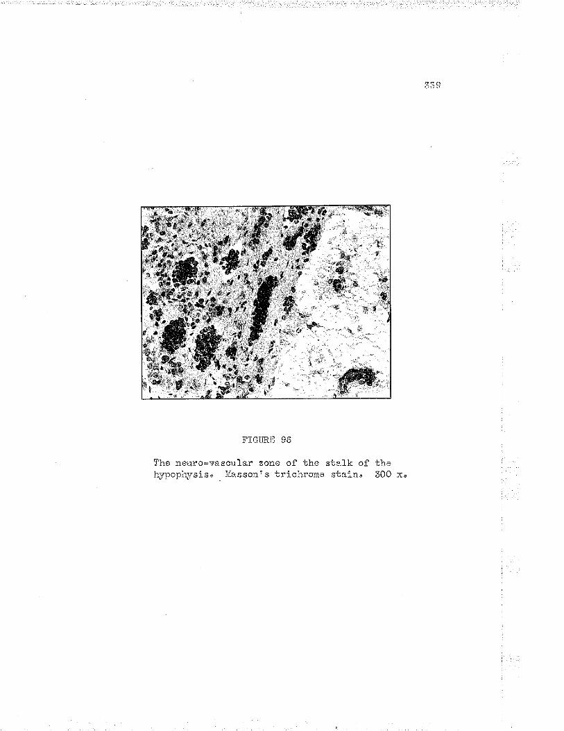
FIGURiI 96
Ihe neuro-vs"scular zone oî the stalk ofh¡'poph¡rsis" Jlfassone s trichro¡re stai n.
the300 x"

340
up to t millimetre in'thiclmess and which is continuou.s with th.e pars
tuberalis on the posterior aspect of the h¡rpophyseal sta.Lk" The zone
extends uprnrarcls almost to the mamillary bod.ies" Inforiorly it is
continuous rvith the pars tuberalis (Cf" Fig" 96), fn tire zoyLe àre blood
vessels, nerve fibres, colLagen fibres and- islands of glandular cells
sirnilar to those seen elsewhere in the pars tu.beraliso The nevve fibres
of the zone are derived chiefly from the perivascular plexuses and are
extrernely complex in the pattorn forned (Cf' Greer,1?),
The bullc of the nerve fibres within the neural sten pass without
interruption from the tractus hypophyseu.s to the neural lobe of the2,
hypophysisSz" The vesseLs of the stal-k havo perivascular sheathesT
v¡hich occupy 50-40 per cont of the volumo of the rnedian eminence and
neural stalk3z" The sheathes of these hypophysio-portal vesssls consist
of ordinary connective tissue in rqhich smooth mu-scle fibres, collagen arrd
reticular fibres may be demonstratea (cf" GreenSz).
Cgncretions anq. s5!ggg: qâte-l!g!" Iitany of the exbraneous
el-ements present in ths hypophysis are mesothelial in origin' Shank1inS8
reports cartilage and bonen Calcified bodies with lanrellated structuro
are usually observed in older subjects. They are foi¡nd in the capsulo
and neu.rohypophysisS8 and are derived frorn mesotheli.r*37 (cf , stranklinS8),
Reference to the oolloid bodies of the hypophysis has been made
on page S25"

341
VTT " TTTE HORÌIOI{ES OF THE I{YPOPiIYSTS
The following discu.ssi-on is a brief sunmary of the material fou-nd
in the äronographs by Cameron and Selye' No attempt Ís ma.de to discuss
the hor¡nones and their proporties, .since such is beyond the scope of the
present work and the su.bjects are well treated i.n the considerably more
authoratat ive r¡¡orks mentioned o
Th". Igsgigg of !þg. pals anteriof. Although the anterior lobe of
the lrypophysi-s iras onl;'r tr,',ro types of cells, as many as fifteen different
hormones have been postu1ated to be prod.uced by the lobe' The existence
.of these horrnones iras not been substantiated and v¡ithin the last ten
years a more rational vier,r has been he1d.41" Only four hornones of the
anterior lobe have been isolated in pure form--the luteotrophic,
sornatotrophic" adrenocorticotrophic a.nd luteinizing hort,ronesu The
foll-icular-stiinulating and thyrotrophic activities are probably due to
separate hornones9" Thel, are al-1 proteins4l. Fossibly the target
glands have secondary actions not understood or appreciated' These
actions may be responsi.ble for some of the results obtained lvhen crude
erbracts of the pars anterior are injected into experiinental animals.
Certain of the actions s.ppear to be produced in part by combinations of
knolvn hormones'
The following list of the aocepted hornones of the pars anterior
have been isolated in pure or alnost pure fonir" The slmonylns are gi'ren
togother v¡ith a few of the properties a¡rd actions of each horrnone' The

342'
e)q)erinei,Lbal basis and ol-inical- observations relating to their use are
not included, but are found in the works of Selye and Cameronu
_F"tfigþ- "tjggl.tjlß horntone' ( Sy". --FST{r f o1 li c 1 e- stinulator,
th¡rlakentrin)" Although this sirbstance has not been isolated., the
highly purified preparations stinulate growth of the gra.nulosa celIs in
tho ova.ries of hypophysecomized. aninalsu The Ìrormono also stimulates
the seminiferous tubules of the testes of intact or hyoophysectomized
arrinals" The role of FSII has been discu.ssed on pages 171, I72' 179 and
180,
Lutgili z ing horyon-e' ( S¡m. --l,tl, Inter st i b ial-c e11- st irnulat ing
hormono, I0SH, chorionic gonadotrophin, metakentrin) " This horrnone has
been isolated in pure forn9 and is a glycoprotein4l" 'r:rr)ren injec'çed., the
hornone stimuLates the trerrsfor¡ration of mature ovaria.n follicles into
corpora lutea and stilnulates the grow-bh of the theca cells and their
pfoduction of the fo1liculoid. hornone9. In the male, the horrlone
stirnulaf es the d.evelopment and the production of horrno¡r.es by the Le¡rdig
cell-s of the testes. Further discussion is found on pages 173¡ Il9' 180
and lBB'
Luteotrop.þig .ÞÆ. (Syri,-*LfU, luteotrophin, marnmotrophin,
pr.olaotinr galactin, lactogenie hormone)" This hornone hel-ps to rnaintain
fuIl-rr developed col.pora lutea and stinulates them to produce the luteoid
hor:nones, It stimulates the mannary glands to secrete rnilk after the
mamnary glands have been brought to fuIl developnent during pregnancyo

343
.å f\:rther discussion is found on page 174'
Cort-ico-Lrophi c. honrrore' (S;m"--ACTH, adreno-corticotrophic
hotmone, ad.renotrophin, corticotrophin, adrenotrophic hormone, oortico-
trophic horrnone)" This hormone has been prepared in pure forrn and is a
protein-like substaJrceo The horinone stimulates the gro-ørbh and hornone
production of the adrenal cortexu It tends to deplete the adrenal cortex
of its lipici and ascorbic acid contentu
Thyrotrophic hon:qgir-g." (Syn,--thyrotrophinu thyreotrophic honnone) "
This horrnone has not been isolated in pure fol:n. It stirnulates the
growbh and the hormone production of the thyroid gland'
Somatotrophic HÆ" (somatotrophin, grov,i'bh hornone) This
principle has been isolated.. As long as the epiphyseal junction
cartilages are still not fused., injections of tÌre hornone cause sl<eletal
growbh, both in length and. thickr,."""9" Injections luill prod.uco
gigantismr if given long enougho and if prolcnged will lead to enfeebled
gigantisn4l'
Other ttactionsti of anterior lobe extracts" Several other Hacti-onsls
aro noted with injections of orude extracts of the anterior lobe of the
hypophysis. -A.s Se1ye points out in his monograph, there has no-b boen a
claím put forLh for a separate hornone to account for each actionn
Sirnply these are the effects of the injection' Sone of the actions have
reasonable expla.nations, others have to wait further investigation and
data" The following is only a list of these actions, based on Selyer s
monograph"

344
I, The ovulation-inducing action2, The antagonistic acticn--antagonistic to certain of the
gonadotrophins3n Thã synergistic action-*alleged to augment the aciion of LH
preparations4, The antiluieogenic action5u The thymotrophic action6. The roaotrophic action7 u The nephrosclerotic actionB. The gluco-corticotrophic¡ mineralo-corticotrophic action¡
lipocortico'b::ophio actiono and testo-corticotrophia actions9" The adrenonedullotrophic action
10" the parath)'rotrophic a.otion11" The ma¡n¡rogenic actionl.2. The glycotropic and anti-insulin actÍon15" The glycostatic action14, The pancreatotroPhic action15, The diabetogenic action16" The anti-di.abetic effect17 " The contra-insular action18, The ketogenic action19, The fatty-liver procìucing actionZo' Ihe preputial gland-sti-¡aulating a-ction
The. Ðre of k pqrå 4egnosgo crucle extracts of the pars
nervosa exhibit three ohief activities: (a) tirey produce a rise in
blood pressure (vasoconstrictor), (¡) they cause uterine contractions
(o.xybocic), and (c) tr."y diminish diuresis9. By d.ifferential
fractionation, two non-crfstalLine fractions have been obtained. Ono
fraction has pressor potency and very 1ow ollrbocic potencyr'¡¡hile the
other has very high o>ybocic potency and only negligible pressol'
activity, No crystalline compounds have yet been obtained but those
amorphous fractions are usually considered. a.s almost pure horr'Lotts4l"
Thus it is generally agreed that there are at Ìeast two distinct
prinoiples of the pars nervosa, the oq¡bocic and vasopressor horinones9"
The anti-diuretic action is probably due to the vasopressor principleg"

345
U-e*_9g.?_qg. ( Sftt. --vasopre s sor princ ipl e u po st 1 ob in-],/,
piiressine vasopressoL'-an'bi-diuretic princi-ple)" The lr.orinone has not
been isolated. Sotne preparations, holt¡everr are very po'cent' The anti-
rliuretic hormone of the pars nervosa is probably identical witho
vasopress in" "
Oxybgcin" (S¡m"*-0xybocic horrnone, ox¡rboci.c posterior-lobe
principleu postlobin-O, pitocin)" The chemioal ancl physical properties
of o,.q,rtocin and. vâ.sopressin are similar anci. make separation cìifficult"
The horrnone has not been isolatedo
The pars i4term:diao Aqueous exbracts of Ure pars internedia
produce di spersion of black pigment granules of amphibia" It has been
sug6ested. that this is due to a ho¡rnone of the pars inter:nedia, to whioh
the narne internedin has beon assignedu Some of the s¡monyms are--
middle-lobe horrnone, meanophore-expanding principle" B-hormone" The
function of intermedin, should it exist, is not knor¡rn in the case of
birds and. marrr-nalso (Cf " Canreron and. setyel)

346
B]BLTOGRAPT{Y
' L. Bucy, Paul 8., The Hy¡lophysis Cerebri" Section XV, in Cytologyand Cellular Patholcg¡ of the Nervous System, Volo IIe1T" Penfield, Editor. New York¡ Paul B. Hoeber Incorporated,LJÓá O
2, Baile¡r, P", The Structure of the Hypophysis Cerebri of Í''lan and ofthe Corunon Laboratory Animals. Section XX, in SpecialCvtologya Vol" If, Second edition, E" V' Cowdryr Editor"Itlew Yorl<: Paul B, I{oeber Inc o " L932 "
3n Rathker, H,, Ueber die EntsteÌrung cÌer Glandul-a Pituitaria'l.liil-l ert s Arch" f " Anat., Physiol, ìr, Ttrissench' i/ied,, lB3B,pp" 482*485, S,uoted by Bucyl.
4. l¡Iaterstono D., Development of ilypophysis Cerobri in lvlan, etco,Trans" Roy. Soco, Edin", 1926, 52aI25. O,uoted by Bucy1"
5q Hamilton, W, J", Jo D. Boyde and H, T'I" Itfoss¡'ran, l{uman Embryology,Cambridge: ïtr" Heffer and Sons Ltd., 1945"
6" Atwellu r;T"" the Development of the Hypophysis Ce::ebri in }ian, withSpecial Reference to the Pars Tuberali-s' i,ner" J. -A.nat,"!926, 37¿L59"
7" Popa, G, and U" Fieldi:rgo The Vassular Lj¡l< between the Pituitarynd the l{¡4pothalamus" The Lancet, 1930, 21,9.e238.
Bo Nikolskaia, S., Die Blutversorgung der l{¡rpophy_se des l,lensc}ron"Ánat. Anzo, 1929, 6J_al3O. Quoted. by Bucyl.
9u Se1yen Ha:rs¡ Toxtbook of Endocrinolcgy. ll{ontråal¡ UniversitJ aeiilontrdat" L947. pp. 914.
1.0. Sunderlarrd., S", The ir{eningeal Relations of the Human HypophysisCerebri. Ju Snat", 1945, 79¡53"
11. TIisloclci, Go 8", The Meningeal Relatíons of the Hypophysis Cerebri,Amer" J' Anat., 1937, 61¿95,
12. Schv¡artz, Hn G., The iriening;ea1 R.elations of the ily¡rophysis Cerebri."A"nat " Rec,, L936" 67 ¿35"

2 t'l ¡?ùat
73, Simnonds, J, P", the Pathological Anatomy and I{istology of theHy¡rophysis. Endocrinolo¿:y and liietabolism, VoI" I, ediiedby L" F. Ba.rker" irlew Yorkt D' Appleton and Company, 1-92
pp.766-802'
!+" Ra.srnussen, A. T.e The ìlleight of the Princi.oal Coraponents of thei{ormal li[a]e Aclult i{uman Hypophysis Cerebriu Amer. Ju årtat',I92B n 4,:::4_zL"
15 " Rasmuss en¡ .Ao T "
16" Lewis, D" and Fn Cu Loe, 0n the Glandular Elements in the PosteriorLobe of ths lIuma"n Hy¡rophysis' 8u11" Johns Hopkins Hospo,197,7 s 4I_224I"
1?,' Rasnussen, JLu To, The fncidense of Tubular GLands and Concretionsin the Ad.ult i{uma.n Hypophysis Cerebrj-. r{nat. Rec", 193"3,55-: 159 '
lBn Cowdry, E' V,, A Te:rtbook of iïistology, Second editron'Phila" ¡ Lea and Febiger
19" Rasmussen, Au T., The Percentage of the Different Types of Cell.s inthe I';Iale Ad.u1t Hutnan Hypophysis. *Amer' J. Path. , I9Z9''Þe263^
2Q, Ras¡russene.A. Tu, The Percentage of the Different T¡4pes of Cells intho Anterior Lobe of the Hypophysis of the Adult Human Female'Amer' Ju Path., 1933, 9:459"
ZL. Rasnussen, A" To, Changes in the Proportion of CeIl Types in theånterior Lobe of the Hunan llypophysis during the FirstNineteen Years of Lif e" "Afloro J' Anat', 1950, 86:75"
22" Rasinussen, å." T', Innervation of the Hypophysis" Endocrinologyo1938,23_2263"
23" Cooper, E" R..4", The llistology of tire more Important }IumanEnd.ocrine Organs at Various Age Groups. London: OxfordUniversity Press, 1925'
24" ivlaximow, Ao 3." and 1&û. Bloom, A Te:ctboolc of Histology, Fifthedition, Phila" ! ï,r. B. Saunders Corapa:tye 1948 ' pp. 700'
25, Rasrm-¡.ssene .4" T'e The ldorphology of the Fars Intermedia of theHriman :iy¡rophysis' Endocrinologyo r-92Bu I2zL29,

348
26, Raslirussene A, T., The Relation of the Basophilic Cells of the liunanIJyoophysis to Blood Pressirve" Endocrinologyr 1956, ?9,2673'
27" Rasnussene /ro To, Ciliated Epitherium and i:.hcous Secreting Cells iathe lluman líypophysis " Ánat. Rec ' u !929 ' 4)t273.
28" Shanklin, ïVin" IvÍ,r 0n tho Pressnce of Cyste in the Hurnan Pituitary.¿ånat" Recne 1949, _1_9,ê;379"
29, Gillmano T., The fncidence of Ciliated Epithelium and i',hcou.s Cellsin the Normal Bantu Pituitaryu S. Afr. J' LIed" Scoe L94O,Ë"2ñvòÐvc
BO" Uonnr-*. and U, Fieldings Á. Portal Circulation from the Pituitaryto the Hypothalamic Region' Ju *A¡aat " , 1930, 6t2e BB
"
6L" Tilneyr F,o The Development and tonstitu,ents of the liuman Ilyoophysis.8u11. Ideurol . Inst' id. Y., 19361 5¡387"
S2. Green, Jo D.r The Histologlr of the lJypophyseal Stalk and lviedi-an¡-minence in irïan l,'urth Special Reference to Blood Vessels, NerveFibres and a Pecu.l-iar lrieurovascu-l-ar Zone in this Regionu*A.nat . F.ec, , 1948, '109;275
"
ß5, Rasm-:-ssen" Ao Toe lhe Proportion of the Various Subdivisions of theNormal- Adult l{uman }lypophysis Cerobri and the Relative itTumber
of the Different Types of Cells in Pars Distalis, r¡ithBi-ometri c Evaluetion of .{ge and Sex Diff erences and SpecialConsj-cLeration of Basophilic Invasion into the InfunciibularProcess' The Pituitaryr Chapto TVe pp'118-150"The Proceedings of the Ássoc" for Research in Nervotts andlr,'iental Dis'¡ Vol" XVfl. Balt"; The -l$illiams and Tr¡ilkinsCompanyr 1938.
34" Severinghau-s, .Ao Eo, The Cybology of the Pituitar¡r Gland.The Pituitarlr, Chàpt"ÏII, pp. 69-11.7" The Proceedi.ngs of the.Assoc. Resn lierv, and irient. Dis', Vol" XVII. Balt"¡ Thel¡iilliams and nïilkins Conpany' 1938'
35" Riocb.g Do lvicK,, Pa-ths of Secretíon from the llypophysis' Chapt. V,pp" 151-17Lo oi the above.
õ6. Ttislocki, G. 8", The Vascular Supply of the Hypophysis Cerebri ofthe Rhesus I'tonkey and jtian. Chapt. Jle pp" 48-68 of the aboven
37 u Shatklin, -t¡fm" lr,/in, The Development and ilistology of Pituitar¡tConcretions i.n it'fan" Anat. Rec." 1946, 942597 '
.2./'t ô1J= J
38" $helklin, T'.¡m" M"o 0n the Presence of carcific Bodiesu car'cilag;e,Bone, Follicular Concretions and the So-Catlecì IlyalineBodies in the iluman Pitu.itary. Anat, Recu, lg48, lQ?;46g"
39' Plaut, A' and Galenson, Eo, Concretions in the Anterior Pitr-iitaryLobe of the äuma¡r Embr;'o and l,Iev¡born, ,Él.mer. J. Path", 1944,29;228,
40" irielchionna, R"Amer ' .T o
41 " Cameron, 3.. Tnï,ondons
H" and R. .An ltloore, The Phar¡mgeal Pitr-ritar¡r Gland"Path"s 1938, L!e763"
, Iìecent Advances in Endoar.inol-ogyr Sixth edition,J" & A" Churchill Ltd", 1947,


