Embed Size (px)
Citation preview
I
YKP
a
ARRAA
KSIICRA
1
hodbeBmo
i(iwd(a
0d
Journal of Ethnopharmacology 136 (2011) 309–315
Contents lists available at ScienceDirect
Journal of Ethnopharmacology
journa l homepage: www.e lsev ier .com/ locate / je thpharm
n vitro and in vivo antioxidant effects of the ethanolic extract of Swertia chirayita
ue Chen, Bo Huang, Jingsheng He, Li Han, Yichao Zhan, Youwei Wang ∗
ey Laboratory of Combinatorial Biosynthesis and Drug Discovery (Wuhan University), Ministry of Education, and Institute of TCM & Natural Products, Wuhan University School ofharmaceutical Sciences, Wuhan 430071, PR China
r t i c l e i n f o
rticle history:eceived 27 September 2010eceived in revised form 13 April 2011ccepted 20 April 2011vailable online 28 April 2011
eywords:wertia chirayitan vitron vivoarbon tetrachloride
a b s t r a c t
Ethnopharmacological relevance: Swertia chirayita, a medicinal herb endemic to the Tibetan region, isused as a special remedy for liver disorders. The hepatoprotective activity of its plant extracts has beenassociated with its antioxidant activity. This paper aims to investigate the in vitro and in vivo antioxidanteffects of Swertia chirayita extracts (SCE).Materials and methods: Antioxidant ability of Swertia chirayita was investigated by employing severalestablished in vitro methods. In vivo antioxidant activity was tested against CCl4-induced toxicity inmice. The levels and activities of malondialdehyde (MDA) and antioxidant enzymes, including superoxidedismutase (SOD), catalase (CAT), and glutathione (GSH), were then assayed using standard procedures.Results: SCE exhibited strong antioxidant ability in vitro. The liver and kidney of CCl4-intoxicated animalsexhibited a significant (p < 0.001) decrease in SOD, CAT, and GSH levels. Additionally, these organs exhib-
adical scavengingntioxidant activity
ited a significant (p < 0.001) increase in MDA level. CCl4 did not exhibit toxicity on mice treated with SCEand Vitamin E. The effects of Swertia chirayita (three dosages) were comparable to those of Vitamin E,except in MDA level in the liver and GSH level in the kidney (p < 0.05).Conclusion: This study suggests that the ethanolic extract of Swertia chirayita possesses in vitro and in vivoantioxidant effects. This supports the traditional use of Swertia chirayita in Tibetan medicine to cure liverdiseases.
© 2011 Elsevier Ireland Ltd. All rights reserved.
. Introduction
Reactive oxygen species (ROS), such as superoxide anion,ydroxyl radicals, and hydrogen peroxide are generated in livingrganisms through numerous metabolic pathways. The oxidativeamage caused by ROS may generate various diseases in the humanody, such as aging, arthritis, cancer, inflammation, and heart dis-ases (Meerson et al., 1982; Busciglio and Yankner, 1995; Abe anderk, 1998). Herbal drugs play an important role in the manage-ent of various diseases, and increasing attention has been focused
n Chinese medicinal plants.Swertia chirayita (Roxb. ex Fleming) Karsten is considered a crit-
cally rare medicinal plant indigenous to the temperate HimalayasGarg, 1987; Samant et al., 1998). In China, it is mainly distributedn the Qinghai-Tibetan Plateau. Swertia chirayita, a medicinal herb
ith Tibetan origin, is used as a special remedy for liver disor-
ers due to its ability to drain heat from the blood and the liverMukherji, 1953). Its extracts are used as hepatoprotective agents,nd its antifungal, antimalarial, anti-inflammatory, and anticar-∗ Corresponding author. Tel.: +86 27 68759323; fax: +86 27 68759010.E-mail addresses: [email protected], [email protected] (Y. Wang).
378-8741/$ – see front matter © 2011 Elsevier Ireland Ltd. All rights reserved.oi:10.1016/j.jep.2011.04.058
cinogenic properties are likewise known (Kirtikar and Basu, 1984;Keil et al., 2000).
The hepatoprotective activity of Swertia chirayita extracts (SCE)has been associated with its antioxidant activity (De et al., 1996;Shanmugasundaram and Venkataraman, 2006; Jain et al., 2008;Sabir and Rocha, 2008; Srivastava and Shivanandappa, 2009;Zeashan et al., 2009; Huang et al., 2010a,b). However, insufficientstudy has been conducted on the role of Swertia chirayita on freeradical management and antioxidant activity. The present workaimed to explore the potential in vitro and in vivo antioxidant prop-erties of the ethanolic extract of Swertia chirayita.
2. Materials and methods
2.1. Chemicals
�,�-Diphenyl-�-picrylhydrazyl (DPPH) and �-Tocopherols(Vitamin E) were purchased from Sigma–Aldrich (St. Louis, MO).2,2′-Azino-bis (3-ethylbenzthiazoline-6-sulphonic acid) (ABTS)
was purchased from Fluka (Menlo Park, CA). Linoleic acid was pur-chased from Alfa Aesar (Ward Hill, MA). Ascorbic acid (VitaminC), gallic acid, rutin, and butylated hydroxytoluene (BHT) werepurchased from the National Institute for the Control of Pharma-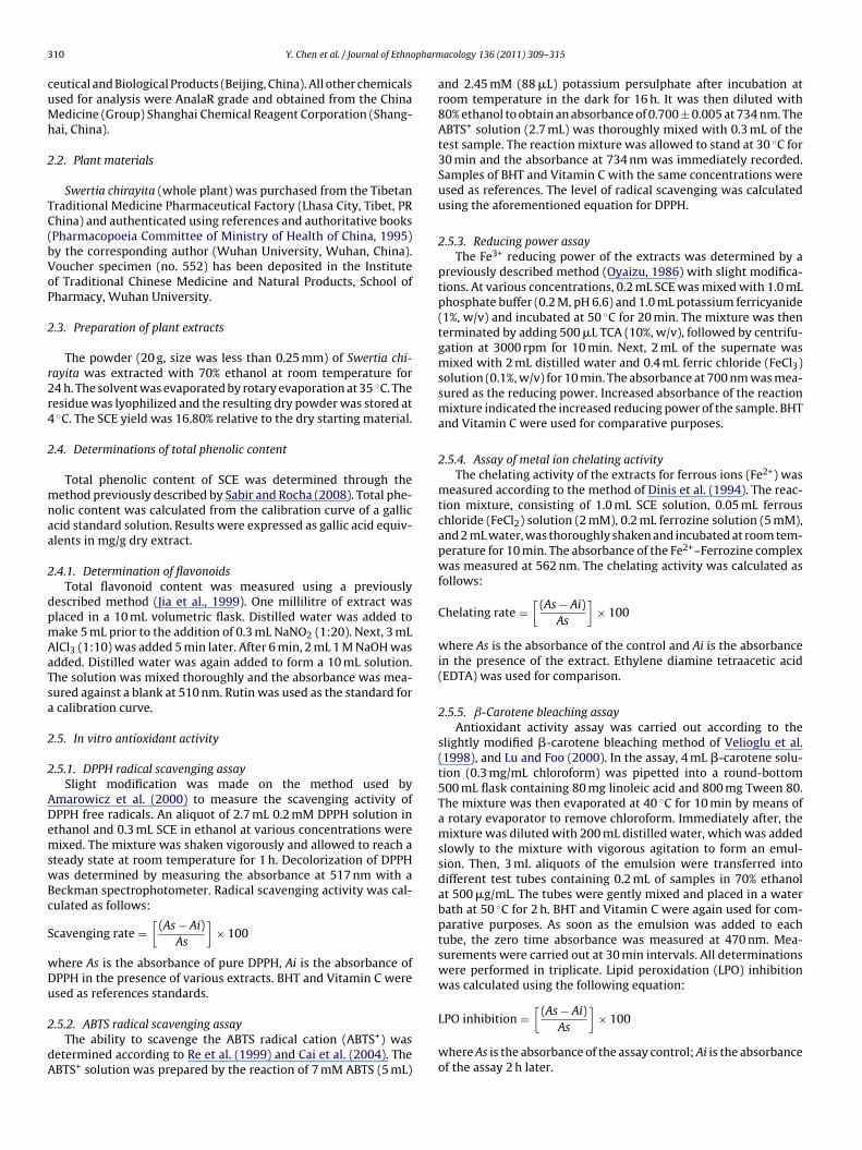
3 pharm
cuMh
2
TC(bVoP
2
r2r4
2
mnaa
2
dpmAaTsa
2
2
ADemswBc
S
wDu
2
dA
10 Y. Chen et al. / Journal of Ethno
eutical and Biological Products (Beijing, China). All other chemicalssed for analysis were AnalaR grade and obtained from the Chinaedicine (Group) Shanghai Chemical Reagent Corporation (Shang-
ai, China).
.2. Plant materials
Swertia chirayita (whole plant) was purchased from the Tibetanraditional Medicine Pharmaceutical Factory (Lhasa City, Tibet, PRhina) and authenticated using references and authoritative booksPharmacopoeia Committee of Ministry of Health of China, 1995)y the corresponding author (Wuhan University, Wuhan, China).oucher specimen (no. 552) has been deposited in the Institutef Traditional Chinese Medicine and Natural Products, School ofharmacy, Wuhan University.
.3. Preparation of plant extracts
The powder (20 g, size was less than 0.25 mm) of Swertia chi-ayita was extracted with 70% ethanol at room temperature for4 h. The solvent was evaporated by rotary evaporation at 35 ◦C. Theesidue was lyophilized and the resulting dry powder was stored at◦C. The SCE yield was 16.80% relative to the dry starting material.
.4. Determinations of total phenolic content
Total phenolic content of SCE was determined through theethod previously described by Sabir and Rocha (2008). Total phe-
olic content was calculated from the calibration curve of a galliccid standard solution. Results were expressed as gallic acid equiv-lents in mg/g dry extract.
.4.1. Determination of flavonoidsTotal flavonoid content was measured using a previously
escribed method (Jia et al., 1999). One millilitre of extract waslaced in a 10 mL volumetric flask. Distilled water was added toake 5 mL prior to the addition of 0.3 mL NaNO2 (1:20). Next, 3 mLlCl3 (1:10) was added 5 min later. After 6 min, 2 mL 1 M NaOH wasdded. Distilled water was again added to form a 10 mL solution.he solution was mixed thoroughly and the absorbance was mea-ured against a blank at 510 nm. Rutin was used as the standard forcalibration curve.
.5. In vitro antioxidant activity
.5.1. DPPH radical scavenging assaySlight modification was made on the method used by
marowicz et al. (2000) to measure the scavenging activity ofPPH free radicals. An aliquot of 2.7 mL 0.2 mM DPPH solution inthanol and 0.3 mL SCE in ethanol at various concentrations wereixed. The mixture was shaken vigorously and allowed to reach a
teady state at room temperature for 1 h. Decolorization of DPPHas determined by measuring the absorbance at 517 nm with aeckman spectrophotometer. Radical scavenging activity was cal-ulated as follows:
cavenging rate =[
(As − Ai)As
]× 100
here As is the absorbance of pure DPPH, Ai is the absorbance ofPPH in the presence of various extracts. BHT and Vitamin C weresed as references standards.
.5.2. ABTS radical scavenging assayThe ability to scavenge the ABTS radical cation (ABTS+) was
etermined according to Re et al. (1999) and Cai et al. (2004). TheBTS+ solution was prepared by the reaction of 7 mM ABTS (5 mL)
acology 136 (2011) 309–315
and 2.45 mM (88 �L) potassium persulphate after incubation atroom temperature in the dark for 16 h. It was then diluted with80% ethanol to obtain an absorbance of 0.700 ± 0.005 at 734 nm. TheABTS+ solution (2.7 mL) was thoroughly mixed with 0.3 mL of thetest sample. The reaction mixture was allowed to stand at 30 ◦C for30 min and the absorbance at 734 nm was immediately recorded.Samples of BHT and Vitamin C with the same concentrations wereused as references. The level of radical scavenging was calculatedusing the aforementioned equation for DPPH.
2.5.3. Reducing power assayThe Fe3+ reducing power of the extracts was determined by a
previously described method (Oyaizu, 1986) with slight modifica-tions. At various concentrations, 0.2 mL SCE was mixed with 1.0 mLphosphate buffer (0.2 M, pH 6.6) and 1.0 mL potassium ferricyanide(1%, w/v) and incubated at 50 ◦C for 20 min. The mixture was thenterminated by adding 500 �L TCA (10%, w/v), followed by centrifu-gation at 3000 rpm for 10 min. Next, 2 mL of the supernate wasmixed with 2 mL distilled water and 0.4 mL ferric chloride (FeCl3)solution (0.1%, w/v) for 10 min. The absorbance at 700 nm was mea-sured as the reducing power. Increased absorbance of the reactionmixture indicated the increased reducing power of the sample. BHTand Vitamin C were used for comparative purposes.
2.5.4. Assay of metal ion chelating activityThe chelating activity of the extracts for ferrous ions (Fe2+) was
measured according to the method of Dinis et al. (1994). The reac-tion mixture, consisting of 1.0 mL SCE solution, 0.05 mL ferrouschloride (FeCl2) solution (2 mM), 0.2 mL ferrozine solution (5 mM),and 2 mL water, was thoroughly shaken and incubated at room tem-perature for 10 min. The absorbance of the Fe2+–Ferrozine complexwas measured at 562 nm. The chelating activity was calculated asfollows:
Chelating rate =[
(As − Ai)As
]× 100
where As is the absorbance of the control and Ai is the absorbancein the presence of the extract. Ethylene diamine tetraacetic acid(EDTA) was used for comparison.
2.5.5. ˇ-Carotene bleaching assayAntioxidant activity assay was carried out according to the
slightly modified �-carotene bleaching method of Velioglu et al.(1998), and Lu and Foo (2000). In the assay, 4 mL �-carotene solu-tion (0.3 mg/mL chloroform) was pipetted into a round-bottom500 mL flask containing 80 mg linoleic acid and 800 mg Tween 80.The mixture was then evaporated at 40 ◦C for 10 min by means ofa rotary evaporator to remove chloroform. Immediately after, themixture was diluted with 200 mL distilled water, which was addedslowly to the mixture with vigorous agitation to form an emul-sion. Then, 3 mL aliquots of the emulsion were transferred intodifferent test tubes containing 0.2 mL of samples in 70% ethanolat 500 �g/mL. The tubes were gently mixed and placed in a waterbath at 50 ◦C for 2 h. BHT and Vitamin C were again used for com-parative purposes. As soon as the emulsion was added to eachtube, the zero time absorbance was measured at 470 nm. Mea-surements were carried out at 30 min intervals. All determinationswere performed in triplicate. Lipid peroxidation (LPO) inhibitionwas calculated using the following equation:
[(As − Ai)
]
LPO inhibition =As× 100
where As is the absorbance of the assay control; Ai is the absorbanceof the assay 2 h later.
pharm
2f
(swpvw7Pimiwwc
att(aaafi
2
2
AgkdwwetP(
2
go
2
aT1n(0bGbporitcoo
Y. Chen et al. / Journal of Ethno
.5.6. Antioxidant activity in a linoleic acid system usingerrothiocyanate (FTC) and thiobarbituric acid (TBA)
The standard method described by Kikuzaki and Nakatani1993) was used to determine antioxidant activity in a linoleic acidystem. Plant extract (400 �g) in 4 mL absolute ethanol was mixedith 4 mL 2.5% linolenic acid in absolute ethanol, 8 mL 0.05 M phos-hate buffer (pH 7.0), and 4 mL water. The mixture I was placed in aial with a screw cap and then placed in a dark oven at 40 ◦C. After-ards, aliquots (0.1 mL) from mixture I were withdrawn and 9.7 mL
5% ethanol and 0.1 mL 30% ammonium thiocyanate were added.recisely 3 min after the addition of 0.1 mL 0.02 M ferrous chloriden 3.5% HCl to the reaction mixture, the absorbance of red color was
easured at 500 nm. Aliquots were withdrawn and assayed in andentical fashion at 24 h intervals until a constant maximum value
as reached. BHT and Vitamin C were used as positive controls,hile a mixture without a plant sample was used as the negative
ontrol.Antioxidant activity of the extracts using TBA was determined
ccording to the method of Kikuzaki and Nakatani (1993). Inhe assay, 2 mL of 20% trichloroacetic acid and 2 mL of 0.67% 2-hiobarbituric acid were added to 1 mL of the sample solutionmixture I) prepared by the FTC method. The mixture was placed inboiling water bath for 10 min, and after cooling, it was centrifugedt 3000 rpm for 20 min. Absorbance of the supernate was measuredt 552 nm. Antioxidant activity was based on the absorbance on thenal day of the FTC method.
.6. In vivo hepatoprotective activity
.6.1. Test animalsMale KM mice (20 ± 2 g) were purchased from the Laboratory
nimal Center of Wuhan University (Wuhan, China). The mice wereroup-housed (10 per cage) with free access to food and water, andept in a regulated environment at 25 ± 1 ◦C under 12 h light/12 hark conditions and in a relative humidity of 30–60%. The miceere fed ad libitum on a diet of standard pellets and water andere acclimated to the housing conditions for three days prior to
xperimentation. The study received clearance from the Institu-ional Animal Ethical Committee (IAEC) of the Committee for theurpose of Control and Supervision of Experiments on AnimalsCPCSEA), Wuhan University (Wuhan, China).
.6.2. Acute toxicity studiesAfter overnight fasting, the mice were administered SCE in
raded doses up to 2000 mg/kg body weight. Animals werebserved for symptoms of toxicity and mortality for 72 h.
.6.3. Carbon tetrachloride (CCl4)-induced oxidative toxicityProtective effect of the SCE treatment against CCl4-induced
cute hepatotoxicity in mice was evaluated in a seven-day study.he animals were randomly divided into six experimental groups of0 mice each (Jaishree and Badami, 2010). The first group served asormal control and, during the experiment, received vehicle only0.3% CMC-Na) (10 mL/kg body weight, p.o.). Group II was given.3% CMC-Na (10 mL/kg body weight, p.o.) solution for six daysefore CCl4 intoxication and served as the toxicity control group.roup III was treated with the standard drug Vitamin E (100 mg/kgody weight, p.o.) daily for six days. Groups IV, V, and VI wererophylactically treated for six days with three different dosesf ethanolic extract suspension (100, 200, and 400 mg/kg/day,espectively, p.o.). On day 6, mice in groups II–VI received anntraperitoneal injection of CCl4 (10 mL/kg b.w. of 10% CCl4 solu-
ion in olive oil) after having been given 0.3% CMC-Na. The normalontrol group was intraperitoneally treated with an equal amountf olive oil alone. The animals were sacrificed 24 h after the CCl4 andlive oil treatments (day 7). The livers and kidneys were carefullyacology 136 (2011) 309–315 311
dissected and cleaned of extraneous tissue. The livers and kidneysare fresh and dark red.
2.6.4. Measurement of antioxidant enzymes and MDA levelsLiver and kidney samples were dissected and the blood
was immediately washed with ice-cold saline. Liver and kidneyhomogenates (10%, w/v) were prepared in cold 50 mM potassiumphosphate buffer (pH 7.4), and the resulting suspension was cen-trifuged at 1000 rpm for 10 min at 4 ◦C. The activity of superoxidedismutase (SOD), catalase (CAT), malondialdehyde (MDA), and glu-tathione (GSH) in the clear supernatant were measured using assaykits (SOD test kits A001-1, CAT test kits A007, MDA test kits A003-2, GSH test kits A006) obtained from the Institute of BiologicalEngineering of Nanjing Jianchen (Nanjing, China), according to themanufacturer’s protocol. SOD activity was estimated by the inhi-bition of spontaneous epinephrine oxidation (Kakkar et al., 1972).Determination of CAT activity was based on the rate of H2O2 reduc-tion (Beer and Seizer, 1952). One unit of CAT reduces 1 mM of H2O2.SOD and CAT activities were expressed in units per mg protein(U/mg protein). MDA level was examined using the 2-thiobarbituricacid (TBA) as previously method described by Yagi and Rastogi(1979) in liver and kidney tissues and expressed as n moles permg protein (nmol/mg protein) using 1,1,3,3-tetraethoxypropane(TEP) as a standard. GSH in liver and kidney tissues was determinedaccording to the Ellman method (Ellaman, 1959), which measuresthe reduction of 5,50-dithio-bis (2-nitrobenzoic acid) (DTNB) (Ell-man’s reagent) by sulfhydryl groups to 2-nitro-5-mercaptobenzoicacid, which has an intense yellow color. The results were expressedin mg per g protein (mg/g protein).
2.7. Statistical analysis
All experiments were done in triplicate and results werereported as mean ± S.D. Data were analyzed by one way ANOVA.Statistically significant effects were further analyzed and meanswere compared using Duncan’s multiple range test. Statistical sig-nificance was determined at p < 0.05.
3. Results and discussion
3.1. Total phenolic and flavonoid content
Ethanol was used as solvent and 16.8% (w/w) bulk materialswere extracted from the plant. Most antioxidant activities fromplant sources are correlated with phenolic-type compounds (Bravo,1998; Moreno et al., 2000; Cai et al., 2004; Jain et al., 2008; Kauret al., 2008; Sabir and Rocha, 2008; Huang et al., 2010a,b). Totalflavonoid content was 4.98 ± 0.40 mg rutin equivalents/g extract inSwertia chirayita (dry weight). Total phenol content was estimatedas 243.02 ± 4.70 mg gallic acid equivalents/g (dry weight) extract.
3.2. In vitro assays
3.2.1. DPPH radical scavenging activityDPPH is a stable, organic free radical extensively used to evaluate
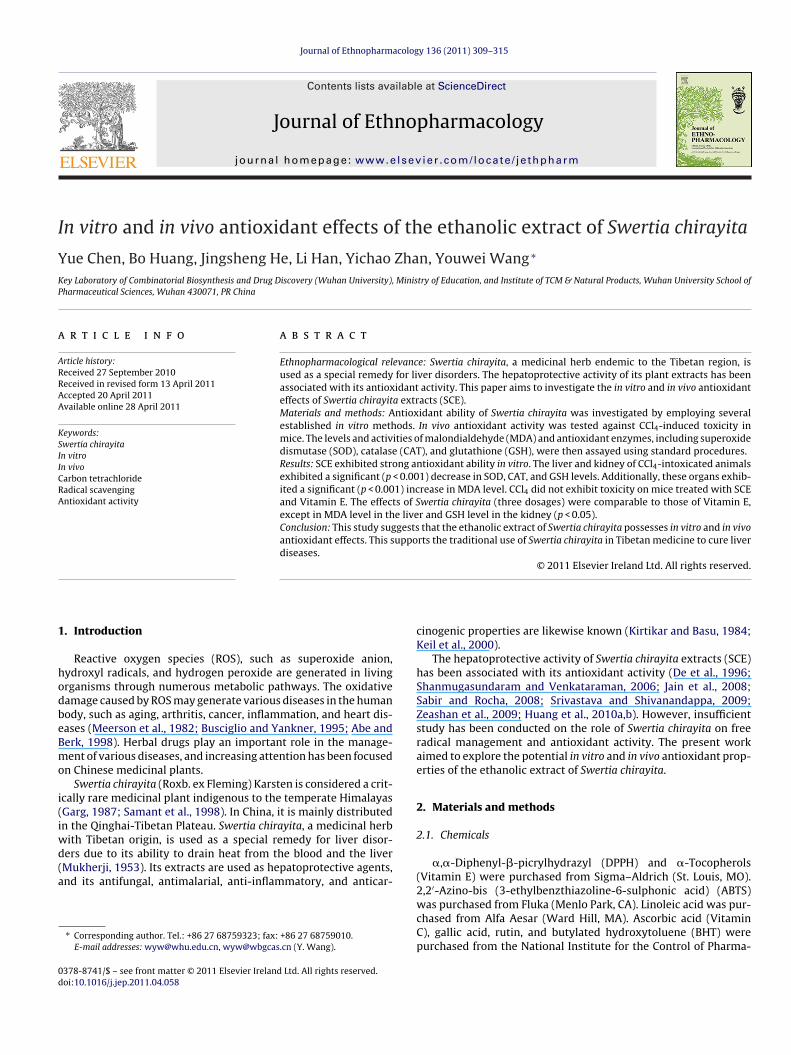
scavenging activity of antioxidants because it is sensitive enoughto detect active ingredients at low concentrations (Oyaizu, 1986;Sanchez-Moreno, 2002). In the DPPH assay, an antioxidant scav-enges the free radicals. In Fig. 1A, the SCE exhibited a steady increasein inhibition percentage at less than 100 �g/mL before eventuallyslowing down. Vitamin C showed an excellent scavenging activ-
ity (IC50 = 5.30 �g/mL). The SCE also presented strong scavengingactivity with an IC50 value of 267.80 �g/mL while BHT had a value of245.80 �g/mL. This showed that the ability of SCE was comparableto that of BHT, but inferior to Vitamin C.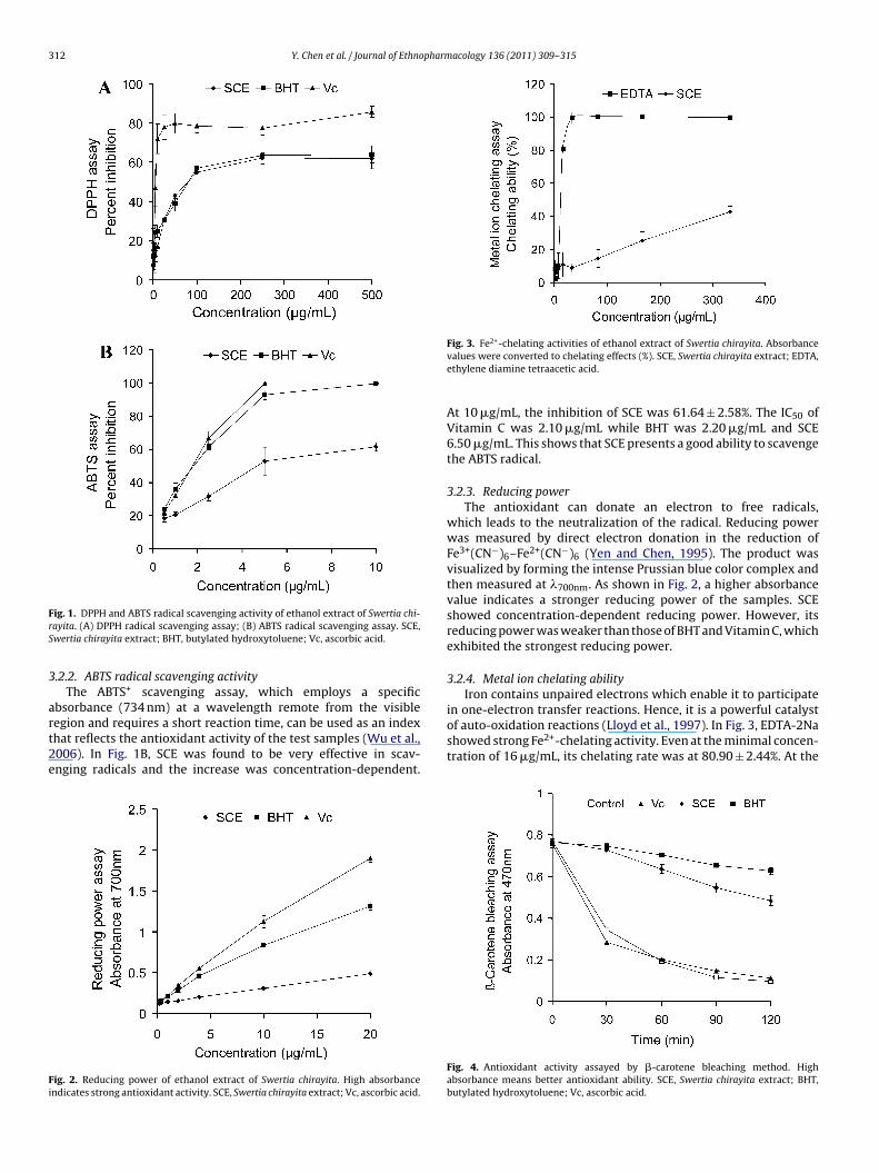
312 Y. Chen et al. / Journal of Ethnopharmacology 136 (2011) 309–315
FrS
3
art2e
Fi
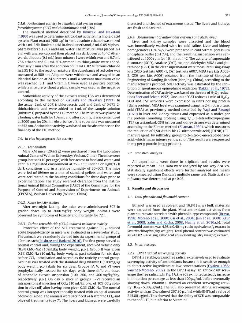
Fig. 3. Fe2+-chelating activities of ethanol extract of Swertia chirayita. Absorbance
in one-electron transfer reactions. Hence, it is a powerful catalystof auto-oxidation reactions (Lloyd et al., 1997). In Fig. 3, EDTA-2Na
ig. 1. DPPH and ABTS radical scavenging activity of ethanol extract of Swertia chi-ayita. (A) DPPH radical scavenging assay; (B) ABTS radical scavenging assay. SCE,wertia chirayita extract; BHT, butylated hydroxytoluene; Vc, ascorbic acid.
.2.2. ABTS radical scavenging activityThe ABTS+ scavenging assay, which employs a specific
bsorbance (734 nm) at a wavelength remote from the visibleegion and requires a short reaction time, can be used as an index
hat reflects the antioxidant activity of the test samples (Wu et al.,006). In Fig. 1B, SCE was found to be very effective in scav-nging radicals and the increase was concentration-dependent.ig. 2. Reducing power of ethanol extract of Swertia chirayita. High absorbancendicates strong antioxidant activity. SCE, Swertia chirayita extract; Vc, ascorbic acid.
values were converted to chelating effects (%). SCE, Swertia chirayita extract; EDTA,ethylene diamine tetraacetic acid.
At 10 �g/mL, the inhibition of SCE was 61.64 ± 2.58%. The IC50 ofVitamin C was 2.10 �g/mL while BHT was 2.20 �g/mL and SCE6.50 �g/mL. This shows that SCE presents a good ability to scavengethe ABTS radical.
3.2.3. Reducing powerThe antioxidant can donate an electron to free radicals,
which leads to the neutralization of the radical. Reducing powerwas measured by direct electron donation in the reduction ofFe3+(CN−)6–Fe2+(CN−)6 (Yen and Chen, 1995). The product wasvisualized by forming the intense Prussian blue color complex andthen measured at �700nm. As shown in Fig. 2, a higher absorbancevalue indicates a stronger reducing power of the samples. SCEshowed concentration-dependent reducing power. However, itsreducing power was weaker than those of BHT and Vitamin C, whichexhibited the strongest reducing power.
3.2.4. Metal ion chelating abilityIron contains unpaired electrons which enable it to participate
showed strong Fe2+-chelating activity. Even at the minimal concen-tration of 16 �g/mL, its chelating rate was at 80.90 ± 2.44%. At the
Fig. 4. Antioxidant activity assayed by �-carotene bleaching method. Highabsorbance means better antioxidant ability. SCE, Swertia chirayita extract; BHT,butylated hydroxytoluene; Vc, ascorbic acid.
Y. Chen et al. / Journal of Ethnopharm
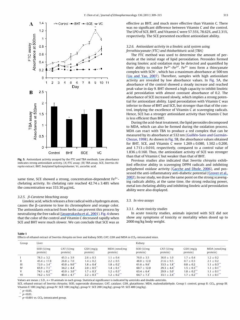
Fig. 5. Antioxidant activity assayed by the FTC and TBA methods. Low absorbanceir
sct
3
cTntS
TE
VSV
ndicates strong antioxidant activity. (A) FTC assay; (B) TBA assay. SCE, Swertia chi-ayita extract; BHT, butylated hydroxytoluene; Vc, ascorbic acid.
ame time, SCE showed a strong, concentration-dependent Fe2+-helating activity. Its chelating rate reached 42.74 ± 3.48% whenhe concentration was 333.30 �g/mL.
.2.5. ˇ-Carotene bleaching assayLinoleic acid, which releases a free radical with a hydrogen atom,
auses the �-carotene to lose its chromophore and orange color.
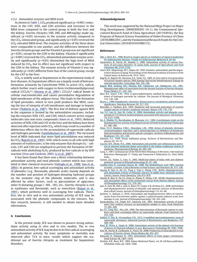
he antioxidants extracted from herbs can prevent this process byeutralizing the free radical (Jayaprakasha et al., 2001). Fig. 4 showshat the color of the control and Vitamin C decreased rapidly whenCE and BHT were much slower. We can conclude that SCE was asable 1ffects of ethanol extract of Swertia chirayita on liver and kidney SOD, CAT, GSH and MDA
Group Liver
SOD (U/mgprotein)
CAT (U/mgprotein)
GSH (mg/gprotein)
MDA (nmol/mgprotein)
I 78.3 ± 3.2 45.3 ± 3.9 2.6 ± 0.3 1.1 ± 0.4II 45.4 ± 17.8 26.8 ± 7.0 1.4 ± 0.2 2.2 ± 0.5III 72.0 ± 3.4** 43.0 ± 9.0*** 1.8 ± 0.4* 1.8 ± 0.2*
IV 65.9 ± 7.1** 34.2 ± 4.4* 2.0 ± 0.5** 1.4 ± 0.1**
V 74.1 ± 8.2*** 43.9 ± 3.0*** 1.7 ± 0.3* 1.2 ± 0.3**
VI 74.2 ± 5.5*** 40.4 ± 4.7** 2.2 ± 0.3*** 1.2 ± 0.2***
alues are mean ± S.D., n = 10 animals in each group. Statistical significance is indicated bCE, ethanol extract of Swertia chirayita; SOD, superoxide dismutase; CAT, catalase; GSHitamin E 100 (mg/kg), group IV: SCE 100 (mg/kg), group V: SCE 200 (mg/kg), group VI: S* p < 0.05.
** p < 0.01.*** p < 0.001 vs. CCl4-intoxicated group.
acology 136 (2011) 309–315 313
effective as BHT, and much more effective than Vitamin C. Therewas no significant difference between Vitamin C and the control.The LPO of SCE, BHT, and Vitamin C were 57.55%, 78.62%, and 2.31%,respectively. The SCE presented excellent antioxidant ability.
3.2.6. Antioxidant activity in a linoleic acid system usingferrothiocyanate (FTC) and thiobarbituric acid (TBA)
The FTC method was used to determine the amount of per-oxide at the initial stage of lipid peroxidation. Peroxides formedduring linoleic acid oxidation may be detected and quantified bytheir ability to oxidize Fe2+–Fe3+. Fe3+ ions form a thiocyanatecomplex with SCN−, which has a maximum absorbance at 500 nm(Liu and Yao, 2007). Therefore, samples with high antioxidantactivity are revealed by low absorbance values. In Fig. 5A, theabsorbance of the control showed a steady increase and reachedpeak value in day 9. BHT showed a high capacity to inhibit linoleicacid peroxidation with almost constant absorbance of 0.2. Theabsorbance of SCE increased slowly, which implies a strong poten-tial for antioxidant ability. Lipid peroxidation with Vitamin C wasinferior to those of BHT and SCE, but stronger than that of the con-trol, implying the excellence of Vitamin C at scavenging radicals.Hence, SCE has a stronger antioxidant activity than Vitamin C butis less efficient than BHT.
During the acid-heat treatment, the lipid peroxides decomposedto MDA, which can also be formed during the oxidation process.MDA can react with TBA to produce a red complex that can bemeasured by its absorbance at 532 nm (Guillén-Sans and Guzmán-Chozas, 1998). As shown in Fig. 5B, the absorbance values obtainedfor BHT, SCE, and Vitamin C were 1.269 ± 0.080, 1.502 ± 0.200,and 1.713 ± 0.010, respectively, compared to a control value of1.836 ± 0.160. Thus, the antioxidant activity of SCE was strongerthan that of Vitamin C but weaker than that of BHT.
Previous studies also indicated that Swertia chirayita exhib-ited effective ability in scavenging DPPH radicals and inhibitingpolyphenol oxidase activity (Gacche and Dhole, 2006), and pos-sessed the anti-inflammatory anti-diabetic potential (Grover et al.,2002). In our study, we draw the same point on the strong scaveng-ing radicals ability, at the same time, the strong reducing power,metal ion chelating ability and inhibiting linoleic acid peroxidationability were also displayed.
3.3. In vivo assays
3.3.1. Acute toxicity studiesIn acute toxicity studies, animals injected with SCE did not
show any symptoms of toxicity or mortality when dosed up to2000 mg/kg body weight.
in CCl4-intoxicated mice.
Kidney
SOD (U/mgprotein)
CAT (U/mgprotein)
GSH (mg/gprotein)
MDA (nmol/mgprotein)
76.0 ± 3.5 36.0 ± 1.0 1.7 ± 0.4 1.2 ± 0.248.8 ± 12.8 21.6 ± 9.5 0.7 ± 0.3 2.1 ± 0.261.6 ± 9.6* 33.5 ± 1.8** 0.8 ± 0.2 1.1 ± 0.3***
60.7 ± 12.0 29.3 ± 4.4** 1.5 ± 0.3*** 1.1 ± 0.1***
63.4 ± 6.4* 29.9 ± 5.0* 1.8 ± 0.2*** 1.1 ± 0.1***
64.7 ± 7.3* 33.1 ± 2.4** 1.7 ± 0.2*** 1.1 ± 0.1***
y asterisks and double-asterisks., glutathione; MDA, malondialdehyde. Group I: control, group II: CCl4, group III:
CE 400 (mg/kg).
3 pharm
3
tMtntCwS((iitnf
lfwrcloitiimaodalbairr
al2otoaoi1fati
4
iaaodd
14 Y. Chen et al. / Journal of Ethno
.3.2. Antioxidant enzymes and MDA levelsAs shown in Table 1, CCl4 produced significant (p < 0.001) reduc-
ions in the CAT, SOD, and GSH activities and increases in theDA lever compared to the control group in both the liver and
he kidney. Swertia chirayita (100, 200, and 400 mg/kg) made sig-ificant (p < 0.05) increases in the enzyme activity compared tohe CCl4-intoxicated group, and significantly (p < 0.05) reversed theCl4-elevated MDA level. The enzyme activities of the three dosesere comparable to one another, and the difference between the
wertia chirayita groups and the Vitamin E group was not significantp > 0.05), except for the GSH in the kidney. Vitamin E significantlyp < 0.001) reversed the CCl4-diminished antioxidant enzyme activ-ty and significantly (p < 0.05) diminished the high level of MDAnduced by CCl4, but its effect was not significant with respect tohe GSH in the kidney. The enzyme activity of Vitamin E was sig-ificantly (p < 0.05) different from that of the control group, except
or the CAT in the liver.CCl4 is widely used as hepatotoxin in the experimental study of
iver diseases. Its hepatotoxic effects are a largely result of biotrans-ormation, primarily by Cytochrome P450, to active CCl3 radical,hich further reacts with oxygen to form trichloromethylperoxyl
adical (CCl3O2•) (Shenoy et al., 2001). CCl3O2
• radical bonds toellular macromolecules and causes peroxidative degradation ofipid membranes of the adipose tissue. This leads to the formationf lipid peroxides, which in turn yield products like MDA, caus-ng the loss of integrity of cell membranes and damage to hepaticissue (Thabrew et al., 1987). The first line of defense against ROSn the organism is formed by the antioxidative enzymes, includ-ng the enzymes SOD, CAT, and GSH, which convert active oxygen
olecules into non-toxic compounds (Ames et al., 1993). Reducedctivities of SOD, GSH and CAT in the liver and the kidney have beenbserved after injection with CCl4, which may result in a number ofeleterious effects due to the accumulation of superoxide radicalsnd hydrogen peroxide (Santhakumari et al., 2003). The increasedevel of MDA indicated that more lipid peroxidation was inducedy CCl4 (Bagchi et al., 1995). SOD, which exists in all cells with highmounts of erythrocytes, is the only enzyme that disrupts O2
− rad-cals. CAT and GSH are employed to prevent the formation of OH−
adicals with abolishing H2O2 produced by free radicals or by SODeaction (Urso and Clarkson, 2003; Li et al., 2007).
It has been found that there was a direct relationship betweenntioxidant activity and total phenolic content which was corre-ated to their chemical structures (Velioglu et al., 1998; Sun et al.,002). In general, free radical scavenging and antioxidant activityf phenolics (e.g., flavonoids, phenolic acids) mainly depends onhe number and position of hydrogen-donating hydroxyl groupsn the aromatic ring of the phenolic molecules, and is alsoffected by other factors, such as glycosylation of aglycones,ther H-donating groups (–NH, –SH), etc., Swertia chirayita is richn xanthones and flavonoids, such as swerchirin (Bajpai et al.,991), which performs well in scavenging free radicals. There-ore, the in vitro and in vivo antioxidant activity of SCE may bessociated with the phenolic compounds in the extracts. Fur-her research, however, is still needed to obtain more detailednformation.
. Conclusions
In the present study, SCE was shown to possess strong antiox-dant activity using in vitro and in vivo models. The in vivontioxidant activity of SCE may be due to its free radical-scavenging
nd antioxidant activity. No toxic symptoms or mortality wasbserved after 72 h in mice, results which support the tra-itional use of Swertia chirayita as treatment for hepatotoxicisorders.acology 136 (2011) 309–315
Acknowledgement
This work was supported by the National Mega Project on MajorDrug Development (2009ZX09301-14-1), the Commonweal Spe-cialized Research Fund of China Agriculture (201103016), the KeyProgram of Natural Science Foundation of Hubei Province of China(2010CBB02301), and the Fundamental Research Funds for the Cen-tral Universities (20103010101000175).
References
Abe, J., Berk, B.C., 1998. Reactive oxygen species as mediators of signal transductionin cardiovascular diseases. Trends in Cardiovascular Medicine 8, 59–64.
Amarowicz, R., Naczk, M., Shahidi, F., 2000. Antioxidant activity of various frac-tions of non-tanin phenolics of canola hulls. Journal of Agricultural and FoodChemistry 48, 2755–2759.
Ames, B.N., Shigenenga, M.T., Hagen, M., 1993. Oxidants, antioxidants and the degen-erative diseases of aging. Proceedings of the National Academy of Sciences of theUnited States of America 90, 7915–7922.
Bagchi, D., Bagchi, M., Hassoun, E.A., Stohs, S.J., 1995. In vitro and in vivo generationof reactive oxygen species, DNA damage and lactate dehydrogenase leakage byselected pesticides. Toxicology 104, 129–140.
Bajpai, M.B., Asthana, R.K., Sharma, N.K., Chatterjee, S.K., Mukherjee, S.K., 1991.Hypoglycemic effect of swerchirin from the hexane fraction of Swertia chirayita.Planta Medica 57, 102–104.
Beer, R.F., Seizer, T.W., 1952. A spectrophotometric method for measuring break-down of hydrogen peroxide by catalase. The Journal of Biological Chemistry115, 130–140.
Bravo, L., 1998. Polyphenols: chemistry, dietary sources, metabolism, and nutritionalsignificance. Nutrition Reviews 56, 317–333.
Busciglio, J., Yankner, B.A., 1995. Apoptosis and increased generation of reactiveoxygen species in Down’s syndrome neurons. Nature 378, 776–779.
Cai, Y.Z., Luo, Q., Sun, M., Corke, H., 2004. Antioxidant activity and phenolic com-pounds of 112 Chinese medicinal plants associated with anticancer. Life Sciences74, 2157–2184.
De, S., Shukla, V.J., Ravishankar, B., Bhavasar, G.C., 1996. A preliminary study on thehepatoprotective activity of methanol extract of Paederia foetida leaf. Fitoterapia67, 106–109.
Dinis, T.C.P., Madeira, V.M.C., Almeidam, L.M., 1994. Action of phenolic derivates(acetoaminophen, salycilate, and 5-aminosalycilate) as inhibitors of membranelipid peroxidation and peroxyl radicals cavengers. Archives of Biochemistry andBiophysics 315, 161–169.
Ellaman, G.L., 1959. Tissue sulfhydryl group. Archives of Biochemistry and Bio-physics 82, 70–72.
Gacche, R.N., Dhole, N.A., 2006. Antioxidant and possible anti-inflammatory poten-tial of selected medicinal plants prescribed in the Indian traditional system ofmedicine. Pharmaceutical Biology 44, 389–395.
Garg, S., 1987. Gentianaceae of the North West Himalaya (A Revision). InternationalBioscience Monograph 17. Today and Tomorrow’s Publication Co., New Delhi,India.
Grover, J.K., Yadav, S., Vats, V., 2002. Medicinal plants of India with anti-diabeticpotential. Journal of Ethnopharmacology 81, 81–100.
Guillén-Sans, R., Guzmán-Chozas, M., 1998. The thiobarbituric acid (TBA) reactionin foods: a review. Critical Reviews in Food Science and Nutrition 38, 315–330.
Huang, B., Ban, X., He, J.S., Tong, J., Tian, J., Wang, Y.W., 2010a. Hepatoprotectiveand antioxidant activity of ethanolic extracts of edible lotus (Nelumbo nuciferaGaertn.) leaves. Food Chemistry 120, 873–878.
Huang, B., Ban, X., He, J.S., Zeng, H., Zhang, P., Wang, Y.W., 2010b. Hepatoprotectiveand antioxidant effects of the methanolic extract from Halenia elliptica. Journalof Ethnopharmacology 131, 276–281.
Jain, A., Soni, M., Deb, L., Jain, A., Rout, S.P., Gupta, V.B., Krishna, K.L., 2008. Antioxidantand hepatoprotective activity of ethanolic and aqueous extracts of Momordicadioica Roxb. leaves. Journal of Ethnopharmacology 115, 61–66.
Jaishree, V., Badami, S., 2010. Antioxidant and hepatoprotective effect of swertia-marin from Enicostemma axillare against d-galactosamine induced acute liverdamage in rats. Journal of Ethnopharmacology 130, 103–106.
Jayaprakasha, G.K., Singh, R.P., Sakariah, K.K., 2001. Antioxidant activity of grapeseed (Vitis vinifera) extracts on peroxidation models in vitro. Food Chemistry73, 285–290.
Jia, Z.S., Tang, M.C., Wu, J.M., 1999. The determination of flavonoid content in mul-berry and their scavenging effects on superoxide radicals. Food Chemistry 64,555–559.
Kakkar, P., Das, B., Visvanathan, P.N., 1972. A modified spectrophotometric assay ofsuperoxide dismutase. Industrial Journal of Biochemistry and Biophysics 197,588–590.
Kaur, R., Arora, S., Singh, B., 2008. Antioxidant activity of the phenol rich fractionsof leaves of Chukrasia tabularis A. Juss. Bioresource Technology 99, 7692–7698.
Keil, M., Hartle, B., Guillaume, A., Psiorz, M., 2000. Production of amarogentin in root
cultures of Swertia chirata. Planta Medica 66, 452–457.Kikuzaki, H., Nakatani, N., 1993. Antioxidant effects of some ginger constituents.Food Science 58, 1407–1410.
Kirtikar, K.R., Basu, B.D., 1984. Indian Medicinal Plants, vol. III. LM Basu publishers,Allahabad, India, pp. 1664–1666.
pharm
L
L
L
L
M
M
M
O
P
R
S
S
Y. Chen et al. / Journal of Ethno
i, X.L., Zhou, A.G., Li, X.M., 2007. Inhibition of Lycium barbarum polysac-charides and Ganoderma lucidum polysaccharides against oxidative injuryinduced by c-irradiation in rat liver mitochondria. Carbohydrate Polymers 69,172–178.
iu, Q., Yao, H.Y., 2007. Antioxidant activities of barley seeds extracts. Food Chemistry102, 732–737.
loyd, R.V., Hanna, P.M., Mason, R.P., 1997. The origin of the hydroxyl radical oxygenin the Fenton reaction. Free Radical Biology and Medicine 22, 885–888.
u, Y.R., Foo, L.Y., 2000. Antioxidant and radical scavenging activities of polyphenolsfrom apple pomace. Food Chemistry 68, 81–85.
eerson, F.Z., Kagan, V.E., Kozlov, Y.P., Belkina, L.M., Arkhipenko, Y.V., 1982. The roleof lipid peroxidation in pathogenesis of ischemic damage and the antioxidantprotection of the heart. Basic Research in Cardiology 77, 465–485.
oreno, M.I.N., Isla, M.I., Sampietro, A.R., 2000. Comparison of the free radical-scavenging activity of propolis from several regions of Argentina. Journal ofEthnopharmacology 71, 109–114.
ukherji, B. (Ed.), 1953. Indian Pharmaceutical Codex, Indigenous Drugs, vol. I. CSIR,New Delhi, pp. 64–65.
yaizu, M., 1986. Studies on products of browning reactions: antioxidant activitiesof products of browning reaction prepared from glucose amine. Japanese Journalof Nutrition 44, 307–315.
harmacopoeia Committee of Ministry of Health of China, 1995. The Drug Standardof Ministry of Health of China. Tibetan Medicine, vol. I. Ministry of Health of PRChina, Beijing, p. 28.
e, R., Pellegrini, N., Proteggente, A., Pannala, A., Yang, M., Rice- Evans, C.A., 1999.Antioxidant activity applying an improved ABTS radical cation decolorizationassay. Free Radical Biology and Medicine 26, 1231–1237.
abir, S.M., Rocha, J.B.T., 2008. Antioxidant and hepatoprotective activity of aqueous
extract of Solanum fastigiatum (false “Jurubeba”) against paracetamol-inducedliver damage in mice. Journal of Ethnopharmacology 120, 226–232.amant, S.S., Dhar, U., Palni, L.M.S., 1998. Medicinal Plants of Indian Himalaya:Diversity, Distribution and Potential Values, vol. 13. Hima-vikas Publication,p. 163. Sanchez-Moreno, C., 2002. Review: methods used to evaluate the free
acology 136 (2011) 309–315 315
radical scavenging activity in foods and biological systems. Food Science andTechnology International 8, 121–137.
Santhakumari, P., Prakasam, A., Pugalendi, K.V., 2003. Modulation of oxidative stressparameters by treatment with piper betle leaf in streptozotocin induced diabeticrats. Indian Journal of Pharmacology 35, 373–378.
Shanmugasundaram, P., Venkataraman, S., 2006. Hepatoprotective and antioxidanteffects of Hygrophila auriculata (K. Schum) Heine Acanthaceae root extract. Jour-nal of Ethnopharmacology 104, 124–128.
Shenoy, K.A., Somayaji, S.N., Bairy, K.L., 2001. Hepatoprotective effects of Ginkgobiloba against carbon tetrachloride induced hepatic injury in rats. Indian Journalof Pharmacology 33, 260–266.
Srivastava, A., Shivanandappa, T., 2009. Hepatoprotective effect of the root extractof Decalepis hamiltonii against carbontetrachloride-induced oxidative stress inrats. Food Chemistry 118, 411–417.
Sun, J., Chu, Y.F., Wu, X.Z., Liu, R.H., 2002. Antioxidant and antiproliferative activitiesof common fruits. Journal of Agricultural and Food Chemistry 50, 7449–7454.
Thabrew, M.I., Joice, P.D.T.M., Rajatissa, W.A., 1987. Comparative study of the effi-cacy of Paetta indica and Osbeckia octandra in the treatment of liver dysfunction.Planta Medica 53, 239–241.
Urso, M.L., Clarkson, P.M., 2003. Oxidative stress, exercise, and antioxidant supple-mentation. Toxicology 189, 41–54.
Velioglu, Y.S., Mazza, G., Gao, L., Oomah, B.D., 1998. Antioxidant activity and totalphenolics in selected fruits, vegetables, and grain products. Journal of Agricul-tural and Food Chemistry 46, 4113–4117.
Wu, L.C, Hsu, H.W., Chen, Y.C., Chiu, C.C., Lin, Y.I., Ho, J.A., 2006. Antioxidant andantiproliferative activities of red pitaya. Food Chemistry 95, 319–327.
Yagi, K., Rastogi, R., 1979. Assay for lipid peroxides in animal tissue by thiobarbituricacid reaction. Analytical Biochemistry 95, 351.
Yen, G.C., Chen, H.Y., 1995. Antioxidant activity of various tea extracts in relation totheir antimutagenicity. Journal of Agriculture and Food Chemistry 43, 27–32.
Zeashan, H., Amresha, G., Singh, S., Rao, C.V., 2009. Hepatoprotective and antiox-idant activity of Amaranthus spinosus against CCl4 induced toxicity. Journal ofEthnopharmacology 125, 364–366.


