Embed Size (px)
Citation preview

Available online at www.sciencedirect.com
Nuclear Medicine and Biology xx (2011) xxx–xxxwww.elsevier.com/locate/nucmedbio
In vivo tracking of 111In-oxine labeled mesenchymal stem cells followinginfusion in patients with advanced cirrhosis
Ali Gholamrezanezhada,⁎, Sahar Mirpoura, Mohammad Bagherib, Mehdi Mohamadnejadb,Kamran Alimoghaddamc, Leila Abdolahzadehb, Mohsen Sagharia, Reza Malekzadehb
aResearch Institute for Nuclear Medicine. Shariati Hospital. Tehran University of Medical Sciences, Tehran, 14114, IranbDigestive Disease Research Center. Shariati Hospital. Tehran University of Medical Sciences, Tehran, 14114, Iran
cHematology and BMT Research Center. Shariati Hospital. Tehran University of Medical Sciences, Tehran, 14114, Iran
Received 25 January 2011; received in revised form 29 March 2011; accepted 30 March 2011
Abstract
Background: Several animal and few human studies suggest the beneficial role of bone marrow mesenchymal stem cells (MSCs) in livercirrhosis. However, little is known about the fate of MSCs after infusion in cirrhotic patients. We evaluated stem cell biodistribution afterperipheral infusion of MSCs in four cirrhotic patients.Methods: After three passages of MSCs, the patients received a total of 250–400×106 cells, of which only 50% of the cells were labeled.Specific activities of 0.21–0.67 MBq/106 cells were maintained for the injected labeled MSCs. Planar whole-body acquisitions (anterior/posterior projections) were acquired immediately following infusion as well as at 2 h, 4 h, 6 h, 24 h, 48 h, 7th and 10th days after cellinfusion.Results: After intravenous infusion, the radioactivity was first observed to accumulate in the lungs. During the following hours to days, theradioactivity gradually increased in the liver and spleen, with spleen uptake exceeding that in the liver in all patients. Region-of-interestanalysis showed that the percentage of cells homing to the liver (following decay and background corrections and geometric meancalculation) increased from 0.0%-2.8% at immediately post-infusion images to 13.0–17.4% in 10th-day post-infusion. Similarly, the residualactivities in the spleen increased from 2.0%-10.2% at immediately post-infusion images to 30.1%-42.2% in 10th-day post-infusion. Duringthe same period, the residual activities in the lungs decreased from 27.0–33.5% to 2.0–5.4%.Conclusion: The infusion of MSCs labeled with 111In-oxine through a peripheral vein is safe in cirrhosis. Cell labeling with 111In-oxine is asuitable method for tracking MSC distribution after infusion.© 2011 Elsevier Inc. All rights reserved.
Keywords: 111In-oxine; Mesenchymal stem cell; Cirrhosis
1. Introduction
Cirrhosis and chronic liver failure are among the mostfrequent causes of morbidity and mortality throughout theworld, with the majority of preventable cases attributed toexcessive alcohol consumption, viral hepatitis or nonalco-holic fatty liver disease [1]. Orthotopic liver transplantationis the standard treatment modality in patients with decom-pensated cirrhosis [2]. However, it has several limitationssuch as shortage of organ donors, high cost, and several
⁎ Corresponding author.E-mail address: [email protected] (A. Gholamrezanezhad).
0969-8051/$ – see front matter © 2011 Elsevier Inc. All rights reserved.doi:10.1016/j.nucmedbio.2011.03.008
complications [2]. Despite progress in medical and surgicaltreatment, alternative strategies are needed to improve theoutcome of patients with cirrhosis.
Stem cell therapy may be a potential alternative to livertransplantation. Bone marrow is a reservoir of various stemcells, including hematopoietic stem cells (HSCs) andmesenchymal stem cells (MSCs). Several studies havesuggested MSC transplantation can reduce liver fibrosis inanimal models of cirrhosis [3–6].
Infusion of the cells directly into the vessels feeding theliver (e.g., portal vein or hepatic artery) may improvedelivery and subsequent engraftment of the cells into theliver. This method requires an invasive procedure forangiography, and may lead to serious adverse effects, such
able 1ge and sex of the patients, the estimated number of infused MSCs, specificctivity and also the labeling efficiency of the samples
atient Age Sex Estimated numberof infused MSCs
Specific activity ofthe injected labeledcells
Labelingefficiency
62 F 270×106 0.67 MBq/106 cells 36.2%17 M 290×106 0.71 MBq/106 cells 38.8%29 M 250×106 0.41 MBq/106 cells 52.0%62 M 400×106 0.21 MBq/106 cells 53.0%
2 A. Gholamrezanezhad et al. / Nuclear Medicine and Biology xx (2011) xxx–xxx
as hemorrhage or contrast nephropathy [2]. In a Phase 1 trial,we have shown MSC transplantation through a peripheralvein is safe and feasible in human cirrhosis [7]. However,there is a concern that after peripheral vein infusion ofMSCs, many of the cells may entrap in the lung during theirfirst pass through this organ. On the other hand, it has beenshown that some MSCs infused through a peripheral veincan escape the lung, engraft into the liver and reverse CCl4-induced acute liver injury [8]. Thus, there is a need to trackMSCs after peripheral vein infusion in cirrhosis, and toprovide information about the biodistribution of the cellsdelivered by such a route, in order to confirm adequatehoming of the injected cells to the target organ.
Among the different methods examined [9–16], magneticresonance imaging (MRI) and radioactive labeling withradiopharmaceuticals are the most widely used methods fortracking infused stem cells [17]. However, some authorsclaim that MRI tracking of stem cells suffers from lowsensitivity [18,19]. An important limitation of MRI is that itmay not distinguish iron-labeled cells from free iron particles.Hence, we chose nuclear medicine techniques to evaluatebiodistribution of stem cells infused via the intravenous route.Three well-established cell labeling radioagents are 18FDG,99mTc-HMPAO and 111In-oxine, with short and longer half-lives, respectively. The time period during which radiola-beled cell distribution can be observed is limited by the decayof their label [20]. Since we were interested in cell homingover several days, we selected 111In as the radionuclide (witha half-life of 67 h). 111In-oxine is an Auger electron emitterwhich internalizes nonspecifically into both normal andmalignant cells [21]. Based on the study of Andersson et al.[22], the decline of intracellular 111In concentration is mostprominent during the first 6 h post-labeling and appears toremain stable thereafter.
We conducted a pilot study on human to evaluate stemcell biodistribution after peripheral infusion of MSCs inpatients with liver cirrhosis by labeling a fraction of theinjected cells using 111In-oxine and imaging the distributionpattern over a 10-day period.
2. Materials and methods
2.1. Patients
Four patients with decompensated liver cirrhosis wereincluded in the study. Informed consent was assigned by thepatients and documented in the medical record. The projectwas approved by the ethics committee and the researchcouncil of Digestive Disease Research Center, TehranUniversity of Medical Sciences [ClinicalTrials.gov ID:NCT00476060].
2.2. Preparation of MSCs
A total of 200 ml bone marrow was aspirated from fourdifferent sites of the iliac crest in the right and left side (50 ml
TAa
P
1234
at each site) of each patient. The procedure of stem cellisolation was performed in a clean room (FS 209 E & ISO 14644). The harvested bone marrow samples were placed insterile tubes and were diluted 1:2 with 2 mM EDTA/PBS.The mononuclear fraction was isolated by density gradientcentrifugation at 435×g for 30 min at room temperatureusing Ficoll–Hypaque solution (Inno-TRAin, Germany) andseeded at a density of 1×10⁶cells/cm2 into T75 cell cultureflasks (Nunc, Austria). The cells were plated in Dulbecco'smodified Eagle's medium-low glucose (DMEM-LG,GIBCO, UK), supplemented with 10% fetal calf serum(Sigma, Germany) and 1% penicillin-streptomycin (Gibco,UK), and cultured at 37°C in a 5% CO2 atmosphere. After 3days, nonadherent cells were removed and the adherent cellswere cultured for another 7 days with media changes every 3days. Cells were grown to confluence, then harvested byincubation with 0.25% trypsin/1mM EDTA (GIBCO, UK),centrifuged at 1200 rpm for 5 min, and subcultured at a 1:3split ratio in new culture flasks. After reaching confluencefor the second time (after 8 days), the harvested cells weredefined as passage 1, and the replated cells were cultured andserially subcultured until passage 3 [23].
At the end of the last passage, theywerewashedwith tyrodesalt (Sigma, Germany) and incubated with M199 medium(Sigma, Germany) for an hour. Cells were detached withtrypsinization and washed with normal saline supplementedwith 1% human serum albumin (Blood Research & Fraction-ation Co, Iran) and heparin three times, and resuspended at 1–1.5×10⁶/ml density in M199. This washing process eliminatestrace amounts of fetal bovine serum as well. Also, bacterio-logical tests were performed on the samples for every passageand at the time of injection. Then, the cells were labeled with111In-oxine before intravenous infusion.
2.3. 111Indium-oxine labeling of MSCs
In each patient, the solution containing the MSCs wasdivided into two equal portions: The first portion was usedfor radiolabeling (hence, only 50% of cells were labeled with111In-oxine [24]) and the second portion infused to thepatients without radiolabeling. For radiolabeling, the firstportion was initially washed with phosphate-buffered saline,mixed and suspended with 111In-oxine at the concentrationof approximately 1.85 MBq/106 cells, and then incubated for30 min at the room temperature.
able 2esidual activity in the liver and spleen at different time points
2 h 4 h 6 h 24 h 48 h 7th day 10th day
atient 1Lung 33.5% 31.1% 19.2% 14.7% 12.0% 4.0% 2.0%Liver 2.8% 4.1% 4.1% 4.3% 4.7% 12.0% 13.5%Spleen 2.0% 3.1% 3.3% 4.0% 5.4% 21.3% 30.1%atient 2Lung 27.0% 26.0% 21.1% 13.0% 12.2% 8.7% 5.4%Liver 0.0% 3.6% 3.6% 3.9% 4.3% 11.0% 13.0%Spleen 2.5% 4.2% 5.1% 7.0% 9.3% 14.0% 42.0%atient 3Lung 29.8% 24.3% 20.1% 13.6% 12.1% 6.8% 2.9%Liver 2.1% 5.1% 5.4% 7.7% 8.2% 9.1% 15.6%Spleen 10.2% 12.7% 15.0% 25.1% 25.7% 37.6% 40.2%atient 4Lung 30.1% 26.9% 20.8% 12.6% 12.0% 5.7% 3.0%Liver 7.9% 9.3% 10.1% 11.9% 11.9% 15.1% 17.4%Spleen 7.3% 8.8% 8.8% 9.1% 14.9% 32.2% 33.0%
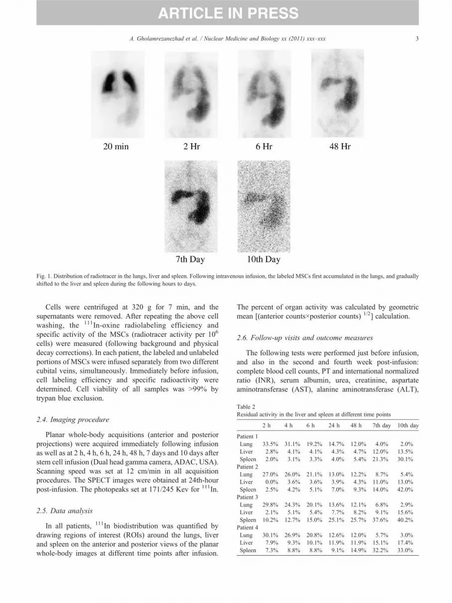
Fig. 1. Distribution of radiotracer in the lungs, liver and spleen. Following intravenous infusion, the labeled MSCs first accumulated in the lungs, and graduallyshifted to the liver and spleen during the following hours to days.
3A. Gholamrezanezhad et al. / Nuclear Medicine and Biology xx (2011) xxx–xxx
Cells were centrifuged at 320 g for 7 min, and thesupernatants were removed. After repeating the above cellwashing, the 111In-oxine radiolabeling efficiency andspecific activity of the MSCs (radiotracer activity per 106
cells) were measured (following background and physicaldecay corrections). In each patient, the labeled and unlabeledportions of MSCs were infused separately from two differentcubital veins, simultaneously. Immediately before infusion,cell labeling efficiency and specific radioactivity weredetermined. Cell viability of all samples was N99% bytrypan blue exclusion.
2.4. Imaging procedure
Planar whole-body acquisitions (anterior and posteriorprojections) were acquired immediately following infusionas well as at 2 h, 4 h, 6 h, 24 h, 48 h, 7 days and 10 days afterstem cell infusion (Dual head gamma camera, ADAC, USA).Scanning speed was set at 12 cm/min in all acquisitionprocedures. The SPECT images were obtained at 24th-hourpost-infusion. The photopeaks set at 171/245 Kev for 111In.
2.5. Data analysis
In all patients, 111In biodistribution was quantified bydrawing regions of interest (ROIs) around the lungs, liverand spleen on the anterior and posterior views of the planarwhole-body images at different time points after infusion.
The percent of organ activity was calculated by geometricmean [(anterior counts×posterior counts) 1/2] calculation.
2.6. Follow-up visits and outcome measures
The following tests were performed just before infusion,and also in the second and fourth week post-infusion:complete blood cell counts, PT and international normalizedratio (INR), serum albumin, urea, creatinine, aspartateaminotransferase (AST), alanine aminotransferase (ALT),
TR
P
P
P
P
Table 3Clinical and laboratory data of the patients during the follow-up visits
Patient 1 (cryptogenic cirrhosis)
Edema PT (s) INR Serumalbumin(g/dl)
Cr(mg/dl)
Totalbilirubin(mg/dl)
Directbilirubin(mg/dl)
AST(IU/ml)
ALT(IU/ml)
AFP(mcg/L)
Hb WBC Plt MELDscore
Baseline 2+ 14.1 1.2 3.5 2.13 1.82 0.53 27 13 3.05 10.5 5400 135000 18Two weeks post-infusion 2+ 13.7 1.2 3.2 2.05 2.03 0.59 40 24 2.83 11.7 5910 71000 18Four weeks post-infusion 1+ 13.7 1.2 3.5 1.83 1.52 0.52 41 27 2.47 12.2 7940 82000 16
Patient 2 (cryptogenic cirrhosis)
Edema PT (s) INR Serumalbumin(g/dl)
Cr(mg/dl)
Totalbilirubin(mg/dl)
Directbilirubin(mg/dl)
AST(IU/ml)
ALT(IU/ml)
AFP(mcg/L)
Hb WBC Plt MELDscore
Baseline 2+ 18 1.7 2.5 0.8 2.7 0.45 73 67 1.89 10.5 3050 31000 16Two weeks post-infusion 2+ 17.6 1.9 2.1 0.57 2.3 0.63 79 53 1.46 9.8 2200 42000 17Four weeks post-infusion 2+ 16.7 1.8 2.1 0.58 2.41 0.63 72 45 1.48 9.9 2290 38000 16
Patient 3 (cryptogenic cirrhosis)
Edema PT (s) INR Serumalbumin(g/dl)
Cr(mg/dl)
Totalbilirubin(mg/dl)
Directbilirubin(mg/dl)
AST(IU/ml)
ALT(IU/ml)
AFP(mcg/L)
Hb WBC Plt MELDscore
Baseline 1+ 15 1.52 3.7 1.2 2.0 – 39 16 4.5 12.7 6300 100000 15Two weeks post-infusion 1+ 16.8 1.6 3.8 1.31 1.56 0.49 38.0 27.0 4.26 14.7 4520 92000 16Four weeks post-infusion 1+ 15.5 1.4 3.5 1.28 2.01 0.57 40.0 28.0 4.20 14.5 4640 84000 15
Patient 4 (hemochromatosis)
Edema PT (s) INR Serumalbumin(g/dl)
Cr(mg/dl)
Totalbilirubin(mg/dl)
Directbilirubin(mg/dl)
AST(IU/ml)
ALT(IU/ml)
AFP(mcg/L)
Hb WBC Plt MELDscore
Baseline 1+ 16.5 1.6 3.0 0.7 3.09 1.8 230 150 1.69 11.0 2700 55000 16Two weeks post-infusion 1+ 16.6 1.6 2.5 0.75 4.07 2.04 265 165 1.69 10.9 2360 50000 14Four weeks post-infusion 1+ N36.7 N6.9 2.7 0.72 5.63 2.78 292 176 1.84 11.5 3190 55000 35
PT, prothrombin time; INR, international normalized ratio; Cr, serum creatinine; AST, aspartate aminotransferase; ALT, alanine aminotransferase; AFP, alphafetoprotein; Hb, hemoglobin; WBC, white blood cells; Plt, platelet; MELD, model for end stage liver disease.
4 A. Gholamrezanezhad et al. / Nuclear Medicine and Biology xx (2011) xxx–xxx
serum alkaline phosphatase, serum total and direct bilirubin,alfa-fetoprotein. In addition, 10 ml of the patients' serumsamples were collected and stored frozen at −70°C at eachfollow-up visits.
Interval histories were taken and physical examinationsperformed at each follow-up visits. Peripheral edema wasgraded as follows: 0: no edema; trace: indention caused bypressure over the dorsum of the foot; 1+: indention at shin;2+: indention at knee; 3+: indention above knee; 4+:generalized edema (indention over hip and low back).
2.7. Evaluation of safety and feasibility
Adverse effects were evaluated at each follow-up visitsaccording to the above-mentioned schedule. Clinical, labora-tory, and safety-related data were prospectively collected.Procedural complications were defined as any hemodynamicinstability during the cell infusion. Major side effects weredefined as development of any of the following complicationsduring the follow up: acute renal failure, worsening hepaticdecompensation that requires urgent liver transplantation,progressive elevation in serum AFP.
3. Results
Table 1 summarizes the patients' age, sex, estimatednumber of infused MSCs (of which only 50% of cells werelabeled), specific activity of injected labeled cells, and alsothe labeling efficiency.
3.1. Whole-body imaging
After intravenous infusion, the radioactivity was firstobserved to accumulate in lungs, and gradually shifted toliver and spleen (Fig. 1). Based on the initial views (thoseprojections which were obtained immediately after infusion),liver and spleen uptake could be distinguished from thebackground activity just in the fourth patient who sufferedfrom a milder form of the disease (as confirmed by his lowerMELD score, as compared to the other three patients). In theother patients, the initial hepatic and splenic accumulation ofradioactivity was visualized only after 2 h (except the thirdpatient who showed delineation of the splenic activity in theimmediately post-infusion images — due to his hugesplenomegaly) (Fig. 1).

5A. Gholamrezanezhad et al. / Nuclear Medicine and Biology xx (2011) xxx–xxx
During the following hours to days, the radioactivitygradually increased in liver and spleen, with spleen uptakeexceeding that in liver in all patients (Fig. 1). ROI analysisshowed that the percentage of cells shifted toward the liver(following decay and background corrections and geometricmean calculation) increased from 0.0%-2.8% in theimmediate post-infusion images to 13.0%-17.4% in 10th-day post-infusion. Similarly, the residual activities in thespleen increased from 2.0%-10.2% at immediately post-infusion images to 30.1%-42.2% in 10th-day post-infusion(Table 2). During the same period, the residual activities inthe lungs decreased from 27.0–33.5% to 2.0–5.4%.
On SPECT images after the 24th hour, the activitydistribution was homogenous throughout the liver in all thepatients, but the splenic activities were nonhomogeneouswith foci of severely decreased or absent radiotracer uptake,even evident in planar imaging (Fig. 1).
No unexpected adverse events or complications wereobserved during stem cell infusion and within 1-monthfollow-up period. Clinical and laboratory data of the patientsat the follow-up visits are summarized in Table 3.
4. Discussion
Our study is the first report of MSCs tracking aftertransplantation in human cirrhosis. This study showed thatafter peripheral vein infusion of MSCs, most of the cells wereinitially entrapped within the lung capillaries during their firstpass, but over the next 48 h, a significant proportion of thecells escaped from the lung capillary system and migrated tothe liver and spleen (Table 2). Furthermore, we showed thatthe cells can be visualized in vivo up to at least 10 days afterinitial infusion, and it is possible to quantify and follow themusing radiolabeling with 111In-oxine. In this study, infusionof the MSCs labeled with 111In-oxine was safe in livercirrhosis patients within 1 month of follow-up.
MRI tracking of the cells labeled with iron particles isanother acceptable method for tracking the cells in regener-ative medicine. However, some authors believe that MRItracking of stem cells suffers from disadvantages such as thelimited number of probes available [18]. Schoepf et al. [25]asserted that with clinical 1.5 T MR equipment and clinicallyapplicable contrast agents, it was not feasible to trace the invivo distribution of intravenously injected hematopoietic cellsto more than one final target organ or to depict the migration ofthe transplanted cells to several subsequent target organs overtime. Based on the study of Kraitchman et al. [19], one of themain disadvantages of MRI tracking is the lower sensitivity ofthe method, compared with radionuclide techniques. In fact,initial cell-labeling techniques were hampered by limitedconcentration of internalized contrast agent,which resulted in alimited sensitivity of MRI to detect the labeled cells. Tocompensate for this limited sensitivity, experimental cell-tracking studies were performed using MR imagers with veryhigh magnetic field strengths of up to 14 T [15].
On the other hand, several studies already reported thatindium- or technetium-labeled stem cell imaging can be usedto monitor the fate of transplanted stem cells [26,27].Radionuclide techniques may provide a noninvasive andsemiquantitative method to sequentially assess the in vivodistribution and homing of MSCs to the intended site [28]. Inour experience, 111In (which has long half-life of 67 h)provides the opportunity to look at cell homing over severaldays. In the current study, the activity continues toaccumulate in the liver and spleen during the entire 10days after infusion.
Kang et al. [29] evaluated the homing and distribution ofperipheral HSCs in patients who were afflicted bymyocardial infarction after injection using 18F-FDG-labeledstem cell PET [29]. They found that after intravenousinjection, the total amount of stem cells in the liver andspleen were 21.3% and 42.1% of the injected dose,respectively, at 4 h post-infusion [29]. Caveliers et al. [30]evaluated stem cell homing after intracoronary injection ofperipheral blood stem cells in patients with chronic ischemiccardiomyopathy. In their study, the ROI analysis in patientsshowed that the respective estimated liver and spleen uptakewas approximately 25% and 3.5% after 12 h.
In our study, the absorbed activity increased graduallyduring the study period; however, the total activity in theliver was less than that reported by Caveliers et al. [30] andKang et al. The differences as compared to our findings (withdecreased hepatic and increased splenic homing) may beexplained by the fact that our patients suffered fromadvanced liver cirrhosis. Splenomegaly which is present incirrhosis, and the hepatofugal flow of the portal veinobserved in portal hypertension, can be responsible fordecreased hepatic homing found in this study [31,32].
Our study has some limitations. It is possible that MSCslabeled with 111In-oxine are phagocytosed by the reticulo-endothelial cells when they are trapped initially in the lungcapillary. Isotopic tracking cannot exclude the possibilitythat 111In-oxine signals were detected from free radioactiveindium released from labeled cells (although based on theprevious reports the extent of such phenomenon is verylimited [22]) or the retoculoendeothelial cells which couldtheoretically phagocytize nonviable MSCs (due to time-dependent cytotoxic effect of the 111In-oxine, after infusion,a limited portion of MSCs would lose viability [23]). In fact,no one can be sure that the observed radioactivity in vivoreveals the presence of viable MSCs in the organs havingradioactivity. However, it should be emphasized that thislimitation is not limited to scintigraphic tracking methods:MR tracking of stem or other cells has a critical flaw that theferromagnetic particles do not represent viable cells in vivo.We simply do not know whether MR signals mean magneticparticles in the “live” cells or “already dead” cells or evenparticles in the engulfed/digested cells.
The beneficial effects of bone marrow stem cells in liverfibrosis have been demonstrated in several animal studies[3–6]. Few human studies have assessed the therapeutic

6 A. Gholamrezanezhad et al. / Nuclear Medicine and Biology xx (2011) xxx–xxx
efficacy of stem cells in the treatment of patients with livercirrhosis [2,7,33–35]. In these limited studies, the cells wereeither administered intravenously [7] or injected into theportal vein [33,34] or hepatic artery [2,33,34]. None of thesestudies addressed the fate of these cells in vivo.
In our study (Table 3), no significant improvement inliver function was noted after 1 month period of follow-up.Our main explanation is the fact that may be longer follow-ups are needed to explore the therapeutic and beneficialeffects of infused MSCs on liver cirrhosis, as theoreticallythey have to generate functionally active hepatocytes (aprocess which is time dependent). Lack of improvement mayalso be explained by the fact that homing of the infused cellsto the target organ (liver) has been occurred in just a limitednumber of MSCs. This explanation is boosted by our studyfindings, which show that up to the 10th post-infusion day,less than 14% of the activity was located in the patients'livers and more than 30–40% of the activity was trapped inthe enlarged spleens. As another explanation it should bekept in mind that may be some degrees of the toxic effects ofthe In-oxine on MSCs would decrease the therapeutic effectsof the therapeutic measure of MSC infusion. Although wehave previously shown that a limited proportion of MSCslose their viability upon binding to In-oxine [23], and in ourstudy just half of the infused cells were labeled withradiotracer and also as low as possible radiotracer wasapplied for radiolabeling of stem cells (the dose with thelowest cytotoxicity, as determined by our previous in vitroexperiment [23]), we can not exclude the negative impact ofIn-oxine on the function and viability of MSCs, however.Furthermore, the efficacy of this potential new treatmentstrategy needs to be evaluated in a larger group of patients.
In conclusion, we found in vivo tracking of MSC with111In-oxine is safe and feasible in cirrhosis. Cell labelingwith 111In-oxine is a suitable method for tracking of celldistribution up to 10 days after infusion.
Acknowledgment
We kindly thank Dr. E. Scott Swenson from Yale Schoolof Medicine, and Dr. Davood Beiki, Dr. MohmmadEftekhari, Dr. Armaghan Fard-Esfehani, and Dr. BabakFallahi from University of Tehran for their valuablecomments. Thanks are also extended to our technologistsin Research Institute for Nuclear Medicine, particularly Ms.Darvish-ha, for image acquisition.
References
[1] Heidelbaugh JJ, Bruderly M. Cirrhosis and chronic liver failure: part I.Diagnosis and evaluation. Am Fam Physician 2006;74:756–62.
[2] Mohamadnejad M, Namiri M, Bagheri M, et al. Phase 1 human trial ofautologous bone marrow-hematopoietic stem cell transplantation inpatients with decompensated cirrhosis. World J Gastroenterol2007;13:3359–63.
[3] Fang B, Shi M, Liao L, et al. Systemic infusion of FLK1(+)mesenchymal stem cells ameliorate carbon tetrachloride-induced liverfibrosis in mice. Transplantation 2004;78:83–8.
[4] Oyagi S, Hirose M, KojimaM, et al. Therapeutic effect of transplantingHGF-treated bone marrow mesenchymal cells into CCl4-injured rats.J Hepatol 2006;44:742–8.
[5] Zhao DC, Lei JX, Chen R, et al. Bone marrow-derived mesenchymalstem cells protect against experimental liver fibrosis in rats. World JGastroenterol 2005;11:3431–40.
[6] Abdel Aziz MT, Atta HM, et al. Therapeutic potential of bone marrow-derived mesenchymal stem cells on experimental liver fibrosis. ClinBiochem 2007;40:893–9.
[7] Mohamadnejad M, Alimoghaddam K, Mohyeddin-Bonab M, et al.Phase 1 trial of autologous bone marrow mesenchymal stem celltransplantation in patients with decompensated liver cirrhosis. ArchIran Med 2007;10:459–66.
[8] Kuo TK, Hung SP, Chuang CH, et al. Stem cell therapy for liverdisease: parameters governing the success of using bone marrowmesenchymal stem cells. Gastroenterology 2008;134:2111–21.
[9] Lewin M, Carlesso N, Tung CH, et al. Tat peptide-derivatizedmagnetic nanoparticles allow in vivo tracking and recovery ofprogenitor cells. Nat Biotechnol 2000;18:410–4.
[10] Hill JM, Dick AJ, Raman VK, et al. Serial cardiac magnetic resonanceimaging of injected mesenchymal stem cells. Circulation 2003;108:1009–14.
[11] Bulte JW, Douglas T, Witwer B, et al. Magnetodendrimers allowendosomal magnetic labeling and in vivo tracking of stem cells. NatBiotechnol 2001;19:1141–7.
[12] Rudelius M, Daldrup-Link HE, Heinzmann U, et al. Highly efficientparamagnetic labelling of embryonic and neuronal stem cells. Eur JNucl Med Mol Imaging 2003;30:1038–44.
[13] Jendelová P, Herynek V, Decroos J, et al. Imaging the fate of implantedbone marrow stromal cells labeled with superparamagnetic nanopar-ticles. Magn Reson Med 2003;50:767–76.
[14] Jendelová P, Herynek V, Urdzíková L, et al. Magnetic resonancetracking of transplanted bone marrow and embryonic stem cells labeledby iron oxide nanoparticles in rat brain and spinal cord. J Neurosci Res2004;76:232–43.
[15] Weissleder R, ChengHC, Bogdanova A, et al. Magnetically labeled cellscan be detected byMR imaging. JMagnReson Imaging 1997;7:258–63.
[16] Yeh TC, Zhang W, Ildstad ST, et al. Intracellular labeling of T-cellswith superparamagnetic contrast agents. Magn Reson Med 1993;30:617–25.
[17] Bindslev L, Haack-Sørensen M, Bisgaard K, et al. Labelling of humanmesenchymal stem cells with indium-111 for SPECT imaging: effecton cell proliferation and differentiation. Eur J Nucl Med Mol Imaging2006;33:1171–7.
[18] Boersma HH, Tromp SC, Hofstra L, et al. Stem cell tracking: reversingthe silence of the lambs. J Nucl Med 2005;46:200–3.
[19] Kraitchman DL, Tatsumi M, Gilson WD, et al. Dynamic imaging ofallogeneic mesenchymal stem cells trafficking to myocardial infarc-tion. Circulation 2005;112:1451–61.
[20] Steindler DA. Stem cells, regenerative medicine, and animal models ofdisease. ILAR J 2007;48:323–38.
[21] Reilly RM, Kiarash R, Cameron RG, et al. 111In-labeled EGF isselectively radiotoxic to human breast cancer cells overexpressingEGFR. J Nucl Med 2000;41:429–38.
[22] Andersson P, Forssell-Aronsson E, Johanson V, et al. Internalization ofindium-111 into human neuroendocrine tumor cells after incubationwithindium-111-DTPA-D-Phe1-octreotide. J Nucl Med 1996;37:2002–6.
[23] Kharaziha P, Hellström PM, Noorinayer B, Farzaneh F, Aghajani K,Jafari F, et al. Improvement of liver function in liver cirrhosis patientsafter autologous mesenchymal stem cell injection: a phase I–II clinicaltrial. Eur J Gastroenterol Hepatol 2009;21:1199–205.
[24] Gholamrezanezhad A, Mirpour S, Majd Ardekani J, et al. Cytotoxicityof 111in-oxine on mesenchymal stem cells: a time dependent adverseeffect. Nucl Med Commun 2009;30:210–6.

7A. Gholamrezanezhad et al. / Nuclear Medicine and Biology xx (2011) xxx–xxx
[25] Schoepf U, Marecos E, Melder R, et al. Intracellular magnetic labelingof lymphocytes for in vivo trafficking studies. Biotechniques 1998;24:6420–651.
[26] Aicher A, Brenner W, Zuhayra M, et al. Assessment of the tissuedistribution of transplanted human endothelial progenitor cells byradioactive labeling. Circulation 2003;107:2134–9.
[27] Barbash IM, Chouraqui P, Baron J, et al. Systemic delivery of bonemarrow-derived mesenchymal stem cells to the infarcted myocardium:feasibility, cell migration, and body distribution. Circulation 2003;108:863–8.
[28] Chin BB, Nakamoto Y, Bulte JW, et al. 111In oxine labelledmesenchymal stem cell SPECT after intravenous administration inmyocardial infarction. Nucl Med Commun 2003;24:1149–54.
[29] Kang WJ, Kang HJ, Kim HS, et al. Tissue distribution of 18F-FDG-labeled peripheral hematopoietic stem cells after intracoronaryadministration in patients with myocardial infarction. J Nucl Med2006;47:1295–301.
[30] Caveliers V, De Keulenaer G, Everaert H, et al. In vivo visualization of111In labeled CD133+ peripheral blood stem cells after intracoronary
administration in patients with chronic ischemic heart disease. Q J NuclMed Mol Imaging 2007;51:61–6.
[31] Shreiner DP, Barlai-Kovach M. Diagnosis of alcoholic cirrhosis withthe right-to-left hepatic lobe ratio: concise communication. J Nucl Med1981;22:116–20.
[32] Bekerman C, Gottschalk A. Diagnostic significance of the relativeuptake of liver compared with spleen in 99mTc-sulfur colloidscintiphotography. J Nucl Med 1971;12:237–40.
[33] Levicar N, Pai M, Habib NA, et al. Long-term clinical results ofautologous infusion of mobilized adult bone marrow derived CD34+cells in patients with chronic liver disease. Cell Prolif 2008;41:115–25.
[34] Gupta DK, Sharma S, Venugopal P, et al. Stem cells as a therapeuticmodality in pediatric malformations. Transplant Proc 2007;39:700–2.
[35] Pan XN, Shen JK, Zhuang YP, Chen XL, Li YX, Chen LJ, et al.Autologous bone marrow stem cell transplantation for treatmentterminal liver diseases. Nan Fang Yi Ke Da Xue Xue Bao 2008;28:1207–9.


