Embed Size (px)
Citation preview

Psychopharmacology (2003) 168:465–474DOI 10.1007/s00213-003-1452-8
O R I G I N A L I N V E S T I G A T I O N
Iain S. McGregor · Clint G. Gurtman ·Kirsten C. Morley · Kelly J. Clemens · Arjan Blokland ·Kong M. Li · Jennifer L. Cornish · Glenn E. Hunt
Increased anxiety and “depressive” symptoms monthsafter MDMA (“ecstasy”) in rats:drug-induced hyperthermia does not predict long-term outcomes
Received: 7 February 2003 / Accepted: 26 February 2003 / Published online: 17 April 2003� Springer-Verlag 2003
Abstract Rationale: There is some uncertainty whetherthe acute hyperthermia caused by MDMA (ecstasy) playsa significant role in determining the long-term neurotoxiceffects on brain 5-HT systems and associated changes inmood and behaviour. Objective: The present studyassessed whether long-term behavioural and cognitivechanges seen in MDMA-treated rats are affected byhyperthermia at the time of drug administration. Meth-od: Male Wistar rats were treated with MDMA (4�5 mg/kg i.p. over 4 h on 2 consecutive days) or vehicle at eithera high ambient temperature (28�C) or a low ambienttemperature (16�C). Eight to 18 weeks later, rats weretested in behavioural measures of anxiety (social interac-tion and emergence tests), a test of cognition (objectrecognition test) and the forced swim test of depression.At the conclusion of behavioural testing the rats werekilled and their brains analysed using HPLC. Re-sults: MDMA treatment caused a clear and consistenthyperthermia at 28�C and hypothermia at 16�C. Monthslater, rats pre-treated with MDMA at either 16 or 28�Cdisplayed increased anxiety in the social interaction andemergence tests and reduced escape attempts and in-
creased immobility in the forced swim test. MDMA pre-treatment was also associated with poorer memory on theobject recognition test, but only in rats given the drug at28�C. Rats pre-treated with MDMA showed loss of 5-HTin the hippocampus, striatum, amygdala and cortex,regardless of body temperature at the time of dosing.However, 5-HIAA loss in the amygdala and hippocampuswas greater in rats pre-treated at 28�C. Dopamine in thestriatum was also depleted in rats given MDMA.Conclusions: These results indicate that hyperthermia atthe time of dosing with MDMA is not necessary toproduce subsequent 5-HT depletion and anxiety in rats.They also extend previous findings of long-term effects ofbrief exposure to MDMA in rats to include apparent“depressive” symptoms in the forced swim model.
Keywords MDMA · Ecstasy · Anxiety · Depression ·Serotonin · 5-HT · Temperature
Introduction
MDMA (3,4 methylenedioxymethamphetamine; ecstasy)is a drug with unique prosocial and euphoric propertiesthat is currently among the most popular illicit recre-ational drugs in the world. For more than 15 years it hasbeen known that MDMA and several related amphet-amine derivatives produce long-term depletion of theneurotransmitter 5-HT in the brains of laboratory animals(Ricaurte et al. 1985, 2000). This loss of 5-HT reflects adistal axotomy of serotonergic neurons originating frommidbrain raphe nuclei (Battaglia et al. 1987; Scanzello etal. 1993; Fischer et al. 1995; Callahan et al. 2001).
MDMA and other amphetamine derivatives can causemajor changes in body temperature, with the environ-mental temperature at the time of dosing determining thedirection of body temperature change. At cooler temper-atures (e.g. 4–20�C), MDMA and methamphetamineinduce a dose-dependent hypothermia (Schmidt et al.
I. S. McGregor ()) · C. G. Gurtman · K. C. Morley · K. J. Clemens ·J. L. CornishSchool of Psychology, University of Sydney,Sydney, 2006 NSW, Australiae-mail: [email protected].: +61-2-93513571Fax: +61-2-93518023
A. BloklandDepartment of Psychology, University of Maastricht,Maastricht, The Netherlands
K. M. LiDepartment of Pharmacology, University of Sydney,Sydney, 2006 NSW, Australia
G. E. HuntDepartment of Psychological Medicine, University of Sydney,Concord Hospital, Sydney, 2139 NSW, Australia

1990; Gordon et al. 1991; Bowyer et al. 1992; Dafters1994; Broening et al. 1995; Marston et al. 1999), while athigh ambient temperatures a hyperthermia is evident(Dafters 1994, 1995; Broening et al. 1995; Malberg andSeiden 1998; Morley et al. 2001; Gurtman et al. 2002).These body temperature changes may influence theneurotoxic action of these drugs. With methamphetamine,the dopamine and 5-HT depleting effects of the drug weregreatly attenuated when it was administered at 4�C, atemperature at which it induces hypothermia in rats(Bowyer et al. 1992, 1994).
Similarly, with MDMA, Malberg and Seiden (1998)reported that 5-HT depletion increased as the ambienttemperature rose to levels at which hyperthermia wasevident. At the lowest ambient temperatures, 20�, 22� and24�C, MDMA caused hypothermia, with no loss of 5-HTevident 2 weeks later. At the highest ambient temperatureof 30�C, a strong hyperthermic response was seen withclear subsequent 5-HT depletion. These results have beencomplemented by pharmacological experiments in whichsuch MDMA and methamphetamine-induced neurotoxic-ity has been prevented or attenuated by co-administrationof a range of drugs that prevent or attenuate hyperthermia(Schmidt et al. 1990; Colado et al. 1993, 1995, 1998,1999, 2001; Farfel and Seiden 1995; Malberg et al. 1996).Such results may have implications for human MDMAusers, given that the drug is frequently consumed in hotcrowded night-clubs and causes hyperthermia in humans(Chadwick et al. 1991; Henry et al. 1992; Mallick andBodenham 1997; Liechti et al. 2000; Parrott 2002).
Nonetheless, careful scrutiny of the literature revealsthat the relationship between ambient temperature hyper-thermia, and MDMA-induced neurotoxicity is not alwayssimple. In some studies, long-term 5-HT depletion hasbeen evident despite clear hypothermia at the time ofMDMA administration. (Farfel and Seiden 1995; Marstonet al. 1999). Similar results have been evident withmethamphetamine (Bowyer et al. 2001) and fenfluramine(Malberg and Seiden 1997). On the other hand, MDMA-induced hyperthermia has sometimes been evident with-out subsequent 5-HT depletion (O’Shea et al. 1998). Itappears then that the relationship between body temper-ature and neurotoxicity induced by substituted am-phetamines is complex. It may be that hyperthermiaexaggerates the 5-HT depletion produced by these drugs,without being necessary or sufficient for its occurrence.
In recent preclinical studies, we have reported thatWistar rats briefly exposed to MDMA in a hot environ-ment showed long-term increases in anxiety-like behav-iour in the social interaction, elevated plus maze andemergence tests 1–3 months post-drug (Morley et al.2001; Gurtman et al. 2002). MDMA treated rats alsodisplayed inferior memory relative to controls in an objectrecognition test of memory (Morley et al. 2001). Post-mortem neurochemical analysis showed that these ratshad approximately 40% loss of 5-HT in cortex, hippo-campus, amygdala and striatum (Gurtman et al. 2002).
In the present study, we further examined these long-term effects of MDMA in rats with a principle focus on
body temperature at the time of dosing as a determinant oflong-term behavioural and neurochemical changes. Tothis end, identical doses of MDMA were given to rats,with one group receiving this dose at a high ambienttemperature (28�C) known to produce acute hyperther-mia, long-term increases in anxiety-like behaviour andlasting 5-HT depletion (Morley et al. 2001; Gurtman et al.2002). The other group received the MDMA at anambient temperature of 16�C, a temperature at whichMDMA induces hypothermia (Schmidt et al. 1990;Gordon et al. 1991; Dafters 1994; Malberg and Seiden1998; Marston et al. 1999). The question of interest waswhether the two groups would differ in anxiety-likebehaviour, memory, and 5-HT depletion measured severalmonths later.
In addition to the social interaction, emergence andobject recognition tests, rats were also tested on the forcedswim test. This is a test of behavioural despair that issensitive to the effects of antidepressant drugs (Cryan etal. 2002). There is increasing concern that heavy MDMAuse may lead to dysregulation of mood, perhaps associ-ated with 5-HT depletion (Topp et al. 1999; Morgan2000; Schifano 2000; Parrott 2001). If this were the casethen it might be expected that rats previously exposed toMDMA would show a “depressive” profile in the forcedswim test, with fewer escape attempts and greaterimmobility.
Materials and methods
Subjects
The subjects were 64 inbred male albino Wistar rats (ConcordHospital breeding facility) aged approximately 60–75 days old andweighing 334€6.88 g at the time of drug treatment. At theconclusion of testing 16–18 weeks after MDMA or vehicleadministration the rats weighed 572€5.32 g.
The rats were housed in groups of eight per cage for theduration of the experiment with food and water freely available.Temperature in the colony room was controlled at 22�C and a 12-hreverse light cycle was in operation. All behavioural testing wasconducted during the dark cycle. All experimentation was approvedby the University of Sydney animal ethics committee.
Drug
(€)3,4-Methylenedioxymethamphetamine was supplied by theAustralian Government Analytical Laboratories (Pymble, N.S.W.,Australia). It was diluted in 0.9% saline and injected IP at a volumeof 1 ml/kg.
Experimental techniques
Drug treatment, body temperatureand locomotor activity measurement
Procedures during acute drug administration closely followed thosedescribed previously (Morley et al. 2001; Gurtman et al. 2002) withrats receiving MDMA or vehicle administration on 2 consecutivedays during which locomotor activity and body temperature weremeasured. Drug treatment of all 64 rats in the study required 2weeks to complete. This meant that there was up to a 2-week
466

difference in the time period between acute drug treatment andsubsequent behavioural and neurochemical tests across rats.
During acute drug administration, rats were placed in standardoperant chambers (30�50�25.5 cm) with aluminium side and backwalls and Perspex front wall and a metal grid floor. The chamberswere placed inside wooden sound attenuation boxes that provideddarkness and masking fan noise during testing. The ambienttemperature in the room in which the test chambers were locatedwas maintained at either 28€1.0�C or 16€1.0�C by means of areverse-cycle room air conditioner. Temperature readings takenwithin the test chambers were always within€1�C of the ambientroom temperature.
Rats given MDMA (n=32) received a 5 mg/kg dose of the drugevery hour for 4 h on each of 2 consecutive days to give acumulative total dose of 40 mg/kg (20 mg/kg per day). This dosingregime is intended to simulate a single weekend of heavy MDMAuse in a human user (Boot et al. 2000; Morley et al. 2001; Gurtmanet al. 2002). Control rats (n=32) received an injection of 0.9%saline at 1 ml/kg every hour for 4 h on each of the 2 treatment days.Each hour, rats were briefly removed from the test chambers toadminister their next injection and to measure body temperature.
Body temperatures were taken using a Braun ThermoscanInstant Thermometer (IRT 1020), which was inserted into the ear ofthe rat (Morley et al. 2001; O’Loinsigh et al. 2001; Gurtman et al.2002). This method provides a rapid and relatively stress-freereading of body temperature in rats that is highly correlated withrectal temperature.
The MDMA and vehicle conditions were split so that half of therats received their treatment at an ambient temperature of 28�Cwhile the other half received the drug at 16�C. This resulted in fourgroups (n=16 per group) that are referred to as MDMA (28�),MDMA (16�), Vehicle (28�) and Vehicle (16�). MDMA andvehicle treated rats were equally represented within each homecage which contained rats from the same temperature condition.
At the conclusion of the 4 h acute dosing sessions, rats werereturned to their normal group housing in the main animal colony at22�C. At this ambient temperature, MDMA produces little if anyeffect on body temperature (Gordon et al. 1991; Malberg andSeiden 1998). Rats pre-treated with MDMA with an identicalregime to that used here (4�5 mg/kg over 4 h) show a return tonormal body temperatures when placed in group housing at thisambient temperature (McGregor and Clemens, unpublished data).
Social interaction test
Approximately 8–10 weeks following acute drug administration,pairs of rats were assessed in the social interaction test. Testingoccurred in a square black Perspex box (52�52�40 cm) dimly litwith red light (40 W). A miniature video camera was mountedabove the box and was connected to a video recorder and monitorin a neighbouring room where the interactions of the rats wererecorded onto tape. The experimenter remained outside the testroom during testing and the test arena was wiped down with 10%ethanol in between each test session.
Testing was performed across 2 consecutive days with each rattested with a different partner on each of the 2 days. Partners wereselected so as to be of approximately equal body weight and fromthe same treatment condition [MDMA (16�), MDMA (28�),Vehicle (16�) or Vehicle (28�)] but from a different home cage.Data for a total of 16 pairs from each condition were obtained.
Each social interaction session lasted for 10 min. The totalduration of social interaction and number of interactions during this10-min period was scored from video using ODLog software(www.macropodsoftware.com) by an observer who was blind togroup assignment. Behaviours that were recorded as socialinteraction included sniffing, adjacent lying, following, crawlingover/under and mutual grooming.
Emergence test
Two days after the social interaction test, rats were tested in theemergence test. The apparatus consists of a black Perspex walledrectangular arena (96�100�40 cm) with a black wooden hide box(24�40�15 cm) placed in the top left corner of the arena. The openpart of the arena was illuminated with red light (40 W) and a videocamera was mounted above the arena and connected to a videorecorder. Note that the use of a black painted apparatus is differentto the white apparatus used in our earlier reports (Morley et al.2001; Gurtman et al. 2002), and resulted in substantially lowerbaseline anxiety in rats.
Rats were initially placed inside the wooden hide box (whichhad a hinged lid through which the rat could be placed inside thebox). Testing continued for 5 min, during which time theexperimenter remained outside the test room.
Subsequent video analysis by an experimenter blind to groupassignment scored the latency of rats to emerge from the hide box,the duration of time spent in the open field and the amount ofrearing. Analysis was accomplished using ODLog data loggingsoftware.
After each test session the apparatus was thoroughly wipeddown with a damp cloth containing 10% ethanol.
Novel object recognition test
Approximately 10–12 weeks after MDMA treatment, 32 rats of the64 rats in the study were tested on the object recognition taskfollowing methods described by Prickaerts and colleagues (Prick-aerts et al. 2002). The decision to proceed with only half of thetreated rats for the remainder of the study was done on the basis ofthe labour intensive nature of scoring the object recognition andforced swim tests and the subsequent HPLC. Four out of the eighthome cages (n=32 rats) were randomly selected for this furthertesting.
The object recognition testing took place in the same blackPerspex box used for the social interaction test (52�52�40 cm)dimly lit with a red light (40 W) and equipped with a miniatureblack and white video camera.
Rats were first further familiarized in the test box without anyobjects for 3 min each. Testing comprised two separate 3 min trials.The first trial involved the rat exploring two identical objects for3 min after which the rat was returned to its home cage. Following adelay of exactly 1 h, the rat was placed back in the arena for 3 minbut this time with two dissimilar objects (one previously exposed,the other novel). The time spent exploring both objects in the firstand second trial was recorded. Exploration of objects was definedas directing the nose towards the object with a distance of no morethan 2 cm and/or touching the object with the nose.
The two sets of objects used in the task were (1) a solid steel barattached to a lamp fitting with a large bolt, and (2) a sugar dispenserfilled with charcoal balls. Both objects had been found in pilotstudies to attract equal inspection times and were heavy enough thatthey did not need to be fixed with adhesives to prevent displace-ment by the rat. These objects were interchanged and counterbal-anced across subjects as novel and familiar. The objects and arenawere thoroughly cleaned with dilute ethanol solution in betweentrials.
Rats were given preliminary tests at a 1-h retention interval oneach of 2 days to allow rats to become familiar with the testingprocedure (Prickaerts et al. 2002). The following day, a third testwas given, and the data for that test used in analysis of groupdifferences. The percentage of time spent inspecting the novelversus the familiar object on the third day of testing was used as theindex of recognition memory.
Forced swim test
At 16–18 weeks following MDMA treatment, the same 32 rats thathad been tested on the object recognition test were assessed on the
467

forced swim test across 3 consecutive days. The methods used weresimilar to those described by Blokland et al (2002) and Cryan et al(2002).
Rats were placed in one of three cylindrical clear Perspex tubes(40 cm high�17 cm diameter) located in a room illuminated withdim red light (40 W). The tubes were filled to a height of 25 cmwith water maintained at a temperature of 23�C. The tubes werecleaned and refilled with fresh water in between each trial. Eachtrial lasted exactly 5 min.
A video camera was mounted near the apparatus which wasconnected to a VCR in a neighbouring room. Here a blind observerscored behaviours in real time using ODlog software. The primarybehavioural variable scored was “immobility” which was definedas “making no movements except for those necessary to keep thenose above water” (Blokland et al 2002; Cryan et al 2002). Theother behaviours analysed were the two active responses of“climbing” and “swimming”, which are known to be sensitive to5-HT acting compounds (Cryan et al 2002).
Neurochemical analysis
Four days after the conclusion of forced swim testing (and 121–135days after the start of the experiment) the 32 rats that had been usedin the object recognition and forced swim tests were decapitatedusing a guillotine, and their brains rapidly removed for neuro-chemical analysis.
Four regions of interest were manually dissected out over dryice using a method derived from that of Harkin and colleagues(Harkin et al. 2001). Samples from the prefrontal cortex, striatum,hippocampus and amygdala were individually placed in centrifugetubes and were stored in a freezer at –80�C until assayed.
Tissue samples were weighed and then homogenized with a500 ml ice-cold solution of 0.2 M perchloric acid containing 0.1%cysteine and 200 nmol/l of internal standard 5-hydroxy-N-methyl-tryptamine (5-HMeT). The homogenate was centrifuged at 15,000 gfor 10 min at 4�C and a 20 ml aliquot of the resulting supernatantfluid was then analysed by high performance liquid chromatogra-phy (HPLC) with electrochemical detection as described previously(Schworer et al. 1987).
Briefly, the HPLC system consisted of a Shimadzu ADVPmodule (Kyoto, Japan) equipped with SIL-10 autoinjector withsample cooler and LC-10 on-line vacuum degassing solventdelivery unit. Chromatographic control, data collection andprocessing were carried out using Shimadzu Class VP datasoftware. The mobile phase consisted of 0.1 mol/l phosphatebuffer (pH 3.0), PIC B-8 octane sulphonic acid (Waters, Australia)0.74 mmol/l, sodium EDTA (0.3 mmol/l) and methanol (12% v/v).The flow rate was maintained at 1 ml/min. Dopamine, 5-HIAA,5-HT and 5-HMeT were separated by a Merck LiChrospher 100RP-18 reversed phase column. Quantification was achieved viaGBC LC-1210 electrochemical detector (Melbourne, Australia)equipped with a glassy carbon working electrode set at +0.75 V.The calibration curve of each standard was obtained by theconcentration versus the area ratio of the standard and internalstandard.
Data analysis
Data for body temperature and locomotor activity during acute drugadministration, for behavioural indices in the social interaction,emergence, object recognition and forced swim tests, and forneurochemical data from the HPLC analysis were performed usinganalysis of variance (ANOVA). The two independent variableswere drug (MDMA versus vehicle) and ambient temperature at thetime of dosing (16�C versus 28�C).
When the overall ANOVA results suggested specific differ-ences between (a) the MDMA (16�) and MDMA (28�) groups, or(b) the Vehicle (16�) and Vehicle (28�) groups, these specificgroups were compared by means of a simple t-test.
The significance level for all statistical tests was set at P<0.05.
Results
Body temperature during drug treatment
The body temperatures in the four groups across the 2days of acute drug treatment are shown in Fig. 1. Thedirection of MDMA effects on body temperature wereentirely dependent on ambient temperature with hyper-thermia evident in rats given MDMA at 28�C andhypothermia in rats given MDMA at 16�C.
Repeated measures ANOVA for day 1 data showed asignificant overall effect of drug [F(1,60)=8.15, P<0.01],of ambient temperature [F(1,60)=170.96, P<0.0001], anda significant drug by temperature interaction [F(1,60)=156.61, P<0.0001].
This pattern of results was similar on day 2 with asignificant overall effect of drug [F(1,60)=7.82, P<0.01],of ambient temperature [F(1,60)=170.84, P<0.0001], anda significant drug by temperature interaction [F(1,60)=112.01, P<0.0001].
Locomotor activity during drug treatment
The data for locomotor activity during acute drugtreatment are also shown in Fig. 1. ANOVA for totalactivity in the 4 h test session on day 1 revealed asignificant effect of drug [F(1,60)=23.74, P<0.0001], butno significant effect of ambient temperature [F(1,60)=2.11, P<0.15] and no drug by temperature interaction(F<1). This pattern of results reflected an overallhyperactivity in the two groups given MDMA.
ANOVA for total activity on day 2 also revealed asignificant effect of drug [F(1,60)=66.28, P<0.0001]. Thisagain reflected greater overall activity in the animalsgiven MDMA. There was also a significant effect ofambient temperature [F(1,60)=13.84, P<0.001] but nodrug by temperature interaction [F(1,60)=2.15, P<0.15].Further analysis showed significantly lower activity in theVehicle (28�) group than Vehicle (16�) group [t(1,31)=3.82, P<0.001] but no significant difference between theMDMA (28�) and MDMA (16�) groups [t(1,31)=1.52,P<0.14].
Social Interaction test
The results of the social interaction tests conductedapproximately 8–10 weeks post-MDMA are shown inTable 1. There was a significant effect of drug pre-treatment [F(1,60)=11.76, P<0.01] on duration of socialinteraction but no significant effect of temperature (F<1)and no significant drug by temperature interaction (F<1).As is shown in Table 1, both MDMA pre-treated groupsshowed less social interaction than vehicle treated groups.
Rats in both MDMA pre-treated groups also showedsignificantly fewer social interaction bouts than controlsas reflected in a significant overall drug effect [F(1,29)=10.46, P<0.01]. Again, there was no significant temper-
468

ature effect or drug by temperature interaction effect(F<1).
Emergence test
The results of the emergence test conduced 8–10 weekspost MDMA are shown in Table 2. Rats in both MDMApre-treated groups took longer to emerge in the open fieldthan controls giving a significant overall effect of drugpre-treatment [F(1,60)=7.37, P<0.01]. There was nosignificant effect of temperature and no drug by temper-ature interaction (F<1).
There was also a significant effect of drug pre-treatment with respect to open field time [F(1.60)=7.18,P<0.01] with MDMA pre-treated rats spending less timein the open field. Again, there was no significant effect oftemperature and no drug by temperature interaction(F<1).
Finally, there was also a significant effect of drug pre-treatment with respect to rearing [F(1,60)=12.59,P<0.001], reflecting lower levels of rearing in the twoMDMA pre-treated groups. There was no significanteffect of temperature or drug by temperature interactionon rearing (F<1.5).
Table 1 Results from the socialinteraction test conducted 10weeks post-MDMA. Data aremean (SEM), and representn=16 per condition. A signifi-cant overall effect of drug
VEH (16�) VEH (28�) MDMA (16�) MDMA (28�) Statistics
Interaction time (s) 131.19 (9.16) 122.33 (5.28) 103.16 (7.16) 99.73 (7.39) AInteractions (number) 59.37 (3.07) 60.81 (1.60) 50.75 (2.90) 51.94 (3.08) A
Fig. 1 Body temperature (left)and locomotor activity (right)on day 1 (a, b) and day 2 (c, d)of acute drug administration.Body temperature data are giv-en for baseline (pre-test) andevery hour for the four hours oftesting. Locomotor activity dataare given as a cumulative valuefor the entire 4-h test. Datarepresent mean€SEM for n=16rats per condition. ** Signifi-cant overall difference betweenMDMA and vehicle groups,ANOVA, P<0.01; # significantdifference between Vehicle16�C and Vehicle 28�C groups,t-test, P<0.05
Table 2 Results from theemergence test conducted 10weeks post-MDMA. Data aremean (SEM), and representn=16 per condition. A signifi-cant overall effect of drug
VEH (16�) VEH (28�) MDMA (16�) MDMA (28�) Statistics
Emergence latency (s) 32.1 (8.62) 24.04 (3.55) 55.73 (17.20) 42.86 (10.86) AOpen time (s) 160.49 (11.50) 166.09 (10.90) 131.44 (13.74) 122.15 (17.82) ARearing (s) 22.40 (3.18) 18.26 (2.67) 12.31 (2.21) 10.27 (1.82) A
469
Novel object recognition test
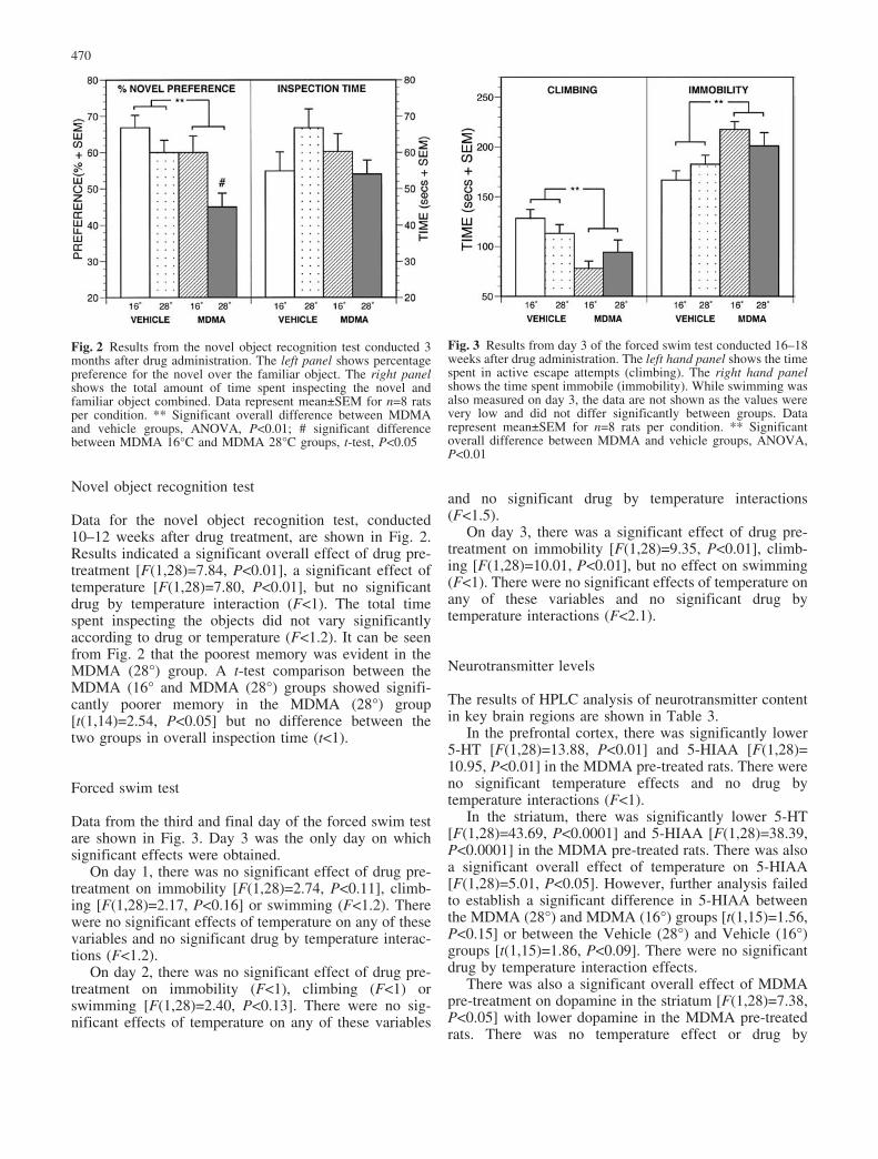
Data for the novel object recognition test, conducted10–12 weeks after drug treatment, are shown in Fig. 2.Results indicated a significant overall effect of drug pre-treatment [F(1,28)=7.84, P<0.01], a significant effect oftemperature [F(1,28)=7.80, P<0.01], but no significantdrug by temperature interaction (F<1). The total timespent inspecting the objects did not vary significantlyaccording to drug or temperature (F<1.2). It can be seenfrom Fig. 2 that the poorest memory was evident in theMDMA (28�) group. A t-test comparison between theMDMA (16� and MDMA (28�) groups showed signifi-cantly poorer memory in the MDMA (28�) group[t(1,14)=2.54, P<0.05] but no difference between thetwo groups in overall inspection time (t<1).
Forced swim test
Data from the third and final day of the forced swim testare shown in Fig. 3. Day 3 was the only day on whichsignificant effects were obtained.
On day 1, there was no significant effect of drug pre-treatment on immobility [F(1,28)=2.74, P<0.11], climb-ing [F(1,28)=2.17, P<0.16] or swimming (F<1.2). Therewere no significant effects of temperature on any of thesevariables and no significant drug by temperature interac-tions (F<1.2).
On day 2, there was no significant effect of drug pre-treatment on immobility (F<1), climbing (F<1) orswimming [F(1,28)=2.40, P<0.13]. There were no sig-nificant effects of temperature on any of these variables
and no significant drug by temperature interactions(F<1.5).
On day 3, there was a significant effect of drug pre-treatment on immobility [F(1,28)=9.35, P<0.01], climb-ing [F(1,28)=10.01, P<0.01], but no effect on swimming(F<1). There were no significant effects of temperature onany of these variables and no significant drug bytemperature interactions (F<2.1).
Neurotransmitter levels
The results of HPLC analysis of neurotransmitter contentin key brain regions are shown in Table 3.
In the prefrontal cortex, there was significantly lower5-HT [F(1,28)=13.88, P<0.01] and 5-HIAA [F(1,28)=10.95, P<0.01] in the MDMA pre-treated rats. There wereno significant temperature effects and no drug bytemperature interactions (F<1).
In the striatum, there was significantly lower 5-HT[F(1,28)=43.69, P<0.0001] and 5-HIAA [F(1,28)=38.39,P<0.0001] in the MDMA pre-treated rats. There was alsoa significant overall effect of temperature on 5-HIAA[F(1,28)=5.01, P<0.05]. However, further analysis failedto establish a significant difference in 5-HIAA betweenthe MDMA (28�) and MDMA (16�) groups [t(1,15)=1.56,P<0.15] or between the Vehicle (28�) and Vehicle (16�)groups [t(1,15)=1.86, P<0.09]. There were no significantdrug by temperature interaction effects.
There was also a significant overall effect of MDMApre-treatment on dopamine in the striatum [F(1,28)=7.38,P<0.05] with lower dopamine in the MDMA pre-treatedrats. There was no temperature effect or drug by
Fig. 2 Results from the novel object recognition test conducted 3months after drug administration. The left panel shows percentagepreference for the novel over the familiar object. The right panelshows the total amount of time spent inspecting the novel andfamiliar object combined. Data represent mean€SEM for n=8 ratsper condition. ** Significant overall difference between MDMAand vehicle groups, ANOVA, P<0.01; # significant differencebetween MDMA 16�C and MDMA 28�C groups, t-test, P<0.05
Fig. 3 Results from day 3 of the forced swim test conducted 16–18weeks after drug administration. The left hand panel shows the timespent in active escape attempts (climbing). The right hand panelshows the time spent immobile (immobility). While swimming wasalso measured on day 3, the data are not shown as the values werevery low and did not differ significantly between groups. Datarepresent mean€SEM for n=8 rats per condition. ** Significantoverall difference between MDMA and vehicle groups, ANOVA,P<0.01
470

temperature interaction for dopamine in the striatum(F<1.6).
In the hippocampus, there was significantly lower5-HT [F(1,28)=37.81, P<0.0001] and 5-HIAA [F(1,28)=27.56, P<0.0001] in MDMA pre-treated rats but nosignificant temperature effect or drug by temperatureinteraction (F<2.7). There was also significantly lower5-HIAA in the MDMA (28�) group than the MDMA (16�)group [t(1,15)=2.49, P<0.05].
Finally, in the amygdala, there was again lower 5-HT[F(1,28)=7.13, P<0.05] and 5-HIAA [F(1,28)=22.46,P<0.001] in MDMA pre-treated rats with no significanttemperature effect or drug by temperature interaction(F<2.7). There was also significantly lower 5-HIAA inthe MDMA (28�) group than the MDMA (16�) group[t(1,15)=2.29, P<0.05].
Discussion
The present study has tested the hypothesis that MDMA-induced hyperthermia influences the long-term 5-HTdepletion and associated changes in emotionality andcognition produced by the drug. For the most part, theresults have been rather surprising, with little apparentinfluence of MDMA-induced hyperthermia on long-termbehavioural and neurochemical outcomes in rats.
The acute changes in body temperature reported herewith MDMA are in line with the existing literature, with aclear and consistent hyperthermia at 28�C (Dafters 1994,1995; Broening et al. 1995; Malberg and Seiden 1998;Morley et al. 2001; Gurtman et al. 2002) and hypothermiaat 16�C (Schmidt et al. 1990; Gordon et al. 1991; Dafters1994; Broening et al. 1995; Marston et al. 1999). Acutehyperactivity was also evident in the MDMA-treated rats,in agreement with our previous reports (Morley et al.2001; Gurtman et al. 2002). Interestingly, ambienttemperature did not influence locomotor activity on thefirst day of drug administration when the test environmentwas novel and exploratory behaviour high. This agreeswith earlier results noting no difference in the hyperki-netic response of rats to MDMA at 11�C and 24�C(Dafters 1994, 1995). On the second day of drugadministration, when the testing environment was no
longer novel, higher temperature was associated withdecreased locomotor activity in vehicle-treated rats, butnot in MDMA-treated animals.
The long-term effects of MDMA on anxiety-likebehaviours were as previously reported (Morley et al.2001; Gurtman et al. 2002), with MDMA pre-treated ratsshowing reduced social interaction relative to controls 8–10 weeks following drug administration. MDMA pre-treated rats also took longer to emerge into a novel openfield, and showed less exploration and rearing in that openfield. This further consolidates the evidence for a long-term anxiogenic effect of MDMA, albeit perhaps only inrat strains that have lower baseline levels of anxiety(Green and McGregor 2002; Mechan et al. 2002). Theinteresting aspect of the present findings is that hyper-thermia at the time of dosing with MDMA does notinfluence these long-term anxiogenic effects.
The observation that MDMA pre-treated rats alsoshowed poorer object recognition memory is also consis-tent with our previous report (Morley et al. 2001). Anabsence of group differences in overall object inspectiontimes between MDMA and vehicle pre-treated groupsshows that the memory impairment was not simply due toa lack of object exploration in MDMA pre-treated rats.Object recognition memory was the only behaviouralmeasure in the present study where rats showing hyper-thermia at the time of MDMA administration had a worseprognosis than those showing hypothermia.
A novel and potentially important finding in thepresent study was the higher immobility and fewer activeescape attempts in MDMA pre-treated rats tested in theforced swim model. This test is thought to be a usefulanimal model of depression, engendering a state of“behavioural despair” that is reversed by antidepressantdrug treatment (Blokland et al. 2002; Cryan et al. 2002).There has been much speculation that heavy MDMA usein humans may lead to a depressed mood, both in the daysimmediately following MDMA administration (Parrottand Lasky 1998) and perhaps more permanently(Schifano et al. 1998; Topp et al. 1999; Morgan 2000;Parrott 2001). The present study, to our knowledge,provides the first preclinical data that reflects on thisissue, and suggests that rats given MDMA 16–18 weekspreviously are more prone to depressive-like symptoms
Table 3 Results of HPLC analysis. Data are mean (SEM) and represent n=8 per condition. All data are in ng/g whole tissue. A significantoverall effect of drug, B significant overall effect of temperature, C significant difference between MDMA (16�) and MDMA (28�) groups
Region Measure Vehicle (16 �) Vehicle (28 �) MDMA (16 �) MDMA (28 �) Statistics
Prefrontal cortex 5-HT 366.4 (16.0) 381.8 (22.4) 298.9 (19.2) 280.4 (33.9) A5-HIAA 145.0 (7.1) 154.0 (9.2) 122.0 (9.8) 108.2 (16.0) A
Striatum 5-HT 399.6 (16.7) 380.2 (21.5) 332.7 (21.7) 315.5 (34.9) A5-HIAA 350.2 (15.3) 313.4 (12.6) 281.7 (5.7) 259.7 (15.4) A, BDA 9473.5 (320.9) 9518.9 (416.1) 8867.6 (296.5) 7852.9 (578.4) A
Hippocampus 5-HT 331.0 (25.3) 337.3 (7.1) 213.9 (19.6) 213.5 (17.8) A5-HIAA 311.3 (24.4) 274.0 (18.0) 196.9 (19.2) 163.4 (11.8) A, C
Amygdala 5-HT 579.6 (45.8) 492.0 (49.5) 457.6 (34.3) 407.5 (23.2) A5-HIAA 367.9 (30.9) 342.7 (30.2) 270.1 (15.2) 213.6 (19.4) A, C
471

when confronted with repeated acute stress. One recentreport indicates that MDMA pre-treated rats show ablunted 5-HT response in the hippocampus and prefrontalcortex to immobilization stress (Matuszewich et al. 2002).Thus the present results might then reflect impairment inthe neurochemical circuitry that underlies active copingresponses.
The neurochemical data reported here agree withnumerous previous studies showing long-term depletionof brain 5-HT and 5-HIAA after MDMA. There was someregional variation evident in this, with more than 40%5-HT depletion in the hippocampus but less than 20% inthe striatum. Some other reports have suggested that thehippocampus may be more vulnerable to the long-termneurotoxic effects of MDMA (Scanzello et al. 1993;Fischer et al. 1995). A significant depletion of dopaminewas also evident in the striatum of rats given MDMA.Although we did not find significant dopamine depletionin a previous study (Gurtman et al. 2002), there is a recentreport of substantial dopaminergic neurotoxicity in mon-keys given short-term exposure to MDMA (Ricaurte et al.2002). In general, the vulnerability to dopaminergic insultfrom MDMA seems to vary greatly across studies andmay be highly dependent upon the dose regime andspecies employed.
The most surprising neurochemical result was thathypothermia at the time of MDMA administration did notprotect much against long-term 5-HT depletion. The onlyneurochemical differences between the MDMA (16�) andMDMA (28�) groups were small and rather subtle: agreater loss of 5-HIAA in the MDMA (28�) group and atendency towards greater dopamine depletion in thisgroup. The lower 5-HIAA in the MDMA (28�) group mayreflect decreased 5-HT uptake and metabolism in theamygdala and hippocampus of these rats, possiblyindicative of a greater degree of neurotoxic damage inthese regions. It is conceivable then that the poorerperformance of MDMA (28�) rats in the novel objectrecognition task may in some way relate to suchdifferences in the hippocampus and amygdala, or possiblythe greater dopamine depletion in this group that isevident in the striatum.
The finding of long-term 5-HT depletion despite anacute hypothermia with MDMA agrees with the results ofMarston et al. (1999) and also with observations of acutehypothermia followed by sizeable 5-HT loss with theother substituted amphetamine drug fenfluramine (Mal-berg and Seiden 1997). However, the present results areinconsistent with previous indications that hypothermiamight offer complete protection against MDMA-induced5-HT depletion (Malberg and Seiden 1998). There areseveral factors that might potentially explain these majordifferences in outcome. One potentially important factorcould be the age of the rats used. Broening and colleagues(1995) reported that MDMA-induced 5-HT depletiondepends not only upon body temperature at the time ofacute treatment but also the age of the rats. Thus 40-day-old male Sprague-Dawley rats were protected fromMDMA induced 5-HT depletion when the drug was
given at 10�C but not 25�C, while 70-day-old rats showedsubstantial 5-HT depletion even when MDMA was givenat 10�C. Since the rats used in the present study were agedapproximately 60–75 days old at the time of dosing, andwere older than those used by Malberg and Seiden (1998),this could be a major factor in explaining our results.
Another important factor could be returning rats togroup housing immediately after MDMA dosing, aprocedure that might prolong hyperthermia and promoteaggregation toxicity (see Green et al. 1995). However atleast one report suggests that individual housing andgroup housing are associated with equivalent long-term5-HT depletion after MDMA given at ambient tempera-tures of 21�C (Broening et al. 1995). We also note thatreturning hypothermic MDMA-treated rats to their homecages at 22�C does not produce hyperthermia in our rats(McGregor and Clemens, unpublished data).
Other factors of likely importance may include thedose regime and time-course of MDMA treatment used,and also the gap between acute treatment with MDMAand neurochemical assessment. Serotonergic parametersshow a gradual recovery in the weeks and months afterMDMA treatment (Scanzello et al. 1993), It might then bethe case that leaving 4 months between MDMA treatmentand neurochemical assessment, as was done in the presentstudy, will allow more time for recovery from 5-HTdepletion and thereby minimise differences in 5-HT and5-HIAA across groups dosed at different ambient tem-peratures.
Finally, it is interesting to speculate on the implica-tions of the present results for human MDMA users.Animal models, such as those used here, allow the long-term effects of MDMA to be assessed in a way that avoidsthe confounds of polydrug use, drug purity and pre-drugpsychopathology that compromise human MDMA re-search (Boot et al. 2000). The present study, in conjunc-tion with its predecessors (Morley et al. 2001; Gurtman etal. 2002), suggests a strong causal link between MDMAexposure and subsequent increases in anxiety, decreasedsocial behaviour, impaired memory and loss of activecoping in response to stress. This is consistent withincreasing evidence from human studies that MDMAusers are more vulnerable to mood disorders and memorydysfunction (Schifano et al. 1998; Topp et al. 1999;Morgan 2000; Parrott 2001). While the advice to humanMDMA users to “chill out” while on the drug isundoubtedly of use in preventing acute hyperthermicreactions that may occasionally be life-threatening, thepresent study raises some doubts that cooler temperatureswill prevent long-term neurotoxicity and related long-term adverse emotional effects of the drug.
Acknowledgements This work was supported by an NH&MRCgrant to Iain S. McGregor and Glenn E. Hunt. We are grateful toGeoffrey van der Plasse for technical assistance and to Darek Figaand Debbie Brookes for animal care.
472

References
Battaglia G, Yeh SY, O’Hearn E, Molliver ME, Kuhar MJ, DeSouza EB (1987) 3,4-Methylenedioxymethamphetamine and3,4-methylenedioxyamphetamine destroy serotonin terminals inrat brain: quantification of neurodegeneration by measurementof [3H]paroxetine-labeled serotonin uptake sites. J PharmacolExp Ther 242:911–916
Blokland A, Lieben C, Deutz NEP (2002) Anxiogenic anddepressive-like effects, but no cognitive deficits, after repeatedmoderate tryptophan depletion in the rat. J Psychopharmacol16:39–49
Boot BP, McGregor LS, Hall W (2000) MDMA (Ecstasy)neurotoxicity: assessing and communicating the risks. Lancet355:1818–1821
Bowyer JF, Tank AW, Newport GD, Slikker W Jr, Ali SF, HolsonRR (1992) The influence of environmental temperature on thetransient effects of methamphetamine on dopamine levels anddopamine release in rat striatum. J Pharmacol Exp Ther260:817–824
Bowyer JF, Davies DL, Schmued L, Broening HW, Newport GD,Slikker W Jr, Holson RR (1994) Further studies of the role ofhyperthermia in methamphetamine neurotoxicity. J PharmacolExp Ther 268:1571–1580
Bowyer JF, Holson RR, Miller DB, O’Callaghan JP (2001)Phenobarbital and dizocilpine can block methamphetamine-induced neurotoxicity in mice by mechanisms that areindependent of thermoregulation. Brain Res 919:179–183
Broening HW, Bowyer JF, Slikker W (1995) Age-dependentsensitivity of rats to the long-term effects of the serotonergicneurotoxicant (€)-3,4-methylenedioxymethamphetamine(MDMA) correlates with the magnitude of the MDMA-inducedthermal response. J Pharmacol Exp Ther 275:325–333
Callahan BT, Cord BJ, Ricaurte GA (2001) Long-term impairmentof anterograde axonal transport along fiber projection originat-ing in the rostral raphe nuclei after treatment with fenfluramineor methylenedioxymethamphetamine. Synapse 40:113–121
Chadwick IS, Curry PD, Linsley A, Freemont AJ, Doran B (1991)Ecstasy, 3-4 methylenedioxymethamphetamine (MDMA), afatality associated with coagulopathy and hyperthermia. J RSoc Med 84:371
Colado MI, Murray TK, Green AR (1993) 5-HT loss in rat brainfollowing 3,4-methylenedioxymethamphetamine (MDMA), p-chloroamphetamine and fenfluramine administration and ef-fects of chlormethiazole and dizocilpine. Br J Pharmacol108:583–589
Colado MI, Williams JL, Green AR (1995) The hyperthermic andneurotoxic effects of “Ecstasy” (MDMA) and 3,4-methylene-dioxyamphetamine (MDA) in the Dark Agouti (DA) rat, amodel of the CYP2D6 poor metabolizer phenotype. Br JPharmacol 115:1281–1289
Colado MI, Granados R, Oshea E, Esteban B, Green AR (1998)Role of hyperthermia in the protective action of clomethiazoleagainst MDMA (Ecstasy)-induced neurodegeneration, compar-ison with the novel nmda channel blocker AR-R15896AR. Br JPharmacol 124:479–484
Colado MI, O’Shea E, Esteban B, Granados R, Green AR (1999) Invivo evidence against clomethiazole being neuroprotectiveagainst MDMA (“Ecstasy”)-induced degeneration of rat brain5-HT nerve terminals by a free radical scavenging mechanism.Neuropharmacology 38:307–314
Colado MI, O’Shea E, Esteban B, Green AR (2001) Studies on theneuroprotective effect of the enantiomers of AR-A008055, acompound structurally related to clomethiazole, on MDMA(“Ecstasy”)-induced neurodegeneration in rat brain. Psycho-pharmacology 157:82–88
Cryan JF, Markou A, Lucki I (2002) Assessing antidepressantactivity in rodents: recent developments and future needs.Trends Pharmacol Sci 23:238–245
Dafters RI (1994) Effect of ambient temperature on hyperthermiaand hyperkinesis induced by 3,4-methylenedioxymetham-
phetamine (MDMA or Ecstasy) in rats. Psychopharmacology114:505–508
Dafters RI (1995) Hyperthermia following MDMA administrationin rats—effects of ambient temperature, water consumption,and chronic dosing. Physiol Behav 58:877–882
Farfel GM, Seiden LS (1995) Role of hypothermia in themechanism of protection against serotonergic toxicity.1. Ex-periments using 3,4-methylenedioxymethamphetamine, di-zocilpine, CGS 9755 and NBQX. J Pharmacol Exp Ther272:860–867
Fischer C, Hatzidimitriou G, Wlos J, Katz J, Ricaurte G (1995)Reorganization of ascending 5-HT axon projections in animalspreviously exposed to the recreational drug (€)3,4-methylene-dioxymethamphetamine (MDMA, “Ecstasy”). J Neurosci15:5476–5485
Gordon CJ, Watkinson WP, O’Callaghan JP, Miller DB (1991)Effects of 3,4-methylenedioxymethamphetamine on autonomicthermoregulatory responses of the rat. Pharmacol BiochemBehav 38:339–344
Green AR, McGregor IS (2002) On the long-term increase inanxiety following central serotonin depletion with MDMA(“Ecstasy”). Psychopharmacology 162:448–450
Green AR, Cross AJ, Goodwin GM (1995) Review of thepharmacology and clinical pharmacology of 3,4-methylene-dioxymethamphetamine (MDMA or Ecstasy). Psychopharma-cology 119:247–260
Gurtman CG, Morley KC, Li KM, Hunt GE, McGregor IS (2002)Increased anxiety in rats after 3,4-methylenedioxymetham-phetamine: association with serotonin depletion. Eur J Phar-macol 446:89–96
Harkin A, Connor TJ, Mulrooney J, Kelly JP, Leonard BE (2001)Prior exposure to methylenedioxyamphetamine (MDA) inducesserotonergic loss and changes in spontaneous exploratory andamphetamine-induced behaviors in rats. Life Sci 68:1367–1382
Henry JA, Jeffreys KJ, Dawling S (1992) Toxicity and deaths from3,4-methylenedioxymethamphetamine (“Ecstasy”). Lancet340:384–387
Liechti ME, Saur MR, Gamma A, Hell D, Vollenweider FX (2000)Psychological and physiological effects of MDMA (“Ecstasy”)after pretreatment with the 5-HT2 antagonist ketanserin inhealthy humans. Neuropsychopharmacology 23:396–404
Malberg JE, Seiden LS (1997) Administration of fenfluramine atdifferent ambient temperatures produces different core temper-ature and 5-HT neurotoxicity profiles. Brain Res 765:101–107
Malberg JE, Seiden LS (1998) Small changes in ambient temper-ature cause large changes in 3,4-methylenedioxymetham-phetamine (MDMA)-induced serotonin neurotoxicity and corebody temperature in the rat. J Neurosci 18:5086–5094
Malberg JE, Sabol KE, Seiden LS (1996) Co-administration ofMDMA with drugs that protect against MDMA neurotoxicityproduces different effects on body temperature in the rat. JPharmacol Exp Ther 278:258–267
Mallick A, Bodenham AR (1997) MDMA induced hyperthermia: asurvivor with an initial body temperature of 42.9 degrees C. JAccident Emer Med 14:336–338
Marston HM, Reid ME, Lawrence JA, Olverman HJ, Butcher SP(1999) Behavioural analysis of the acute and chronic effects ofMDMA treatment in the rat. Psychopharmacology 144:67–76
Matuszewich L, Filon ME, Finn DA, Yamamoto BK (2002)Altered forebrain neurotransmitter responses to immobilizationstress following 3,4-methylenedioxymethamphetamine. Neuro-science 110:41–48
Mechan AO, Moran PM, Elliott JM, Young AMJ, Joseph MH,Green AR (2002) A study of the effects of a single neurotoxicdose of 3,4-methylenedioxymethamphetamine (MDMA; Ecsta-sy) on the subsequent long-term behaviour of rats in the plusmaze and open field. Psychopharmacology 159:167–175
Morgan MJ (2000) Ecstasy (MDMA): a review of its possiblepersistent psychological effects. Psychopharmacology152:230–248
Morley KC, Gallate JE, Hunt GE, Mallet PE, McGregor IS (2001)Increased anxiety and impaired memory in rats 3 months after
473

administration of 3,4-methylenedioxymethamphetamine (“Ec-stasy”). Eur J Pharmacol 433:91–99
O’Loinsigh ED, Boland G, Kelly JP, O’Boyle KM (2001)Behavioural, hyperthermic and neurotoxic effects of 3,4-methylenedioxymethamphetamine analogues in the Wistar rat.Prog Neuro-Psychopharmacol Biol Psychiatry 25:621–638
O’Shea E, Granados R, Esteban B, Colado MI, Green AR (1998)The relationship between the degree of neurodegeneration ofrat brain 5-HT nerve terminals and the dose and frequency ofadministration of MDMA (Ecstasy). Neuropharmacology37:919–926
Parrott AC (2001) Human psychopharmacology of Ecstasy(MDMA): a review of 15 years of empirical research. HumPsychopharmacol 16:557–577
Parrott AC (2002) Recreational Ecstasy/MDMA, the serotoninsyndrome, and serotonergic neurotoxicity. Pharmacol BiochemBehav 71:837–844
Parrott AC, Lasky J (1998) Ecstasy (MDMA) effects upon moodand cognition—before, during and after a Saturday night dance.Psychopharmacology 139:261–268
Prickaerts J, van Staveren WCG, Sik A, Markerink-van Ittersum M,Niewohner U, van der Staay FJ, Blokland A, de Vente J (2002)Effects of two selective phosphodiesterase type 5 inhibitors,sildenafil and vardenafil, on object recognition memory andhippocampal cyclic GMP levels in the rat. Neuroscience113:351–361
Ricaurte G, Bryan G, Strauss L, Seiden L, Schuster C (1985)Hallucinogenic amphetamine selectively destroys brain seroto-nin nerve terminals. Science 229:986–988
Ricaurte GA, McCann UD, Szabo Z, Scheffel U (2000) Toxico-dynamics and long-term toxicity of the recreational drug, 3,4-
methylenedioxymethamphetamine (MDMA, “Ecstasy”). Toxi-col Lett 112:143–146
Ricaurte G, Yuan J, Hatzidimitriou G, Cord BJ, McCann UD(2002) Severe dopaminergic neurotoxicity in primates after acommon recreational dose regimen of MDMA (“Ecstasy”).Science 297: 2260–2263
Scanzello CR, Hatzidimitriou G, Martello AL, Katz JL, RicaurteGA (1993) Serotonergic recovery after (€)3,4-(methylene-dioxy) methamphetamine injury: observations in rats. J Phar-macol Exp Ther 264:1484–1491
Schifano F (2000) Potential human neurotoxicity of MDMA(“Ecstasy”): Subjective self-reports, evidence from an Italiandrug addiction centre and clinical case studies. Neuropsychobi-ology 42:25–33
Schifano F, Difuria L, Forza C, Minicuci N, Bricolo R (1998)MDMA (Ecstasy) consumption in the context of polydrugabuse—a report on 150 patients. Drug Alcohol Depend 52:85–90
Schmidt CJ, Black CK, Abbate GM, Taylor VL (1990) Methylene-dioxymethamphetamine-induced hyperthermia and neurotoxic-ity are independently mediated by 5-HT2 receptors. Brain Res529:85–90
Schworer H, Racke K, Kilbinger H (1987) Cholinergic modulationof the release of 5-hydroxytryptamine from the guinea pigileum. Naunyn-Schmiedeberg’s Arch Pharmacol 336:127–132
Topp L, Hando J, Dillon P, Roche A, Solowij N (1999) Ecstasy usein Australia: patterns of use and associated harm. Drug AlcoholDepend 55:105–115
474

![Synthetic studies and pharmacological evaluations on the MDMA ([] Ecstasy') antagonist nantenine](https://img.pdfslide.net/doc/110x75/632f14ebe68c6e65e90a94a6/synthetic-studies-and-pharmacological-evaluations-on-the-mdma-ecstasy-antagonist-1680031827.jpg)


