Embed Size (px)
Citation preview
Journal of Molecular Graphics and Modelling 28 (2009) 336–346
Induced-fit docking studies of the active and inactive states of proteintyrosine kinases
Haizhen Zhong *, Ly M. Tran, Jenna L. Stang
Department of Chemistry, The University of Nebraska at Omaha, DSC362, 6001 Dodge Street, Omaha, NE 68182, USA
A R T I C L E I N F O
Article history:
Received 25 February 2009
Received in revised form 21 August 2009
Accepted 25 August 2009
Available online 31 August 2009
Keywords:
Protein kinase
Imatinib
Erlotinib
Inhibitor design
Loop movement
A B S T R A C T
Inhibition of tyrosine kinases (such as the epidermal growth factor receptor, EGFR, and/or Abelson
leukemia virus protein kinase, ABL) represents a major advancement in the treatment of solid tumors,
supported by the clinical administration of gefitinib, erlotinib, imatinib, and dasatinib. The identification
of the binding interactions in the EGFR/ligands and the ABL/ligands complexes can facilitate the
structure-based design of new tyrosine kinase inhibitors. We carried out induced-fit docking studies of
18 structurally diverse kinase inhibitors against the EGFR, the active and inactive states of the ABL
protein. Our docking data show that the induced-fit docking (IFD) protocol can successfully reproduce
the native poses of ligands from different sources. The binding interactions and the docked poses are
consistent with the available experimental data. Our results indicate that imatinib is a weak binder to the
active state of ABL but a strong binder to EGFR. The increased sensitivity of erlotinib to EGFR might be
attributed to Cys797 of EGFR. In addition to Cys797, other important residues for kinase inhibitor design
include Thr790, Met793, Lys745 and Asp855 of EGFR; and Thr315, Met318, Asp381 and Glu286 of the
ABL. The minimum number of H-bonds required for the ligand binding provides a reasonable
explanation to the effectiveness of nilotinib against most imatinib resistant mutants.
� 2009 Elsevier Inc. All rights reserved.
Contents lists available at ScienceDirect
Journal of Molecular Graphics and Modelling
journal homepage: www.elsev ier .com/ locate /JMGM
1. Introduction
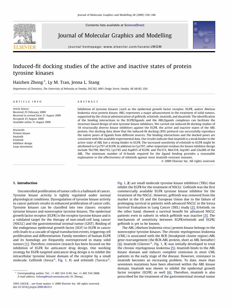
Uncontrolled proliferation of tumor cells is a hallmark of cancer.Tyrosine kinase activity is tightly regulated under normalphysiological conditions. Dysregulation of tyrosine kinase activityin cancer patients results in enhanced proliferation of cancer cells.Tyrosine kinases can be classified into two classes: receptortyrosine kinases and nonreceptor tyrosine kinases. The epidermalgrowth factor receptor (EGFR) is the receptor tyrosine kinase and isa validated target for the therapy of non-small-cell lung cancer(NSCLC) and the gastrointestinal stromal tumor (GIST). Binding ofthe endogenous epidermal growth factor (EGF) to EGFR in cancercells leads to a cascade of signal transduction events, triggering cellproliferation and differentiation. Family members of human EGFRand its homologs are frequently overexpressed in many solidtumors [1]. Therefore, extensive research has been focused on theinhibition of EGFR for anticancer drug design. One workingstrategy for EGFR-targeted anticancer drug design is to inhibit theintracellular tyrosine kinase domain of the receptor by a smallmolecule. Gefitinib (Iressa1, Fig. 1, 1) and erlotinib (Tarceca1,
* Corresponding author. Tel.: +1 402 554 3145; fax: +1 402 554 3888.
E-mail address: [email protected] (H. Zhong).
1093-3263/$ – see front matter � 2009 Elsevier Inc. All rights reserved.
doi:10.1016/j.jmgm.2009.08.012
Fig. 1, 2) are small molecule tyrosine kinase inhibitors (TKIs) thatinhibit the EGFR for the treatment of NSCLC. Gefitinib was the firstcommercially available EGFR tyrosine kinase inhibitor for thetreatment of the NSCLC. However, gefitinib was removed from themarket in the US and the European Union due to the failure ofprolonging survival in patients with advanced NSCLC in the IressaSurvival Evaluation in Lung Cancer (ISEL) study [2]. Erlotinib, onthe other hand, showed a survival benefit for advanced NSCLCpatients even in subsets in which gefitinib was inactive [3]. Themechanism of sensitivity between EGFR/erlotinib and EGFR/gefitinib is yet to be known.
The ABL (Abelson leukemia virus) protein kinase belongs to thenonreceptor tyrosine kinases. The chronic myelogenous leukemia(CML) is associated with the BCR (breakpoint cluster region)-ABLgene rearrangement (the BCR-ABL protein, short as ABL hereafter)[4]. Imatinib (Gleevec1, Fig. 1, 3) was initially developed to treatthe chronic myelogenous leukemia [5]. Imatinib binds to the ABLkinase domain and induces complete remission in most CMLpatients in the early stage of the disease. However, resistance toimatinib becomes an increasing problem. To date, more thanseventeen mutations have been observed within the ABL kinasedomain. Imatinib was shown to inhibit the epidermal growthfactor receptor (EGFR) as well [6]. Therefore, imatinib is alsoprescribed for the treatment of the gastrointestinal stromal tumor
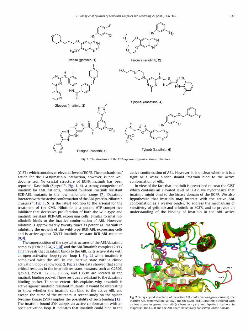
Fig. 1. The structures of the FDA-approved tyrosine kinase inhibitors.
Fig. 2. X-ray crystal structures of the active ABL conformation (green cartoon), the
inactive ABL conformation (yellow), and the EGFR (red). Dasatinib is colored with
carbon atoms in green, imatinib (carbons in cyan), and lapatinib (carbons in
magenta). The EGFR and the ABL share structurally conserved kinase domain.
H. Zhong et al. / Journal of Molecular Graphics and Modelling 28 (2009) 336–346 337
(GIST), which contains an elevated level of EGFR. The mechanism ofaction for the EGFR/imatinib interaction, however, is not welldocumented. No crystal structure of EGFR/imatinib has beenreported. Dasatinib (Sprycel1, Fig. 1, 4), a strong competitor ofimatinib for CML patients, inhibited fourteen imatinib resistantBCR-ABL mutants in the low nanomolar range [7]. Dasatinibinteracts with the active conformation of the ABL protein. Nilotinib(Tasigna1, Fig. 1, 5) is the latest addition to the arsenal for thetreatment of the CML. Nilotinib is a potent ATP-competitiveinhibitor that decreases proliferation of both the wild-type andimatinib resistant BCR-ABL expressing cells. Similar to imatinib,nilotinib binds to the inactive conformation of ABL. However,nilotinib is approximately twenty times as potent as imatinib ininhibiting the growth of the wild-type BCR-ABL expressing cellsand is active against 32/33 imatinib resistant BCR-ABL mutants[8,9].
The superposition of the crystal structures of the ABL/dasatinibcomplex (PDB id: 2GQG [10]) and the ABL/imatinib complex (2HYY[11]) reveals that dasatinib binds to the ABL in its active state withan open activation loop (green loop 1, Fig. 2) while imatinib iscomplexed with the ABL in the inactive state with a closedactivation loop (yellow loop 2, Fig. 2). Our data showed that somecritical residues in the imatinib resistant mutants, such as G250E,Q252H, Y253F, E255K, E355G, and F359V are located in theimatinib binding pocket. These residues are distant to the dasatinibbinding pocket. To some extent, this explains why dasatinib isactive against imatinib resistant mutants. It would be interestingto know whether the imatinib can bind to the active ABL andescape the curse of the mutants. A recent study on the spleentyrosine kinase (SYK) implies the possibility of such binding [12].The imatinib-bound SYK adopts an active conformation with anopen activation loop. It indicates that imatinib could bind to the
active conformation of ABL. However, it is unclear whether it is atight or a weak binder should imatinib bind to the activeconformation of ABL.
In view of the fact that imatinib is prescribed to treat the GISTwhich contains an elevated level of EGFR, we hypothesize thatimatinib might bind to the kinase domain of the EGFR. We alsohypothesize that imatinib may interact with the active ABLconformation as a weaker binder. To address the mechanism ofsensitivity of gefitinib and erlotinib to EGFR, and to provide anunderstanding of the binding of imatinib in the ABL active
H. Zhong et al. / Journal of Molecular Graphics and Modelling 28 (2009) 336–346338
conformation, we conducted induced-fit docking (IFD) studiesusing Schrodinger’s IFD module for the ABL/dasatinib; ABL/imatinib in the inactive and active conformations of ABL, andthe wild-type EGFR/lapatinib complexes. Lapatinib (Tykerb1,Fig. 1, 6) is an EGFR inhibitor for the treatment of solid tumorssuch as breast and lung cancers. In order to provide structuralinformation for the design of next generation of TKIs, we selected atotal of 18 TKIs with structurally diverse scaffolds and performedthe induced-fit docking (IFD) of these TKIs to the active, inactiveABL conformations and EGFR proteins. The docking affinity and thebinding interactions between the three model proteins and theligands are reported herein.
We applied the IFD method in the Schrodinger [13] to study thebinding affinity of protein/ligand complexes. Docking programs,such as DOCK [14], FlexX [15], GOLD [16], Autodock [17] and Glide[18] have been used in structure-based drug design to predict thebinding affinity and optimize small molecule drug candidates. Onecommon feature of these tools is that a flexible ligand is docked to arigid receptor binding pocket. However, it becomes increasinglyclear that the crucial information from the protein structuralflexibility should be included in the drug design process [19].Several new computational tools have been developed to addressthe protein plasticity by means of the ‘‘induced-fit’’ model. The‘‘Induced-Fit Docking’’ (IFD) module [13] from the Schrodinger Inc.has been reported to be a robust and accurate method to accountfor both ligand and receptor flexibility. The average ligand root-mean-square deviation (RMSD) for the traditional rigid receptordocking for 21 cases was 5.5 A, while the RMSD from theSchrodinger’s IFD module was 1.4 A [20]. The protein plasticityis taken into account in the IFD by iteratively combining rigidreceptor docking using Glide program with sampling side chaindegrees of freedom in the receptor while allowing minor backbonemovements through the minimization using the Prime program.
2. Methods
2.1. Computational protein structure preparation
The coordinates for all protein–ligand complexes were obtainedfrom the RCSB Protein Data Bank (PDB). Included in this study arethe active ABL/dasatinib (2GQG) [10]; inactive ABL/imatinib(2HYY) [11]; and EGFR/lapatinib (1XKK) [21]. 2GQG was used tostudy the interactions between active ABL conformation andligands, while 2HYY for inactive ABL conformation and ligands.Protein structures of 2GQG and 2HYY were prepared using theSchrodinger’s Protein Preparation Wizard module [13]. Hydrogenatoms were added and the side chain structures of Gln and Asnwere flipped if necessary in order to provide maximum degree ofhydrogen bond interactions. There are five sections of missingresidues in the crystal structure 1XKK. The missing residues ofsection 1 (734EGEK737) and section 2 (750ATSP753) were modeledfrom 2ITT, the complex of the EGFR mutant L858R/AEE788 [22].The missing residues of section 3 (868EYHAEGGK875) were modeledfrom crystal structure 2GS6, the complex of EGFR/ATP [23]. Thefourth section of missing residues (988HLPSPTD994) was modeledfrom the homologous sequence from 1XO0 [24] using thehomology modeling module in MOE [25]. The last section ofmissing residues in crystal structure 1XKK (1005EDMDD1009) wasbuilt from 2ITO, the complex of EGFR G719S mutant/gefitinib [22].Crystal structures of 2ITT, 2GS6, 2ITO were structurally super-imposed to 1XKK using the DaliLite program [26]. The coordinatesfrom the corresponding regions in 2ITT, 2GS6, and 2ITO wereadopted for the missing residues in 1XKK, following withminimization on the affected regions while holding the remainingresidues fixed. The subsequent 1XKK model was then subject to theProtein Preparation Wizard module in Schrodinger [13]. All
proteins were minimized using the OPLS force field in theMacroModel module in Schrodinger with backbone atoms beingfixed.
2.2. Induced-fit docking
The protein structures of 2GQG, 2HYY and 1XKK were appliedwith the induced-fit docking (IFD) method in the Schrodingersoftware suite [13]. The structurally diverse 18 tyrosine kinaseinhibitors were selected from our previous review paper (Figs. 1and 5) [27]. All ligands were prepared using LigPrep and wereoptimized with the OPLS force field in the MacroModel module inSchrodinger [13]. The optimization of ligands with the well-knownMMFF force field [28] might generate conformationally differentligand structures. This might influence the docking results in asubtle way. But the difference in results derived from OPLS orMMFF may not be profound due to the fact that during the dockingprocess the ligand optimized from either the OPLS or the MMFFforce field will change its conformation to find the best fit to theprotein binding pocket. The IFD protocol was adopted from ourrecent paper [29]. In short, ligands were docked to the rigid proteinusing the soften-potential docking in the Glide program with thevan der Waals radii scaling of 0.7 for the proteins. The resulting top20 poses of ligands were used to sample the protein plasticity usingthe Prime program in the Schrodinger suite. Residues having atleast one atom within 5 A of any of the 20 ligand poses were subjectto a conformational search and minimization while residuesoutside the zone were held fixed. In this way, the flexibility ofproteins was taken into account. The resulting 20 new receptorconformations were taken forward for redocking. In this redockingstage, Glide docking parameters were set to the default hardpotential function, i.e., the van der Waals radii scaling is 1.0. TheGlide XP (extra precision) was used for all docking calculations[30]. The binding affinity of each complex was reported in theGlideScore. The more negative the GlideScore, the more favorablethe binding. All graphical pictures were made using the Pymolprogram [31].
2.3. Glide docking
The rigid receptor docking using the Glide program was carriedout against the three receptors using the same set of ligands. Thescaling factor for protein van der Waals radii was 1.0 in thereceptor grid generation. The ligands in the active sites were usedas the centroid to generate the grid files for the docking. The defaultgrid size was adopted from the Glide program. No constraints wereapplied for all the docking studies.
3. Results and discussion
EGFR under normal physiology conditions is activated by theendogenous EGF or the transforming growth factor a (TGF-a) atthe extracellular domain. The binding activates the intracellularkinase domain and triggers a cascade of signal transduction eventsinvolving the regulation of cell proliferation. Many inhibitors havebeen designed to inhibit the kinase domain of EGFR. Gefitinib anderlotinib are one of the successful stories. However, the sensitivityof gefitinib and erlotinib to the EGFR is quite different. Erlotinibshowed a survival benefit in advanced NSCLC patients in whichgefitinib was not active.
EGFR and ABL share 37.8% of sequence identity and 59.3% ofsequence similarity as revealed in the multiple sequence align-ment from the ClustalW [32]. Fig. 2 showed that EGFR and theactive state of the ABL share structurally similar kinase domain.EGFR adopts a similar activation loop (red loop 3, Fig. 2) asthe active state of ABL (green loop 1, Fig. 2). The a-helices and the
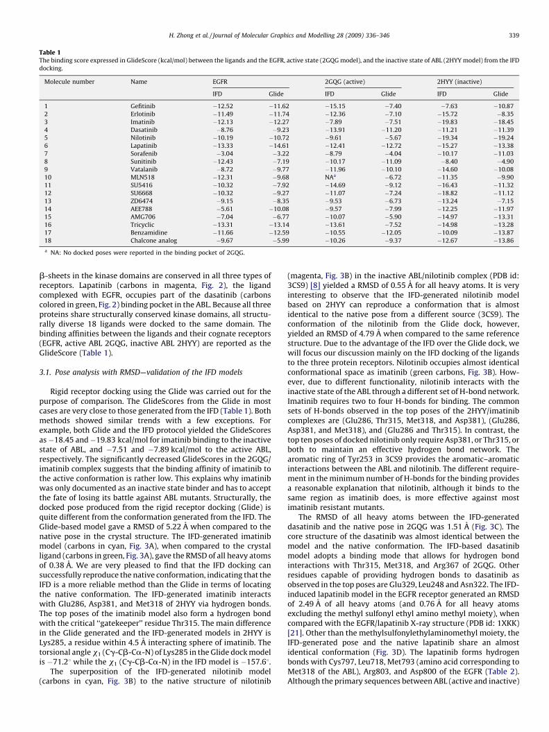
Table 1The binding score expressed in GlideScore (kcal/mol) between the ligands and the EGFR, active state (2GQG model), and the inactive state of ABL (2HYY model) from the IFD
docking.
Molecule number Name EGFR 2GQG (active) 2HYY (inactive)
IFD Glide IFD Glide IFD Glide
1 Gefitinib �12.52 �11.62 �15.15 �7.40 �7.63 �10.87
2 Erlotinib �11.49 �11.74 �12.36 �7.10 �15.72 �8.35
3 Imatinib �12.13 �12.27 �7.89 �7.51 �19.83 �18.45
4 Dasatinib �8.76 �9.23 �13.91 �11.20 �11.21 �11.39
5 Nilotinib �10.19 �10.72 �9.61 �5.67 �19.34 �19.24
6 Lapatinib �13.33 �14.61 �12.41 �12.72 �15.27 �13.38
7 Sorafenib �3.04 �3.22 �8.79 �4.04 �10.17 �11.03
8 Sunitinib �12.43 �7.19 �10.17 �11.09 �8.40 �4.90
9 Vatalanib �8.72 �9.77 �11.96 �10.10 �14.60 �10.08
10 MLN518 �12.31 �9.68 NAa �6.72 �11.35 �9.90
11 SU5416 �10.32 �7.92 �14.69 �9.12 �16.43 �11.32
12 SU6668 �10.32 �9.27 �11.07 �7.24 �18.82 �11.12
13 ZD6474 �9.15 �8.35 �9.53 �6.73 �13.24 �7.15
14 AEE788 �5.61 �10.08 �9.57 �7.99 �12.25 �11.97
15 AMG706 �7.04 �6.77 �10.07 �5.90 �14.97 �13.31
16 Tricyclic �13.31 �13.14 �13.61 �7.52 �14.98 �13.28
17 Benzamidine �11.66 �12.59 �10.55 �12.05 �10.09 �13.87
18 Chalcone analog �9.67 �5.99 �10.26 �9.37 �12.67 �13.86
a NA: No docked poses were reported in the binding pocket of 2GQG.
H. Zhong et al. / Journal of Molecular Graphics and Modelling 28 (2009) 336–346 339
b-sheets in the kinase domains are conserved in all three types ofreceptors. Lapatinib (carbons in magenta, Fig. 2), the ligandcomplexed with EGFR, occupies part of the dasatinib (carbonscolored in green, Fig. 2) binding pocket in the ABL. Because all threeproteins share structurally conserved kinase domains, all structu-rally diverse 18 ligands were docked to the same domain. Thebinding affinities between the ligands and their cognate receptors(EGFR, active ABL 2GQG, inactive ABL 2HYY) are reported as theGlideScore (Table 1).
3.1. Pose analysis with RMSD—validation of the IFD models
Rigid receptor docking using the Glide was carried out for thepurpose of comparison. The GlideScores from the Glide in mostcases are very close to those generated from the IFD (Table 1). Bothmethods showed similar trends with a few exceptions. Forexample, both Glide and the IFD protocol yielded the GlideScoresas�18.45 and�19.83 kcal/mol for imatinib binding to the inactivestate of ABL, and �7.51 and �7.89 kcal/mol to the active ABL,respectively. The significantly decreased GlideScores in the 2GQG/imatinib complex suggests that the binding affinity of imatinib tothe active conformation is rather low. This explains why imatinibwas only documented as an inactive state binder and has to acceptthe fate of losing its battle against ABL mutants. Structurally, thedocked pose produced from the rigid receptor docking (Glide) isquite different from the conformation generated from the IFD. TheGlide-based model gave a RMSD of 5.22 A when compared to thenative pose in the crystal structure. The IFD-generated imatinibmodel (carbons in cyan, Fig. 3A), when compared to the crystalligand (carbons in green, Fig. 3A), gave the RMSD of all heavy atomsof 0.38 A. We are very pleased to find that the IFD docking cansuccessfully reproduce the native conformation, indicating that theIFD is a more reliable method than the Glide in terms of locatingthe native conformation. The IFD-generated imatinib interactswith Glu286, Asp381, and Met318 of 2HYY via hydrogen bonds.The top poses of the imatinib model also form a hydrogen bondwith the critical ‘‘gatekeeper’’ residue Thr315. The main differencein the Glide generated and the IFD-generated models in 2HYY isLys285, a residue within 4.5 A interacting sphere of imatinib. Thetorsional angle x1 (Cg-Cb-Ca-N) of Lys285 in the Glide dock modelis �71.28 while the x1 (Cg-Cb-Ca-N) in the IFD model is �157.68.
The superposition of the IFD-generated nilotinib model(carbons in cyan, Fig. 3B) to the native structure of nilotinib
(magenta, Fig. 3B) in the inactive ABL/nilotinib complex (PDB id:3CS9) [8] yielded a RMSD of 0.55 A for all heavy atoms. It is veryinteresting to observe that the IFD-generated nilotinib modelbased on 2HYY can reproduce a conformation that is almostidentical to the native pose from a different source (3CS9). Theconformation of the nilotinib from the Glide dock, however,yielded an RMSD of 4.79 A when compared to the same referencestructure. Due to the advantage of the IFD over the Glide dock, wewill focus our discussion mainly on the IFD docking of the ligandsto the three protein receptors. Nilotinib occupies almost identicalconformational space as imatinib (green carbons, Fig. 3B). How-ever, due to different functionality, nilotinib interacts with theinactive state of the ABL through a different set of H-bond network.Imatinib requires two to four H-bonds for binding. The commonsets of H-bonds observed in the top poses of the 2HYY/imatinibcomplexes are (Glu286, Thr315, Met318, and Asp381), (Glu286,Asp381, and Met318), and (Glu286 and Thr315). In contrast, thetop ten poses of docked nilotinib only require Asp381, or Thr315, orboth to maintain an effective hydrogen bond network. Thearomatic ring of Tyr253 in 3CS9 provides the aromatic–aromaticinteractions between the ABL and nilotinib. The different require-ment in the minimum number of H-bonds for the binding providesa reasonable explanation that nilotinib, although it binds to thesame region as imatinib does, is more effective against mostimatinib resistant mutants.
The RMSD of all heavy atoms between the IFD-generateddasatinib and the native pose in 2GQG was 1.51 A (Fig. 3C). Thecore structure of the dasatinib was almost identical between themodel and the native conformation. The IFD-based dasatinibmodel adopts a binding mode that allows for hydrogen bondinteractions with Thr315, Met318, and Arg367 of 2GQG. Otherresidues capable of providing hydrogen bonds to dasatinib asobserved in the top poses are Glu329, Leu248 and Asn322. The IFD-induced lapatinib model in the EGFR receptor generated an RMSDof 2.49 A of all heavy atoms (and 0.76 A for all heavy atomsexcluding the methyl sulfonyl ethyl amino methyl moiety), whencompared with the EGFR/lapatinib X-ray structure (PDB id: 1XKK)[21]. Other than the methylsulfonylethylaminomethyl moiety, theIFD-generated pose and the native lapatinib share an almostidentical conformation (Fig. 3D). The lapatinib forms hydrogenbonds with Cys797, Leu718, Met793 (amino acid corresponding toMet318 of the ABL), Arg803, and Asp800 of the EGFR (Table 2).Although the primary sequences between ABL (active and inactive)
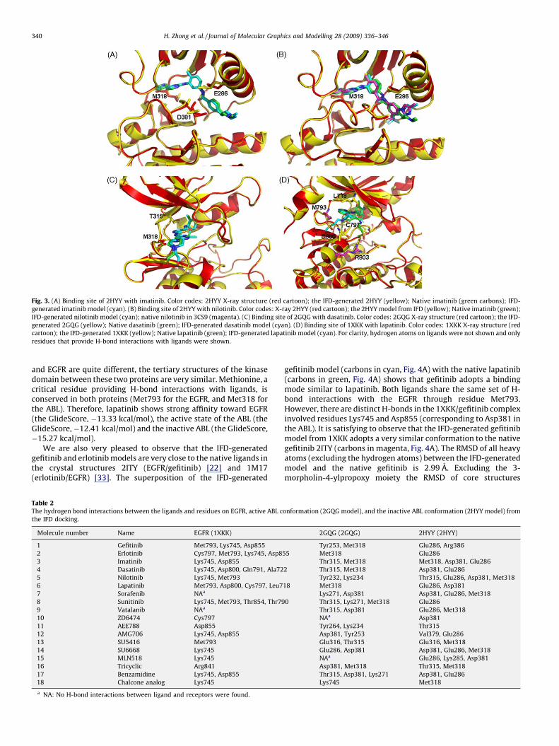
Fig. 3. (A) Binding site of 2HYY with imatinib. Color codes: 2HYY X-ray structure (red cartoon); the IFD-generated 2HYY (yellow); Native imatinib (green carbons); IFD-
generated imatinib model (cyan). (B) Binding site of 2HYY with nilotinib. Color codes: X-ray 2HYY (red cartoon); the 2HYY model from IFD (yellow); Native imatinib (green);
IFD-generated nilotinib model (cyan); native nilotinib in 3CS9 (magenta). (C) Binding site of 2GQG with dasatinib. Color codes: 2GQG X-ray structure (red cartoon); the IFD-
generated 2GQG (yellow); Native dasatinib (green); IFD-generated dasatinib model (cyan). (D) Binding site of 1XKK with lapatinib. Color codes: 1XKK X-ray structure (red
cartoon); the IFD-generated 1XKK (yellow); Native lapatinib (green); IFD-generated lapatinib model (cyan). For clarity, hydrogen atoms on ligands were not shown and only
residues that provide H-bond interactions with ligands were shown.
H. Zhong et al. / Journal of Molecular Graphics and Modelling 28 (2009) 336–346340
and EGFR are quite different, the tertiary structures of the kinasedomain between these two proteins are very similar. Methionine, acritical residue providing H-bond interactions with ligands, isconserved in both proteins (Met793 for the EGFR, and Met318 forthe ABL). Therefore, lapatinib shows strong affinity toward EGFR(the GlideScore, �13.33 kcal/mol), the active state of the ABL (theGlideScore,�12.41 kcal/mol) and the inactive ABL (the GlideScore,�15.27 kcal/mol).
We are also very pleased to observe that the IFD-generatedgefitinib and erlotinib models are very close to the native ligands inthe crystal structures 2ITY (EGFR/gefitinib) [22] and 1M17(erlotinib/EGFR) [33]. The superposition of the IFD-generated
Table 2The hydrogen bond interactions between the ligands and residues on EGFR, active ABL c
the IFD docking.
Molecule number Name EGFR (1XKK)
1 Gefitinib Met793, Lys745, Asp855
2 Erlotinib Cys797, Met793, Lys745, Asp8
3 Imatinib Lys745, Asp855
4 Dasatinib Lys745, Asp800, Gln791, Ala72
5 Nilotinib Lys745, Met793
6 Lapatinib Met793, Asp800, Cys797, Leu7
7 Sorafenib NAa
8 Sunitinib Lys745, Met793, Thr854, Thr79
9 Vatalanib NAa
10 ZD6474 Cys797
11 AEE788 Asp855
12 AMG706 Lys745, Asp855
13 SU5416 Met793
14 SU6668 Lys745
15 MLN518 Lys745
16 Tricyclic Arg841
17 Benzamidine Lys745, Asp855
18 Chalcone analog Lys745
a NA: No H-bond interactions between ligand and receptors were found.
gefitinib model (carbons in cyan, Fig. 4A) with the native lapatinib(carbons in green, Fig. 4A) shows that gefitinib adopts a bindingmode similar to lapatinib. Both ligands share the same set of H-bond interactions with the EGFR through residue Met793.However, there are distinct H-bonds in the 1XKK/gefitinib complexinvolved residues Lys745 and Asp855 (corresponding to Asp381 inthe ABL). It is satisfying to observe that the IFD-generated gefitinibmodel from 1XKK adopts a very similar conformation to the nativegefitinib 2ITY (carbons in magenta, Fig. 4A). The RMSD of all heavyatoms (excluding the hydrogen atoms) between the IFD-generatedmodel and the native gefitinib is 2.99 A. Excluding the 3-morpholin-4-ylpropoxy moiety the RMSD of core structures
onformation (2GQG model), and the inactive ABL conformation (2HYY model) from
2GQG (2GQG) 2HYY (2HYY)
Tyr253, Met318 Glu286, Arg386
55 Met318 Glu286
Thr315, Met318 Met318, Asp381, Glu286
2 Thr315, Met318 Asp381, Glu286
Tyr232, Lys234 Thr315, Glu286, Asp381, Met318
18 Met318 Glu286, Asp381
Lys271, Asp381 Asp381, Glu286, Met318
0 Thr315, Lys271, Met318 Glu286
Thr315, Asp381 Glu286, Met318
NAa Asp381
Tyr264, Lys234 Thr315
Asp381, Tyr253 Val379, Glu286
Glu316, Thr315 Glu316, Met318
Glu286, Asp381 Asp381, Glu286, Met318
NAa Glu286, Lys285, Asp381
Asp381, Met318 Thr315, Met318
Thr315, Asp381, Lys271 Asp381, Glu286
Lys745 Met318
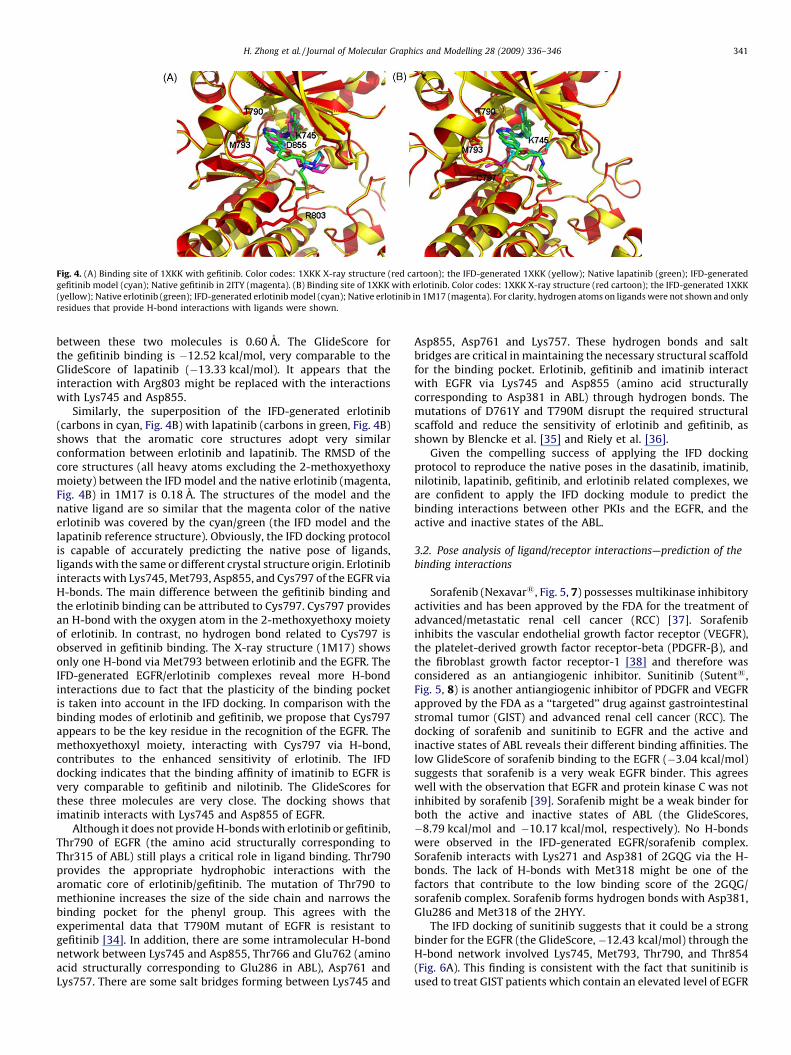
Fig. 4. (A) Binding site of 1XKK with gefitinib. Color codes: 1XKK X-ray structure (red cartoon); the IFD-generated 1XKK (yellow); Native lapatinib (green); IFD-generated
gefitinib model (cyan); Native gefitinib in 2ITY (magenta). (B) Binding site of 1XKK with erlotinib. Color codes: 1XKK X-ray structure (red cartoon); the IFD-generated 1XKK
(yellow); Native erlotinib (green); IFD-generated erlotinib model (cyan); Native erlotinib in 1M17 (magenta). For clarity, hydrogen atoms on ligands were not shown and only
residues that provide H-bond interactions with ligands were shown.
H. Zhong et al. / Journal of Molecular Graphics and Modelling 28 (2009) 336–346 341
between these two molecules is 0.60 A. The GlideScore forthe gefitinib binding is �12.52 kcal/mol, very comparable to theGlideScore of lapatinib (�13.33 kcal/mol). It appears that theinteraction with Arg803 might be replaced with the interactionswith Lys745 and Asp855.
Similarly, the superposition of the IFD-generated erlotinib(carbons in cyan, Fig. 4B) with lapatinib (carbons in green, Fig. 4B)shows that the aromatic core structures adopt very similarconformation between erlotinib and lapatinib. The RMSD of thecore structures (all heavy atoms excluding the 2-methoxyethoxymoiety) between the IFD model and the native erlotinib (magenta,Fig. 4B) in 1M17 is 0.18 A. The structures of the model and thenative ligand are so similar that the magenta color of the nativeerlotinib was covered by the cyan/green (the IFD model and thelapatinib reference structure). Obviously, the IFD docking protocolis capable of accurately predicting the native pose of ligands,ligands with the same or different crystal structure origin. Erlotinibinteracts with Lys745, Met793, Asp855, and Cys797 of the EGFR viaH-bonds. The main difference between the gefitinib binding andthe erlotinib binding can be attributed to Cys797. Cys797 providesan H-bond with the oxygen atom in the 2-methoxyethoxy moietyof erlotinib. In contrast, no hydrogen bond related to Cys797 isobserved in gefitinib binding. The X-ray structure (1M17) showsonly one H-bond via Met793 between erlotinib and the EGFR. TheIFD-generated EGFR/erlotinib complexes reveal more H-bondinteractions due to fact that the plasticity of the binding pocketis taken into account in the IFD docking. In comparison with thebinding modes of erlotinib and gefitinib, we propose that Cys797appears to be the key residue in the recognition of the EGFR. Themethoxyethoxyl moiety, interacting with Cys797 via H-bond,contributes to the enhanced sensitivity of erlotinib. The IFDdocking indicates that the binding affinity of imatinib to EGFR isvery comparable to gefitinib and nilotinib. The GlideScores forthese three molecules are very close. The docking shows thatimatinib interacts with Lys745 and Asp855 of EGFR.
Although it does not provide H-bonds with erlotinib or gefitinib,Thr790 of EGFR (the amino acid structurally corresponding toThr315 of ABL) still plays a critical role in ligand binding. Thr790provides the appropriate hydrophobic interactions with thearomatic core of erlotinib/gefitinib. The mutation of Thr790 tomethionine increases the size of the side chain and narrows thebinding pocket for the phenyl group. This agrees with theexperimental data that T790M mutant of EGFR is resistant togefitinib [34]. In addition, there are some intramolecular H-bondnetwork between Lys745 and Asp855, Thr766 and Glu762 (aminoacid structurally corresponding to Glu286 in ABL), Asp761 andLys757. There are some salt bridges forming between Lys745 and
Asp855, Asp761 and Lys757. These hydrogen bonds and saltbridges are critical in maintaining the necessary structural scaffoldfor the binding pocket. Erlotinib, gefitinib and imatinib interactwith EGFR via Lys745 and Asp855 (amino acid structurallycorresponding to Asp381 in ABL) through hydrogen bonds. Themutations of D761Y and T790M disrupt the required structuralscaffold and reduce the sensitivity of erlotinib and gefitinib, asshown by Blencke et al. [35] and Riely et al. [36].
Given the compelling success of applying the IFD dockingprotocol to reproduce the native poses in the dasatinib, imatinib,nilotinib, lapatinib, gefitinib, and erlotinib related complexes, weare confident to apply the IFD docking module to predict thebinding interactions between other PKIs and the EGFR, and theactive and inactive states of the ABL.
3.2. Pose analysis of ligand/receptor interactions—prediction of the
binding interactions
Sorafenib (Nexavar1, Fig. 5, 7) possesses multikinase inhibitoryactivities and has been approved by the FDA for the treatment ofadvanced/metastatic renal cell cancer (RCC) [37]. Sorafenibinhibits the vascular endothelial growth factor receptor (VEGFR),the platelet-derived growth factor receptor-beta (PDGFR-b), andthe fibroblast growth factor receptor-1 [38] and therefore wasconsidered as an antiangiogenic inhibitor. Sunitinib (Sutent1,Fig. 5, 8) is another antiangiogenic inhibitor of PDGFR and VEGFRapproved by the FDA as a ‘‘targeted’’ drug against gastrointestinalstromal tumor (GIST) and advanced renal cell cancer (RCC). Thedocking of sorafenib and sunitinib to EGFR and the active andinactive states of ABL reveals their different binding affinities. Thelow GlideScore of sorafenib binding to the EGFR (�3.04 kcal/mol)suggests that sorafenib is a very weak EGFR binder. This agreeswell with the observation that EGFR and protein kinase C was notinhibited by sorafenib [39]. Sorafenib might be a weak binder forboth the active and inactive states of ABL (the GlideScores,�8.79 kcal/mol and �10.17 kcal/mol, respectively). No H-bondswere observed in the IFD-generated EGFR/sorafenib complex.Sorafenib interacts with Lys271 and Asp381 of 2GQG via the H-bonds. The lack of H-bonds with Met318 might be one of thefactors that contribute to the low binding score of the 2GQG/sorafenib complex. Sorafenib forms hydrogen bonds with Asp381,Glu286 and Met318 of the 2HYY.
The IFD docking of sunitinib suggests that it could be a strongbinder for the EGFR (the GlideScore, �12.43 kcal/mol) through theH-bond network involved Lys745, Met793, Thr790, and Thr854(Fig. 6A). This finding is consistent with the fact that sunitinib isused to treat GIST patients which contain an elevated level of EGFR
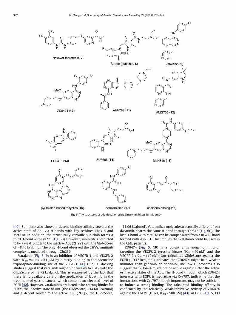
Fig. 5. The structures of additional tyrosine kinase inhibitors in this study.
H. Zhong et al. / Journal of Molecular Graphics and Modelling 28 (2009) 336–346342
[40]. Sunitinib also shows a decent binding affinity toward theactive state of ABL via H-bonds with key residues Thr315 andMet318. In addition, the structurally versatile sunitinib forms athird H-bond with Lys271 (Fig. 6B). However, suninitib is predictedto be a weak binder to the inactive ABL (2HYY) with the GlideScoreof �8.40 kcal/mol. The only H-bond observed the 2HYY/sunitinibcomplex is mediated through Glu286.
Vatalanib (Fig. 5, 9) is an inhibitor of VEGFR-1 and VEGFR-2with IC50 values <0.1 mM by directly binding to the adenosinetriphosphate-binding site of the VEGFRs [41]. Our IFD dockingstudies suggest that vatalanib might bind weakly to EGFR with theGlideScore of �8.72 kcal/mol. This is supported by the fact thatthere is no available data on the application of lapatinib in thetreatment of gastric cancer, which contains an elevated level ofEGFR [42]. However, vatalanib is predicted to be a strong binder for2HYY, the inactive state of ABL (the GlideScore, �14.60 kcal/mol)and a decent binder to the active ABL (2GQG, the GlideScore,
�11.96 kcal/mol). Vatalanib, a molecule structurally different fromdasatinib, shares the same H-bond through Thr315 (Fig. 6C). Thelost H-bond with Met318 can be compensated from a new H-bondformed with Asp381. This implies that vatalanib could be used inthe CML patients.
ZD6474 (Fig. 5, 10) is a potent antiangiogenic inhibitortargeting the VEGFR-2 tyrosine kinase (IC50 = 40 nM) and theVEGRR-3 (IC50 = 110 nM). Our calculated GlideScore against theEGFR (�9.15 kcal/mol) indicates that ZD6474 might be a weakerinhibitor than gefitinib or erlotinib. The low GlideScores alsosuggest that ZD6474 might not be active against either the activeor inactive states of the ABL. The H-bond through which ZD6424interacts with EGFR is mediating via Cys797, indicating that theinteractions with Cys797, though important, may not be sufficientto induce a strong binding. The calculated binding affinity isconfirmed by the relatively weak inhibitive activity of ZD6474against the EGFR1 (HER1, IC50 = 500 nM) [43]. AEE788 (Fig. 5, 11)
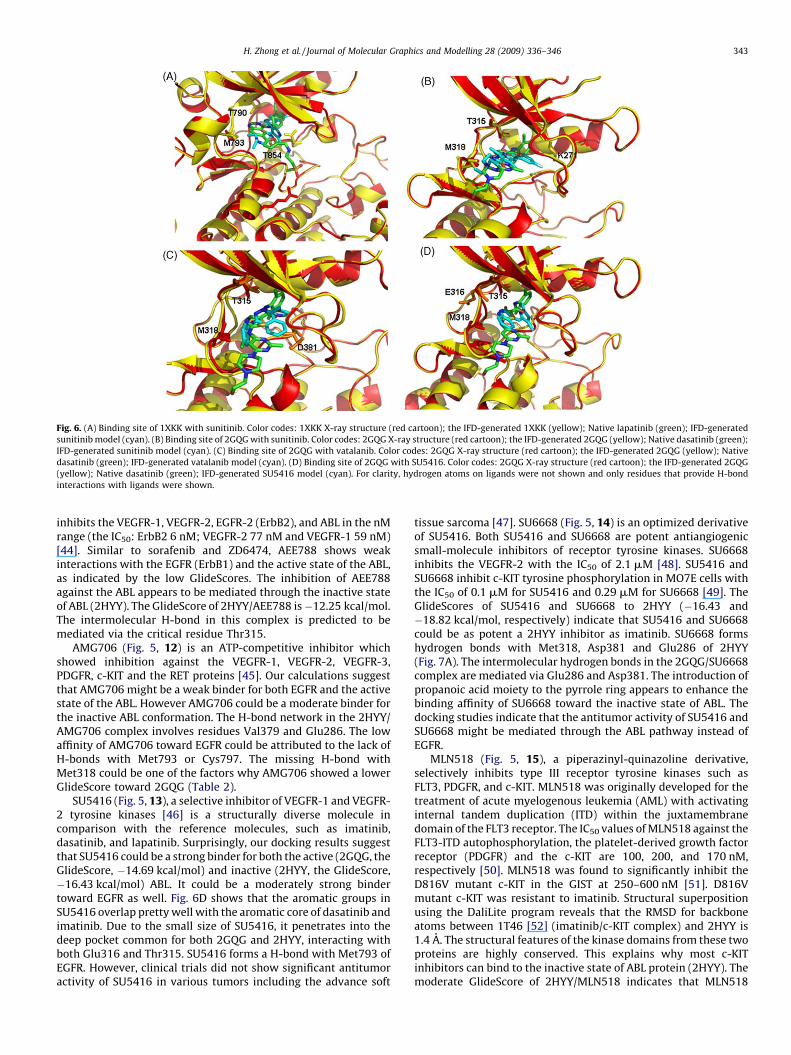
Fig. 6. (A) Binding site of 1XKK with sunitinib. Color codes: 1XKK X-ray structure (red cartoon); the IFD-generated 1XKK (yellow); Native lapatinib (green); IFD-generated
sunitinib model (cyan). (B) Binding site of 2GQG with sunitinib. Color codes: 2GQG X-ray structure (red cartoon); the IFD-generated 2GQG (yellow); Native dasatinib (green);
IFD-generated sunitinib model (cyan). (C) Binding site of 2GQG with vatalanib. Color codes: 2GQG X-ray structure (red cartoon); the IFD-generated 2GQG (yellow); Native
dasatinib (green); IFD-generated vatalanib model (cyan). (D) Binding site of 2GQG with SU5416. Color codes: 2GQG X-ray structure (red cartoon); the IFD-generated 2GQG
(yellow); Native dasatinib (green); IFD-generated SU5416 model (cyan). For clarity, hydrogen atoms on ligands were not shown and only residues that provide H-bond
interactions with ligands were shown.
H. Zhong et al. / Journal of Molecular Graphics and Modelling 28 (2009) 336–346 343
inhibits the VEGFR-1, VEGFR-2, EGFR-2 (ErbB2), and ABL in the nMrange (the IC50: ErbB2 6 nM; VEGFR-2 77 nM and VEGFR-1 59 nM)[44]. Similar to sorafenib and ZD6474, AEE788 shows weakinteractions with the EGFR (ErbB1) and the active state of the ABL,as indicated by the low GlideScores. The inhibition of AEE788against the ABL appears to be mediated through the inactive stateof ABL (2HYY). The GlideScore of 2HYY/AEE788 is�12.25 kcal/mol.The intermolecular H-bond in this complex is predicted to bemediated via the critical residue Thr315.
AMG706 (Fig. 5, 12) is an ATP-competitive inhibitor whichshowed inhibition against the VEGFR-1, VEGFR-2, VEGFR-3,PDGFR, c-KIT and the RET proteins [45]. Our calculations suggestthat AMG706 might be a weak binder for both EGFR and the activestate of the ABL. However AMG706 could be a moderate binder forthe inactive ABL conformation. The H-bond network in the 2HYY/AMG706 complex involves residues Val379 and Glu286. The lowaffinity of AMG706 toward EGFR could be attributed to the lack ofH-bonds with Met793 or Cys797. The missing H-bond withMet318 could be one of the factors why AMG706 showed a lowerGlideScore toward 2GQG (Table 2).
SU5416 (Fig. 5, 13), a selective inhibitor of VEGFR-1 and VEGFR-2 tyrosine kinases [46] is a structurally diverse molecule incomparison with the reference molecules, such as imatinib,dasatinib, and lapatinib. Surprisingly, our docking results suggestthat SU5416 could be a strong binder for both the active (2GQG, theGlideScore, �14.69 kcal/mol) and inactive (2HYY, the GlideScore,�16.43 kcal/mol) ABL. It could be a moderately strong bindertoward EGFR as well. Fig. 6D shows that the aromatic groups inSU5416 overlap pretty well with the aromatic core of dasatinib andimatinib. Due to the small size of SU5416, it penetrates into thedeep pocket common for both 2GQG and 2HYY, interacting withboth Glu316 and Thr315. SU5416 forms a H-bond with Met793 ofEGFR. However, clinical trials did not show significant antitumoractivity of SU5416 in various tumors including the advance soft
tissue sarcoma [47]. SU6668 (Fig. 5, 14) is an optimized derivativeof SU5416. Both SU5416 and SU6668 are potent antiangiogenicsmall-molecule inhibitors of receptor tyrosine kinases. SU6668inhibits the VEGFR-2 with the IC50 of 2.1 mM [48]. SU5416 andSU6668 inhibit c-KIT tyrosine phosphorylation in MO7E cells withthe IC50 of 0.1 mM for SU5416 and 0.29 mM for SU6668 [49]. TheGlideScores of SU5416 and SU6668 to 2HYY (�16.43 and�18.82 kcal/mol, respectively) indicate that SU5416 and SU6668could be as potent a 2HYY inhibitor as imatinib. SU6668 formshydrogen bonds with Met318, Asp381 and Glu286 of 2HYY(Fig. 7A). The intermolecular hydrogen bonds in the 2GQG/SU6668complex are mediated via Glu286 and Asp381. The introduction ofpropanoic acid moiety to the pyrrole ring appears to enhance thebinding affinity of SU6668 toward the inactive state of ABL. Thedocking studies indicate that the antitumor activity of SU5416 andSU6668 might be mediated through the ABL pathway instead ofEGFR.
MLN518 (Fig. 5, 15), a piperazinyl-quinazoline derivative,selectively inhibits type III receptor tyrosine kinases such asFLT3, PDGFR, and c-KIT. MLN518 was originally developed for thetreatment of acute myelogenous leukemia (AML) with activatinginternal tandem duplication (ITD) within the juxtamembranedomain of the FLT3 receptor. The IC50 values of MLN518 against theFLT3-ITD autophosphorylation, the platelet-derived growth factorreceptor (PDGFR) and the c-KIT are 100, 200, and 170 nM,respectively [50]. MLN518 was found to significantly inhibit theD816V mutant c-KIT in the GIST at 250–600 nM [51]. D816Vmutant c-KIT was resistant to imatinib. Structural superpositionusing the DaliLite program reveals that the RMSD for backboneatoms between 1T46 [52] (imatinib/c-KIT complex) and 2HYY is1.4 A. The structural features of the kinase domains from these twoproteins are highly conserved. This explains why most c-KITinhibitors can bind to the inactive state of ABL protein (2HYY). Themoderate GlideScore of 2HYY/MLN518 indicates that MLN518
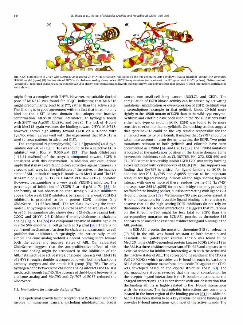
Fig. 7. (A) Binding site of 2HYY with SU6668. Color codes: 2HYY X-ray structure (red cartoon); the IFD-generated 2HYY (yellow); Native imatinib (green); IFD-generated
SU6668 model (cyan). (B) Binding site of 2HYY with chalcone analog. Color codes: 2HYY X-ray structure (red cartoon); the IFD-generated 2HYY (yellow); Native imatinib
(green); IFD-generated chalcone analog model (cyan). For clarity, hydrogen atoms on ligands were not shown and only residues that provide H-bond interactions with ligands
were shown.
H. Zhong et al. / Journal of Molecular Graphics and Modelling 28 (2009) 336–346344
might form a complex with 2HYY. However, no suitable dockedpose of MLN518 was found for 2GQG, indicating that MLN518might predominantly bind to 2HYY, rather than the active state.This finding is in good agreement with the fact that imatinib onlybind to the c-KIT kinase domain that adopts the inactiveconformation. MLN518 forms intermolecular hydrogen bondswith 2HYY via Asp381, Glu286, and Lys285. The lack of H-bondwith Met318 again weakens the binding toward 2HYY. MLN518,however, shows high affinity toward EGFR via a H-bond withLys745, which agrees well with the experiment that MLN518 isused to treat patients in advanced GIST.
The compound N-phenylpyrido[10,20:1,5]pyrazolo[3,4-d]pyr-imidine derivative (Fig. 5, 16) was found to be a selective EGFRinhibitor with IC50 at 0.050 mM [53]. The high GlideScore(�13.31 kcal/mol) of the tricyclic compound toward EGFR isconsistent with this observation. In addition, our calculationsimply that it may exert its inhibitory activities against tumors viaa second pathway, i.e., the inhibition of the active, or the inactivestate of ABL, or both through H-bonds with Met318 and Thr315.Benzamidine (Fig. 5, 17) is a latest VEGFR-2 (KDR) inhibitor.However, benzamidine is a very weak VEGFR-2 inhibitor. Thepercentage of inhibition of VEGFR-2 at 10 mM is 7% [54]. Inconformity of our observation that strong VEGFR-2 inhibitorsappear to be weak EGFR inhibitors, benzamidine, a weak VEGFR-2inhibitor, is predicted to be a potent EGFR inhibitor (theGlideScore, �11.66 kcal/mol). The residues involving the inter-molecular hydrogen bonds in EGFR/benzamidine are Lys745 andAsp855. Benzamidine also shows decent GlideScore against both2GQG and 2HYY. 2,6-Dichloro-40-methylchalcone, a chalconeanalog (Fig. 5, 18) [55] is a compound capable of inhibiting 98.2%in vitro SVR endothelial cell growth at 3 mg/ml [56]. There is noconfirmed mechanism of actions for chalcone and curcumin as cellproliferation inhibitors. Surprisingly, the structurally muchsimple chalcone analog yielded a decent binding score towardboth the active and inactive states of ABL. Our calculatedGlideScores suggest that the antiproliferative effect of thischalcone analog might be attributed to the inhibition of theABL in its inactive or active states. Chalcone interacts with Met318of 2HYY through a double hydrogen bond with both the backbonecarbonyl oxygen and the amide NH group (Fig. 7B). The onlyhydrogen bond between the chalcone analog interacts and EGFR ismediated through Lys745. The absence of the H-bond between thechalcone analog and Met793 and Cys797 of EGFR reduced theGlideScore.
3.3. Implications for molecule design of TKIs
The epidermal growth factor receptor (EGFR) has been found toinvolve in numerous cancers, including glioblastomas, breast
cancer, non-small-cell lung cancer (NSCLC), and GISTs. Thederegulation of EGFR kinase activity can be caused by activatingmutations, amplification or overexpression of EGFR. Gefitinib wasa serendipitous example in that gefitinib binds 20-fold moretightly to the L858R mutant of EGFR than to the wild-type enzyme.Gefitinib and erlotinib have been used in the NSCLC patients witheither wild-type or mutant EGFR. EGFR was found to be moresensitive to erlotinib than to gefitinib. Our docking studies suggestthat cysteine-797 could be the key residue responsible for theenhanced sensitivity of erlotinib. It implies that Cys797 should betaken into account in drug design targeting the EGFR. Two pointmutations resistant to both gefitinib and erlotinib have beendocumented as T790M [34] and D761Y [57]. The T790M mutationis located at the gatekeeper position in the kinase domain. Someirreversible inhibitors such as CL-387785, HKI-272, EKB-569 andCL-1033 seem to irreversibly inhibit EGFR T790 mutant by forminga covalent bond with cysteine-797 of EGFR [58]. This supports ourfinding that Cys797 is critical for ligand binding to EGFR. Inaddition, Met793, Lys745 and Asp855 appear to be importantresidues for ligand binding. Almost all the high scoring ligandsinteract with one or more of these residues. Lysine-745 (Lys745)and aspartate-855 (Asp855) form a salt bridge, not only providingscaffold for the binding pocket, but also interacting with ligands viaH-bond interactions [59]. Methionine-793 also provides essentialH-bond interactions for favorable ligand binding. It is relieving toobserve that all the high scoring EGFR inhibitors do not rely onthreonine-790 for H-bond interactions. It suggests that mutationson the threonine-790 might be less fatal to EGFR than thecorresponding mutation on BCR-ABL protein, as threonine-315appears to be one of the essential residues for ligand binding in theABL protein.
In BCR-ABL protein, the mutation threonine-315 to isoleucine(T315I) in the ABL was found resistant to both imatinib anddasatinib. The ‘‘gatekeeper’’ residue Thr315 was found to beMet120 in the cAMP-dependent protein kinases (CDKs). Met318 inthe ABL is a three-residue downstream of Thr315 and appears to bea critical residue for inhibitors interacting with both the active andthe inactive states of ABL. The corresponding residue in the CDKs isVal120 (CDKs) which provides an H-bond through its backboneNH. A pharmacophore map of small molecule PKI against the CDKswas developed based on the crystal structure 1ATP [60]. Thepharmacophore studies revealed that the major contribution forthe receptor–ligand interactions is the H-bond interactions, not thecharged interactions. This is consistent with our observation thatthe binding affinity is highly related to the H-bond interactionswith the receptor. The hydrophobic interactions are commonlylocated in the inner region of the binding pocket [61]. In addition,Asp381 has been shown to be a key residue for ligand binding as itprovides H-bond interactions with most of the active ligands. The
H. Zhong et al. / Journal of Molecular Graphics and Modelling 28 (2009) 336–346 345
similar importance can be found in the corresponding residueAsp184 in the CDKs.
4. Conclusions
Protein plasticity has been documented to play a critical role inligand binding, especially in protein kinases and HIV protease,which undergo significant conformational changes upon ligandbinding. Conformational changes include not only side chaindisplacement but also adjustment of loop structures upon thebinding, as indicated in Figs. 3–7. It is obvious that there is a closerelationship between the accuracy of the binding (both scoring andRMS deviation) and the inclusion of conformational changesduring complexation. The induced-fit docking module has beenshown to outperform the rigid receptor Glide dock. However, thegain in accuracy does not come without a price tag. While Glidedock can dock a database of ligands to a protein active site, the IFDmethod only docks one ligand at a time and it sometimes takesmore than 2 h per task. The IFD method, however, does show betterresults in reproducing the native conformations and should beconsidered in the drug design phase. The cost of the IFD docking isnot uncommon. The flexible docking based on normal modesmethod yielded a better performance than the rigid receptordocking [62]. However, the average run times of the normal modesbased docking is ‘‘especially remarkable’’ [62]. It is remarkable toobserve that the IFD docking protocol in this paper can reproducethe native poses for ligands from different sources of origin, such asdasatinib, imatinib, nilotinib, lapatinib, gefitinib, and erlotinib. Thebinding poses and hydrogen bond interactions of all 18 structurallydiverse TKIs are consistent with available experimental data.
The ‘‘gatekeeper’’ residue threonine (Thr315 of the ABL, andThr790 of EGFR) is a very critical residue for ligand binding.Mutations of this critical residue often cause the decreased bindingaffinity. An effective strategy appears to deploy other residues thatprovide the essential H-bond interactions. Residues important forthe EGFR binding include Cys797, Met793 and Lys745; and for theABL, Met318, Asp381 and Glu286. Our docking studies indicatethat imatinib might be a weak binder to the active state of ABL. Theincreased sensitivity of erlotinib might be attributed to the H-bondinteraction between the methoxyethoxyl moiety of erlotinib andCys797. It implies that Cys797 should be taken into account in drugdesign targeting the EGFR. The minimum number of H-bondsrequired for the binding is important as well. The differentrequirement in the number of H-bonds for the binding provides areasonable explanation that nilotinib is more effective thanimatinib against most imatinib resistant mutants.
Acknowledgements
The authors are indebted to Prof. J.K. Wood for his helpfulreview of this manuscript. This work has been in part supported bythe Research Corporation and the UCR grant of the University ofNebraska at Omaha.
References
[1] Y. Yarden, A. Ullrich, Growth factor receptor tyrosine kinase, Annu. Rev. Biochem.57 (1988) 443–478.
[2] N. Thatcher, A. Chang, P. Parikh, P.J. Rodrigues, T. Ciuleanu, J. von Pawel, S.Thongprasert, E.H. Tan, K. Pemberton, V. Archer, K. Carroll, Gefitinib plus bestsupportive care in previously treated patients with refractory advanced non-small-cell lung cancer: results from a randomised, placebo-controlled, multi-centre study (Iressa Survival Evaluation in Lung Cancer), Lancet 366 (2005) 1527–1537.
[3] B.C. Cho, C.K. Im, M.S. Park, S.K. Kim, J. Chang, J.P. Park, H.J. Choi, Y.J. Kim, S.J. Shin,J.H. Sohn, H. Kim, J.H. Kim, Phase II study of erlotinib in advanced non-small-celllung cancer after failure of gefitinib, J. Clin. Oncol. 25 (2007) 2528–2533.
[4] B.J. Druker, C.L. Sawyers, H. Kantarjian, D.J. Resta, S.F. Reese, J.M. Ford, R. Capde-ville, M. Talpaz, Activity of a specific inhibitor of the BCR-ABL tyrosine kinase in
the blast crisis of chronic myeloid leukemia and acute lymphoblastic leukemiawith the Philadelphia chromosome, N. Engl. J. Med. 344 (2001) 1038–1042.
[5] B.J. Druker, M. Talpaz, D.J. Resta, B. Peng, E. Buchdunger, J.M. Ford, N.B. Lydon, H.Kantarjian, R. Capdeville, S. Ohno-Jones, C.L. Sawyers, Efficacy and safety of aspecific inhibitor of the BCR-ABL tyrosine kinase in chronic myeloid leukemia, N.Engl. J. Med. 344 (2001) 1031–1037.
[6] G.D. Demetri, M. von Mehren, C.D. Blanke, A.D. Van den Abbeele, B. Eisenberg, P.J.Roberts, M.C. Heinrich, D.A. Tuveson, S. Singer, M. Janicek, J.A. Fletcher, S.G.Silverman, S.L. Silberman, R. Capdeville, B. Kiese, B. Peng, S. Dimitrijevic, B.J.Druker, C. Corless, C.D.M. Fletcher, H. Joensuu, Efficacy and safety of imatinibmesylate in advanced gastrointestinal stromal tumors, N. Engl. J. Med. 347 (2002)472–480.
[7] N.P. Shah, C. Tran, F.Y. Lee, P. Chen, D. Norris, C.L. Sawyers, Overriding imatinibresistance with a novel ABL kinase inhibitor, Science 305 (2004) 399–401.
[8] E. Weisberg, P. Manley, W. Breitenstein, J. Bruggen, S. Cowan-Jacob, A. Ray, B.Huntly, D. Fabbro, G. Fendrich, E. Hall-Meyers, A. Kung, J. Mestan, G.Q. Daley, L.Callahan, L. Catley, C. Cavazza, M. Azam, D. Neuberg, R.D. Wright, D.G. Gilliland,J.D. Griffin, Characterization of AMN107, a selective inhibitor of native and mutantBcr-Abl, Cancer Cell 7 (2005) 129–141.
[9] E. Weisberg, P. Manley, J. Mestan, S. Cowan-Jacob, A. Ray, J.D. Griffin, AMN(nilotinib): a novel and selective inhibitor of BCR-ABL, Br. J. Cancer 94 (2006)1765–1769.
[10] J.S. Tokarski, J. Newitt, C.Y.J. Chang, J.D. Cheng, M. Wittekind, S.E. Kiefer, K. Kish,F.Y.F. Lee, R. Borzillerri, L.J. Lombardo, D. Xie, Y. Zhang, H.E. Klei, The structure ofdasatinib (BMS-354825) bound to activated ABL kinase domain elucidates itsinhibitory activity against imatinib-resistant ABL mutants, Cancer Res. 66 (2006)5790–5797.
[11] S.W. Cowan-Jacob, G. Fendrich, A. Floersheimer, P. Furet, J. Liebetanz, G. Rummel,P. Rheinberger, M. Centeleghe, D. Fabbro, P.W. Manley, Structural biology con-tributions to the discovery of drugs to treat chronic myelogenous leukemia, ActaCrystallogr., Sect. D 63 (2007) 80–93.
[12] S. Atwell, J.M. Adams, J. Badger, M.D. Buchanan, I.K. Feil, K.J. Froning, X. Gao, J.Hendle, K. Keegan, B.C. Leon, H.J. Muller-Dieckmann, V.L. Nienaber, B.W. Noland,K. Post, K.R. Rajashankar, A. Ramos, M. Russell, S.K. Burley, S.G. Buchanan, A novelmode of Gleevec (STI-571 Imatinib) binding is revealed by the structure of spleentyrosine kinase (Syk), J. Biol. Chem. 279 (2004) 55827–55832.
[13] Schrodinger, LLC: Portland, OR, 2007, Web address: www.schrodinger.com.[14] I.D. Kuntz, J.M. Blaney, S.J. Oatley, R. Langridge, T.E. Ferrin, A geometric approach
to macromolecule–ligand interactions, J. Mol. Biol. 161 (1982) 269–288.[15] M. Rarey, B. Kramer, T. Lengauer, G. Klebe, A fast flexible docking method using an
incremental construction algorithm, J. Mol. Biol. 261 (1996) 470–489.[16] G. Jones, P. Willett, R.C. Glen, A.R. Leach, R. Taylor, Development and validation of
a genetic algorithm for flexible docking, J. Mol. Biol. 267 (1997) 727–748.[17] G.M. Morris, D.S. Goodsell, R.S. Halliday, R. Huey, W.E. Hart, R.K. Belew, A.J. Olson,
Automated docking using a Lamarckian genetic algorithm and an empiricalbinding free energy function, J. Comput. Chem. 19 (1998) 1639–1662.
[18] R.A. Friesner, J.L. Banks, R.B. Murphy, T.A. Halgren, J.J. Klicic, D.T. Mainz, M.P.Repasky, E.H. Knoll, M. Shelley, J.K. Perry, D.E. Shaw, P. Francis, P.S. Shenkin, Glide:a new approach for a rapid, accurate docking and scoring. 1. Method andassessment of docking accuracy, J. Med. Chem. 47 (2004) 1739–1749.
[19] H.A. Carlson, Protein flexibility and drug design: how to hit a moving target, Curr.Opin. Chem. Biol. 6 (2002) 447–452.
[20] W. Sherman, T. Day, M.P. Jacobson, R.A. Friesner, R. Farid, Novel procedure formodeling ligand/receptor induced fit effects, J. Med. Chem. 49 (2006) 534–553.
[21] E.R. Wood, A.T. Truesdale, O.B. McDonald, D. Yuan, A. Hassell, S.H. Dickerson, B.Ellis, C. Pennisi, E. Horne, K. Lackey, K.J. Alligood, D.W. Rusnak, T.M. Gilmer, L.M.Shewchuk, A unique structure for epidermal growth factor receptor bound toGW572016 (Lapatinib): relationships among protein conformation, inhibitor off-rate, and receptor activity in tumor cells, Cancer Res. 64 (2004) 6652–6659.
[22] C.H. Yun, T.J. Boggon, Y. Li, S. Woo, H. Greulich, M. Meyerson, M.J. Eck, Structuresof lung cancer-derived egfr mutants and inhibitor complexes: mechanism ofactivation and insights into differential inhibitor sensitivity, Cancer Cell 11 (2007)217–227.
[23] X. Zhang, J. Gureasko, K. Shen, P.A. Cole, J. Kuriyan, An allosteric mechanism foractivation of the kinase domain of epidermal growth factor receptor, Cell 125(2006) 1137–1149.
[24] K. Ghosh, C.K. Lau, F. Guo, A.M. Segall, G.D. van Duyne, Peptide trapping of theHolliday junction intermediate in Cre-loxP site-specific recombination, J. Biol.Chem. 280 (2005) 8290–8299.
[25] MOE software, Chemical Computing Group Inc., Montreal, Canada, Web address:http://www.chemcomp.com.
[26] J. Holm, J. Park, DaliLite workbench for protein structure comparison, Bioinfor-matics 16 (2000) 566–567, World wide web address: http://www.ebi.ac.uk/Tools/dalilite/index.html.
[27] H. Zhong, J.P. Bowen, Molecular design and clinical development of VEGFR kinaseinhibitors, Curr. Top. Med. Chem. 7 (2007) 1379–1393.
[28] T.A. Halgren, Merck molecular force field. I. Basis, form, scope, parameterization,and performance of MMFF94, J. Comp. Chem. 490–519.
[29] A.L. Bowman, Z. Nikolovska-Coleska, H. Zhong, S. Wang, H.A. Carlson, Smallmolecule inhibitors of the MDM2-p53 interaction discovered by ensemble-basedreceptor models, J. Am. Chem. Soc. 129 (2007) 12809–12814.
[30] R.A. Friesner, R.B. Murphy, M.P. Repasky, L.L. Frye, J.R. Greenwood, T.A. Halgren,P.C. Sanschagrin, D.T. Mainz, Extra precision Glide: docking and scoring incor-porating a model of hydrophobic enclosure for protein–ligand complexes, J. Med.Chem. 49 (2006) 6177–6196.
H. Zhong et al. / Journal of Molecular Graphics and Modelling 28 (2009) 336–346346
[31] PYMOL, Delano Scientific LLC, San Carlos, CA, USA, Web address: http://pymol.sourceforge.net.
[32] ClustalW server, the Swiss Institute of Bioinformatics, Basel, Switzerland, WorldWide Web: http://www.ch.embnet.org/software/ClustalW.html.
[33] J. Stamos, M.X. Sliwkowski, C. Eigenbrot, Structure of the epidermal growth factorreceptor kinase domain alone and in complex with a 4-anilinoquinazolineinhibitor, J. Biol. Chem. 277 (2002) 46265–46272.
[34] S. Kobayashi, T.J. Boggon, T. Dayaram, P.A. Janne, O. Kocher, M. Meyerson, B.E.Johnson, M.J. Eck, D.G. Tenen, B. Halmos, EGFR mutation and resistance of non-small-cell lung cancer to gefitinib, N. Engl. J. Med. 352 (2005) 786–792.
[35] S. Blencke, A. Ullrichs, H. Daub, Mutation of threonine 766 in the epidermalgrowth factor receptor reveals a hotspot for resistance formation against selectivetyrosine kinase inhibitors, J. Biol. Chem. 278 (2003) 15435–15440.
[36] G.J. Riley, K.A. Politi, V.A. Miller, W. Pao, Update on epidermal growth factorreceptor mutations in non-small cell lung cancer, Clin. Cancer Res. 12 (2006)7232–7241.
[37] O. Hahn, W. Stadler, Sofafenib, Curr. Opin. Oncol. 18 (2006) 615–621.[38] S.M. Wilhelm, D.S. Chien, BAY 43-9006: preclinical data, Curr. Pharm. Des. 8
(2002) 2255–2257.[39] S.M. Wilhelm, C. Carter, L.Y. Tang, D. Wilkie, A. McNabola, H. Rong, C. Chen, X.M.
Zhang, P. Vincent, M. McHugh, Y.C. Cao, J. Shujath, S. Gawlak, D. Eveleigh, B.Rowley, L. Liu, L. Adnane, M. Lynch, D. Auclair, I. Taylor, R. Gedrich, A. Vozne-sensky, B. Riedl, L.E. Post, G. Bollag, P.A. Trail, BAY 43-9006 exhibits broadspectrum oral antitumor activity and targets the RAF/MEK/ERK pathway andreceptor tyrosine kinases involved in tumor progression and angiogenesis, CancerRes. 64 (2004) 7099–7109.
[40] G.D. Demetri, A.T. van Oosterom, C.R. Garrett, M.E. Blackstein, M.H. Shah, J.Verweij, G. McArthur, I.R. Judson, M.C. Heinrich, J.A. Morgan, J. Desai, D.C. Fletcher,S. George, C.L. Bello, X. Huang, C.M. Baum, P.G. Casali, Lancet 368 (2006) 1329–1338.
[41] G. Bold, K.-H. Altmann, J. Frei, M. Lang, P.W. Manley, P. Traxler, B. Wietfeld, J.Bruggen, E. Buchdunger, R. Cozens, S. Ferrari, P. Furet, F. Hofmann, G. Martiny-Baron, J. Mestan, J. Rosel, M. Sills, D. Stover, F. Acemoglu, E. Boss, R. Emmenegger,L. Lasser, E. Masso, R. Roth, C. Schlachter, W. Vetterli, D. Wyss, J.M. Wood, Newanilinophthalazines as potent and orally well absorbed inhibitors of the VEGFreceptor tyrosine kinases useful as antagonists of tumor-driven angiogenesis, J.Med. Chem. 43 (2000) 2310–2323.
[42] J.C. Becker, C. Mueller-Tidow, H. Serve, W. Domschke, T. Pohle, Role of receptortyrosine kinases in gastric cancer: new targets for a selective therapy, World J.Gastroenterol. 12 (2006) 3297–3305.
[43] S.R. Wedge, D.J. Ogilvie, M. Dukes, J. Kendrew, R. Chester, J.A. Jackson, S.J. Boffey,P.J. Valentine, J.O. Curwen, H.L. Musgrove, G.A. Graham, G.D. Hughes, A.P. Thomas,E.S.E. Stokes, B. Curry, G.H.P. Richmond, P.F. Wadsworth, A.L. Bigley, L.F. Henne-quin, ZD6474 inhibits vascular endothelial growth factor signaling, angiogenesis,and tumor growth following oral administration, Cancer Res. 62 (2002) 4645–4655.
[44] P. Traxler, P.R. Allegrini, R. Brandt, J. Brueggen, R. Cozens, D. Fabbro, K. Grossios,H.A. Lane, P. McSheehy, J. Mestan, T. Meyer, C. Tang, M. Wartmann, J. Wood, G.Caravatti, AEE788: a dual family epidermal growth factor receptor/ErbB2 andvascular endothelial growth factor receptor tyrosine kinase inhibitor with anti-tumor and antiangiogenic activity, Cancer Res. 64 (2004) 4931–4941.
[45] B. Askew, J. Adams, S. Booker, G. Chen, L.V. Dipietro, D. Elbaum, J. Germain, S.D.Geuns-Meyer, G.J. Habgood, M. Handley, Q. Huang, T. Kim, A. Li, N. Nishimura, R.Nomak, V.F. Patel, B. Riahi, J.L. Kim, X. Ning, K. Yang, C.C. Yuan, Substitutedalkylamine derivatives and methods of use, Amgen, Inc., U.S. patent 6,878,714(2005).
[46] T.A.T. Fong, L.K. Shawver, L. Sun, C. Tang, H. App, T.J. Powell, Y.H. Kim, R. Schreck,X.Y. Wang, W. Risau, A. Ullrich, K.P. Hirth, G. McMahon, SU5416 is a potent andselective inhibitor of the vascular endothelial growth factor receptor (Flk-1/KDR)
that inhibits tyrosine kinase catalysis, tumor vascularization, and growth ofmultiple tumor types, Cancer Res. 59 (1999) 99–106.
[47] J.V. Heymach, J. Desai, J. Manola, D.W. Davis, D.J. McConkey, D. Harmon, D.P. Ryan, G.Goss, T. Quigley, A.D. Van den Abbeele, S.G. Silverman, S. Connors, J. Folkman, C.D.M.Fletcher, G.D. Demetri, Phase II study of the antiangiogenic agent SU5416 in patientswith advanced soft tissue sarcomas, Clin. Cancer Res. 10 (2004) 5732–5740.
[48] A.D. Laird, J.G. Christensen, G.M. Li, J. Carver, K. Smith, X.H. Xin, K.G. Moss, S.G.Louie, D.B. Mendel, J.M. Cherrington, SU6668 inhibits Flk-1/KDR and PDGFRß invivo, resulting in rapid apoptosis of tumor vasculature and tumor regression inmice, FASEB J. 16 (2002) 681–690.
[49] B.D. Smolich, H.A. Yuen, K.A. West, F.J. Giles, M. Albitar, J.M. Cherrington, Theantiangiogenic protein kinase inhibitors SU5416 and SU6668 inhibit the SCFreceptor (c-kit) in a human myeloid leukemia cell line and in acute myeloidleukemia blasts, Blood 97 (2001) 1413–1421.
[50] L.M. Kelly, J.C. Yu, C.L. Boulton, M. Apatira, J. Li, C.M. Sullivan, I. Williams, S.M.Amaral, D.P. Curley, N. Duclos, D. Neuberg, R.M. Scarborough, A. Pandey, S.Hollenbach, K. Abe, N.A. Lokker, D.G. Gilliland, N.A. Giese, CT53518, a novelselective FLT3 antagonist for the treatment of acute myelogenous leukemia(AML), Cancer Cell 1 (2002) 421–432.
[51] A.S. Corbin, I.J. Griswold, P.L. Rosee, K.W.H. Yee, M.C. Heinrich, C.L. Reimer, B.J.Druker, M.W.N. Deininger, Sensitivity of oncogenic KIT mutants to the kianseinhibtors MLN518 and PD180970, Blood 104 (2004) 3754–3757.
[52] C.D. Mol, D.R. Dougan, T.R. Schneider, R.J. Skene, M.L. Kraus, D.N. Scheibe, G.P.Snell, H. Zou, B.C. Sang, K.P. Wilson, Structural basis for the autoinhibition and STI-571 inhibition of c-Kit tyrosine kinase, J. Biol. Chem. 279 (2004) 31655–31663.
[53] M.J. Alberti, E.P. Auten, K.E. Lackey, O.B. McDonald, E.R. Wood, F. Preugschat, G.J.Cutler, L. Kane-Carson, W. Liu, D.K. Jung, Discovery and in vitro evaluation ofpotent kinase inhibitors: Pyrido[10 ,20:1,5]pyrazolo[3,4-d]pyrimidines, Bioorg.Med. Chem. Lett. 15 (2005) 3778–3781.
[54] H. Nakamura, Y. Sasaki, M. Uno, T. Yoshikawa, T. Asano, H.S. Ban, H. Fukazawa, M.Shibuya, Y. Uehara, Synthesis and biological evaluation of benzamides andbenzamidines as selective inhibitors of VEGFR tyrosine kinase, Bioorg. Med.Chem. Lett. 16 (2006) 5127–5131.
[55] T.P. Robinson, T. Ehlers, R.B. Hubbard, X. Bai, J.L. Arbiser, D.J. Goldsmith, J.P.Bowen, Design, synthesis, and biological evaluation of angiogenesis inhibitors:aromatic enone and dienone analogues of curcumin, Bioorg. Med. Chem. Lett. 13(2003) 115–117.
[56] T.P. Robinson, R.B. Hubbard IV, T.J. Ehlers, J.L. Arbiser, D.J. Goldsmith, J.P. Bowen,Synthesis and biological evaluation of aromatic enones related to curcumin,Bioorg. Med. Chem. 13 (2005) 4007–4013.
[57] M.N. Balak, Y. Gong, G.J. Riely, R. Somwar, A.R. Li, M.F. Zakowski, A. Chiang, G.Yang, O. Ouerfelli, M.G. Kris, M. Ladanyi, V.A. Miller, W. Pao, Novel D761Y andcommon secondary T790M mutations in epidermal growth factor receptor-mutant lung adenocarcinomas with acquired resistance to kinase inhibitors, Clin.Cancer Res. 12 (2006) 6494–6501.
[58] S.V. Sharma, D.W. Bell, J. Settleman, D.A. Haber, Epidermal growth factor receptormutations in lung cancer, Nat. Rev. Cancer 7 (2007) 169–181.
[59] A. Kumar, E.T. Petri, B. Halmos, T.J. Boggon, Structure and clinical relevance of theepidermal growth factor receptor in human cancer, J. Clin. Oncol. 26 (2008) 1742–1751.
[60] J. Zheng, E.A. Trafny, D.R. Knighton, N.H. Xuong, S.S. Taylor, L.F. Ten Eyck, J.M.Sowadski, 2.2 A refined crystal structure of the catalytic subunit of cAMP-depen-dent protein kinase complexed with MnATP and a peptide inhibitor, Acta Crystal-logr., Sect.D 49 (1993) 362–365.
[61] M.J. McGregor, A pharmacophore map of small molecule protein kinase inhibi-tors, J. Chem. Inf. Model. 47 (2007) 2374–2382.
[62] A. May, M. Zacharias, Protein–ligand docking accounting for receptor side chainand global flexibility in normal modes: evaluation on kinase inhibitor crossdocking, J. Med. Chem. 51 (2008) 3499–3506.


