Embed Size (px)
Citation preview
Inhibiting AKT Phosphorylation Employing Non-CytotoxicAnthraquinones Ameliorates TH2 Mediated AllergicAirways Disease and Rhinovirus ExacerbationCaio Cesar de Souza Alves1,2,3, Adam Collison2,3, Luke Hatchwell2,3, Maximilian Plank3, Matthew Morten2,3,Paul S. Foster3, Sebastian L. Johnston4, Cristiane França da Costa5, Mauro Vieira de Almeida5, HenriqueCouto Teixeira1, Ana Paula Ferreira1*, Joerg Mattes2,3,6*
1 Department of Parasitology, Microbiology and Immunology, Institute of Biological Sciences, Federal University of Juiz de Fora, Minas Gerais, Brazil,2 Experimental&Translational Respiratory Group, University of Newcastle and Hunter Medical Research Institute, Newcastle, Australia, 3 Priority ResearchCentre for Asthma and Respiratory Diseases, University of Newcastle and Hunter Medical Research Institute, Newcastle, Australia, 4 Airway Disease InfectionSection, National Heart and Lung Institute, Medical Research Council & Asthma UK Centre in Allergic Mechanisms of Asthma, Imperial College London,London, United Kingdom, 5 Department of Chemistry, Federal University of Juiz de Fora, Minas Gerais, Brazil, 6 Paediatric Respiratory & Sleep Medicine Unit,Newcastle Children’s Hospital, Newcastle, Australia
Abstract
Background: Severe asthma is associated with T helper (TH) 2 and 17 cell activation, airway neutrophilia andphosphoinositide-3-kinase (PI3K) activation. Asthma exacerbations are commonly caused by rhinovirus (RV) andalso associated with PI3K-driven inflammation. Anthraquinone derivatives have been shown to reduce PI3K-mediated AKT phosphorylation in-vitro.Objective: To determine the anti-inflammatory potential of anthraquinones in-vivo.Methods: BALB/c mice were sensitized and challenged with crude house dust mite extract to induce allergic airwaysdisease and treated with mitoxantrone and a novel non-cytotoxic anthraquinone derivative. Allergic mice were alsoinfected with RV1B to induce an exacerbation.Results: Anthraquinone treatment reduced AKT phosphorylation, hypoxia-inducible factor-1α and vascularendothelial growth factor expression, and ameliorated allergen- and RV-induced airways hyprereactivity, neutrophilicand eosinophilic inflammation, cytokine/chemokine expression, mucus hypersecretion, and expression of TH2proteins in the airways. Anthraquinones also boosted type 1 interferon responses and limited RV replication in thelung.Conclusion: Non-cytotoxic anthraquinone derivatives may be of therapeutic benefit for the treatment of severe andRV-induced asthma by blocking pro-inflammatory pathways regulated by PI3K/AKT.
Citation: Cesar de Souza Alves C, Collison A, Hatchwell L, Plank M, Morten M, et al. (2013) Inhibiting AKT Phosphorylation Employing Non-CytotoxicAnthraquinones Ameliorates TH2 Mediated Allergic Airways Disease and Rhinovirus Exacerbation . PLoS ONE 8(11): e79565. doi:10.1371/journal.pone.0079565
Editor: Junji Yodoi, Institute for Virus Research, Laboratory of Infection and Prevention, Japan
Received June 18, 2012; Accepted October 2, 2013; Published November 5, 2013
Copyright: © 2013 Cesar de Souza Alves et al. This is an open-access article distributed under the terms of the Creative Commons Attribution License,which permits unrestricted use, distribution, and reproduction in any medium, provided the original author and source are credited.
Funding: This study was supported by the National Health Medical Research Council (NH&MRC) (J.M., P.F.), the Hunter Medical Research Institute (J.M.,P.F.), the Hunter Children's Research Foundation (J.M., P.F.) and an NH&MRC Health Professional Research Fellowship (J.M.). C.C.S.A., C.F.C., H.C.T.,M.V.A. and A.P.F. were supported by CAPES, CNPq and FAPEMIG grants. S.L.J. was supported by a Chair from Asthma UK (CH1155). This work wassupported in part by MRC Centre grant G1000758 and ERC FP7 Advanced Grant 233015 (to S.L.J). The funders had no role in study design, datacollection and analysis, decision to publish, or preparation of the manuscript.
Competing interests: The authors have declared that no competing interests exist.
* E-mail: [email protected] (JM); [email protected] (APF)
Introduction
It is estimated that asthma affects more than 300 millionpeople worldwide [1]. Asthma is characterized by airwayshyperreactivity (AHR) and inflammation, mucus hypersecretion,and an aberrant T cell response [2]. Accumulation of
eosinophils, neutrophils and lymphocytes in the airwayscorrelate with disease severity and anti-inflammatorytreatments ameliorate episodic airways obstruction, which isthe clinical hallmark of asthma [3,4]. Airways inflammation istightly regulated and involves the release of interleukin (IL)-13and IL-5 produced by T helper type 2 (TH2) cells and natural
PLOS ONE | www.plosone.org 1 November 2013 | Volume 8 | Issue 11 | e79565

killer T cells, IL-17 by TH17 cells, as well as chemokines byresidential lung and innate immune cells [5–7]. IL-13 alsoinduces mucus production in a signal transducer and activatorof transcription 6 (STAT6)-dependent manner and is bothsufficient and required for the development of AHR and gobletcell hyperplasia [8–10]. Acute asthma exacerbations are amajor disease burden and predominantly caused by infectionwith rhinovirus (RV) -the common cold virus- [11–14].Importantly some asthmatics display impaired innate immuneresponses with deficient type 1 and 3 interferon (IFN)responses upon RV infection [15,16]. Corticosteroids are themainstream anti-inflammatory treatment used in asthmaticsand have been effective in reducing eosinophilic inflammation.However, their therapeutic efficacy in preventing neutrophilicand RV-induced inflammation and symptoms is thought to belimited [17]. Furthermore, side effects can be observed inasthmatics requiring high dose or systemic corticosteroidtreatment. In some patients asthma is steroid-resistant andthen difficult to control [18,19]. Thus novel therapeuticstrategies for severe asthma and RV-induced exacerbationsare required.
Mitoxantrone belongs to the clinically useful topoisomerase2-targeting anthraquinones that is used to treat metastaticbreast cancer, leukemia, and lymphoma [20–22]. Mitoxantronedisrupts DNA synthesis and repair causing single and double-strand breaks by intercalating the DNA through hydrogenbinding [23] resulting in non-specific cytotoxicity [20–22]. Inpatients with relapsing multiple sclerosis mitoxantrone may beof therapeutic benefit as a potent immunosuppressive agenttargeting proliferating macrophages, B and T lymphocytes[24,25]. Recently topoisomerase 2-independent effects ofmitoxantrone have also been described that may be ofrelevance for therapeutically modulating the aberrant immuneresponse observed in asthma and RV-induced exacerbations.Specifically, mitoxantrone inhibited hypoxia-inducible factor(HIF)-1α and vascular endothelial growth factor (VEGF)expression through dephosphorylation of AKT in transformedcell lines suggesting that mitoxantrone regulates thephosphoinositide-3-kinase (PI3K)/AKT pathway [26].
Importantly several lines of evidence suggest that inhibitionof the PI3K effector pathway may be a potential therapeuticstrategy for asthma and RV-induced exacerbations [27–29].Recently it has been shown that the TH2 cytokine IL-25promotes angiogenesis, at least in part, by increasing VEGF/VEGF receptor expression through PI3K/and Erk/MAPKpathways [30]. Osteopontin is upregulated by epithelial cellsand macrophages in the lungs of asthmatics that in turnactivates PI3K/AKT downstream signaling pathways to induceIL-13, AHR, mucus hypersecretion, and pro-matrixmetalloproteinase-9 in the lungs [31]. Receptor-mediated mastcell growth, differentiation, homing to their target tissues,survival and activation are also controlled, to varying degrees,by PI3K-driven pathways [32]. Finally, PI3K/AKT signaling isrequired for maximal RV-induced neutrophilic airwayinflammation in an experimental mouse model and RV-inducedIL-8 expression by airway epithelial cells, likely via its essentialrole in virus internalization [33,34].
In this study, the effects of mitoxantrone and a novel non-cytotoxic anthraquinone derivative (O,O´-didodecanoyl-1,4-dihydroxyanthraquinone) on allergic airways disease (AAD)and RV-induced exacerbation was investigated. We show thatanthraquinone treatment reduced AKT phosphorylation, HIF-αand VEGF expression, and ameliorated allergen- and RV-induced airways hyprereactivity, neutrophilic and eosinophilicinflammation, cytokine/chemokine expression, mucushypersecretion, and expression of TH2 factors in the airways.Anthraquinones also boosted type 1 interferon (IFN) responsesand limited RV replication in the lung.
Methods
AnimalsMale BALB/c mice, 6-8 weeks old, were obtained from the
Specific Pathogen Free Facility of the University of Newcastle.The Animal Care and Ethics Committee of the University ofNewcastle, Australia approved all experiments.
Preparation of anthraquinone derivatives1,4-dihydroxyanthraquinone was dissolved in
dimethylacetamide and pyridine at 0°C and dodecanoylchloride added. The reaction mixture was stirred at 0°C to roomtemperature for 24 hrs. The light yellow precipitate was washedwith hexane. Mitoxantrone (Quiral Quimica do Brasil S.A., Juizde Fora, MG, Brazil) and its analog was solubilized in DMSO(Sigma, USA), never exceeding 0.1% (v/v) and diluted in 0.9%sterile saline.
Induction of AAD and rhinovirus-induced exacerbationMice were intranasally sensitized with house dust mite
extract (HDM; 50μg daily at day 0, 1 and 2) followed byintranasal challenges (5μg daily from day 14 to day 17)delivered in 50μl of 0.9% sterile saline. Non-sensitized micereceived sterile saline only. Mice were euthanized 24hrs afterthe last allergen challenge.
Four groups of mice were studied: non-allergic (SALINE),HDM allergic and vehicle treated (VEHICLE, DMSO 0.1%),HDM allergic and mitoxantrone treated (MITOXANTRONE1mg/kg as previously described[35]), HDM allergic and analogtreated (ANALOG 1mg/kg) mice. Treatments (100mcl/mouse/day) were administered intraperitoneally from day 12 to day 17during HDM challenges and after HDM sensitization. In anotherseries of experiments mice were intranasally infected withminor group RV (RV1B) – 50µl containing 1x10⁷ virions – orUV-inactivated RV1B at day 18 which was 24hrs after the lastHDM challenge to exacerbate preexisting AAD. Mice wereeuthanized 24hrs after the RV1B infection.
AHR measurementAHR was invasively assessed in separate groups of
anesthetized mice by measurement of total lung resistance inresponse to increasing doses of methacholine as previouslydescribed[36]. Percentage increase over baseline (water) inresponse to nebulized methacholine was calculated.
Non-Cytotoxic Anthraquinones Inhibit AAD
PLOS ONE | www.plosone.org 2 November 2013 | Volume 8 | Issue 11 | e79565
Collection of bronchoalveolar lavage fluidTwenty-four hours after the last HDM challenge,
bronchoalveolar lavage (BAL) fluid was performed bycannulating the trachea and instillation of 800 µl of Hank'sBuffered Salt Solution (HBSS) three times.
Total and differential cell countsBAL fluid was centrifuged at 800 x g for 10 min at 4°C and
cell-free supernatant was collected. Cells were resuspended in100µl of HBSS and total number of viable cells was determinedby trypan blue exclusion in a Neubauer cell chamber.Cytospins were prepared and slides were stained with May-Grunwald-Giemsa. Differential cell counts were determinedfrom a total of 200 cells per slide.
Quantitative Real time PCRLower airway tissue from the left lung of each animal was
separated by blunt dissection and stored in RNA later (Ambion,USA) at -80°C. Total RNA was extracted from the airwaysaccording to the manufacturer's instructions (mirVana m/miRNA isolation kit, Ambion, USA). The primers used weresynthesized by Sigma (Table 1). qRT-PCRs reactions wereperformed using the SYBR® Green (Kappa Biosystems, USA)according to manufacturer's instructions. After amplification allsamples were subjected to dissociation curve analysis in orderto validate the absence of nonspecific products and primerdimers. RNA was normalized to expression levels ofHypoxanthine-guanine phosphoribosyl transferase (HPRT) andrelative expression was calculated with the 2-ΔΔCt method.
Airway morphology analysisThe right lower lung lobe from each animal was fixed in 10%
buffered formalin and the samples were subjected to routine
histologic procedures and then stained with periodic acid-Schiff(PAS) to identify mucus glycoconjugates, Toluidine blue toidentify mast cells, or Carbol’s chromotrope-hematoxylin toidentify eosinophils. Cells were identified by morphologicalcriteria and quantified by counting ten high-powered fields(HPF) in each slide.
Measurement of cytokinesPeribronchial lymph nodes were excised, filtered and
cultured in the presence of HDM (50μg/ml) for 6 days. Levels ofIL-13, IL-5 and IFN-γ in supernatants were determined byELISA (BD Biosciences Pharmingen, USA) according to themanufacturer's instructions. CD4+ T-cells were isolated fromthe draining lymph nodes using an Auto Macs Pro (MiltenyiBiotec, USA) according to the manufacturer’s instructions.Levels of IL-4 and IL-13 were measured concurrently bymultiplex using the Novex platform (Invitrogen, USA) accordingto the manufacturer’s instructions before being quantified usinga Bioplex (Biorad, USA) luminex system. Whole mouse lungswere homogenized with a Tissue Tearor (Biospec Products,USA) on ice in lysis buffer.
Flow cytometryTo prepare single-cell suspensions from whole lung and
lymph nodes, tissues were gently mashed through 100µm cellstrainers (BD Falcon). Red blood cells were removed usinglysis buffer (4.15g ammonium chloride, 1g sodium hydrogencarbonate, 0.0185g EDTA in 500ml of dH2O). Cells werecounted and the Fc receptor was blocked. Cell surfaceexpression of CD4 (PE), CD8 (PerCP), TCRβ (FITC), CD3e(APC), CD19 (PerCP), CD11b (PerCP), CD11c (FITC), F4/80(APC) and MHCII (PE) (all antibodies from Pharmingen, USA)was determined by flow cytometry analysis with a FACSCanto
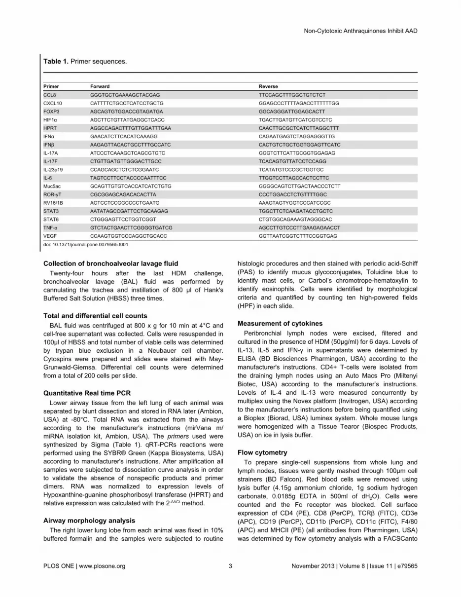
Table 1. Primer sequences.
Primer Forward ReverseCCL8 GGGTGCTGAAAAGCTACGAG TTCCAGCTTTGGCTGTCTCTCXCL10 CATTTTCTGCCTCATCCTGCTG GGAGCCCTTTTAGACCTTTTTTGGFOXP3 AGCAGTGTGGACCGTAGATGA GGCAGGGATTGGAGCACTTHIF1α AGCTTCTGTTATGAGGCTCACC TGACTTGATGTTCATCGTCCTCHPRT AGGCCAGACTTTGTTGGATTTGAA CAACTTGCGCTCATCTTAGGCTTTIFNα GAACATCTTCACATCAAAGG CAGAATGAGTCTAGGAGGGTTGIFNβ AAGAGTTACACTGCCTTTGCCATC CACTGTCTGCTGGTGGAGTTCATCIL-17A ATCCCTCAAAGCTCAGCGTGTC GGGTCTTCATTGCGGTGGAGAGIL-17F CTGTTGATGTTGGGACTTGCC TCACAGTGTTATCCTCCAGGIL-23p19 CCAGCAGCTCTCTCGGAATC TCATATGTCCCGCTGGTGCIL-6 TAGTCCTTCCTACCCCAATTTCC TTGGTCCTTAGCCACTCCTTCMuc5ac GCAGTTGTGTCACCATCATCTGTG GGGGCAGTCTTGACTAACCCTCTTROR-yT CGCGGAGCAGACACACTTA CCCTGGACCTCTGTTTTGGCRV16/1B AGTCCTCCGGCCCCTGAATG AAAGTAGTYGGTCCCATCCGCSTAT3 AATATAGCCGATTCCTGCAAGAG TGGCTTCTCAAGATACCTGCTCSTAT6 CTGGGAGTTCCTGGTCGGT CTGTGGCAGAAAGTAGGGCACTNF-α GTCTACTGAACTTCGGGGTGATCG AGCCTTGTCCCTTGAAGAGAACCTVEGF CCAAGTGGTCCCAGGCTGCACC GGTTAATCGGTCTTTCCGGTGAG
doi: 10.1371/journal.pone.0079565.t001
Non-Cytotoxic Anthraquinones Inhibit AAD
PLOS ONE | www.plosone.org 3 November 2013 | Volume 8 | Issue 11 | e79565

flow cytometer using commercially available Abs from BDBiosciences. Cells gated by forward- and side-scatterparameters were analyzed using FACSDiva software.
p-AKT Western blotLevels of p-AKT were determined by western blotting in
whole cell protein lysates isolated from lung homogenates.Protein samples at 45 μg/lane underwent electrophoresis on a10% SDS-polyacrylamide gel and were electroblotted ontoPVDF. Membranes were blocked for 2h at room temperature inTBS containing 5% bovine serum albumin (Sigma), themembrane was incubated for 2h at room temperature withmonoclonal anti p-AKT (1:300 in a TBST solution made up with10ng/ml of B-Actin). After washing the membrane 3x for 5minin TBST the membrane was incubated with HEP-conjugatedsecondary antibody (1:5000 in TBST) for 1h at roomtemperature. The membrane was incubated with LuminataCresecendo Western HRP Substrate (Millipore) and visualizedon a Fujifilm LAS-4000 using Image reader LAS-4000.
Determination of PIP3 activityLevels of active phosphatidylinositol-(3,4,5)-trisphosphate
(PIP3) were determined by ELISA (Echelon Biosciences, USA)according to the manufacturer’s instructions.
Statistical analysisNumerical data were analyzed for normal distribution
employing the Kolmogorov-Smirnov test. Subsequently, theunpaired t test was used for parametric data or Mann Whitneytest for nonparametric data. The significance level accepted forthe tests was p<0.05. Data are expressed as mean ± standarderror of the mean (SEM).
Results
Treatment with anthraquinones ameliorates hallmarkfeatures of AAD
The ability of mitoxantrone to intercalate with the DNAthrough hydrogen binding was precluded by synthesizing anovel anthraquinone derivative, O,O´-didodecanoyl-1,4-dihydroxyanthraquinone (Figure 1A). Consequently, this analogdid not exhibit any in-vitro cytotoxicity on transformedmacrophage cell lines or cytotoxic effects in-vivo (data notshown). In order to investigate the anti-inflammatory propertiesof mitoxantrone and its analog on AAD, we sensitized andchallenged BALB/c mice with HDM via the airway route whichresulted in the development of AHR (Figure 1B) and increasedcellularity in BAL fluid (Figure 1C) consisting of eosinophils,lymphocytes, and neutrophils (Figure 1D). Treatment withmitoxantrone or its non-cytotoxic analog significantly reducedAHR and airways inflammation (Figure 1C and D). To furtherinvestigate the effect of mitoxantrone and its analog onaccumulation of lymphocyte subsets in the lungs andperibronchial lymph nodes (PBLN) FACS analysis wasperformed. Both mitoxantrone and its analog impairedrecruitment of T cells (CD3+), CD4+ and CD8+ T helper cells,and CD19+ B cells into the lungs (Figure 1E) while those cells
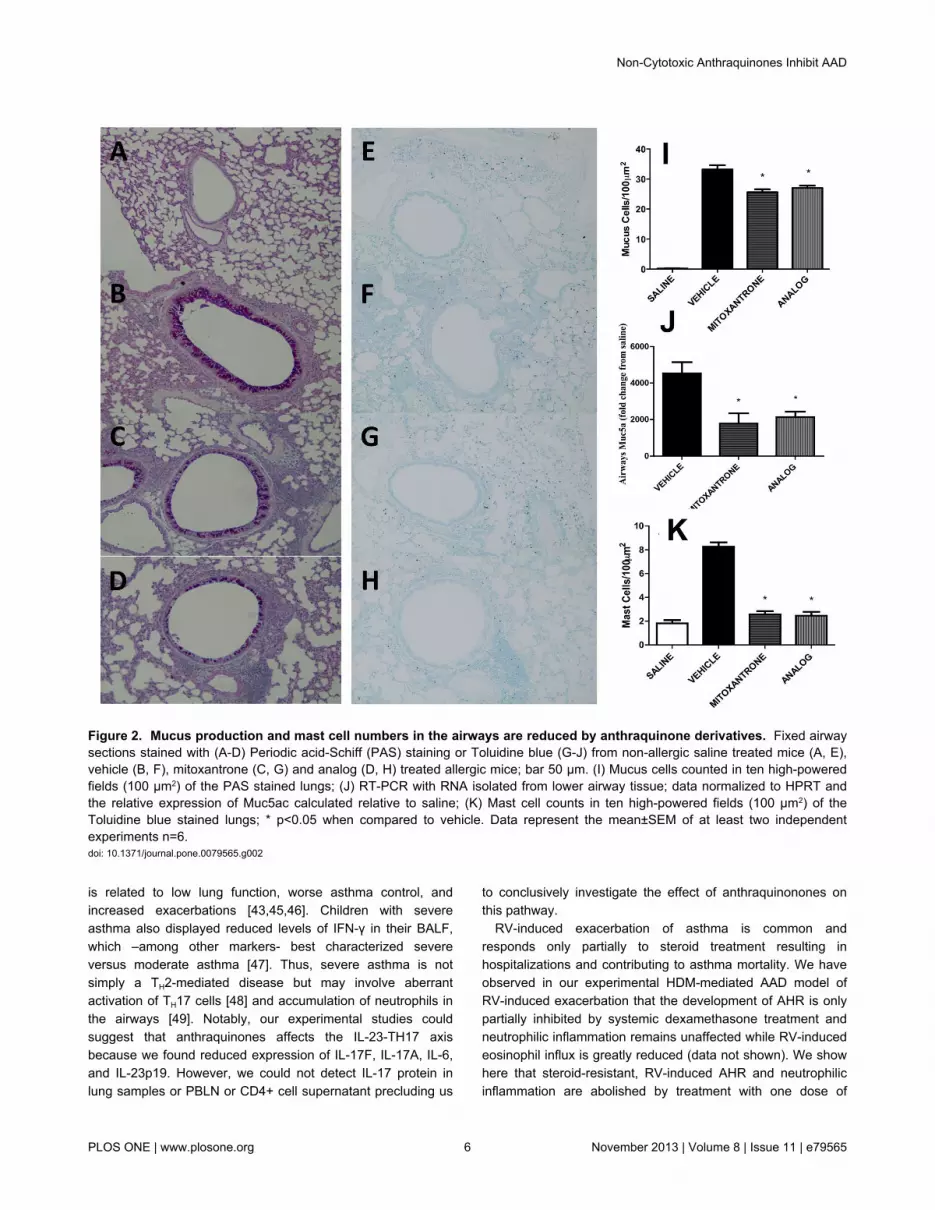
accumulated in the PBLN (Figure 1F). Mucus hypersecretion,Muc5ac expression, and mast cell influx were also significantlyreduced upon mitoxantrone or analog treatment (Figure 2 A toK).
Anthraquinones impair expression of pro-inflammatorycytokines along with regulatory transcriptional factorsin the lung
IL-13 and IL-5 release from in-vitro HDM-stimulated PBLNcells was significantly impaired in allergic mice treated with theantraquinone analog while IFN-γ release was increased (Figure3 A to C). HDM-stimulated CD4+ T cells isolated from micetreated with the antraquinone analog released lower levels ofthe archetypal TH2 cytokines IL-4 and IL-13 (Figure 3 D to E).Expression of cytokines promoting TH17 immunity such asIL-17F, IL-17A, IL-6, and IL-23p19 were also reduced (Figure 3F to I) but we were not able to detect IL-17 protein in CD4+ Tcell or PBLN supernatants, or lung homogenates. Other pro-inflammatory factors such as TNF-α, CCL8 and CXCL10 werealso reduced in analog treated allergic mice (Figure 3J and K).Same results were observed with mitoxantrone-treated mice(data not shown). The reduction in TH2 and T17 cytokineexpression upon analog treatment was associated withimpaired expression of STAT6, STAT3, and ROR-γT (Figure4A to C).
p-AKT phosphorylation, HIF-1α and VEGF expressionare limited by anthraquinone treatment
In accordance with in vitro observations [26] anthraquinonetreatment of allergic mice reduced HIF-1α and VEGFexpression along with reduced levels of p-AKT and active PIP3
in the lungs (Figure 5A to D).
RV-induced exacerbation of AAD and RV replication arereduced by anthraquinone treatment
AHR is further exacerbated by RV1B infection of allergicmice as compared to mice exposed to UV-inactivated RV1B(Figure 6A). Notably, treatment with one dose of analog 24hrsbefore RV exposure resulted in marked attenuation of AHR tolevels that are comparable to allergic mice exposed to UV-inactivated RV1B (Figure 6A). This was associated withinhibition of RV-induced exacerbation of eosinophilic andneutrophilic airways inflammation (Figure 6B). Interestinglyanthraquinone treatment also impaired RV1B replication(Figure 6C) and increased expression of innate antiviral type 1IFNs (Figure 6D and E).
Discussion
In the present work, treatment with O,O´-didodecanoyl-1,4-dihydroxyanthraquinone, a new non-cytotoxic anthraquinonederivative and analog of mitoxantrone, reduced thecharacteristic hallmark features of AAD including AHR, airwaysinflammation with eosinophils, neutrophils and mast cells, T-cell recruitment into the lung, expression of TH2 and TH17transcriptional factors, release of TH2 cytokines, mucushypersecretion, and RV-induced exacerbation. Moreover,
Non-Cytotoxic Anthraquinones Inhibit AAD
PLOS ONE | www.plosone.org 4 November 2013 | Volume 8 | Issue 11 | e79565

anthraquinone treatment boosted antiviral IFN responses andlimited RV replication.
There is a need for improved therapies for patient withsevere asthma as it accounts for 5% to 10% of cases andcauses 50% of total asthma-related health costs [35]. Forinstance, humanized antibodies that ameliorate TH2 effectorpathways by blocking circulating IgE (omalizumab) or IL-5(mepolizumab) reduced exacerbations and need for other
medication and improved asthma control [37–41]. Anti-IL-13treatment (lebrikizumab) was associated with improved lungfunctions [42].
Severe asthma is also associated with increased IL-17Aproduction [43] and studies in experimental models suggest acritical role for complement-mediated regulation of the IL-23-TH17 axis by enhancing IL-13-driven responses [44]. IL-17-expressing cells may promote neutrophilic inflammation, which
Figure 1. Anthraquinone derivative suppresses AHR and inflammation. (A) Scheme of chemical synthesis of theanthraquinone derivative O,O´-didodecanoyl-1,4-dihydroxyanthraquinone. (B) AHR, (C) total and (D) differential number of BALFcells, (E) T (CD4, CD8) and B (CD19) cell numbers in lung homogenates and (F) peribronchial lymph node cells in HDM sensitizedand challenged mice treated with 1 mg/kg of mitoxantrone or analog. * p<0.05 when compared to vehicle. Data represent the mean±SEM of at least two independent experiments n=6.doi: 10.1371/journal.pone.0079565.g001
Non-Cytotoxic Anthraquinones Inhibit AAD
PLOS ONE | www.plosone.org 5 November 2013 | Volume 8 | Issue 11 | e79565
is related to low lung function, worse asthma control, andincreased exacerbations [43,45,46]. Children with severeasthma also displayed reduced levels of IFN-γ in their BALF,which –among other markers- best characterized severeversus moderate asthma [47]. Thus, severe asthma is notsimply a TH2-mediated disease but may involve aberrantactivation of TH17 cells [48] and accumulation of neutrophils inthe airways [49]. Notably, our experimental studies couldsuggest that anthraquinones affects the IL-23-TH17 axisbecause we found reduced expression of IL-17F, IL-17A, IL-6,and IL-23p19. However, we could not detect IL-17 protein inlung samples or PBLN or CD4+ cell supernatant precluding us
to conclusively investigate the effect of anthraquinonones onthis pathway.
RV-induced exacerbation of asthma is common andresponds only partially to steroid treatment resulting inhospitalizations and contributing to asthma mortality. We haveobserved in our experimental HDM-mediated AAD model ofRV-induced exacerbation that the development of AHR is onlypartially inhibited by systemic dexamethasone treatment andneutrophilic inflammation remains unaffected while RV-inducedeosinophil influx is greatly reduced (data not shown). We showhere that steroid-resistant, RV-induced AHR and neutrophilicinflammation are abolished by treatment with one dose of
Figure 2. Mucus production and mast cell numbers in the airways are reduced by anthraquinone derivatives. Fixed airwaysections stained with (A-D) Periodic acid-Schiff (PAS) staining or Toluidine blue (G-J) from non-allergic saline treated mice (A, E),vehicle (B, F), mitoxantrone (C, G) and analog (D, H) treated allergic mice; bar 50 μm. (I) Mucus cells counted in ten high-poweredfields (100 µm2) of the PAS stained lungs; (J) RT-PCR with RNA isolated from lower airway tissue; data normalized to HPRT andthe relative expression of Muc5ac calculated relative to saline; (K) Mast cell counts in ten high-powered fields (100 µm2) of theToluidine blue stained lungs; * p<0.05 when compared to vehicle. Data represent the mean±SEM of at least two independentexperiments n=6.doi: 10.1371/journal.pone.0079565.g002
Non-Cytotoxic Anthraquinones Inhibit AAD
PLOS ONE | www.plosone.org 6 November 2013 | Volume 8 | Issue 11 | e79565

Figure 3. TH1/2/17 cytokine and chemokine expression upon treatment with anthraquinone derivative. (A) IL-13, (B) IL-5,and (C) IFN-λ release from peribronchial lymph node cells cultured in the presence of HDM (50µg/ml). (D) IL-4 and (E) IL-13 releaseform CD4+ T-cells cultured in the presence of HDM (50µg/ml). RT-PCRs with RNA isolated from lower airway tissue; datanormalized to HPRT and the relative expression of (F) IL-17A, (G) IL-17F, (H) IL-6, (I) IL-23p19, (J) TNF-α, (K) CCL8 and (L)CXCL10 was calculated relative to saline. * p<0.05 when compared to vehicle. Data represent the mean±SEM of at least twoindependent experiments n=6.doi: 10.1371/journal.pone.0079565.g003
Non-Cytotoxic Anthraquinones Inhibit AAD
PLOS ONE | www.plosone.org 7 November 2013 | Volume 8 | Issue 11 | e79565

mitoxantrone analog before RV exposure. This effect may beexplained by a boost in IFN production with subsequentinhibition of RV replication (Figure 6) as a consequence oftherapeutically modulating TH cell effector pathways. Thisconclusion is supported by studies in asthmatics that showedan inverse relationship between RV replication and IFNproduction [50]. Alternatively the deficiency in IFN productionupon RV infection -which is observed in some asthmatics[15,16]- may promote aberrant T helper cell activation [51].
Kinases such as p38 mitogen-activated protein kinase, Junkinases and PI3K play a key role in regulating inflammatorygene expression in asthmatics [52]. PI3K activation also resultsin reduced steroid sensitivity through phosphorylation of AKTand subsequent reduced histone deacetylase 2 activity [53].PI3K inhibitors against isoforms attenuate antigen-inducedairway inflammation in murine models [29,54] and mightreverse steroid resistance in severe asthmatics [52].Unselective PI3K inhibitors however are likely to be toxic [55].
Mitoxantrone has recently been shown to block PI3Ksignaling through dephosphorylation of AKT in-vitro resulting inreduced HIF-1α and VEGF expression [26]. We have shownhere that a non-cytotoxic anthraquinone derivative attenuatesthe PI3K signaling pathway in-vivo despite its inability to disruptDNA synthesis and repair due to removal of the hydrogenmolecules during chemical synthesis (Figure 1A). Interestingly,Dang et al have recently demonstrated that HIF-1α plays animportant role in the alternate induction of TH17 cells [56].
Furthermore inhibition of HIF-1α attenuated OVA-induced AHRand inflammation via VEGF suppression in bronchial epithelium[57], which is a critical effector molecule in Th2 driven allergicdisease in the lung [58,59].
Recently Emodin and Citreorosein, two naturally occurringanthraquinone derivatives, have been shown to suppress IgE-mediated anaphylactic reaction, mast cell activation, andleukotriene generation [60,61]. This was associated withblockage of antigen-triggered phosphorylation of Syk, areceptor-proximal tyrosine kinase targeted by anthraquinones,which regulates the PI3K pathway through the adaptor proteinNTAL [62] and plays a key role in NF-κB-mediated expressionof COX-2 and pro-inflammatory cytokines.
Interestingly, Syk is also phosphorylated upon RV binding toits cell surface receptor and regulates clathrin-mediated RVendocytosis through the activation of the PI3K/AKT signalingpathway [63]. Our results highlight the in-vivo relevance of thispathway by demonstrating limited RV replication, AHR andairways inflammation in allergic mice treated withanthraquinone derivatives.
The PI3K/AKT signaling pathway is also activated by HDMallergen Der p1 binding to protease-activated receptors [64]and LPS (e.g. in crude HDM extract) sensing by TLR4 [65].Thus employing non-cytotoxic anthraquinone derivatives maybe of therapeutic benefit for the treatment of both allergen- andRV-triggered severe asthma (Figure 7).
Figure 4. Expression of transcription factors after anthraquinone derivative. BALB/c mice were sensitized and challengedwith HDM intranasally and treated with 1 mg/kg of analog. RT-PCRs with RNA isolated from lower airway tissue; data normalized toHPRT and the relative expression of (A) STAT6, (B) STAT3, (C) ROR-γT and (D) FOXP3 was calculated relative to expression insaline. * p<0.05 when compared to vehicle. Data represent the mean±SEM of at least two different experiments n=6.doi: 10.1371/journal.pone.0079565.g004
Non-Cytotoxic Anthraquinones Inhibit AAD
PLOS ONE | www.plosone.org 8 November 2013 | Volume 8 | Issue 11 | e79565

Figure 5. Phosporylated AKT, PIP3, HIF-1α, and VEGF expression after anthraquinone derivative treatment. (A) Protein lunglysates were isolated from allergic mice and levels of p-AKT1/2/3 were determined by western blotting. Signal strength of p-AKTwhen compared to total actin level. (B) PIP3 activity in lung lysates. (C-D) RT-PCRs with RNA isolated from lower airway tissue; datanormalized to HPRT and the relative expression of (C) HIF-1α and (D) VEGF was calculated to saline expression levels. * p<0.05when compared to vehicle. Data represent the mean±SEM of at least two independent experiments n=6.doi: 10.1371/journal.pone.0079565.g005
Non-Cytotoxic Anthraquinones Inhibit AAD
PLOS ONE | www.plosone.org 9 November 2013 | Volume 8 | Issue 11 | e79565

Figure 6. Rhinovirus-induced exacerbation of AAD is ameliorated by anthraquinone derivative treatment. 24hrs after thelast HDM challenge, allergic mice were infected with 50µl of RV1B (RV) or UV-inactivated RV1B (UVRV). (A) 24hrs after RV1Binfection AHR was determined. (B) Differential number of cells in the BALF. RT-PCRs with RNA isolated from lower airway tissue;data normalized to HPRT, (C) the absolute copy numbers of RV, and the relative expression of (D) IFN-α and (E) IFN-β wascalculated relative UVRV expression levels. * p<0.05 when compared to vehicle+RV. Data represent the mean±SEM of at least twoindependent experiments n=6.doi: 10.1371/journal.pone.0079565.g006
Non-Cytotoxic Anthraquinones Inhibit AAD
PLOS ONE | www.plosone.org 10 November 2013 | Volume 8 | Issue 11 | e79565

Figure 7. Proposed mechanism of O,O´-didodecanoyl-1,4-dihydroxyanthraquinone action. doi: 10.1371/journal.pone.0079565.g007
Non-Cytotoxic Anthraquinones Inhibit AAD
PLOS ONE | www.plosone.org 11 November 2013 | Volume 8 | Issue 11 | e79565
Acknowledgements
We appreciate the technical assistance from Ana Pereira deSiqueira, Jason Girkin, Leon Sokulsky, Jane Grehan, and thestaff from the Animal Care Facility of the contributing institutes.
Author Contributions
Conceived and designed the experiments: JM APF MVA CFCCCSA PSF. Performed the experiments: CCSA AC LH MPMM. Analyzed the data: CCSA AC LH MP JM. Contributedreagents/materials/analysis tools: JM SLJ APF MVA CFC.Wrote the manuscript: CCSA AC JM HCT PSF APF MVA CFC.
References
1. Dunn JJ, Woolstenhulme RD, Langer J, Carroll KC (2004) Sensitivity ofrespiratory virus culture when screening with R-mix fresh cells. J ClinMicrobiol 42: 79-82. doi:10.1128/JCM.42.1.79-82.2004. PubMed:14715735.
2. Bochner BS, Undem BJ, Lichtenstein LM (1994) Immunologicalaspects of allergic asthma. Annu Rev Immunol 12: 295-335. doi:10.1146/annurev.iy.12.040194.001455. PubMed: 8011284.
3. Kay AB (2005) The role of eosinophils in the pathogenesis of asthma.Trends Mol Med 11: 148-152. doi:10.1016/j.molmed.2005.02.002.PubMed: 15823751.
4. Broide DH (2008) Immunologic and inflammatory mechanisms thatdrive asthma progression to remodeling. J Allergy Clin Immunol 121:560-570. doi:10.1016/j.jaci.2008.01.031. PubMed: 18328887.
5. Lloyd C (2002) Chemokines in allergic lung inflammation. Immunology105: 144-154. doi:10.1046/j.1365-2567.2002.01344.x. PubMed:11872089.
6. Holgate ST, Roberts G, Arshad HS, Howarth PH, Davies DE (2009)The role of the airway epithelium and its interaction with environmentalfactors in asthma pathogenesis. Proc Am Thorac Soc 6: 655-659. doi:10.1513/pats.200907-072DP. PubMed: 20008870.
7. Lambrecht BN, Hammad H (2009) Biology of lung dendritic cells at theorigin of asthma. Immunity 31: 412-424. doi:10.1016/j.immuni.2009.08.008. PubMed: 19766084.
8. Wills-Karp M LJ, Xu X, Schofield B, Neben TY, Karp CL et al. (1998)Interleukin-13: central mediator of allergic asthma. Science 282:2258-2261. doi:10.1126/science.282.5397.2258. PubMed: 9856949.
9. Kuperman DA, Huang X, Koth LL, Chang GH, Dolganov GM et al.(2002) Direct effects of interleukin-13 on epithelial cells cause airwayhyperreactivity and mucus overproduction in asthma. Nat Med, 8: 885–9. PubMed: 12091879.
10. Mattes J, Yang M, Siqueira A, Clark K, MacKenzie J et al. (2001) IL-13induces airways hyperreactivity independently of the IL-4R alpha chainin the allergic lung. J Immunol 167: 1683-1692. PubMed: 11466392.
11. Johnston SL, Pattemore PK, Sanderson G, Smith S, Lampe F et al.(1995) Community study of role of viral infections in exacerbations ofasthma in 9-11 year old children. Bmj 310: 1225-1229. doi:10.1136/bmj.310.6989.1225. PubMed: 7767192.
12. Friedlander SL, Busse WW (2005) The role of rhinovirus in asthmaexacerbations. Clin Infect Dis 116: 267-273. PubMed: 16083778.
13. Sears MR (2008) Epidemiology of asthma exacerbations. J Allergy ClinImmunol 122: 662-670; quiz: 19014756.
14. Miller EK (2010) New human rhinovirus species and their significancein asthma exacerbation and airway remodeling. Immunol Allergy ClinNorth Am 30: 541-552, vii doi:10.1016/j.iac.2010.08.007. PubMed:21029937.
15. Wark PA, Johnston SL, Bucchieri F, Powell R, Puddicombe S et al.(2005) Asthmatic bronchial epithelial cells have a deficient innateimmune response to infection with rhinovirus. J Exp Med 201: 937-947.doi:10.1084/jem.20041901. PubMed: 15781584.
16. Contoli M, Message SD, Laza-Stanca V, Edwards MR, Wark PA et al.(2006) Role of deficient type III interferon-lambda production in asthmaexacerbations. Nat Med 12: 1023-1026. doi:10.1038/nm1462. PubMed:16906156.
17. Grünberg K, Sharon RF, Sont JK; In 't Veen JC, Van Schadewijk WA,et al. (2001) Rhinovirus-induced airway inflammation in asthma: effectof treatment with inhaled corticosteroids before and duringexperimental infection. Am J Respir Crit Care Med 164: 1816-1822. doi:10.1164/ajrccm.164.10.2102118. PubMed: 11734429. doi:10.1164/ajrccm.164.10.2102118 PubMed: 11734429
18. Leung DY, Szefler SJ (1998) New insights into steroid resistantasthma. Pediatr Allergy Immunol Off Publ European Society OfPediatric Allergy And Immunology 9: 3-12. doi:10.1111/j.1399-3038.1998.tb00293.x. PubMed: 9560836.
19. Leung DY, Szefler SJ (1997) Diagnosis and management of steroid-resistant asthma. Clin Chest Med 18: 611-625. doi:10.1016/S0272-5231(05)70405-6. PubMed: 9329880.
20. Alberts DS, Peng YM, Bowden GT, Dalton WS, Mackel C (1985)Pharmacology of mitoxantrone: mode of action and pharmacokinetics.Invest New Drugs 3: 101-107. PubMed: 4040505.
21. Nitiss JL (2009) Targeting DNA topoisomerase II in cancerchemotherapy. Nat Rev Cancer 9: 338-350. doi:10.1038/nrc2607.PubMed: 19377506.
22. Faulds D, Balfour JA, Chrisp P, Langtry HD (1991) Mitoxantrone. Areview of its pharmacodynamic and pharmacokinetic properties, andtherapeutic potential in the chemotherapy of cancer. Drugs 41:400-449. doi:10.2165/00003495-199141030-00007. PubMed: 1711446.
23. Smith IE (1983) Mitoxantrone (novantrone): a review of experimentaland early clinical studies. Cancer Treat Rev 10: 103-115. doi:10.1016/S0305-7372(83)80014-0. PubMed: 6347376.
24. Neuhaus O, Kieseier BC, Hartung HP (2004) Mechanisms ofmitoxantrone in multiple sclerosis--what is known? J Neurol Sci 223:25-27. doi:10.1016/j.jns.2004.04.015. PubMed: 15261556.
25. Fox EJ (2006) Management of worsening multiple sclerosis withmitoxantrone: a review. Clinical Therapeutics 28: 461-474. doi:10.1016/j.clinthera.2006.04.013. PubMed: 16750460.
26. Toh YM, Li TK (2011) Mitoxantrone inhibits HIF-1alpha expression in atopoisomerase II-independent pathway. Clin Cancer Res Off J AmAssoc Cancer Res 17: 5026-5037. doi:10.1158/1078-0432.CCR-11-0235.
27. Kwak YG, Song CH, Yi HK, Hwang PH, Kim JS et al. (2003)Involvement of PTEN in airway hyperresponsiveness and inflammationin bronchial asthma. J Clin Invest 111: 1083-1092. doi:10.1172/JCI16440. PubMed: 12671058.
28. Lee CT, Risom T, Strauss WM (2006) MicroRNAs in mammaliandevelopment. Birth Defects Res C Embryo TODAY 78: 129-139. doi:10.1002/bdrc.20072. PubMed: 16847889.
29. Takeda M, Ito W, Tanabe M, Ueki S, Kato H et al. (2009) Allergicairway hyperresponsiveness, inflammation, and remodeling do notdevelop in phosphoinositide 3-kinase γ–deficient mice. J Allergy ClinImmunol 123: 805-812. doi:10.1016/j.jaci.2008.11.047. PubMed:19232703.
30. Corrigan CJ, Wang W, Meng Q, Fang C, Wu H et al. (2011) T-helpercell type 2 (Th2) memory T cell-potentiating cytokine IL-25 has thepotential to promote angiogenesis in asthma. Proc Natl Acad Sci U S A108: 1579-1584. doi:10.1073/pnas.1014241108. PubMed: 21205894.
31. Simoes DC, Xanthou G, Petrochilou K, Panoutsakopoulou V, RoussosC et al. (2009) Osteopontin deficiency protects against airwayremodeling and hyperresponsiveness in chronic asthma. Am J RespirCrit Care Med 179: 894-902. doi:10.1164/rccm.200807-1081OC.PubMed: 19234104.
32. Kim JH, Choi C, Benveniste EN, Kwon D (2008) TRAIL induces MMP-9expression via ERK activation in human astrocytoma cells. BiochemBiophys Res Commun 377: 195-199. doi:10.1016/j.bbrc.2008.09.095.PubMed: 18834856.
33. Newcomb DC, Sajjan US, Nagarkar DR, Wang Q, Nanua S et al.(2008) Human rhinovirus 1B exposure induces phosphatidylinositol 3-kinase-dependent airway inflammation in mice. Am J Respir Crit CareMed 177: 1111-1121. doi:10.1164/rccm.200708-1243OC. PubMed:18276942.
34. Newcomb DC, Sajjan U, Nanua S, Jia Y, Goldsmith AM et al. (2005)Phosphatidylinositol 3-kinase is required for rhinovirus-induced airwayepithelial cell interleukin-8 expression. J Biol Chem 280: 36952-36961.doi:10.1074/jbc.M502449200. PubMed: 16120607.
35. Thomson NC, Chaudhuri R, Spears M (2011) Emerging therapies forsevere asthma. BMC Med 9: 102. doi:10.1186/1741-7015-9-102.PubMed: 21896202.
36. Weckmann M, Collison A, Simpson JL, Kopp MV, Wark PAB et al.(2007) Critical link between TRAIL and CCL20 for the activation of TH2cells and the expression of allergic airway disease. Nat Med 13:1308-1315. doi:10.1038/nm1660. PubMed: 17934471.
37. Busse W, Corren J, Lanier BQ, McAlary M, Fowler-Taylor A et al.(2001) Omalizumab, anti-IgE recombinant humanized monoclonal
Non-Cytotoxic Anthraquinones Inhibit AAD
PLOS ONE | www.plosone.org 12 November 2013 | Volume 8 | Issue 11 | e79565

antibody, for the treatment of severe allergic asthma. J Allergy ClinImmunol 108: 184-190. doi:10.1067/mai.2001.117880. PubMed:11496232.
38. Busse WW, Morgan WJ, Gergen PJ, Mitchell HE, Gern JE et al. (2011)Randomized trial of omalizumab (anti-IgE) for asthma in inner-citychildren. N Engl J Med 364: 1005-1015. doi:10.1056/NEJMoa1009705.PubMed: 21410369.
39. Lanier B, Bridges T, Kulus M, Taylor AF, Berhane I et al. (2009)Omalizumab for the treatment of exacerbations in children withinadequately controlled allergic (IgE-mediated) asthma. J Allergy ClinImmunol 124: 1210-1216. doi:10.1016/j.jaci.2009.09.021. PubMed:19910033.
40. Haldar P, Brightling CE, Hargadon B, Gupta S, Monteiro W (2009)Mepolizumab and exacerbations of refractory eosinophilic asthma. NEngl J Med 360: 973-984. doi:10.1056/NEJMoa0808991. PubMed:19264686.
41. Nair P, Pizzichini MM, Kjarsgaard M, Inman MD, Efthimiadis A et al.(2009) Mepolizumab for prednisone-dependent asthma with sputumeosinophilia. N Engl J Med 360: 985-993. doi:10.1056/NEJMoa0805435. PubMed: 19264687.
42. Corren J, Lemanske RF, Hanania NA, Korenblat PE, Parsey MV et al.(2011) Lebrikizumab Treatment in Adults with Asthma. N Engl J Med 3:3. PubMed: 21812663.
43. Al-Ramli W, Prefontaine D, Chouiali F, Martin JG, Olivenstein R et al.(2009) TH17-associated cytokines (IL-17A and IL-17F)in severeasthma. J Allergy Clin Immunol 123.
44. Lajoie S, Lewkowich IP, Suzuki Y, Clark JR, Sproles AA et al. (2010)Complement-mediated regulation of the IL-17A axis is a central geneticdeterminant of the severity of experimental allergic asthma. NatImmunol 11: 928-935. doi:10.1038/ni.1926. PubMed: 20802484.
45. Simpson JL, Scott R, Boyle MJ, Gibson PG (2006) Inflammatorysubtypes in asthma: assessment and identification using inducedsputum. Respirology 11: 54-61. doi:10.1111/j.1440-1843.2006.00784.x.PubMed: 16423202.
46. Hastie AT, Moore WC, Meyers DA, Vestal PL, Li H et al. (2010)Analyses of asthma severity phenotypes and inflammatory proteins insubjects stratified by sputum granulocytes. J Allergy Clin Immunol 125:1028-1036 e1013 doi:10.1016/j.jaci.2010.02.008. PubMed: 20398920.
47. Fitzpatrick AM, Higgins M, Holguin F, Brown LA, Teague WG (2010)The molecular phenotype of severe asthma in children. J Allergy ClinImmunol 125: 851-857 e818 doi:10.1016/j.jaci.2010.01.048. PubMed:20371397.
48. Wang Q, Miller DJ, Bowman ER, Nagarkar DR, Schneider D et al.(2011) MDA5 and TLR3 initiate pro-inflammatory signaling pathwaysleading to rhinovirus-induced airways inflammation andhyperresponsiveness. PLOS Pathog 7: e1002070. PubMed: 21637773.
49. Wenzel SE, Szefler SJ, Leung DY, Sloan SI, Rex MD et al. (1997)Bronchoscopic evaluation of severe asthma. Persistent inflammationassociated with high dose glucocorticoids. Am J Respir Crit Care Med156: 737-743. doi:10.1164/ajrccm.156.3.9610046. PubMed: 9309987.
50. Message SD, Laza-Stanca V, Mallia P, Parker HL, Zhu J et al. (2008)Rhinovirus-induced lower respiratory illness is increased in asthma andrelated to virus load and Th1/2 cytokine and IL-10 production. Proc NatlAcad Sci U S A 105: 13562-13567. doi:10.1073/pnas.0804181105.PubMed: 18768794.
51. Papadopoulos NG, Stanciu LA, Papi A, Holgate ST, Johnston SL(2002) A defective type 1 response to rhinovirus in atopic asthma.Thorax 57: 328-332. doi:10.1136/thorax.57.4.328. PubMed: 11923551.
52. Barnes PJ (2012) Severe asthma: advances in current managementand future therapy. J Allergy Clin Immunol 129: 48-59. doi:10.1016/j.jaci.2011.11.006. PubMed: 22196524.
53. To Y, Ito K, Kizawa Y, Failla M, Ito M et al. (2010) Targetingphosphoinositide-3-kinase-delta with theophylline reversescorticosteroid insensitivity in chronic obstructive pulmonary disease.Am J Respir Crit Care Med 182: 897-904. doi:10.1164/rccm.200906-0937OC. PubMed: 20224070.
54. Lee KS, Park SJ, Kim SR, Min KH, Jin SM et al. (2006)Phosphoinositide 3-kinase-δ inhibitor reduces vascular permeability ina murine model of asthma. J Allergy Clin Immunol 118: 403-409. doi:10.1016/j.jaci.2006.04.041. PubMed: 16890765.
55. Rommel C, Camps M, Ji H (2007) PI3K delta and PI3K gamma:partners in crime in inflammation in rheumatoid arthritis and beyond?Nat Rev Immunol 7: 191-201. doi:10.1038/nri2036. PubMed:17290298.
56. Dang Eric V, Barbi J, Yang H-Y, Jinasena D, Yu H et al. (2011) Controlof TH17/Treg Balance by Hypoxia-Inducible Factor 1. Cell 146:772-784. doi:10.1016/j.cell.2011.07.033. PubMed: 21871655.
57. Kim SR, Lee KS, Park HS, Park SJ, Min KH et al. (2010) HIF-1αinhibition ameliorates an allergic airway disease via VEGF suppressionin bronchial epithelium. Eur J Immunol 40: 2858-2869. doi:10.1002/eji.200939948. PubMed: 20827786.
58. Lee CG, Ma B, Takyar S, Ahangari F, DelaCruz C et al. (2011) Studiesof Vascular Endothelial Growth Factor in Asthma and ChronicObstructive Pulmonary Disease. Proc Am Thorac Soc 8: 512-515. doi:10.1513/pats.201102-018MW. PubMed: 22052929.
59. Lee CG, Link H, Baluk P, Homer RJ, Chapoval S et al. (2004) Vascularendothelial growth factor (VEGF) induces remodeling and enhancesTH2-mediated sensitization and inflammation in the lung. Nat Med 10:1095-1103. doi:10.1038/nm1105. PubMed: 15378055.
60. Lu Y, Yang JH, Li X, Hwangbo K, Hwang SL et al. (2011) Emodin, anaturally occurring anthraquinone derivative, suppresses IgE-mediatedanaphylactic reaction and mast cell activation. Biochem Pharmacol 82:1700-1708. doi:10.1016/j.bcp.2011.08.022. PubMed: 21907188.
61. Lu Y, Li Y, Jahng Y, Son JK, Chang HW (2012) Citreorosein inhibitsdegranulation and leukotriene C(4) generation through suppression ofSyk pathway in mast cells. Mol Cell Biochem 365: 333-341. doi:10.1007/s11010-012-1273-3. PubMed: 22395859.
62. Kambayashi T, Koretzky GA (2007) Proximal signaling events in Fcepsilon RI-mediated mast cell activation. J Allergy Clin Immunol 119:544-553; quiz: 17336609.
63. Lau C, Wang X, Song L, North M, Wiehler S et al. (2008) Sykassociates with clathrin and mediates phosphatidylinositol 3-kinaseactivation during human rhinovirus internalization. J Immunol 180:870-880. PubMed: 18178826.
64. Shi J, Luo Q, Chen F, Chen D, Xu G et al. (2010) Induction of IL-6 andIL-8 by house dust mite allergen Der p1 in cultured human nasalepithelial cells is associated with PAR/PI3K/NFkappaB signaling. ORLJ Otorhinolaryngol Relat Spec 72: 256-265. doi:10.1159/000312687.PubMed: 20733339.
65. Bauerfeld CP, Rastogi R, Pirockinaite G, Lee I, Hüttemann M et al.(2012) TLR4-mediated AKT activation is MyD88/TRIF dependent andcritical for induction of oxidative phosphorylation and mitochondrialtranscription factor A in murine macrophages. J Immunol 188:2847-2857. doi:10.4049/jimmunol.1102157. PubMed: 22312125.
Non-Cytotoxic Anthraquinones Inhibit AAD
PLOS ONE | www.plosone.org 13 November 2013 | Volume 8 | Issue 11 | e79565


