Embed Size (px)
Citation preview

at SciVerse ScienceDirect
Biomaterials 33 (2012) 4443e4450
Contents lists available
Biomaterials
journal homepage: www.elsevier .com/locate/biomater ia ls
Effect of surface-functionalized nanoparticles on the elongation phaseof beta-amyloid (1e40) fibrillogenesis
Ho-Man Chan a, Lehui Xiao a, Kai-Ming Yeung a, See-Lok Ho a, Dan Zhao a, Wing-Hong Chan a,Hung-Wing Li a,b,*aDepartment of Chemistry, Hong Kong Baptist University, Kowloon Tong, Hong Kong, PR ChinabCentre for Surface Analysis and Research, Hong Kong Baptist University, Kowloon Tong, Hong Kong, PR China
a r t i c l e i n f o
Article history:Received 16 January 2012Accepted 6 March 2012Available online 27 March 2012
Keywords:NanoparticleSelf-assemblyPeptideSurface modification
* Corresponding author. Department of Chemistry, HKowloon Tong, Hong Kong, PR China. Tel.: þ852 3411
E-mail address: [email protected] (H.-W. Li).
0142-9612/$ e see front matter � 2012 Elsevier Ltd.doi:10.1016/j.biomaterials.2012.03.024
a b s t r a c t
The influence of nanoparticles of various sizes and surface functionalities on the self-assembling fibril-logenesis of beta-amyloid (1e40) peptide was investigated. Functionalized nanoparticles includingquantum dots and gold nanoparticles were co-incubated with monomeric Ab1e40 peptides under seed-mediated growth method to study their influences on the elongation phase of the fibrillogenesis. It isobserved that charge-to-surface area ratio of the nanoparticles and the functional moiety and electro-static charges of the conjugated ligands on the particle surfaces took crucial regulatory role in the Ab1e40
fibrillogenesis.� 2012 Elsevier Ltd. All rights reserved.
1. Introduction
Alzheimer’s disease (AD) is a neurodegenerative disorderprimarily found in elderly. The formation of self-assemblingneurotoxic fibrous beta-amyloid (Ab) plaques in brain is believedas a pathological hallmark of AD [1e3]. Controlling the nucleation/elongation-growth processes of monomeric Ab peptides intoinsoluble oligomers, protofibrils and fibrils are reckoned asa potential therapy to AD [4,5]. While nanomaterials are nowwidely designed and applied as drug delivering vehicles for ther-apeutics and diagnostics purposes [6], precautionary selection ofnanomaterials is highly recommended as the vehicle itselfmaybe cytotoxic in nature otherwise aggravated the conditions.Several recent studies have provided insight that nanomaterialsof various sizes, shapes, compositions and functionalities maysignificantly intervene with the self-assembling mechanism ofamyloid peptides, drastically either promoting or inhibiting thefibrillogenesis [7e14]. For instance, Wu et al. reported that 20 nmTiO2 nanoparticles could promote the growth of Ab by shorteningthe nucleation process [12]. Cabaleiro-Lago et al. demonstrated thatco-polymeric NIPAM:BAM nanoparticles of 40 nm with differenthydrophobicities inhibited the growth of Ab fibrils by adsorbing the
ong Kong Baptist University,7065; fax: þ852 3411 7348.
All rights reserved.
monomers onto the particle surface and thus depleting the solutionmonomer concentration [9]. They have also reported that cationicamino-modified polystyrene nanoparticles of 57e180 nm inducedboth acceleration and retardation effects on Ab fibrillation based onthe concentration and coverage of peptide monomers on theparticle surfaces [8]. More recently, Yoo and coworkers have pre-sented that functionalized quantum dots (QDs) non-specificallyinteracted with the Ab monomers, interrupting the nucleationprocess and consequently inhibiting the growth of Ab fibrils [14].Also, a recent paper from Majzik and coworkers has drawn to ourattention that the covalent interaction between the gold nano-particles and the cysteine-modified beta-amyloid peptideshindered the formation of the polypeptide chain structure [15].There were also numerous reports that drawn our attentiondescribing the roles of nanomaterials on the controlled fibrillationof other self-assembling proteins and peptides, such as humanserum albumin (HSA) [11], islet amyloid polypeptide (IAPP) [7], andhuman b2-microglobulin (b2m) [10]. In view of the reported results,it is believed that surface areas, compositions and functionalities ofnanoparticles play significant regulatory role on controlling the Abfibrillation process and worth in depth investigations.
Accordingly, the behind mechanism of interactions betweennanoparticles and peptide monomers were still not clearly under-stood. Researchers proposed that particles acted to reduce therate of nucleation, yet the elongation phase were unaffected oncecritical nuclei were formed [9]. On the contrary, our previouswork illustrated that N-acetyl-L-cysteine-capped quantum dots

H.-M. Chan et al. / Biomaterials 33 (2012) 4443e44504444
(NAC-QDs) with hydrodynamic diameter of ca. 3 nm could effec-tively quench both nucleation and elongation process of the Abfibrillogenesis at any time points in the seed-mediated growth ofbeta amyloid (1e40) (Ab1e40) by blocking active sites of the seedfibrils or monomers [13]. The inhibitory effect is concentrationdependent, and a remarkable inhibition towards 50 mM of Abpeptide was observed when the dosage of NAC-QDs increased from10�9 to 10�7
M. This preliminary result showed that nanomaterialsnot only regulate the nucleation but also the elongation phase ofbeta-amyloid fibrillogenesis.
Based on our previous findings that small inorganic nano-particles may disturb Ab1e40 fibrillogenesis, we explored the effectof inorganic nanoparticles of various properties on regulating theelongation of Ab1e40 fibrils under physiological conditions. Here,nanoparticles were co-incubated with the monomeric Ab1e40peptides for seed-mediated growth such that the elongationphase in the amyloidogenesis was dominant. Water-soluble CdTeQDs of different sizes (ca. 2e4 nm) and gold nanoparticles (AuNPs)of ca. 15 nmwere adopted as the nanoparticles of interest. The QDsand AuNPs were synthesized and capped with the thiolated ligandsN-acetyl-L-cysteine (NAC) and 3-mercaptopropionic acid (MPA) viathe previously reported hydrothermal method [16] and modifiedFrens method [17] respectively. The effect of particle sizes, surfacecharges, functionality and compositions toward beta-amyloidfibrillogenesis was investigated. The as-formed fibrils were char-acterized with total internal reflection fluorescence microscopy(TIRFM), for visualizing the length of the Thioflavin T (ThT) labeledfibrils and monitoring the real-time fibril growth; and transmissionelectron microscopy (TEM), for studying the morphology andinteraction between nanoparticles and fibrils. These imagingtechniques provide advantages over conventional ThT fluorescenceassay with smaller sample volume consumption; and better capa-bility to reveal fibril length, density and general morphologicalstructures of each single fibril instead of bulk fluorescence intensityreadout [18]. Furthermore, plasmonic nanoparticles of larger sizemay significantly scatter incident light and absorb the emittedfluorescence from ThT due to their broad absorption wavelengthsrespectively.
This work studied the critical regulatory roles of both thenanoparticles and surface functionalized ligands on the extent offibrillation and the fibril morphologies of the Ab1e40 peptides in theelongation phase. Particles of different dimensions, materials andsurface charges were applied to interact with the peptide mono-mers and seeds. The fibrillogenesis of Ab1e40 peptides controlled inthe presence of nanomaterials was then monitored with spec-troscopy and microscopy techniques.
2. Materials and methods
2.1. Materials
Tellurium (reagent powder, 99.8%), cadmium chloride hydrate (CdCl2�H2O),sodium borohydride (NaBH4), hydrogen tetrachloroaurate (III) hydrate (HAuCl4),trisodium citrate, 3-mercaptopropionic acid (MPA), N-acetyl-L-cysteine (NAC), Thi-oflavin T (ThT), sodium phosphate monobasic and sodium phosphate dibasic werepurchased from SigmaeAldrich. All chemicals were used as receivedwithout furtherpurification. Beta-amyloid (1e40) (Ab1e40) was purchased from Invitrogen (Lot:25315-01S, Biosource, Camarillo CA, USA).
2.2. Synthesis and characterization of MPA- and NAC-capped quantum dots
The detail procedures to synthesize high quality water soluble NAC, MPA-capped CdTe quantum dots (QD) were similar to those reported before [16]. Atypical experiment procedure for synthesizing NAC capped CdTe QDs was describedas follow. In brief, the ice-cold aqueous NaHTe solution was prepared by mixingNaBH4 (80 mg) with Te (127 mg) at a molar ratio of 2 : 1 in DI water (Millipore, USA)in the presence of N2. CdCl2 (12.5 mmol/L) and NAC (15 mmol/L) were mixedtogether in 40 mL DI water in an ice-cold bath for 30 min. The pH of this precursorsolution was adjusted to pH 9.5 with NaOH. Freshly prepared NaHTe solution was
then injected to the N2 saturated precursor solutionwithmolar ratio of Cd/NAC/Te at1 : 1.2 : 0.2. Finally, 35 mL of the mixture solution was added into a 40 mL Teflon-lined stainless steel autoclave and kept at 200 �C for 30 min. To remove thoseunreacted reagents, cold 2-propanol was added to the reaction mixture to precipi-tate NAC-capped CdTe QDs and then rinsed with DI water. The UVevisible adsorp-tion and fluorescence spectra were measured by a Cary 300 UVevisiblespectrophotometer (Varian, Inc., USA) and a Perkin Elmer LS-50B luminescencespectrometer (Buckinghamshire, U.K.) respectively. The concentration of NAC-capped CdTe QDs was determined according to the method reported before [19].Zeta potential of these QDs was measured by Zetasizer Nano (Malvern InstrumentsLtd., U.K.).
2.3. Preparation of citrate-, MPA- and NAC-stabilized gold nanoparticles
Distilled water used in the preparation of solution and synthesis of nano-particles and was filtered with 0.22 mm nylon membrane twice prior to use. Allglasswares were cleaned with aqua regia and rinsed thoroughly with distilled waterbefore use. Citrate stabilized gold nanoparticle (citrate-AuNP) was synthesized bythe modified Frens method [17]. A 50 mL of solution containing 1 mM HAuCl4 wasbrought to boil. Under continuous heating and vigorous stirring, 5 mL of 38.8 mM
sodium citrate was added to the vortex of the stirring solution. Boiling wascontinued for 10 min and the colour of the solution change from pale yellow to redwine. The heating source was removed and stirring was continued for an additional15 min. The solution was cooled to room temperature. Gold nanoparticles func-tionalized with MPA and NAC were prepared by place exchange reactions. In brief,the pH of the as-prepared citrate-AuNP was tuned to approximately pH 7 withammonia solution. The AuNP hydrosols were then added with 5 � 10�5
M MPA andNAC dissolved in absolute ethanol under vigorous stirring respectively. The solutionwas stirred for 2 h at room temperature. UVevis spectroscopy measurements of theAuNP solution yielded an absorbance maximum centered at ca. 520 nm (Fig. S2) andthe size of the nanoparticles was characterized by TEM measurement (Fig. S3). Theconcentrations of all synthesized AuNP were estimated using the absorbance of thesurface plasmon resonance peak [20].
2.4. Preparation of beta-amyloid (1e40) fibrils
The beta-amyloid (1e40) (Ab1e40) fibrils were prepared as reported previously[13,21]. Stock Ab1e40 solutionwas prepared by dissolving in 400 mL of ice-cold 0.02%ammonia solution and stored at �80 �C before use. To prepare the seed fibrils, thestock solution was diluted with to 57.7 mM with 50 mM phosphate buffer (pH 7.4,with 100 mM NaCl). After a brief sonication (about 5e10 s), the reaction solutionwasincubated at 37 �C water bath for 24 h. The reaction solution was centrifuged at 4 �Cfor 1 h at 1.6� 104g. The supernatant solutionwas discarded and the precipitate wasresuspended in phosphate buffer with 0.05% NaN3 and stored at �18 �C. Prior toeach experiment, sample of the seed fibrils was sonicated thrice for 5 s and was thenadded to the phosphate buffer solution containing monomer peptide (57.7 mM) witha final concentration of ca. 10 mg/mL. The reaction solution was incubated in a waterbath at 37 �C for 1 h. The fibril was diluted with buffer solution to appropriateconcentration and labeled with the thioflavin T (ThT) dye. In general, 5 mL of dilutedand labeled fibril solution was sandwiched between a pair of cover glasses (No.1,22 � 22 sq. mm, Menzel-Gläser, Germany, pre-cleaned with NaOH and distilledwater) for imaging under fluorescence microscope.
In the QDs and AuNPs modulation experiments, different size and ligand pro-tected CdTe QDs and AuNPs of 10 nM was added to the seed-mediated beta-amyloidpeptide reaction mixture solution and incubated at 37 �C. For the TIRFM assay,sample solution of 1 mL was aliqouted, diluted with buffer solution and labeled withThT in the time-lapse experiment at different time points (t ¼ 0, 15, 30, 45, 60,120 min) for kinetics monitoring. For the ThT fluorescence measurement assay, theAb peptide (50 mM), Ab seeds and gold nanoparticles (10 nM)were suspended in ThTsolution instead of PB buffer, and incubated in the quartz cuvette at 37 �Cwith gentleagitation. The fluorescence signals from the as-formed ThT labeled fibrils weremeasured in the luminescence lifetime scanning spectrometer (PTI Time MasterModel C-720) every 10 min up to 2 h.
2.5. Fluorescence imaging and data analysis
The prism-type total internal reflection fluorescence microscopic imagingsystem was similar to the setup described before [13,21]. In brief, a fused silicaIsosceles Brewster Prism (CVI Laser, USA) was mounted on an Olympus IX71inverted microscope. An evanescent field was generated on the interface betweenthe sample solution and the surface of a cover glass. A 445 nm diode laser (Newport,USA) was used to excite the fluorescent labeling dye ThT binding to the amyloidfibrils. A band pass filter (HQ 480/40, Chroma Technology Corp., USA) and an oil-immersion 60 � (PlanApo, N.A. 1.42) objective were used. Fluorescence imageswere captured by an electron-multiplying charge-coupled device (EMCCD) camera(PhotonMax 512, Princeton Instrument, USA) with 100 ms exposure time. The pixelsize of the EMCCD is 16 mm. The length of the fibrils was measured with the free-domain software Image J (http://rsbweb.nih.gov/ij/). A hundred fibrils weremeasured with the Freehand line and Measure function of the software in each

H.-M. Chan et al. / Biomaterials 33 (2012) 4443e4450 4445
condition. The distribution of real-time fibril length of Ab was fitted with the soft-ware OriginPro 8 and Igor Pro.
2.6. Transmission electron microscopy (TEM)
Fibril sample and AuNP solutions of 5 mL was dropped on a carbon-coatedcopper grid (T200H-Cu, Electron Microscopy Sciences, Washington, USA) anddried for 5 min. The grid with amyloid fibrils was further negatively stained with 2%uranyl acetate for another 5 min. The dried sample was examined by a Technai G2Transmission Electron Microscope (FEI, USA) with an acceleration voltage of 200 kV,and a JEM 2100 TEM (JEOL, Japan) with an acceleration voltage of 210 kV for highsolution TEM images respectively.
3. Results and discussions
3.1. Regulation of beta-amyloid growth by functionalized quantumdots of various sizes
CdTe QDs of various sizes were synthesized to investigate thesize effect of nanoparticles on amyloid fibrillogenesis. MPA and NACwere functionalized onto the surface of the QDs as stabilizers. Withrespect to the pKa value of the eCOOH group of the surface-bounded MPA (pKa z 6.6) and NAC (pKa z 4.1) [22,23], thenanoparticles were anionic at physiological pH. The negativelycharged carboxylate end groups of the ligands allowed welldispersion and better stabilization of QDs by electrostatic repulsion.Characterization of the MPA- and NAC-QDs was shown in Table 1.The sizes and concentrations of the QDs were estimated from theUVevis absorption spectra [19], and the zeta-potential measure-ments confirmed that all QDs were stable in aqueous solution withnegative charges. As according to our previous study, functional-ized QDs (ca. 3 nm) showed threshold response to the fibrillation ofAb1e40 ranging from 10�9 to 10�7
M. In this work, the concentrationof nanoparticles was kept constant at 10�8
M to investigate theeffect of nanoparticle sizes and surface charges to the regulation ofAb growth. As demonstrated in our previous work that elongationprocess of Ab1e40 fibrils was completed in an hour under seed-mediated growth [13,21], the incubation time of the mixture ofAb1e40 peptides and nanoparticles was kept at 1 h throughoutunless otherwise specified.
Fig. 1 showed the fluorescence and TEM images of Ab1e40 fibrilsgrew in the presence of MPA- and NAC-QDs of three sizes respec-tively. It revealed that the amyloid fibrillation was inhibited by10 nM of 2.8 nm NAC-QDs, however, not by 2.2 nm MPA-QDs(Fig. 1A and D). This effect could be explained by the same argu-ment as proposed in our previous work that NAC, as comparedwithMPA, offers more hydrogen bonding (H-bond) sites to interactwith Ab1e40 peptides [13]. The small NAC-QDs blocked the elon-gation site of the Ab1e40 peptides and seeds, and prohibited theself-assembling fibrillation. The 2.2 nm MPA-QDs, albeit owningsimilar charges as the 2.8 nm NAC-QDs, did not observably affectthe growth of Ab1e40 fibrils. Nevertheless, the inhibitory effect was
Table 1Absorption maximum, calculated diameter and zeta potential of MPA and NAC-capped CdTe quantum dots in aqueous solution.
lmax/nm Diameter/nma z/mV
MPA-QD 1 494 2.2 �25.2MPA-QD 2 542 3.1 �36.6MPA-QD 3 577 3.5 �29.8NAC-QD 1 519 2.8 �29.6NAC-QD 2 536 3.1 �39.9NAC-QD 3 589 3.8 �28.7
a Theoretical diameter of the stock QDs were calculated according to the equationreported previously, in which d ¼ (9.8127 � 10�7)lmax
3 � (1.7147 � 10�3)lmax2 þ (1.0064)lmax � 194.84 [18], in which lmax is the maximum absorptionwavelength.
noticed when the concentration of MPA-QDs increased to 10�7M or
higher (data not shown). We presumed that MPA possess a weakerinteraction with the Ab1e40 peptide than NAC, and hence the MPA-QDs were relatively less effective in inhibiting the fibrillogenesisalthough bearing similar surface charges and sizes as the NAC-QDs.Seemingly, surface ligands possessed more H-bond sites that mayinteract with the peptides.
There was no inhibition observed on the growth of Ab1e40peptides in the presence of medium size MPA- and NAC-QDs. Thefibrillation patterns of Ab1e40 fibrils, regarding for the fibril lengthand fibril number, grew in the presence of medium size 3.1 nmNAC-QDs and 3.1 nm MPA-QDs (Fig. 1B and E) were very similar tothe control fibrils. Both NAC- and MPA-QDs of 3.1 nm wereconcluded neither fibrillation inhibitor nor promoter. In principle,the number of ligands conjugated on the particle surfaces, and thusthe available H-bond sites for interaction with peptides, shouldincrease with the extended size and surface area of the AuNPs. Asevidenced from the zeta-potential measurements, the medium sizeMPA- and NAC-QDs carried more negative surface charges than thesmall QDs, confirming that more ligands were bound onto thesurface of the particles. However, it is revealed the relatively highdensity of negative charges on the surface of the particles may repelthe Ab1e40 peptides with net charges of 3� and thus retarded theinteractions between QDs and Ab1e40. The above results provedthat despite the increase in sites for H-bond interactions shouldtheoretically further facilitate the blockage of amyloid elongationsites, the high density of surface charges generated also by theelevated number of surface ligands may counteract the conceivablehydrogen bonds between particles and peptides, and accordinglyoffset the inhibitory effect of the NAC- and MPA- QDs to thefibrillogenesis.
Unexpectedly, TIRFM imaging observed that Ab1e40 fibrillo-genesis was promoted in the batches of Ab1e40 peptides incubatedwith larger size of NAC-QDs (3.8 nm) and MPA-QDs (3.5 nm)respectively. The measured zeta potentials of the large QDs werehigher than those of medium size. We presumed that the charge tosurface area ratio of the conjugated QDwas reduced in the presenceof limited amount of ligands but enlarged particle sizes, and thus,the electrostatic repulsions of large QDs to peptides were attenu-ated. Here, we found the peptides spontaneously assembled intolarge and high-density aggregates with large QDs (Fig. 1C and F). Inorder to validate for the formation of the amyloid clusters in thepresence of large QDs, the real-time seed-mediated fibrillation ofAb1e40 peptides was monitored. Fluorescence images were taken att ¼ 0, 15, 30, 45 and 60 min respectively as shown in Fig. 2.
Large fibrillar aggregates appeared notably earlier in the pres-ence of QDs, meaning that the interactions between the QDs andfibrils were highly favorable. To review if the QDs interrupted thefibrillation rate of Ab1e40, the length of a hundred control and QDs-interacted fibrils were measured from the TIRFM images respec-tively. The number of pixels occupied by the fibrils allowed esti-mation of the length of each Ab1e40 fibril. It was worth noting thatthe individual cluster-forming fibrils cannot be well resolved underfluorescence microscopy as they appeared as a single large brightcluster, and thus the length of the fibrils inside the cluster cannot beanalyzed while only the length of the free non-clustered fibrilswere measured and compared in this work. Hereby, the lengths offibrils in the presence of MPA-QDs (6.7 mm � 0.04 mm,mean� standard error of mean, n¼ 100, hereinafter) and NAC-QDs(8.0 mm � 0.1 mm) were slightly shorter than the control fibrils(10.4 mm � 0.3 mm) after 1 h of incubation. We expected thatthe QDs, which have similar sizes as the Ab seeds, maybe largeenough to serve as additional nucleation sites. Monomeric Ab1e40interacted through hydrogen bonds and adsorbed onto the func-tionalized surface of the QDs. The concentration of monomeric

Fig. 1. Fluorescence (left) and TEM (right) images of Ab1e40 fibrils formed after incubating (A) 2.8 nm, (B) 3.1 nm, (C) 3.8 nm NAC-capped QDs; and (D) 2.2 nm, (E) 3.1 nm, (F) 3.5 nmMPA-capped QDs with Ab1e40 seed mediated growth solution for 1 h at 37 �C. Scale bar in fluorescence and TEM images are 16 mm and 0.2 mm respectively.
Fig. 2. Real-time monitoring of the seed-mediated fibrillation of Ab1e40 with and without the addition of QDs. Occurrence of large Ab1e40 aggregates was earlier in the presence ofboth MPA- and NAC-capped QDs compared to the control. Scale bar is 20 mm.
H.-M. Chan et al. / Biomaterials 33 (2012) 4443e44504446

H.-M. Chan et al. / Biomaterials 33 (2012) 4443e4450 4447
Ab1e40 in free solution was reduced [8], and thus relatively shorterfibrils were generated in the presence of large QDs.
3.2. Regulation of beta-amyloid growth by functionalized goldnanoparticles
To further explore the factors controlling the Ab1e40 growth andthe role of nanoparticles played in the regulation of fibrillogenesis,we extended the size scale of the nanoparticles from QDs smallerthan 5 nm to gold nanoparticles of 15 nm in the study. The surfacecharges and functional ligands on the surface of the particles werefound to be comparable to those used in the study of QDs-Ab1e40interaction. Recent studies on the effect of particles towards thegrowth of protein fibril limited in mostly polymeric or semi-conducting metallic nanoparticles of drastic sizes (either below5 nm or above 40 nm) [8e11]. However, the interaction betweenamyloid peptide and metallic nanoparticles ranging from 10 to40 nm, which were commonly used in bioassays, were not repor-ted. AuNP is chosen to interact with amyloid peptide for its well-known biocompatibility, high monodispersity and simply fabrica-tion method compared to other nanoparticles [24,25]. Further-more, the derivatization of AuNP with different ligands such asaliphatic thiols and cysteine residues through AueS bondwerewellestablished and hence the surface charges and functionality of theAuNP could be easily regulated [26,27]. Previous report revealedthat protein such as bovine serum albumin (BSA) appeared to bindspontaneously to the surface of citrate-stabilized gold nano-particles (citrate-AuNP) even though both BSA and citrate-AuNPwere negatively charged and should repel from each other atphysiological pH [28]. It was proposed that the positive lysineresidues of BSA interact strongly with the negative charge from thecitrate ion by salt-bridge interaction and facilitated strong binding.Similarly, monomeric Ab1e40 peptides, although with a net chargeof 3e, bearing three positive and 18 hydrophobic residues shouldhave high potential to bind to the AuNP surface through hydrogenbondings or other electrostatic interactions and hence intervenedwith the fibrillation mechanism of beta-amyloid.
Herein, monomeric Ab1e40 peptides were co-incubated with15 nmAuNPs stabilized by citrate, MPA and NAC into amyloid fibrilsusing the same conditions as described in the former sectionrespectively. Since the acidity of the stock AuNP solution maydisturb the overall pH of the Ab1e40 growth solution and so doAuNP has a high flocculation rate at low pH [29], all AuNP solutionsused were therefore adjusted to approximately pH 7 prior toexperiments. Table 2 showed the characterization of the function-alized AuNPs used in this study. The conjugation of MPA and NACwas achieved by ligand displacement method, in which citrate ionelectrostatically stabilized on the AuNP surface was displaced by
Table 2Absorptionmaximum, calculated diameter, measured diameter and zeta potential ofcitrate, MPA and NAC-stabilized gold nanoparticles in aqueous solution at pH 7.
lmax/nm Diameter/nma Diameter/nmb z/mV
Citrate-AuNP 519 16.90 14.05 � 1.2 �38.7MPA-AuNP 520 16.10 14.49 � 1.2 �48.7NAC-AuNP 520 16.44 14.40 � 1.1 �25.0
a Theoretical diameter of the AuNP were calculated according to the equationreported previously, in which d ¼ [Aspr � 5.89 � 10�6/(Cau � e�4.75)](1/0.314) [19], inwhich Aspr is the absorbance of the surface plasmon resonance peak, Cau is the initialconcentration of the gold ion used in the synthesis.
b TEMmeasured diameter of the AuNP was verified by fitting the size distributionof 100 AuNPs with the Gaussian distribution and hence obtained the averagediameter. The TEM images and the size distributions of citrate-, MPA- andNAC-AuNP were shown in the supporting information.
the thiolated ligands respectively. The ligands covalently bound tothe surface of the AuNP through strong AueS bonds and resulted ina charged monolayer surface of AuNPs as proven by the decline inabsorbance in the UVevis absorption spectra (Fig. S2 in supportinginformation). The particles were remained spherical and mono-disperse after the surface modifications as evaluated by the TEMimaging. As shown in Fig. S3 in the supporting information, theMPA- and NAC-AuNPs shared comparable diameter as the stockcitrate-AuNP, displaying that the ligand exchange reaction did notreduce the stability of the nanoparticles. Neither flocculation wasobserved from TEM nor positive zeta-potentials (which implying tothe instability of the colloids) were measured in all batches ofsamples confirming that the AuNPs have both successfully func-tionalized with the thiolated ligands and possessed with highmonodispersity to interact with monomeric Ab1e40 peptides.
Preliminarily, the fibrillation kinetics of the seed-mediatedgrowth of 50 mM Ab1e40 peptides in the absence and presence of10�8
M citrate-, MPA- and NAC-AuNPs were studied with the typicalThT fluorescence assays. Fig. S4 showed the corresponding real-time fluorescence spectra of the amyloid fibrils grew in the pres-ence of ThT and the corresponding colloids. Fluorescenceenhancement at lem ¼ 480 nmwas observed over 2 h of incubationin all cases (Fig. S5) except in the presence of NAC-AuNP (Fig. S4D),indicating that citrate and MPA functionalized AuNPs did notinhibit the fibrillation. However, it is worth noting that the peakshapes for all nanoparticles-interacted Ab1e40 fibrils (Fig. S4BeD)was significantly different from that of the control (Fig. S4A),implicating that AuNPs may interfere with the spectral character-istics of the ThT fluorescence spectra by scattering the excitationlight source or absorbing the emitted ThT fluorescence (quenching)as mentioned. Therefore, we reckoned that one should neither (i)serve ThT fluorescence assay as the sole explanatory experiment tomanifest the regulatory role of AuNPs on Ab1e40 fibrillation, nor (ii)conclude that NAC-AuNPs limited the growth of Ab1e40 fibrils basedon the unexpected differences in ThT emission spectrum. Forextended verification on the fibril-particles interaction, fluores-cence and electron microscopic imaging were exercised forauthentic examination of the resultant fibril lengths andmorphologies.
Fig. 3 showed the length distributions, fluorescence and TEMimages of the Ab1e40 fibrils grew for 1 h in the absence andpresence of nanoparticles respectively. As similar to the case ofQDs, fluorescence images showed that Ab has a tendency to for-m cluster-like aggregates in the presence of nanoparticles. Theappearance frequency and density of the amyloid cluster wasin the following order: NAC-AuNP > Citrate-AuNP > MPA-AuNP > Control. The cluster formation was induced by the intro-duction of the functionalized nanoparticles rather than the ligandcompounds, as proven by the control experiments incubatingligand and Ab1e40 peptides only that no cluster-like aggregateswere observed in the fibrils grew from the Ab1e40 peptides withsodium citrate, MPA and NAC respectively (Fig. S6). The real-timegrowth of amyloid fibrils in the absence and presence of func-tionalized AuNPs were shown in Fig. 4. The fibril length measuredin fluorescence images showed no significant deviation amongcontrol and the existence of three different nanoparticles. The 1-h-incubated fibril lengths were measured to be 11.1 mm � 0.2 mm(control), 11.0 mm � 0.2 mm (citrate-AuNPs interacted fibrils),8.6 mm � 0.1 mm (MPA-AuNPs interacted fibrils) and10.5 mm � 0.2 mm (NAC-AuNPs interacted fibrils) respectively. Theoverall distribution of the length of 100 fibrils surveyed in each ofthe concerned conditions did not significantly deviated from that ofcontrol (Fig. S7e10). Although the surface charges of the AuNPswere similar to that of the QDs, the variation in length was notas obvious as the case of QDs since the size of the AuNPs are larger
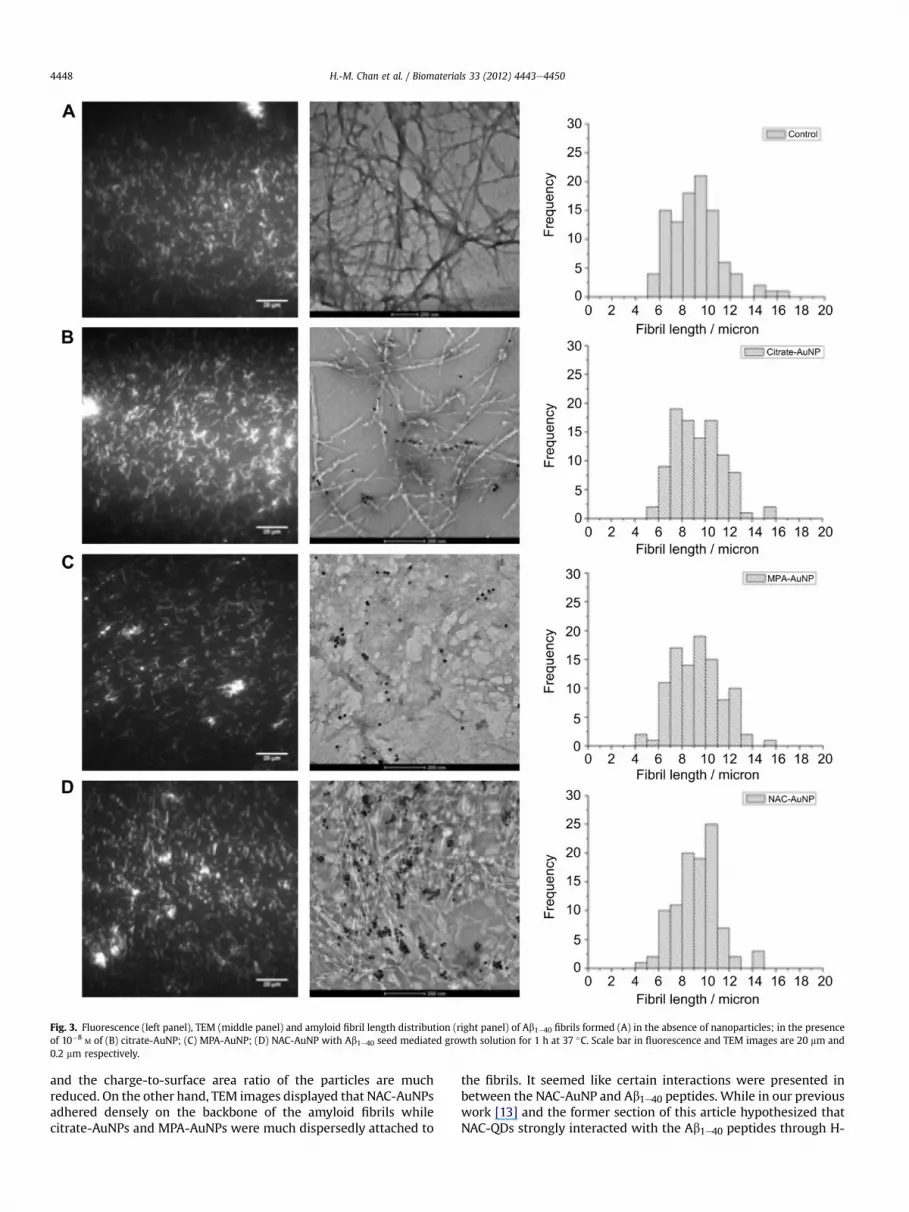
Fig. 3. Fluorescence (left panel), TEM (middle panel) and amyloid fibril length distribution (right panel) of Ab1e40 fibrils formed (A) in the absence of nanoparticles; in the presenceof 10�8
M of (B) citrate-AuNP; (C) MPA-AuNP; (D) NAC-AuNP with Ab1e40 seed mediated growth solution for 1 h at 37 �C. Scale bar in fluorescence and TEM images are 20 mm and0.2 mm respectively.
H.-M. Chan et al. / Biomaterials 33 (2012) 4443e44504448
and the charge-to-surface area ratio of the particles are muchreduced. On the other hand, TEM images displayed that NAC-AuNPsadhered densely on the backbone of the amyloid fibrils whilecitrate-AuNPs and MPA-AuNPs were much dispersedly attached to
the fibrils. It seemed like certain interactions were presented inbetween the NAC-AuNP and Ab1e40 peptides. While in our previouswork [13] and the former section of this article hypothesized thatNAC-QDs strongly interacted with the Ab1e40 peptides through H-

Fig. 4. Fibrillation kinetics of Ab1e40 was monitored by measuring the fibril length at different time points by TIRFM under the addition of functionalized AuNPs at 10�8M: (B) no
particles, (,) Citrate-AuNP, (C) MPA-AuNP, and (-) NAC-AuNP. The fibrillation kinetics follows the pseudo first order kinetics.
H.-M. Chan et al. / Biomaterials 33 (2012) 4443e4450 4449
bond, we herein testified if the proposed mechanismwas also validfor NAC-AuNPs.
The occurrence of hydrogen bonding interactions betweenfunctionalized AuNPs andmonomeric Ab1e40 peptides were provedby UVevis spectroscopy. Fig. S11 showed the UVevis spectra offunctionalized AuNPs dispersed in (i) DMSO, (ii) DMSO with Ab1e40peptides, (iii) ethanol (EtOH), and (iv) EtOH with Ab1e40 peptidesrespectively. Here, no notable differences in the spectral shape andthe lmax were displayed when functionalized AuNPs weredispersed in either DMSO or DMSO with monomeric Ab1e40peptides. We deduced that DMSO, as a strong polar aproticsolvent, interacted with the ligands and the Ab1e40 peptides indi-vidually. The solvent environment obstructed and shielded theinteractions between nanoparticles and peptides and hence, evenwith the addition of Ab1e40 monomers, no obvious change in lmaxof the AuNP spectrumwas achieved. Interestingly, the situationwastotally different when the particles were suspended in EtOH.Nanoparticles were found aggregated in EtOH since the surfacecharges of the functionalized AuNPs were neutralized. The particleswere agglomerated and resulted in broad UVevis spectra withlmax > 600 nm for both MPA-AuNP and NAC-AuNP. However, withthe addition of Ab1e40 monomers, the spectra maxima were wellpreserved at w530 nm. Comparing with the absorption maxima ofnanoparticles in EtOH only, a significant blue shift of 128 nm and96 nm in lmax was observed for MPA-AuNP and NAC-AuNPrespectively after exposing the colloids to monomeric Ab1e40peptides. It was expected that the peptide molecules adsorbed onthe surface of the nanoparticles provided extra stabilization to theparticles and avoided aggregation. While EtOH diluted the elec-trostatic charges of the particles, the potential interactions betweenthe functionalized AuNPs and the Ab peptides were therefore likelyto be hydrogen bondings.
The above arguments provided evidences that the interactionsbetween Ab1e40 peptides and AuNPs were highly correlated to theligand functionality and surface charges of the particles, in whichthe cluster-like aggregates were found in the batches of function-alized AuNP with more H-bond sites and less negative surfacecharges (e.g. NAC-AuNP). We inferred that the NAC-AuNP, whichoffers more hydrogen bonding sites and less negatively chargedsurface, favored a stronger interaction with the amyloid fibrils; on
the other hand, the citrate-AuNP and MPA-AuNP with highlynegative surface charges discouraged the interaction between theparticles and the Ab1e40 peptides. This hypothesis also explainedthe dissimilarity in spectral characteristics of the ThT fluorescencespectra of Ab1e40 peptides interacted with NAC-AuNPs (Fig. S4D).Since NAC conjugated strongly with the Ab1e40 peptides, theintermolecular distances between the ThT molecules and the NAC-AuNPs were expected to be close. The fluorescence emitted fromthe fibril-bound ThT maybe absorbed and quenched by the AuNPsand thus the fluorescence enhancement at lem ¼ 480 nm was notprojected as expected. It also supports that TIRFM imaging is moresuitable than conventional ThT fluorescence assays for the study ofamyloid growth involving other fluorescent dyes or nanoparticles.
Our results showed there is no significant influence on thelength and kinetics of Ab1e40 fibrillogenesis in the presenceof functionalized AuNPs. The phenomenon also agreed wellwith the mechanism proposed in the preceding reports fromCabaleiroeLago and coworkers [8]. Under a high peptide to nano-particle surface area ratio (5000 peptide against 1 nanoparticle),a remarkably large fraction of the Ab1e40 monomer was present inthe solution instead of on the particle surface. Thus, the fibrillationof Ab1e40 in solution, instead of that on particle surface, dominatedthe fibrillation pathway and hence the fibrillation patterns ofAb1e40 were not eminently disturbed in the presence of 15 nmAuNPs.
4. Conclusions
Nanoparticles with various sizes, surface charges and potentialhydrogen-bonding sites act differently in the elongation phase ofthe Ab1e40 fibrillogenesis. Slight change in sizes and charges ofsmall nanoparticles may induce contrary effect on amyloid fibril-lation. It has been shown that many nanoparticles are small enoughto pass through different barriers in the body and are regarded asvaluable therapeutic materials, for example, in the treatment ofAlzheimer’s disease [30]. Knowing that nanoparticles of variousfunctionalities play critical regulatory role on the fibrillation ofAb1e40 peptide, our findings provide insight that well-controllingthe surface functionality, size and concentration of nanoparticles

H.-M. Chan et al. / Biomaterials 33 (2012) 4443e44504450
is very crucial and essential in the development of nanomaterialsfor future biological and biomedical applications.
Acknowledgements
This work was fully supported by grants from the UniversityGrant Council of Hong Kong Special Administrative Region,China (HKBU 201208) and Faculty Research Grant from Hong KongBaptist University (FRG2/08-09/073) and (FRG2/09-10/037). TheFEI-Technai G2 TEM used in this work was supported by theCenter for Surface Analysis and Research (CSAR) with fundingfrom the Special Equipment Grant from the University GrantCommittee of the Hong Kong Special Administrative Region, China(SEG_HKBU06). We thank Mr. Benson Leung (HKBU) and Mr. RoyHo (HKUST) for TEM measurement. We also thank Dr. Kai-ChungLau and Miss Queeny Lo of the City University of Hong Kong forzeta-potential measurement.
Appendix A. Supplementary material
Supplementary material associated with this article can befound, in the online version, at doi:10.1016/j.biomaterials.2012.03.024.
References
[1] Caughey B, Lansbury PT. Protofibrils, pores, fibrils, and neurodegeneration:separating the responsible protein aggregates from the innocent bystanders.Annu Rev Neurosci 2003;26:267e98.
[2] Pepys MB. Amyloidosis. Annu Rev Med 2006;57:223e41.[3] Rauk A. The chemistry of alzheimer’s disease. Chem Soc Rev 2009;38:
2698e715.[4] Hamley IW. Peptide fibrillization. Angew Chem-Int Edit 2007;46:8128e47.[5] Selkoe DJ. Normal and abnormal biology of the beta-amyloid precursor
protein. Annu Rev Neurosci 1994;17:489e517.[6] Stark WJ. Nanoparticles in biological systems. Angew Chem Int Edit 2011;50:
1242e58.[7] Cabaleiro-Lago C, Lynch I, Dawson KA, Linse S. Inhibition of iapp and iapp
(20e29) fibrillation by polymeric nanoparticles. Langmuir 2010;26:3453e61.[8] Cabaleiro-Lago C, Quinlan-Pluck F, Lynch I, Dawson KA, Linse S. Dual effect of
amino modified polystyrene nanoparticles on amyloid beta protein fibrilla-tion. ACS Chem Neurosci 2010;1:279e87.
[9] Cabaleiro-Lago C, Quinlan-Pluck F, Lynch I, Lindman S, Minogue AM, Thulin E,et al. Inhibition of amyloid beta protein fibrillation by polymeric nano-particles. J Am Chem Soc 2008;130:15437e43.
[10] Linse S, Cabaleiro-Lago C, Xue WF, Lynch I, Lindman S, Thulin E, et al.Nucleation of protein fibrillation by nanoparticles. Proc Natl Acad Sci U S A2007;104:8691e6.
[11] Vannoy CH, Leblanc RM. Effects of dhla-capped cdse/zns quantum dots on thefibrillation of human serum albumin. J Phys Chem B 2010;114:10881e8.
[12] Wu WH, Sun X, Yu YP, Hu J, Zhao L, Liu Q, et al. Tio2 nanoparticles promotebeta-amyloid fibrillation in vitro. Biochem Biophys Res Commun 2008;373:315e8.
[13] Xiao LH, Zhao D, Chan WH, Choi MMF, Li HW. Inhibition of beta 1-40 amyloidfibrillation with n-acetyl-l-cysteine capped quantum dots. Biomaterials 2010;31:91e8.
[14] Yoo SI, Yang M, Brender JR, Subramanian V, Sun K, Joo NE, et al. Inhibition ofamyloid peptide fibrillation by inorganic nanoparticles: functional similaritieswith proteins. Angew Chem Int Edit 2011;50:5110e5.
[15] Majzik A, Fulop L, Csapo E, Bogar F, Martinek T, Penke B, et al. Functionali-zation of gold nanoparticles with amino acid, beta-amyloid peptides andfragment. Colloid Surf B-Biointerfaces 2010;81:235e41.
[16] Zhao D, He ZK, Chan WH, Choi MMF. Synthesis and characterization of high-quality water-soluble near-infrared-emitting cdte/cds quantum dots cappedby N-acetyl-L-cysteine via hydrothermal method. J Phys Chem C 2009;113:1293e300.
[17] Grabar KC, Freeman RG, Hommer MB, Natan MJ. Preparation and character-iation of au colloid monolayers. Anal Chem 1995;67:735e43.
[18] Ban T, Morigaki K, Yagi H, Kawasaki T, Kobayashi A, Yuba S, et al. Real-timeand single fibril observation of the formation of amyloid beta spheruliticstructures. J Biol Chem 2006;281:33677e83.
[19] Yu WW, Qu LH, Guo WZ, Peng XG. Experimental determination of theextinction coefficient of cdte, cdse, and cds nanocrystals. Chem Mat 2003;15:2854e60.
[20] Haiss W, Thanh NTK, Aveyard J, Fernig DG. Determination of size andconcentration of gold nanoparticles from UV-Vis spectra. Anal Chem 2007;79:4215e21.
[21] Man BYW, Chan HM, Leung CH, Chan DSH, Bai LP, Jiang ZH, et al. Group 9metal-based inhibitors of beta-amyloid (1e40) fibrillation as potential ther-apeutic agents for alzheimer’s disease. Chem Sci 2011;2:917e21.
[22] Choi MMF, Douglas AD, Murray RW. Ion-pair chromatographic separation ofwater-soluble gold monolayer-protected clusters. Anal Chem 2006;78:2779e85.
[23] Park JJ, Weiger MC, Lacerda S, Pristinski D, Becker ML, Douglas JF, et al.Characterization of non-equilibrium nanoparticle adsorption on a modelbiological substrate. Langmuir 2010;26:4822e30.
[24] Daniel MC, Astruc D. Gold nanoparticles: assembly, supramolecular chemistry,quantum-size-related properties, and applications toward biology, catalysis,and nanotechnology. Chem Rev 2004;104:293e346.
[25] Murphy CJ, Gole AM, Stone JW, Sisco PN, Alkilany AM, Goldsmith EC, et al.Gold nanoparticles in biology: beyond toxicity to cellular imaging. Acc ChemRes 2008;41:1721e30.
[26] Love JC, Estroff LA, Kriebel JK, Nuzzo RG, Whitesides GM. Self-assembledmonolayers of thiolates on metals as a form of nanotechnology. Chem Rev2005;105:1103e69.
[27] Weisbecker CS, Merritt MV, Whitesides GM. Molecular self-assembly ofaliphatic thiols on gold colloids. Langmuir 1996;12:3763e72.
[28] Brewer SH, GlommWR, Johnson MC, Knag MK, Franzen S. Probing bsa bindingto citrate-coated gold nanoparticles and surfaces. Langmuir 2005;21:9303e7.
[29] Mayya KS, Patil V, Sastry M. On the stability of carboxylic acid derivatized goldcolloidal particles: the role of colloidal solution ph studied by opticalabsorption spectroscopy. Langmuir 1997;13:3944e7.
[30] Liu G, Garrett MR, Men P, Zhu XW, Perry G, Smith MA. Nanoparticle and othermetal chelation therapeutics in alzheimer disease. Bba-Mol Basis Dis 2005;1741:246e52.


