Embed Size (px)
Citation preview
INFECTION AND IMMUNITY,0019-9567/01/$04.0010 DOI: 10.1128/IAI.69.2.931–936.2001
Feb. 2001, p. 931–936 Vol. 69, No. 2
Copyright © 2001, American Society for Microbiology. All Rights Reserved.
Inhibition of p38 Mitogen-Activated Protein Kinase AugmentsLipopolysaccharide-Induced Cell Proliferation in CD14-
Expressing Chinese Hamster Ovary CellsDIPSHIKHA CHAKRAVORTTY, YUTAKA KATO, TSUYOSHI SUGIYAMA, NAOKI KOIDE,
MYA MYA MU, TOMOAKI YOSHIDA, AND TAKASHI YOKOCHI*
Department of Microbiology and Immunology and Division of Bacterial Toxin, Research Center for Infectious Disease,Aichi Medical University School of Medicine, Nagakute, Aichi 480-1195, Japan
Received 7 August 2000/Returned for modification 28 September 2000/Accepted 8 November 2000
CD14-expressing Chinese hamster ovary (CD14-CHO) cells, established by transfection of human CD14DNA, acquired high responsiveness to lipopolysaccharide (LPS) through membrane-bound CD14 expression.LPS induced DNA synthesis and activated a series of mitogen-activated protein (MAP) kinases, extracellularsignal-regulated kinase 1/2 (Erk1/2), p38, and c-Jun N-terminal kinase/stress-activated protein kinase, inCD14-CHO cells but not in mock-transfected CHO cells. Anti-CD14 antibody completely abrogated bothLPS-induced DNA synthesis and LPS-induced phosphorylation of those MAP kinases, suggesting a critical roleof membrane-bound CD14 in LPS signaling. A p38 MAP kinase inhibitor, SB203580, markedly augmentedLPS-induced DNA synthesis in CD14-CHO cells, whereas an Erk1/2 inhibitor, PD98059, had no affect. On theother hand, SB203580 exhibited no effect on epidermal growth factor-induced DNA synthesis in CD14-CHOcells, although PD98059 inhibited it significantly. The activation and inactivation of p38 MAP kinase withdominant negative and dominant positive mutants also suggested the participation of p38 MAP kinase inLPS-induced DNA synthesis. It was therefore suggested that the activation of p38 MAP kinase can negativelyregulate LPS-induced cell proliferation in CD14-CHO cells.
Lipopolysaccharide (LPS) is present on the outer mem-branes of all gram-negative bacteria and known to cause thesystemic inflammatory response syndrome septic shock anddisseminated intravascular coagulation, leading finally to mul-tiorgan failure (1, 21, 24, 33). The activation of monocytes/macrophages by LPS leads to inflammatory response via vari-ous cellular signaling events. LPS binds to monocytes/macrophages via membrane-bound CD14, which is theglycosylphosphatidylinositol-linked glycoprotein (37). The in-tegration of membrane-bound CD14 renders various cell typeshighly sensitive to LPS. In fact, Chinese hamster ovary (CHO)cells which are transfected with the CD14 gene and expressmembrane-bound CD14 acquire the high responsiveness toLPS (7–9, 13–15, 18, 20, 22, 31, 38, 39). Membrane-boundCD14-expressing CHO (CD14-CHO) cells can respond to alow concentration of LPS and exhibit various responses, suchas release of arachidonic acid metabolites (8), translocation ofnuclear factor kappa B (NF-kB) (5, 9, 23) and production ofinterleukin 6 (10, 15), like LPS-responsive monocytes/macro-phages. CD14-CHO cells may provide an experimental systemuseful for LPS signaling. LPS signaling is transduced by intracel-lular signal pathways using NF-kB and a series of mitogen-acti-vated protein (MAP) kinases. In particular, the phosphorylationof three major MAP kinases i.e., extracellular signal-regulatedkinase 1/2 (Erk1/2), p38, and c-Jun N-terminal kinase/stress-acti-vated protein kinase (JNK/SAPK), is rapidly induced by LPS intarget cells such as macrophages (34, 35). There is no report on
the activation of MAP kinases in LPS-stimulated CD14-CHOcells. In the present study, we examined whether MAP kinaseswere activated in LPS-stimulated CD14-CHO cells and whatfunction the activation of MAP kinases had in those cells. Herewe discuss the relationship between the activation of p38 MAPkinases and cell proliferation in LPS-stimulated CD14-CHO cells.
MATERIALS AND METHODS
Materials. LPS from Escherichia coli O55:B5 and epidermal growth factor(EGF) were obtained from Sigma Chemical Co., St. Louis, Mo. SB203580,PD98059, and 5-nitro-2-(3-phenylpropylamino)benzoic acid (NPPB) were pur-chased from Calbiochem, San Diego, Calif. They were dissolved in dimethylsulfoxide and diluted in the culture medium. Anti-human CD14 antibody CLB-MON/1 was purchased from Nichirei (Tokyo, Japan).
Establishment of CD14-CHO cells. CHO-K1 fibroblasts, obtained from theAmerican Type Culture Collection (Manassas, Va.), were maintained in Ham’sF-12 (Sigma) containing 5% heat-inactivated fetal calf serum and antibiotics.The plasmid carrying human CD14 DNA was a kind gift from R. J. Ulevitch, TheScripps Research Institute, La Jolla, Calif. CHO cells were transfected with theCD14 plasmid by the lipofection method (8). CD14-CHO cells were selectedpositively by using anti-CD14 antibody-coated beads and further cultured withthe addition of Geneticin (750 mg/ml). CD14-CHO cells were maintained inHam’s F-12 with 5% fetal calf serum. CHO cells transfected by the control vectorplasmid served as mock-transfected control CHO cells.
Laser flow cytometric analysis of CD14 expression and LPS binding. CD14-CHO cells were incubated with a 1:200 dilution of fluorescein isothiocyanate(FITC)-conjugated anti-human CD14 monoclonal antibody (Coulter, Miami,Fla.) or 1 mg of FITC-conjugated LPS (Sigma) per ml at 4°C for 1 h. The cellswere washed and suspended in phosphate-buffered saline. Fluorescence wasanalyzed by a laser flow cytometer (FACScaliber; Becton Dickinson, San Jose,Calif.).
DNA synthesis. DNA synthesis in CD14-CHO cells was assayed by [3H]thy-midine incorporation into the nucleus. Cells (3 3 104/100 ml) were plated in96-well plates and incubated with LPS (1 mg/ml or various concentrations) in thepresence or absence of anti-CD14 antibody (10 mg/ml) for 24 h. [3H]thymidine(1 mCi/well; Amersham, London, England) was added to the cultures. Eighteenhours later, the cells were harvested on glass fiber filters Radioactivity was
* Corresponding author. Mailing address: Department of Microbi-ology and Immunology, Aichi Medical University School of Medicine,Aichi 480-1195, Japan. Fax: 81 (561) 63-9187. Phone: 81 (561) 62-3311.E-mail: [email protected].
931
determined as counts per minute in a Beckman b counter. In some experiments,CD14-CHO or mock-transfected control CHO cells were also pretreated with 20mM SB203580 or PD98059, or various concentrations of SB203580, for 1 h beforeLPS treatment.
Immunoblotting. CD14-CHO cells were seeded in 35-mm-diameter plasticdishes (4 3 105 cells/dish) and incubated with LPS (0.1 mg/ml) for 30 min. Cellswere lysed in the lysis buffer and boiled for 5 min at 100°C as described previously(3). Aliquots (20 mg/lane) containing equal amounts of protein were electropho-resed under reducing conditions in a 4 to 20% gradient polyacrylamide gel andtransferred to a polyvinylidene difluoride membrane filter. The membranes weretreated with 5% bovine serum albumin for 1 h to block nonspecific binding,rinsed, and incubated with a panel of rabbit polyclonal antibodies against Erk1/2,phospho-Erk1/2, p38, phospho-p38, JNK/SAPK, and phospho-JNK/SAPK (NewEngland Biolabs, Beverly, Mass.) for 1 h. The membranes were further treatedwith a 1:3,000 dilution of horseradish peroxidase-conjugated protein G for 1 h.Immune complexes were detected with an enhanced chemiluminescence sub-strate (New England Nuclear, Boston, Mass.) and exposed to Kodak XAR X-rayfilm. A prestained molecular weight standard kit (Nippon Bio-Rad, Tokyo,Japan) was used as a reference. In some experiments, CD14-CHO cells werepretreated with 20 mM SB203580 or PD98059 for 1 h before LPS treatment.
Transfection of dominant negative mutants of Erk1/2, p38, and JNK/SAPKand a dominant positive mutant of MKK6. Dominant negative mutants ofErk1/2 (17), p38 (17), and JNK/SAPK (16) and a dominant positive mutant ofMAP kinase kinase 6 (MKK6) (16) were kind gifts from J. D. Lee, The ScrippsResearch Institute. CD14-CHO cells were transfected with 5 mg of dominantnegative mutants by the Lipofectamine (GIBCO-BRL, Gaithersburg, Md.)method (16, 17) for 8 h, and then the culture medium was replaced by Ham’sF-12 containing 5% fetal calf serum. After 48 h of incubation, transfected cellswere used for experiments. CD14-CHO cells were also transfected with a dom-inant positive mutant of MKK6 (5 mg/ml). To confirm overexpression of trans-fected MKK6, phospho-p38 was analyzed by immunoblotting using anti-phos-pho-p38 antibody. Anti-Flag (M2) antibody (Sigma) was also used to testtransfection efficiency.
Statistical analysis. Results are expressed as means 6 standard deviations oftriplicate determinations. Statistical significance was determined by Student’s ttest.
RESULTS
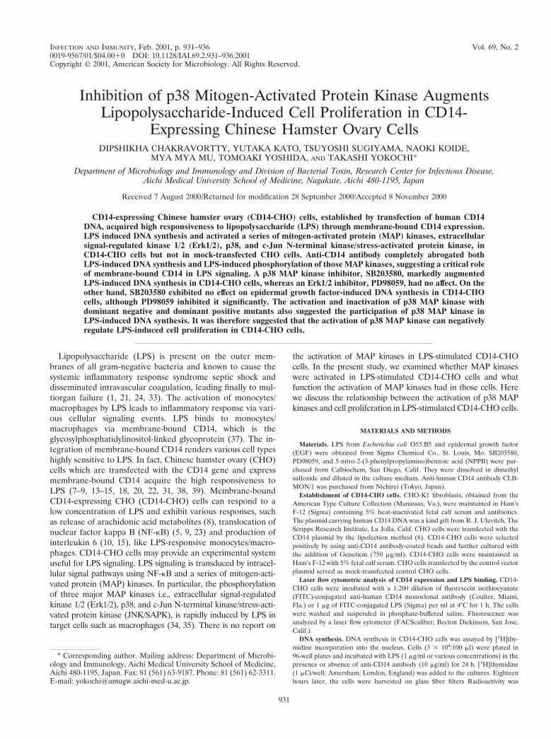
Establishment of CHO cells constitutively expressing mem-brane-bound CD14. The expression of membrane-boundCD14 on CD14-CHO cells, which were transfected with hu-man CD14 DNA, was analyzed by laser flow cytometry (Fig. 1).CD14-CHO cells expressed a high level of membrane-boundCD14. However, no significant CD14 expression was detectedon mock-transfected control CHO cells. In an assay of thebinding of FITC-conjugated LPS, the fluorescence intensity onCD14-CHO cells was approximately 10 times as high as that onmock-transfected CHO cells, suggesting enhanced binding ofLPS to CD14-CHO cells.
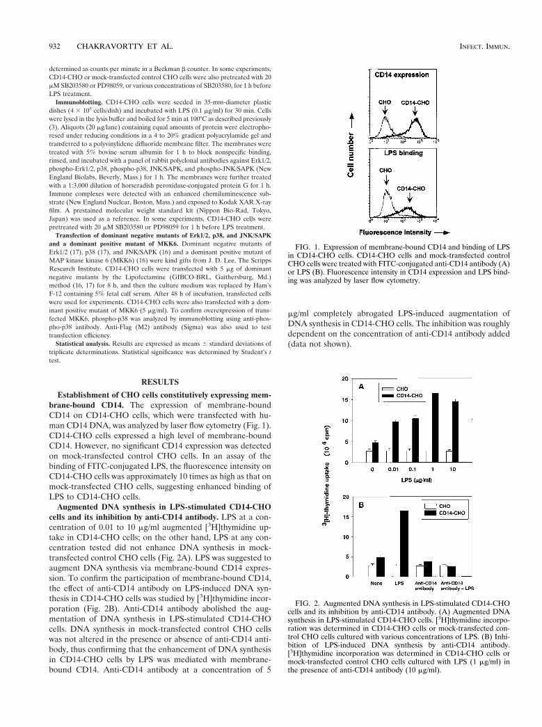
Augmented DNA synthesis in LPS-stimulated CD14-CHOcells and its inhibition by anti-CD14 antibody. LPS at a con-centration of 0.01 to 10 mg/ml augmented [3H]thymidine up-take in CD14-CHO cells; on the other hand, LPS at any con-centration tested did not enhance DNA synthesis in mock-transfected control CHO cells (Fig. 2A). LPS was suggested toaugment DNA synthesis via membrane-bound CD14 expres-sion. To confirm the participation of membrane-bound CD14,the effect of anti-CD14 antibody on LPS-induced DNA syn-thesis in CD14-CHO cells was studied by [3H]thymidine incor-poration (Fig. 2B). Anti-CD14 antibody abolished the aug-mentation of DNA synthesis in LPS-stimulated CD14-CHOcells. DNA synthesis in mock-transfected control CHO cellswas not altered in the presence or absence of anti-CD14 anti-body, thus confirming that the enhancement of DNA synthesisin CD14-CHO cells by LPS was mediated with membrane-bound CD14. Anti-CD14 antibody at a concentration of 5
mg/ml completely abrogated LPS-induced augmentation ofDNA synthesis in CD14-CHO cells. The inhibition was roughlydependent on the concentration of anti-CD14 antibody added(data not shown).
FIG. 1. Expression of membrane-bound CD14 and binding of LPSin CD14-CHO cells. CD14-CHO cells and mock-transfected controlCHO cells were treated with FITC-conjugated anti-CD14 antibody (A)or LPS (B). Fluorescence intensity in CD14 expression and LPS bind-ing was analyzed by laser flow cytometry.
FIG. 2. Augmented DNA synthesis in LPS-stimulated CD14-CHOcells and its inhibition by anti-CD14 antibody. (A) Augmented DNAsynthesis in LPS-stimulated CD14-CHO cells. [3H]thymidine incorpo-ration was determined in CD14-CHO cells or mock-transfected con-trol CHO cells cultured with various concentrations of LPS. (B) Inhi-bition of LPS-induced DNA synthesis by anti-CD14 antibody.[3H]thymidine incorporation was determined in CD14-CHO cells ormock-transfected control CHO cells cultured with LPS (1 mg/ml) inthe presence of anti-CD14 antibody (10 mg/ml).
932 CHAKRAVORTTY ET AL. INFECT. IMMUN.
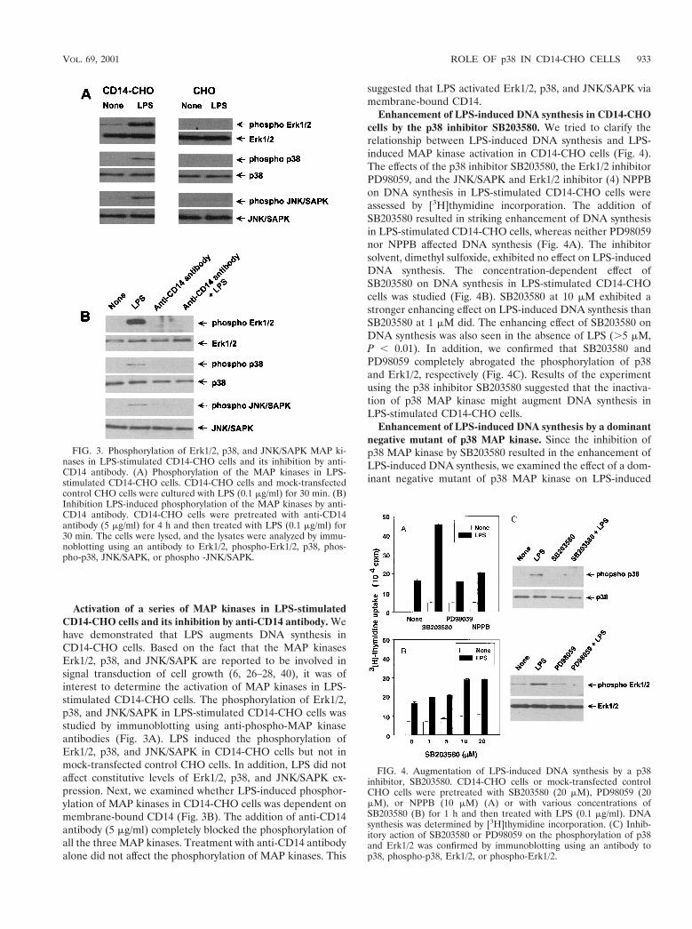
Activation of a series of MAP kinases in LPS-stimulatedCD14-CHO cells and its inhibition by anti-CD14 antibody. Wehave demonstrated that LPS augments DNA synthesis inCD14-CHO cells. Based on the fact that the MAP kinasesErk1/2, p38, and JNK/SAPK are reported to be involved insignal transduction of cell growth (6, 26–28, 40), it was ofinterest to determine the activation of MAP kinases in LPS-stimulated CD14-CHO cells. The phosphorylation of Erk1/2,p38, and JNK/SAPK in LPS-stimulated CD14-CHO cells wasstudied by immunoblotting using anti-phospho-MAP kinaseantibodies (Fig. 3A). LPS induced the phosphorylation ofErk1/2, p38, and JNK/SAPK in CD14-CHO cells but not inmock-transfected control CHO cells. In addition, LPS did notaffect constitutive levels of Erk1/2, p38, and JNK/SAPK ex-pression. Next, we examined whether LPS-induced phosphor-ylation of MAP kinases in CD14-CHO cells was dependent onmembrane-bound CD14 (Fig. 3B). The addition of anti-CD14antibody (5 mg/ml) completely blocked the phosphorylation ofall the three MAP kinases. Treatment with anti-CD14 antibodyalone did not affect the phosphorylation of MAP kinases. This
suggested that LPS activated Erk1/2, p38, and JNK/SAPK viamembrane-bound CD14.
Enhancement of LPS-induced DNA synthesis in CD14-CHOcells by the p38 inhibitor SB203580. We tried to clarify therelationship between LPS-induced DNA synthesis and LPS-induced MAP kinase activation in CD14-CHO cells (Fig. 4).The effects of the p38 inhibitor SB203580, the Erk1/2 inhibitorPD98059, and the JNK/SAPK and Erk1/2 inhibitor (4) NPPBon DNA synthesis in LPS-stimulated CD14-CHO cells wereassessed by [3H]thymidine incorporation. The addition ofSB203580 resulted in striking enhancement of DNA synthesisin LPS-stimulated CD14-CHO cells, whereas neither PD98059nor NPPB affected DNA synthesis (Fig. 4A). The inhibitorsolvent, dimethyl sulfoxide, exhibited no effect on LPS-inducedDNA synthesis. The concentration-dependent effect ofSB203580 on DNA synthesis in LPS-stimulated CD14-CHOcells was studied (Fig. 4B). SB203580 at 10 mM exhibited astronger enhancing effect on LPS-induced DNA synthesis thanSB203580 at 1 mM did. The enhancing effect of SB203580 onDNA synthesis was also seen in the absence of LPS (.5 mM,P , 0.01). In addition, we confirmed that SB203580 andPD98059 completely abrogated the phosphorylation of p38and Erk1/2, respectively (Fig. 4C). Results of the experimentusing the p38 inhibitor SB203580 suggested that the inactiva-tion of p38 MAP kinase might augment DNA synthesis inLPS-stimulated CD14-CHO cells.
Enhancement of LPS-induced DNA synthesis by a dominantnegative mutant of p38 MAP kinase. Since the inhibition ofp38 MAP kinase by SB203580 resulted in the enhancement ofLPS-induced DNA synthesis, we examined the effect of a dom-inant negative mutant of p38 MAP kinase on LPS-induced
FIG. 3. Phosphorylation of Erk1/2, p38, and JNK/SAPK MAP ki-nases in LPS-stimulated CD14-CHO cells and its inhibition by anti-CD14 antibody. (A) Phosphorylation of the MAP kinases in LPS-stimulated CD14-CHO cells. CD14-CHO cells and mock-transfectedcontrol CHO cells were cultured with LPS (0.1 mg/ml) for 30 min. (B)Inhibition LPS-induced phosphorylation of the MAP kinases by anti-CD14 antibody. CD14-CHO cells were pretreated with anti-CD14antibody (5 mg/ml) for 4 h and then treated with LPS (0.1 mg/ml) for30 min. The cells were lysed, and the lysates were analyzed by immu-noblotting using an antibody to Erk1/2, phospho-Erk1/2, p38, phos-pho-p38, JNK/SAPK, or phospho -JNK/SAPK.
FIG. 4. Augmentation of LPS-induced DNA synthesis by a p38inhibitor, SB203580. CD14-CHO cells or mock-transfected controlCHO cells were pretreated with SB203580 (20 mM), PD98059 (20mM), or NPPB (10 mM) (A) or with various concentrations ofSB203580 (B) for 1 h and then treated with LPS (0.1 mg/ml). DNAsynthesis was determined by [3H]thymidine incorporation. (C) Inhib-itory action of SB203580 or PD98059 on the phosphorylation of p38and Erk1/2 was confirmed by immunoblotting using an antibody top38, phospho-p38, Erk1/2, or phospho-Erk1/2.
VOL. 69, 2001 ROLE OF p38 IN CD14-CHO CELLS 933
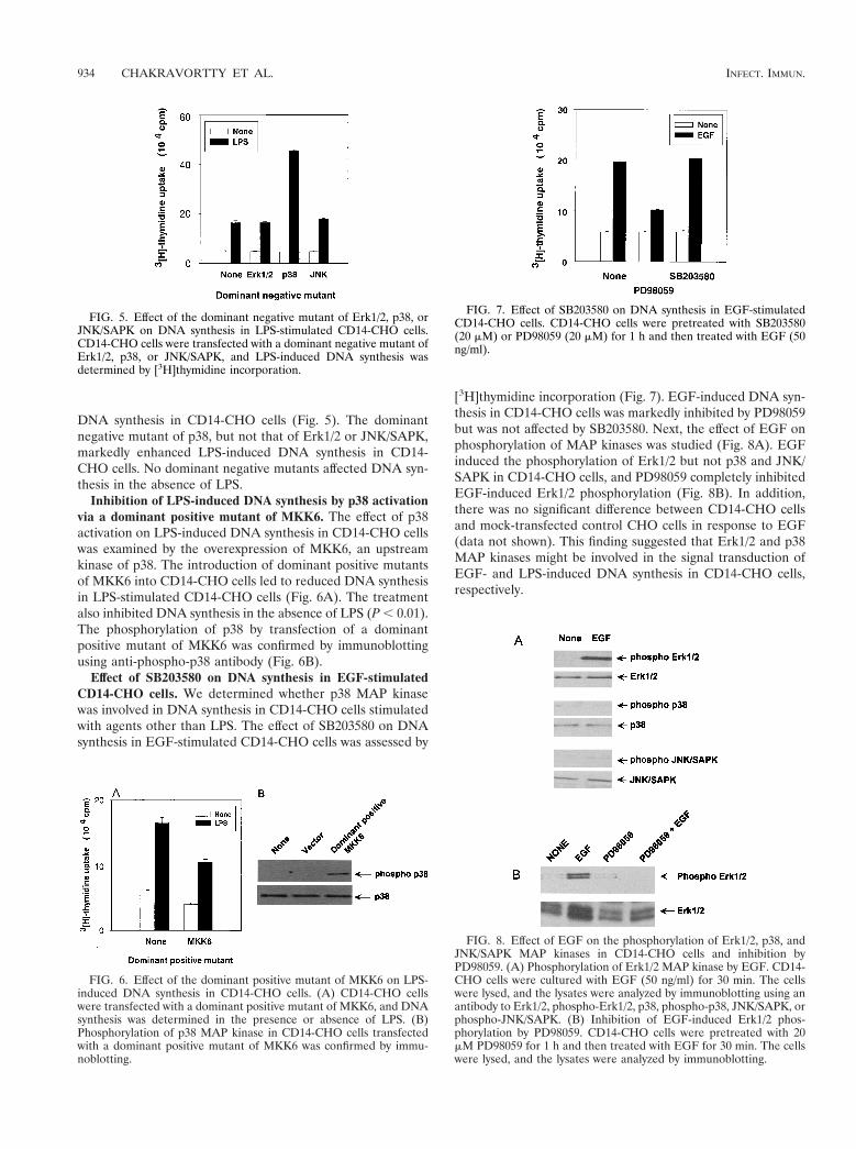
DNA synthesis in CD14-CHO cells (Fig. 5). The dominantnegative mutant of p38, but not that of Erk1/2 or JNK/SAPK,markedly enhanced LPS-induced DNA synthesis in CD14-CHO cells. No dominant negative mutants affected DNA syn-thesis in the absence of LPS.
Inhibition of LPS-induced DNA synthesis by p38 activationvia a dominant positive mutant of MKK6. The effect of p38activation on LPS-induced DNA synthesis in CD14-CHO cellswas examined by the overexpression of MKK6, an upstreamkinase of p38. The introduction of dominant positive mutantsof MKK6 into CD14-CHO cells led to reduced DNA synthesisin LPS-stimulated CD14-CHO cells (Fig. 6A). The treatmentalso inhibited DNA synthesis in the absence of LPS (P , 0.01).The phosphorylation of p38 by transfection of a dominantpositive mutant of MKK6 was confirmed by immunoblottingusing anti-phospho-p38 antibody (Fig. 6B).
Effect of SB203580 on DNA synthesis in EGF-stimulatedCD14-CHO cells. We determined whether p38 MAP kinasewas involved in DNA synthesis in CD14-CHO cells stimulatedwith agents other than LPS. The effect of SB203580 on DNAsynthesis in EGF-stimulated CD14-CHO cells was assessed by
[3H]thymidine incorporation (Fig. 7). EGF-induced DNA syn-thesis in CD14-CHO cells was markedly inhibited by PD98059but was not affected by SB203580. Next, the effect of EGF onphosphorylation of MAP kinases was studied (Fig. 8A). EGFinduced the phosphorylation of Erk1/2 but not p38 and JNK/SAPK in CD14-CHO cells, and PD98059 completely inhibitedEGF-induced Erk1/2 phosphorylation (Fig. 8B). In addition,there was no significant difference between CD14-CHO cellsand mock-transfected control CHO cells in response to EGF(data not shown). This finding suggested that Erk1/2 and p38MAP kinases might be involved in the signal transduction ofEGF- and LPS-induced DNA synthesis in CD14-CHO cells,respectively.
FIG. 5. Effect of the dominant negative mutant of Erk1/2, p38, orJNK/SAPK on DNA synthesis in LPS-stimulated CD14-CHO cells.CD14-CHO cells were transfected with a dominant negative mutant ofErk1/2, p38, or JNK/SAPK, and LPS-induced DNA synthesis wasdetermined by [3H]thymidine incorporation.
FIG. 6. Effect of the dominant positive mutant of MKK6 on LPS-induced DNA synthesis in CD14-CHO cells. (A) CD14-CHO cellswere transfected with a dominant positive mutant of MKK6, and DNAsynthesis was determined in the presence or absence of LPS. (B)Phosphorylation of p38 MAP kinase in CD14-CHO cells transfectedwith a dominant positive mutant of MKK6 was confirmed by immu-noblotting.
FIG. 7. Effect of SB203580 on DNA synthesis in EGF-stimulatedCD14-CHO cells. CD14-CHO cells were pretreated with SB203580(20 mM) or PD98059 (20 mM) for 1 h and then treated with EGF (50ng/ml).
FIG. 8. Effect of EGF on the phosphorylation of Erk1/2, p38, andJNK/SAPK MAP kinases in CD14-CHO cells and inhibition byPD98059. (A) Phosphorylation of Erk1/2 MAP kinase by EGF. CD14-CHO cells were cultured with EGF (50 ng/ml) for 30 min. The cellswere lysed, and the lysates were analyzed by immunoblotting using anantibody to Erk1/2, phospho-Erk1/2, p38, phospho-p38, JNK/SAPK, orphospho-JNK/SAPK. (B) Inhibition of EGF-induced Erk1/2 phos-phorylation by PD98059. CD14-CHO cells were pretreated with 20mM PD98059 for 1 h and then treated with EGF for 30 min. The cellswere lysed, and the lysates were analyzed by immunoblotting.
934 CHAKRAVORTTY ET AL. INFECT. IMMUN.
DISCUSSION
In this study we have demonstrated that LPS augmentsDNA synthesis and activates a series of MAP kinases, Erk1/2,p38, and JNK/SAPK, in CD14-CHO cells via membrane-bound CD14. Furthermore, a p38-specific inhibitor, SB203580,strikingly enhanced LPS-induced DNA synthesis in CD14-CHO cells. This suggests that the activation of p38 MAP ki-nase inhibits LPS-induced DNA synthesis in CD14-CHO cellsand that the inhibition of p38 MAP kinase removes the inhib-itory effect. This idea is also supported by the results of exper-iments with the dominant negative mutant of p38 and thedominant positive mutant of MKK6. Thus, LPS-activated p38MAP kinase may negatively regulate the proliferative activityof CD14-CHO cells. This is to our knowledge the first reporton the inhibitory action of p38 MAP kinase on LPS-inducedcell proliferation. On the other hand, Erk1/2 did not seem tobe involved in LPS-induced DNA synthesis of CD14-CHOcells, although Erk1/2 plays a pivotal role in EGF-inducedDNA synthesis of CD14-CHO cells. Therefore, p38 MAP ki-nase is a key molecule in regulation of LPS-induced cell growthin CD14-CHO cells.
The p38 MAP kinase signal pathway plays an essential rolein regulation of many cellular processes, including prolifera-tion, differentiation, and cell death (6, 27–29, 40). Previouslywe reported that LPS reduced DNA synthesis in LPS-sensitivebovine aortic endothelial cells (3) and that SB203580 pre-vented LPS-induced reduction of DNA synthesis in those cells(3). The activation of p38 also regulates DNA synthesis nega-tively in bovine aortic endothelial cells. Although different celltypes respond differentially to LPS in DNA synthesis, the in-hibition of p38 MAP kinase essentially seems to have an en-hancing effect on DNA synthesis. It has been reported that p38MAP kinase provides a negative signal on cell proliferation ofvarious cell types by various stimuli other than LPS (25–27, 30).On the other hand, the constitutive activation of p38 is re-ported to inhibit DNA synthesis in various cell types (6, 32).The exact role of p38 in cell proliferation requires furthercharacterization.
Cell surface expression of CD14 provides the high respon-siveness to LPS on CD14-CHO cells (7–9, 13–15, 18, 20, 22, 31,38, 39). CD14-CHO fibroblasts become responsive to an ex-tremely low concentration of LPS (8). Therefore, CD14-CHOcells have been studied as a model of LPS-responsive phago-cytic and nonphagocytic cells since they appear to containmany elements responsible for LPS-induced signaling. In par-ticular, CD14-CHO cells have been used for LPS signaling forNF-kB activation (5, 9), interleukin 6 production (10, 15),arachidonic acid release (8), and endotoxin tolerance (11). Inthe present study, we found that LPS induced the activation ofa series of MAP kinases, Erk1/2, p38, and JNK/SAPK, inCD14-CHO cells. There is the first report on the activation ofMAP kinases in LPS-stimulated CD14-CHO cells. Our exper-imental system using CD14-CHO cells might provide a newmodel for characterization of LPS-induced MAP kinase acti-vation.
LPS modulates the growth of gingival fibroblasts in the pres-ence of growth factors (12). LPS also acts as a mitogen inchicken embryo (36) and intestinal (2) fibroblasts. In CD14-CHO cells, LPS induces the up-regulation of an apparently
extracellular protein, H411, which contains EGF-like repeatsand promotes growth (10). Microinjection of mRNA of itshighly homologous protein led to an autocrine/paracrine stim-ulation of DNA synthesis (19). LPS may induce DNA synthesisin CD14-CHO cells by up-regulation of H411 proteins. Al-though the biological significance of LPS-induced DNA syn-thesis in CD14-CHO cells is unknown, this system might beuseful for characterization of the growth and proliferation ofmacrophages by stimulation of LPS.
ACKNOWLEDGMENTS
This work was supported in part by a Grant-in-Aid for ScientificResearch from the Ministry of Education, Science, Sports and Cultureof Japan and Daiko Foundation.
We thank M. Naruse for laser flow cytometric analysis.
REFERENCES
1. Bone, R. C. 1993. Gram-negative sepsis: a dilemma of modern medicine.Clin. Microbiol. Rev. 6:57–68.
2. Chakravortty, D., and K. S. Kumar. 1997. Induction of cell proliferation andcollagen synthesis in human small intestinal lamina propria fibroblasts bylipopolysaccharide: possible involvement of nitric oxide. Biochem. Biophys.Res. Commun. 240:458–463.
3. Chakravortty, D., Y. Kato, N. Koide, T. Sugiyama, M. Kawai, M. Fukada, T.Yoshida, and T. Yokochi. 2000. Extracellular matrix components preventlipopolysaccharide-induced bovine arterial endothelial cell injury by inhibit-ing p38 mitogen-activated protein kinase. Thromb. Res. 98:187–193.
4. Chan, E. D., and D. W. Riches. 1998. Potential role of the JNK/SAPK signaltransduction pathway in the induction of iNOS by TNF-alpha. Biochem.Biophys. Res. Commun. 253:790–796.
5. Delude, R. L., M. J. Fenton, R. Savedra, Jr., P. Y. Perera, S. N. Vogel, R.Thieringer, and D. T. Golenbock. 1994. CD14-mediated translocation ofnuclear factor-kappa B induced by lipopolysaccharide does not require ty-rosine kinase activity. J. Biol. Chem. 269:22253–22260.
6. Diehl, N. L., H. Enslen, K. A. Fortner, C. Merritt, N. Stetson, C. Charland,R. A. Flavell, R. J. Davis, and M. Rincon. 2000. Activation of the p38mitogen-activated protein kinase pathway arrests cell cycle progression anddifferentiation of immature thymocytes in vivo. J. Exp. Med. 191:321–334.
7. Durieux, J. J., N. Vita, O. Popescu, F. Guette, J. Calzada-Wack, R. Munker,R. E. Schmidt, J. Lupker, P. Ferrara, and H. W. Ziegler-Heitbrock. 1994.The two soluble forms of the lipopolysaccharide receptor, CD14: character-ization and release by normal human monocytes. Eur. J. Immunol. 24:2006–2012.
8. Golenbock, D. T., Y. Liu, F. H. Millham, M. W. Freeman, and R. A. Zoeller.1993. Surface expression of human CD14 in Chinese hamster ovary fibro-blasts imparts macrophage-like responsiveness to bacterial endotoxin.J. Biol. Chem. 268:22055–22059.
9. Hamann, L., R. R. Schumann, H. D. Flad, L. Brade, E. T. Rietschel, and A. J.Ulmer. 2000. Binding of lipopolysaccharide (LPS) to CHO cells does notcorrelate with LPS-induced NF-kappaB activation. Eur. J. Immunol. 30:211–216.
10. Heine, H., R. L. Delude, B. G. Monks, T. Espevik, and D. T. Golenbock. 1999.Bacterial lipopolysaccharide induces expression of the stress response geneshop and H411. J. Biol. Chem. 274:21049–21055.
11. Heine, H., C. J. Kirschning, E. Lien, B. G. Monks, M. Rothe, and D. T.Golenbock. 1999. Cells that carry A null allele for toll-like receptor 2 arecapable of responding to endotoxin. J. Immunol. 162:6971–6975.
12. Hill, S. J., and J. L. Ebersole. 1996. The effect of lipopolysaccharide ongrowth factor-induced mitogenesis in human gingival fibroblasts. J. Peri-odontol. 67:1274–1280.
13. Hoshino, K., O. Takeuchi, T. Kawai, H. Sanjo, T. Ogawa, Y. Takeda, K.Takeda, and S. Akira. 1999. Toll-like receptor 4 (TLR4)-deficient mice arehyporesponsive to lipopolysaccharide: evidence for TLR4 as the Lps geneproduct. J. Immunol. 162:3749–3752.
14. Ingalls, R. R., and D. T. Golenbock. 1995. CD11c/CD18, a transmembranesignaling receptor for lipopolysaccharide. J. Exp. Med. 181:1473–1479.
15. Iwagaki, A., M. Porro, and M. Pollack. 2000. Influence of synthetic antien-dotoxin peptides on lipopolysaccharide (LPS) recognition and LPS-inducedproinflammatory cytokine responses by cells expressing membrane-boundCD14. Infect. Immun. 68:1655–1663.
16. Kato, Y., V. V. Kravchenko, R. I. Tapping, J. Han, R. J. Ulevitch, J.-D. Lee.1997. BMK1/ERK5 regulates serum-induced early gene expression throughtranscription factor MEF2C. EMBO J. 16:7054–7066.
17. Kato, Y., M. Zhao, A. Morikawa, T. Sugiyama, D. Chakravortty, N. Koide, T.Yoshida, R. I. Tapping, Y. Young, T. Yokochi, and J-D. Lee. 2000. Bigmitogen-activated kinase regulates multiple members of the MEF2 protein
VOL. 69, 2001 ROLE OF p38 IN CD14-CHO CELLS 935
family. J. Biol. Chem. 275:18534–18540.18. Kirschning, C. J., H. Wesche, T. Merrill Ayres, and M. Rothe. 1998. Human
toll-like receptor 2 confers responsiveness to bacterial lipopolysaccharide. J.Exp. Med. 188:2091–2097.
19. Lecka-Czernik, B., C. K. Lumpkin, Jr. and S. Goldstein. 1995. An overex-pressed gene transcript in senescent and quiescent human fibroblasts encod-ing a novel protein in the epidermal growth factor-like repeat family stim-ulates DNA synthesis. Mol. Cell. Biol. 15:120–128.
20. Lien, E., T. K. Means, H. Heine, A. Yoshimura, S. Kusumoto, K. Fukase,M. J. Fenton, M. Oikawa, N. Qureshi, B. Monks, R. W. Finberg, R. R.Ingalls, and D. T. Golenbock. 2000. Toll-like receptor 4 imparts ligand-specific recognition of bacterial lipopolysaccharide. J. Clin. Investig. 105:497–504.
21. Martin, M. A. 1991. Epidemiology and clinical impact of gram-negativesepsis. Infect. Dis. Clin. North. Am. 5:739–752.
22. Means, T. K., E. Lien, A. Yoshimura, S. Wang, D. T. Golenbock, and M. J.Fenton. 1999. The CD14 ligands lipoarabinomannan and lipopolysaccharidediffer in their requirement for Toll-like receptors. J. Immunol. 163:6748–6755.
23. Medvedev, A. E., T. Flo, R. R. Ingalls, D. T. Golenbock, G. Teti, S. N. Vogel,and T. Espevik. 1998. Involvement of CD14 and complement receptors CR3and CR4 in nuclear factor-kappaB activation and TNF production inducedby lipopolysaccharide and group B streptococcal cell walls. J. Immunol.16:4535–4542.
24. Morrison, D. C., and J. L. Ryan. 1979. Bacterial endotoxins and host im-mune responses. Adv. Immunol. 28:293–450.
25. Murakami-Mori, K., S. Mori, and S. Nakamura. 1999. p38 MAP kinase is anegative regulator for ERK1/2-mediated growth of AIDS-associated Kapo-si’s sarcoma cells. Biochem. Biophys. Res. Commun. 264:676–682.
26. Nagata, Y., and K. Tadokoro. 1999. Requirement of activation of JNK andp38 for environmental stress-induced erythroid differentiation and apoptosisand of inhibition of ERK for apoptosis. Blood 94:853–863.
27. Oh, C. D., S. H. Chang, Y. M. Yoon, S. J. Lee, Y. S. Lee, S. S. Kang, and J. S.Chun. 2000. Opposing role of mitogen-activated protein kinase subtypes,erk-1/2 and p38, in the regulation of chondrogenesis of mesenchymes.J. Biol. Chem. 275:5613–5619.
28. Orsini, M. J., V. P. Krymskaya, A. J. Eszterhas, J. L. Benovic, R. A. Panet-tieri, Jr., and R. B. Penn. 1999. MAPK superfamily activation in humanairway smooth muscle: mitogenesis requires prolonged p42/p44 activation.Am. J. Physiol. 277:L479–L488.
29. Page, K., and M. B. Hershenson. 2000. Mitogen-activated signaling and cell
cycle regulation in airway smooth muscle. Front. Biosci. 5:D258–D267.30. Philips, A., P. Roux, V. Coulon, J. M. Bellanger, A. Vie, M. L. Vignais, and
J. M. Blanchard. 2000. Differential effect of Rac and Cdc42 on p38 kinaseactivity and cell cycle progression of nonadherent primary mouse fibroblasts.J. Biol. Chem. 275:5911–5917.
31. Pollack, M., C. A. Ohl, D. T. Golenbock, F. Di Padova, L. M. Wahl, N. L.Koles, G. Guelde, and B. G. Monks. 1997. Dual effects of LPS antibodies oncellular uptake of LPS and LPS-induced proinflammatory functions. J. Im-munol. 159:3519–3530.
32. Puri, P. L., Z. Wu, P. Zhang, L. D. Wood, K. S. Bhakta, J. Han, J. R.Feramisco, M. Karin, and J. Y. Wang. 2000. Induction of terminal differen-tiation by constitutive activation of p38 MAP kinase in human rhabdomyo-sarcoma cells. Genes Dev. 14:574–584.
33. Rietschel, E. T., and H. Brade. 1992. Bacterial endotoxins. Sci. Am. 67:54–61.
34. Sanghera, J. S., S. L. Weinstein, M. Aluwalia, J. Girn, and S. L. Pelech. 1996.Activation of multiple proline-directed kinases by bacterial lipopolysaccha-ride in murine macrophages. J. Immunol. 156:4457–4465.
35. Swantek, J. L., M. H. Cobb, and T. D. Geppert. 1997. Jun N-terminalkinase/stress-activated protein kinase (JNK/SAPK) is required for lipopoly-saccharide stimulation of tumor necrosis factor alpha (TNF-a) translation:glucocorticoids inhibit TNF-a translation by blocking JNK/SAPK. Mol. Cell.Biol. 17:6274–6282.
36. Vaheri, A., E. Ruoslahti, M. Sarvas, and M. Nurminen. 1973. Mitogeniceffect by lipopolysaccharide and pokeweed lectin on density-inhibited chickembryo fibroblasts. J. Exp. Med. 138:1356–1364.
37. Wright, S. D., R. A. Ramos, P. S. Tobias, R. J. Ulevitch, and J. C. Mathison.1990. CD14, a receptor for complexes of lipopolysaccharide (LPS) and LPSbinding protein. Science 249:1431–1433.
38. Yamamoto, H., K. Hanada, and M. Nishijima. 1997. Involvement of diacyl-glycerol production in activation of nuclear factor kappaB by a CD14-me-diated lipopolysaccharide stimulus. Biochem. J. 325:223–228.
39. Yang, R. B., M. R. Mark, A. Gray, A. Huang, M. H. Xie, M. Zhang, A.Goddard, W. I. Wood, A. L. Gurney, and P. J. Godowski. 1998. Toll-likereceptor-2 mediates lipopolysaccharide-induced cellular signalling. Nature395:284–288.
40. Yoon, Y. M., C. D. Oh, D. Y. Kim, Y. S. Lee, J. W. Park, T. L. Huh, S. S.Kang, and J. S. Chun. 2000. Epidermal growth factor negatively regulateschondrogenesis of mesenchymal cells by modulating the protein kinase C-alpha, Erk-1, and p38 MAPK signaling pathways. J. Biol. Chem. 275:12353–12359.
Editor: R. N. Moore
936 CHAKRAVORTTY ET AL. INFECT. IMMUN.


