Embed Size (px)
Citation preview
Inhibition of platelet aggregation by olive oil phenols
via cAMP-phosphodiesterase
Mario Dell’Agli*, Omar Maschi, Germana V. Galli, Rossana Fagnani, Esther Dal Cero,
Donatella Caruso and Enrica Bosisio
Research centre for the characterization and safe use of natural compounds-‘Giovanni Galli’,
Department of Pharmacological Sciences, University of Milan, Italy
(Received 23 May 2007 – Revised 8 August 2007 – Accepted 24 August 2007 – First published online 11 October 2007)
The aim of the present study was to confirm that olive oil phenols reduce human platelet aggregability and to verify the hypothesis that cAMP- and
cGMP- phosphodiesterases (PDE) could be one of the targets of the biological effect. Four extracts from oils characterized by a high phenol con-
tent (HPE), and low phenol levels (LPE) were prepared and analyzed quali- and quantitatively by HPLC-UV and electrospray ionization–MS/MS.
Human washed platelets stimulated with thrombin were used for the aggregation assay. Human platelet cAMP-PDE and recombinant PDE5A1
were used as enzyme source. Platelet aggregation and enzyme activity were assayed in the presence of HPE, LPE and individual phenols.
The phenol content of HPE ranged between 250 and 500 mg/kg, whereas the LPE content was 46 mg/kg. The compounds identified were hydro-
xytyrosol (HT), tyrosol (TY), oleuropein aglycone (OleA) and the flavonoids quercetin (QU), luteolin (LU) and apigenin (AP). OleA was the most
abundant phenol (range 23·3 to 37·7 %) and LU was the most abundant flavonoid in the extracts. Oil extracts inhibited platelet aggregation with an
50% inhibitory concentration interval of 1·23–11·2mg/ml. The inhibitory effect of individual compounds (10mM) including homovanillyl alcohol
(HVA) followed this order: OleA . LU . HT ¼ TY ¼ QU ¼ HVA, while AP was inactive. All the extracts inhibited cAMP-PDE, while no sig-
nificant inhibition of PDE5A1 (50mg/ml) was observed. All the flavonoids and OleA inhibited cAMP-PDE, whereas HT, TY, HVA (100mM) were
inactive. Olive oil extracts and part of its phenolic constituents inhibit platelet aggregation; cAMP-PDE inhibition is one mechanism through which
olive oil phenols inhibit platelet aggregation.
Olive oil extracts: Platelet aggregation: Flavonoids: Phosphodiesterases: Phenols
A Mediterranean-style diet, where olive oil is the main source offat, has been associated with a reduced risk of CVD1,2. Healthbenefits of olive oil have been in part attributed to minorphenol components, whose composition varies qualitativelyand quantitatively depending on several factors such as the cul-tivar, stage of fruit ripeness and region of cultivation. In addition,the agronomic conditions and the process of oil extractionstrongly affect the quality of olive oil and its content of phenols3.
The type of phenols in olive oil include flavonoids, in par-ticular luteolin (LU) and apigenin (AP), and secoiridoidsoleuropein aglycone (OleA), ligstroside aglycone and theirhydrolysis products hydroxytyrosol (HT), and tyrosol (TY),respectively. Olive oil phenols possess antioxidant propertiesand influence many biological activities that may, at least par-tially, account for the observed effects of olive oil on the car-diovascular system. Some of these include: (1) inhibition ofLDL oxidation4 – 8,(2) production of nitric oxide9 and (3) thedown regulation of intercellular adhesion molecule-1 and vas-cular cell adhesion molecule-1 expression in endothelialcells10,11 in the presence of olive oil phenols. Platelet aggrega-tion, which often accompanies and aggravates CVD, was
inhibited by olive oil phenols12, although this remains some-what controversial13. Tight regulation of platelet functionand platelet–vessel interaction is an essential requisite forintact vessel physiology. Platelet activation is regulated by anumber of physiological activators (thromboxane A2, vaso-pressin, ADP, thrombin, serotonin) and inhibitors (endo-thelium-derived relaxing factor, prostaglandin inhibitor-2).Platelet antagonists inhibit platelet function by increasing theintracellular levels of cyclic nucleotides cAMP and cGMPthrough the activation of the respective cyclases. Cyclicnucleotide levels are down regulated by degradation throughphosphodiesterases (PDE). Platelets contain mainly PDE3,which preferentially hydrolyzes cAMP as substrate, andPDE5 which uses preferentially cGMP as substrate14. PDE3is inhibited by the binding of cGMP; therefore platelet PDEinhibition is reasonably considered a therapeutic tool to treatvascular diseases. Indeed PDE inhibitors are currently usedas anti-aggregating agents and in the treatment of arterialocclusive diseases15.
Inhibition of platelet aggregation has been demonstrated forsome olive oil phenols16. The exact nature of the mechanisms
*Corresponding author: Dr Mario Dell’Agli, fax þ39-0250318391, email [email protected]
Abbreviations: AP, apigenin; ESI, electrospray ionisation; HPE, high phenol extract; HT, hydroxytyrosol; HVA, homovanillyl alcohol; IC50, concentration that
reduces the effect by 50 %; LPE, low phenol extract; LU, luteolin; OleA, oleuropein aglycone; PDE, phosphodiesterase; QU, quercetin; TY, tyrosol.
British Journal of Nutrition (2008), 99, 945–951 doi: 10.1017/S0007114507837470q The Authors 2007
British
Journal
ofNutrition
involved in the modulation of the platelet activity remainsunclear. PDE inhibition has been speculated as one factor inthis regard16, but there are no supporting data thus far.
In this paper we describe the effects of phenolic extractsobtained from five commercially available olive oils on aggrega-tion of human platelets and on cAMP- and cGMP-PDE activi-ties. The olive oils were chosen on the basis of their phenolcontent: four extracts (extracts A–D) were obtained from oilscharacterized by high phenol content (high phenol extract,HPE), while the fifth extract (extract E) was obtained from anolive oil with low phenol levels (low phenol extract, LPE).Each extract was analyzed qualitatively and quantitatively byHPLC-UV and electrospray ionization (ESI)–MS/MS inorder to verify whether compositional differences affectedbiological activity.
Experimental methods
Reagents
Culture medium Dulbecco’s modified Eagle’s medium, tryp-sin, protease inhibitors, and all chemical reagents forcell culture were purchased from Sigma Aldrich (Milan,Italy). Penicillin, streptomycin, and L-glutamine were fromGIBCO (Grand Island, NY, USA); foetal calf serum was pro-vided by Mascia Brunelli S.p.A. (Milan, Italy). COS-7 cellline was purchased from ATCC (Manassas, VA, USA). Super-fect reagent for transient transfections was obtained fromQiagen GmbH (Hilden, Germany). The expression plasmidpcDNA3 containing the full-length cDNA of PDE5A1 was akind gift of Prof. C. S. Lin (Department of Urology, Univer-sity of California, San Francisco, CA). [3H]cGMP and[3H]cAMP were from Amersham Pharmacia Biotech (Amer-sham Place, Little Chalfont, Buckinghamshire, UK). DEAE-Sephadex A25 was from Pharmacia (Uppsala, Sweden).cGMP, cAMP, AMP, Crotalus adamanteus snake venom,quercetin (QU), aminophylline, phloretin, homovanillyl alco-hol (HVA) and TY were purchased from Sigma Aldrich. Sil-denafil was provided by Sequoia Research Products (Oxford,UK). AP and LU were purchased from Extrasynthese (Lyon,France). OleA was obtained from oleuropein glucoside (Extra-synthese, Lyon, France) by enzymatic digestion17, and thepurity (99 %) was confirmed by both TLC and ESI–MS anal-ysis. HT was from Cayman Chemical Company (Tallinn,Estonia). All compounds used for the analytical determi-nations and for the biological assays were of HPLC puritygrade.
Preparation and quantification of HPE and LPE total phenols
Extracts A–E, obtained from olive oils available fromvarious drugstores, were prepared according to the methodof Montedoro et al.18 with minor modifications. Briefly,100 g olive oil was delipidized with hexane (100 ml) andextracted twice with methanol–water (80 : 20 v/v; 100 ml)for 20 min on a mechanical shaker. The collected methanolicphases were then taken to dryness under N2 and the extractsstored at 2208C until analysis. Spectrophotometric analysesof total phenols reactive to Folin-Ciocalteu19, expressedas oleuropein equivalents, were carried out as described20
(Fig. 1).
Evaluation of HPE and LPE by HPLC-UV, GC-MS and ESI–MS/MS
HPLC-UV analyses were performed on a JASCO instrumentPU 980 using a LiChroCARTw 4·0 £ 250 mm (5mm)HPLC-Cartridge Lichrosorbw RP-18 column (Merck, Darm-stadt, Germany) at flow rate 1 ml/min. The UV-VIS detector(Jasco mod 875-UV; Jasco Europe, Italy) was set at 278 nm.A gradient elution was performed using water acidified with2 % acetic acid (A) and methanol (B). The gradient programwas: 0–3 min, 90 % (A); 3–13 min, from 90 % to 80 % (A);13–15 min, 80 % (A); 15–20 min, from 80 % to 60 % (A);20–30 min, from 60 % to 50 % (A); 30–50 min, from 50 %to 0 % (A); 50–60 min, 0 % (A); 60–65 min, from 0 % to90 % (A). Aliquots of HPE and LPE were dissolved in ethanol.OleA, HT, AP, QU, and LU were identified by comparison ofthe retention times and mass spectra with those of authenticstandards. In the case of unresolved peaks, identification wasobtained by co-injection of the extract with the authenticstandards.
Quantitative determination of the single phenols was carriedout by ESI–MS/MS using a linear ion-trap mass spectrometer(LTQ; Thermo Finnigan, USA) equipped with an ESI sourceoperating in the negative mode. The operating parametersfor AP, LU, and QU were as follows: source voltage 5 kV;source current 80mA, capillary temperature and voltagewere 2508C and 46 V, respectively; sheath gas and auxiliarygas flow were 75 and 25 arbitrary units, respectively; tubelens offset 25 V. For the quantification of OleA some modifi-cations of the analytical parameters were used and in particu-lar the capillary voltage was 35 V, sheath gas and auxiliary gasflow were 80 and 5 arbitrary units, respectively and tube lensoffset 15 V. Collision energy was set at 50, 45 and 40 % of 5 Vfor AP, LU, and QU, respectively. All analyses were carriedout by direct injection, through a loop injector.
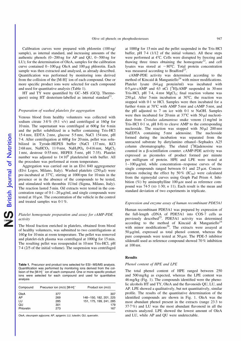
Fig. 1. Levels of phenols in olive oil extracts. A–D are extracts from oils with
high phenol content (HPE), E is from oil with low phenol content (LPE).
Quantitative determination of the single phenols (apigenin (AP), ; luteolin
(LU), ; quercetin (QU), ; oleuropein aglycone (OleA), ) was carried out
by electrospray ionization (ESI)–MS/MS equipped with an ESI source oper-
ating in the negative mode. Hydroxytyrosol (HT), ; and tyrosol (TY),
were quantified by GC–MS using deuterium-labelled compounds as internal
standards. Results represent the mean and SD of at least three injections.
Results were determined as phenols reactive to Folin-Ciocalteu, expressed
as oleuropein equivalents. * Not detectable.
M. Dell’Agli et al.946
British
Journal
ofNutrition
Calibration curves were prepared with phloretin (100 ng/sample), as internal standard, and increasing amounts of theauthentic phenols (0–200 ng for AP and QU, 0–500 ng forLU); for the determination of OleA, samples for the calibrationcurve contained 0–100mg OleA and 100mg phloretin. Eachsample was then extracted and analysed, as already described.Quantification was performed by monitoring ions derivedfrom the collision of the [M-H]- ion of each compound. One ormore specific product ions were selected for each compoundand used for quantitative analysis (Table 1).
HT and TY were quantified by GC–MS (GCQ, Thermo-quest) using HT deuterium-labelled as internal standard21.
Preparation of washed platelets for aggregation
Venous blood from healthy volunteers was collected withsodium citrate 3·8 % (9:1 v/v) and centrifuged at 160 g for10 min. The supernatant was centrifuged at 600 g for 6 minand the pellet solubilized in a buffer containing Tris-HCl15·4 mM, EDTA 2 mM, glucose 5·5 mM, NaCl 154 mM, pH7·4. After centrifugation at 600 g for 20 min, pellet was solu-bilized in Tyrode-HEPES buffer (NaCl 137 mM, KCl2·68 mM, NaHCO3 11·9 mM, NaH2PO4 0·416 mM, MgCl21 mM, destrose 0·55 mM, HEPES 5 mM, pH 7·35). Plateletnumber was adjusted to 1£108 platelets/ml with buffer. Allthe procedure was performed at room temperature.
Aggregation was carried out in an Elvi 840 aggregometer(Elvi Logos, Milano, Italy). Washed platelets (250ml) werepre-incubated at 378C, stirring at 1000 rpm for 10 min in thepresence or in the absence of the compounds to be tested,and stimulated with thrombin 1U/ml (Sigma, Milano, Italy).The reaction lasted 5 min. Oil extracts were tested in the con-centration range of 0·1–20mg/ml, and single compounds weretested at 10mM. The concentration of the vehicle in the controland treated samples was 0·1 %.
Platelet homogenate preparation and assay for cAMP-PDEactivity
The blood fraction enriched in platelets, obtained from bloodof healthy volunteers, was submitted to two centrifugations at160 g for 10 min at room temperature. The pellet was removedand platelet-rich plasma was centrifuged at 1000 g for 15 min.The resulting pellet was resuspended in 10 mM Tris-HCl, pH7·4 (2/5 of the initial volume). The suspension was centrifuged
at 1000 g for 15 min and the pellet suspended in the Tris-HClbuffer, pH 7·4 (1/12 of the initial volume). All these stepswere performed at 48C. Cells were disrupted by freezing andthawing three times obtaining the homogenate22, and celllysates was stored at 2808C. Total protein concentrationwas measured according to Bradford23.
cAMP-PDE activity was determined according to themethod of Kincaid & Manganiello24 with minor modifications.Platelet lysate (64mg protein/ml) was incubated with0·5mM-cAMP and 63 nCi [3H]cAMP suspended in 30 mM
Tris-HCl, pH 7·4, 4 mM MgCl2; final reaction volume was250ml. After 5 min incubation at 308C, the reaction wasstopped with 0·1 M HCl. Samples were then incubated for afurther 4 min at 708C with AMP 5 mM and cAMP 5 mM, andthe pH adjusted to 7 on ice with 0·1 M NaOH. Sampleswere then incubated for 20 min at 378C with 50ml nucleoti-dase from Crotalus adamanteus snake venom (1 mg/ml inTris-HCl 0·1 M, pH 8·0) to cleave AMP to the correspondingnucleoside. The reaction was stopped with 50ml 200 mM
NaEDTA containing 5 mM adenosine. The nucleosideformed during the incubation was separated from theunreacted substrate by dietylamino ethanol–Sephadex A25column chromatography. The eluted [3H]adenosine wascounted in a b-scintillation counter. cAMP-PDE activity wasexpressed as picomoles of product formed per minuteper milligram of protein. HPE and LPE were tested at1–100mg/ml, while concentration–response curves of thesingle compounds ranged between 0·1 and 25mM. Concen-trations reducing the effect by 50 % (IC50) were calculatedfrom the sigmoydal curves using Graph Pad Prism 4. Inhi-bition (%) by aminophylline 100mM used as reference com-pound was 74·5 (SD 1·30; n 11). Each result is the mean andstandard deviation of two experiments in triplicate.
Expression and enzyme assay of human recombinant PDE5A1
Human recombinant PDE5A1 was prepared by expression ofthe full-length cDNA of PDE5A1 into COS-7 cells aspreviously described25. PDE5A1 activity was determinedaccording to the method of Kincaid & Manganiello24
with minor modifications26. The extracts were assayed at50mg/ml, expressed as total phenol content, whereas thepure compounds were tested at 50mM. The PDE-5 inhibitorsildenafil used as reference compound showed 70 % inhibitionat 100 nM.
Results
Phenol content of HPE and LPE
The total phenol content of HPE ranged between 250and 500 mg/kg as expected, whereas the LPE content was46 mg/kg (Fig. 1). The compounds identified were the pheno-lic alcohols HT and TY, OleA and the flavonoids QU, LU, andAP. LPE showed a qualitatively, but not quantitatively, similarprofile. The results of the quantitative determination of theidentified compounds are shown in Fig. 1. OleA was themost abundant phenol present in the extracts (range 23·3 to37·7 %) and LU was the most abundant flavonoid in all theextracts analyzed. LPE showed the lowest amount of OleAand LU, while AP and QU were undetectable.
Table 1. Precursor and product ions selected for ESI–MS/MS analysis.Quantification was performed by monitoring ions derived from the col-lision of the [M-H]2 ion of each compound. One or more specific productions were selected for each compound and used for quantitativeanalysis
Compound Precursor ion (m/z) [M-H]– Product ion (m/z)
OleA 377 307AP 269 148–150, 182, 201, 225LU 285 151, 175, 199, 241, 285QU 301 179Phloretin 273 167
OleA, oleuropein aglycone; AP, apigenin; LU, luteolin; QU, quercetin.
Olive oil phenols on phosphodiesterases 947
British
Journal
ofNutrition
Effect of oil extracts and single phenols on plateletaggregation and PDE activities
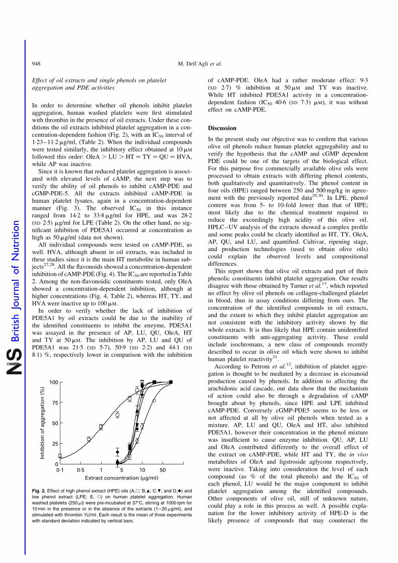
In order to determine whether oil phenols inhibit plateletaggregation, human washed platelets were first stimulatedwith thrombin in the presence of oil extracts. Under these con-ditions the oil extracts inhibited platelet aggregation in a con-centration-dependent fashion (Fig. 2), with an IC50 interval of1·23–11·2mg/ml, (Table 2). When the individual compoundswere tested similarly, the inhibitory effect obtained at 10mM
followed this order: OleA . LU . HT ¼ TY ¼ QU ¼ HVA,while AP was inactive.
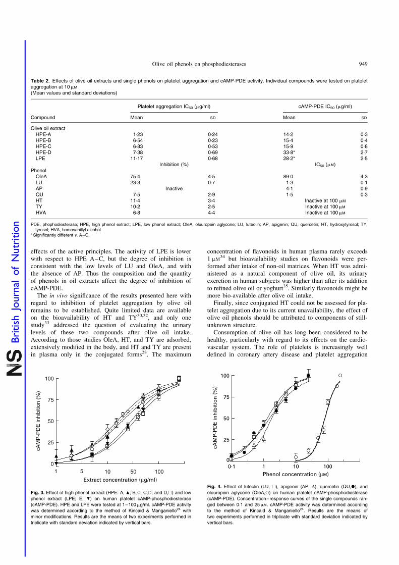
Since it is known that reduced platelet aggregation is associ-ated with elevated levels of cAMP, the next step was toverify the ability of oil phenols to inhibit cAMP-PDE andcGMP-PDE-5. All the extracts inhibited cAMP-PDE inhuman platelet lysates, again in a concentration-dependentmanner (Fig. 3). The observed IC50 in this instanceranged from 14·2 to 33·8mg/ml for HPE, and was 28·2(SD 2·5) mg/ml for LPE (Table 2). On the other hand, no sig-nificant inhibition of PDE5A1 occurred at concentration ashigh as 50mg/ml (data not shown).
All individual compounds were tested on cAMP-PDE, aswell. HVA, although absent in oil extracts, was included inthese studies since it is the main HT metabolite in human sub-jects27,28. All the flavonoids showed a concentration-dependentinhibition of cAMP-PDE (Fig. 4). The IC50 are reported in Table2. Among the non-flavonoidic constituents tested, only OleAshowed a concentration-dependent inhibition, although athigher concentrations (Fig. 4, Table 2), whereas HT, TY, andHVA were inactive up to 100mM.
In order to verify whether the lack of inhibition ofPDE5A1 by oil extracts could be due to the inability ofthe identified constituents to inhibit the enzyme, PDE5A1was assayed in the presence of AP, LU, QU, OleA, HTand TY at 50mM. The inhibition by AP, LU and QU ofPDE5A1 was 21·5 (SD 5·7), 50·9 (SD 2·2) and 44·1 (SD
8·1) %, respectively lower in comparison with the inhibition
of cAMP-PDE. OleA had a rather moderate effect: 9·3(SD 2·7) % inhibition at 50mM and TY was inactive.While HT inhibited PDE5A1 activity in a concentration-dependent fashion (IC50 40·6 (SD 7·3) mM), it was withouteffect on cAMP-PDE.
Discussion
In the present study our objective was to confirm that variousolive oil phenols reduce human platelet aggregability and toverify the hypothesis that the cAMP and cGMP dependentPDE could be one of the targets of the biological effect.For this purpose five commercially available olive oils wereprocessed to obtain extracts with differing phenol contents,both qualitatively and quantitatively. The phenol content infour oils (HPE) ranged between 250 and 500 mg/kg in agree-ment with the previously reported data29,30. In LPE, phenolcontent was from 5- to 10-fold lower than that of HPE;most likely due to the chemical treatment required toreduce the exceedingly high acidity of this olive oil.HPLC–UV analysis of the extracts showed a complex profileand some peaks could be clearly identified as HT, TY, OleA,AP, QU, and LU, and quantified. Cultivar, ripening stage,and production technologies (used to obtain olive oils)could explain the observed levels and compositionaldifferences.
This report shows that olive oil extracts and part of theirphenolic constituents inhibit platelet aggregation. Our resultsdisagree with those obtained by Turner et al.13, which reportedno effect by olive oil phenols on collagen-challenged plateletin blood, thus in assay conditions differing from ours. Theconcentration of the identified compounds in oil extracts,and the extent to which they inhibit platelet aggregation arenot consistent with the inhibitory activity shown by thewhole extracts. It is thus likely that HPE contain unidentifiedconstituents with anti-aggregating activity. These couldinclude isochromans, a new class of compounds recentlydescribed to occur in olive oil which were shown to inhibithuman platelet reactivity31.
According to Petroni et al.12, inhibition of platelet aggre-gation is thought to be mediated by a decrease in eicosanoidproduction caused by phenols. In addition to affecting thearachidonic acid cascade, our data show that the mechanismof action could also be through a degradation of cAMPbrought about by phenols, since HPE and LPE inhibitedcAMP-PDE. Conversely cGMP-PDE5 seems to be less ornot affected at all by olive oil phenols when tested as amixture. AP, LU and QU, OleA and HT, also inhibitedPDE5A1, however their concentration in the phenol mixturewas insufficient to cause enzyme inhibition. QU, AP, LUand OleA contributed differently to the overall effect ofthe extract on cAMP-PDE, while HT and TY, the in vivometabolites of OleA and ligstroside aglycone respectively,were inactive. Taking into consideration the level of eachcompound (as % of the total phenols) and the IC50 ofeach phenol, LU would be the major component to inhibitplatelet aggregation among the identified compounds.Other components of olive oil, still of unknown nature,could play a role in this process as well. A possible expla-nation for the lower inhibitory activity of HPE-D is thelikely presence of compounds that may counteract the
Fig. 2. Effect of high phenol extract (HPE) oils (A,A; B,O; C,P; and D,V) and
low phenol extract (LPE; E, W) on human platelet aggregation. Human
washed platelets (250ml) were pre-incubated at 378C, stirring at 1000 rpm for
10 min in the presence or in the absence of the extracts (1–20mg/ml), and
stimulated with thrombin 1U/ml. Each result is the mean of three experiments
with standard deviation indicated by vertical bars.
M. Dell’Agli et al.948
British
Journal
ofNutrition
effects of the active principles. The activity of LPE is lowerwith respect to HPE A–C, but the degree of inhibition isconsistent with the low levels of LU and OleA, and withthe absence of AP. Thus the composition and the quantityof phenols in oil extracts affect the degree of inhibition ofcAMP-PDE.
The in vivo significance of the results presented here withregard to inhibition of platelet aggregation by olive oilremains to be established. Quite limited data are availableon the bioavailability of HT and TY30,32, and only onestudy33 addressed the question of evaluating the urinarylevels of these two compounds after olive oil intake.According to those studies OleA, HT, and TY are adsorbed,extensively modified in the body, and HT and TY are presentin plasma only in the conjugated forms28. The maximum
concentration of flavonoids in human plasma rarely exceeds1mM
34 but bioavailability studies on flavonoids were per-formed after intake of non-oil matrices. When HT was admi-nistered as a natural component of olive oil, its urinaryexcretion in human subjects was higher than after its additionto refined olive oil or yoghurt35. Similarly flavonoids might bemore bio-available after olive oil intake.
Finally, since conjugated HT could not be assessed for pla-telet aggregation due to its current unavailability, the effect ofolive oil phenols should be attributed to components of still-unknown structure.
Consumption of olive oil has long been considered to behealthy, particularly with regard to its effects on the cardio-vascular system. The role of platelets is increasingly welldefined in coronary artery disease and platelet aggregation
Table 2. Effects of olive oil extracts and single phenols on platelet aggregation and cAMP-PDE activity. Individual compounds were tested on plateletaggregation at 10mM
(Mean values and standard deviations)
Platelet aggregation IC50 (mg/ml) cAMP-PDE IC50 (mg/ml)
Compound Mean SD Mean SD
Olive oil extractHPE-A 1·23 0·24 14·2 0·3HPE-B 6·54 0·23 15·4 0·4HPE-C 6·83 0·53 15·9 0·8HPE-D 7·38 0·69 33·8* 2·7LPE 11·17 0·68 28·2* 2·5
Inhibition (%) IC50 (mM)Phenol
OleA 75·4 4·5 89·0 4·3LU 23·3 0·7 1·3 0·1AP Inactive 4·1 0·9QU 7·5 2·9 1·5 0·3HT 11·4 3·4 Inactive at 100 mM
TY 10·2 2·5 Inactive at 100mM
HVA 6·8 4·4 Inactive at 100mM
PDE, phophodiesterase; HPE, high phenol extract; LPE, low phenol extract; OleA, oleuropein aglycone; LU, luteolin; AP, apigenin; QU, quercetin; HT, hydroxytyrosol; TY,tyrosol; HVA, homovanillyl alcohol.
* Significantly different v. A–C.
Fig. 3. Effect of high phenol extract (HPE: A, O; B,S; C,W; and D,A) and low
phenol extract (LPE; E, P) on human platelet cAMP-phosphodiesterase
(cAMP-PDE). HPE and LPE were tested at 1–100mg/ml. cAMP-PDE activity
was determined according to the method of Kincaid & Manganiello24 with
minor modifications. Results are the means of two experiments performed in
triplicate with standard deviation indicated by vertical bars.
Fig. 4. Effect of luteolin (LU, A), apigenin (AP, D), quercetin (QU,X), and
oleuropein aglycone (OleA,W) on human platelet cAMP-phosphodiesterase
(cAMP-PDE). Concentration–response curves of the single compounds ran-
ged between 0·1 and 25mM. cAMP-PDE activity was determined according
to the method of Kincaid & Manganiello24. Results are the means of
two experiments performed in triplicate with standard deviation indicated by
vertical bars.
Olive oil phenols on phosphodiesterases 949
British
Journal
ofNutrition
is shown to be one underlying mechanism in its pathogen-esis. Phenolic components of commonly consumed foods,in Mediterranean countries at least, such as red wine andolive oil rich in these constituents, are shown to inhibit pla-telet aggregation thus potentially reducing the putative riskof vascular diseases. Data presented here show one mechan-ism, cAMP-PDE inhibition, by which olive oil phenols inhi-bit platelet aggregation.
Acknowledgements
Prof. C. S. Lin, Department of Urology, University of Califor-nia, San Francisco, CA, USA, for the kind supply of PDE5A1cDNA, and Mrs M. P. Pasini for generous financial support aregratefully acknowledged. The authors thank Prof. C. Galli forthe access to the aggregometer, Dr S. Barbieri for the help inthe aggregation assay, and Mr A. Toia for technical assistance.The authors thank Dr A. Sanghvi for valuable comments.
References
1. Estruch R, Martinez-Gonzalez MA, Corella D, et al. (2006)
Effects of a Mediterranean-style diet on cardiovascular risk fac-
tors: a randomized trial. Ann Intern Med 145, 1–11.
2. Serra-Majem L, Roman B & Estruch R (2006) Scientific evi-
dence of interventions using the Mediterranean diet: a systema-
tic review. Nutr Rev 64, S27–S47.
3. Servili M & Montedoro G (2002) Contribution of phenolic com-
pounds to virgin olive oil quality. Eur J Lipid Sci Tech 104,
602–613.
4. Visioli F, Bellomo G, Montedoro G & Galli C (1995) Low den-
sity lipoprotein oxidation is inhibited in vitro by olive oil con-
stituents. Atherosclerosis 117, 25–32.
5. Caruso D, Berra B, Giavarini F, Cortesi N, Fedeli E & Galli G
(1999) Effect of virgin olive oil phenolic compounds on in vitro
oxidation of human low density lipoproteins. Nutr Metab Cardi-
ovasc Dis 9, 102–107.
6. Andrikopoulos NK, Kaliora AC, Assimopoulou AN &
Papageorgiou VP (2002) Inhibitory activity of minor polyphe-
nolic and non polyphenolic constituents of olive oil against in
vitro low-density lipoprotein oxidation. J Med Food 5, 1–7.
7. Benkhalti F, Legssyer A, Gomez P, Paz E, Lopez-Miranda J,
Perez-Jimenez F & el Boustani ES (2003) Effects of virgin
olive oil phenolic compounds on LDL oxidation and vasorelaxa-
tion activity. Therapie 58, 133–137.
8. Ferroni F, Maccaglia A, Pietraforte D, Turco L & Minetti M
(2004) Phenolic antioxidants and the protection of low density
lipoprotein from peroxynitrite-mediated oxidations at physio-
logic CO2. J Agric Food Chem 52, 2866–2874.
9. Visioli F, Bellosta S & Galli C (1998) Oleuropein, the bitter
principle of olives, enhances nitric oxide production by mouse
macrophages. Life Sci 62, 541–546.
10. Carluccio MA, Siculella L, Ancora MA, Massaro M, Scoditti E,
Storelli C, Visioli F, Distante A & De Caterina R (2003) Olive
oil and red wine antioxidant polyphenols inhibit endothelial
activation: antiatherogenic properties of Mediterranean diet
phytochemicals. Arterioscler Thromb Vasc Biol 23, 622–629.
11. Dell’Agli M, Fagnani R, Mitro N, et al. (2006) Minor com-
ponents of olive oil modulate proatherogenic adhesion mole-
cules involved in endothelial activation. J Agric Food Chem
54, 3259–3264.
12. Petroni A, Blasevich M, Salami M, Papini N, Montedoro GF &
Galli C (1995) Inhibition of platelet aggregation and eicosanoid
production by phenolic components of olive oil. Thromb Res 78,
151–160.
13. Turner R, Etienne N, Alonso MG, de Pascual-Teresa S, Mini-
hane AM, Weinberg PD & Rimbach G (2005) Antioxidant
and anti-atherogenic activities of olive oil phenolics. Int J
Vitam Nutr Res 75, 61–70.
14. Haslam RJ, Dickinson NT & Jang EK (1999) Cyclic nucleotides
and phosphodiesterases in platelets. Thromb Haemost 82,
412–423.
15. Bender AT & Beavo JA (2006) Cyclic nucleotide phosphodi-
esterases: molecular regulation to clinical use. Pharmacol Rev
58, 488–520.
16. Natella F, Nardini M, Virgili F & Scaccini C (2006) Role
of dietary polyphenols in the platelet aggregation network -
a review of the in vitro studies. Curr Topics Nutr Res 4,
1–21.
17. Limiroli RC, Ottolina G, Marsilio V, Bianchi G & Zetta L
(1995) 13C NMR characterisation of new oleuropein aglycons.
J Chem Soc Perkin Trans 1, 1519–1523.
18. Montedoro GS, Baldioli M & Miniati E (1992) Simple and
hydrolizable phenolic compounds in virgin olive oil. 1. Their
extraction, separation, quantitative and semiquantitative evalu-
ation by HPLC. J Agric Food Chem 40, 1571–1576.
19. Folin O & Ciocalteu V (1927) On tyrosine and tryptophane
determinations in proteins. J Biol Chem 73, 627–650.
20. Cardoso SM, Guyot S, Marnet N, Lopes-da-Silva JA, Renard
CMGC & Coimbra MA (2005) Characterization of phenolic
extracts from olive pulp and olive pomace by electrospray
mass spectrometry. J Sci Food Agric 85, 21–32.
21. Visioli F, Caruso D, Galli C, Viappiani S, Galli G & Sala A
(2000) Olive oils rich in natural catecholic phenols decrease iso-
prostane excretion in humans. Biochem Biophys Res Commun
278, 797–799.
22. Giovanazzi S, Accomazzo MR, Letari O, Oliva D & Nicosia S
(1997) Internalization and down-regulation of the prostacyclin
receptor in human platelets. Biochem J 325, 71–77.
23. Bradford MM (1976) A rapid and sensitive method for
the quantitation of microgram quantities of protein utilizing
the principle of protein-dye binding. Anal Biochem 72,
248–254.
24. Kincaid RL & Manganiello VC (1988) Assay of cyclic nucleo-
tide phosphodiesterase using radiolabeled and fluorescent sub-
strates. Methods Enzymol 159, 457–470.
25. Lin CS, Lau A, Tu R & Lue TF (2000) Expression of three iso-
forms of cGMP-binding cGMP-specific phosphodiesterase
(PDE5) in human penile cavernosum. Biochem Biophys Res
Commun 268, 628–635.
26. Dell’Agli M, Galli GV, Vrhovsek U, Mattivi F & Bosisio E
(2005) In vitro inhibition of human cGMP-specific phosphodi-
esterase-5 by polyphenols from red grapes. J Agric Food
Chem 53, 1960–1965.
27. Visioli F, Galli C, Bornet F, Mattei A, Patelli R, Galli G &
Caruso D (2000) Olive oil phenolics are dose-dependently
absorbed in humans. FEBS Lett 468, 159–160.
28. Miro-Casas E, Covas MI, Farre M, Fito M, Ortuno J, Weinbren-
ner T, Roset P & de la Torre R (2003) Hydroxytyrosol disposi-
tion in humans. Clin Chem 49, 945–952.
29. Visioli F & Galli C (2002) Biological properties of olive oil
phytochemicals. Crit Rev Food Sci Nutr 42, 209–221.
30. Vissers MN, Zock PL & Katan MB (2004) Bioavailability and
antioxidant effects of olive oil phenols in humans: a review. Eur
J Clin Nutr 58, 955–965.
31. Togna GI, Togna AR, Franconi M, Marra C & Guiso M (2003)
Olive oil isochromans inhibit human platelet reactivity. J Nutr
133, 2532–2536.
32. Bonanome A, Pagnan A, Caruso D, Toia A, Xamin A, Fedeli E,
Berra B, Zamburlini A, Ursini F & Galli G (2000) Evidence of
M. Dell’Agli et al.950
British
Journal
ofNutrition
postprandial absorption of olive oil phenols in humans. Nutr
Metab Cardiovasc Dis 10, 111–120.
33. Miro-Casas E, Covas MI, Fito M, Farre-Albadalejo M,
Marrugat J & de la Torre R (2003) Tyrosol and hydroxytyrosol
are absorbed from moderate and sustained doses of virgin olive
oil in humans. Eur J Clin Nutr 57, 186–190.
34. Scalbert A & Williamson G (2000) Dietary intake and bioavail-
ability of polyphenols. J Nutr 130, S2073–S2085.
35. Visioli F, Galli C, Grande S, Colonnelli K, Patelli C, Galli G &
Caruso D (2003) Hydroxytyrosol excretion differs between rats
and humans and depends on the vehicle of administration. J Nutr
133, 2612–2615.
Olive oil phenols on phosphodiesterases 951
British
Journal
ofNutrition