Embed Size (px)
Citation preview
Page 1/19
Innovative Method of Alopecia Treatment by UsingAdipose-derived Stromal Vascular Fraction (SVF) Cellssun jong kim
Research Center on Medicine Sciences Health and Society: Centre de Recherche Medecine Sciences SanteSante Mentale Societe https://orcid.org/0000-0001-5501-7246MyungJin Kim
Research Center on Medicine Sciences Health and Society: Centre de Recherche Medecine Sciences SanteSante Mentale SocieteYoungJun Lee
Research Centre on Medicine Sciences Health and Society: Centre de Recherche Medecine Sciences SanteSante Mentale SocieteJoochan Lee
Research Center on Medicine Sciences Health and Society: Centre de Recherche Medecine Sciences SanteSante Mentale SocieteJi Hyang Kim
Research Center on Medicine Sciences Health and Society: Centre de Recherche Medecine Sciences SanteSante Mentale SocieteDo Ha Kim
Research Center on Medicine Sciences Health and Society: Centre de Recherche Medecine Sciences SanteSante Mentale SocieteYoung Hoo Do
Research Center on Medicine Sciences Health and Society: Centre de Recherche Medecine Sciences SanteSante Mentale SocieteJun Woo Choi
Research Center on Medicine Sciences Health and Society: Centre de Recherche Medecine Sciences SanteSante Mentale SocieteSung Ill Chung
Research Center on Medicine Sciences Health and Society: Centre de Recherche Medecine Sciences SanteSante Mentale SocieteByung- rok Do ( [email protected] )
Hurim BioCell Inc
Short report
Keywords: Adipose derived stem cells (ADSCs), Stromal Vascular Fractions (SVFs), Androgenic Alopecia (AGA),Hair loss, Baldness

Page 2/19
Posted Date: June 1st, 2021
DOI: https://doi.org/10.21203/rs.3.rs-533152/v1
License: This work is licensed under a Creative Commons Attribution 4.0 International License. ReadFull License
Page 3/19
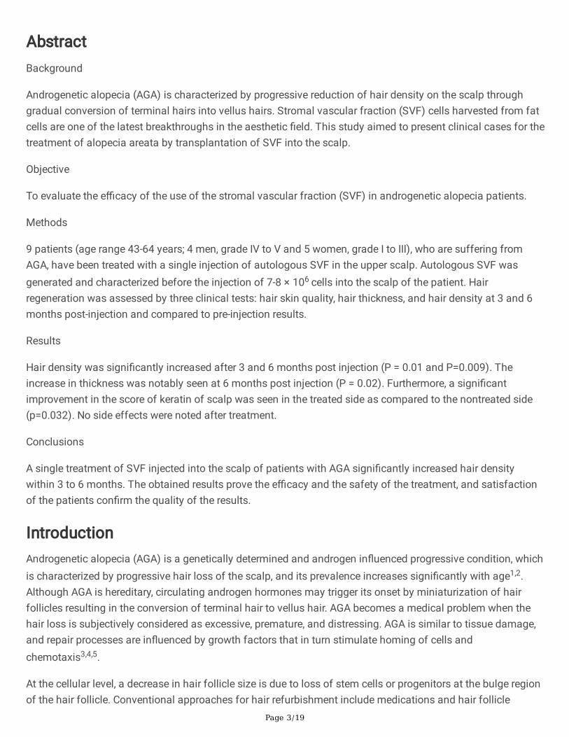
AbstractBackground
Androgenetic alopecia (AGA) is characterized by progressive reduction of hair density on the scalp throughgradual conversion of terminal hairs into vellus hairs. Stromal vascular fraction (SVF) cells harvested from fatcells are one of the latest breakthroughs in the aesthetic �eld. This study aimed to present clinical cases for thetreatment of alopecia areata by transplantation of SVF into the scalp.
Objective
To evaluate the e�cacy of the use of the stromal vascular fraction (SVF) in androgenetic alopecia patients.
Methods
9 patients (age range 43-64 years; 4 men, grade IV to V and 5 women, grade I to III), who are suffering fromAGA, have been treated with a single injection of autologous SVF in the upper scalp. Autologous SVF wasgenerated and characterized before the injection of 7-8 × 10⁶ cells into the scalp of the patient. Hairregeneration was assessed by three clinical tests: hair skin quality, hair thickness, and hair density at 3 and 6months post-injection and compared to pre-injection results.
Results
Hair density was signi�cantly increased after 3 and 6 months post injection (P = 0.01 and P=0.009). Theincrease in thickness was notably seen at 6 months post injection (P = 0.02). Furthermore, a signi�cantimprovement in the score of keratin of scalp was seen in the treated side as compared to the nontreated side(p=0.032). No side effects were noted after treatment.
Conclusions
A single treatment of SVF injected into the scalp of patients with AGA signi�cantly increased hair densitywithin 3 to 6 months. The obtained results prove the e�cacy and the safety of the treatment, and satisfactionof the patients con�rm the quality of the results.
IntroductionAndrogenetic alopecia (AGA) is a genetically determined and androgen in�uenced progressive condition, whichis characterized by progressive hair loss of the scalp, and its prevalence increases signi�cantly with age1,2.Although AGA is hereditary, circulating androgen hormones may trigger its onset by miniaturization of hairfollicles resulting in the conversion of terminal hair to vellus hair. AGA becomes a medical problem when thehair loss is subjectively considered as excessive, premature, and distressing. AGA is similar to tissue damage,and repair processes are in�uenced by growth factors that in turn stimulate homing of cells andchemotaxis3,4,5.
At the cellular level, a decrease in hair follicle size is due to loss of stem cells or progenitors at the bulge regionof the hair follicle. Conventional approaches for hair refurbishment include medications and hair follicle
Page 4/19
transplantation surgery6. However, these strategies are mostly ineffective due to drawbacks including highcost, numerous side effects, unsatisfactory results, and the requirement for long-lasting use of medicines. Inaddition, their e�cacy is dependent on gender. Contemporary therapies with promising results are required thatshould be effective in both sexes, and the outcomes should be long‐lasting.
Treatment options for men and women with hair loss include medical therapy, hair transplant surgery, low-levellaser therapy, hair systems, micropigmentation of the scalp, and topical concealer �bers7,8. These options arenot without shortcomings, and thus researchers are always looking for new and alternative therapies9. Oneemerging area of clinical and scienti�c focus lies in exploring the role of adipose tissue (fat), and speci�callyautologous adipose transplantation10, in the complex, hair growth cycle. Although platelet- rich plasma (PRP)injections are frequently used for AGA patients and overall results suggest that it has a good therapeuticeffect11, the treatment has to be repeated several times leading to poor compliance of the patient.
The repair of hair follicles in AGA could be improved using a combination of regenerative cells (i.e., stromalvascular fraction (SVF)) as a source of growth factors. Adipose tissue-derived SVF contains non culturedregenerative cells, such as mesenchymal stem cells (MSCs) or adipose derived stem cells(ADSCs), that can“home” to the site of injury12,13,14. Abundant research has supported the fact that adipose is biologically active,complex, and an important tissue. In the context of the scalp, adipocyte lineage cells support the stem cellniche and help drive the hair growth cycle15. Shin et al documented the role of ADSC-conditioned media inpromoting hair growth in female pattern alopecia16,17.
Today, autologous fat is transplanted primarily for an esthetic and reconstructive volume effect, andtraditionally, rates of graft retention have been widely varied. A number of strategies have been applied toincrease this rate of graft take. One such strategy is to enrich the adipose with SVF, a heterogeneous group ofgenerally well-characterized multinucleated cells that can be reliably extracted from adipose by usingautomated systems. These cells work largely by paracrine mechanisms to support adipocyte viability.
SVF is applied to restore hair growth because it contains several types of regenerative cells such as MSCs thatare highly proliferative, have multi-lineage differentiation potential, and are immunomodulatory andimmunosuppressive14. Furthermore, the cells in SVF also secrete various growth factors and proteins whichcan perform several functions including activation of hair follicles18. SVF can have multiple effects onminiaturized hair follicles by homing to the hair follicles and by their paracrine effects. Stromal vascularfraction‐based treatment for AGA can open a new avenue for the development of therapies for hairrestoration19.
In a group of AGA patients, SVF was injected into the affected area of scalp. After 6 months, the patients wereassessed by a physician, and patient global assessment scores were recorded. Photographs of the affectedareas were taken. The results show an increase in hair density, decreased hair fall, and improvement inphysician and patient global assessment score after SVF treatment.
Materials & Methods
Subjects
Page 5/19
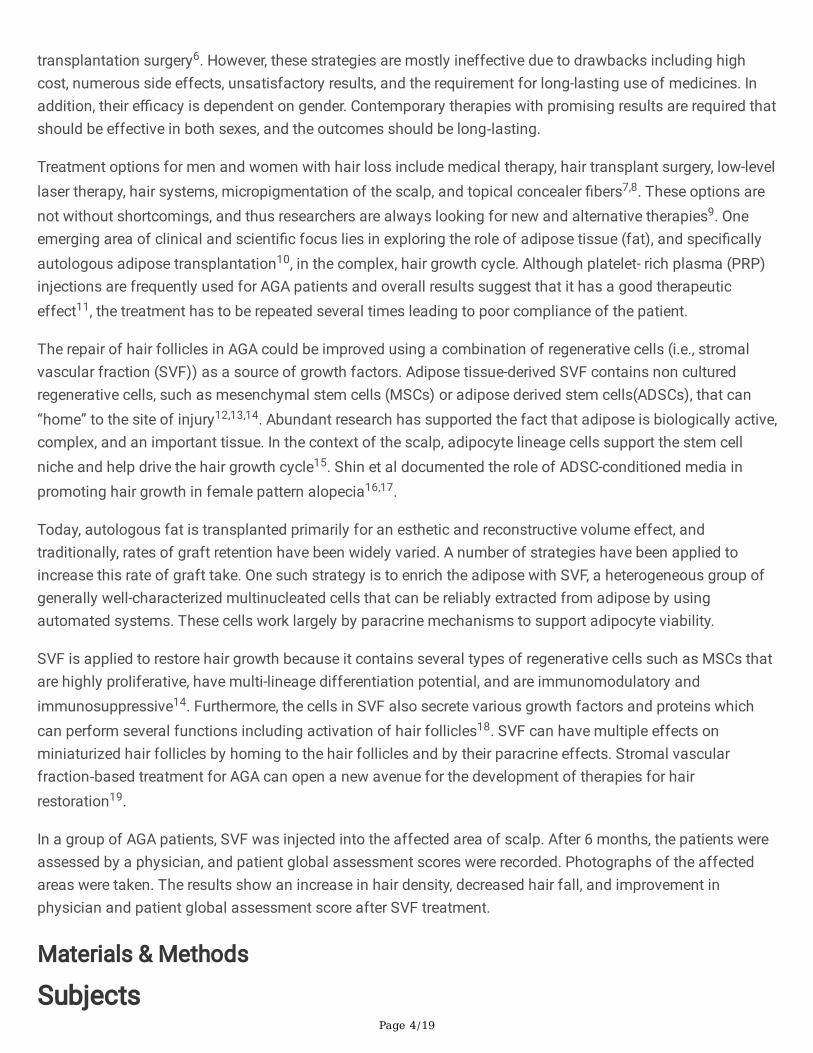
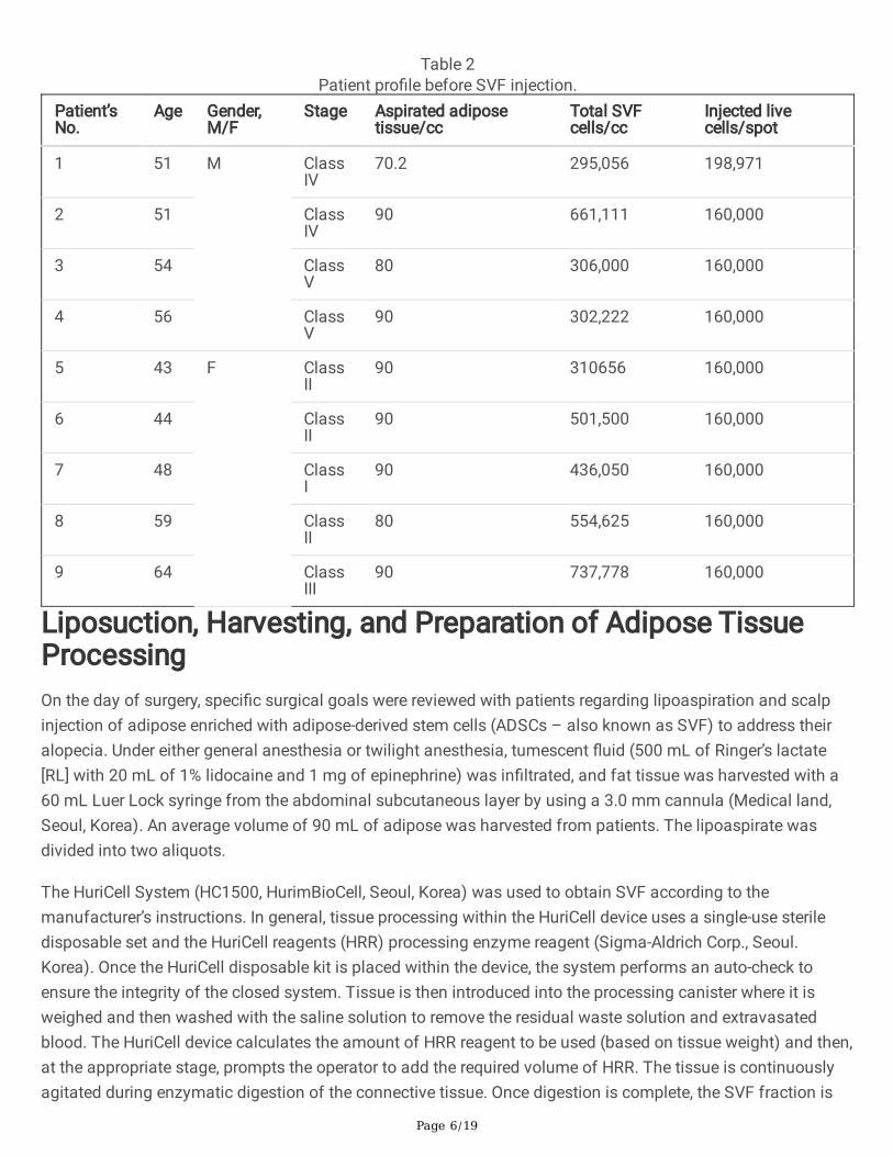
The medical records of patients were reviewed to collect subjects treated with only SVF at the plastic surgeryclinic (TOP Plastic Surgery, Seoul, Korea). A thorough medical and hair history was collected, and a physicalexamination was performed to diagnose male or female pattern alopecia. Patients in this clinical series werehealthy men and woman aged 43–64 years with androgenic baldness rated using the Norwood–Hamiltongrades and Ludwig scale. Over a period of 6 months, a total of nine subjects (4 men and 5 women) wereenrolled. The investigators reviewed the inclusion and exclusion criteria (Table 1) to screen patientsaccordingly. Importantly, patients who reported the administration of any agent aimed at affecting hair growthwithin 6 months prior to presentation, regardless of whether they were prescribed by a physician or obtainedover the counter, were excluded from participation. All patients had a body mass index (BMI) within normallimits. All patients were considered generally healthy, and no patients had comorbidities such as diabetes orhigh blood pressure. Written informed consent for the procedure, including photographing and publication in amedical and scienti�c journal for educational purposes, was obtained from all subjects, and the protocol wasreviewed and approved by the hospital’s Internal Medical Advisory Committee. Table 2 summarizes patientdemographics, including the amount of adipose harvested, processed, and injected SVF.
Table 1Inclusion and exclusion criteria for enrollment of patients in the study.
Inclusion Male and female patients with androgenic alopecia
Age of 43–64 years, provide written informed consent and comply with the studyrequirements
Norwood-Hamilton grades II-VI or Ludwig Class I-III
Active hair loss within last 12 months
No clinically signi�cant disease or abnormal laboratory results at the time of screening visit
Patient has adequate abdominal or other subcutaneous adipose tissue accessible by syringe-based lipoharvest
For women of child-bearing potential: negative pregnancy test at screening visit
Exclusion Patients with in�ammation, infection, malignancy, allegoric disease, autoimmune disease,pregnancy, diabetes and on current anticoagulant therapy.
Subject who has previously failed or has been deemed nonresponsive to a previousexperimental hair loss treatment, prior surgery in the treatment area and subject who has asensitive, irritated, or abraded scalp area
Clinically signi�cant medical or psychiatric illness currently or within 30 days of studyscreening as determined by the investigator
Any disease or condition (medical or surgical) that in the opinion of the investigator, mightcompromise hematologic, cardiovascular, pulmonary, renal, gastrointestinal, hepatic, orcentral nervous system function; or any condition that would place the subject at increasedrisk
Page 6/19
Table 2Patient pro�le before SVF injection.
Patient’sNo.
Age Gender,M/F
Stage Aspirated adiposetissue/cc
Total SVFcells/cc
Injected livecells/spot
1 51 M ClassIV
70.2 295,056 198,971
2 51 ClassIV
90 661,111 160,000
3 54 ClassV
80 306,000 160,000
4 56 ClassV
90 302,222 160,000
5 43 F ClassII
90 310656 160,000
6 44 ClassII
90 501,500 160,000
7 48 ClassI
90 436,050 160,000
8 59 ClassII
80 554,625 160,000
9 64 ClassIII
90 737,778 160,000
Liposuction, Harvesting, and Preparation of Adipose TissueProcessingOn the day of surgery, speci�c surgical goals were reviewed with patients regarding lipoaspiration and scalpinjection of adipose enriched with adipose-derived stem cells (ADSCs – also known as SVF) to address theiralopecia. Under either general anesthesia or twilight anesthesia, tumescent �uid (500 mL of Ringer’s lactate[RL] with 20 mL of 1% lidocaine and 1 mg of epinephrine) was in�ltrated, and fat tissue was harvested with a60 mL Luer Lock syringe from the abdominal subcutaneous layer by using a 3.0 mm cannula (Medical land,Seoul, Korea). An average volume of 90 mL of adipose was harvested from patients. The lipoaspirate wasdivided into two aliquots.
The HuriCell System (HC1500, HurimBioCell, Seoul, Korea) was used to obtain SVF according to themanufacturer’s instructions. In general, tissue processing within the HuriCell device uses a single-use steriledisposable set and the HuriCell reagents (HRR) processing enzyme reagent (Sigma-Aldrich Corp., Seoul.Korea). Once the HuriCell disposable kit is placed within the device, the system performs an auto-check toensure the integrity of the closed system. Tissue is then introduced into the processing canister where it isweighed and then washed with the saline solution to remove the residual waste solution and extravasatedblood. The HuriCell device calculates the amount of HRR reagent to be used (based on tissue weight) and then,at the appropriate stage, prompts the operator to add the required volume of HRR. The tissue is continuouslyagitated during enzymatic digestion of the connective tissue. Once digestion is complete, the SVF fraction is
Page 7/19
pumped into a centrifuge chamber where it is washed and concentrated. The �nal cell product can then beaspirated from the chamber in a volume of 13 mL.
The stromal cell fraction was �ltered through a 70 µm cell strainer (BD Biosciences, Inc., San Jose, CA, USA).The number and relative viability of SVF recovered from tissue processing samples in each study weredetermined using a Semiautomated Cell Counter (ADAM MC Cell Counter; NanoEnTek, Seoul, Korea).
Phenotyping SVFExpression of surface markers on SVFs was determined by AttuneTM NxT Flow Cytometer (Thermo FischerScienti�c). SVF was stained for the surface expression of CD31, CD34, CD45, CD73, CD90, and CD105 usingeach speci�c anti-human antibody (BD Pharmingen, CA) for �ow cytometric analysis. Isotype control stainingwas performed with IgG1-FITC and IgG2b-PE. Data represents the percentage of positive cells for each markeranalyzed on SVFs and are means ± SD.
Treatment and Evaluation of AGAThe SVF was injected into the scalp of the patient according to the following procedure: (1) the upper frontal,biparietal, and upper pyramidal area were �rst treated with the aseptic chlorhexidine without local anesthesia;(2) to reach the hair follicle area, the injection into the scalp area was performed with the following attributes:3cc syringe; gauge, 30; and depth, 4 mm ; (3) 0.15 ml per injection was delivered perpendicularly, separated by2 cm in a square shape all over the scalp marked previously; a total of 4 ml were injected in 48 spots; (4) afterthe injection was administered, the needle was kept in the scalp for 2 s. After the transplantation, the patientwas prescribed nonsteroid anti-in�ammatory and cephalexin antibiotics for 3 days. Patients were advised notto shower until 24 h after the procedure, not to sunbathe until after 1 week, and not to engage in sports untilafter 1 week; however, return to work can be on the same day. Follow-up for hair evaluation was based on thehair cycles and was performed 1, 3 and 6 months after injection, using Aroma Smart Wizard system (ASW200,Aram Huvis, Seoul, Korea).
Measurements and statistical analysisHair density (hair count per cm2) was calculated by counting the total number of hairs in the target area. Hairthickness was calculated as the average diameter of hairs, and scalp status, keratin of scalp, scalp sensitivity,scalp sebum, hair pore status, and cuticle status measured automatically on the Smart Wizard System. Meandifferences were then tested by paired t-tests. It should be noted that the sample size decreases over time, soless weight should be given to observed differences at time points. Wilcoxon signed rank tests were applied todetect the difference in the rates between different groups. P values of less than 0.05 were consideredsigni�cant.
ResultsIn the current study, patients enrolled were divided into two groups: Male group (n = 4) and female group (n = 5). Given that current clinical practice guidelines on the treatment of AGA, we treated �nasteride 1 mg,dutasteride 0.5 mg for men and 3% minoxidil foam for women.
Page 8/19
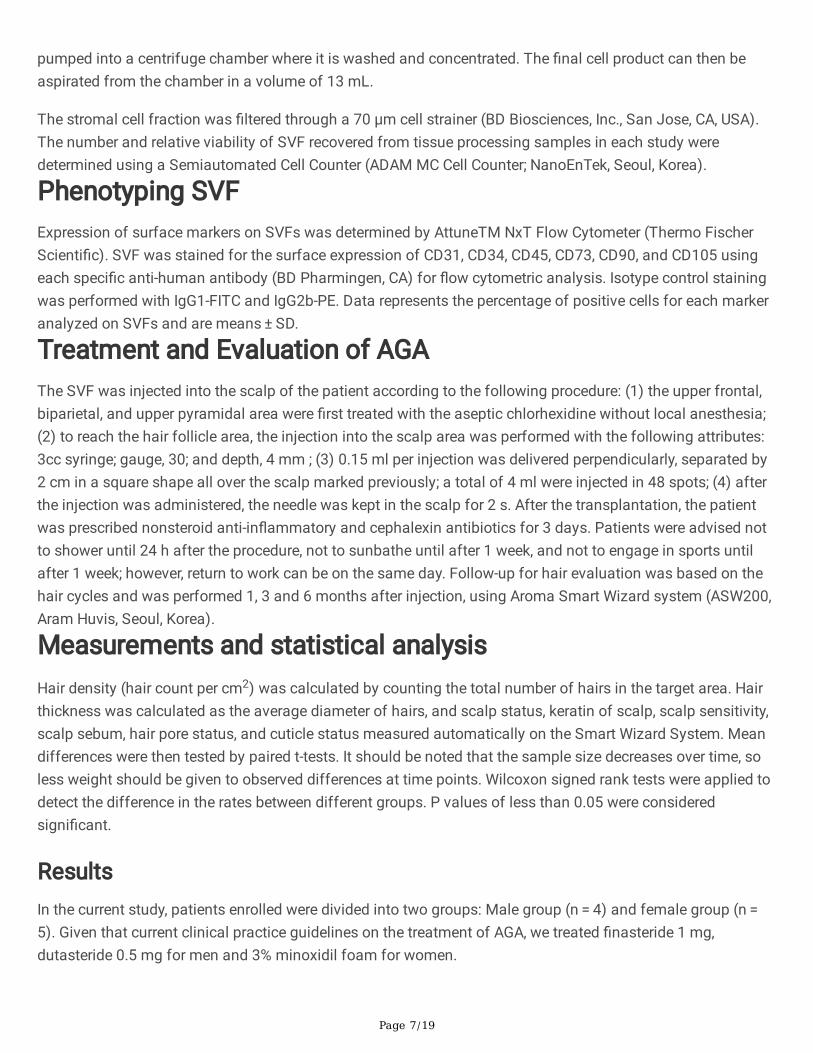
First, based on a joint statement of the International Federation for Adipose Therapeutics and Science (IFATS)and the International Society for Cellular Therapy (ISCT) published in 2013, which point out the minimalphenotypic criteria to characterize the uncultured SVF population from adipose tissue, these freshly isolatedcells were characterized (Table 3). The immunophenotyping of the transplanted cells showed a clearlyheterogeneous population expressing not only the mesenchymal stem cell markers but also thepanhematopoietic/monocyte/macrophage/endothelial/pericyte markers along with particularly high levels ofCD3419.
Table 3Immunophenotyping of cell surface markers
expressed by total nucleated SVF cellsMarker Percentage of gated
(Means ± SD, n = 9)
Characterization
CD31 33.88 ± 11.45 Endothelial
CD34 55.65 ± 11.85 Hematopoetic
CD45 2.33 ± 2.06 Immunological
CD73 12.53 ± 13.39 Mesenchymal
CD90 58.52 ± 11.19 Mesenchymal
CD105 10.03 ± 8.44 Mesenchymal
.
Page 9/19
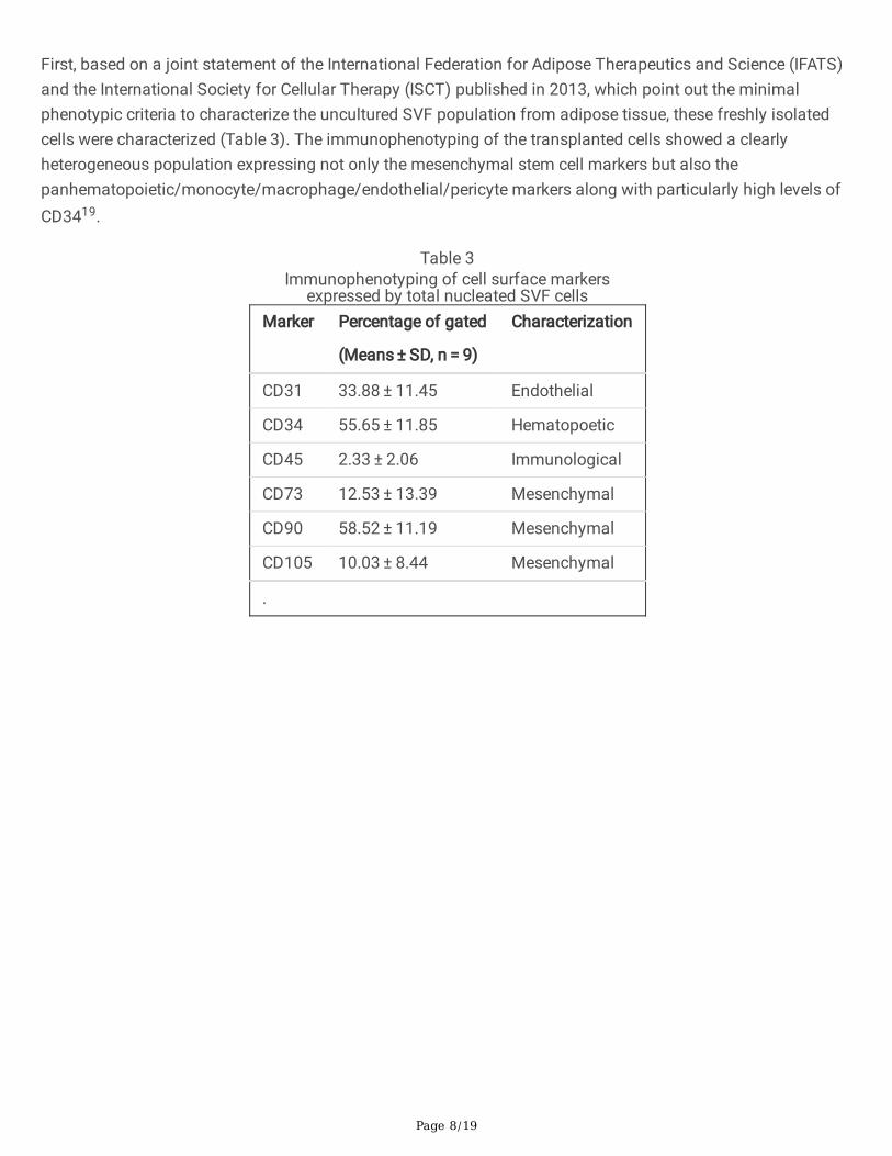
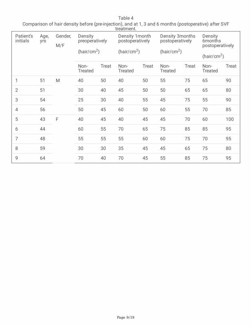
Table 4Comparison of hair density before (pre-injection), and at 1, 3 and 6 months (postoperative) after SVF
treatment.Patient’sinitials
Age,yrs
Gender,
M/F
Densitypreoperatively
(hair/cm2)
Density 1monthpostoperatively
(hair/cm2)
Density 3monthspostoperatively
(hair/cm2)
Density6monthspostoperatively
(hair/cm2)
Non-Treated
Treat Non-Treated
Treat Non-Treated
Treat Non-Treated
Treat
1 51 M 40 50 40 50 55 75 65 90
2 51 30 40 45 50 50 65 65 80
3 54 25 30 40 55 45 75 55 90
4 56 50 45 60 50 60 55 70 85
5 43 F 40 45 40 45 45 70 60 100
6 44 60 55 70 65 75 85 85 95
7 48 55 55 55 60 60 75 70 95
8 59 30 30 35 45 45 65 75 80
9 64 70 40 70 45 55 85 75 95
Page 10/19
Table 5Comparison of hair diameter before (pre-injection), and at 1, 3 and 6 months(postoperative) after SVF
treatment.Patient’sinitials
Age,yrs
Gender,
M/F
Thicknesspreoperatively
(mm)
Thickness1monthpostoperatively
(mm)
Thickness3monthpostoperatively
(mm)
Thickness6monthpostoperatively
(mm)
Non-Treated
Treat Non-Treated
Treat Non-Treated
Treat Non-Treated
Treat
1 51 M 0.022 0.028 0.028 0.031 0.044 0.042 0.047 0.055
2 51 0.028 0.023 0.03 0.023 0.033 0.029 0.047 0.045
3 54 0.018 0.021 0.028 0.029 0.032 0.031 0.049 0.051
4 56 0.029 0.027 0.03 0.033 0.022 0.044 0.033 0.047
5 43 F 0.034 0.028 0.03 0.031 0.04 0.046 0.06 0.063
6 44 0.033 0.035 0.037 0.036 0.038 0.038 0.053 0.056
7 48 0.034 0.025 0.036 0.038 0.048 0.052 0.066 0.082
8 59 0.029 0.038 0.038 0.046 0.04 0.048 0.062 0.063
9 64 0.06 0.038 0.06 0.04 0.061 0.065 0.063 0.068
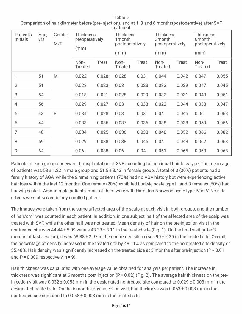
Patients in each group underwent transplantation of SVF according to individual hair loss type. The mean ageof patients was 53 ± 1.22 in male group and 51.5 ± 3.43 in female group. A total of 3 (30%) patients had afamily history of AGA, while the 6 remaining patients (70%) had no AGA history but were experiencing activehair loss within the last 12 months. One female (20%) exhibited Ludwig scale type III and 3 females (60%) hadLudwig scale II. Among male patients, most of them were with Hamilton-Norwood scale type IV or V. No sideeffects were observed in any enrolled patient.
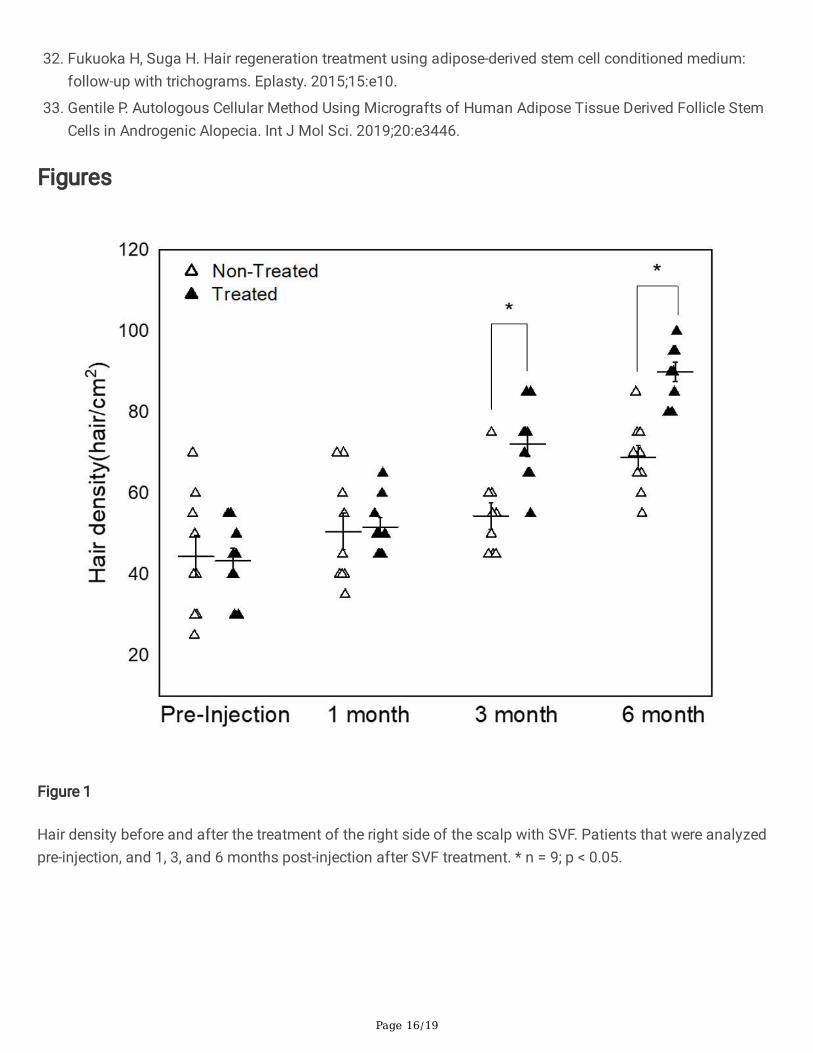
The images were taken from the same affected area of the scalp at each visit in both groups, and the numberof hair/cm2 was counted in each patient. In addition, in one subject, half of the affected area of the scalp wastreated with SVF, while the other half was not treated. Mean density of hair on the pre-injection visit in thenontreated site was 44.44 ± 5.09 versus 43.33 ± 3.11 in the treated site (Fig. 1). On the �nal visit (after 3months of last session), it was 68.88 ± 2.97 in the nontreated site versus 90 ± 2.35 in the treated site. Overall,the percentage of density increased in the treated site by 48.11% as compared to the nontreated site density of35.48%. Hair density was signi�cantly increased on the treated side at 3 months after pre-injection (P = 0.01and P = 0.009 respectively, n = 9).
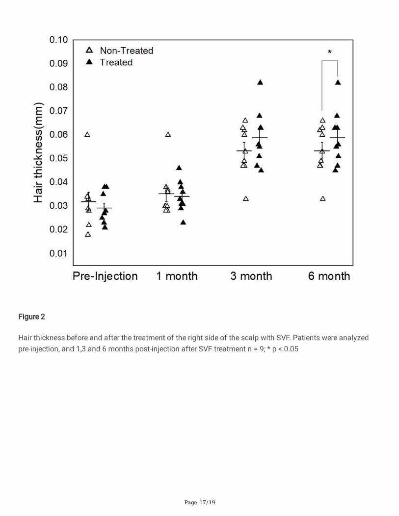
Hair thickness was calculated with one average value obtained for analysis per patient. The increase inthickness was signi�cant at 6 months post injection (P = 0.02) (Fig. 2). The average hair thickness on the pre-injection visit was 0.032 ± 0.053 mm in the designated nontreated site compared to 0.029 ± 0.003 mm in thedesignated treated site. On the 6 months post-injection visit, hair thickness was 0.053 ± 0.003 mm in thenontreated site compared to 0.058 ± 0.003 mm in the treated site.
Page 11/19
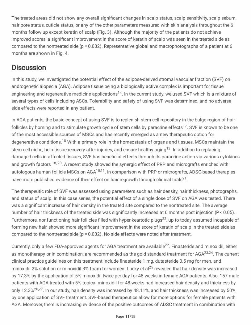
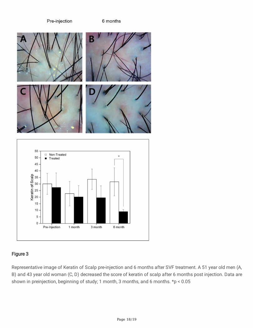
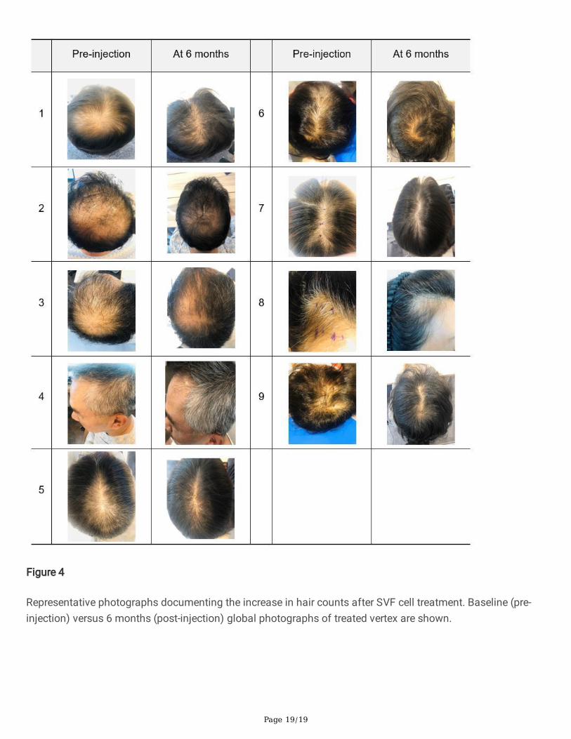
The treated areas did not show any overall signi�cant changes in scalp status, scalp sensitivity, scalp sebum,hair pore status, cuticle status, or any of the other parameters measured with skin analysis throughout the 6months follow up except keratin of scalp (Fig. 3). Although the majority of the patients do not achieveimproved scores, a signi�cant improvement in the score of keratin of scalp was seen in the treated side ascompared to the nontreated side (p = 0.032). Representative global and macrophotographs of a patient at 6months are shown in Fig. 4.
DiscussionIn this study, we investigated the potential effect of the adipose-derived stromal vascular fraction (SVF) onandrogenetic alopecia (AGA). Adipose tissue being a biologically active complex is important for tissueengineering and regenerative medicine applications14. In the current study, we used SVF which is a mixture ofseveral types of cells including ASCs. Tolerability and safety of using SVF was determined, and no adverseside effects were reported in any patient.
In AGA patients, the basic concept of using SVF is to replenish stem cell repository in the bulge region of hairfollicles by homing and to stimulate growth cycle of stem cells by paracrine effects17. SVF is known to be oneof the most accessible sources of MSCs and has recently emerged as a new therapeutic option fordegenerative conditions.18 With a primary role in the homeostasis of organs and tissues, MSCs maintain thestem cell niche, help tissue recovery after injuries, and ensure healthy aging13. In addition to replacingdamaged cells in affected tissues, SVF has bene�cial effects through its paracrine action via various cytokinesand growth factors 18, 20. A recent study showed the synergic effect of PRP and micrografts enriched withautologous human follicle MSCs on AGA10,11. In comparison with PRP or micrografts, ADSC-based therapieshave more published evidence of their effect on hair regrowth through clinical trials21.
The therapeutic role of SVF was assessed using parameters such as hair density, hair thickness, photographs,and status of scalp. In this case series, the potential effect of a single dose of SVF on AGA was tested. Therewas a signi�cant increase of hair density in the treated site compared to the nontreated site. The averagenumber of hair thickness of the treated side was signi�cantly increased at 6 months post injection (P < 0.05).Furthermore, nonfunctioning hair follicles �lled with hyper-keartotic plugs22, up to today assumed incapable offorming new hair, showed more signi�cant improvement in the score of keratin of scalp in the treated side ascompared to the nontreated side (p = 0.032). No side effects were noted after treatment.
Currently, only a few FDA-approved agents for AGA treatment are available22. Finasteride and minoxidil, eitheras monotherapy or in combination, are recommended as the gold standard treatment for AGA23,24. The currentclinical practice guidelines on this treatment include �nasteride 1 mg, dutasteride 0.5 mg for men, andminoxidil 2% solution or minoxidil 3% foam for women. Lucky et al25 revealed that hair density was increasedby 17.3% by the application of 5% minoxidil twice per day for 48 weeks in female AGA patients. Also, 157 malepatients with AGA treated with 5% topical minoxidil for 48 weeks had increased hair density and thickness byonly 12.3%26,27. In our study, hair density was increased by 48.11%, and hair thickness was increased by 50%by one application of SVF treatment. SVF-based therapeutics allow for more options for female patients withAGA. Moreover, there is increasing evidence of the positive outcomes of ADSC treatment in combination with
Page 12/19
human follicle stem cells in hair regrowth.27,54,55 Although many laboratory experiments and animal studieshave investigated the effects of ADSC on hair growth and identi�ed its positive effect in promoting hairregeneration, only a few clinical trials have investigated the effects of ADSC-based therapies on the hair cyclein humans28,29.
According to recent data, promotion of hair growth via ADSCs can be enhanced by combining it with minoxidil,which stimulates the motility of ADSCs and increases the secretion of growth factors and paracrinesignaling29. This result might suggest that ASCs migrated close to the injection site and enabled hair growth.Alternatively, ASCs might be capable of migration by making use of the local circulation. The differentiation aswell as production and secretion of growth factors that activate neighboring cells are also mentioned asrelevant functions of ADSCs. Compared with healthy individuals, in patients with AGA, the expression ofvascular endothelial growth factor (VEGF), keratinocyte growth factor (KGF), epidermal Growth Factor (EGF),and transforming growth factor-β1 (TGF-β1) is disturbed in the hair follicles, and this affects the hair cycledifferently depending on the age and sex30. Therefore, it is clinically important to personalize the optimalconcentration, dosage, and frequency of adipose derived stem cell (ADSC)-based therapies. Indeed, clinicaltrials have shown that the e�cacy of ADSC-based therapies in AGA treatment is dependent of a number ofdifferent variables, such as the type of formulation, presence of combined treatments, and delivery methods ofADSC-based therapies,22,31,33.
The proposed strategy can provide not only a treatment for AGA patients but also be helpful in thedevelopment and success of tissue engineering and regenerative medicine applications. In addition, the resultsof this study will open a new avenue in dermatology for the treatment of patients with AGA. Taking everythinginto consideration, we believe that the hallmarks of tissue damage are also present in AGA. Addressing thecombination of both cellular as well as intercellular aspects of wound repair as an alternative treatment of AGAseems to deserve further attention.
ConclusionsThis initial data experience demonstrates that scalp stem cell-enriched grafting may represent a promisingalternative approach for treating baldness in men and women. A single treatment of SVF injected in the scalpof patients with AGA signi�cantly increased hair density within 6 months. Further research is required todetermine the optimal treatment regimen.
AbbreviationsADSCs: Adipose-derived stem cells; MSCs: mesenchymal stem cells; AGA: androgenetic alopecia; SVF: stromalvascular fraction; PRP: platelet- rich plasma; RL: Ringer’s lactate; HRR: HuriCell reagents; VEGF: vascularendothelial growth factor; KGF: keratinocyte growth factor; EGF ; epidermal growth factor; TGF-β1:transforming growth factor-β1
DeclarationsAcknowledgements
Page 13/19
The authors would like to acknowledge all parties that participated in this study.
Funding
This work was supported by HurimBioCell research fund (HBC202104) for collection, analysis, andinterpretation of data.
Availability of data and materials
All data generated or analyzed during this study are included in this published article.
Authors’ contributions
SJ kim, IL Chung and BR Do contributed to conception, study design, and conduction of the study. MJ Kim, YJLee, JW Choi, and JH Kim contributed to experimentation and data collection. YH Do contributed to dataanalysis and interpretation. JC Lee contributed to manuscript writing and editing. DH Kim and Ill Chungcontributed to patient selection and sample procurement. All authors read and approved the �nal manuscript.
Ethics approval and consent to participate
The study was conducted in strict adherence to the tenets of the Declaration of Helsinki, and it wasretrospectively registered in https://cris.nih.go.kr/ (Identi�er: KCT0005880). The protocol was approved by theinstitutional review board of Korea National Institute For Bioethics Policy (KNIFBP) as previously under theacademic regulations of the public institution. All patients provided written informed consent and freshsamples were procured by Dr. Sung Ill Chung (plastic surgeon) from Top Plastic surgery hospital at Gangnam-gu, Seoul, Korea. The �rst patient was enrolled in Nov. 2020.
Consent for publication
Written informed consent for publication of their clinical details and/or clinical images was obtained from thepatient/parent/guardian/relative of the patient. A copy of the consent form is available for review by the Editorof this journal.
Competing interests
The authors declare that they have no competing interests related to this study.
Author details
1 Department of Bioconvergence, Hurim BioCell Inc., Seoul, Korea,2 Central Research Institute, Hurim BioCellInc., Seoul, Korea, 3Top Plastic surgery, Gangnam-gu, Seoul, Korea, 4Colleage of Natural Science, HanyangUniversity, Seoul, Korea
References1. Hamilton JB. Patterned loss of hair in man: types and incidence. Ann N Y Acad Sci. 1951;53:708–728.
Page 14/19
2. Norwood O. Male pattern baldness: classi�cation and incidence. South Med J. 1975;68:1359–1365.
3. Otberg N, Finner AM, Shapiro J. Androgenetic alopecia. Endocrinol Metab Clin North Am. 2007;36:379–398.
4. Stough D, Stenn K, Haber R, et al. Psychological effect, pathophysiology, and management ofandrogenetic alopecia in men. May Clin Proc. 2005;80:1316–1322.
5. Krupa Shankar DS, Chakravarthi M, Shilpakar R. Male androgenetic alopecia: population base study in1,005 subjects. Int J Tricology. 2009;1:131–133.
�. Perez-Meza D, Niedbalski R. Complications in hair restoration surgery. Oral Maxillofac Surg Clin North Am.2009;21:119–148.
7. Blumeyer A, Tosti A, Messenger A, et al. Evidence-based (S3) guideline for the treatment of androgeneticalopecia in women and in men. J Dtsch Dermatol Ges. 2011;9:1-57.
�. Rossi A, Anzalone A, Fortuna MC, et al. Multi-therapies in androgenetic alopecia: review and clinicalexperiences. Dermatol Ther. 2016;29:424-432.
9. Van Dongen JA, Stevens HP, Harmsen MC, van der Lei B. Mechanical micronization of lipoaspirates:squeeze and emulsi�cation techniques. Plast Reconstr Surg. 2017;139:1369-1370.
10. Zhu M, Zhou Z, Chen Y, et al. Supplementation of fat grafts with adipose-derived regenerative cellsimproves long-term graft retention. Ann Plast Surg. 2010;64:222–228.
11. Hieronymus PS, Simone D, Julia dB. Introducing Platelet-Rich Stroma: Platelet- Rich Plasma (PRP) andStromal Vascular Fraction (SVF) Combined for the Treatment of Androgenetic Alopecia. Aesthet Surg J.2018;13:811-822.
12. Chu DT, Nguyen TPT, Tien NLB, et al. Adipose tissue stem cells for therapy: an update on the progress ofisolation, culture, storage, and clinical application. J Clin Med. 2019;8:917.
13. Mizuno H, Tobita M, Uysal AC. Concise review: adipose-derived stem cells as a novel tool for futureregenerative medicine. Stem Cells 2012;30:804–10.
14. Gentile P, Scioli MG, Bielli A, Orlandi A, Cervelli V. Concise review: the use of adipose-derived stromalvascular fraction cells and platelet rich plasma in regenerative plastic surgery. STEM CELLS. 2017;35:117-134.
15. Blanpain C, Lowry WE, Geoghegan A, Polak L, Fuchs E. Self-renewal, multipotency, and the existence oftwo cell populations within an epithelial stem cell niche. Cell. 2004;118:635–48.
1�. Shin H, Won CH, Chung WK, et al. Up-to-date clinical trials of hair regeneration using conditioned media ofadipose-derived stem cells in male and female pattern hair loss. Curr Stem Cell Res Ther. 2017;12:524-530.
Page 15/19
17. Narita K, Fukuoka Sekiyama H, Suga H, Harii K. Sequential Scalp Assessment in Hair RegenerationTherapy Using an Adipose-Derived Stem Cell-Conditioned Medium. Dermatol Surg. 2020;46:819-825.
1�. Kilroy GE, Foster SJ, Wu X, Ruiz J, et al. Cytokine pro�le of human adipose-derived stem cells: expressionof angiogenic, hematopoietic, and pro-in�ammatory factors. J Cell Physiol 2007;212:702–9.
19. Bourin P, Bunnell BA, Casteilla L, et al. Stromal cells from the adipose tissue derived stromal vascularfraction and culture expanded adipose tissuederived stromal/stem cells: a joint statement of theInternational Federation for Adipose Therapeutics and Science (IFATS) and the International Society forCellular Therapy (ISCT). Cytotherapy. 2013;15(6):641–8. https://doi.org/10.1016/j.jcyt.2013.02.006.
20. Ramdasi S, Tiwari SK. Growth factors and cytokines secreted in conditioned media by mesenchymal stemcells-promising possible therapeutic approach for hair regeneration. J Stem Cells. 2016;11:201-211.
21. Ibrahim ZA, Elmaadawi IH, Mohamed BM, et al. Stem cell therapy as a novel therapeutic intervention forresistant cases of alopecia areata and androgenetic alopecia. J Dermatol Treat. 2016;24:1–10.
22. Tinoco A, Gonçalves J, Silva C, Loureiro A, Gomes AC, Cavaco-Paulo A, Ribeiro A. Keratin-based Particlesfor Protection and Restoration of Hair Properties. Int J Cosmet Sci. 2018;40:408-419.
23. Kelly Y, Blanco A, Tosti A. Androgenetic alopecia: an update of treatment options. Drugs. 2016;76:1349-1364.
24. Olsen EA, Dunlap FE, Funicella T, et al. A randomized clinical trial of 5% topical minoxidil versus 2% topicalminoxidil and placebo in the treatment of androgenetic alopecia in men. J Am Acad Dermatol.2002;47:377-385.
25. Lucky AW, Piacquadio DJ, Ditre CM, et al. A randomized, placebo-controlled trial of 5% and 2% topicalminoxidil solutions in the treatment of female pattern hair loss. J Am Acad Dermatol 2004;50:541–553.
2�. Fertig RM, Gamret AC, Darwin E, et al. Sexual side effects of 5-α- reductase inhibitors �nasteride anddutasteride: a comprehensive review. Dermatol Online J. 2017;23:13030/qt24k8q743.
27. Goren A, Naccarato T. Minoxidil in the treatment of androgenetic alopecia. Dermatol Ther.2018;31:e12686.
2�. David PM, Craig Z, Marcos S, Ganesh K, Edward B, Eric D. Hair follicle growth by stromal vascular fractionenhanced adipose transplantation in baldness. Stem Cells Cloning. 2017;6;10:1-10.
29. Ghazal B, Ijaz H, Fridoon JA, Mahmood SC. Stromal Vascular Fraction-Enriched Platelet-Rich PlasmaTherapy Reverses the Effects of Androgenetic Alopecia. J Cosmet Dermatol. 2020;19:1078-1085.
30. Kubanov AA, Gallyamova YA, Korableva OA. The study of growth factors in patients with androgenicalopecia. J Biomed Pharmacol. 2017;10:1219-1228
31. Young JT, Sang YL, Cho AR, Kim YS. A Randomized, Double-Blind, Vehicle-Controlled Clinical Study of HairRegeneration Using Adipose-Derived Stem Cell Constituent Extract in Androgenetic Alopecia. Stem CellsTransl Med. 2020;18:1-11.
Page 16/19
32. Fukuoka H, Suga H. Hair regeneration treatment using adipose-derived stem cell conditioned medium:follow-up with trichograms. Eplasty. 2015;15:e10.
33. Gentile P. Autologous Cellular Method Using Micrografts of Human Adipose Tissue Derived Follicle StemCells in Androgenic Alopecia. Int J Mol Sci. 2019;20:e3446.
Figures
Figure 1
Hair density before and after the treatment of the right side of the scalp with SVF. Patients that were analyzedpre-injection, and 1, 3, and 6 months post-injection after SVF treatment. * n = 9; p < 0.05.
Page 17/19
Figure 2
Hair thickness before and after the treatment of the right side of the scalp with SVF. Patients were analyzedpre-injection, and 1,3 and 6 months post-injection after SVF treatment n = 9; * p < 0.05
Page 18/19
Figure 3
Representative image of Keratin of Scalp pre-injection and 6 months after SVF treatment. A 51 year old men (A,B) and 43 year old woman (C, D) decreased the score of keratin of scalp after 6 months post injection. Data areshown in preinjection, beginning of study; 1 month, 3 months, and 6 months. *p < 0.05
Page 19/19
Figure 4
Representative photographs documenting the increase in hair counts after SVF cell treatment. Baseline (pre-injection) versus 6 months (post-injection) global photographs of treated vertex are shown.