Embed Size (px)
Citation preview

Intensity Modulated Radiation Therapy (IMRT): A New Promising Technology in Radiation Oncology
BIN S. TEH, SHIAO Y. WOO, E. BRIAN BUTLER
Department of Radiology/Radiation Oncology, Baylor College of Medicine, Houston, Texas, USA
Key Words. Intensity modulated radiation therapy · Conformal radiotherapy · SMART boost · Central nervous system tumor · Head and neck cancer · Prostate cancer · Reirradiation
ABSTRACT
Intensity modulated radiation therapy (IMRT) is anew technology in radiation oncology that delivers radia-tion more precisely to the tumor while relatively sparingthe surrounding normal tissues. It also introduces newconcepts of inverse planning and computer-controlledradiation deposition and normal tissue avoidance in con-trast to the conventional trial-and-error approach. IMRThas wide application in most aspects of radiation oncologybecause of its ability to create multiple targets and multi-ple avoidance structures, to treat different targets simulta-neously to different doses as well as to weight targets and
avoidance structures according to their importance. Bydelivering radiation with greater precision, IMRT hasbeen shown to minimize acute treatment-related mor-bidity, making dose escalation feasible which may ulti-mately improve local tumor control. IMRT has alsointroduced a new accelerated fractionation scheme knownas SMART (simultaneous modulated accelerated radia-tion therapy) boost. By shortening the overall treatmenttime, SMART boost has the potential of improving tumorcontrol in addition to offering patient convenience andcost savings. The Oncologist1999;4:433-442
The Oncologist 1999;4:433-442
Correspondence: E. Brian Butler, M.D., Baylor College of Medicine, One Baylor Plaza, 165B, Houston, Texas 77030-3498,USA. Telephone: 713-790-2637; Fax: 713-793-1300; e-mail: [email protected] Accepted for publication October 9, 1999.©AlphaMed Press 1083-7159/99/$5.00/0
INTRODUCTION
Together with surgery and chemotherapy, radiotherapyplays an important role in oncology, both in the definitive andpalliative aspects of treatment. Three major aspects of radia-tion oncology are potentially important advances in cancertreatment. The first is a multidisciplinary therapeutic approachwith important clinical applications, the second is the progressin the physics and dosimetry of radiotherapy that has clinicalramification, and the third is genetic radiotherapy that will betranslated to clinical practice [1]. Intensity modulated radiationtherapy (IMRT) is the product of advances in the technologyof radiotherapy to deliver radiation more precisely to the tumorwhile relatively limiting dose to the surrounding normal tis-sues. The purpose of this paper is to discuss the new conceptof IMRT, its application in radiation oncology and potentialbenefits over conventional radiotherapy.
IMRT—A R EVOLUTIONARY CONCEPT
In radiotherapy the single most important limiting factor isthe normal tissue radiation tolerance, and the objective of opti-mal radiotherapy is to deliver the maximum radiation dose to the
tumor while keeping the dose to the surrounding normal struc-tures below tolerance [2]. To date this aim is achieved by the useof multiple treatment fields, choice of beam energies and modal-ities, weighting of different beams as well as the use of wedgesand tissue compensators. In addition, two-dimensional cus-tomized blocks are routinely used to shield the normal struc-tures. All of these conventional treatment-planning processesare approaches by trial and error. For example, a radiation oncol-ogist will first place a radiation treatment field and then evaluatethe dose. If the dose is not acceptable, a new field or other mod-ification has to be made. This trial-and-error process has to berepeated until the optimal dose and coverage is achieved. This istime-consuming and sometimes no optimal plan can be reached.
IMRT combines two advanced concepts to deliver 3D con-formal radiation therapy: A) inverse treatment planning withoptimization by computer and B) computer-controlled intensitymodulation of the radiation beam during treatment [3].
Inverse Treatment Planning with Computer OptimizationIn contrast to the conventional trial-and-error approach, a
radiation oncologist defines the tumor and the radiation dose
by guest on March 3, 2016
http://theoncologist.alphamedpress.org/
Dow
nloaded from

that he wants around the tumor. The computer, using a math-ematical optimization technique known as simulated anneal-ing, will determine the optimal treatment fields. In addition,one can also define where one does not want the depositionof radiation (e.g., dose-limiting critical surrounding normaltissues). Figure 1 shows a smiling face demonstrating howradiation can be deposited in almost any pattern.
Computer-Controlled Intensity Modulation of the RadiationBeam During Treatment
IMRT (NOMOS Peacock system; Sewickley, PA) hasevolved from computer tomography (CT) concepts. A CTscan delivers uniform radiation exposure to the patient as itrotates around the patient’s contour in a slice-by-slice fashion.
Due to varying attenuation among multiple tissues, a nonuni-form radiation dose exits the patient and hits the detector.The detector feeds this information to the computer, whichprocesses it to create the sliced scan images. The PeacockIMRT system, on the other hand, starts with the target volume,where it places a uniform, conformal dose around the tumor.The computer then “backprojects” through the patient’s tissueto the linear accelerator source and finds the nonuniform radi-ation exposure that must be delivered by the linear acceleratorto give this conformal dose pattern. The system, like the CTscan, uses a slice-by-slice, arc-rotation approach. The conceptof the Peacock system is demonstrated in Figure 2.
IMRT is a customized or individualized radiotherapyaccording to patient’s location of tumor and anatomical struc-tures, i.e., each patient has his or her own “unique” treatmentplan. Blocks, compensators and wedges are obviated. Withthe availability of a powerful computer, planning time isshort. Treatment delivery is also very efficient as there is noneed for different energies of photons or mixed photon andelectron beams. A special multileaf collimating systemknown as multileaf intensity modulating collimator (MIMiC)(Fig. 3) is used to deliver spatially nonuniform radiationexposure to the patient to create a relatively uniform dosedistribution at the target.
CONSIDERATIONS IN THE DELIVERY OF IMRTThe more conformal a radiation treatment approach, the
less error is allowed in patient set-up and treatment planning.In simpler terms, one cannot use conformal radiotherapy totreat a “moving target,” as the incidence of “missing” thetarget is very high! Also, small movements can result in
434 Intensity Modulated Radiation Therapy for Cancer Treatment
Figure 1. Shows a smiling face demonstrating how many smallbeams of radiation can be deposited to produce the final uniformpattern.
Figure 2. Demonstrates the concept of the Peacock system, an inverse planning system.
Computed tomography Conformal therapy
Target
Intensitymodulator
Radiationsource
Detectors
Projection
Radiationsource
by guest on March 3, 2016
http://theoncologist.alphamedpress.org/
Dow
nloaded from

significant deviations from calculated doses based on aninstantaneous image. Thus, patient and organ movements areof crucial concern when delivering conformal radiation ther-apy. The ideal site to start IMRT is the brain where the cen-tral nervous system (CNS) tumors are encased in the cranium.The only factor is patient’s movement that can be minimizedby either the Peacock “talon” system (invasive fixationdevice) or a special reinforced mask (noninvasive immobi-lization device). The talon system uses two self-tapping skullscrews/sockets attached to the skull. The talon body is thensecured to the screws/sockets. The body is then rigidly fixedto support the structure on the treatment table to achieve goodpatient immobilization [4].
In addition to the above immobilization techniques, anintraoral stent or bite-block is used for organ immobilizationwhen treating head and neck cancers. Oral stents also serve thepurpose of normal tissue avoidance, e.g., protecting oraltongue in treating the cancersof the hard palate.
Treating prostate cancersposes a challenge in bothpatient and organ move-ments. Due to anatomic andphysical constraint, an inva-sive fixation technique forthe pelvis was found not to befeasible. Prior experiencewith immobilization tech-niques enabled the develop-ment of a fiducial systemlinked to a treatment box.The patient lies prone in anevacuated beanbag that con-forms both to the patient’sbody contour and the treat-ment box. Each patient has
his own box with the fitted beanbag throughout the treatment.This has helped in solving the problem of patient movement.
The next problem is the previously well-documentedmovement of the prostate itself [5]. This problem is signifi-cant with regard to using IMRT to treat prostate cancer.Prostate motion during radiotherapy can lead to underdosingthe target (prostate) and/or overdosing critical normal struc-tures (rectum and bladder). A rectal catheter with an inflatedballoon was developed to minimize the prostate motion.IMRT can then be delivered with more confidence. To date,only a limited attempt has been made to use IMRT in treatinglung cancer, as it has proved difficult to immobilize lung andchest wall movement satisfactorily due to respiratory motion.
APPLICATION OF IMRT AND ITS POTENTIAL
ADVANTAGES
Once the problem of patient and organ motion isresolved, IMRT can be applied to various tumors at varioussites, either for definitive or palliative treatment intent.
To Create Multiple TargetsMultiple treatment targets can be created and treated at the
same time. A good example is multiple brain metastases. Forpatients who have prior whole brain radiotherapy and need boostirradiation, or patients with metastatic radioresistant tumors,e.g., renal cell carcinoma or melanoma, multiple brain metas-tases can be treated simultaneously to high-dose radiation whileother parts of the brain are avoided as shown in Figure 4. Thishas the benefit of decreasing treatment-related CNS toxicity.
Teh, Woo, Butler 435
Figure 3. MIMiC (Multileaf Intensity Modulating Collimator), a specialmultileaf collimating system.
Figure 4. Shows multiple brainmetastases alone treated simul-taneously with IMRT.
by guest on March 3, 2016
http://theoncologist.alphamedpress.org/
Dow
nloaded from

Besides creating multiple targets of the same origin, IMRTalso enables the planning and treatment of multiple targets ofdifferent origins. A good example is in head and neck cancersas shown in Figure 5. The primary target (red) includes all pal-pable and imaging documented abnormal tumor and lymphnodes. The secondary target (purple) includes all at-risksubclinical disease sites, e.g., all draining lymphatics, per-ineural routes, etc. Different targets can then be treatedsimultaneously with different fraction sizes and differenttotal doses. This has significant implication in radiobiolog-ical control of gross versus subclinical disease. Anotherexample is using IMRT to treat two completely unrelatedtumors simultaneously as demonstrated in a patient withpituitary adenoma and chemodectoma (Fig. 6).
To Create Multiple Critical Avoidance Normal Structures There are many critical normal structures surrounding
the targets, especially when treating head and neck or braintumors. These structures include optic nerves and chiasm,lens, lacrimal glands, salivary glands, mandible, temporallobes, brain stem and spinal cord. These structures usuallyhave lower radiation tolerance, much lower than the tumor-icidal radiation dose. Typical radiation thresholds used inthese avoidance structures are shown in Table 1. IMRTallows the creation of these avoidance structures on thetreatment plan as well as the designation of their respectiveradiation thresholds. This has led to the concept of confor-mal avoidance, i.e., limiting the radiation to the surround-ing structures below the designated threshold limit (Fig. 5).
WeightingsIn addition to conformal treatment and avoidance, the
Peacock IMRT system allows differential weightings on bothtargets and avoidance structures (Table 2). Generally the tar-get (tumor) is weighted more than the avoidance structures.However, in cases where a tumor surrounds a critical struc-ture, e.g., optic chiasm, and the patient does not want to acceptthe treatment complication of losing his vision, the avoidancestructure is then weighted more than the target. The ability ofdifferential weighting has increased the versatility of IMRTand can be used to tailor to patient’s needs and wishes.
New Accelerated Fractionation SchemeWith the advent of IMRT and its capability to treat mul-
tiple targets simultaneously to different doses, a new accel-erated fractionation scheme is introduced. It is known assimultaneous modulated accelerated radiation therapy(SMART) boost [4]. SMART boost can be applied to vari-ous sites including head and neck, brain and prostate. Theprinciple is to treat two different targets with different frac-tion sizes to different total doses. A good example is the
treatment of head and neck cancers. Accelerated repopulationof tumor clonogens during conventional fractionated radio-therapy has been recognized as an important cause of treat-ment failure in head and neck cancers, especially if the overall
436 Intensity Modulated Radiation Therapy for Cancer Treatment
Figure 5. Primary target (red) includes all palpable and imaging documentedabnormal tumor and lymph nodes.Secondary target (purple) includes all at-risk subclinical disease sites, e.g., all draining lymphatics. It illustrates sparingof mandible, spinal cord and parotids.
Figure 6. Patient with pituitary adenoma and chemodectoma—two targetstreated simultaneously with IMRT.
by guest on March 3, 2016
http://theoncologist.alphamedpress.org/
Dow
nloaded from

treatment time is prolonged. Both laboratory and clinical data[6-8] support this hypothesis. The reduction of overall treat-ment time has the potential for improving tumor control byminimizing tumor clonogen regeneration. Various purelyaccelerated treatment schedules have been used, e.g., PolishTrial (CAIR) [9], CHART [10], Massachusetts General
Hospital accelerated split course [11] and M.D. AndersonConcomitant Boost [12] (Table 3). These trials have sug-gested improved tumor control. By applying SMART boostby the use of IMRT, the primary target (palpable and imagingdocumented gross tumors and lymph nodes) and the sec-ondary targets (at risk microscopic disease sites, e.g., draining
Teh, Woo, Butler 437
Table 1. Normal structures in the head and neck with their assigned radiation thresholds
Normal structures Radiation thresholds Normal structures Radiation thresholds(Gy) (Gy)
Brain stem 50 Optic chiasm 45
Lacrimal glands 30 Parotid (ipsilateral) 35
Lens 12 Parotid (contralateral) 25
Retina 45 Spinal cord 40
Optic nerves 45 Mandible 58
Table 2. Radiation deposition and avoidance
Number of fractions 25 over 37 days (inclusive) Planning parameter
Target Prescribed dose
Total Daily Target weight
Target 1 6,000 cGy 240 cGy 1.0
Target 2 5,000 cGy 200 cGy 1.0
Nontarget structures Maximum dose Target weight
Mandible 5,800 cGy 1.0
Right parotid gland 2,500 cGy 1.0
Left parotid gland 2,500 cGy 1.0
Brain stem 5,000 cGy 0.1
Spinal cord 4,000 cGy 0.5
Table 3. Various accelerated treatment schedules
Radiotherapy Dose Total dose Overallschedules fractions (cGy) duration
Standard fractionation 35 fractions ×200 cGy daily = 7,000 7 weeks
***CAIR 35 fractions ×200 cGy daily = 7,000 5 weeks
***CHART 36 fractions ×150 cGy TID = 5,400 12 days
***MGH accelerated split course 42 fractions ×160 cGy BID = 6,720 6 weeks
†MDACC concomitant boost 30 fractions × 180 cGy daily+
12 fractions ×150 cGy daily = 7,200 6 weeks
‡SMART boost technique 25 fractions × 240 cGy (Target 1) = 6,000
25 fractions ×200 cGy (Target 2) = 5,000 5 weeks
***Continuous accelerated radiotherapy.***Continuous hyperfractionated accelerated radiotherapy.***Massachusetts General Hospital.†M.D. Anderson Cancer Center.‡Simultaneous modulated accelerated radiation therapy.
by guest on March 3, 2016
http://theoncologist.alphamedpress.org/
Dow
nloaded from

lymphatics) are treated simul-taneously to total doses of 60Gy and 50 Gy, respectivelyover five weeks. Daily fractionsizes are 2.4 Gy and 2.0 Gy,respectively. As it is completedin five weeks compared to theconventional seven weeks, it isan accelerated scheme.SMART has the benefit ofdelivering only once-dailytreatments, five days a weekcompared to other acceleratedschemes usually involvingtwice or three-times-a-day ortreating over the weekend. It iscost saving and offers patientconvenience. More impor-tantly, it has the radiobiologi-cal advantage of overcoming
the accelerated repopulation of clonogens during radiationtherapy that may lead to improved treatment outcome.
CLINICAL OUTCOME —THE BAYLOR COLLEGE
OF MEDICINE EXPERIENCE
More than 700 patients have been treated with IMRTat the Baylor College of Medicine since March 1994 todate. Four major subsets were presented and discussed.These include: A) CNS; B) head and neck; C) prostate, andD) previously irradiated patients.
CNSBoth pediatric and adult benign and malignant brain
tumors have been treated with IMRT. Table 4 shows variousCNS tumors treated with IMRT.
Eleven patients with medulloblastoma received IMRT asa posterior fossa boost after the initial craniospinal irradiation,while the remaining 68 patients were irradiated with IMRTfor the full course. Total prescribed doses ranged from 36 Gyto 64 Gy. Noninvasive immobilization was used in 35 patientsand invasive immobilization was used in the other 45 patients.Median follow-up was 24 months.
Figure 7 shows an axial plan of a patient with infratentor-ial ependymoma. The patient had gross total resection followedby full course IMRT. Temporal lobes and cranial nerve VIII aretwo important avoidance normal structures. With the capabilityof conformal avoidance by IMRT, the mean doses to temporallobes and CN VIII are 21.7 Gy and 18.7 Gy, respectively, muchbelow their tolerance threshold. Figure 8 shows an axial plan ofa pituitary adenoma treated with IMRT. It illustrates thesparing of optic chiasm, temporal lobes, orbits and brain stem.
Treatment-related toxicity was limited to mild headachein ten patients with two patients requiring steroids. Twopatients with meningioma had symptoms from persistentperitumoral edema that was present before radiotherapy,requiring an increase in the steroid dose. Three patients hadlocal scalp asymptomatic erythema. None of the patients
438 Intensity Modulated Radiation Therapy for Cancer Treatment
Table 4. Various CNS tumors treatedwith IMRT
Types n ofof tumor patients
A) Pediatric
Medulloblastoma 11
Craniopharyngioma 10
Astrocytoma 6
Ependymoma 5
Germ cell tumor 3
Brain stem glioma 2
Ganglioglioma 2
B) Adult
Meningioma 22
Pituitary adenoma 14
Neuroma 4
Total 79
Figure 7. Shows an axial plan of a patient with infratentorialependymoma (purple), sparing CN VIII (blue).
Figure 8. Shows an axial plan of a pituitary adenoma (red) treated withIMRT. It illustrates the sparing of optic chiasm (turquoise), temporal lobes(blue), orbits, and brain stem (green).
by guest on March 3, 2016
http://theoncologist.alphamedpress.org/
Dow
nloaded from

with invasive immobilization developed any infection atlocal wound sites.
Two patients with optic nerve sheath meningioma havereported improvement in vision. Four patients (medullo-blastoma, brain stem glioma, ganglioglioma and recurrentmalignant meningioma) showed progression of disease.The remaining 76 patients (median follow-up of 30 months)showed no progression thus far.
Head and Neck CancersTwenty patients with primary head and neck cancers
(Table 5) were treated with IMRT using the SMART boosttechnique with parotid preser-vation [4]. The treatment fieldsencompassed two simultane-ous targets with primary targetincluding palpable and imag-ing detected disease and sec-ondary target including areasat risk for microscopic disease,e.g., lymphatic compartments.Daily fractions of 2.4 Gy and 2Gy were prescribed to the pri-mary and secondary targets toa total dose of 60 Gy and 50Gy, respectively. The overalltreatment course was over fiveweeks (daily treatment) ratherthan the usual seven weeks
(daily treatment) or the more-than-daily treatment in otheraccelerated schemes. With the IMRT inverse planning sys-tem, we were able to limit the radiation dose to the parotidglands. For midline tumors the parotids were limited to 25Gy. For a unilateral tumor the ipsilateral parotid was limitedto 35 Gy and the contralateral gland to 25 Gy. Figure 5 showsan axial treatment plan of an oropharyngeal primary treatedwith SMART boost. It demonstrates two targets and spar-ing of parotid glands, mandible and spinal cord. RadiationTherapy Oncology Group (RTOG) acute toxicity gradingcriteria were used to evaluate surrounding normal tissueeffects. Subjective salivary function was also assessed.Tumor response was evaluated by clinical examination, CTor magnetic resonance imaging (MRI) at periodic follow-up.
Sixteen of 20 (80%) patients completed the treatmentwithin 40 days. Two patients took up to 50 days to completethe treatment because of acute toxicity while two other patientscompleted the treatment in more than 50 days due to noncom-pliance rather than treatment-related side effects. Four separateorgan systems were assessed using RTOG acute radiationmorbidity scoring criteria [13]. Sixteen patients (80%) hadRTOG grade 3 mucositis while ten patients (50%) had grade 3
pharyngitis. All patients recovered well after completion ofradiotherapy. Encouragingly, more than half of the patients(55%) had grade 1 or less toxicity with the salivary gland; nopatient had higher than grade 2 toxicity [4].
As xerostomia is a subjective report of symptom, thepatients were asked to grade this symptom (none, mild,moderate and severe or complete). Eleven patients (55%)reported mild or no mouth dryness. Nine patients had mod-erate symptoms but none reported severe or complete xeros-tomia. Only one patient with moderate xerostomia wasprescribed with pilocarpine. Time to relief of xerostomiavaried from one to six months.A completely normal foodintake was observed in sevenpatients.
Initial tumor response toSMART boost using IMRTis shown in Table 6. Nineteenpatients (95%) had a completeresponse (CR) while onepatient had a partial response(PR). Median follow-up wassix months. Two of the 19patients with local regional CRwere found to have lung metastases at follow-up. There was nolocal regional recurrence or marginal miss.
Prostate CancerFifty men aged 56 to 82 with a mean of 70.8 years were
included in the study. Clinical stages ranged from T1c to T3a.Median Gleason combined score was 6.5 (4-9). Mean pre-treatment prostate-specific antigen (PSA) was 11.8 (3.6-60.0).All patients had negative metastatic work-up including chestx-ray, bone scan and CT of pelvis. Twenty-five (50%) patientshad hormonal manipulation prior to and during radiotherapy.Patients treated with IMRT were prescribed a dose of 70 Gy in35 fractions over 50-55 days. This is compared to the only ran-domized trial in this dose range, i.e., 70 Gy (conventional in 35fractions over 50-55 days) and 78 Gy (six-field conformal in39 fractions over 55-60 days) [14]. Patients were immobilizedin a prone position in an evacuated “beanbag” contained in abox. Rectal balloon was used daily to minimize prostate move-ment. Patients treated with the conventional and six-field con-formal radiotherapy were in supine treatment position withoutrectal balloon immobilization. The RTOG scoring system wasused to assess acute toxicity. Median follow-up was 5.5months (1 to 12 months).
Figure 9 shows an axial treatment plan of prostate can-cer treated with IMRT. It illustrates well how use of the rec-tal balloon minimized the prostate motion as well as thehighly conformal radiation dose coverage of tumor sparing
Teh, Woo, Butler 439
Table 5. Tumor site
Tumor site n of patients
Nasopharynx 3
Paranasal sinus 1
Oral cavity 1
Oropharynx 12
Larynx 3
AJCC stage n of patients
I 0
II 2
III 7
IV 10
NA 1
Table 6. Head and neck cancers treatedwith SMART boost—tumor response
Follow-up
Range 2-20 months
Mean 6.9 months
Median 6 months
Response to treatment
CR – 19 (including two patients withlocal CR but had lung metastases)
PR - 1
by guest on March 3, 2016
http://theoncologist.alphamedpress.org/
Dow
nloaded from

the rectum. Mean doses to prostate, seminal vesicles, rec-tum and bladder were 75.8 Gy, 73.7 Gy, 34.2 Gy and 23.3Gy, respectively. Although the prescribed dose for patientstreated with IMRT was 70 Gy, the mean dose to the prostatewas 75.8 Gy. There is dose inhomogeneity when the IMRTdosimetry is reviewed. At our institution we have used equiv-alent uniform dose (EUD) for reporting in order to give amore accurate delivered dose representation. EUD is found tobe very similar to the mean dose in the treatment of prostatecancer. Thus, a higher dose is delivered with IMRT whencompared to that of conventional treatment for prostate can-cer despite the same prescribed dose. A mean dose of approx-imately 76 Gy over 35 fractions delivers a daily fraction sizeof approximately 2.17 Gy. Moderate dose escalation is desiredas there is evidence to suggest dose response [15]. Higher radi-ation dose delivered with 3D conformal radiotherapy has been
shown to improve treatment outcome of prostate cancer [15].This is also a form of an accelerated fractionation scheme as76 Gy is delivered over seven weeks in 35 fractions. Thehigher daily fraction size of 2.17 Gy is also well tolerated bypatients as shown by the acute toxicity profile.
Acute genitourinary (GU) and gastrointestinal (GI) tox-icity are shown in Table 7. There was a statistical difference(p < 0.001) for both GU and lower GI grade 0, 1, 2 toxicitybetween the IMRT and conventional or six-field conformalgroup. There was no grade 3 or higher toxicity noted in theIMRT group. Of note was the very low incidence of grade2 GI toxicity in the IMRT group (14%) compared to theconventional (60%) and the six-field conformal group(66.7%) [16].
It is too early to assess late toxicity or biochemical con-trol (especially since 50% of the patients received androgen
440 Intensity Modulated Radiation Therapy for Cancer Treatment
Figure 9. Shows an axial treatment plan of prostate cancer treated with IMRT. It illustrates how the rectal balloon minimizes theprostate motion as well as the highly conformal radiation dose coverage of prostate gland sparing the rectum.
Table 7. Acute GU and lower GI toxicity secondary to radiotherapy for prostate cancer—a comparison
Total patients GU-RTOG grade GI-RTOG graden (%) 0 1 2 0 1 2
Conventional 30 (100) 2 (6.7) 12 (40.0) 13 (43.3) 4 (13.3) 7 (23.3) 18 (60.0)
(3 pts [10.0] > grade 2) (1 pt [3.3] > grade 2)
6-field conformal 30 (100) 2 (6.7) 17 (56.7) 9 (30.0) 5 (16.7) 4 (13.3) 20 (66.7)
(2 pts [6.7] > grade 2) (1 pt [3.3] > grade 2)
IMRT 50 (100) 26 (52.0) 9 (18.0) 15 (30.0) 37 (74.0) 6 (12.0) 7 (14.0)
by guest on March 3, 2016
http://theoncologist.alphamedpress.org/
Dow
nloaded from
ablation). However, there already is an emerging trend sug-gesting that PSA nadir was reached early post-radiotherapycompared to nadir after conventional treatment. To date nopatient has had a biochemical failure (three consecutiverises in PSA).
Recurrent Tumor Previously IrradiatedTen patients with recurrent carcinoma in the head and
neck were reirradiated to provide further local control aswell as symptom palliation. Five patients had recurrent car-cinoma of nasopharynx while the remaining consisted ofrecurrent carcinoma involving maxillary sinus, nasal cav-ity, medial canthus, oropharynx and posterior pharyngealwall. All patients received prior radiotherapy ranging from30 Gy to 67 Gy. They were all referred to our center as itwas felt that no further radiation could be delivered safelyto the local recurrence with conventional radiotherapy.Additional radiation was delivered with IMRT. The doseranged from 16 Gy to 66 Gy. Toxicity was graded usingRTOG morbidity scores, and tumor response was evaluatedby clinical examination, CT and/or MRI. Follow-upsranged from 2 to 12 months.
All patients were able to complete planned reirradiationat the scheduled time frame. There was no grade 3 or higherRTOG toxicity in mucosa, pharynx, skin or salivary glandsthat required split from treatment. All patients achieved theaim of palliation of local symptoms such as pain, epistaxisor discomfort. Quality of life has improved during and oncompletion of radiotherapy. Only half of the patients showedno progression of disease at the last follow-up.
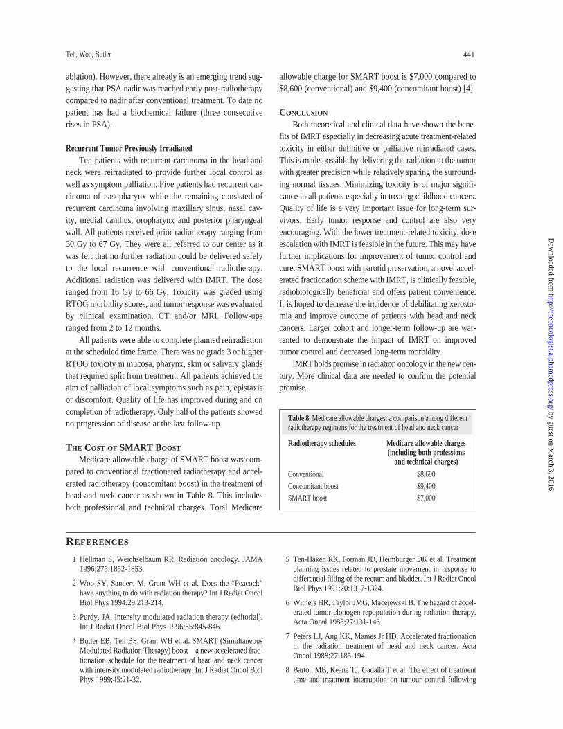
THE COST OF SMART BOOST
Medicare allowable charge of SMART boost was com-pared to conventional fractionated radiotherapy and accel-erated radiotherapy (concomitant boost) in the treatment ofhead and neck cancer as shown in Table 8. This includesboth professional and technical charges. Total Medicare
allowable charge for SMART boost is $7,000 compared to$8,600 (conventional) and $9,400 (concomitant boost) [4].
CONCLUSION
Both theoretical and clinical data have shown the bene-fits of IMRT especially in decreasing acute treatment-relatedtoxicity in either definitive or palliative reirradiated cases.This is made possible by delivering the radiation to the tumorwith greater precision while relatively sparing the surround-ing normal tissues. Minimizing toxicity is of major signifi-cance in all patients especially in treating childhood cancers.Quality of life is a very important issue for long-term sur-vivors. Early tumor response and control are also veryencouraging. With the lower treatment-related toxicity, doseescalation with IMRT is feasible in the future. This may havefurther implications for improvement of tumor control andcure. SMART boost with parotid preservation, a novel accel-erated fractionation scheme with IMRT, is clinically feasible,radiobiologically beneficial and offers patient convenience.It is hoped to decrease the incidence of debilitating xerosto-mia and improve outcome of patients with head and neckcancers. Larger cohort and longer-term follow-up are war-ranted to demonstrate the impact of IMRT on improvedtumor control and decreased long-term morbidity.
IMRT holds promise in radiation oncology in the new cen-tury. More clinical data are needed to confirm the potentialpromise.
Teh, Woo, Butler 441
Table 8. Medicare allowable charges: a comparison among differentradiotherapy regimens for the treatment of head and neck cancer
Radiotherapy schedules Medicare allowable charges (including both professions
and technical charges)
Conventional $8,600
Concomitant boost $9,400
SMART boost $7,000
REFERENCES
1 Hellman S, Weichselbaum RR. Radiation oncology. JAMA1996;275:1852-1853.
2 Woo SY, Sanders M, Grant WH et al. Does the “Peacock”have anything to do with radiation therapy? Int J Radiat OncolBiol Phys 1994;29:213-214.
3 Purdy, JA. Intensity modulated radiation therapy (editorial).Int J Radiat Oncol Biol Phys 1996;35:845-846.
4 Butler EB, Teh BS, Grant WH et al. SMART (SimultaneousModulated Radiation Therapy) boost—a new accelerated frac-tionation schedule for the treatment of head and neck cancerwith intensity modulated radiotherapy. Int J Radiat Oncol BiolPhys 1999;45:21-32.
5 Ten-Haken RK, Forman JD, Heimburger DK et al. Treatmentplanning issues related to prostate movement in response todifferential filling of the rectum and bladder. Int J Radiat OncolBiol Phys 1991;20:1317-1324.
6 Withers HR, Taylor JMG, Macejewski B. The hazard of accel-erated tumor clonogen repopulation during radiation therapy.Acta Oncol 1988;27:131-146.
7 Peters LJ, Ang KK, Mames Jr HD. Accelerated fractionationin the radiation treatment of head and neck cancer. ActaOncol 1988;27:185-194.
8 Barton MB, Keane TJ, Gadalla T et al. The effect of treatmenttime and treatment interruption on tumour control following
by guest on March 3, 2016
http://theoncologist.alphamedpress.org/
Dow
nloaded from

radical radiation therapy of laryngeal cancer. Radiat Ther Oncol1992;23:137-143.
9 Maciejewski B, Skladowski K, Pilecki B et al. Randomizedclinical trial on accelerated 7 days per week fractionation inradiotherapy for head and neck cancer: preliminary report onacute toxicity. Radiother Oncol 1996;40:137-145.
10 Sanders MI, Dische S, Grosch EJ et al. Experience withCHART. Int J Radiat Oncol Biol Phys 1991;21:871-878.
11 Wang CC. Improved local control for advanced oropharyn-geal carcinoma following twice daily radiation therapy. Am JClin Oncol 1985;8:512-516.
12 Ang KK, Peters LJ, Weber RS et al. Concomitant boost radio-therapy schedules in the treatment of carcinoma of theoropharynx and nasopharynx. Int J Radiat Oncol Biol Phys1990;19:1339-1345.
13 Cox JD, Stetz J, Pajak TF. Toxicity criteria of the RadiationTherapy Oncology Group (RTOG) and the EuropeanOrganization for Research and Treatment of Cancer (EORTC)(editorial). Int J Radiat Oncol Biol Phys 1995;31:1341-1346.
14 Pollack A, Zagars GK, Starkschall G et al. Conventional vs.conformal radiotherapy for prostate cancer: preliminaryresults of dosimetry and acute toxicity. Int J Radiat OncolBiol Phys 1996;34:555-564.
15 Zelefsky MJ, Leibel SA, Kutcher GJ et al. Three-dimensionalconformal radiotherapy and dose escalation: where do westand? Semin Radiat Oncol 1998;8:107-114.
16 Teh BS, Uhl BM, Augspurger ME et al. Intensity modulatedradiotherapy (IMRT) for localized prostate cancer: preliminaryresults of acute toxicity compared to conventional and six fieldapproach. Int J Radiat Oncol Biol Phys 1998;42(suppl 1):219.
442 Intensity Modulated Radiation Therapy for Cancer Treatment
by guest on March 3, 2016
http://theoncologist.alphamedpress.org/
Dow
nloaded from


