Embed Size (px)
Citation preview

www.elsevier.com/locate/ynimg
NeuroImage 25 (2005) 267–277
Intentional false responding shares neural substrates with response
conflict and cognitive control
Jennifer Maria Nunez,a,1 B.J. Casey,b Tobias Egner,c Todd Hare,a and Joy Hirschc,d,*
aDepartment of Neurology and Neuroscience, Weill Medical College of Cornell University, New York, NY 10021, USAbSackler Institute for Developmental Psychobiology, Weill Medical College of Cornell University, New York, NY 10021, USAcDepartment of Radiology, Center for Neurobiology and Behavior of Columbia University, Functional MRI Research Center, New York, NY 10032, USAdDepartment of Psychology, Center for Neurobiology and Behavior of Columbia University, Functional MRI Research Center, New York, NY 10032, USA
Received 19 August 2004; revised 13 October 2004; accepted 14 October 2004
Available online 13 January 2005
The ability to deceive others is a high-level social and cognitive
function. It has been suggested that response conflict and cognitive
control increase during deceptive acts but this hypothesis has not been
evaluated directly. Using fMRI, we tested this prediction for the
execution of an intentional false response. Subjects were instructed to
respond truthfully or falsely to a series of yes/no questions that were
also varied in autobiographical and nonautobiographical content to
further examine the influence of personal relevance when lying. We
observed an interference effect (longer reaction times for false versus
true responses) that was accompanied by increased activation within
the anterior cingulate, caudate and thalamic nuclei, and dorsolateral
prefrontal cortex (DLPFC), a circuit that has been implicated in
response conflict and cognitive control. Behavioral and neural effects
were more robust when falsifying autobiographical responses relative
to nonautobiographical responses. Furthermore, a correlation between
reaction time and left caudate activity supported the presence of
increased response inhibition when falsifying responses. When pre-
sented with self-relevant (autobiographical) stimuli regardless of
response condition, the mesial prefrontal and posterior cingulate
cortices were recruited. Neural activity within these two regions and
the anterior cingulate cortex (ACC) also showed correlations with self-
report personality measures from the Psychopathic Personality
Inventory (PPI). Overall, we conclude that the process of interference
is inherent to the act of falsifying information and that the amount of
conflict induced and cognitive control needed to successfully execute
false responses is greater when dealing with personal information.
D 2004 Elsevier Inc. All rights reserved.
Keywords: Response conflict; Cognitive control; Deception
1053-8119/$ - see front matter D 2004 Elsevier Inc. All rights reserved.
doi:10.1016/j.neuroimage.2004.10.041
* Corresponding author. Fax: +1 212 342 0855.
E-mail addresses: [email protected] (J.M. Nunez)8
[email protected] (J. Hirsch).1 Editorial correspondence address: fMRI Research Center, Columbia
University, Neurological Institute B-41, PO Box 108, 710 West 168th
Street, New York, NY 10032, USA.
Available online on ScienceDirect (www.sciencedirect.com).
Introduction
Many attempts have been made to define markers of deceptive
behavior using both behavioral (body posture, speech patterns, and
response latency) (DePaulo et al., 2003; Seymour et al., 2000;
Zuckerman et al., 1981) and physiological measures (heart rate,
skin conductance, and pupil diameter) (Dionisio et al., 2001;
Godert et al., 2001; Podlesny and Raskin, 1977). Nevertheless, the
subjective nature of experimenter evaluations and differences in the
nature of each individual’s level of moral and ethical behavior
produce a considerable amount experimental variability for such
measures (Ben-Shakhar and Dolev, 1996; Bradley and Cullen,
1993; DePaulo et al., 2003; Kircher and Raskin, 1988). However,
the availability of techniques such as functional magnetic
resonance imaging (fMRI) allows for a direct examination of the
neural activity that underlies deceptive behavior.
Recent functional imaging studies have employed a variety of
techniques to identify the neural correlates of deceptive behavior.
For example, one study used a modified version of the Guilty
Knowledge Test (Lykken, 1959, 1960) in which subjects were
asked to lie about their possession of a specific target stimulus
while answering truthfully to all other stimuli (Langleben et al.,
2002). A second study examined the patterns of neural activity
between spontaneous and memorized lies (Ganis et al., 2003), and
yet another used a forced choice task in which subjects were asked
to feign memory impairment such that some trials were answered
incorrectly while others were answered correctly (Lee et al., 2002).
These studies have identified a variety of brain regions within
frontal, temporal, and parietal cortices yet the regions identified
between studies have been highly variable and lacking consistent
activation across all studies (Ganis et al., 2003; Langleben et al.,
2002; Lee et al., 2002; Spence et al., 2001). This may be due in
part to the large variability among experimental designs as well as
their broad and behaviorally complex definitions of a deceptive act.
Thus, our study attempted to target specific elements of deceptive
behavior such as conflict, response inhibition, and higher level
cognitive control using an ecologically relevant question and

J.M. Nunez et al. / NeuroImage 25 (2005) 267–277268
answer paradigm in combination with a simple and clearly defined
deceptive act.
Our approach was based upon three current theories of
deception including information manipulation theory (IMT)
(McCornack, 1992, 1997), interpersonal deception theory (IDT)
(Buller and Burgoon, 1996), and Zuckerman’s four-factor model
(Zuckerman et al., 1981). All of these posit that there is an increase
in the amount of cognitive control during deceptive behavior and
recent fMRI studies have confirmed that there is an increase in
neural activity within a number of cortical regions implicated in
control during a deceptive act (Ganis et al., 2003; Langleben et al.,
2002). The need for increased bcontrolled behaviorQ is also
featured as one of the four factors in Zuckerman’s model and
can be viewed as a product of the neural processes of conflict
resolution and cognitive control (Botvinick et al., 2001; Braver et
al., 2003; Cohen et al., 1990; MacDonald et al., 2000). Interest-
ingly, the processes of conflict monitoring and higher level
cognitive control have been associated with the anterior cingulate
cortex (ACC) and dorsolateral prefrontal cortex (DLPFC), respec-
tively, and both of these regions have been identified as being
active during deceptive behavior using fMRI (Ganis et al., 2003;
Langleben et al., 2002). Accordingly, deceptive behavior lends
itself to study within the context of interference and conflict
resolution since when generating a lie or a false response, one must
know the truth, resist the impulse to answer truthfully, and further
generate an alternative but appropriate response. Although the
processes of increased conflict and cognitive control are believed
to be inherent to deceptive behavior (Ganis et al., 2003; Langleben
et al., 2002; Lee et al., 2002; Spence et al., 2001), they have not
been the target of direct study in previous functional imaging work.
Furthermore, the presence of an interference effect during
deceptive behavior has not been validated with behavioral
measures such as reaction time, and thus whether or not
interference occurs during a deceptive act remains unconfirmed.
Perhaps the simplest deceptive act as described by Coleman and
Kay (1981) is the lie, which is defined by three basic features. The
first feature is the falsehood itself, for example, the communication
of the falsehood. Second is the awareness that a false act/utterance
has been committed, and third that this act has been committed
intentionally (e.g., it is not a mistake). Thus, in the current study, we
varied true versus false responses while maintaining the basic
elements of a lie such that (1) subjects intentionally give a false
response, (2) subjects were aware they were answering incorrectly,
and (3) their responses were not the result of a mistake.
Although this task may satisfy Coleman and Kay’s (1981)
definition of a lie, the intentional delivery of false information in and
of itself is merely a distillation of the deceptive process, which in
real life is often complicated by personal circumstance. For
example, one’s incentive to lie may vary substantially depending
upon whether or not the lie is of no personal consequence versus a
scenario in which a successful lie leads to reward or deters
punishment. Therefore, we added the additional dimension of
personal (autobiographical) and impersonal (nonautobiographical)
question types to investigate how self-relevant information interacts
with levels of conflict and control processes when falsifying
information. Recent functional imaging work has shown that tasks
of an autobiographical versus nonautobiographical nature show
distinct neural activity, which is indicative of emotional processing,
and thus these manipulations afford us a simple entry point into the
emotive aspects of deceptive behavior (Fink et al., 1996; Maguire
and Frith, 2003; Piefke et al., 2003; Vogeley and Fink, 2003).
Furthermore, within the field of deception, there is interest as to
whether or not certain classifiable personality types show a
propensity towards deceptive behavior. Thus, we attempted to
correlate changes in magnetic resonance signal in brain regions,
which exhibited a main effect for false versus true comparisons with
various sociopathic personality traits as determined by the Psycho-
pathic Personality Inventory (PPI) (Lilienfeld and Andrews, 1996).
We predicted greater activation in the ACC and DLPFC regions
for false over true responses, similar to that seen in classic
interference tasks such as the Stroop and flanker paradigms
(Botvinick et al., 1999; Eriksen and Eriksen, 1974; MacLeod,
1991; Stroop, 1935), and increased reaction times for false
responses indicating the presence of an interference effect. We
also predicted an interaction between autobiographical questions
and falsifying information such that neural activity within those
brain regions, which regulate response inhibition and higher level
cognitive control, will show larger increases in neural activity
when falsifying autobiographical information (versus nonautobio-
graphical) since personal information is more readily accessible
and highly practiced, presumably making it more difficult to
suppress prepotent truthful responses. Furthermore, differences in
the levels of conflict and cognitive control as indicated by
functional activity may correlate with personality traits that are
characteristic of high or low levels of self-control or emotionality.
Methods
Subjects
Twenty healthy volunteers (10 males and 10 females) ranging
from 20 to 34 years of age (mean age 26.0 years, SD = 4)
participated in the functional imaging experiment. Consent
procedures were performed according to institutional guidelines
and all subjects were right-handed with normal neurological
histories and no contraindication for MRI.
Procedure
Stimulus development
One hundred and twenty yes/no questions were generated for
pilot testing. All stimuli used for both the piloting and functional
experimentation were generated and presented with E-Prime/IFIS
(Psychology Software Tools, Inc., Pittsburgh, PA; MRI Devices
Corporation, Gainesville, FL) software and hardware. Target
stimuli included questions that were easily audible, could be
delivered within a 2-s presentation period, and successfully
answered within a 2-s response period. Eight subjects, four males
and four females (not those referenced for functional scanning),
participated in the pilot study. The four different experimental
conditions were counterbalanced both within and across runs using
a pseudo-randomized Latin square design (Fig. 1A). Only those
questions that were answered correctly (as instructed) for 75% of
the trials and still retained a content-matched pair were included.
The final result was 72 yes/no questions, which comprised two
different categories, autobiographical and nonautobiographical,
and were represented by 36 content-matched pairs (Table 1).
Experimental design
All subjects provided their truthful responses for the 72 test
questions used during the imaging experiments two or more days

Table 1
Sample of stimulus questions, matched for content
Autobiographical Nonautobiographical
Do you own a laptop computer? Is a laptop computer portable?
Have you ever told a lie? Do people ever lie?
Can you ride a bicycle? Does a bicycle have six wheels?
Were you born in New York City? Is New York City in Ohio?
There was a significant difference ( P V 0.001) in salience ratings between
autobiographical (1.7 F 0.07 SEM) and nonautobiographical questions
(1.4 F 0.07 SEM). There was no significant difference ( P V 0.683) in
confidence ratings between autobiographical (3.9 F 0.03 SEM) and
nonautobiographical questions (3.9 F 0.02 SEM).
J.M. Nunez et al. / NeuroImage 25 (2005) 267–277 269
prior to the imaging session to reduce priming effects. Within this
questionnaire, subjects were also required to rate each question for
both its emotional salience as well as their confidence in their
answer. During the scanning sessions, the 72 yes/no questions were
presented via headphones and were answered using a push-button
response pad. Each question was answered once truthfully and once
falsely across the experiment yielding four different experimental
conditions (Fig. 1A) and 144 total stimuli. Stimuli and question
blocks were equally dispersed across three separate functional runs.
Each run consisted of eight question blocks, six questions per block,
four blocks requiring truthful answers and four blocks requiring
false answers (Fig. 1B). All questions within a single question block
required the same response type as cued by a rear projection screen,
which displayed a bTQ to indicate truthful responses and an bFQ toindicate false responses. The cue for the desired response type
appeared on the screen 2 s prior to the onset of the question block to
allow the subject time to prepare and respond appropriately. Each
question was presented during a 2-s presentation period, which was
followed by a 2-s silent response period in which the subject was
instructed to answer either byesQ or bnoQ using their push-button
response pad. For each subject, a visual reminder was presented
during all questions blocks to indicate how to respond, for example,
the index finger indicates a byesQ response and the thumb indicates a
bnoQ response. In between questions blocks, subjects were
presented with 10-s rest periods in which a fixation cross was
displayed on the screen. All subjects performed a 2-min practice
session in the scanner during their T1 anatomical scans in order to
familiarize themselves with the question presentation and response
procedures. Questions presented during the practice session were
not included in the experimental task.
The four different conditions, true autobiographical (TRUE
AUTO), false autobiographical (FALSE AUTO), true nonautobio-
graphical (TRUE NONAUTO), and false nonautobiographical
(FALSE NONAUTO), were counterbalanced both across and
within runs using a pseudo-randomized Latin square distribution.
Once this order was established, question order was held constant
Fig. 1. Experimental paradigm. (A) Conditions were varied as a function of
the type of response required (TRUE versus FALSE) and as a function of
the type of question presented (AUTOBIOGRAPHICAL versus NON-
AUTOBIOGRAPHICAL). (B) Eight stimulus blocks (six yes/no questions
per block) were alternated with rest periods across three separate runs. A
visual cue directed subject’s responses for each block and was presented 2 s
prior to question onset (indicated by gray and black lines). Ta, true
autobiographical; Fa, false autobiographical; Tn, true nonautobiographical;
and Fn, false nonautobiographical.
among subjects and was further counterbalanced to avoid order
effects by inverting true and false responses for each question
block for half of the subjects tested. Furthermore, byesQ and bnoQpush-button responses were counterbalanced between the thumb
and index fingers across subjects and the orders of both condition
and finger assignment were counterbalanced across males and
females.
Finally, each subject completed the Psychopathic Personality
Inventory (PPI), a self-report questionnaire designed to assess major
personality traits associated with psychopathy in noncriminal
populations (Lilienfeld and Andrews, 1996). Scored questions
from this personality inventory can be divided into eight different
subscales, each of which is characterized by different personality
traits. For example, subscales of particular interest for this study are
those which are characterized by either certain types of deceptive
behavior (such as the bMachiavellian egocentricityQ subscale,
exemplified by frequent bwhite liesQ and manipulative tendencies)
or those which are characterized by high or low levels of controlled
behavior (such as the bCarefree nonplanfulnessQ subscale, exem-
plified by poorly controlled and reckless behavior). Half of the
subjects completed this personality styles inventory prior to their
scanning session and half completed it following their session.
Image acquisition
All images were acquired using a 1.5-T (General Electric)
whole-body MRI scanner with a standard head coil. An initial set
of axial T1-weighted images, 19 slices, was acquired in preparation
for the acquisition of the axial functional images. T2*-weighted
images using a gradient echo, echo planar imaging (EPI) pulse
sequence (TR = 2000 ms, TE = 60 ms, FA2 = 608, FOV = 19, 64 �64 matrix, skip 0 mm, six disabled acquisitions at the beginning of
each scan) were then acquired. Functional run duration was 5 min
and 14 s during which 19 contiguous, 5.0-mm-thick oblique–axial
images, oriented parallel to the anterior–posterior commissural
plane, were acquired with a total of 157 brain volumes. The in-
plane resolution was 3.0 � 3.0 mm and the approximate size of
each volume element, or voxel, was 45 mm3. The 19 slices covered
the entire cortex for all subjects. Upon the completion of
experimental runs, a 124 slice, three-dimensional axial set (T1-
weighted, 3-D spoiled gradient echo images) was acquired to
supply a high-resolution structural image.
2 Flip angle. In an attempt to improve the MR signal to noise ratio, five
individuals were scanned using a TE = 40 ms and a FA = 908. However,these parameters showed no significant gain in signal to noise as compared
to the original TE (60 ms) and FA (608) indicated in the Methods section.
Thus, remaining subjects were scanned according to the original scanning
parameters. All other scanning parameters remained constant.
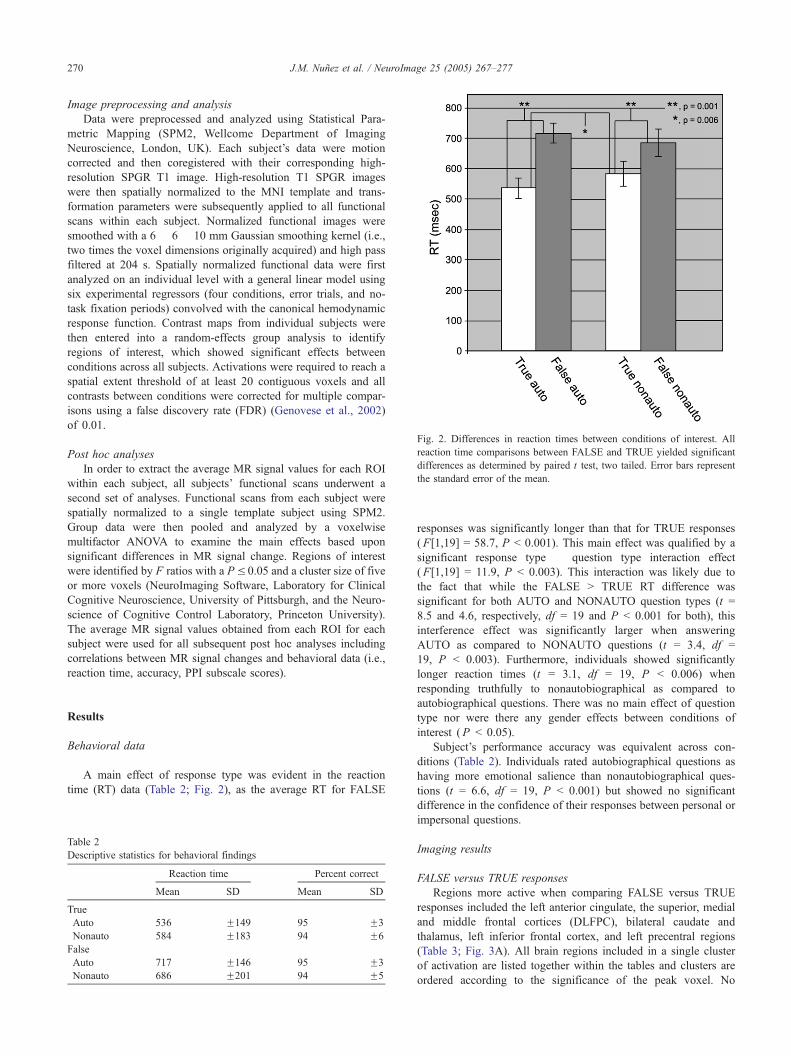
Fig. 2. Differences in reaction times between conditions of interest. All
reaction time comparisons between FALSE and TRUE yielded significant
differences as determined by paired t test, two tailed. Error bars represent
the standard error of the mean.
J.M. Nunez et al. / NeuroImage 25 (2005) 267–277270
Image preprocessing and analysis
Data were preprocessed and analyzed using Statistical Para-
metric Mapping (SPM2, Wellcome Department of Imaging
Neuroscience, London, UK). Each subject’s data were motion
corrected and then coregistered with their corresponding high-
resolution SPGR T1 image. High-resolution T1 SPGR images
were then spatially normalized to the MNI template and trans-
formation parameters were subsequently applied to all functional
scans within each subject. Normalized functional images were
smoothed with a 6 � 6 � 10 mm Gaussian smoothing kernel (i.e.,
two times the voxel dimensions originally acquired) and high pass
filtered at 204 s. Spatially normalized functional data were first
analyzed on an individual level with a general linear model using
six experimental regressors (four conditions, error trials, and no-
task fixation periods) convolved with the canonical hemodynamic
response function. Contrast maps from individual subjects were
then entered into a random-effects group analysis to identify
regions of interest, which showed significant effects between
conditions across all subjects. Activations were required to reach a
spatial extent threshold of at least 20 contiguous voxels and all
contrasts between conditions were corrected for multiple compar-
isons using a false discovery rate (FDR) (Genovese et al., 2002)
of 0.01.
Post hoc analyses
In order to extract the average MR signal values for each ROI
within each subject, all subjects’ functional scans underwent a
second set of analyses. Functional scans from each subject were
spatially normalized to a single template subject using SPM2.
Group data were then pooled and analyzed by a voxelwise
multifactor ANOVA to examine the main effects based upon
significant differences in MR signal change. Regions of interest
were identified by F ratios with a P V 0.05 and a cluster size of five
or more voxels (NeuroImaging Software, Laboratory for Clinical
Cognitive Neuroscience, University of Pittsburgh, and the Neuro-
science of Cognitive Control Laboratory, Princeton University).
The average MR signal values obtained from each ROI for each
subject were used for all subsequent post hoc analyses including
correlations between MR signal changes and behavioral data (i.e.,
reaction time, accuracy, PPI subscale scores).
Results
Behavioral data
A main effect of response type was evident in the reaction
time (RT) data (Table 2; Fig. 2), as the average RT for FALSE
Table 2
Descriptive statistics for behavioral findings
Reaction time Percent correct
Mean SD Mean SD
True
Auto 536 F149 95 F3
Nonauto 584 F183 94 F6
False
Auto 717 F146 95 F3
Nonauto 686 F201 94 F5
responses was significantly longer than that for TRUE responses
(F[1,19] = 58.7, P b 0.001). This main effect was qualified by a
significant response type � question type interaction effect
(F[1,19] = 11.9, P b 0.003). This interaction was likely due to
the fact that while the FALSE N TRUE RT difference was
significant for both AUTO and NONAUTO question types (t =
8.5 and 4.6, respectively, df = 19 and P b 0.001 for both), this
interference effect was significantly larger when answering
AUTO as compared to NONAUTO questions (t = 3.4, df =
19, P b 0.003). Furthermore, individuals showed significantly
longer reaction times (t = 3.1, df = 19, P b 0.006) when
responding truthfully to nonautobiographical as compared to
autobiographical questions. There was no main effect of question
type nor were there any gender effects between conditions of
interest (P b 0.05).
Subject’s performance accuracy was equivalent across con-
ditions (Table 2). Individuals rated autobiographical questions as
having more emotional salience than nonautobiographical ques-
tions (t = 6.6, df = 19, P b 0.001) but showed no significant
difference in the confidence of their responses between personal or
impersonal questions.
Imaging results
FALSE versus TRUE responses
Regions more active when comparing FALSE versus TRUE
responses included the left anterior cingulate, the superior, medial
and middle frontal cortices (DLFPC), bilateral caudate and
thalamus, left inferior frontal cortex, and left precentral regions
(Table 3; Fig. 3A). All brain regions included in a single cluster
of activation are listed together within the tables and clusters are
ordered according to the significance of the peak voxel. No
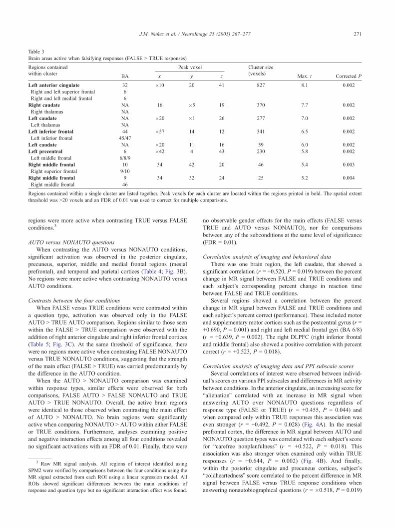
Table 3
Brain areas active when falsifying responses (FALSE N TRUE responses)
Regions contained Peak voxel Cluster size
within clusterBA x y z
(voxels)Max. t Corrected P
Left anterior cingulate 32 �10 20 41 827 8.1 0.002
Right and left superior frontal 6
Right and left medial frontal 6
Right caudate NA 16 �5 19 370 7.7 0.002
Right thalamus NA
Left caudate NA �20 �1 26 277 7.0 0.002
Left thalamus NA
Left inferior frontal 44 �57 14 12 341 6.5 0.002
Left inferior frontal 45/47
Left caudate NA �20 11 16 59 6.0 0.002
Left precentral 6 �42 4 43 230 5.8 0.002
Left middle frontal 6/8/9
Right middle frontal 10 34 42 20 46 5.4 0.003
Right superior frontal 9/10
Right middle frontal 9 34 32 24 25 5.2 0.004
Right middle frontal 46
Regions contained within a single cluster are listed together. Peak voxels for each cluster are located within the regions printed in bold. The spatial extent
threshold was N20 voxels and an FDR of 0.01 was used to correct for multiple comparisons.
J.M. Nunez et al. / NeuroImage 25 (2005) 267–277 271
regions were more active when contrasting TRUE versus FALSE
conditions.3
AUTO versus NONAUTO questions
When contrasting the AUTO versus NONAUTO conditions,
significant activation was observed in the posterior cingulate,
precuneus, superior, middle and medial frontal regions (mesial
prefrontal), and temporal and parietal cortices (Table 4; Fig. 3B).
No regions were more active when contrasting NONAUTO versus
AUTO conditions.
Contrasts between the four conditions
When FALSE versus TRUE conditions were contrasted within
a question type, activation was observed only in the FALSE
AUTO N TRUE AUTO comparison. Regions similar to those seen
within the FALSE N TRUE comparison were observed with the
addition of right anterior cingulate and right inferior frontal cortices
(Table 5; Fig. 3C). At the same threshold of significance, there
were no regions more active when contrasting FALSE NONAUTO
versus TRUE NONAUTO conditions, suggesting that the strength
of the main effect (FALSE N TRUE) was carried predominantly by
the difference in the AUTO condition.
When the AUTO N NONAUTO comparison was examined
within response types, similar effects were observed for both
comparisons, FALSE AUTO N FALSE NONAUTO and TRUE
AUTO N TRUE NONAUTO. Overall, the active brain regions
were identical to those observed when contrasting the main effect
of AUTO N NONAUTO. No brain regions were significantly
active when comparing NONAUTO N AUTO within either FALSE
or TRUE conditions. Furthermore, analyses examining positive
and negative interaction effects among all four conditions revealed
no significant activations with an FDR of 0.01. Finally, there were
3 Raw MR signal analysis. All regions of interest identified using
SPM2 were verified by comparisons between the four conditions using the
MR signal extracted from each ROI using a linear regression model. All
ROIs showed significant differences between the main conditions of
response and question type but no significant interaction effect was found.
no observable gender effects for the main effects (FALSE versus
TRUE and AUTO versus NONAUTO), nor for comparisons
between any of the subconditions at the same level of significance
(FDR = 0.01).
Correlation analysis of imaging and behavioral data
There was one brain region, the left caudate, that showed a
significant correlation (r = +0.520, P = 0.019) between the percent
change in MR signal between FALSE and TRUE conditions and
each subject’s corresponding percent change in reaction time
between FALSE and TRUE conditions.
Several regions showed a correlation between the percent
change in MR signal between FALSE and TRUE conditions and
each subject’s percent correct (performance). These included motor
and supplementary motor cortices such as the postcentral gyrus (r =
+0.690, P = 0.001) and right and left medial frontal gyri (BA 6/8)
(r = +0.639, P = 0.002). The right DLPFC (right inferior frontal
and middle frontal) also showed a positive correlation with percent
correct (r = +0.523, P = 0.018).
Correlation analysis of imaging data and PPI subscale scores
Several correlations of interest were observed between individ-
ual’s scores on various PPI subscales and differences in MR activity
between conditions. In the anterior cingulate, an increasing score for
balienationQ correlated with an increase in MR signal when
answering AUTO over NONAUTO questions regardless of
response type (FALSE or TRUE) (r = +0.455, P = 0.044) and
when compared only within TRUE responses this association was
even stronger (r = +0.492, P = 0.028) (Fig. 4A). In the mesial
prefrontal cortex, the difference in MR signal between AUTO and
NONAUTO question types was correlated with each subject’s score
for bcarefree nonplanfulnessQ (r = +0.522, P = 0.018). This
association was also stronger when examined only within TRUE
responses (r = +0.644, P = 0.002) (Fig. 4B). And finally,
within the posterior cingulate and precuneus cortices, subject’s
bcoldheartednessQ score correlated to the percent difference in MR
signal between FALSE versus TRUE response conditions when
answering nonautobiographical questions (r = �0.518, P = 0.019)
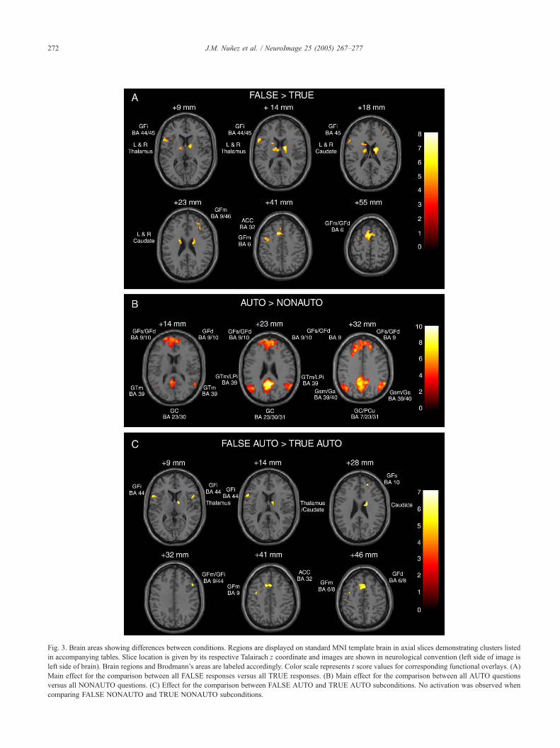
Fig. 3. Brain areas showing differences between conditions. Regions are displayed on standard MNI template brain in axial slices demonstrating clusters listed
in accompanying tables. Slice location is given by its respective Talairach z coordinate and images are shown in neurological convention (left side of image is
left side of brain). Brain regions and Brodmann’s areas are labeled accordingly. Color scale represents t score values for corresponding functional overlays. (A)
Main effect for the comparison between all FALSE responses versus all TRUE responses. (B) Main effect for the comparison between all AUTO questions
versus all NONAUTO questions. (C) Effect for the comparison between FALSE AUTO and TRUE AUTO subconditions. No activation was observed when
comparing FALSE NONAUTO and TRUE NONAUTO subconditions.
J.M. Nunez et al. / NeuroImage 25 (2005) 267–277272
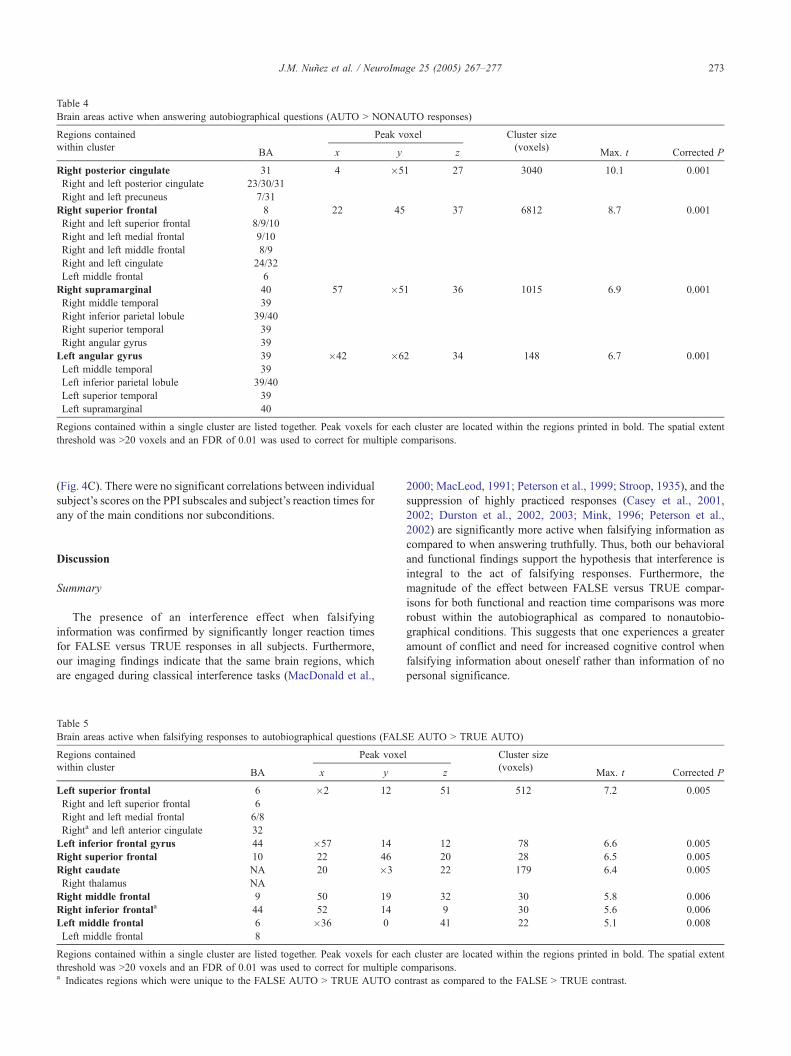
Table 4
Brain areas active when answering autobiographical questions (AUTO N NONAUTO responses)
Regions contained Peak voxel Cluster size
within clusterBA x y z
(voxels)Max. t Corrected P
Right posterior cingulate 31 4 �51 27 3040 10.1 0.001
Right and left posterior cingulate 23/30/31
Right and left precuneus 7/31
Right superior frontal 8 22 45 37 6812 8.7 0.001
Right and left superior frontal 8/9/10
Right and left medial frontal 9/10
Right and left middle frontal 8/9
Right and left cingulate 24/32
Left middle frontal 6
Right supramarginal 40 57 �51 36 1015 6.9 0.001
Right middle temporal 39
Right inferior parietal lobule 39/40
Right superior temporal 39
Right angular gyrus 39
Left angular gyrus 39 �42 �62 34 148 6.7 0.001
Left middle temporal 39
Left inferior parietal lobule 39/40
Left superior temporal 39
Left supramarginal 40
Regions contained within a single cluster are listed together. Peak voxels for each cluster are located within the regions printed in bold. The spatial extent
threshold was N20 voxels and an FDR of 0.01 was used to correct for multiple comparisons.
J.M. Nunez et al. / NeuroImage 25 (2005) 267–277 273
(Fig. 4C). There were no significant correlations between individual
subject’s scores on the PPI subscales and subject’s reaction times for
any of the main conditions nor subconditions.
Discussion
Summary
The presence of an interference effect when falsifying
information was confirmed by significantly longer reaction times
for FALSE versus TRUE responses in all subjects. Furthermore,
our imaging findings indicate that the same brain regions, which
are engaged during classical interference tasks (MacDonald et al.,
Table 5
Brain areas active when falsifying responses to autobiographical questions (FALS
Regions contained Peak voxe
within clusterBA x y
Left superior frontal 6 �2 12
Right and left superior frontal 6
Right and left medial frontal 6/8
Righta and left anterior cingulate 32
Left inferior frontal gyrus 44 �57 14
Right superior frontal 10 22 46
Right caudate NA 20 �3
Right thalamus NA
Right middle frontal 9 50 19
Right inferior frontala 44 52 14
Left middle frontal 6 �36 0
Left middle frontal 8
Regions contained within a single cluster are listed together. Peak voxels for eac
threshold was N20 voxels and an FDR of 0.01 was used to correct for multiple ca Indicates regions which were unique to the FALSE AUTO N TRUE AUTO co
2000; MacLeod, 1991; Peterson et al., 1999; Stroop, 1935), and the
suppression of highly practiced responses (Casey et al., 2001,
2002; Durston et al., 2002, 2003; Mink, 1996; Peterson et al.,
2002) are significantly more active when falsifying information as
compared to when answering truthfully. Thus, both our behavioral
and functional findings support the hypothesis that interference is
integral to the act of falsifying responses. Furthermore, the
magnitude of the effect between FALSE versus TRUE compar-
isons for both functional and reaction time comparisons was more
robust within the autobiographical as compared to nonautobio-
graphical conditions. This suggests that one experiences a greater
amount of conflict and need for increased cognitive control when
falsifying information about oneself rather than information of no
personal significance.
E AUTO N TRUE AUTO)
l Cluster size
z(voxels)
Max. t Corrected P
51 512 7.2 0.005
12 78 6.6 0.005
20 28 6.5 0.005
22 179 6.4 0.005
32 30 5.8 0.006
9 30 5.6 0.006
41 22 5.1 0.008
h cluster are located within the regions printed in bold. The spatial extent
omparisons.
ntrast as compared to the FALSE N TRUE contrast.
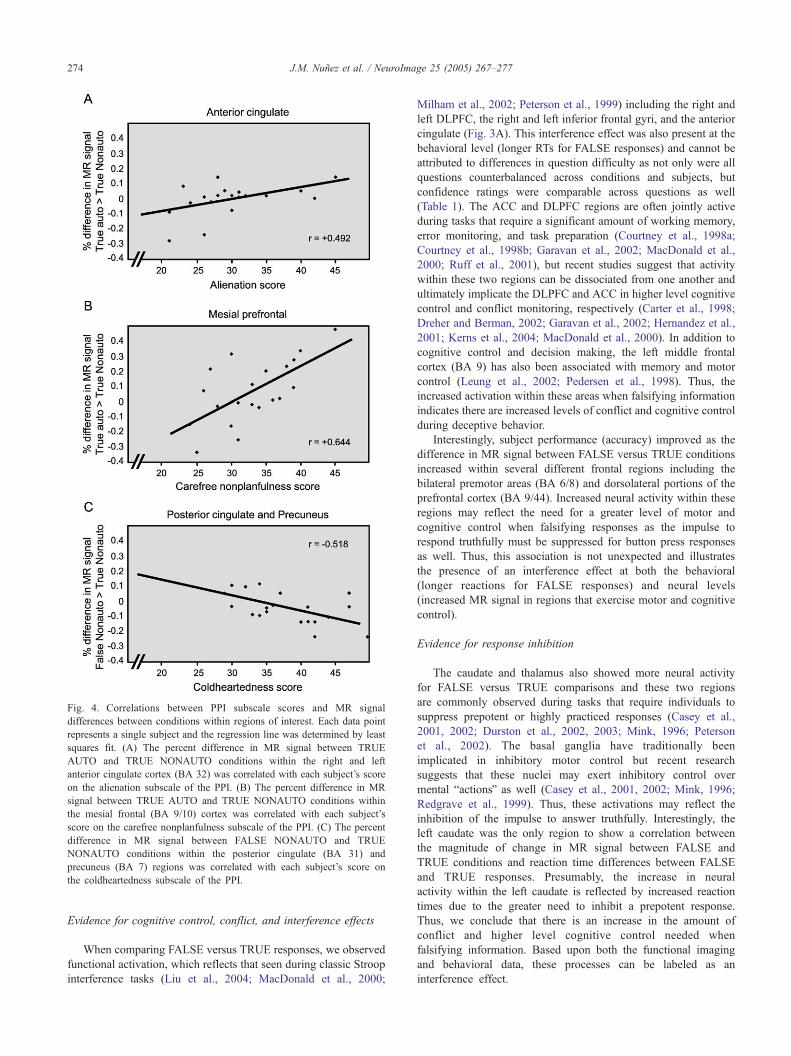
Fig. 4. Correlations between PPI subscale scores and MR signal
differences between conditions within regions of interest. Each data point
represents a single subject and the regression line was determined by least
squares fit. (A) The percent difference in MR signal between TRUE
AUTO and TRUE NONAUTO conditions within the right and left
anterior cingulate cortex (BA 32) was correlated with each subject’s score
on the alienation subscale of the PPI. (B) The percent difference in MR
signal between TRUE AUTO and TRUE NONAUTO conditions within
the mesial frontal (BA 9/10) cortex was correlated with each subject’s
score on the carefree nonplanfulness subscale of the PPI. (C) The percent
difference in MR signal between FALSE NONAUTO and TRUE
NONAUTO conditions within the posterior cingulate (BA 31) and
precuneus (BA 7) regions was correlated with each subject’s score on
the coldheartedness subscale of the PPI.
J.M. Nunez et al. / NeuroImage 25 (2005) 267–277274
Evidence for cognitive control, conflict, and interference effects
When comparing FALSE versus TRUE responses, we observed
functional activation, which reflects that seen during classic Stroop
interference tasks (Liu et al., 2004; MacDonald et al., 2000;
Milham et al., 2002; Peterson et al., 1999) including the right and
left DLPFC, the right and left inferior frontal gyri, and the anterior
cingulate (Fig. 3A). This interference effect was also present at the
behavioral level (longer RTs for FALSE responses) and cannot be
attributed to differences in question difficulty as not only were all
questions counterbalanced across conditions and subjects, but
confidence ratings were comparable across questions as well
(Table 1). The ACC and DLPFC regions are often jointly active
during tasks that require a significant amount of working memory,
error monitoring, and task preparation (Courtney et al., 1998a;
Courtney et al., 1998b; Garavan et al., 2002; MacDonald et al.,
2000; Ruff et al., 2001), but recent studies suggest that activity
within these two regions can be dissociated from one another and
ultimately implicate the DLPFC and ACC in higher level cognitive
control and conflict monitoring, respectively (Carter et al., 1998;
Dreher and Berman, 2002; Garavan et al., 2002; Hernandez et al.,
2001; Kerns et al., 2004; MacDonald et al., 2000). In addition to
cognitive control and decision making, the left middle frontal
cortex (BA 9) has also been associated with memory and motor
control (Leung et al., 2002; Pedersen et al., 1998). Thus, the
increased activation within these areas when falsifying information
indicates there are increased levels of conflict and cognitive control
during deceptive behavior.
Interestingly, subject performance (accuracy) improved as the
difference in MR signal between FALSE versus TRUE conditions
increased within several different frontal regions including the
bilateral premotor areas (BA 6/8) and dorsolateral portions of the
prefrontal cortex (BA 9/44). Increased neural activity within these
regions may reflect the need for a greater level of motor and
cognitive control when falsifying responses as the impulse to
respond truthfully must be suppressed for button press responses
as well. Thus, this association is not unexpected and illustrates
the presence of an interference effect at both the behavioral
(longer reactions for FALSE responses) and neural levels
(increased MR signal in regions that exercise motor and cognitive
control).
Evidence for response inhibition
The caudate and thalamus also showed more neural activity
for FALSE versus TRUE comparisons and these two regions
are commonly observed during tasks that require individuals to
suppress prepotent or highly practiced responses (Casey et al.,
2001, 2002; Durston et al., 2002, 2003; Mink, 1996; Peterson
et al., 2002). The basal ganglia have traditionally been
implicated in inhibitory motor control but recent research
suggests that these nuclei may exert inhibitory control over
mental bactionsQ as well (Casey et al., 2001, 2002; Mink, 1996;
Redgrave et al., 1999). Thus, these activations may reflect the
inhibition of the impulse to answer truthfully. Interestingly, the
left caudate was the only region to show a correlation between
the magnitude of change in MR signal between FALSE and
TRUE conditions and reaction time differences between FALSE
and TRUE responses. Presumably, the increase in neural
activity within the left caudate is reflected by increased reaction
times due to the greater need to inhibit a prepotent response.
Thus, we conclude that there is an increase in the amount of
conflict and higher level cognitive control needed when
falsifying information. Based upon both the functional imaging
and behavioral data, these processes can be labeled as an
interference effect.

J.M. Nunez et al. / NeuroImage 25 (2005) 267–277 275
Augmented conflict and cognitive control for personal information
When analyses were restricted to question type (AUTO and
NONAUTO), a significant effect was observed between FALSE N
TRUE responses only when comparing autobiographical questions
(FALSE AUTO N TRUE AUTO). Thus, an increase in neural
activity was observed across both question types when falsifying
information but the effect was more robust when examining
autobiographical questions alone. This was accompanied by a
larger difference in FALSE N TRUE reaction time for autobio-
graphical questions. These results are important for several
reasons. First, they provide evidence that the simple element of
personal relevance noticeably influences patterns of behavioral and
neural activity within the context of deceptive behavior. Second,
they suggest that there is a difference in the amount of conflict one
experiences when falsifying personal information as well as the
amount of cognitive control needed to successfully implement a
response. And finally, future functional imaging studies of
deception may benefit by exploiting the increased power self-
relevant stimuli affords and thus greatly enhance the sensitivity of
experimental designs.
Brain areas that are engaged during self- versus
non-self-discriminations
Interestingly, when contrasting the AUTO versus NONAUTO
conditions, we observed robust and distinct activations in the
superior and medial frontal regions (mesial prefrontal), the
posterior cingulate, precuneus, and temporal and parietal cortices
(Table 4; Fig. 3B). The pattern of functional activation seen for
this comparison is strikingly similar to that observed in a recent
study (Greene et al., 2001), which contrasted the brain
activations evoked when individuals were faced with different
kinds of moral dilemmas. The moral dilemmas in Greene’s study
were divided into bmoral–personal Q and bmoral–impersonalQconditions and each decision was categorized according to how
closely (personally) the subject would be involved in committing
a morally questionable act. The authors suggest that the different
emotional response elicited by each of the dilemma types was
responsible for this pattern of neural activity and indeed these
regions have all been implicated in emotional processing (Lane
et al., 1997a, 1997b; Maddock, 1999; Maddock et al., 2003).
Although these regions may be involved during emotional or
moral reasoning processes, our experiment employed a task in
which the stimuli differed only between self- versus non-self-
attributes, and nevertheless we observed the same pattern of
neural activity. However, subjects within our study consistently
rated autobiographical questions as more emotionally salient than
nonautobiographical questions, and thus the possibility remains
that the recruitment of these regions could be due to either the
self-referential content of the stimuli or their emotional salience.
But the difference in emotional salience ratings between the two
categories was small (Table 1) and recent imaging studies which
have focused on autobiographical processing, not emotional nor
ethical content, have produced equally similar results (Vogeley
and Fink, 2003; Vogeley et al., 2004). Thus, based upon our
findings and recent autobiographical research, the pattern of
brain activation seen for AUTO versus NONAUTO comparisons
is most likely due to the differences in the self-referential
content of the stimuli rather than differences in emotional or
moral reasoning.
Relationships between personality traits and imaging findings
Within the regions identified above, three showed correlations
with the subscales of the Psychopathic Personality Inventory
(Lilienfeld and Andrews, 1996), including the anterior cingulate,
mesial prefrontal, and posterior cingulate/precuneus regions. In the
anterior cingulate, the difference in MR signal between AUTO
versus NONAUTO conditions correlated with each subject’s
balienationQ score and this association was even stronger when
compared within TRUE responses (Fig. 4A) but did not retain
significance when compared within FALSE responses. Individuals
who score highly on the balienationQ subscale tend to have various
paranoid or insecure personality features. For example, they often
feel like they have a lot of bad luck or that they are not as
successful as they could be because others perceive them unfairly,
and in general, they feel misunderstood by others. Increased neural
activity within the ACC when answering autobiographical as
compared to nonautobiographical questions may reflect the
experience of a higher level of conflict when these individuals
are faced with questions that are of a self-relevant nature (AUTO)
due to their naturally underlying issues with self-perception and
tendency towards feelings of persecution.
A similar association was shown within the mesial prefrontal
cortex (BA 9/10) for MR signal differences between AUTO versus
NONAUTO conditions and subject’s bcarefree nonplanfulnessQscores. This correlation was also stronger when compared within
TRUE AUTO and TRUE NONAUTO conditions (Fig. 4B) and did
not retain significance when comparing FALSE AUTO and FALSE
NONAUTO conditions. The mesial prefrontal area has been
implicated in both self-identity and emotional processing (Gilboa,
2004; Lane et al., 1997a, 1997b; Piefke et al., 2003; Vogeley and
Fink, 2003; Vogeley et al., 2004) and thus differences in levels of
neural activity as a function of self and nonself are expected. A high
score for the bcarefree nonplanfulnessQ subscale would be common
for individuals who are less inclined to monitor or evaluate their own
behavior, generally irresponsible, give up easily, often make the
same mistakes over and over again and have very little foresight,
planning skills, or thought for others. Thus, subjects who displayed
stronger tendencies for these traits showed more neural activity
within the mesial prefrontal cortex when having to evaluate truthful
statements about themselves versus nonself statements.
Finally, within the posterior cingulate (BA 23/30/31) and
precuneus cortices (BA 7), the difference in MR signal between
FALSE NONAUTO and TRUE NONAUTO conditions showed a
negative correlation with subject’s bcoldheartednessQ scores (Fig.
4C). Individuals who score highly on this scale are relatively
unemotional, guiltless, do not have many deep interpersonal
relationships or attachments, and are generally not concerned with
others’ feelings nor their effect on others. Our results support
previous studies (Maddock et al., 2001; Vogeley and Fink, 2003;
Vogeley et al., 2004), which suggest that these regions are
responding to the self-relevance of the stimuli and one possibility
is that individuals who score highly on the bcoldheartednessQsubscale experience a lesser degree of self-identification when
lying about matters that do not concern them. Thus, individuals
who are generally coldhearted may find it easier to engage in
deceptive behaviors when it involves matters that are of no
personal consequence. Interestingly, although the mesial prefrontal
cortex and posterior cingulate/precuneus regions are often seen
jointly active during tasks that incorporate autobiographical versus
nonautobiographical stimuli, we observed a dissociation in their

J.M. Nunez et al. / NeuroImage 25 (2005) 267–277276
patterns of functional activity and individual’s scores on the
bcoldheartednessQ and bcarefree nonplanfulnessQ subscales of the
PPI. This suggests that even within the general category of self-
referential stimuli, these two regions may be further specialized in
either their response to similar stimuli (e.g., AUTO versus
NONAUTO when responding truthfully; Fig. 4B) or when
different response patterns are required to the same stimuli (e.g.,
FALSE versus TRUE responding to nonautobiographical ques-
tions; Fig. 4C). Thus, although both regions clearly process self-
referential stimuli, the mesial prefrontal region appears to be more
highly specialized for self- versus non-self-comparisons whereas
the posterior cingulate/precuneus region shows broader capabilities
for discrimination between stimuli which are not self-relevant.
Conclusion
This study demonstrates that there is increased neural activity
within the anterior cingulate, dorsolateral prefrontal cortex, and
caudate and thalamic nuclei when individuals answer falsely as
compared to truthfully. Based upon these functional results and the
behavioral findings (longer RTs for FALSE versus TRUE
responses), we conclude that interference, similar to that observed
during the Stroop and other tasks that measure various aspects of
response inhibition and choice selection, is inherent to the act of
falsifying information and thus certain types of deceptive behavior.
Furthermore, both functional activity and reaction time data
showed greater differences between FALSE and TRUE compar-
isons for autobiographical stimuli as compared to nonautobio-
graphical stimuli (although the interaction effect was not
significant for functional activity). Thus, not only is there an
interference effect when falsifying information, but the amount of
conflict and cognitive control needed to successfully execute false
responses is greater when individuals are faced with self-relevant
circumstances. Also, the presence of a correlation for reaction time
differences and MR signal differences within the left caudate
nucleus between FALSE versus TRUE conditions similarly
suggests there is an increased need for response inhibition when
falsifying responses. And finally, certain measurable personality
traits, as determined by the Psychopathic Personality Inventory,
show a relationship to patterns of neural activity within the anterior
cingulate, mesial prefrontal cortex, and posterior cingulate/precu-
neus regions and indicate that regions that are engaged when
processing self-referential information (mesial prefrontal cortex
and posterior cingulate/precuneus regions) may be further speci-
alized to handle different aspects of autobiographical information.
In conclusion, this is the first study to demonstrate the presence of
an interference effect at both the behavioral and functional levels
using a deception-based paradigm, thus providing a new founda-
tion for future deception research as well as presenting a new
medium for the study of conflict and cognitive control processes.
Acknowledgments
This work was supported by the Cornell/Rockefeller/Memorial
Sloan-Kettering Tri-Institutional MD/PhD program, NIH MSTP
grant GM07739, and the Charles A. Dana Foundation. This work
has been included as a partial fulfillment of the requirements for a
PhD degree granted to the first author. The authors would like to
thank Scott O. Lilienfeld for supplying the Psychopathic Person-
ality Inventory and Olaf Andersen, George Reeke, and Rae Silver
for their invaluable input, thoughtful criticism, and overall support
of this work.
References
Ben-Shakhar, G., Dolev, K., 1996. Psychophysiological detection through
the guilty knowledge technique: effects of mental countermeasures.
J. Appl. Psychol. 81, 273–281.
Botvinick, M., Nystrom, L.E., Fissell, K., Carter, C.S., Cohen, J.D., 1999.
Conflict monitoring versus selection-for-action in anterior cingulate
cortex. Nature 402, 179–181.
Botvinick, M.M., Braver, T.S., Barch, D.M., Carter, C.S., Cohen, J.D.,
2001. Conflict monitoring and cognitive control. Psychol. Rev. 108,
624–652.
Bradley, M.T., Cullen, M.C., 1993. Polygraph lie detection on real events in
a laboratory setting. Percept. Mot. Skills 76, 1051–1058.
Braver, T.S., Reynolds, J.R., Donaldson, D.I., 2003. Neural mechanisms of
transient and sustained cognitive control during task switching. Neuron
39, 713–726.
Buller, D.B., Burgoon, J.K., 1996. Interpersonal deception theory.
Commun. Theory 3, 203–242.
Carter, C.S., Braver, T.S., Barch, D.M., Botvinick, M.M., Noll, D., Cohen,
J.D., 1998. Anterior cingulate cortex, error detection, and the online
monitoring of performance. Science 280, 747–749.
Casey, B.J., Forman, S.D., Franzen, P., Berkowitz, A., Braver, T.S.,
Nystrom, L.E., Thomas, K.M., Noll, D.C., 2001. Sensitivity of
prefrontal cortex to changes in target probability: a functional MRI
study. Hum. Brain Mapp. 13, 26–33.
Casey, B.J., Thomas, K.M., Davidson, M.C., Kunz, K., Franzen, P.L., 2002.
Dissociating striatal and hippocampal function developmentally with a
stimulus–response compatibility task. J. Neurosci. 22, 8647–8652.
Cohen, J.D., Dunbar, K., McClelland, J.L., 1990. On the control of
automatic processes: a parallel distributed processing account of the
Stroop effect. Psychol. Rev. 97, 332–361.
Coleman, L., Kay, P., 1981. Prototype semantics: the English word lie.
Language 57, 26–44.
Courtney, S.M., Petit, L., Haxby, J.V., Ungerleider, L.G., 1998a. The role of
prefrontal cortex in working memory: examining the contents of con-
sciousness. Philos. Trans. R. Soc. Lond., B Biol. Sci. 353, 1819–1828.
Courtney, S.M., Petit, L., Maisog, J.M., Ungerleider, L.G., Haxby, J.V.,
1998b. An area specialized for spatial working memory in human
frontal cortex. Science 279, 1347–1351.
DePaulo, B.M., Lindsay, J.J., Malone, B.E., Muhlenbruck, L., Charlton, K.,
Cooper, H., 2003. Cues to deception. Psychol. Bull. 129, 74–118.
Dionisio, D.P., Granholm, E., Hillix, W.A., Perrine, W.F., 2001. Differ-
entiation of deception using pupillary responses as an index of cognitive
processing. Psychophysiology 38, 205–211.
Dreher, J.C., Berman, K.F., 2002. Fractionating the neural substrate of
cognitive control processes. Proc. Natl. Acad. Sci. U. S. A. 99,
14595–14600.
Durston, S., Thomas, K.M., Worden, M.S., Yang, Y., Casey, B.J., 2002. The
effect of preceding context on inhibition: an event-related fMRI study.
NeuroImage 16, 449–453.
Durston, S., Davidson, M.C., Thomas, K.M., Worden, M.S., Tottenham, N.,
Martinez, A., Watts, R., Ulug, A.M., Casey, B.J., 2003. Parametric
manipulation of conflict and response competition using rapid mixed-
trial event-related fMRI. NeuroImage 20, 2135–2141.
Eriksen, B.A., Eriksen, C.W., 1974. Effects of noise letters upon the
identification of a target letter in a nonsearch task. Percept. Psychophys.
16, 143–149.
Fink, G.R., Markowitsch, H.J., Reinkemeier, M., Bruckbauer, T., Kessler,
J., Heiss, W.D., 1996. Cerebral representation of one’s own past:
neural networks involved in autobiographical memory. J. Neurosci.
16, 4275–4282.

J.M. Nunez et al. / NeuroImage 25 (2005) 267–277 277
Ganis, G., Kosslyn, S.M., Stose, S., Thompson, W.L., Yurgelun-Todd,
D.A., 2003. Neural correlates of different types of deception: an fMRI
investigation. Cereb. Cortex 13, 830–836.
Garavan, H., Ross, T.J., Murphy, K., Roche, R.A., Stein, E.A., 2002.
Dissociable executive functions in the dynamic control of behavior:
inhibition, error detection, and correction. NeuroImage 17, 1820–1829.
Genovese, C.R., Lazar, N.A., Nichols, T., 2002. Thresholding of statistical
maps in functional neuroimaging using the false discovery rate.
NeuroImage 15, 870–878.
Gilboa, A., 2004. Autobiographical and episodic memory-one and the
same? Evidence from prefrontal activation in neuroimaging studies.
Neuropsychologia 42, 1336–1349.
Godert, H.W., Rill, H.G., Vossel, G., 2001. Psychophysiological differen-
tiation of deception: the effects of electrodermal liability and mode of
responding on skin conductance and heart rate. Int. J. Psychophysiol.
40, 61–75.
Greene, J.D., Sommerville, R.B., Nystrom, L.E., Darley, J.M., Cohen, J.D.,
2001. An fMRI investigation of emotional engagement in moral
judgment. Science 293, 2105–2108.
Hernandez, A.E., Dapretto, M., Mazziotta, J., Bookheimer, S., 2001.
Language switching and language representation in Spanish-English
bilinguals: an fMRI study. NeuroImage 14, 510–520.
Kerns, J.G., Cohen, J.D., MacDonald III, A.W., Cho, R.Y., Stenger, V.A.,
Carter, C.S., 2004. Anterior cingulate conflict monitoring and adjust-
ments in control. Science 303, 1023–1026.
Kircher, J.C., Raskin, D.C., 1988. Human versus computerized evalua-
tions of polygraph data in a laboratory setting. J. Appl. Psychol. 73,
291–302.
Lane, R.D., Reiman, E.M., Ahern, G.L., Schwartz, G.E., Davidson, R.J.,
1997a. Neuroanatomical correlates of happiness, sadness, and disgust.
Am. J. Psychiatry 154, 926–933.
Lane, R.D., Reiman, E.M., Bradley, M.M., Lang, P.J., Ahern, G.L.,
Davidson, R.J., Schwartz, G.E., 1997b. Neuroanatomical correlates of
pleasant and unpleasant emotion. Neuropsychologia 35, 1437–1444.
Langleben, D.D., Schroeder, L., Maldjian, J.A., Gur, R.C., McDonald, S.,
Ragland, J.D., O’Brien, C.P., Childress, A.R., 2002. Brain activity
during simulated deception: an event-related functional magnetic
resonance study. NeuroImage 15, 727–732.
Lee, T.M.C., Liu, H., Tan, L., Chan, C.C.H., Mahankali, S., Feng, C., Hou,
J., Fox, P.T., Gao, J., 2002. Lie detection by functional magnetic
resonance imaging. Hum. Brain Mapp. 15, 157–164.
Leung, H.C., Gore, J.C., Goldman-Rakic, P.S., 2002. Sustained mnemonic
response in the human middle frontal gyrus during on-line storage of
spatial memoranda. J. Cogn. Neurosci. 14, 659–671.
Lilienfeld, S.O., Andrews, B.P., 1996. Development and preliminary
validation of a self-report measure of psychopathic personality traits
in noncriminal populations. J. Pers. Assess. 66, 488–524.
Liu, X., Banich, M.T., Jacobson, B.L., Tanabe, J.L., 2004. Common and
distinct neural substrates of attentional control in an integrated Simon
and spatial Stroop task as assessed by event-related fMRI. NeuroImage
22, 1097–1106.
Lykken, D.T., 1959. The GSR in the detection of guilt. J. Appl. Psychol. 43,
385–388.
Lykken, D.T., 1960. The validity of the guilty knowledge technique: the
effect of faking. J. Appl. Psychol. 44, 258–262.
MacDonald III, A.W., Cohen, J.D., Stenger, V.A., Carter, C.S., 2000.
Dissociating the role of the dorsolateral prefrontal and anterior cingulate
cortex in cognitive control. Science 288, 1835–1838.
MacLeod, C.M., 1991. Half a century of research on the Stroop effect: an
integrative review. Psychol. Bull. 109, 163–203.
Maddock, R.J., 1999. The retrosplenial cortex and emotion: new insights
from functional neuroimaging of the human brain. Trends Neurosci. 22,
310–316.
Maddock, R.J., Garrett, A.S., Buonocore, M.H., 2001. Remembering
familiar people: the posterior cingulate cortex and autobiographical
memory retrieval. Neuroscience 104, 667–676.
Maddock, R.J., Garrett, A.S., Buonocore, M.H., 2003. Posterior cingulate
cortex activation by emotional words: fMRI evidence from a valence
decision task. Hum. Brain Mapp. 18, 30–41.
Maguire, E.A., Frith, C.D., 2003. Lateral asymmetry in the hippocampal
response to the remoteness of autobiographical memories. J. Neurosci.
23, 5302–5307.
McCornack, S.A., 1992. Information manipulation theory. Commun.
Monogr. 59, 1–16.
McCornack, S.A., 1997. The generation of deceptive messages: laying the
groundwork for a viable theory of interpersonal deception. In: Greene,
J.O. (Ed.), Message Production: Advances in Communication Theory.
Erlbaum, Mahwah, NJ, pp. 91–126.
Milham, M.P., Erickson, K.I., Banich, M.T., Kramer, A.F., Webb, A.,
Wszalek, T., Cohen, N.J., 2002. Attentional control in the aging
brain: insights from an fMRI study of the Stroop task. Brain Cogn.
49, 277–296.
Mink, J.W., 1996. The basal ganglia: focused selection and inhibition of
competing motor programs. Prog. Neurobiol. 50, 381–425.
Pedersen, J.R., Johannsen, P., Bak, C.K., Kofoed, B., Saermark, K., Gjedde,
A., 1998. Origin of human motor readiness field linked to left middle
frontal gyrus by MEG and PET. NeuroImage 8, 214–220.
Peterson, B.S., Skudlarski, P., Gatenby, J.C., Zhang, H., Anderson, A.W.,
Gore, J.C., 1999. An fMRI study of Stroop word-color interference:
evidence for cingulate subregions subserving multiple distributed
attentional systems. Biol. Psychiatry 45, 1237–1258.
Peterson, B.S., Kane, M.J., Alexander, G.M., Lacadie, C., Skudlarski, P.,
Leung, H.C., May, J., Gore, J.C., 2002. An event-related functional
MRI study comparing interference effects in the Simon and Stroop
tasks. Brain Res., Cogn. Brain Res. 13, 427–440.
Piefke, M., Weiss, P.H., Zilles, K., Markowitsch, H.J., Fink, G.R., 2003.
Differential remoteness and emotional tone modulate the neural
correlates of autobiographical memory. Brain 126, 650–668.
Podlesny, J.A., Raskin, D.C., 1977. Physiological measures and the
detection of deception. Psychol. Bull. 84, 782–799.
Redgrave, P., Prescott, T.J., Gurney, K., 1999. The basal ganglia: a
vertebrate solution to the selection problem. Neuroscience 89,
1009–1023.
Ruff, C.C., Woodward, T.S., Laurens, K.R., Liddle, P.F., 2001. The role of
the anterior cingulate cortex in conflict processing: evidence from
reverse Stroop interference. NeuroImage 14, 1150–1158.
Seymour, T.L., Seifert, C.M., Shafto, M.G., Mosmann, A.L., 2000. Using
response time measures to assess bguilty knowledgeQ. J. Appl. Psychol.85, 30–37.
Spence, S.A., Farrow, T.F.D., Herford, A.E., Wilkinson, I.D., Zheng, Y.,
Woodruff, P.W.R., 2001. Behavioural and functional anatomical
correlates of deception in human. NeuroReport 12, 2849–2853.
Stroop, J.R., 1935. Studies of interference in serial verbal reactions. J. Exp.
Psychol. 18, 643–662.
Vogeley, K., Fink, G.R., 2003. Neural correlates of the first-person-
perspective. Trends Cogn. Sci. 7, 38–42.
Vogeley, K., May, M., Ritzl, A., Falkai, P., Zilles, K., Fink, G.R., 2004.
Neural correlates of first-person perspective as one constituent of
human self-consciousness. J. Cogn. Neurosci. 16, 817–827.
Zuckerman, M., DePaulo, B.M., Rosenthal, R., 1981. Verbal and nonverbal
communication of deception. In: Berkowitz, L. (Ed.), Advances in
Experimental Social Psychology. Academic Press, New York, pp. 1–59.