Embed Size (px)
Citation preview
Intentional and Incidental Self-Control in Ventrolateral PFC
Jessica R. Cohen1, Elliot T. Berkman2 & Matthew D. Lieberman3
1Helen Wills Neuroscience Institute, University of California, Berkeley2Department of Psychology, University of Oregon
3Department of 1Psychology, University of California, Los Angeles
August 30, 2011
The ability to exert self-control over one’s thoughts and behaviors is crucial to successfully navigatingthe real world in a variety of domains, such as motor control (remaining in your seat during a boringlecture instead of jumping up and running outside), control over risky behavior (taking a sure optionso as not to risk losing money), control over immediate temptation (choosing to delay a payment soas to receive a larger one at a later date), and emotional control (remaining composed and suppressingthe desire to yell at someone who angered you). Each of these examples requires different actions tosuccessfully exert control over the more desirable yet detrimental action. There are multiple clinicaldisorders that are related to impairments in control such as ADHD, substance abuse, and pathologicalgambling. Given the serious problems that may occur if one has difficulty exerting behavioral or affectiveself-control, it is critical to understand the mechanisms behind successful self-control and how they arechanged when self-control ability is impaired.
As can be noted by the above examples, self-control is a broad concept that has been defined inmany different manners. A good, general definition is that self-control is “the overriding or inhibiting ofautomatic, habitual, or innate behaviors, urges, emotions, or desires that would otherwise interfere withgoal directed behavior” (Muraven et al., 2006). As is noted from this definition, many different methodscan be used to study self-control, ranging from inhibiting a motor response to regulating an emotion tosuppressing the temptation to eat sweets. In addition to these explicit, intentional forms of self-control,it is possible to exert control without an explicit goal to do so (i.e., automatically or incidentally) giventhe right situation. For example, in priming paradigms participants are not explicitly aware that they sawa prime, but the implicit encoding of primes can cause incidental behavioral control. Additionally, it ispossible to implicitly or incidentally regulate an affective response without awareness (for a review, seeBerkman & Lieberman, 2009).
It has been asserted that self-control ability may be like a muscle: it is a limited resource that canbe fatigued with use, or trained to increase stamina (Muraven, 2010; Muraven & Baumeister, 2000;Muraven et al., 1999). Evidence for this assertion can be found in studies in which participants wererequired to first exert self-control in one domain, and then subsequently exert self-control in a differentdomain. The domains used were quite varied and included motor control (the stop-signal paradigm orsqueezing a handgrip), controlling one’s temptation to eat sweets or drink alcohol, and emotional control.It was consistently found that participants who were required to exert control two times in a row wereworse on the second control task than were those who performed a difficult task that did not requireself-control as their first task (such as solving mathematical problems or typing a paragraph quickly
1
without feedback; for a review, see Muraven & Baumeister, 2000). Moreover, not only was this self-control fatigue alleviated when participants practiced exerting self-control over an extended period oftime (Muraven et al., 1999), but baseline self-control ability improved with practice (Muraven, 2010).It is important to note that the type of self-control practiced did not matter; self-control was improvedacross domains.
This research implies that multiple forms of self-control may be subsumed under one general controlmechanism. Therefore, it is natural to turn to brain systems to determine whether different forms ofcontrol utilize the same, or at least overlapping, neural networks.
This chapter reviews the literature exploring the neural basis of self-control and asserts that the rightventrolateral prefrontal cortex (rVLPFC) is a neural region commonly recruited across many differentforms of self-control. As used here, self-control is operationalized as inhibitory impulse control. Thisis one of multiple subprocesses of executive, or cognitive, control (Lenartowicz et al., 2010; Sabb et al.,2008). This operationalization is motivated by the hypothesis addressed here that the rVLPFC underliesinhibitory control, thus for the remainder of this chapter we will use the term “self-control” to refer toinhibitory impulse control.
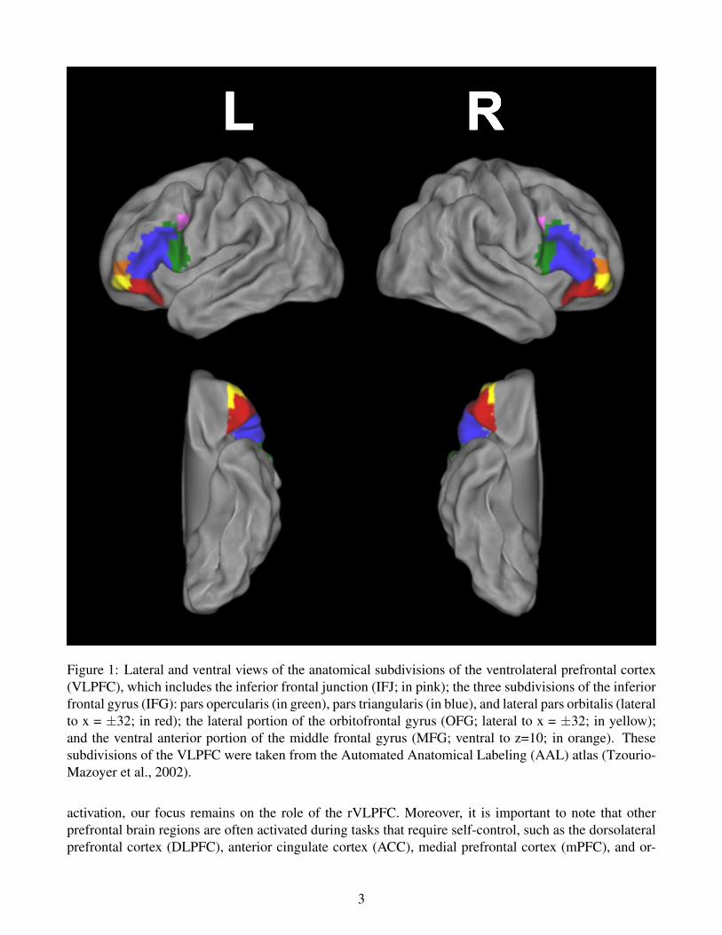
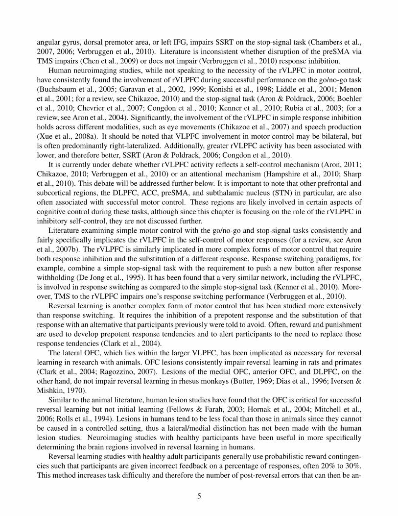
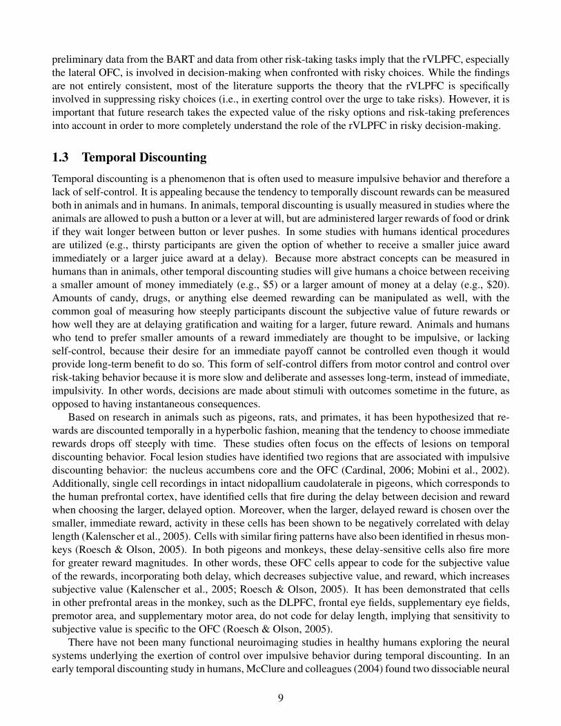
It is important to note that “ventrolateral prefrontal cortex” is a broad term that covers a wide swathof brain (Figure 1). We purposely use such a broad term to be consistent with the literature, whichrefers to a range of distinct brain regions that fall into the ventral and the lateral prefrontal cortex. Theseregions include the inferior frontal junction (IFJ), the inferior frontal gyrus (IFG), the lateral orbitofrontalgyrus (OFG), and the ventral anterior middle frontal gyrus (MFG). The IFG can be further divided intothree subregions: the pars opercularis, pars triangularis, and pars orbitalis (Duvernoy, 1991). The IFJis defined as the junction of the inferior frontal sulcus and the inferior precentral sulcus (Derrfuss et al.,2004). When studies we discuss below report activity in subregions within the VLPFC we specify wherein the VLPFC the activity was localized.
In this chapter we will first discuss the common activation of the rVLPFC across many different formsof self-control. We will first focus on explicit, intentional self-control such as motor control, control overrisky behavior, the ability to delay gratification, and intentional emotion regulation. While self-controlliterature mostly limits itself to discussing these and other examples of intentional self-control, we arguethat implicit, incidental processes can be considered self-regulation, or self-control, as well. Therefore,we will then focus on incidental self-control including incidental affect regulation, incidental behavioralcontrol, and incidental pain regulation. Subsequently, we will discuss relevant anatomical and functionalconnections of the rVLPFC. Finally, we will review other hypothesized roles of the rVLPFC and attemptto resolve conflicting theories. We will conclude by stating that the rVLPFC is a brain region central toexecutive control that has different subdivisions with different roles. One main role of the rVLPFC is toexert self-control over behaviors.
1 The rVLPFC and Intentional Self-ControlThe rVLPFC is a strong candidate for a brain region that is central to exerting self-control. It is commonlyactivated across many different tasks requiring different forms of behavioral and affective self-control.It is also in a central location and well-connected to regions that may carry out control-related phenom-ena, such as motor control or emotional control. Historically, there has been a focus on the role of therVLPFC in intentional forms of self-control. While activity in healthy participants is sometimes bilateral,the VLPFC is significantly active in the left hemisphere less often than in the right hemisphere (see Figure2 and Table 1), and lesion and TMS studies point to the right, but not the left, VLPFC as being necessaryfor control (Aron et al., 2003; Chambers et al., 2007). Thus, while we mention relevant left VLPFC
2
L R
Figure 1: Lateral and ventral views of the anatomical subdivisions of the ventrolateral prefrontal cortex(VLPFC), which includes the inferior frontal junction (IFJ; in pink); the three subdivisions of the inferiorfrontal gyrus (IFG): pars opercularis (in green), pars triangularis (in blue), and lateral pars orbitalis (lateralto x = ±32; in red); the lateral portion of the orbitofrontal gyrus (OFG; lateral to x = ±32; in yellow);and the ventral anterior portion of the middle frontal gyrus (MFG; ventral to z=10; in orange). Thesesubdivisions of the VLPFC were taken from the Automated Anatomical Labeling (AAL) atlas (Tzourio-Mazoyer et al., 2002).
activation, our focus remains on the role of the rVLPFC. Moreover, it is important to note that otherprefrontal brain regions are often activated during tasks that require self-control, such as the dorsolateralprefrontal cortex (DLPFC), anterior cingulate cortex (ACC), medial prefrontal cortex (mPFC), and or-
3
bitofrontal cortex (OFC). However, these regions may be recruited for other, non-inhibitory self-control-related task demands, such as rule monitoring (DLPFC; Bunge, 2004), performance/conflict monitoring(ACC; Botvinick et al., 2004), or the processing of emotions or rewards (mPFC/OFC; Elliott et al., 2000;Ochsner & Gross, 2005). Therefore, the role of these regions in self-control related tasks will not bediscussed in depth here.
1.1 Motor ControlThe control of motor responses is an oft-studied form of self-control. Generally, motor response inhibi-tion is studied using the go/no-go (Casey et al., 1997b) and the stop-signal (Logan, 1994) tasks, in whichan intended motor response simply has to be suppressed. Motor control can also be more complex, how-ever, requiring the substitution of a novel response in addition to the suppression of the intended response.This is typically studied using response switching, or stop-change, paradigms (De Jong et al., 1995) orreversal learning paradigms (Clark et al., 2004).
Simple response inhibition tasks require participants to exert motor self-control by inhibiting a buttonpress to a stimulus when they perceive a signal to stop their response. By altering the proportion ofstimuli that are associated with stop-signals, the level of prepotency of responding can be manipulated.It becomes more difficult to inhibit a response when there are fewer stop-signals interspersed amongthe go stimuli. The dependent variables in the go/no-go task are number of commission errors (i.e.,responding to a no-go stimulus) and number of omission errors (i.e., not responding to a go stimulus).The dependent variable in the stop-signal task is stop-signal reaction time (SSRT). SSRT is a measureof the time a participant needs to be able to inhibit his or her intended response. While go/no-go andstop-signal tasks are fairly similar, there is one key difference between them: the signal to stop. In thego/no-go task, the stop-signal is the stimulus itself (i.e., an “X” in a string of other letters that require aresponse). In the stop-signal task, the stop-signal is a signal that occurs after the onset of the primarygo stimulus (i.e., a color change or an auditory tone). Given the difference in the stop-signals of the twotasks, it has been asserted that they may measure slightly different forms of motor control. It is possiblethat the go/no-go task may actually evaluate response selection ability, since the signal to withhold aresponse is given before the response is initiated. The stop-signal task, on the other hand, does notproduce the signal to stop until after the go stimulus has been displayed, and consequently an intendedmotor response has already been initiated. This task may thus asses response inhibition ability (Rubiaet al., 2001). Regardless of these differences, both tasks require motor control and neuroimaging resultsare quite similar across them.
Research in monkeys has pointed to a critical role of the VLPFC for motor control. In one study,lesions to the inferior frontal convexity, which corresponds to the human VLPFC, but not to the mPFC,impaired performance on go/no-go tasks (Iversen & Mishkin, 1970). In another study, single-cell record-ing in macaque monkeys found that inferior DLPFC neurons (analogous to the human VLPFC) respondedselectively to either go or to no-go stimuli (Sakagami & Niki, 1994).
Lesion and transcranial magnet stimulation (TMS) studies in humans have confirmed the animalresearch that the rVLPFC is necessary in order to exert motor control. It has been found that lesions inthe right inferior frontal gyrus (IFG) of the VLPFC impaired motor control and, critically, the extent ofthe lesions was positively correlated with longer SSRTs. The extent of the damage to no other regionsin the frontal lobes, including those of the left IFG, correlated with SSRT (Aron et al., 2003). Otherstudies in patients with focal lesions to the frontal lobes have implicated the pre-supplementary motorarea (preSMA) as a second area necessary for successful response inhibition performance (Floden &Stuss, 2006; Picton et al., 2007). A series of TMS studies have confirmed the results from the lesionstudies. They have found that temporary disruption of the right IFG, but not middle frontal gyrus (MFG),
4
angular gyrus, dorsal premotor area, or left IFG, impairs SSRT on the stop-signal task (Chambers et al.,2007, 2006; Verbruggen et al., 2010). Literature is inconsistent whether disruption of the preSMA viaTMS impairs (Chen et al., 2009) or does not impair (Verbruggen et al., 2010) response inhibition.
Human neuroimaging studies, while not speaking to the necessity of the rVLPFC in motor control,have consistently found the involvement of rVLPFC during successful performance on the go/no-go task(Buchsbaum et al., 2005; Garavan et al., 2002, 1999; Konishi et al., 1998; Liddle et al., 2001; Menonet al., 2001; for a review, see Chikazoe, 2010) and the stop-signal task (Aron & Poldrack, 2006; Boehleret al., 2010; Chevrier et al., 2007; Congdon et al., 2010; Kenner et al., 2010; Rubia et al., 2003; for areview, see Aron et al., 2004). Significantly, the involvement of the rVLPFC in simple response inhibitionholds across different modalities, such as eye movements (Chikazoe et al., 2007) and speech production(Xue et al., 2008a). It should be noted that VLPFC involvement in motor control may be bilateral, butis often predominantly right-lateralized. Additionally, greater rVLPFC activity has been associated withlower, and therefore better, SSRT (Aron & Poldrack, 2006; Congdon et al., 2010).
It is currently under debate whether rVLPFC activity reflects a self-control mechanism (Aron, 2011;Chikazoe, 2010; Verbruggen et al., 2010) or an attentional mechanism (Hampshire et al., 2010; Sharpet al., 2010). This debate will be addressed further below. It is important to note that other prefrontal andsubcortical regions, the DLPFC, ACC, preSMA, and subthalamic nucleus (STN) in particular, are alsooften associated with successful motor control. These regions are likely involved in certain aspects ofcognitive control during these tasks, although since this chapter is focusing on the role of the rVLPFC ininhibitory self-control, they are not discussed further.
Literature examining simple motor control with the go/no-go and stop-signal tasks consistently andfairly specifically implicates the rVLPFC in the self-control of motor responses (for a review, see Aronet al., 2007b). The rVLPFC is similarly implicated in more complex forms of motor control that requireboth response inhibition and the substitution of a different response. Response switching paradigms, forexample, combine a simple stop-signal task with the requirement to push a new button after responsewithholding (De Jong et al., 1995). It has been found that a very similar network, including the rVLPFC,is involved in response switching as compared to the simple stop-signal task (Kenner et al., 2010). More-over, TMS to the rVLPFC impairs one’s response switching performance (Verbruggen et al., 2010).
Reversal learning is another complex form of motor control that has been studied more extensivelythan response switching. It requires the inhibition of a prepotent response and the substitution of thatresponse with an alternative that participants previously were told to avoid. Often, reward and punishmentare used to develop prepotent response tendencies and to alert participants to the need to replace thoseresponse tendencies (Clark et al., 2004).
The lateral OFC, which lies within the larger VLPFC, has been implicated as necessary for reversallearning in research with animals. OFC lesions consistently impair reversal learning in rats and primates(Clark et al., 2004; Ragozzino, 2007). Lesions of the medial OFC, anterior OFC, and DLPFC, on theother hand, do not impair reversal learning in rhesus monkeys (Butter, 1969; Dias et al., 1996; Iversen &Mishkin, 1970).
Similar to the animal literature, human lesion studies have found that the OFC is critical for successfulreversal learning but not initial learning (Fellows & Farah, 2003; Hornak et al., 2004; Mitchell et al.,2006; Rolls et al., 1994). Lesions in humans tend to be less focal than those in animals since they cannotbe caused in a controlled setting, thus a lateral/medial distinction has not been made with the humanlesion studies. Neuroimaging studies with healthy participants have been useful in more specificallydetermining the brain regions involved in reversal learning in humans.
Reversal learning studies with healthy adult participants generally use probabilistic reward contingen-cies such that participants are given incorrect feedback on a percentage of responses, often 20% to 30%.This method increases task difficulty and therefore the number of post-reversal errors that can then be an-
5
alyzed in event-related fMRI designs. Moreover, reversals tend to occur after a range of correct responsesin a row (e.g., anywhere between 10 and 15 correct responses). Taken together, these approaches ensurethat a reversal is not predictable (Cools et al., 2002). Reversal learning studies consistently find thatthe last incorrect post-reversal trial before a successful response switch (a final reversal error) activatesthe lateral OFC/VLPFC more than correct trials, incorrect trials where participants did not subsequentlychange their response, or control tasks not requiring a decision to be made (Cools et al., 2002; Freyeret al., 2009; Kringelbach & Rolls, 2003; O’Doherty et al., 2003; Remijnse et al., 2005). VLPFC activ-ity has also been noted when looking at all incorrect post-reversal trials as compared to correct trials(Mitchell et al., 2009). In one of these studies that focused on the neural response to errors, it was foundthat the rVLPFC was active only for the last incorrect trial before a behavioral reversal as compared tocorrect trials, but not for initial errors after a reversal (when the response was not subsequently changed)or for probabilistic errors as compared to correct trials (Cools et al., 2002). In other words, the rVLPFCwas active only when the participants realized that their prepotent response had to be inhibited, but notfor errors generally. Instead of specifically focusing on final reversal errors, some studies have examinedepochs of reversal learning tasks and compared neural activity on post-reversal trials with that during ini-tial learning, when there is no prepotent response that must be suppressed. These studies have also foundthat there is more rVLPFC activity for post-reversal trials as compared to initial learning trials (Ghahre-mani et al., 2010; Xue et al., 2008b). Taken together, these studies indicate that the role of the rVLPFCin reversal learning, like that in simple motor control tasks, may be to exert behavioral self-control overa prepotent motor response.
The reversal learning literature supports the theory that the ventral PFC, and specifically the OFC,can be functionally separated into lateral and medial regions. It is theorized that the medial OFC tracksdynamic reward contingencies, while the lateral OFC exerts behavioral control based on the realizationthat those contingencies have changed (Elliott et al., 2000).
As can be noted from the above review of motor control literature, evidence consistently supportsa role for the rVLPFC, the IFG and lateral OFC in particular, in the behavioral control of prepotentresponses, whether the task requirements are simple and only entail the suppression of a motor response,or more complex and additionally necessitate the substitution of a novel motor response.
1.2 Risk-Taking BehaviorRisk-taking behavior can manifest itself in many different manners, such as substance use, gambling,or driving without a seatbelt. There is a sense that people who engage in any risky action are behavingimpulsively, or lacking the self-control necessary to take the more difficult but more responsible action.While some of the processes behind control over risk-taking behavior may be similar to those requiredin motor control (i.e., relatively rapid decision-making and suppression of the prepotent, easier, or moredesirable response), a major difference between these two types of control is the addition of externalrewards in risk-taking. This may change the subjective experience of the participants, as well as thestrategies used to exert self-control. Additionally, while the decision-making is fairly rapid in both motorcontrol and control over risk-taking behavior, motor control usually occurs on the order of milliseconds,while control over risky behavior can occur on the order of milliseconds or seconds. Risk-taking isoften studied in the laboratory using tasks that invoke gambling behavior because they can use simplestimuli and the potential for being rewarded with money is universally appealing. While participants canalternately be given self-report questionnaires, many do not fill them out accurately due to lack of insightor self-presentational concerns (Lejuez et al., 2002).
The Iowa Gambling Task (IGT; Bechara et al., 1994) is one of the earliest gambling tasks used toassess risky behavior. In this task, participants choose cards from four different decks. Two of these
6
decks are “advantageous” and associated with small rewards and small losses, with an overall gain. Theother two decks are “disadvantageous” and associated with large rewards and large losses, but an overallloss. Patients with ventromedial prefrontal cortex (VMPFC) lesions tend to make risky choices that resultin potentially higher gains in the short term but an overall lower payoff (Bechara, 2004; Bechara et al.,1994, 1998). It has been hypothesized that this tendency to make maladaptive, risky decisions is dueto the impairment of emotional circuitry after VMPFC lesions (the somatic marker hypothesis; Bechara,2004; Damasio, 1994). This theory states that healthy decision-making requires a link between autonomicresponses to risky and emotional stimuli and control regions in the brain, such as the VMPFC. However,poor performance on the IGT has also been seen in patients with DLPFC lesions (Fellows & Farah,2005; Manes et al., 2002), a region not associated with risky decision-making or emotional circuitry.Therefore, whether this poor performance is due to risky impulses or to other processes hypothesized tobe involved in the task, such as learning outcome probabilities, long-term strategy development (Maneset al., 2002; Wu et al., 2004), or reversal learning (Dunn et al., 2006; Fellows & Farah, 2005) is underdebate. However, it is understood that this is not a task that purely measures the lack of self-controlassociated with risky behavior. Even given the potential confounds of the IGT, a few neuroimagingstudies have been conducted examining the neural regions involved in successful performance. In aPET study, overall earnings on the IGT were correlated with the magnitude of regional cerebral bloodflow in the rVLPFC, as well as in the right anterior insula and the right head of the caudate nucleus(Ernst et al., 2002). In other words, participants who were able to suppress the impulsive urge to chooseshort-term higher gains so that they could maximize long-term higher gains utilized a right-lateralizednetwork including the rVLPFC more than participants who responded based on those impulsive urges.As mentioned before, it is important to realize that self-control over risky behavior may be confoundedwith learning, strategy development, or reversal learning in this task.
In an attempt to separate the processes involved in risky decision-making from the confounding pro-cesses found in the IGT, Rogers and colleagues developed the Cambridge Gamble Task (CGT; Rogerset al., 1999a) and the Cambridge Risk Task (CRT; Rogers et al., 1999b). In these tasks, each trial is inde-pendent so there is no learning or strategy development that can occur. A token (worth a variable amountof points) is hidden behind one of many red and blue boxes on the computer monitor. Participants mustchoose the color of the box it is hidden behind. The proportion of red:blue boxes is manipulated in orderto make some choices riskier than others. Importantly, in order to maximize one’s winnings, a participantmust inhibit the risky but more appealing choice of gaining more points in order to make the safer bet.The researchers found that patients with OFC lesions (including lateral OFC) were slower and made morerisky, maladaptive decisions on the CGT than did healthy control participants and patients with DLPFCand mPFC lesions, who performed equivalently to controls (Rogers et al., 1999a). A subsequent studyfound that patients with both VMPFC and insula lesions (including the posterior VLPFC) were riskierthan control participants, but that only the patients with insula lesions did not adjust their risk-takingbased on probabilities and as a result went bankrupt more than both controls and patients with VMPFClesions (Clark et al., 2008). In a PET study with the CRT, greater rVLPFC activity was associated withdecision-making on trials that involved making a decision about riskier options (i.e., trials in which theratio of red:blue boxes was 4:2 or 5:1) as compared to safer options (i.e., trials in which the ratio was3:3; Rogers et al., 1999b). Unfortunately, no analyses were conducted based on participant choice, sono conclusions can be drawn regarding whether this rVLPFC activity was related to choosing the safeoption, choosing the risky option, or the decision-making process in general.
Other studies examining risky decision-making with gambling tasks appear to have somewhat incon-sistent results. In a meta-analysis, Krain and colleagues (2006) concluded that the lateral OFC and themedial PFC are both generally involved in risky decision-making, but neither are associated specificallywith making risky choices (i.e., with more impulsivity) or safe choices (i.e., with more self-control).
7
Contrary to this conclusion, it has been found that regions within the VLPFC were active specificallywhen participants made safe, as compared to risky, choices (Matthews et al., 2004). Moreover, a studywith lesion patients found that participants with VLPFC lesions (some were bilateral and some were con-fined to the left hemisphere) made riskier choices than participants with non-frontal lesions and controls(Floden et al., 2008). Alternately, however, it has also been found that the right OFC/VLPFC was moreactive for risky as compared to safe trials (Cohen et al., 2005; Ernst et al., 2004; Eshel et al., 2007). Itis critical to point out, however, that many studies exploring risky decision-making either focus on thedecision phase without taking choice into account (Ernst et al., 2004), or control for the expected valueof the decision, meaning that it is not detrimental to choose the riskier option. These studies, therefore,may be measuring risk preference more than control over negative, risky impulses (Cohen et al., 2005).To support this, studies specifically examining risk-preference (as opposed to the neural correlates ofrisk-taking when it is accompanied by negative consequences) have found that there is more activity inbilateral lateral OFC/IFG (Engelmann & Tamir, 2009) and more connectivity between the right IFG andthe anterior insula (Cox et al., 2010) in risk-seeking individuals than in risk-averse individuals. Anotherstudy examining risk preference found that the rIFG was more active when making less risky choices,but only in risk-averse participants, and that it was more active if people were more risk-averse thanrisk-seeking, but only on trials that were relatively low-risk (Christopoulos et al., 2009). These findingsimply that it is critical to take risk preference into account, as the decision-making process is different inrisk-averse and risk-seeking individuals.
In addition to examining the overall neural response during risky decision making, some studies haveexamined individual differences and have found that lateral OFC activity was negatively correlated withnumber of risky choices (Eshel et al., 2007) and positively correlated with risk aversion (Tobler et al.,2007), both relationships indicating that a tendency toward making safer choices is related to lateral OFCactivity.
Another task that has been used to explore risky decision-making is the Balloon Analogue Risk Task(BART; Hunt et al., 2005; Lejuez et al., 2002). In this task, participants are told to inflate a balloonworth a small amount of money (e.g., 10 cents) with a button press. The button press inflates the balloonand increases its worth by a constant amount (e.g., 5 cents). When it is inflated too much, however,the balloon explodes and the participant loses all the money gained on that trial. If the participant endsthe trial before the balloon explodes, the balloon’s worth is added to a pool of winnings. The averagenumber of pumps before an explosion and the amount of money each inflation is worth can be varied tostudy the nuances of risky behavior. This task is an appealing alternative to gambling tasks because itis simpler, provides immediate feedback, and, as sometimes occurs in the real world, risky behavior isrewarded up to a point before it is punished (Lejuez et al., 2002). Behaviorally, number of pumps hasbeen associated with a variety of self-reported risk-taking and impulsive behaviors in healthy adults, suchas smoking, drinking, drug use, gambling, stealing, unprotected sex, not using seatbelts, and impulsivity-related subscales of the Barratt Impulsiveness Scale, the Eysenck Impulsiveness Scale, and the SensationSeeking Scale (Lejuez et al., 2002). Crucially, the relationship between risky performance on the BARTand responses on the self-report scales was specific to risk-taking; it was not correlated with anxiety,depression, or empathy (Lejuez et al., 2002).
The neural correlates of the BART are beginning to be explored. There has been one published studyexploring risky decision-making on the BART in healthy adults (Rao et al., 2008). The purpose of thisstudy was to examine the differences between active risky behavior and passive risky behavior (whenthe computer instructed participants what action to take). Therefore, the authors did not investigate thedifferences in safe versus risky decisions. Some preliminary data suggests that the rVLPFC is active,along with the ACC, DLPFC, parietal and occipital regions, the basal ganglia, and hippocampus, whensuppressing risky responding in order to cash out on the BART (Cohen & Poldrack, 2009). Therefore,
8
preliminary data from the BART and data from other risk-taking tasks imply that the rVLPFC, especiallythe lateral OFC, is involved in decision-making when confronted with risky choices. While the findingsare not entirely consistent, most of the literature supports the theory that the rVLPFC is specificallyinvolved in suppressing risky choices (i.e., in exerting control over the urge to take risks). However, it isimportant that future research takes the expected value of the risky options and risk-taking preferencesinto account in order to more completely understand the role of the rVLPFC in risky decision-making.
1.3 Temporal DiscountingTemporal discounting is a phenomenon that is often used to measure impulsive behavior and therefore alack of self-control. It is appealing because the tendency to temporally discount rewards can be measuredboth in animals and in humans. In animals, temporal discounting is usually measured in studies where theanimals are allowed to push a button or a lever at will, but are administered larger rewards of food or drinkif they wait longer between button or lever pushes. In some studies with humans identical proceduresare utilized (e.g., thirsty participants are given the option of whether to receive a smaller juice awardimmediately or a larger juice award at a delay). Because more abstract concepts can be measured inhumans than in animals, other temporal discounting studies will give humans a choice between receivinga smaller amount of money immediately (e.g., $5) or a larger amount of money at a delay (e.g., $20).Amounts of candy, drugs, or anything else deemed rewarding can be manipulated as well, with thecommon goal of measuring how steeply participants discount the subjective value of future rewards orhow well they are at delaying gratification and waiting for a larger, future reward. Animals and humanswho tend to prefer smaller amounts of a reward immediately are thought to be impulsive, or lackingself-control, because their desire for an immediate payoff cannot be controlled even though it wouldprovide long-term benefit to do so. This form of self-control differs from motor control and control overrisk-taking behavior because it is more slow and deliberate and assesses long-term, instead of immediate,impulsivity. In other words, decisions are made about stimuli with outcomes sometime in the future, asopposed to having instantaneous consequences.
Based on research in animals such as pigeons, rats, and primates, it has been hypothesized that re-wards are discounted temporally in a hyperbolic fashion, meaning that the tendency to choose immediaterewards drops off steeply with time. These studies often focus on the effects of lesions on temporaldiscounting behavior. Focal lesion studies have identified two regions that are associated with impulsivediscounting behavior: the nucleus accumbens core and the OFC (Cardinal, 2006; Mobini et al., 2002).Additionally, single cell recordings in intact nidopallium caudolaterale in pigeons, which corresponds tothe human prefrontal cortex, have identified cells that fire during the delay between decision and rewardwhen choosing the larger, delayed option. Moreover, when the larger, delayed reward is chosen over thesmaller, immediate reward, activity in these cells has been shown to be negatively correlated with delaylength (Kalenscher et al., 2005). Cells with similar firing patterns have also been identified in rhesus mon-keys (Roesch & Olson, 2005). In both pigeons and monkeys, these delay-sensitive cells also fire morefor greater reward magnitudes. In other words, these OFC cells appear to code for the subjective valueof the rewards, incorporating both delay, which decreases subjective value, and reward, which increasessubjective value (Kalenscher et al., 2005; Roesch & Olson, 2005). It has been demonstrated that cellsin other prefrontal areas in the monkey, such as the DLPFC, frontal eye fields, supplementary eye fields,premotor area, and supplementary motor area, do not code for delay length, implying that sensitivity tosubjective value is specific to the OFC (Roesch & Olson, 2005).
There have not been many functional neuroimaging studies in healthy humans exploring the neuralsystems underlying the exertion of control over impulsive behavior during temporal discounting. In anearly temporal discounting study in humans, McClure and colleagues (2004) found two dissociable neural
9
systems involved when participants were choosing between a smaller monetary reward sooner or a largermonetary reward later. They found one network, including limbic areas such as the ventral striatum,medial OFC, and mPFC, that was active for all trials in which an immediate choice was available. Theyfound another network, including the rVLPFC/lateral OFC and DLPFC, that was active for all trials inwhich two delayed options were offered. Crucially, these areas were more active during difficult, ascompared to easy, decisions. Difficult trials were defined as those in which the magnitude of the twooptions was relatively similar and there was more variability in participant responses. When comparingthe relative activation of these two networks during trials in which one option was immediate, the lateralprefrontal network was more active than the limbic network when the delayed option was chosen, whilethere was a trend toward more activity in the limbic network as compared to the lateral prefrontal networkwhen the immediate option was chosen. Similar regions were found to underlie temporal discountingbehavior when participants were deciding between smaller primary rewards sooner (juice or water) orlarger primary rewards later (McClure et al., 2007).
In a second study, methamphetamine abusers were compared to healthy adult control participants.Similarly to the study conducted by McClure and colleagues (2004), when difficult choices were com-pared to easy choices there was significantly more activity in the rVLPFC for difficult choices in bothparticipant groups. Furthermore, participants who discounted delayed options less on difficult trials hadgreater activity in the VLPFC (in the left hemisphere) than those with steeper discounting curves (Mon-terosso et al., 2007). In a third study that also explored the relationship between one’s tendency todiscount rewards temporally and VLPFC activity, it was found that participants who exhibited less tem-poral discounting of rewards had more bilateral VLPFC activity (localized to the IFG) than those whodiscounted delayed rewards more (Wittmann et al., 2007). A fourth key study confirmed those findings; itwas found that the lateral OFC was the only brain region to correlate with the tendency to choose larger,delayed rewards (Boettiger et al., 2007).
Lastly, it was found in a delayed reward task that participants who chose to delay the receipt oflarge immediate rewards to ultimately avoid large losses and end up with larger rewards at a delay hadneural activity in a network including bilateral VLPFC, as well as DLPFC, dorsal premotor area, parietalregions, and subcortical regions (Tanaka et al., 2004).
While only a small number of neuroimaging studies have explored the neural correlates of temporaldiscounting in healthy humans, what does exist suggests that the rVLPFC is involved when exerting self-control over impulsive urges and behavior and a decision is made to delay gratification for a larger payoffin the future.
1.4 Emotion RegulationIt is often adaptive to be in touch with and be able to express one’s own emotions. However, there aresome situations in which that is not appropriate, such as if a person falls and hurts him or herself in amanner that a bystander finds amusing. In that situation, it is beneficial to be able to exert self-control overone’s emotions. Emotion regulation, the process by which people influence their emotional experienceand expression (Gross, 1998), is a method that has been studied in order to understand the mechanismsbehind self-control over affective processes. Prefrontal lesions, specifically VMPFC lesions, have beenshown to cause impairments in both emotion expression and emotion regulation (Anderson et al., 2006;Barrash et al., 2000). Moreover, it is possible that some mental disorders, such as anxiety or depression,may in part be caused by the inability to regulate affect. Thus, there has been much interest in discoveringhow successful emotion regulation can be exerted (Gross, 1998). Emotion regulation differs from theafore-mentioned types of intentional self-control because there is no behavioral outcome measure.
The regulation of one’s emotions can be explicit and intentional, or implicit and unintentional (Mauss
10
et al., 2007). This section will focus on intentional emotion regulation, in which participants are activelytrying to regulate their emotional experiences. Multiple strategies may be used in order to control emo-tions. Gross (1998; 2002) developed a process model of emotion regulation that separates control overone’s feelings into two broad categories: antecedent-focused and response-focused. Antecedent-focusedstrategies are used to alter one’s appraisal of a situation before an emotion is experienced. These may in-clude strategies that are not directly relevant to emotion regulation per se, such as avoiding a situation thatmay bring about an emotional response or changing a situation so an emotional response is not elicited.Other strategies more specifically act on inhibiting the occurrence of a soon-to-be experienced emotion,such as the deliberate deployment of attentional resources away from the emotion-eliciting stimulus or thecognitive reappraisal of a situation so it is not as emotionally salient. Response-focused strategies suchas distraction, on the other hand, focus on changing an emotion after it has already been experienced.This can be achieved via direct modulation of a current affective state, either by suppression or enhance-ment (Gross, 1998, 2002). This section will focus on literature examining both antecedent-focused andresponse-focused strategies in intentional emotion regulation that are utilized after the emotional stimu-lus has been experienced (i.e., cognitive reappraisal of emotional stimuli to reduce their emotionality orsuppression of already-experienced negative emotions, but not avoidance of an emotional stimulus beforeit has been experienced). Unintentional emotion regulation will be discussed below.
There is a large amount of literature focusing on the neural correlates of intentional emotion regu-lation. Most commonly, participants view images that are neutral or elicit negative emotions and areasked to use a technique called cognitive reappraisal to decrease the intensity of the emotion felt towardthe negative images (Goldin et al., 2008; Harenski & Hamann, 2006; Kim & Hamann, 2007; Levesqueet al., 2003; McRae et al., 2010; Ochsner et al., 2004; Phan et al., 2005; Wager et al., 2008). In cognitivereappraisal, participants are trained to redefine an image in a non-emotional, less negative manner. Forexample, an image of a person with a gruesome bullet wound may be described as an image of an actorin a movie covered in fake blood. In these studies, the rVLPFC, including the lateral OFC and IFG, isconsistently implicated when suppressing as compared to maintaining a negative emotional reaction to animage (Harenski & Hamann, 2006; Kim & Hamann, 2007; McRae et al., 2010; Ochsner et al., 2004; Phanet al., 2005; Wager et al., 2008). Similar involvement of the rVLPFC has been noted when reappraisingsad or negatively valenced films (Goldin et al., 2008; Levesque et al., 2003) and when suppressing anx-iety resulting from the anticipation of shocks (Kalisch et al., 2005). This finding is consistent not onlyacross different emotions and stimuli, but across strategies as well. In one study comparing cognitivereappraisal to expressive suppression (the inhibition of facial expressions and verbal utterances relatedto an emotion), the rVLPFC was involved during both reappraisal and suppression. Interestingly, thetimecourse of rVLPFC involvement was different across the two strategies. In this study, participantswere shown film clips for 15 seconds and told to either reappraise or suppress their natural emotionalreaction to the film clips. While the rVLPFC was involved early in the trial for reappraisal (0-4.5 sec), itwas involved later in the trial for suppression (10.5-15 sec; Goldin et al., 2008). Another study comparedcognitive reappraisal with cognitive distraction (participants were asked to perform a concurrent memorytask). This study found that the rVLPFC was involved in both techniques and there was no differencein rVLPFC activity across reappraisal and distraction (McRae et al., 2010). These studies indicate thatwhile the rVLPFC is involved during emotional self-control utilizing multiple strategies, it is differen-tially involved based on the specific strategy used; presumably it is only active when the self-control isbeing implemented. All the above studies highlight that the rVLPFC may play a role in exerting self-control across a variety of forms of emotion regulation, in addition to a variety of forms of self-control.It is important to note that the rVLPFC is not the only brain area active during emotional self-control.Other prefrontal regions such as the mPFC, ACC, and DLPFC are commonly active, as are subcorticalregions such as the amygdala. Additionally, there is often activity seen in the left VLPFC as well (Goldin
11
et al., 2008; Harenski & Hamann, 2006; Kalisch et al., 2005; Kim & Hamann, 2007; Levesque et al.,2003; McRae et al., 2010; Ochsner et al., 2004; Phan et al., 2005; Wager et al., 2008). However, a recentreview (Berkman & Lieberman, 2009) indicates that the rVLPFC is the most commonly activated regionacross different emotion regulation tasks.
Importantly, these findings are specific to decreasing emotions. In one study where participants wereinstructed to increase negative emotions there was less activity in the right-lateralized network seen whendecreasing negative emotions, and more activity in the left amygdala. This upholds findings that theamygdala is involved in the subjective experience of negative emotions (Ochsner et al., 2004). Interest-ingly, a negative relationship has been found between rVLPFC and amygdala activity, implying that therVLPFC may play a role in suppressing the amygdala’s natural response to negative emotions (Bankset al., 2007; Hariri et al., 2000, 2003; Lieberman et al., 2007; Phan et al., 2005; Wager et al., 2008).A second study comparing decreasing and increasing both negative and positive emotions found thatrVLPFC was involved in decreasing both negative and positive emotions (although more so for negativeemotions), but not for increasing negative or positive emotions (Kim & Hamann, 2007).
It has been asserted that many of the neural activations that are thought to be associated with cognitivereappraisal may in fact be due to eye movements and not emotion regulation (van Reekum et al., 2007).However, this study was done in older adults (ages 61-65), who have been shown to have different patternsof brain activity (Winecoff et al., 2011) and functional connectivity (Urry et al., 2006) during emotionregulation than younger adults. Additionally, participants were cued whether to reappraise the emotionalstimuli four seconds after each stimulus was initially presented, while most emotion regulation studiesgive participants instructions before the stimuli appear on the screen (Goldin et al., 2008; Harenski &Hamann, 2006; Kalisch et al., 2005; Kim & Hamann, 2007; Levesque et al., 2003; McRae et al., 2010;Ochsner et al., 2004; Phan et al., 2005; Wager et al., 2008). This procedural difference could have causeda strategy shift and therefore the eye gaze results may be specific to the age group and procedure utilizedin this study (van Reekum et al., 2007).
Critically, a relationship between the magnitude of rVLPFC activity and self-reported decrease innegative emotions has been found, indicating that this region is involved in the control of emotions(Ochsner et al., 2004; Phan et al., 2005; Wager et al., 2008). Increased negative coupling between theVLPFC and the amygdala during cognitive reappraisal has also been associated with less self-reportednegative affect, indicating that there is a behavioral correlate to the antagonistic relationship betweenthese two regions (Banks et al., 2007). Therefore, the rVLPFC is not only consistently active whenpeople suppress their emotions in a variety of contexts, but its magnitude is related to the degree ofemotional self-control as well.
2 The rVLPFC and Incidental Self-ControlTraditional theories of self-control assume that it is a deliberate act. In this view, engaging in an act ofself-control requires at least: an intention to regulate one’s behavior, awareness of the fact that self-controlis occurring, and expenditure of limited top-down cognitive control resources (here called “effortful”processes). However, these assumptions have recently been challenged based on both behavioral (Custers& Aarts, 2010) and neural (van Gaal et al., 2010) evidence. There is now a growing body of evidencethat self-control (and particularly emotional self-control) can be engaged without attention, outside ofawareness, and with little effortful processing. This section will briefly review studies providing insightsinto the neural bases of this kind of incidental self-control, which we termed earlier as “incidental self-regulation” (Berkman & Lieberman, 2009).
12
2.1 Defining Incidental Self-ControlA preliminary challenge is simply defining incidental self-control. This is particularly important giventhat participants by definition cannot report on something that occurred outside of their awareness. Assuch, self-report can only provide an indirect measure that self-control occurred by indexing pre- to post-control change in a response channel. If incidental self-control occurs outside of subjective awareness,how are we to measure whether it occurred at all?
Drawing insight from affective science, the best evidence for incidental control can be obtained us-ing a combination of multimethod assessment, peripheral, and central physiology (cf., Cacioppo et al.,2000; Gross & John, 1998). We maintain that emotion reactivity and regulation can be measured with-out self-report so long as other channels of emotion responding are isolated by the task and measured.Nonetheless, researchers must be cautious in determining the presence of or change in an emotional statebased on neuroimaging data alone. Many of the studies reviewed in this chapter do employ multimethodassessment, but many do not. In light of this, there are four special considerations for determining thatincidental self-control has occurred (Berkman & Lieberman, 2009).
1. The habitual or prepotent response must be established in the relevant domain during some trialsnot requiring self-control. For example, participants must produce a go response in a go/no-go taskor an affective response in an emotion regulation task following some stimulus (e.g., the go signalor an affective image) in the absence of attempts to exert control. This is particularly important inthe affective domain, as researchers occasionally employ nominally “affective stimuli” without amanipulation check and assume that viewing those stimuli will produce an affective response.
2. A condition measuring self-control should produce reductions of the prepotent response (e.g., no-go trials or reductions in affective responding) under conditions where those responses would haveotherwise occurred.
3. An “incidental manipulation check” should be used following the experiment to ensure that partic-ipants (1) did not intend to control the prepotent response and (2) were not aware that self-controlmight have occurred. This second condition—awareness—is important for eliminating the effectsof demand characteristics. For example, participants who are repeatedly asked about their distresslevel following exposure to affective stimuli may become aware that some aspect of the task isexpected to reduce their distress, even if they do not intend to control it.
4. The alternative explanations of task difficulty and distraction in particular, but also several others,must be ruled out as possible causes of the self-control effect. For example, demonstrating thatparticipants successfully withhold a go response on no-go trials when they are also blasted withextremely loud bursts of white noise is not evidence that white noise produces incidental no-goresponses. Likewise, showing that participants who view affective stimuli during a complicatedsplit-attention paradigm fail to produce an amygdala response on those trials is not evidence thatsplit-attention unintentionally reduces emotional reactions to emotional stimuli outside of aware-ness.
It should be noted that the processes that launch incidental self-control need not occur entirely outsideof awareness; we claim only that incidental self-control remains unintentional and outside of awareness.In many cases, an entirely intentional process results in unintended and incidental self-control. In thissense, incidental processes are similar to behavioral automaticities, which are thoughts or behaviors thatoccur outside of awareness and without deliberate intent but can be initiated by consciously-perceivedprimes such as a word search (Bargh, 1984). As with incidental self-control, what matters is not whether
13
the stimulus or action that triggers the automatic behavior is perceived, but whether the actor knows therelationship between the trigger and the automatic behavior.
Below, we review neuropsychological studies on a variety of topics that meet these four criteria forincidental self-control. Nearly all of the studies reviewed find activation in the rVLPFC during incidentalself-control. The topics include: incidental affect regulation produced by labeling, contextual task de-mands, or trait-driven spontaneous regulation; incidental behavioral control using priming of inhibitionor relationship maintenance motives; and incidental regulation of pain responses based on beliefs abouta placebo or use of religious prayer.
2.2 Incidental Self-Control of EmotionOne of the most direct demonstrations of incidental affect regulation comes from studies using the af-fect labeling paradigm (Hariri et al., 2000; Lieberman et al., 2007). In this paradigm, participants arepresented with an emotion face target and are instructed to identify the emotion depicted in the face bymatching it to a similar emotional face (‘match’) or to a linguistic label for the emotion (‘label’). Inboth cases the comparison is made by selecting one of two options (either emotional faces or emotionwords) that best corresponds to the target face. A task matching geometric shapes is used as a controlcondition. In both cases participants are attending to affective features of the stimulus. The critical dif-ference between them is whether the comparison emotional information is represented visually (faces) orlinguistically (words).
Results from these studies consistently find greater activation in rVLPFC during ‘label’ than ‘match’,and greater bilateral amygdala activity in ‘match’ than ‘label’. In one study, connectivity analyses re-vealed greater inverse connectivity between the amygdala and the rVLPFC during ‘label’ than ‘match’(Hariri et al., 2000). Together, this pattern of findings suggests an inhibitory relationship between amyg-dala and rVLPFC that is specifically engaged during the processing of linguistic affective information.
How is affect labeling a form of emotion regulation? It is not, according to conventional definitions ofemotion regulation, because it lacks both the intention to reduce affective experience and the awarenessthat the reduction is occurring. However, although those differences qualify affect labeling as a distinctstrategy phenomenologically, we suggest that it is similar to intentional emotion regulation in several keyways. Across studies, labeling has been shown to reduce affective responding in three response chan-nels. Labeling elicits similar reductions in subsequent self-reported affect and physiological responding(Lieberman et al., 2011; Tabibnia et al., 2008), and shares a similar (but not identical) pattern of neuralactivity with intentional emotion regulation.
Another type of incidental affect regulation occurs when contextual factors alter an affective responseoutside of awareness. Even without self-reports of emotional experience, this effect can be observed us-ing neuroimaging by identifying limbic system activity in one condition, and then a pattern of decreasedactivity in those same limbic structures coupled with increased prefrontal cortical activity in the othercondition. This conclusion would be further bolstered by showing parallel effects in additional affect re-sponse channels. Together, these findings would suggest that some process beyond simple disengagementfrom the affective stimuli is involved in the limbic reductions.
For example, Berkman and colleagues examined the impact of intentional control of a motor response(i.e., a form of behavioral self-control) on the brain activity of participants while they viewed affectiveimages (Berkman et al., 2009). In this study, participants viewed negative emotional images while per-forming a go/no-go task. Importantly, the affective content of the images—facial expressions—wasentirely irrelevant to the task, which required behavioral responses based on the gender of the face. Thecontextual factor of whether intentional motor control was engaged produced incidental affect regulation.Specifically, the amygdala responses that were otherwise present when participants viewed negative im-
14
ages without motor control (on go trials) were significantly reduced during motor control (on successfulno-go trials). Additionally, rVLPFC activity was increased during motor control trials relative to non-control trials, and was significantly and negatively correlated with amygdala activity during motor controltrials in the negative emotional condition. The fact that the behavioral self-control seemed to “spill-over”into the affective domain via rVLPFC activation supports the hypothesis of that region acting as a centraland domain-general locus of self-control in the brain.
In another example of how contextual factors can induce incidental affect regulation, Hare, Tot-tenham, Davidson, Glover, and Casey (2005) used an emotion go/no-go task to examine the effect ofcontextual information on neural responses to fear stimuli. In the task, the go trials were fearful facestimuli. Those fearful go faces were intermixed with occasional no-go trials, which were indicated byeither neutral or happy face stimuli. The authors observed robust amygdala activation in response to thefearful faces in blocks when they were paired with neutral no-go stimuli. However, in blocks when thefearful faces were paired with happy no-go faces, the authors found a relative reduction in amygdalaactivity and an increase in caudate and VLPFC during the fear trials. There was also an increase in re-sponse time to the fearful trials in this condition. Here, a contextual factor—whether the occasional no-gotrials were neutral or happy—altered the neural affective response to fearful face stimuli that were iden-tical across the conditions. This study provides intriguing evidence that contextual factors can produceregulation-like effects in the absence of any intention to control one’s emotions.
In a recent study using a novel paradigm, researchers showed that another contextual factor—one’sown facial expression—may also generate incidental reductions in affective responding (Lee et al., 2008).Participants in this study produced an emotional facial expression that was either congruent or incongru-ent with one that they were viewing. For example, smiling while looking at a happy face is congruent,whereas smiling while looking at a fear face is incongruent. This incongruence automatically produceswhat the authors termed “emotional expression interference” between the participants’ own expressionand the one they are viewing. Despite the fact that participants were not instructed to nor reportedintentionally regulating their emotion, the emotional expression interference task produced inhibition ofemotional expressions (measured with facial electromyography; EMG) and also recruited a brain networkimplicated in motor self-control, including rVLPFC and preSMA. This line of research is consistent withthe facial feedback hypothesis (James, 1890; Strack et al., 1988) that mimicking a facial expression canunintentionally alter emotion experience.
We note that the rostral ACC has also been implicated in incidental affect regulation (Egner et al.,2008; Etkin et al., 2006; Vuilleumier et al., 2001) and in regulation of pain (see below), but we willnot discuss these studies further here as they are not relevant to the role of the rVLPFC in incidentalself-control.
One final way of measuring incidental affect regulation is to leverage individual differences in the ten-dency to spontaneously engage in emotion regulation, particularly in a context when there is no explicitinstruction or awareness of emotion regulation. People who repeatedly engage in intentional emotionregulation in a particular situation (e.g., when interacting with one’s boss) over time may develop a cue-response association whereby incidental emotion regulation is triggered by contextual cues in a relativelyautomatic fashion and outside of awareness in a process similar to habit formation (Aarts & Dijksterhuis,2000). Though these individuals still undoubtedly engage in deliberate emotion regulation in their ev-eryday lives, they may also use incidental emotion regulation at a greater level than others. Additionally,studies comparing those high in daily emotion regulation use to those low in daily use can be usefulnot only in elucidating the neural systems involved in incidental emotion regulation, but may also fur-ther our understanding of the difference between the capacity to self-regulate (i.e., the effectiveness ofself-regulation when one is prompted to engage it) and the tendency to self-regulate (i.e., the likelihoodof self-regulation being engaged during daily life, regardless of one’s capacity to wield it; Berkman &
15
Lieberman, 2009).For example, the effect of the “emotional expression interference” in the study described above (Lee
et al., 2008) was moderated by trait-level emotion regulation such that those who report higher levels ofdaily emotion regulation show even greater activation in rVLPFC and even more behavioral inhibition ofemotional facial expressions than those who report lower levels of daily emotion regulation. A relatedstudy found that those who report higher daily levels of one form of emotion regulation, reappraisal, showreduced amygdala activity and increased rVLPFC activity during passive viewing of negative emotionalfaces compared to those who report lower levels (Drabant et al., 2009). One suggestion of studies likethese is that those who tend to engage in emotion regulation in daily life may over time develop the abilityto engage in relatively low-effort and unintentional incidental self-regulation.
2.3 Incidental Self-Control of BehaviorSeveral studies show that incidental self-control of behavior also recruits the rVLPFC. The majority ofthese use an unconscious priming paradigm, whereby a cue that has been consciously associated withmotor inhibition during training (e.g., response inhibition during a stop-signal task) is subsequently dis-played very briefly (e.g., for 32 ms) during an ongoing task as brain activity is recorded. Experimentersusing this paradigm typically ensure that participants are unable to see the primes by using backwardsmasking and/or a visual discrimination task. In a series of such studies, van Gaal and colleagues usedelectroencephalography (EEG) to record neural activity as participants completed a go/no-go task withno-go trials that were both above and below the visual discrimination threshold (van Gaal et al., 2008).In other words, on some no-go trials participants could consciously recognize the no-go signal and onothers the no-go signal was only accessible unconsciously. Consistent with the idea that behavioral inhi-bition can be primed outside of awareness, the researchers found that participants withheld a behavioralresponse more frequently on unconscious no-go trials than on go trials and that response latency duringunconscious no-go trials was significantly slower compared to conscious go trials. Source localizationidentified increases in right lateral prefrontal cortex during unconscious no-go trials and furthermore,the magnitude of this activation was correlated with the magnitude of the behavioral slowing of the goresponse on unconscious no-go trials. In a subsequent study, the same group replicated these findingsusing fMRI and found that unconscious no-go trials recruited activation in bilateral VLPFC as well asthe preSMA (van Gaal et al., 2010). Another study also found that left VLPFC was active during uncon-sciously presented incongruent behavioral primes (Lau & Passingham, 2007).
A recent study capitalized on the tendency for people in committed romantic relationships to automat-ically devalue potential alternative partners in order to study incidental control over romantic attraction(Meyer et al., 2011). Individuals who are committed to maintaining a romantic relationship report be-ing disinterested when shown images of possible alternative partners. This occurs outside of subjectiveawareness (Ritter et al., 2010). Meyer and colleagues used fMRI to record neural activity while roman-tically committed participants viewed a series of images of potential attractive relationship partners andmade a judgment about each one. The rVLPFC was more active on trials when committed participantssuccessfully derogated the alternative option compared to when they failed to engage in romantic self-control. Furthermore, a greater level of commitment to one’s current romantic partner was associatedwith greater levels of control-related rVLPFC activation. This study not only provides another exampleof how the rVLPFC is recruited for self-control outside of awareness, but also illustrates the practicalvalue of incidental self-control in protecting pair-bonding relationships. To the extent that encounterswith potential alternative partners are common and that conscious exertions of self-control draw upon alimited resource, it would be highly adaptive to be able to recruit self-control without deliberate intentand outside of awareness in the service of maintaining long-term relationships.
16
2.4 Incidental Self-Control of PainPlacebo analgesia refers to self-induced pain relief that one attributes to an external source that actuallyhas no effect. By definition, then, placebo effects are a form of self-control that occurs without intentionand outside of awareness. Numerous studies using fMRI have implicated the rVLPFC and other regions(e.g., the rostral ACC) as being critical for placebo-induced pain relief. For example, an early PET studyobserved increased activation in rVLPFC and rostral ACC during painful heat stimulation with a placebocompared to without (Petrovic et al., 2002). Another PET study found that rVLPFC activation increasedsignificantly across a three-week placebo intervention for irritable bowel syndrome and was correlatedwith the magnitude of reductions in participant pain reports (Lieberman et al., 2004). Intriguingly, onestudy found an analgesic effect of viewing religious images among devout Catholics (compared to athe-ists) during painful stimulation, and that this effect was associated with rVLPFC activation (Wiech et al.,2008).
Several other studies have replicated these results implicating rVLPFC using various manipulationsof momentary pain stimulation (Kong et al., 2006; Wager et al., 2004), and even the distress associatedwith viewing aversive images (Petrovic et al., 2005). Each of these studies stresses the importance ofsubjective expectations prior to the experience of pain or distress over deliberately attempting to controlit after the fact. One potential implication is that placebo effects are a kind of implicit “pre-regulation”of distress, which occur without intention, typically outside of awareness, and may be less effortful thanpost-hoc deliberate self-control.
2.5 SummaryWe reviewed studies on incidental self-control in three domains: affect, behavior, and pain. Amongthese, we surveyed studies using a variety of experimental paradigms including affect labeling, emotionalexpression interference, threat to close relationships, visual masking, and placebo manipulations. In eachcase, some prepotent response (affective, behavioral, or pain) that would have otherwise been present wasshown to be either reduced or absent, and these effects occurred with no instruction to engage in self-control and outside of the conscious experience of the participant. The rVLPFC was recruited in nearlyall of these cases of incidental self-control, and the magnitude of rVLPFC activation was frequentlyassociated with reductions in the various response channels when they were measured. In several studies,the rostral ACC was observed to be co-active with the rVLPFC during instances of incidental self-control.
What do these results tell us about the role of the rVLPFC in self-control more broadly? First,and most importantly, they provide diverse examples of how self-control can be successfully recruitedwithout intention and outside of awareness, and implicate the rVLPFC in this process. It is possiblethat self-control is similar to the intention to act, in that it is often, but not necessarily, accompanied bythe subjective experience of awareness (e.g., Libet et al., 1983). The fact that the rVLPFC is recruitedregardless of whether the act of self-control rises to the level of awareness or “feels” intentional impliesthat this brain region may be involved in other aspects of self-control.
For example, it is consistent with the data reviewed above that the rVLPFC represents the concept ofself-control and flexibly applies that concept as appropriate to an ongoing task. The data from primingstudies in particular (van Gaal et al., 2008, 2010) suggest that possibility. This observation converges witha wealth of behavioral evidence (for a review, see Custers & Aarts, 2010) that self-control (and cognitivecontrol more generally) can become automated over time through repeated use (akin to motor learning)and associated with antecedent cues. Once this happens, self-control may be triggered automatically bycues in the environment and proceed outside of awareness. The studies reviewed here demonstrate thevaried forms that those cues may take, such as priming of a learned stop-signal, viewing of images that
17
threaten one’s romantic relationship, or an exposure to a painful stimulus combined with a placebo cuethat one believes to be associated with analgesia.
If it is true that rVLPFC activation indexes the representational “strength” of the concept of self-control but not awareness or intention of that self-control, an important unanswered set of questionsregards how and where the conscious experience of intentional self-control is activated. Might therebe a threshold of activation in the rVLPFC, above which self-control becomes effortful, or is effortonly experienced upon the recruitment of another region that is only involved in intentional, and notincidental, self-control? The answer to these questions might bear on another set of questions aboutthe difference in quality between intentional and incidental forms of self-control. For example, canincidental self-control be as effective as intentional self-control though it recruits a different or possiblyreduced network? Future studies can adapt the paradigms reviewed above to address these questions andmany others regarding the neural pathways underlying incidental self-control and their relation to thoseinvolved in intentional forms.
3 Synthesizing the LiteratureAs is noted above, the rVLPFC is commonly active in a variety of forms of both intentional and incidentalself-control (for a summary of all VLPFC maxima for all discussed forms of self-control, see Figure2 and Table 1). Just as behavioral literature has theorized that self-control is like a muscle and can befatigued (or trained) across domains (for a review, see Muraven & Baumeister, 2000), some neuroimagingliterature has focused on the common role of the rVLPFC in multiple forms of self-control. Moreover,there is a body of literature exploring whether populations across the lifespan (children do not yet have afully developed PFC and older adults have prefrontal atrophy) and populations with impaired self-control(e.g., people with ADHD, people who abuse substances, or compulsive gamblers) have impaired rVLPFCactivity on tasks requiring acts of self-control.
Most of this research focuses on motor control and its relation to other forms of self-control. For ex-ample, conjunction analyses have found overlap within the rVLPFC when comparing response inhibitionon the go/no-go task to a flanker task, which requires the suppression of irrelevant distracting information(Bunge et al., 2002; Wager et al., 2005), to set shifting during the Wisconsin Card Sorting Test (Konishiet al., 1999), and to an incompatible stimulus-response task that requires participants to press left fora right arrow and vice versa (Wager et al., 2005). Additionally, a meta-analysis found overlap in therVLPFC across the Wisconsin Card Sorting Test, task-switching, and go/no-go tasks across 49 studies(Buchsbaum et al., 2005).
Preliminary research conducted by Cohen and Poldrack (unpublished data) has attempted to equatethree different forms of self-control in 24 healthy adult participants: motor control (using the stop-signaltask), risk-taking (using the BART), and emotion regulation (using a cognitive reappraisal paradigm).BOLD data during each of these tasks was examined within the rVLPFC, which was defined as in Figure1. When contrasting rVLPFC activity during successful response inhibition with that during successfulresponse execution during the stop-signal task, we found significant activity in a large portion of therVLPFC ROI. When exploring self-control on the BART (defined as cashing out on the current balloonas compared to continuing to inflate the balloon regardless of the increasing risk of an explosion), againmost of the rVLPFC was active, save for the pars triangularis portion of the IFG and the most anteriorVLPFC subregions. For the emotion regulation task we limited our analysis to the 21 participants whorated the images they were to regulate as at least a 5 out of 7 in a post-scan rating of negativity. This wasto ensure that viewing the images was a sufficiently negative experience so cognitive reappraisal could beused to decrease that initial negative reaction. We focused on rVLPFC regions that increased with greater
18
Study # rVLPFC # lVLPFC Study # rVLPFC # lVLPFCFoci Foci Foci Foci
Intentional Motor Control Temporal DiscountingAron & Poldrack (2006) 5 0 Boettiger et al. (2007) 1 0Boehler et al. (2010) 2 2 McClure et al. (2004) 1 0Buchsbaum et al. (2005) 1 0 Tanaka et al. (2004) 1 0Chikazoe et al. (2007) 2 0 Total 3 0Congdon et al. (2010) 2 0Cools et al. (2002) 1 0 Intentional Emotion RegulationFreyer et al. (2009) 0 1 Goldin et al. (2008) 4 3Garavan et al. (1999) 1 0 Ochsner et al. (2004) 4 4Ghahremani et al. (2010) 1 0 Phan et al. (2005) 2 1Kenner et al. (2010) 0 1 Wager et al. (2008) 3 1Kringelbach et al. (2003) 1 2 Total 13 9Liddle et al. (2001) 1 1Menon et al. (2001) 0 1 Incidental Affect RegulationMitchell et al. (2009) 1 1 Hare et al. (2005) 1 1ODoherty et al. (2003) 1 0 Hariri et al. (2000) 2 0Remijnse et al. (2005) 2 2 Lieberman et al. (2007) 4 0Rubia et al. (2003) 2 0 Total 7 1Xue et al. (2008a) 4 0Xue et al. (2008b) 1 2 Incidental Pain RegulationTotal 28 13 Kong et al. (2006) 4 2
Lieberman et al. (2004) 2 0Incidental Behavioral Control Petrovic et al. (2002) 1 0Meyer et al. (2011) 1 1 Petrovic et al. (2005) 2 2vanGaal et al. (2010) 1 0 Wager et al. (2004) 1 1Total 2 1 Wiech et al. (2009) 1 0
Total 11 5Risk-TakingChristopoulos et al. (2009) 1 0Ernst et al. (2002) 4 1Eshel et al. (2007) 1 0Tobler et al. (2007) 1 0Total 7 1
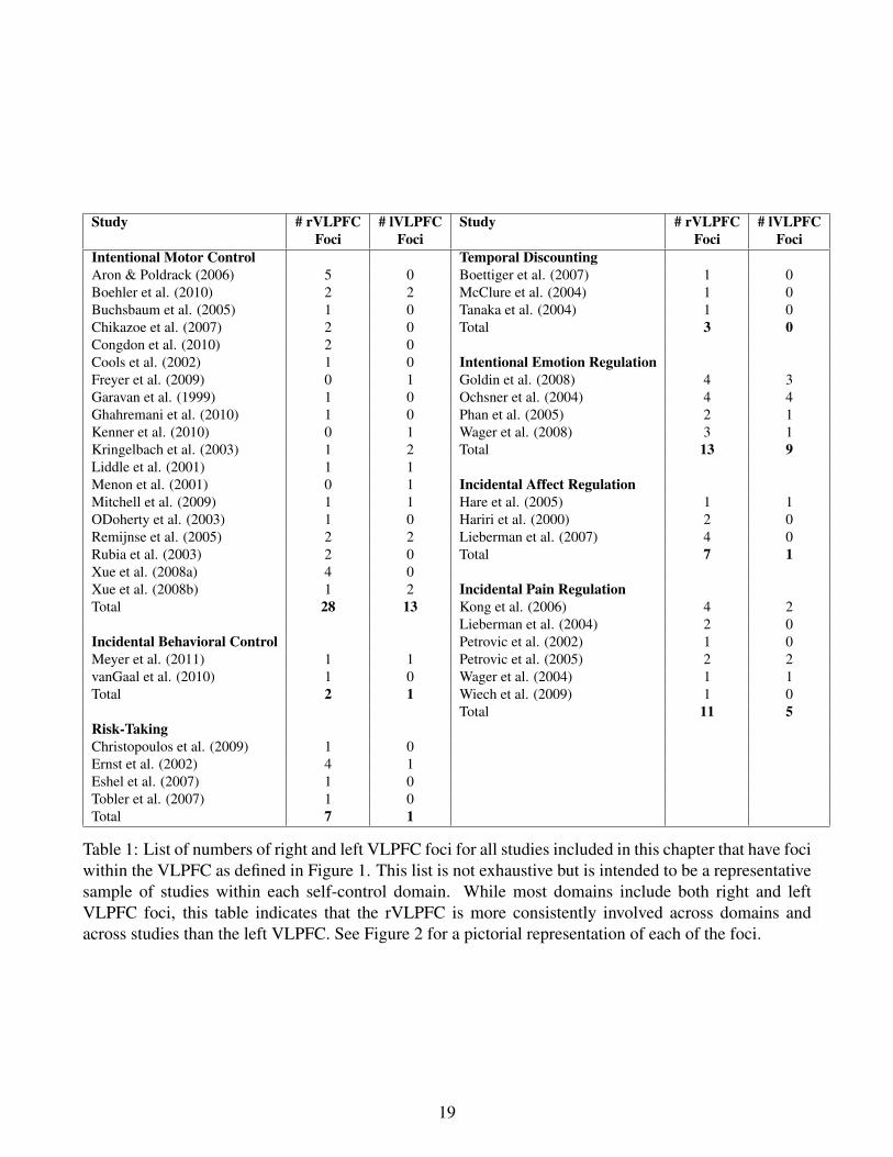
Table 1: List of numbers of right and left VLPFC foci for all studies included in this chapter that have fociwithin the VLPFC as defined in Figure 1. This list is not exhaustive but is intended to be a representativesample of studies within each self-control domain. While most domains include both right and leftVLPFC foci, this table indicates that the rVLPFC is more consistently involved across domains andacross studies than the left VLPFC. See Figure 2 for a pictorial representation of each of the foci.
19
L R
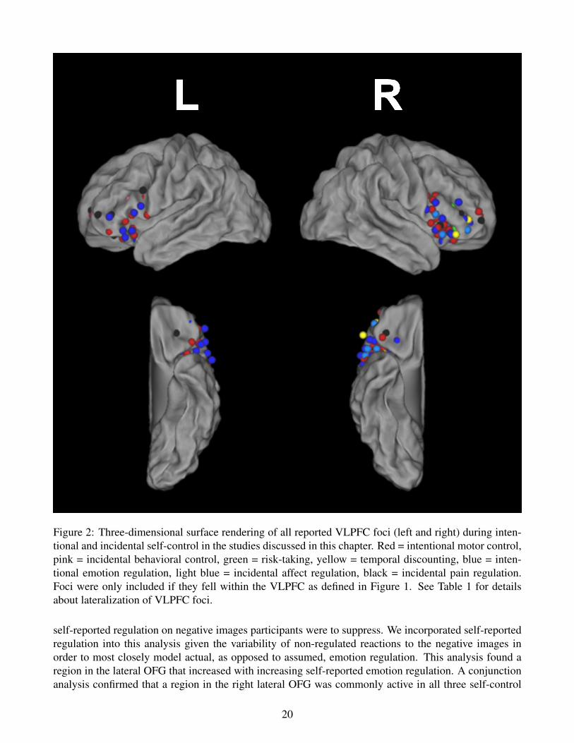
Figure 2: Three-dimensional surface rendering of all reported VLPFC foci (left and right) during inten-tional and incidental self-control in the studies discussed in this chapter. Red = intentional motor control,pink = incidental behavioral control, green = risk-taking, yellow = temporal discounting, blue = inten-tional emotion regulation, light blue = incidental affect regulation, black = incidental pain regulation.Foci were only included if they fell within the VLPFC as defined in Figure 1. See Table 1 for detailsabout lateralization of VLPFC foci.
self-reported regulation on negative images participants were to suppress. We incorporated self-reportedregulation into this analysis given the variability of non-regulated reactions to the negative images inorder to most closely model actual, as opposed to assumed, emotion regulation. This analysis found aregion in the lateral OFG that increased with increasing self-reported emotion regulation. A conjunctionanalysis confirmed that a region in the right lateral OFG was commonly active in all three self-control
20
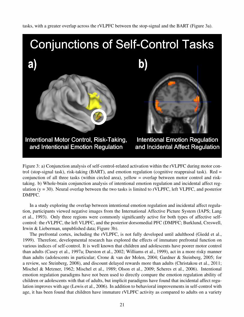
tasks, with a greater overlap across the rVLPFC between the stop-signal and the BART (Figure 3a).
Intentional Motor Control, Risk-Taking,and Intentional Emotion Regulation
Intentional Emotion Regulationand Incidental Affect Regulation
Conjunctions of Self-Control Tasksa) b)
Figure 3: a) Conjunction analysis of self-control-related activation within the rVLPFC during motor con-trol (stop-signal task), risk-taking (BART), and emotion regulation (cognitive reappraisal task). Red =conjunction of all three tasks (within circled area), yellow = overlap between motor control and risk-taking. b) Whole-brain conjunction analysis of intentional emotion regulation and incidental affect reg-ulation (y = 30). Neural overlap between the two tasks is limited to rVLPFC, left VLPFC, and posteriorDMPFC.
In a study exploring the overlap between intentional emotion regulation and incidental affect regula-tion, participants viewed negative images from the International Affective Picture System (IAPS; Langet al., 1993). Only three regions were commonly significantly active for both types of affective self-control: the rVLPFC, the left VLPFC, and the posterior dorsomedial PFC (DMPFC; Burklund, Creswell,Irwin & Lieberman, unpublished data; Figure 3b).
The prefrontal cortex, including the rVLPFC, is not fully developed until adulthood (Giedd et al.,1999). Therefore, developmental research has explored the effects of immature prefrontal function onvarious indices of self-control. It is well known that children and adolescents have poorer motor controlthan adults (Casey et al., 1997a; Durston et al., 2002; Williams et al., 1999), act in a more risky mannerthan adults (adolescents in particular; Crone & van der Molen, 2004; Gardner & Steinberg, 2005; fora review, see Steinberg, 2008), and discount delayed rewards more than adults (Christakou et al., 2011;Mischel & Metzner, 1962; Mischel et al., 1989; Olson et al., 2009; Scheres et al., 2006). Intentionalemotion regulation paradigms have not been used to directly compare the emotion regulation ability ofchildren or adolescents with that of adults, but implicit paradigms have found that incidental affect regu-lation improves with age (Lewis et al., 2006). In addition to behavioral improvements in self-control withage, it has been found that children have immature rVLPFC activity as compared to adults on a variety
21
of self-control tasks. For example, children have been shown to have decreased rVLPFC activity as com-pared to adults during successful simple motor control (Bunge et al., 2002; Durston et al., 2002; Rubiaet al., 2007). Furthermore, a recent study found that neural activity in the rVLPFC, along with otherregions such as the striatum and right STN, predicted both age and response inhibition ability (as mea-sured by SSRT) on the stop-signal task (Cohen et al., 2010). This indicated that the rVLPFC is criticallyinvolved in successful motor control and that rVLPFC activity is decreased in children, who have notyet fully developed their self-control ability. In literature examining the development of reward-seekingbehavior, children and adolescents have immature patterns of activity in the lateral OFC as compared toadults (Eshel et al., 2007; Galvan et al., 2006). The developmental literature implies that the developmentof the rVLPFC is related to the increase in self-control ability seen as children and adolescents becomeadults.
On the other end of the aging spectrum, it has been found that as people age their brains atrophy,particularly in the prefrontal cortex (Cabeza, 2001). Therefore, some aging research has examined theeffects of prefrontal degeneration on self-control. Much of the research has focused on either motor con-trol or emotion regulation. Older adults have longer SSRTs in the stop-signal task (Williams et al., 1999)and fewer correct inhibitions in the go/no-go task (Nielson et al., 2002) than younger adults. Further,brain imaging results from one study revealed decreased activity in multiple brain regions, includingrVLPFC, in older adults as compared to younger adults (Nielson et al., 2002). In contrast, older adultshad greater activity in left VLPFC than younger adults, potentially indicating the recruitment of addi-tional regions, specifically the reduction of hemispheric asymmetry (Cabeza, 2002), as compensation forpoorer functioning in the rVLPFC. These results were specific to response inhibition, as there were nodifferences in prefrontal activity during go trials across age (Nielson et al., 2002). In the field of emotionprocessing it has been noted that older adults show a “positivity effect” when remembering stimuli, inthat they selectively remember positive, as compared to negative, stimuli more so than younger adults.One theory is that this positivity effect occurs due to increased emotion regulation in older adults (Mather& Carstensen, 2005; Nashiro et al., In Press). Very little research has directly compared the neural re-gions involved in emotion regulation in older and younger adults. A recent study that did compare thetwo groups found that attempted emotion regulation resulted in greater activity in left, but not right, IFGin younger as compared to older adults (Winecoff et al., 2011). However, older adults also reported lesssuccessful emotion regulation than younger adults, so future studies where equivalent regulation is foundacross age groups should be conducted before drawing conclusions about neural differences due to agingin emotion regulation.
In addition to the normal functional trajectory of the rVLPFC during acts of self-control across thelifespan, it has been shown that rVLPFC activity has important implications for a variety of psychiatricillnesses that are related to impulsivity. Research has demonstrated decreased rVLPFC activity duringsimple motor control tasks in people with ADHD (Booth et al., 2005; Durston et al., 2006; Rubia et al.,1999) and obsessive compulsive disorder (Roth et al., 2007). A recent review of the simple responseinhibition literature noted that not only is right IFG activity decreased in people with ADHD as com-pared to controls, but that right IFG volume is also reduced relative to controls (Chambers et al., 2009).Additionally, it was demonstrated that compulsive gamblers performed worse on a reversal learning taskand had less rVLPFC activity than did healthy controls (de Ruiter et al., 2009). Furthermore, individualswho abuse methamphetamine were shown to have less rVLPFC activity on an incidental affect labelingtask (Payer et al., 2008), as well as a trend toward lower right IFG gray matter concentration in individu-als who abuse methamphetamine than in healthy control participants (Payer et al., 2011). This literatureunderscores that not only do people with impulsivity-related disorders have behavioral problems withself-control, but the neural mechanisms underlying self-control may be impaired as well.
22
4 rVLPFC ConnectionsThe rVLPFC is well positioned to be a key neural region involved in exerting self-control. It is anatom-ically associated with other prefrontal control regions, such as the DLPFC, mPFC, ACC, and OFC(Miller & Cohen, 2001). Research utilizing Diffusion Tensor Imaging (DTI) in humans has indicatedthat there are white matter connections between the rVLPFC and the ventral caudate (Leh et al., 2007),the preSMA, and the STN (Aron et al., 2007a). A study comparing functional to structural connectivitysimilarly found that the rVLPFC and the preSMA were connected using both functional connectivity anddiffusion-weighted techniques (Johansen-Berg et al., 2004). The preSMA has been associated with con-flict detection and the basal ganglia, including the caudate and the STN, have been associated with thecontrol of motor responses. Connections between these regions, therefore, could be the means throughwhich the rVLPFC becomes aware of a conflict between a goal-directed intention and a prepotent im-pulse (through the preSMA) and then sends a signal to exert behavioral self-control over the impulse(through the basal ganglia). Functional connectivity analyses using Granger causality mapping providedevidence that not only are the preSMA and the rVLPFC connected, but that the preSMA, along with thecerebellum and the thalamus, cause changes related to conflict/error processing in rVLPFC activity (Ide& Li, 2011). Moreover, Granger causality supports the theory that the rVLPFC projects to the preSMA,which in turn projects to the basal ganglia/STN (Duann et al., 2009).
Additionally, there is evidence that the rVLPFC is functionally connected to the amygdala, possiblyvia the mPFC, which has reciprocal connections with both structures (Banks et al., 2007; Hariri et al.,2000, 2003; Lieberman et al., 2007). Lastly, there is evidence that the lateral OFC is functionally con-nected to a large network of dorsal prefrontal and dorsal parietal regions (Cohen et al., 2005).
Studies with macaque monkeys have more directly examined structural brain connections. Evidenceexists that the cytoarchitecture of the macaque VLPFC is similar to that in humans, specifically themacaque inferior arcuate sulcus and its surrounding cortex (Ongur & Price, 2000; Petrides et al., 2005).It is therefore assumed that this area in macaques is the monkey homologue to the human VLPFC andthat the anatomical connections found in this region in macaques may also be found in humans. In themonkey, the inferior arcuate sulcus is connected to the lateral and medial OFC, the DMPFC, the DLPFC,the ACC, the insula, the supplementary, premotor and primary motor areas, and areas of the superiortemporal lobe (Deacon, 1992). Moreover, there are architectonic and connectivity subdivisions withinthe macaque VLPFC, implying that different subregions may have different functional roles (Gerbellaet al., 2010).
Other research investigating the primate lateral OFC, which is a part of the larger VLPFC, has foundthat the lateral OFC receives sensory input directly from the primary taste cortex, indirectly from visualareas via the inferior temporal cortex, and from somatosensory areas such as the primary and secondarysensory cortices and the insula. The lateral OFC sends output to the hypothalamus, the periaqueductalgray area, and the striatum, especially the ventral caudate. Furthermore, it has reciprocal connectionswith the amygdala, cingulate cortex, premotor area, and DLPFC (Kringelbach & Rolls, 2004). To furthersupport the theory that the connections found in monkeys also exist in humans, research has demonstratedthat connections with some of these regions, such as the ACC, DLPFC, preSMA, amygdala, and ventralcaudate, have been noted in humans as well (Aron et al., 2007a; Leh et al., 2007; Lieberman et al., 2007;Miller & Cohen, 2001).
23
5 Other rVLPFC RolesIt is important to point out that there are multiple theories about the function of the rVLPFC. Activityin this region has been associated with cognitive processes as diverse as self-control (as discussed inthis chapter), attention to unexpected stimuli (Corbetta & Shulman, 2002; Hampshire et al., 2010; Iariaet al., 2008; Sharp et al., 2010), various aspects of memory (Courtney et al., 1996; Dove et al., 2008;Kostopoulos & Petrides, 2003; Rizzuto et al., 2005), and the interpretation of emotions (Kober et al.,2008). For example, it has been proposed that rVLPFC activity is specifically related to stimulus-driven,bottom-up attention and automatic alerting to unexpected, salient stimuli (Corbetta & Shulman, 2002;Iaria et al., 2008). Recent research comparing stop-signal performance to performance on equivalenttasks that do not require motor inhibition but still have random (i.e., unexpected), infrequent “signals”has found equivalent rVLPFC activity when inhibiting one’s response in reaction to a stop-signal as whenencountering a signal giving instructions to respond (Hampshire et al., 2010) or to be ignored (Sharp et al.,2010). However, participants responded more slowly when they heard a signal that was to be ignored(Sharp et al., 2010), leaving open the possibility that a response inhibition mechanism was occurring (andthen overcome) on those trials. There is also evidence that the rVLPFC is involved in target detectionand thus responds to the relevant aspects of a stimulus (Hampshire et al., 2009). In line with this findingbut in a different cognitive domain, the rVLPFC has been implicated during memory retrieval when onemust differentiate between relevant and irrelevant aspects of a stimulus (Kostopoulos & Petrides, 2003)or when participants are engaging in active, goal-directed retrieval (Dove et al., 2008). Additionally,the rVLPFC has been associated with both object-oriented (Courtney et al., 1996) and spatially oriented(Rizzuto et al., 2005) working memory.
Clearly the rVLPFC has been associated with many different roles. These are not necessarily mutu-ally exclusive. First, there is evidence that different foci within the rVLPFC are associated with motorcontrol (specifically the right ventral IFG) and attention to unexpected stimuli (specifically the right dor-sal IFG/inferior frontal junction; Aron, 2011; Chikazoe, 2010; Chikazoe et al., 2009; Verbruggen et al.,2010). A recent meta-analysis further supports the finding that across many studies the locus of rVLPFCactivity during motor control and during reorienting to unexpected stimuli, while partially overlapping,can be dissociated (Levy & Wagner, 2011). The possibility of functional subdivisions within the rVLPFCis strengthened by the finding that there are different structural patterns within VLPFC subdivisions inthe macaque (Gerbella et al., 2010). Second, many purported roles of the rVLPFC involve goal-directedselection, be it whether or not to respond to a stimulus, how to regulate one’s emotions, whether to be-have in a risky or impatient manner, or selectively remembering relevant items. Thus, it is possible thatthe rVLPFC is generally involved in goal-directed behavior, while different subregions are specificallyinvolved in various processed that fall under that umbrella.
In addition to the afore-mentioned theories about the specific function(s) of the rVLPFC, general the-ories of prefrontal functioning have been proposed that may explain the varied processes that the rVLPFCappears to be involved in. McIntosh (2000; 2004) has proposed a theory of “neural context” that statesthat dynamic neural interactions, which can rapidly and transiently change based on the current envi-ronment, are more important for cognition than the magnitude of neural activity of any single region.This theory would predict that the role of an individual region such as the rVLPFC can change basedon its current pattern of functional connectivity (i.e., what other brain regions to which is it connectedand with which it is interacting). This is a distinct possibility with the rVLPFC, given that it is highlyinterconnected with other brain regions (see “rVLPFC Connections” section above). Other theories havesimilarly proposed that the role of prefrontal cortical regions can change based on the current cogni-tive environment (Duncan, 2001; Miller & Cohen, 2001). Evidence supporting all of these connectivitytheories comes in part from single unit recordings demonstrating that prefrontal neurons can adaptively
24
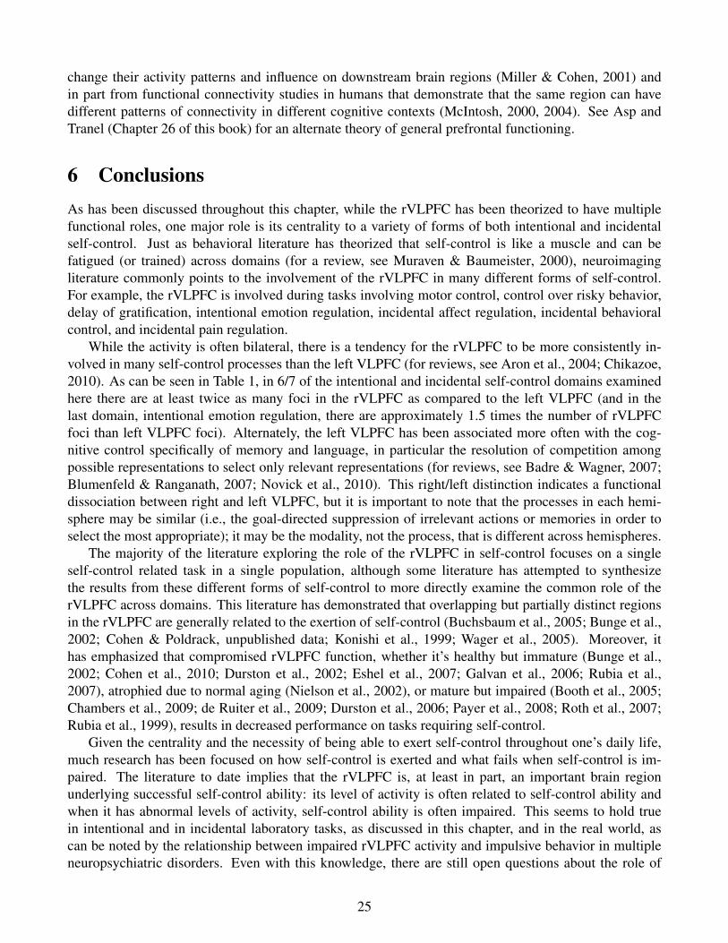
change their activity patterns and influence on downstream brain regions (Miller & Cohen, 2001) andin part from functional connectivity studies in humans that demonstrate that the same region can havedifferent patterns of connectivity in different cognitive contexts (McIntosh, 2000, 2004). See Asp andTranel (Chapter 26 of this book) for an alternate theory of general prefrontal functioning.
6 ConclusionsAs has been discussed throughout this chapter, while the rVLPFC has been theorized to have multiplefunctional roles, one major role is its centrality to a variety of forms of both intentional and incidentalself-control. Just as behavioral literature has theorized that self-control is like a muscle and can befatigued (or trained) across domains (for a review, see Muraven & Baumeister, 2000), neuroimagingliterature commonly points to the involvement of the rVLPFC in many different forms of self-control.For example, the rVLPFC is involved during tasks involving motor control, control over risky behavior,delay of gratification, intentional emotion regulation, incidental affect regulation, incidental behavioralcontrol, and incidental pain regulation.
While the activity is often bilateral, there is a tendency for the rVLPFC to be more consistently in-volved in many self-control processes than the left VLPFC (for reviews, see Aron et al., 2004; Chikazoe,2010). As can be seen in Table 1, in 6/7 of the intentional and incidental self-control domains examinedhere there are at least twice as many foci in the rVLPFC as compared to the left VLPFC (and in thelast domain, intentional emotion regulation, there are approximately 1.5 times the number of rVLPFCfoci than left VLPFC foci). Alternately, the left VLPFC has been associated more often with the cog-nitive control specifically of memory and language, in particular the resolution of competition amongpossible representations to select only relevant representations (for reviews, see Badre & Wagner, 2007;Blumenfeld & Ranganath, 2007; Novick et al., 2010). This right/left distinction indicates a functionaldissociation between right and left VLPFC, but it is important to note that the processes in each hemi-sphere may be similar (i.e., the goal-directed suppression of irrelevant actions or memories in order toselect the most appropriate); it may be the modality, not the process, that is different across hemispheres.
The majority of the literature exploring the role of the rVLPFC in self-control focuses on a singleself-control related task in a single population, although some literature has attempted to synthesizethe results from these different forms of self-control to more directly examine the common role of therVLPFC across domains. This literature has demonstrated that overlapping but partially distinct regionsin the rVLPFC are generally related to the exertion of self-control (Buchsbaum et al., 2005; Bunge et al.,2002; Cohen & Poldrack, unpublished data; Konishi et al., 1999; Wager et al., 2005). Moreover, ithas emphasized that compromised rVLPFC function, whether it’s healthy but immature (Bunge et al.,2002; Cohen et al., 2010; Durston et al., 2002; Eshel et al., 2007; Galvan et al., 2006; Rubia et al.,2007), atrophied due to normal aging (Nielson et al., 2002), or mature but impaired (Booth et al., 2005;Chambers et al., 2009; de Ruiter et al., 2009; Durston et al., 2006; Payer et al., 2008; Roth et al., 2007;Rubia et al., 1999), results in decreased performance on tasks requiring self-control.
Given the centrality and the necessity of being able to exert self-control throughout one’s daily life,much research has been focused on how self-control is exerted and what fails when self-control is im-paired. The literature to date implies that the rVLPFC is, at least in part, an important brain regionunderlying successful self-control ability: its level of activity is often related to self-control ability andwhen it has abnormal levels of activity, self-control ability is often impaired. This seems to hold truein intentional and in incidental laboratory tasks, as discussed in this chapter, and in the real world, ascan be noted by the relationship between impaired rVLPFC activity and impulsive behavior in multipleneuropsychiatric disorders. Even with this knowledge, there are still open questions about the role of
25
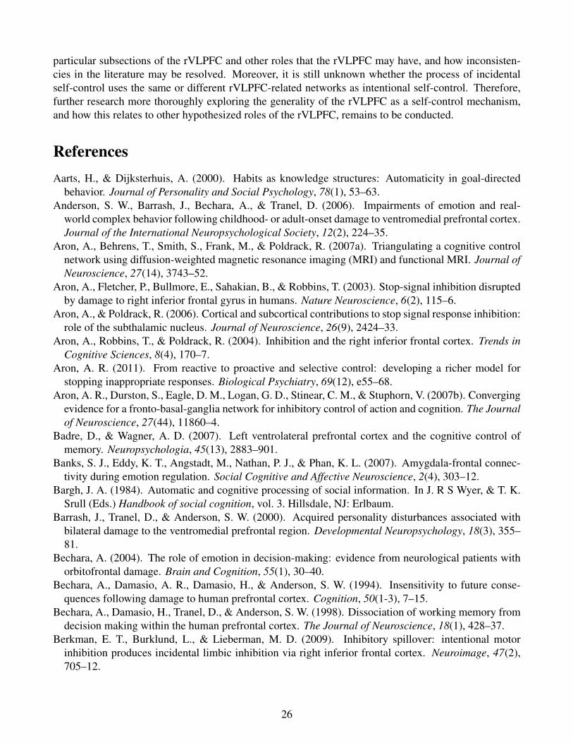
particular subsections of the rVLPFC and other roles that the rVLPFC may have, and how inconsisten-cies in the literature may be resolved. Moreover, it is still unknown whether the process of incidentalself-control uses the same or different rVLPFC-related networks as intentional self-control. Therefore,further research more thoroughly exploring the generality of the rVLPFC as a self-control mechanism,and how this relates to other hypothesized roles of the rVLPFC, remains to be conducted.
ReferencesAarts, H., & Dijksterhuis, A. (2000). Habits as knowledge structures: Automaticity in goal-directed
behavior. Journal of Personality and Social Psychology, 78(1), 53–63.Anderson, S. W., Barrash, J., Bechara, A., & Tranel, D. (2006). Impairments of emotion and real-
world complex behavior following childhood- or adult-onset damage to ventromedial prefrontal cortex.Journal of the International Neuropsychological Society, 12(2), 224–35.
Aron, A., Behrens, T., Smith, S., Frank, M., & Poldrack, R. (2007a). Triangulating a cognitive controlnetwork using diffusion-weighted magnetic resonance imaging (MRI) and functional MRI. Journal ofNeuroscience, 27(14), 3743–52.
Aron, A., Fletcher, P., Bullmore, E., Sahakian, B., & Robbins, T. (2003). Stop-signal inhibition disruptedby damage to right inferior frontal gyrus in humans. Nature Neuroscience, 6(2), 115–6.
Aron, A., & Poldrack, R. (2006). Cortical and subcortical contributions to stop signal response inhibition:role of the subthalamic nucleus. Journal of Neuroscience, 26(9), 2424–33.
Aron, A., Robbins, T., & Poldrack, R. (2004). Inhibition and the right inferior frontal cortex. Trends inCognitive Sciences, 8(4), 170–7.
Aron, A. R. (2011). From reactive to proactive and selective control: developing a richer model forstopping inappropriate responses. Biological Psychiatry, 69(12), e55–68.
Aron, A. R., Durston, S., Eagle, D. M., Logan, G. D., Stinear, C. M., & Stuphorn, V. (2007b). Convergingevidence for a fronto-basal-ganglia network for inhibitory control of action and cognition. The Journalof Neuroscience, 27(44), 11860–4.
Badre, D., & Wagner, A. D. (2007). Left ventrolateral prefrontal cortex and the cognitive control ofmemory. Neuropsychologia, 45(13), 2883–901.
Banks, S. J., Eddy, K. T., Angstadt, M., Nathan, P. J., & Phan, K. L. (2007). Amygdala-frontal connec-tivity during emotion regulation. Social Cognitive and Affective Neuroscience, 2(4), 303–12.
Bargh, J. A. (1984). Automatic and cognitive processing of social information. In J. R S Wyer, & T. K.Srull (Eds.) Handbook of social cognition, vol. 3. Hillsdale, NJ: Erlbaum.
Barrash, J., Tranel, D., & Anderson, S. W. (2000). Acquired personality disturbances associated withbilateral damage to the ventromedial prefrontal region. Developmental Neuropsychology, 18(3), 355–81.
Bechara, A. (2004). The role of emotion in decision-making: evidence from neurological patients withorbitofrontal damage. Brain and Cognition, 55(1), 30–40.
Bechara, A., Damasio, A. R., Damasio, H., & Anderson, S. W. (1994). Insensitivity to future conse-quences following damage to human prefrontal cortex. Cognition, 50(1-3), 7–15.
Bechara, A., Damasio, H., Tranel, D., & Anderson, S. W. (1998). Dissociation of working memory fromdecision making within the human prefrontal cortex. The Journal of Neuroscience, 18(1), 428–37.
Berkman, E. T., Burklund, L., & Lieberman, M. D. (2009). Inhibitory spillover: intentional motorinhibition produces incidental limbic inhibition via right inferior frontal cortex. Neuroimage, 47(2),705–12.
26
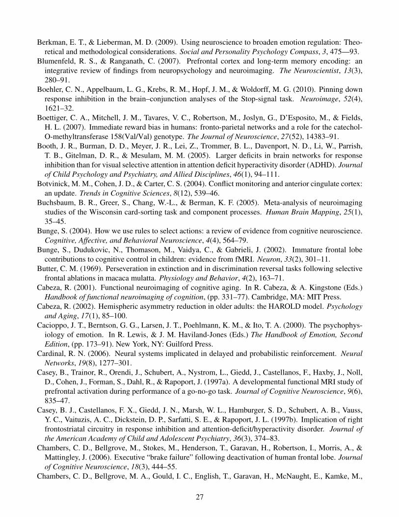
Berkman, E. T., & Lieberman, M. D. (2009). Using neuroscience to broaden emotion regulation: Theo-retical and methodological considerations. Social and Personality Psychology Compass, 3, 475—93.
Blumenfeld, R. S., & Ranganath, C. (2007). Prefrontal cortex and long-term memory encoding: anintegrative review of findings from neuropsychology and neuroimaging. The Neuroscientist, 13(3),280–91.
Boehler, C. N., Appelbaum, L. G., Krebs, R. M., Hopf, J. M., & Woldorff, M. G. (2010). Pinning downresponse inhibition in the brain–conjunction analyses of the Stop-signal task. Neuroimage, 52(4),1621–32.
Boettiger, C. A., Mitchell, J. M., Tavares, V. C., Robertson, M., Joslyn, G., D’Esposito, M., & Fields,H. L. (2007). Immediate reward bias in humans: fronto-parietal networks and a role for the catechol-O-methyltransferase 158(Val/Val) genotype. The Journal of Neuroscience, 27(52), 14383–91.
Booth, J. R., Burman, D. D., Meyer, J. R., Lei, Z., Trommer, B. L., Davenport, N. D., Li, W., Parrish,T. B., Gitelman, D. R., & Mesulam, M. M. (2005). Larger deficits in brain networks for responseinhibition than for visual selective attention in attention deficit hyperactivity disorder (ADHD). Journalof Child Psychology and Psychiatry, and Allied Disciplines, 46(1), 94–111.
Botvinick, M. M., Cohen, J. D., & Carter, C. S. (2004). Conflict monitoring and anterior cingulate cortex:an update. Trends in Cognitive Sciences, 8(12), 539–46.
Buchsbaum, B. R., Greer, S., Chang, W.-L., & Berman, K. F. (2005). Meta-analysis of neuroimagingstudies of the Wisconsin card-sorting task and component processes. Human Brain Mapping, 25(1),35–45.
Bunge, S. (2004). How we use rules to select actions: a review of evidence from cognitive neuroscience.Cognitive, Affective, and Behavioral Neuroscience, 4(4), 564–79.
Bunge, S., Dudukovic, N., Thomason, M., Vaidya, C., & Gabrieli, J. (2002). Immature frontal lobecontributions to cognitive control in children: evidence from fMRI. Neuron, 33(2), 301–11.
Butter, C. M. (1969). Perseveration in extinction and in discrimination reversal tasks following selectivefrontal ablations in macaca mulatta. Physiology and Behavior, 4(2), 163–71.
Cabeza, R. (2001). Functional neuroimaging of cognitive aging. In R. Cabeza, & A. Kingstone (Eds.)Handbook of functional neuroimaging of cognition, (pp. 331–77). Cambridge, MA: MIT Press.
Cabeza, R. (2002). Hemispheric asymmetry reduction in older adults: the HAROLD model. Psychologyand Aging, 17(1), 85–100.
Cacioppo, J. T., Berntson, G. G., Larsen, J. T., Poehlmann, K. M., & Ito, T. A. (2000). The psychophys-iology of emotion. In R. Lewis, & J. M. Haviland-Jones (Eds.) The Handbook of Emotion, SecondEdition, (pp. 173–91). New York, NY: Guilford Press.
Cardinal, R. N. (2006). Neural systems implicated in delayed and probabilistic reinforcement. NeuralNetworks, 19(8), 1277–301.
Casey, B., Trainor, R., Orendi, J., Schubert, A., Nystrom, L., Giedd, J., Castellanos, F., Haxby, J., Noll,D., Cohen, J., Forman, S., Dahl, R., & Rapoport, J. (1997a). A developmental functional MRI study ofprefrontal activation during performance of a go-no-go task. Journal of Cognitive Neuroscience, 9(6),835–47.
Casey, B. J., Castellanos, F. X., Giedd, J. N., Marsh, W. L., Hamburger, S. D., Schubert, A. B., Vauss,Y. C., Vaituzis, A. C., Dickstein, D. P., Sarfatti, S. E., & Rapoport, J. L. (1997b). Implication of rightfrontostriatal circuitry in response inhibition and attention-deficit/hyperactivity disorder. Journal ofthe American Academy of Child and Adolescent Psychiatry, 36(3), 374–83.
Chambers, C. D., Bellgrove, M., Stokes, M., Henderson, T., Garavan, H., Robertson, I., Morris, A., &Mattingley, J. (2006). Executive “brake failure” following deactivation of human frontal lobe. Journalof Cognitive Neuroscience, 18(3), 444–55.
Chambers, C. D., Bellgrove, M. A., Gould, I. C., English, T., Garavan, H., McNaught, E., Kamke, M.,
27
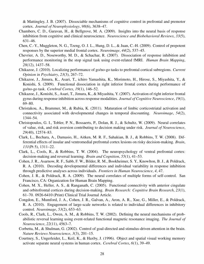
& Mattingley, J. B. (2007). Dissociable mechanisms of cognitive control in prefrontal and premotorcortex. Journal of Neurophysiology, 98(6), 3638–47.
Chambers, C. D., Garavan, H., & Bellgrove, M. A. (2009). Insights into the neural basis of responseinhibition from cognitive and clinical neuroscience. Neuroscience and Biobehavioral Reviews, 33(5),631–46.
Chen, C.-Y., Muggleton, N. G., Tzeng, O. J. L., Hung, D. L., & Juan, C.-H. (2009). Control of prepotentresponses by the superior medial frontal cortex. Neuroimage, 44(2), 537–45.
Chevrier, A. D., Noseworthy, M. D., & Schachar, R. (2007). Dissociation of response inhibition andperformance monitoring in the stop signal task using event-related fMRI. Human Brain Mapping,28(12), 1437–58.
Chikazoe, J. (2010). Localizing performance of go/no-go tasks to prefrontal cortical subregions. CurrentOpinion in Psychiatry, 23(3), 267–72.
Chikazoe, J., Jimura, K., Asari, T., ichiro Yamashita, K., Morimoto, H., Hirose, S., Miyashita, Y., &Konishi, S. (2009). Functional dissociation in right inferior frontal cortex during performance ofgo/no-go task. Cerebral Cortex, 19(1), 146–52.
Chikazoe, J., Konishi, S., Asari, T., Jimura, K., & Miyashita, Y. (2007). Activation of right inferior frontalgyrus during response inhibition across response modalities. Journal of Cognitive Neuroscience, 19(1),69–80.
Christakou, A., Brammer, M., & Rubia, K. (2011). Maturation of limbic corticostriatal activation andconnectivity associated with developmental changes in temporal discounting. Neuroimage, 54(2),1344–54.
Christopoulos, G. I., Tobler, P. N., Bossaerts, P., Dolan, R. J., & Schultz, W. (2009). Neural correlatesof value, risk, and risk aversion contributing to decision making under risk. Journal of Neuroscience,29(40), 12574–83.
Clark, L., Bechara, A., Damasio, H., Aitken, M. R. F., Sahakian, B. J., & Robbins, T. W. (2008). Dif-ferential effects of insular and ventromedial prefrontal cortex lesions on risky decision-making. Brain,131(Pt 5), 1311–22.
Clark, L., Cools, R., & Robbins, T. W. (2004). The neuropsychology of ventral prefrontal cortex:decision-making and reversal learning. Brain and Cognition, 55(1), 41–53.
Cohen, J. R., Asarnow, R. F., Sabb, F. W., Bilder, R. M., Bookheimer, S. Y., Knowlton, B. J., & Poldrack,R. A. (2010). Decoding developmental differences and individual variability in response inhibitionthrough predictive analyses across individuals. Frontiers in Human Neuroscience, 4, 47.
Cohen, J. R., & Poldrack, R. A. (2009). The neural correlates of multiple forms of self-control. SanFrancisco, CA: Organization for Human Brain Mapping.
Cohen, M. X., Heller, A. S., & Ranganath, C. (2005). Functional connectivity with anterior cingulateand orbitofrontal cortices during decision-making. Brain Research: Cognitive Brain Research, 23(1),61–70. 0926-6410 (Print) Clinical Trial Journal Article.
Congdon, E., Mumford, J. A., Cohen, J. R., Galvan, A., Aron, A. R., Xue, G., Miller, E., & Poldrack,R. A. (2010). Engagement of large-scale networks is related to individual differences in inhibitorycontrol. Neuroimage, 53(2), 653–63.
Cools, R., Clark, L., Owen, A. M., & Robbins, T. W. (2002). Defining the neural mechanisms of prob-abilistic reversal learning using event-related functional magnetic resonance imaging. The Journal ofNeuroscience, 22(11), 4563–7.
Corbetta, M., & Shulman, G. (2002). Control of goal-directed and stimulus-driven attention in the brain.Nature Reviews Neuroscience, 3(3), 201–15.
Courtney, S., Ungerleider, L., Keil, K., & Haxby, J. (1996). Object and spatial visual working memoryactivate separate neural systems in human cortex. Cerebral Cortex, 6(1), 39–49.
28
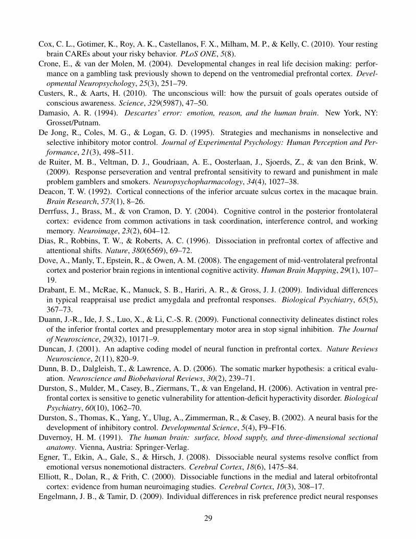
Cox, C. L., Gotimer, K., Roy, A. K., Castellanos, F. X., Milham, M. P., & Kelly, C. (2010). Your restingbrain CAREs about your risky behavior. PLoS ONE, 5(8).
Crone, E., & van der Molen, M. (2004). Developmental changes in real life decision making: perfor-mance on a gambling task previously shown to depend on the ventromedial prefrontal cortex. Devel-opmental Neuropsychology, 25(3), 251–79.
Custers, R., & Aarts, H. (2010). The unconscious will: how the pursuit of goals operates outside ofconscious awareness. Science, 329(5987), 47–50.
Damasio, A. R. (1994). Descartes’ error: emotion, reason, and the human brain. New York, NY:Grosset/Putnam.
De Jong, R., Coles, M. G., & Logan, G. D. (1995). Strategies and mechanisms in nonselective andselective inhibitory motor control. Journal of Experimental Psychology: Human Perception and Per-formance, 21(3), 498–511.
de Ruiter, M. B., Veltman, D. J., Goudriaan, A. E., Oosterlaan, J., Sjoerds, Z., & van den Brink, W.(2009). Response perseveration and ventral prefrontal sensitivity to reward and punishment in maleproblem gamblers and smokers. Neuropsychopharmacology, 34(4), 1027–38.
Deacon, T. W. (1992). Cortical connections of the inferior arcuate sulcus cortex in the macaque brain.Brain Research, 573(1), 8–26.
Derrfuss, J., Brass, M., & von Cramon, D. Y. (2004). Cognitive control in the posterior frontolateralcortex: evidence from common activations in task coordination, interference control, and workingmemory. Neuroimage, 23(2), 604–12.
Dias, R., Robbins, T. W., & Roberts, A. C. (1996). Dissociation in prefrontal cortex of affective andattentional shifts. Nature, 380(6569), 69–72.
Dove, A., Manly, T., Epstein, R., & Owen, A. M. (2008). The engagement of mid-ventrolateral prefrontalcortex and posterior brain regions in intentional cognitive activity. Human Brain Mapping, 29(1), 107–19.
Drabant, E. M., McRae, K., Manuck, S. B., Hariri, A. R., & Gross, J. J. (2009). Individual differencesin typical reappraisal use predict amygdala and prefrontal responses. Biological Psychiatry, 65(5),367–73.
Duann, J.-R., Ide, J. S., Luo, X., & Li, C.-S. R. (2009). Functional connectivity delineates distinct rolesof the inferior frontal cortex and presupplementary motor area in stop signal inhibition. The Journalof Neuroscience, 29(32), 10171–9.
Duncan, J. (2001). An adaptive coding model of neural function in prefrontal cortex. Nature ReviewsNeuroscience, 2(11), 820–9.
Dunn, B. D., Dalgleish, T., & Lawrence, A. D. (2006). The somatic marker hypothesis: a critical evalu-ation. Neuroscience and Biobehavioral Reviews, 30(2), 239–71.
Durston, S., Mulder, M., Casey, B., Ziermans, T., & van Engeland, H. (2006). Activation in ventral pre-frontal cortex is sensitive to genetic vulnerability for attention-deficit hyperactivity disorder. BiologicalPsychiatry, 60(10), 1062–70.
Durston, S., Thomas, K., Yang, Y., Ulug, A., Zimmerman, R., & Casey, B. (2002). A neural basis for thedevelopment of inhibitory control. Developmental Science, 5(4), F9–F16.
Duvernoy, H. M. (1991). The human brain: surface, blood supply, and three-dimensional sectionalanatomy. Vienna, Austria: Springer-Verlag.
Egner, T., Etkin, A., Gale, S., & Hirsch, J. (2008). Dissociable neural systems resolve conflict fromemotional versus nonemotional distracters. Cerebral Cortex, 18(6), 1475–84.
Elliott, R., Dolan, R., & Frith, C. (2000). Dissociable functions in the medial and lateral orbitofrontalcortex: evidence from human neuroimaging studies. Cerebral Cortex, 10(3), 308–17.
Engelmann, J. B., & Tamir, D. (2009). Individual differences in risk preference predict neural responses
29
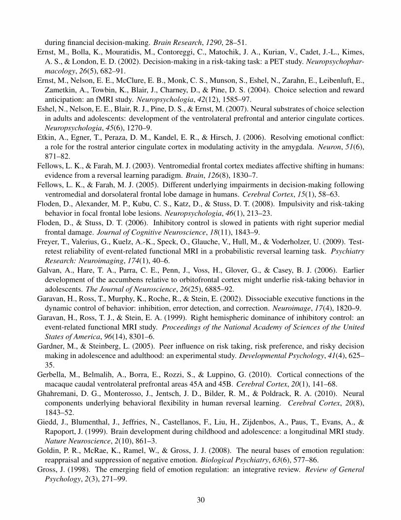
during financial decision-making. Brain Research, 1290, 28–51.Ernst, M., Bolla, K., Mouratidis, M., Contoreggi, C., Matochik, J. A., Kurian, V., Cadet, J.-L., Kimes,
A. S., & London, E. D. (2002). Decision-making in a risk-taking task: a PET study. Neuropsychophar-macology, 26(5), 682–91.
Ernst, M., Nelson, E. E., McClure, E. B., Monk, C. S., Munson, S., Eshel, N., Zarahn, E., Leibenluft, E.,Zametkin, A., Towbin, K., Blair, J., Charney, D., & Pine, D. S. (2004). Choice selection and rewardanticipation: an fMRI study. Neuropsychologia, 42(12), 1585–97.
Eshel, N., Nelson, E. E., Blair, R. J., Pine, D. S., & Ernst, M. (2007). Neural substrates of choice selectionin adults and adolescents: development of the ventrolateral prefrontal and anterior cingulate cortices.Neuropsychologia, 45(6), 1270–9.
Etkin, A., Egner, T., Peraza, D. M., Kandel, E. R., & Hirsch, J. (2006). Resolving emotional conflict:a role for the rostral anterior cingulate cortex in modulating activity in the amygdala. Neuron, 51(6),871–82.
Fellows, L. K., & Farah, M. J. (2003). Ventromedial frontal cortex mediates affective shifting in humans:evidence from a reversal learning paradigm. Brain, 126(8), 1830–7.
Fellows, L. K., & Farah, M. J. (2005). Different underlying impairments in decision-making followingventromedial and dorsolateral frontal lobe damage in humans. Cerebral Cortex, 15(1), 58–63.
Floden, D., Alexander, M. P., Kubu, C. S., Katz, D., & Stuss, D. T. (2008). Impulsivity and risk-takingbehavior in focal frontal lobe lesions. Neuropsychologia, 46(1), 213–23.
Floden, D., & Stuss, D. T. (2006). Inhibitory control is slowed in patients with right superior medialfrontal damage. Journal of Cognitive Neuroscience, 18(11), 1843–9.
Freyer, T., Valerius, G., Kuelz, A.-K., Speck, O., Glauche, V., Hull, M., & Voderholzer, U. (2009). Test-retest reliability of event-related functional MRI in a probabilistic reversal learning task. PsychiatryResearch: Neuroimaging, 174(1), 40–6.
Galvan, A., Hare, T. A., Parra, C. E., Penn, J., Voss, H., Glover, G., & Casey, B. J. (2006). Earlierdevelopment of the accumbens relative to orbitofrontal cortex might underlie risk-taking behavior inadolescents. The Journal of Neuroscience, 26(25), 6885–92.
Garavan, H., Ross, T., Murphy, K., Roche, R., & Stein, E. (2002). Dissociable executive functions in thedynamic control of behavior: inhibition, error detection, and correction. Neuroimage, 17(4), 1820–9.
Garavan, H., Ross, T. J., & Stein, E. A. (1999). Right hemispheric dominance of inhibitory control: anevent-related functional MRI study. Proceedings of the National Academy of Sciences of the UnitedStates of America, 96(14), 8301–6.
Gardner, M., & Steinberg, L. (2005). Peer influence on risk taking, risk preference, and risky decisionmaking in adolescence and adulthood: an experimental study. Developmental Psychology, 41(4), 625–35.
Gerbella, M., Belmalih, A., Borra, E., Rozzi, S., & Luppino, G. (2010). Cortical connections of themacaque caudal ventrolateral prefrontal areas 45A and 45B. Cerebral Cortex, 20(1), 141–68.
Ghahremani, D. G., Monterosso, J., Jentsch, J. D., Bilder, R. M., & Poldrack, R. A. (2010). Neuralcomponents underlying behavioral flexibility in human reversal learning. Cerebral Cortex, 20(8),1843–52.
Giedd, J., Blumenthal, J., Jeffries, N., Castellanos, F., Liu, H., Zijdenbos, A., Paus, T., Evans, A., &Rapoport, J. (1999). Brain development during childhood and adolescence: a longitudinal MRI study.Nature Neuroscience, 2(10), 861–3.
Goldin, P. R., McRae, K., Ramel, W., & Gross, J. J. (2008). The neural bases of emotion regulation:reappraisal and suppression of negative emotion. Biological Psychiatry, 63(6), 577–86.
Gross, J. (1998). The emerging field of emotion regulation: an integrative review. Review of GeneralPsychology, 2(3), 271–99.
30
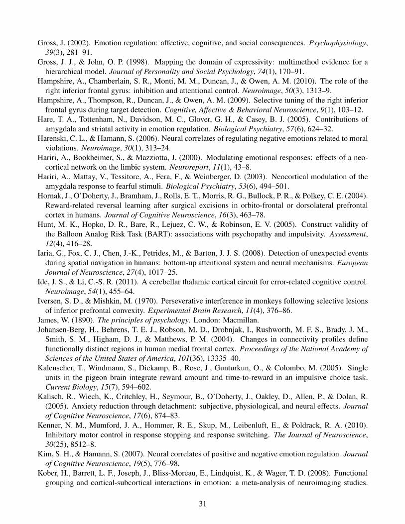
Gross, J. (2002). Emotion regulation: affective, cognitive, and social consequences. Psychophysiology,39(3), 281–91.
Gross, J. J., & John, O. P. (1998). Mapping the domain of expressivity: multimethod evidence for ahierarchical model. Journal of Personality and Social Psychology, 74(1), 170–91.
Hampshire, A., Chamberlain, S. R., Monti, M. M., Duncan, J., & Owen, A. M. (2010). The role of theright inferior frontal gyrus: inhibition and attentional control. Neuroimage, 50(3), 1313–9.
Hampshire, A., Thompson, R., Duncan, J., & Owen, A. M. (2009). Selective tuning of the right inferiorfrontal gyrus during target detection. Cognitive, Affective & Behavioral Neuroscience, 9(1), 103–12.
Hare, T. A., Tottenham, N., Davidson, M. C., Glover, G. H., & Casey, B. J. (2005). Contributions ofamygdala and striatal activity in emotion regulation. Biological Psychiatry, 57(6), 624–32.
Harenski, C. L., & Hamann, S. (2006). Neural correlates of regulating negative emotions related to moralviolations. Neuroimage, 30(1), 313–24.
Hariri, A., Bookheimer, S., & Mazziotta, J. (2000). Modulating emotional responses: effects of a neo-cortical network on the limbic system. Neuroreport, 11(1), 43–8.
Hariri, A., Mattay, V., Tessitore, A., Fera, F., & Weinberger, D. (2003). Neocortical modulation of theamygdala response to fearful stimuli. Biological Psychiatry, 53(6), 494–501.
Hornak, J., O’Doherty, J., Bramham, J., Rolls, E. T., Morris, R. G., Bullock, P. R., & Polkey, C. E. (2004).Reward-related reversal learning after surgical excisions in orbito-frontal or dorsolateral prefrontalcortex in humans. Journal of Cognitive Neuroscience, 16(3), 463–78.
Hunt, M. K., Hopko, D. R., Bare, R., Lejuez, C. W., & Robinson, E. V. (2005). Construct validity ofthe Balloon Analog Risk Task (BART): associations with psychopathy and impulsivity. Assessment,12(4), 416–28.
Iaria, G., Fox, C. J., Chen, J.-K., Petrides, M., & Barton, J. J. S. (2008). Detection of unexpected eventsduring spatial navigation in humans: bottom-up attentional system and neural mechanisms. EuropeanJournal of Neuroscience, 27(4), 1017–25.
Ide, J. S., & Li, C.-S. R. (2011). A cerebellar thalamic cortical circuit for error-related cognitive control.Neuroimage, 54(1), 455–64.
Iversen, S. D., & Mishkin, M. (1970). Perseverative interference in monkeys following selective lesionsof inferior prefrontal convexity. Experimental Brain Research, 11(4), 376–86.
James, W. (1890). The principles of psychology. London: Macmillan.Johansen-Berg, H., Behrens, T. E. J., Robson, M. D., Drobnjak, I., Rushworth, M. F. S., Brady, J. M.,
Smith, S. M., Higham, D. J., & Matthews, P. M. (2004). Changes in connectivity profiles definefunctionally distinct regions in human medial frontal cortex. Proceedings of the National Academy ofSciences of the United States of America, 101(36), 13335–40.
Kalenscher, T., Windmann, S., Diekamp, B., Rose, J., Gunturkun, O., & Colombo, M. (2005). Singleunits in the pigeon brain integrate reward amount and time-to-reward in an impulsive choice task.Current Biology, 15(7), 594–602.
Kalisch, R., Wiech, K., Critchley, H., Seymour, B., O’Doherty, J., Oakley, D., Allen, P., & Dolan, R.(2005). Anxiety reduction through detachment: subjective, physiological, and neural effects. Journalof Cognitive Neuroscience, 17(6), 874–83.
Kenner, N. M., Mumford, J. A., Hommer, R. E., Skup, M., Leibenluft, E., & Poldrack, R. A. (2010).Inhibitory motor control in response stopping and response switching. The Journal of Neuroscience,30(25), 8512–8.
Kim, S. H., & Hamann, S. (2007). Neural correlates of positive and negative emotion regulation. Journalof Cognitive Neuroscience, 19(5), 776–98.
Kober, H., Barrett, L. F., Joseph, J., Bliss-Moreau, E., Lindquist, K., & Wager, T. D. (2008). Functionalgrouping and cortical-subcortical interactions in emotion: a meta-analysis of neuroimaging studies.
31
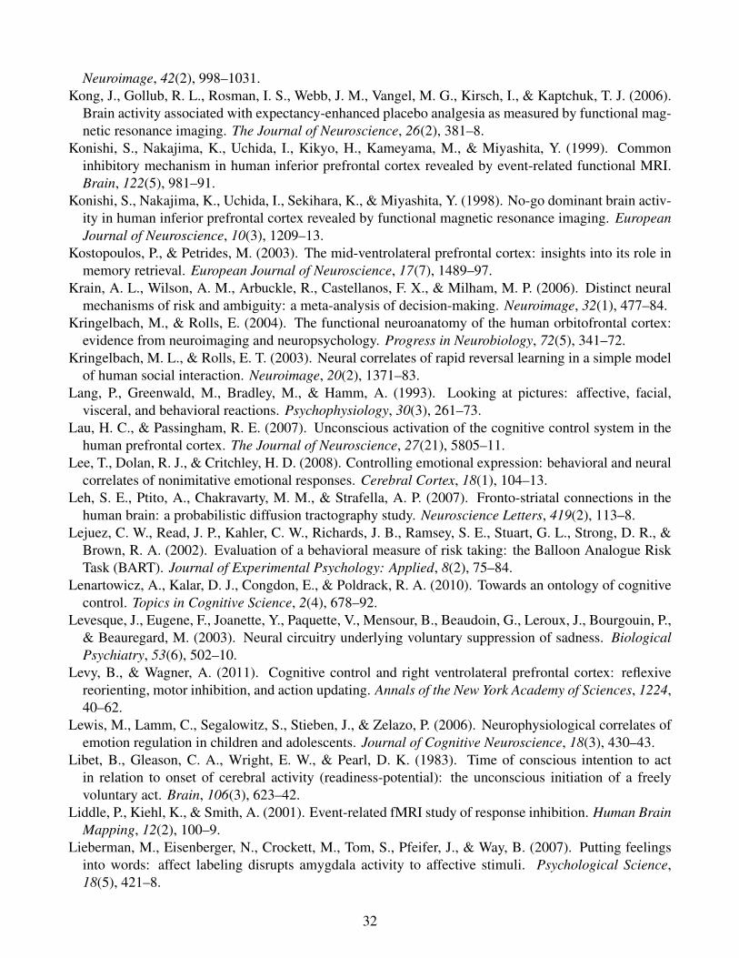
Neuroimage, 42(2), 998–1031.Kong, J., Gollub, R. L., Rosman, I. S., Webb, J. M., Vangel, M. G., Kirsch, I., & Kaptchuk, T. J. (2006).
Brain activity associated with expectancy-enhanced placebo analgesia as measured by functional mag-netic resonance imaging. The Journal of Neuroscience, 26(2), 381–8.
Konishi, S., Nakajima, K., Uchida, I., Kikyo, H., Kameyama, M., & Miyashita, Y. (1999). Commoninhibitory mechanism in human inferior prefrontal cortex revealed by event-related functional MRI.Brain, 122(5), 981–91.
Konishi, S., Nakajima, K., Uchida, I., Sekihara, K., & Miyashita, Y. (1998). No-go dominant brain activ-ity in human inferior prefrontal cortex revealed by functional magnetic resonance imaging. EuropeanJournal of Neuroscience, 10(3), 1209–13.
Kostopoulos, P., & Petrides, M. (2003). The mid-ventrolateral prefrontal cortex: insights into its role inmemory retrieval. European Journal of Neuroscience, 17(7), 1489–97.
Krain, A. L., Wilson, A. M., Arbuckle, R., Castellanos, F. X., & Milham, M. P. (2006). Distinct neuralmechanisms of risk and ambiguity: a meta-analysis of decision-making. Neuroimage, 32(1), 477–84.
Kringelbach, M., & Rolls, E. (2004). The functional neuroanatomy of the human orbitofrontal cortex:evidence from neuroimaging and neuropsychology. Progress in Neurobiology, 72(5), 341–72.
Kringelbach, M. L., & Rolls, E. T. (2003). Neural correlates of rapid reversal learning in a simple modelof human social interaction. Neuroimage, 20(2), 1371–83.
Lang, P., Greenwald, M., Bradley, M., & Hamm, A. (1993). Looking at pictures: affective, facial,visceral, and behavioral reactions. Psychophysiology, 30(3), 261–73.
Lau, H. C., & Passingham, R. E. (2007). Unconscious activation of the cognitive control system in thehuman prefrontal cortex. The Journal of Neuroscience, 27(21), 5805–11.
Lee, T., Dolan, R. J., & Critchley, H. D. (2008). Controlling emotional expression: behavioral and neuralcorrelates of nonimitative emotional responses. Cerebral Cortex, 18(1), 104–13.
Leh, S. E., Ptito, A., Chakravarty, M. M., & Strafella, A. P. (2007). Fronto-striatal connections in thehuman brain: a probabilistic diffusion tractography study. Neuroscience Letters, 419(2), 113–8.
Lejuez, C. W., Read, J. P., Kahler, C. W., Richards, J. B., Ramsey, S. E., Stuart, G. L., Strong, D. R., &Brown, R. A. (2002). Evaluation of a behavioral measure of risk taking: the Balloon Analogue RiskTask (BART). Journal of Experimental Psychology: Applied, 8(2), 75–84.
Lenartowicz, A., Kalar, D. J., Congdon, E., & Poldrack, R. A. (2010). Towards an ontology of cognitivecontrol. Topics in Cognitive Science, 2(4), 678–92.
Levesque, J., Eugene, F., Joanette, Y., Paquette, V., Mensour, B., Beaudoin, G., Leroux, J., Bourgouin, P.,& Beauregard, M. (2003). Neural circuitry underlying voluntary suppression of sadness. BiologicalPsychiatry, 53(6), 502–10.
Levy, B., & Wagner, A. (2011). Cognitive control and right ventrolateral prefrontal cortex: reflexivereorienting, motor inhibition, and action updating. Annals of the New York Academy of Sciences, 1224,40–62.
Lewis, M., Lamm, C., Segalowitz, S., Stieben, J., & Zelazo, P. (2006). Neurophysiological correlates ofemotion regulation in children and adolescents. Journal of Cognitive Neuroscience, 18(3), 430–43.
Libet, B., Gleason, C. A., Wright, E. W., & Pearl, D. K. (1983). Time of conscious intention to actin relation to onset of cerebral activity (readiness-potential): the unconscious initiation of a freelyvoluntary act. Brain, 106(3), 623–42.
Liddle, P., Kiehl, K., & Smith, A. (2001). Event-related fMRI study of response inhibition. Human BrainMapping, 12(2), 100–9.
Lieberman, M., Eisenberger, N., Crockett, M., Tom, S., Pfeifer, J., & Way, B. (2007). Putting feelingsinto words: affect labeling disrupts amygdala activity to affective stimuli. Psychological Science,18(5), 421–8.
32
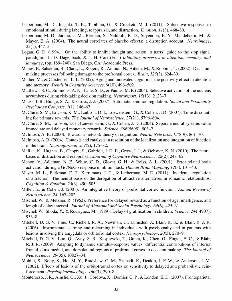
Lieberman, M. D., Inagaki, T. K., Tabibnia, G., & Crockett, M. J. (2011). Subjective responses toemotional stimuli during labeling, reappraisal, and distraction. Emotion, 11(3), 468–80.
Lieberman, M. D., Jarcho, J. M., Berman, S., Naliboff, B. D., Suyenobu, B. Y., Mandelkern, M., &Mayer, E. A. (2004). The neural correlates of placebo effects: a disruption account. Neuroimage,22(1), 447–55.
Logan, G. D. (1994). On the ability to inhibit thought and action: a users’ guide to the stop signalparadigm. In D. Dagenbach, & T. H. Carr (Eds.) Inhibitory processes in attention, memory, andlanguage, (pp. 189–240). San Diego, CA: Academic Press.
Manes, F., Sahakian, B., Clark, L., Rogers, R., Antoun, N., Aitken, M., & Robbins, T. (2002). Decision-making processes following damage to the prefrontal cortex. Brain, 125(3), 624–39.
Mather, M., & Carstensen, L. L. (2005). Aging and motivated cognition: the positivity effect in attentionand memory. Trends in Cognitive Sciences, 9(10), 496–502.
Matthews, S. C., Simmons, A. N., Lane, S. D., & Paulus, M. P. (2004). Selective activation of the nucleusaccumbens during risk-taking decision making. Neuroreport, 15(13), 2123–7.
Mauss, I. B., Bunge, S. A., & Gross, J. J. (2007). Automatic emotion regulation. Social and PersonalityPsychology Compass, 1(1), 146–67.
McClure, S. M., Ericson, K. M., Laibson, D. I., Loewenstein, G., & Cohen, J. D. (2007). Time discount-ing for primary rewards. The Journal of Neuroscience, 27(21), 5796–804.
McClure, S. M., Laibson, D. I., Loewenstein, G., & Cohen, J. D. (2004). Separate neural systems valueimmediate and delayed monetary rewards. Science, 306(5695), 503–7.
McIntosh, A. R. (2000). Towards a network theory of cognition. Neural Networks, 13(8-9), 861–70.McIntosh, A. R. (2004). Contexts and catalysts: a resolution of the localization and integration of function
in the brain. Neuroinformatics, 2(2), 175–82.McRae, K., Hughes, B., Chopra, S., Gabrieli, J. D. E., Gross, J. J., & Ochsner, K. N. (2010). The neural
bases of distraction and reappraisal. Journal of Cognitive Neuroscience, 22(2), 248–62.Menon, V., Adleman, N. E., White, C. D., Glover, G. H., & Reiss, A. L. (2001). Error-related brain
activation during a Go/NoGo response inhibition task. Human Brain Mapping, 12(3), 131–43.Meyer, M. L., Berkman, E. T., Karremans, J. C., & Lieberman, M. D. (2011). Incidental regulation
of attraction: The neural basis of the derogation of attractive alternatives in romantic relationships.Cognition & Emotion, 25(3), 490–505.
Miller, E., & Cohen, J. (2001). An integrative theory of prefrontal cortex function. Annual Review ofNeuroscience, 24, 167–202.
Mischel, W., & Metzner, R. (1962). Preference for delayed reward as a function of age, intelligence, andlength of delay interval. Journal of Abnormal and Social Psychology, 64(6), 425–31.
Mischel, W., Shoda, Y., & Rodriguez, M. (1989). Delay of gratification in children. Science, 244(4907),933–8.
Mitchell, D. G. V., Fine, C., Richell, R. A., Newman, C., Lumsden, J., Blair, K. S., & Blair, R. J. R.(2006). Instrumental learning and relearning in individuals with psychopathy and in patients withlesions involving the amygdala or orbitofrontal cortex. Neuropsychology, 20(3), 280–9.
Mitchell, D. G. V., Luo, Q., Avny, S. B., Kasprzycki, T., Gupta, K., Chen, G., Finger, E. C., & Blair,R. J. R. (2009). Adapting to dynamic stimulus-response values: differential contributions of inferiorfrontal, dorsomedial, and dorsolateral regions of prefrontal cortex to decision making. The Journal ofNeuroscience, 29(35), 10827–34.
Mobini, S., Body, S., Ho, M.-Y., Bradshaw, C. M., Szabadi, E., Deakin, J. F. W., & Anderson, I. M.(2002). Effects of lesions of the orbitofrontal cortex on sensitivity to delayed and probabilistic rein-forcement. Psychopharmacology, 160(3), 290–8.
Monterosso, J. R., Ainslie, G., Xu, J., Cordova, X., Domier, C. P., & London, E. D. (2007). Frontoparietal
33
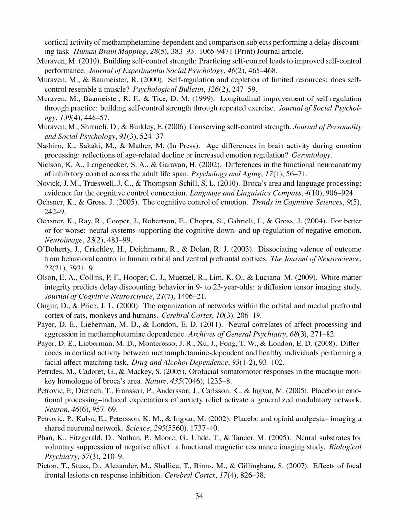
cortical activity of methamphetamine-dependent and comparison subjects performing a delay discount-ing task. Human Brain Mapping, 28(5), 383–93. 1065-9471 (Print) Journal article.
Muraven, M. (2010). Building self-control strength: Practicing self-control leads to improved self-controlperformance. Journal of Experimental Social Psychology, 46(2), 465–468.
Muraven, M., & Baumeister, R. (2000). Self-regulation and depletion of limited resources: does self-control resemble a muscle? Psychological Bulletin, 126(2), 247–59.
Muraven, M., Baumeister, R. F., & Tice, D. M. (1999). Longitudinal improvement of self-regulationthrough practice: building self-control strength through repeated exercise. Journal of Social Psychol-ogy, 139(4), 446–57.
Muraven, M., Shmueli, D., & Burkley, E. (2006). Conserving self-control strength. Journal of Personalityand Social Psychology, 91(3), 524–37.
Nashiro, K., Sakaki, M., & Mather, M. (In Press). Age differences in brain activity during emotionprocessing: reflections of age-related decline or increased emotion regulation? Gerontology.
Nielson, K. A., Langenecker, S. A., & Garavan, H. (2002). Differences in the functional neuroanatomyof inhibitory control across the adult life span. Psychology and Aging, 17(1), 56–71.
Novick, J. M., Trueswell, J. C., & Thompson-Schill, S. L. (2010). Broca’s area and language processing:evidence for the cognitive control connection. Language and Linguistics Compass, 4(10), 906–924.
Ochsner, K., & Gross, J. (2005). The cognitive control of emotion. Trends in Cognitive Sciences, 9(5),242–9.
Ochsner, K., Ray, R., Cooper, J., Robertson, E., Chopra, S., Gabrieli, J., & Gross, J. (2004). For betteror for worse: neural systems supporting the cognitive down- and up-regulation of negative emotion.Neuroimage, 23(2), 483–99.
O’Doherty, J., Critchley, H., Deichmann, R., & Dolan, R. J. (2003). Dissociating valence of outcomefrom behavioral control in human orbital and ventral prefrontal cortices. The Journal of Neuroscience,23(21), 7931–9.
Olson, E. A., Collins, P. F., Hooper, C. J., Muetzel, R., Lim, K. O., & Luciana, M. (2009). White matterintegrity predicts delay discounting behavior in 9- to 23-year-olds: a diffusion tensor imaging study.Journal of Cognitive Neuroscience, 21(7), 1406–21.
Ongur, D., & Price, J. L. (2000). The organization of networks within the orbital and medial prefrontalcortex of rats, monkeys and humans. Cerebral Cortex, 10(3), 206–19.
Payer, D. E., Lieberman, M. D., & London, E. D. (2011). Neural correlates of affect processing andaggression in methamphetamine dependence. Archives of General Psychiatry, 68(3), 271–82.
Payer, D. E., Lieberman, M. D., Monterosso, J. R., Xu, J., Fong, T. W., & London, E. D. (2008). Differ-ences in cortical activity between methamphetamine-dependent and healthy individuals performing afacial affect matching task. Drug and Alcohol Dependence, 93(1-2), 93–102.
Petrides, M., Cadoret, G., & Mackey, S. (2005). Orofacial somatomotor responses in the macaque mon-key homologue of broca’s area. Nature, 435(7046), 1235–8.
Petrovic, P., Dietrich, T., Fransson, P., Andersson, J., Carlsson, K., & Ingvar, M. (2005). Placebo in emo-tional processing–induced expectations of anxiety relief activate a generalized modulatory network.Neuron, 46(6), 957–69.
Petrovic, P., Kalso, E., Petersson, K. M., & Ingvar, M. (2002). Placebo and opioid analgesia– imaging ashared neuronal network. Science, 295(5560), 1737–40.
Phan, K., Fitzgerald, D., Nathan, P., Moore, G., Uhde, T., & Tancer, M. (2005). Neural substrates forvoluntary suppression of negative affect: a functional magnetic resonance imaging study. BiologicalPsychiatry, 57(3), 210–9.
Picton, T., Stuss, D., Alexander, M., Shallice, T., Binns, M., & Gillingham, S. (2007). Effects of focalfrontal lesions on response inhibition. Cerebral Cortex, 17(4), 826–38.
34
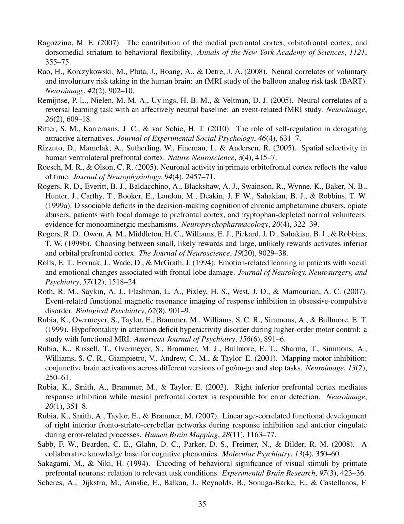
Ragozzino, M. E. (2007). The contribution of the medial prefrontal cortex, orbitofrontal cortex, anddorsomedial striatum to behavioral flexibility. Annals of the New York Academy of Sciences, 1121,355–75.
Rao, H., Korczykowski, M., Pluta, J., Hoang, A., & Detre, J. A. (2008). Neural correlates of voluntaryand involuntary risk taking in the human brain: an fMRI study of the balloon analog risk task (BART).Neuroimage, 42(2), 902–10.
Remijnse, P. L., Nielen, M. M. A., Uylings, H. B. M., & Veltman, D. J. (2005). Neural correlates of areversal learning task with an affectively neutral baseline: an event-related fMRI study. Neuroimage,26(2), 609–18.
Ritter, S. M., Karremans, J. C., & van Schie, H. T. (2010). The role of self-regulation in derogatingattractive alternatives. Journal of Experimental Social Psychology, 46(4), 631–7.
Rizzuto, D., Mamelak, A., Sutherling, W., Fineman, I., & Andersen, R. (2005). Spatial selectivity inhuman ventrolateral prefrontal cortex. Nature Neuroscience, 8(4), 415–7.
Roesch, M. R., & Olson, C. R. (2005). Neuronal activity in primate orbitofrontal cortex reflects the valueof time. Journal of Neurophysiology, 94(4), 2457–71.
Rogers, R. D., Everitt, B. J., Baldacchino, A., Blackshaw, A. J., Swainson, R., Wynne, K., Baker, N. B.,Hunter, J., Carthy, T., Booker, E., London, M., Deakin, J. F. W., Sahakian, B. J., & Robbins, T. W.(1999a). Dissociable deficits in the decision-making cognition of chronic amphetamine abusers, opiateabusers, patients with focal damage to prefrontal cortex, and tryptophan-depleted normal volunteers:evidence for monoaminergic mechanisms. Neuropsychopharmacology, 20(4), 322–39.
Rogers, R. D., Owen, A. M., Middleton, H. C., Williams, E. J., Pickard, J. D., Sahakian, B. J., & Robbins,T. W. (1999b). Choosing between small, likely rewards and large, unlikely rewards activates inferiorand orbital prefrontal cortex. The Journal of Neuroscience, 19(20), 9029–38.
Rolls, E. T., Hornak, J., Wade, D., & McGrath, J. (1994). Emotion-related learning in patients with socialand emotional changes associated with frontal lobe damage. Journal of Neurology, Neurosurgery, andPsychiatry, 57(12), 1518–24.
Roth, R. M., Saykin, A. J., Flashman, L. A., Pixley, H. S., West, J. D., & Mamourian, A. C. (2007).Event-related functional magnetic resonance imaging of response inhibition in obsessive-compulsivedisorder. Biological Psychiatry, 62(8), 901–9.
Rubia, K., Overmeyer, S., Taylor, E., Brammer, M., Williams, S. C. R., Simmons, A., & Bullmore, E. T.(1999). Hypofrontality in attention deficit hyperactivity disorder during higher-order motor control: astudy with functional MRI. American Journal of Psychiatry, 156(6), 891–6.
Rubia, K., Russell, T., Overmeyer, S., Brammer, M. J., Bullmore, E. T., Sharma, T., Simmons, A.,Williams, S. C. R., Giampietro, V., Andrew, C. M., & Taylor, E. (2001). Mapping motor inhibition:conjunctive brain activations across different versions of go/no-go and stop tasks. Neuroimage, 13(2),250–61.
Rubia, K., Smith, A., Brammer, M., & Taylor, E. (2003). Right inferior prefrontal cortex mediatesresponse inhibition while mesial prefrontal cortex is responsible for error detection. Neuroimage,20(1), 351–8.
Rubia, K., Smith, A., Taylor, E., & Brammer, M. (2007). Linear age-correlated functional developmentof right inferior fronto-striato-cerebellar networks during response inhibition and anterior cingulateduring error-related processes. Human Brain Mapping, 28(11), 1163–77.
Sabb, F. W., Bearden, C. E., Glahn, D. C., Parker, D. S., Freimer, N., & Bilder, R. M. (2008). Acollaborative knowledge base for cognitive phenomics. Molecular Psychiatry, 13(4), 350–60.
Sakagami, M., & Niki, H. (1994). Encoding of behavioral significance of visual stimuli by primateprefrontal neurons: relation to relevant task conditions. Experimental Brain Research, 97(3), 423–36.
Scheres, A., Dijkstra, M., Ainslie, E., Balkan, J., Reynolds, B., Sonuga-Barke, E., & Castellanos, F.
35
(2006). Temporal and probabilistic discounting of rewards in children and adolescents: effects of ageand ADHD symptoms. Neuropsychologia, 44(11), 2092–103.
Sharp, D. J., Bonnelle, V., Boissezon, X. D., Beckmann, C. F., James, S. G., Patel, M. C., & Mehta, M. A.(2010). Distinct frontal systems for response inhibition, attentional capture, and error processing.Proceedings of the National Academy of Sciences of the United States of America, 107(13), 6106–11.
Steinberg, L. (2008). A social neuroscience perspective on adolescent risk-taking. Developmental Re-view, 28(1), 78–106.
Strack, F., Martin, L. L., & Stepper, S. (1988). Inhibiting and facilitating conditions of the human smile:a nonobtrusive test of the facial feedback hypothesis. Journal of Personality and Social Psychology,54(5), 768–77.
Tabibnia, G., Lieberman, M. D., & Craske, M. G. (2008). The lasting effect of words on feelings: wordsmay facilitate exposure effects to threatening images. Emotion, 8(3), 307–17.
Tanaka, S. C., Doya, K., Okada, G., Ueda, K., Okamoto, Y., & Yamawaki, S. (2004). Prediction ofimmediate and future rewards differentially recruits cortico-basal ganglia loops. Nature Neuroscience,7(8), 887–93.
Tobler, P. N., O’Doherty, J. P., Dolan, R. J., & Schultz, W. (2007). Reward value coding distinct fromrisk attitude-related uncertainty coding in human reward systems. Journal of Neurophysiology, 97(2),1621–32.
Tzourio-Mazoyer, N., Landeau, B., Papathanassiou, D., Crivello, F., Etard, O., Delcroix, N., Mazoyer,B., & Joliot, M. (2002). Automated anatomical labeling of activations in SPM using a macroscopicanatomical parcellation of the MNI MRI single-subject brain. Neuroimage, 15(1), 273–89.
Urry, H. L., van Reekum, C. M., Johnstone, T., Kalin, N. H., Thurow, M. E., Schaefer, H. S., Jackson,C. A., Frye, C. J., Greischar, L. L., Alexander, A. L., & Davidson, R. J. (2006). Amygdala andventromedial prefrontal cortex are inversely coupled during regulation of negative affect and predictthe diurnal pattern of cortisol secretion among older adults. The Journal of Neuroscience, 26(16),4415–25.
van Gaal, S., Ridderinkhof, K. R., Fahrenfort, J. J., Scholte, H. S., & Lamme, V. A. F. (2008). Frontalcortex mediates unconsciously triggered inhibitory control. The Journal of Neuroscience, 28(32),8053–62.
van Gaal, S., Ridderinkhof, K. R., Scholte, H. S., & Lamme, V. A. F. (2010). Unconscious activation ofthe prefrontal no-go network. The Journal of Neuroscience, 30(11), 4143–50.
van Reekum, C. M., Johnstone, T., Urry, H. L., Thurow, M. E., Schaefer, H. S., Alexander, A. L., &Davidson, R. J. (2007). Gaze fixations predict brain activation during the voluntary regulation ofpicture-induced negative affect. Neuroimage, 36(3), 1041–55.
Verbruggen, F., Aron, A. R., Stevens, M. A., & Chambers, C. D. (2010). Theta burst stimulation dis-sociates attention and action updating in human inferior frontal cortex. Proceedings of the NationalAcademy of Sciences of the United States of America, 107(31), 13966–71.
Vuilleumier, P., Armony, J. L., Driver, J., & Dolan, R. J. (2001). Effects of attention and emotion on faceprocessing in the human brain: an event-related fMRI study. Neuron, 30(3), 829–41.
Wager, T., Sylvester, C., Lacey, S., Nee, D., Franklin, M., & Jonides, J. (2005). Common and uniquecomponents of response inhibition revealed by fMRI. Neuroimage, 27(2), 323–40.
Wager, T. D., Davidson, M. L., Hughes, B. L., Lindquist, M. A., & Ochsner, K. N. (2008). Prefrontal-subcortical pathways mediating successful emotion regulation. Neuron, 59(6), 1037–50.
Wager, T. D., Rilling, J. K., Smith, E. E., Sokolik, A., Casey, K. L., Davidson, R. J., Kosslyn, S. M., Rose,R. M., & Cohen, J. D. (2004). Placebo-induced changes in FMRI in the anticipation and experience ofpain. Science, 303(5661), 1162–7.
Wiech, K., Farias, M., Kahane, G., Shackel, N., Tiede, W., & Tracey, I. (2008). An fMRI study measuring
36
analgesia enhanced by religion as a belief system. Pain, 139(2), 467–76.Williams, B., Ponesse, J., Schachar, R., Logan, G., & Tannock, R. (1999). Development of inhibitory
control across the life span. Developmental Psychology, 35(1), 205–13.Winecoff, A., Labar, K. S., Madden, D. J., Cabeza, R., & Huettel, S. A. (2011). Cognitive and neural
contributors to emotion regulation in aging. Social Cognitive and Affective Neuroscience, 6(2), 165–76.
Wittmann, M., Leland, D. S., & Paulus, M. P. (2007). Time and decision making: differential contributionof the posterior insular cortex and the striatum during a delay discounting task. Experimental BrainResearch, 179(4), 643–53.
Wu, G., Zhang, J., & Gonzalez, R. (2004). Decision under risk. In D. Koehler, & N. Harvey (Eds.)Handbook of judgment and decision making, (pp. 399–423). Oxford, UK: Blackwell.
Xue, G., Aron, A. R., & Poldrack, R. A. (2008a). Common neural substrates for inhibition of spoken andmanual responses. Cerebral Cortex, 18(8), 1923–32.
Xue, G., Ghahremani, D. G., & Poldrack, R. A. (2008b). Neural substrates for reversing stimulus-outcome and stimulus-response associations. The Journal of Neuroscience, 28(44), 11196–204.
37


![[Incidental thyroid carcinoma: a multicentric experience]](https://img.pdfslide.net/doc/110x75/633bbd1a0d778cea7b08563a/incidental-thyroid-carcinoma-a-multicentric-experience.jpg)