Embed Size (px)
Citation preview

Inter- and Intra-Host Viral Diversity in a Large SeasonalDENV2 OutbreakCamila Malta Romano1*, Michael Lauck2, Felipe S. Salvador1, Celia Rodrigues Lima1, Lucy S. Villas-Boas1,
Evaldo Stanislau A. Araujo3, Jose Eduardo Levi1, Claudio Sergio Pannuti1, David O’Connor2, Esper
Georges Kallas4
1 Instituto de Medicina Tropical de Sao Paulo e Faculdade de Medicina, Departamento de Molestias Infecciosas e Parasitarias (LIMHC), Universidade de Sao Paulo, Sao
Paulo, Brazil, 2 Department of Pathology and Laboratory Medicine, University of Wisconsin–Madison, Madison, Wisconsin, United States of America, 3 Hospital das Clınicas
da Faculdade de Medicina, Universidade de Sao Paulo, Sao Paulo, Brazil, 4 Faculdade de Medicina, Disciplina de Imunologia Clınica e Alergia (LIM-60), Universidade de Sao
Paulo, Sao Paulo, Brazil
Abstract
Background: High genetic diversity at both inter- and intra-host level are hallmarks of RNA viruses due to the error-pronenature of their genome replication. Several groups have evaluated the extent of viral variability using different RNA virusdeep sequencing methods. Although much of this effort has been dedicated to pathogens that cause chronic infections inhumans, few studies investigated arthropod-borne, acute viral infections.
Methods and Principal Findings: We deep sequenced the complete genome of ten DENV2 isolates from representativeclassical and severe cases sampled in a large outbreak in Brazil using two different approaches. Analysis of the consensusgenomes confirmed the larger extent of the 2010 epidemic in comparison to a previous epidemic caused by the sameviruses in another city two years before (genetic distance = 0.002 and 0.0008 respectively). Analysis of viral populationswithin the host revealed a high level of conservation. After excluding homopolymer regions of 454/Roche generatedsequences, we found 10 to 44 variable sites per genome population at a frequency of .1%, resulting in very low intra-hostgenetic diversity. While up to 60% of all variable sites at intra-host level were non-synonymous changes, only 10% of inter-host variability resulted from non-synonymous mutations, indicative of purifying selection at the population level.
Conclusions and Significance: Despite the error-prone nature of RNA-dependent RNA-polymerase, dengue virusesmaintain low levels of intra-host variability.
Citation: Romano CM, Lauck M, Salvador FS, Lima CR, Villas-Boas LS, et al. (2013) Inter- and Intra-Host Viral Diversity in a Large Seasonal DENV2 Outbreak. PLoSONE 8(8): e70318. doi:10.1371/journal.pone.0070318
Editor: Cecilio Lopez-Galındez, Centro Nacional de Microbiologıa - Instituto de Salud Carlos III, Spain
Received February 5, 2013; Accepted June 18, 2013; Published August , 2013
Copyright: � 2013 Romano et al. This is an open-access article distributed under the terms of the Creative Commons Attribution License, which permitsunrestricted use, distribution, and reproduction in any medium, provided the original author and source are credited.
Funding: The present work was supported by Fundacao de Amparo a Pesquisa do Estado de Sao Paulo (FAPESP grant #10/12313-5 to CMR) and by theConselho Nacional de Desenvolvimento Cientifico e Tecnologico, Brazilian Ministry of Science and Technology (CNPq, grant #476088/2009-7 to EGK). URL ofFAPESP: www.fapesp.br; URL of CNPq: www.cnpq.br; URL of Coordenacao de Aperfeicoamento de Pessoal de Nıvel Superior: www.capes.gov.br. The funders hadno role in study design, data collection and analysis, decision to publish, or preparation of the manuscript. This work was also made possible in part by NationalInstitutes of Health grant R01 AI077376-04A1. The research was conducted, in part, at a facility constructed with support from Research Facilities ImprovementProgram grants RR15459-01 and RR020141-01.
Competing Interests: The authors state that the co-author Dr Esper George Kallas is currently a PLOS ONE Editorial Board member and also declare that thisdoes not alter the authors’ adherence to all the PLOS ONE policies on sharing data and materials.
* E-mail: [email protected]
Introduction
Dengue is the most frequent arthropod-borne human viral
infection in tropical and sub-tropical regions, with one hundred
million people at risk annually. In the last two decades, Brazil has
been responsible for more than 60% of the total reported dengue
fever cases in the Americas, experiencing severe yearly outbreaks
of serotypes 1 to 3 [1]. In 2008, serotype 4 re-emerged in the
northern region of Brazil [2] and rapidly spread to other regions,
causing small outbreaks in at least six different states. Today, the
country experiences outbreaks of all four serotypes. For this study,
samples were collected during a large outbreak in the State of Sao
Paulo coastal region during the first semester of 2010, with more
than 108,000 reported cases, including 81 deaths associated with
the severe forms of the disease [3]. This epidemic affected a
geographic area nearby the city of Santos, with the greatest
number of cases reported in five nearby cities (Figure 1).
Dengue has limited intra-genotype diversity, which is modulat-
ed by two main processes: (i) the host’s immune response, which
exerts a selective pressure on the virus, and (ii) the bottlenecks at
transmission (two in this case, the vertebrate-invertebrate alternate
cycle) [4]. As a result, a balance between variability gain due to
mutations in each host and the maintenance of these mutations
through natural selection is shaping the resulting viral diversity
observed at the population level.
Our current understanding of dengue intra-host diversity has
been mostly limited to the analysis of single genes of dengue
serotypes 1 to 3 using conventional cloning and Sanger sequencing
methods [5–7]. Most studies agree that the level of intra-host
diversity is relatively high with mean pairwise differences reaching
PLOS ONE | www.plosone.org 1 August 2013 | Volume 8 | Issue 8 | e70318
2

up to 1.67%. Recently, a study conducted by Thai et al. [8]
assessed dengue diversity by sampling a larger number of clones
while using more rigorous methods to validate the mutations and
reported much lower intra-host variability estimates than previ-
ously reported (0.008%,).In the last decade, deep sequencing
technologies have emerged capable of sequencing individual
molecules directly from PCR amplicons without the need for
cloning thereby enabling researchers to study viral quasispecies
diversity with unprecedented resolution. Work by our group and
others has employed 454 pyrosequencing, a method for deep
sequencing, for whole-genome shotgun sequencing of RNA viruses
causing chronic infections, such as hepatitis C virus (HCV),
human immunodeficiency virus (HIV), and simian immunodefi-
ciency virus (SIV) [9–14], but studies on the evolutionary
dynamics of acute infecting viruses within individuals are limited
[15]. Here, we employed pyrosequencing in combination with a
transposon-based fragmentation method to capture inter and
intra-host diversity of near full-length DENV-2 genomes covering
the entire open reading frame (ORF) in representative samples
obtained during the 2010 outbreak in cities located in the State of
Sao Paulo coastline [3]. By sequencing viruses sampled from 10
infected individuals we were able to confirm that the level of intra-
host viral variability is much lower than previously reported,
reaching approximately 0.002%, and determined the level of
variability throughout four months of the outbreak.
Materials and Methods
Ethics StatementThe current project was conducted after Hospital das Clınicas -
University of Sao Paulo’s Institutional Review Board (CAPPesq)
approval, under protocol #0652/09. Written informed consent
was obtained from all participants. Written informed consent was
also obtained for children through their parents.
Patients and SamplesSerum samples were obtained from patients with clinically
suspected dengue fever treated at the Ana Costa Hospital in
Santos, coastal region in the State of Sao Paulo, Brazil, during the
2010 DENV2 epidemic [16]. Dengue infections were confirmed
and serotypes were determined by using commercially available
rapid tests (NS1, IgG, and IgM) as well as multiplex reverse
transcriptase polymerase chain reaction (RT-PCR) [17]. Patients
were selected to be representative of the outbreak. Therefore,
samples were obtained from cases diagnosed from February
through May, 2010, from either classic dengue fever cases and
severe cases.
To determine the viral load, RNA was extracted from 140 ml of
plasma using the Qiagen Viral RNA mini kit (Qiagen, Valencia,
USA) according to the manufacturer’s instructions. RT-PCRs
were performed in duplicate using SuperScriptH III PlatinumHSYBRH Green One-Step RT-qPCR with ROX kit (Invitrogen,
Inc., EUA) and pan-dengue primers covering all 4 serotypes [18].
An internal control (Bovine Diarrhea Virus - BVDV, a flavivirus
grown in cell culture) was added to the samples before extraction
and also submitted to a parallel Real-time PCR assay. Supernatant
from DENV-3 cell cultures was included as external control in
every RT-qPCR run.
Several samples were also isolated in cell culture using the
established C6/36 Aedes albopictus cell line (ATCC number CRL-
1660). After the onset of a cytopathic effect (CPE) associated with
DENV infection, supernatants were collected and subjected to
RT-PCR in order to confirm the serotype. To discriminate
Figure 1. Geographic region of 2010 Santos epidemic. Southern Sao Paulo coastal region where viruses from this study were sampled are inred in the Sao Paulo map. Full black and red circles in the region focused represent the cities affected. The size of the circles specifies the magnitudeof the epidemic in each city, also given by the number of reported cases in Santos (St), Praia Grande (PG), Guaruja (Gu), Sao Vicente (SV) and Cubatao(Cu).doi:10.1371/journal.pone.0070318.g001
Intra- and Inter-Host DENV2 Diversity
PLOS ONE | www.plosone.org 2 August 2013 | Volume 8 | Issue 8 | e70318

between primary and secondary dengue virus infections, the
antigen-binding avidity of specific IgG was measured using a
modified Dengue ELISA IgG kit (Focus Technologies) [19].
RNA Extraction and Deep-sequencing500 ml of plasma from acutely ill patients were centrifuged at
3,000 rpm for 5 min to remove cellular debris followed by
centrifugation at 14,000 rpm for 60 min to concentrate the virus.
To evaluate if the viral population diversity found in plasma is
maintained after isolation in insect cell culture, we isolated viruses
from one acutely infected patient and sequenced the viruses (i)
direct from plasma and (ii) after one-week culture in C6/36 cell
line. Viral RNA was extracted from 200 ml of plasma or cell
culture supernatant containing the concentrated virus using the
QIAamp Viral RNA Mini Kit (Qiagen, Valencia, USA) according
to the manufacturer’s instructions. Primers were designed to
amplify the complete open reading frame with three to six
overlapping PCR amplicons of approximately 2–4 kb (See Table
S1 for primers sequences used), based on conserved regions
identified after a multiple sequence alignment of complete
DENV2 genomes of the American/Asian genotype. Viral RNA
was reverse transcribed and amplified using the SuperScript III
high-fidelity one-step reverse transcription-PCR (RT-PCR) kit
(Invitrogen, Life Technologies, Carlsbad, CA) with the following
conditions: 50uC for 30 min; 94uC for 2 min; 40 cycles of 94uC for
15 s, 55uC for 30 s, and 68uC for 2–4 min; and 68uC for 5 min.
Following amplification, PCR amplicon bands were isolated using
gel electrophoresis (1% agarose) and purified using a Qiagen
MinElute gel extraction kit (Qiagen, Valencia, CA). Each
amplicon was quantified using Quant-IT HS reagents (Invitrogen,
Life Technologies, Carlsbad, CA), and all amplicons from a single
viral genome were pooled together at equimolar ratios. Each pool
was then quantitated, and approximately 50 ng of each was
fragmented, using a Nextera DNA sample prep kit (Roche
Titanium compatible; Epicentre Biotechnologies, Madison, WI)
according to the manufacturer’s protocol. Final libraries repre-
senting each genome were characterized for average size by use of
a high-sensitivity DNA Bioanalyzer chip on a model 2100
Bioanalyzer (Agilent Technologies, Loveland, CO) and were
quantified with Quant-IT HS reagents (Invitrogen, Life Technol-
ogies, Carlsbad, CA). Libraries were then subjected to emulsion
PCR, and enriched DNA beads were loaded onto a picotiter plate
and pyrosequenced with a Roche/454 GS Junior sequencer using
titanium chemistry (454 Life Sciences, Branford, CT).
Pyrosequencing data were analyzed using CLC Genomics
Workbench 5.5 (CLC Bio, Aarhus, Denmark). Initially, reads were
trimmed to remove short and low-quality reads. Using a
phylogenetically related DENV2 genome as reference (GenBank
ID GU131864), reads were assembled and the resulting consensus
sequences were then used as each subject’s sequence in a reference
guided alignment to assess intra-host variability. The single
nucleotide polymorphism (SNP) analysis was performed using
CLC’s SNP analysis tool, applying the following parameters:
window length = 7, maximum gap and mismatch count = 2,
minimum central quality base = 30, minimum average quality
for window bases = 25, minimum coverage =6100, and minimum
variant frequency = 1% (defined as the percentage of nucleotides
that differ from the reference). Parameters used in this analysis
were chosen to include variants more likely to be biologically
significant, based on previous studies that used Roche/454 data
[12,14,20]. The 1% frequency cut-off both maximizes the chance
of detecting minor variants while at the same time reducing the
probability to detect errors generated in vitro [12,14]. Becker and
co-workers used this cut-off as well as cut-offs below 1% for
comparison and showed that if the coverage is higher than 1006,
variants found at 1% frequency are most likely real, and the lower
the coverage is, the higher the probability of the variant to be an
artifact [20]. Also, by choosing high-quality values for both the
central base (the SNP base) and the window bases (bases
surrounding the central SNP base, both upstream and down-
stream), as well as having a minimum coverage of 6100, only
areas of high quality and high coverage were considered for SNP
calling. To avoid erroneous base calls in homopolymer regions, we
mapped all the homopolymers through the genome by searching
for regions of three or more identical consecutive nucleotides, and
the deletions and insertions (DIPs) and SNPs that mapped in these
regions were dismissed from Roche/454 generated sequences due
to the high probability of miscalls.
In addition to the samples sequenced in 454 Roche, one
different plasma sample was sequenced on the Illumina MiSeq
using direct sequencing as previously described [14]. Briefly, after
isolation of RNA from cell-free plasma and DNase I treatment,
double stranded DNA was generated using the Superscript
double-stranded cDNA Synthesis kit (Invitrogen, Carlsbad, CA,
USA) primed with random hexamers thereby omitting any
amplification. Approximately 1 ng of cDNA was then fragmented
using the Nextera DNA Sample Preparation kit (Illumina, San
Diego, CA, USA), quantified and subjected to deep sequencing on
the Illumina MiSeq. Sequencing data were analyzed applying the
same parameters previously used for the pyrosequencing data
using CLC Genomics Workbench 5.5.
Sanger SequencingTo compare the variability obtained by deep sequencing
methods to traditional cloning and Sanger sequencing, the capsid
gene and part of the prM gene of the sample ACS538 were
subjected to traditional Sanger sequencing. Viral RNA was
extracted as described above and single strand cDNA was
generated with random hexamers using the High Capacity cDNA
Reverse Transcription Kit (Invitrogen, Life Technologies, Carls-
bad, CA) followed by amplification with the TaqPlatinum Hi
Fidelity kit (Invitrogen, Life Technologies, Carlsbad, CA) using
primers described in Table S1. The resulting 615-nucleotide (nt)
fragment was cloned into the pCR-TOPO TA vector (Invitrogen,
Life Technologies, Carlsbad, CA) and transformed into E. coli
DH5alpha quimio-competent cells. Thirty-one clones were
sequenced on the ABI3100 using Big-Dye7 Terminator (Applied
Biosystems, Warrington,UK), with M13 forward and M13 reverse
primers. Chromatograms were analyzed in CodonCode Aligner
v.3.0 (available at http://www.codoncode.com/) with a Phred
quality score of 20 as cut-off for trimming of low-quality sequences
and manually aligned and inspected using SeAl (http://tree.bio.
ed.ac.uk/software/seal/).
Phylogenetic AnalysisConsensus complete genome sequences obtained by sequencing
on the GS Junior and the MiSeq were aligned to globally sampled
DENV2 genomes (See Table S2 for a description of the sequences
used) using Muscle [21]. Bayesian phylogenetic trees were
constructed for 124 DENV2 genomes using MrBayes v.3.1.2
[22]. Two independent runs of 50 million steps each were done
using GTR with gamma-distributed rate variation substitution
model and a proportion of invariant sites as suggested by Akaike’s
information criterion (AICc) in jModeltest [23]. Sampled trees
were summarized and the consensus tree was visualized in FigTree
v1.3 (http://tree.bio.ed.ac.uk/). Selection pressures were evaluat-
ed by comparing the rate of non-synonymous changes per non-
synonymous site (dN) to the rate of synonymous changes per
Intra- and Inter-Host DENV2 Diversity
PLOS ONE | www.plosone.org 3 August 2013 | Volume 8 | Issue 8 | e70318

synonymous sites (dS) inferred for entire polyprotein as well as for
specific sites (by codon) of all the consensus genomes by single-
likelihood ancestor counting (SLAC), using the open-source
HyPhy package [24]. Genetic diversity was also estimated for
the consensus complete genomes at both the nucleotide and amino
acid levels using Mega 5 [25]. MacClade v4.08 was used to assess
nucleotide and amino acid substitutions along the branches [26].
Results
Epidemiological and Evolutionary Patterns of SantosDENV2
American/Asian DENV-2 complete genomes consensus were
reconstructed from overlapping RT-PCR amplicons deep se-
quenced on the Roche/454 GS Junior instrument. Additionally,
one plasma sample from an individual with classical dengue was
sequenced on the Illumina MiSeq by direct sequencing [27].
Therefore, viral complete genomes were generated from ten
infected patients that manifested either classical fever (eight
individuals) or severe dengue (two individuals), and also from
one sample cultured in C6/36 cell line. Disease outcome was
determined according to the WHO’s 2009 Dengue Guidelines
[28], where three possible classifications were adopted: (i) Dengue
without warning signals (classical), (ii) Dengue with warning signals
(classical WS), and (iii) severe Dengue. Table 1 summarizes all
samples used in this study as well as key clinical and laboratory
relevant features. There was no evident correlation between age,
gender or viral load and disease outcome. The complete genomes
of all DENV2 viruses were submitted to GenBank, under IDs
JX286516 to JX286526 (Table 1).
To determine the extent of DENV variability at a population
level, we examined the consensus sequence of all viruses. We were
not able to identify specific nucleotide or amino acid substitutions
that distinguished classical and severe cases. Nucleotide pairwise
distance calculated for all consensus genomes showed high degree
of conservation, with a maximum distance observed between
DGV37 and DGV69 (0.0044). Selection pressure analysis
performed across the whole coding region and also by codon
revealed no signs of positive selection (w = 0.03) as well as no
codon under selection. The mean genetic diversity of the
population (p) across the entire polyprotein was 0.002 for
nucleotide and 0.0008 for amino acid sequence, indicative of
protein conservation. Changes in 84 nucleotide sites were
distributed throughout the coding region, and only nine were
non-synonymous with three of them exclusively found in ACS542
(prM, NS3 and NS4a) (Figure 2B). Surprisingly, while the
envelope gene exhibited low variability at inter-host level, the
NS5 showed higher variability among samples (Figure 2A). It is
also interesting to observe that the variability among DENV2
population sampled in this outbreak was much higher than that
observed in viruses that circulated in a 2008 epidemic in Ribeirao
Preto, a city located in the North of the State of Sao Paulo
(represented by light green squares 2B), even though they belong
to the same lineage.
Phylogenetic AnalysisTo evaluate Dengue viruses in an epidemiological context, a
Bayesian phylogenetic tree was constructed using 124 complete
genomes comprising all five DENV-2 human genotypes and the
eleven genomes sequenced from the 2010 DENV2 Santos
outbreak (Figure 3A) (GenBank accession numbers are listed in
table S2). All genomes sequenced in this study clustered with
viruses that circulated in previous outbreaks registered in Sao
Paulo and Rio de Janeiro States in 2008, but separated from those
that circulated in older epidemics [16,29]. Based on our results,
Brazilian DENV viruses are not monophyletic, with viruses from
Cuba, USA and the Dominican Republic clustering with those
sequences.
The eleven 2010 DENV-2 genomes sequenced here segregated
into two sub-populations, named clade 1 (DGV34, DGV69,
DGV106, ACS538, ACS542 and ACS721) and clade 2 (DGV37,
DGV91, ACS46p, ACS46sn and ACS380) (Figure 3B). By
mapping all changes along the branches leading to them we
found that viruses from clade 2 accumulated eleven nucleotides
changes (eight C-T and two G-A transitions and one C-A
transversion) in relation to the clade 1. One of them, a transition of
cytosine to thymine at position 180 of the envelope gene led to an
Table 1. Clinical and laboratorial information of DENV-2 samples.
Patient GenBank IDAge(years) Gender
Clinicalmanifestation
Primary XSecondary Platelets/ml
Samplingday* VL
Samplingdate
ACS46 JX286516 55 Female Classical 2nd 113000 1 2.8E6 03/01/2010
ACS46sn$ JX286517 – – – – – – – –
ACS380* JX286526 67 Female Classical 2nd 124000 2 6.6E6 03/30/2010
ACS538 JX286518 62 Female Severe 2nd 43000 3 9.2E6 04/10/2010
ACS542 JX286519 1 Male Severe ND 24000 3 4.5E6 04/12/2010
ACS721 JX286521 34 Female Classical ND 191000 4 1.18E7 05/04/2010
DGV34 JX286522 50 Male Classical 2nd 158000 3 1.4E7 02/24/2010
DGV37 JX286520 46 Female Classical 2nd 132000 2 8.9E5 02/24/2010
DGV69 JX286525 56 Male Classical 2nd 122000 2 2.07E7 03/09/2010
DGV91 JX286523 45 Female Classical 2nd 233000 3 1.2E6 03/24/2010
DGV106& JX286524 82 Female Classical 2nd 10000 4 4E5 04/15/2010
$supernatant of sample ACS46 cultured viruses in C6/36 (1week).
&Although the low platelets number, the patient had no additional evidence of severity.*Days after the onset.VL- viral load in copy number/ml.Nd- not done.doi:10.1371/journal.pone.0070318.t001
Intra- and Inter-Host DENV2 Diversity
PLOS ONE | www.plosone.org 4 August 2013 | Volume 8 | Issue 8 | e70318

amino acid replacement (Thr to Ile). We also detected variability
in NS1, NS3, and NS4b but found no evidence of association with
virulence or pathogenicity.
Deep Sequencing of DENV2 GenomesOn average, 44,000 reads per sample (620.800) were generated
with an average coverage of ,1226 reads per nucleotide in
Roche/454 (Table 2). Between 0.1 and 1.7% of the reads could
not be mapped to Dengue genomes. Blast analysis revealed that
99% of the unspecific reads result from bacteria, while none of the
remaining reads matched to the human genome. Less than
0.001% of the unmapped reads come from DENV.
Homopolymer regions can induce erroneous base calls on the
Roche/454 platform, therefore all SNPs and DIPs that mapped to
those regions were disregarded in subsequent analyses. Between 32
and 85 DIPs for each sample sequenced were recovered by 454/
Roche and only two DIPs by Illumina. More than 90% of 454/
Roche DIPs mapped to homopolymer regions and about 80% of
them were identified as deletions in homopolymers of A, followed
by a much lower proportion of Ts, Cs and Gs, respectively (Figure
S1). The presence of DIPs was correlated to the size of the
homopolymer and most DIPs occurred in regions of more than 5
nucleotides (Figure S1, right top graph). A total of 624
homopolymers (ranging from 3 to 6 consecutive bases) were
Figure 2. Inter-host variability. A- The line graph summarizes the level of accumulated variability per genome region across the Santos consensusviruses. B- Variability (synonymous changes) among consensus sequences sampled in Santos (light blue squares) compared to the variability amongviruses from previous epidemics in 2008 in Ribeirao Preto, SP (green squares). Non-synonymous changes are represented in dark blue in bothpopulations.doi:10.1371/journal.pone.0070318.g002
Intra- and Inter-Host DENV2 Diversity
PLOS ONE | www.plosone.org 5 August 2013 | Volume 8 | Issue 8 | e70318

found along the DENV2 genome, resulting in 2178 sites excluded
from the analysis of intra-host genetic diversity (Table 2).
The level of intrahost variability obtained through Illumina
direct sequencing was also evaluated for the sample ACS380 and
was comparable to the observed through 454 pyrosequencing
(Table 2, Tables S3 and S4).
Sanger Sequencing of Capsid and prMIn parallel, the entire capsid and part of the prM gene of
ACS538 (400 base pairs) was amplified using similar conditions
and 31 clones were sequenced. The resulting 12378 nucleotides
sequenced revealed twelve changes randomly distributed among
the clones. One change was observed twice, but the remaining
eleven were singletons. None of the changes were detected by deep
sequencing and the unique variable site in the capsid recovered by
deep sequencing was not observed in Sanger sequences probably
due to the very low frequency (1.6%).
Low Intra-host Genetic Diversity of DENV2Overall, the two deep sequencing methods revealed between 10
and 44 variable sites per genome, resulting in approximately
0.002% of variability in all sequenced sites (Table 2 and see also
Table S3 for details of frequency and coverage of all SNPs).
DGV37, ACS46 and ACS721 had the highest conservation, with
Figure 3. Bayesian phylogenetic tree of 124 DENV2 complete genomes. A-The tree shows the eleven Brazilian viruses sequenced in thisstudy (two-color highlighted cluster at the top) and globally sampled DENV2 genomes. Blue branches represent Brazilian viruses sampled in previousepidemics. Posterior probability of all key nodes is depicted. B- Clusters of viruses sequenced during this study. The blue bar in the branch leading toclade 2 represents the amino acid change at position T180I in envelope gene.doi:10.1371/journal.pone.0070318.g003
Intra- and Inter-Host DENV2 Diversity
PLOS ONE | www.plosone.org 6 August 2013 | Volume 8 | Issue 8 | e70318
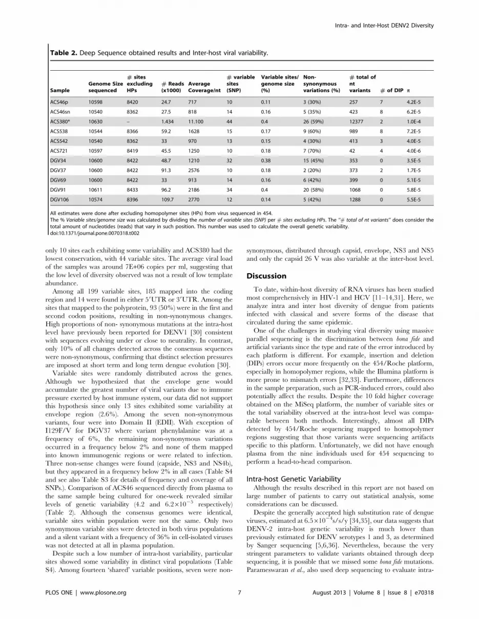
only 10 sites each exhibiting some variability and ACS380 had the
lowest conservation, with 44 variable sites. The average viral load
of the samples was around 7E+06 copies per ml, suggesting that
the low level of diversity observed was not a result of low template
abundance.
Among all 199 variable sites, 185 mapped into the coding
region and 14 were found in either 59UTR or 39UTR. Among the
sites that mapped to the polyprotein, 93 (50%) were in the first and
second codon positions, resulting in non-synonymous changes.
High proportions of non- synonymous mutations at the intra-host
level have previously been reported for DENV1 [30] consistent
with sequences evolving under or close to neutrality. In contrast,
only 10% of all changes detected across the consensus sequences
were non-synonymous, confirming that distinct selection pressures
are imposed at short term and long term dengue evolution [30].
Variable sites were randomly distributed across the genes.
Although we hypothesized that the envelope gene would
accumulate the greatest number of viral variants due to immune
pressure exerted by host immune system, our data did not support
this hypothesis since only 13 sites exhibited some variability at
envelope region (2.6%). Among the seven non-synonymous
variants, four were into Domain II (EDII). With exception of
I129F/V for DGV37 where variant phenylalanine was at a
frequency of 6%, the remaining non-synonymous variations
occurred in a frequency below 2% and none of them mapped
into known immunogenic regions or were related to infection.
Three non-sense changes were found (capside, NS3 and NS4b),
but they appeared in a frequency below 2% in all cases (Table S4
and see also Table S3 for details of frequency and coverage of all
SNPs.). Comparison of ACS46 sequenced directly from plasma to
the same sample being cultured for one-week revealed similar
levels of genetic variability (4.2 and 6.261025 respectively)
(Table 2). Although the consensus genomes were identical,
variable sites within population were not the same. Only two
synonymous variable sites were detected in both virus populations
and a silent variant with a frequency of 36% in cell-isolated viruses
was not detected at all in plasma population.
Despite such a low number of intra-host variability, particular
sites showed some variability in distinct viral populations (Table
S4). Among fourteen ‘shared’ variable positions, seven were non-
synonymous, distributed through capsid, envelope, NS3 and NS5
and only the capsid 26 V was also variable at the inter-host level.
Discussion
To date, within-host diversity of RNA viruses has been studied
most comprehensively in HIV-1 and HCV [11–14,31]. Here, we
analyze intra and inter host diversity of dengue from patients
infected with classical and severe forms of the disease that
circulated during the same epidemic.
One of the challenges in studying viral diversity using massive
parallel sequencing is the discrimination between bona fide and
artificial variants since the type and rate of the error introduced by
each platform is different. For example, insertion and deletion
(DIPs) errors occur more frequently on the 454/Roche platform,
especially in homopolymer regions, while the Illumina platform is
more prone to mismatch errors [32,33]. Furthermore, differences
in the sample preparation, such as PCR-induced errors, could also
potentially affect the results. Despite the 10 fold higher coverage
obtained on the MiSeq platform, the number of variable sites or
the total variability observed at the intra-host level was compa-
rable between both methods. Interestingly, almost all DIPs
detected by 454/Roche sequencing mapped to homopolymer
regions suggesting that those variants were sequencing artifacts
specific to this platform. Unfortunately, we did not have enough
plasma from the nine individuals used for 454 sequencing to
perform a head-to-head comparison.
Intra-host Genetic VariabilityAlthough the results described in this report are not based on
large number of patients to carry out statistical analysis, some
considerations can be discussed.
Despite the generally accepted high substitution rate of dengue
viruses, estimated at 6.561024s/s/y [34,35], our data suggests that
DENV-2 intra-host genetic variability is much lower than
previously estimated for DENV serotypes 1 and 3, as determined
by Sanger sequencing [5,6,36]. Nevertheless, because the very
stringent parameters to validate variants obtained through deep
sequencing, it is possible that we missed some bona fide mutations.
Parameswaran et al., also used deep sequencing to evaluate intra-
Table 2. Deep Sequence obtained results and Inter-host viral variability.
SampleGenome Sizesequenced
# sitesexcludingHPs
# Reads(x1000)
AverageCoverage/nt
# variablesites(SNP)
Variable sites/genome size(%)
Non-synonymousvariations (%)
# total ofntvariants # of DIP p
ACS46p 10598 8420 24.7 717 10 0.11 3 (30%) 257 7 4.2E-5
ACS46sn 10540 8362 27.5 818 14 0.16 5 (35%) 423 8 6.2E-5
ACS380* 10630 – 1.434 11.100 44 0.4 26 (59%) 12377 2 1.0E-4
ACS538 10544 8366 59.2 1628 15 0.17 9 (60%) 989 8 7.2E-5
ACS542 10540 8362 33 970 13 0.15 4 (30%) 413 3 4.0E-5
ACS721 10597 8419 45.5 1250 10 0.18 7 (70%) 42 4 4.0E-6
DGV34 10600 8422 48.7 1210 32 0.38 15 (45%) 353 0 3.5E-5
DGV37 10600 8422 91.3 2576 10 0.18 2 (20%) 373 2 1.7E-5
DGV69 10600 8422 33 913 14 0.16 6 (42%) 399 0 5.1E-5
DGV91 10611 8433 96.2 2186 34 0.4 20 (58%) 1068 0 5.8E-5
DGV106 10574 8396 109.7 2770 12 0.14 5 (42%) 1288 0 5.5E-5
All estimates were done after excluding homopolymer sites (HPs) from virus sequenced in 454.The % Variable sites/genome size was calculated by dividing the number of variable sites (SNP) per # sites excluding HPs. The ‘‘# total of nt variants’’ does consider thetotal amount of nucleotides (reads) that vary in such position. This number was used to calculate the overall genetic variability.doi:10.1371/journal.pone.0070318.t002
Intra- and Inter-Host DENV2 Diversity
PLOS ONE | www.plosone.org 7 August 2013 | Volume 8 | Issue 8 | e70318

host DENV2 variability in Nicaragua, but although they also
noticed low intra-host variability, they observed 10 to 1006more
variable sites across the genome compared to our results, with no
evidences of mixed infection [37]. However, about 3 out 9 variable
sites described by the authors as putative hotspots map to
homopolymer regions and may be related to 454 errors.
By considering the number of reads with variability at any given
site per total coverage at this site, the mean diversity of intra-host
viral population was 4.861025, which is consistent with previous
results reporting DENV1 intra-host variability as 7.261025 [8].
Interestingly, while Thai et al. used cloning and Sanger sequencing
to evaluate variability, they also applied rigorous methods to
validate variants eventually leading to results comparable to our
study.
With few exceptions, most studies estimating DENV variability
have used cloning and Sanger sequencing [5–8,36]. In this study,
we compared the variability obtained by deep sequencing to that
recovered by cloning and Sanger sequencing. Although we are
aware that an appropriate comparison is not possible here due to
the low number of clones sequenced, the high number of changes
observed in Sanger sequencing (, 0,1%) resembled previously
reported results using the same method, but clearly contrasted to
the results seen by deep sequencing here and by others [5,7,8,37].
Importantly, Descloux and group evaluated the sequence variation
produced by their in vitro system and found an error frequency of
0.1% [5], which is exactly the SNP frequency found here and in
the previous studies that used Sanger.
Two SNPs found by Sanger (both from A to G) were also
present in 454/Roche reads. However, they mapped in homo-
polymer regions of 3 and 4 Adenines and were in frequencies of
0.37 and 0.4% respectively (data not shown). Thus, according to
the parameters adopted here to consider a variant to be true (i.e.
1% frequency, quality phred score above 30 and, most impor-
tantly, the exclusion of homopolymers), these variants were
dismissed. We therefore suspect that much of the changes reported
by studies that used Sanger sequencing without posterior
validation of the variants represent artifacts generated during
cDNA synthesis and PCR amplification [12,38], which are more
prone to be eliminated by deep sequencing analysis due to the high
coverage and filter parameters. More rigorous experiments must
be performed to evaluate how much of Sanger detected variants
are due to experimental errors.
Arboviruses usually harbor less nucleotide and amino acid
variation than observed in other RNA viruses due to alternate
replication in mammalian and mosquito cells [39–41]. This
alternation between hosts keeps arboviruses in high-fitness peaks
that rarely overlap and only tolerate very few changes. In this
scenario, genetically stable viruses would have evolutionary
advantages over those that rapidly accumulate variants. In our
case, the specialization of DENV to a single mammalian
environment after urbanization may indeed have led to the
selection of very specific genetic traits, further reducing optimal
fitness peaks. Additional studies are needed however to determine
how dengue restore adequate levels of diversity.
It is not unusual to use in-vitro cell culture to amplify viruses
obtained from infected individuals prior to genetic and molecular
studies [42,43]. However, although one week of virus culture is
probably not sufficient for the emergence of adaptive changes, the
amount of variability that is gained or lost at the intra-population
level during in vitro culture is largely unknown. To address this
question, we compared deep sequenced viruses directly from
human plasma to the same viruses after one-week of culture in
C6/36 mosquito cells. As expected, the consensus sequences were
identical, but the sites showing variability at the intra-host level
were different. These findings indicate that different (low) selection
pressures are exerted by C6/36 culture [44], which ultimately
impacts on the generation of distinct viral populations. However,
the influence of distinct selection pressure imposed by mammalian
and invertebrate hosts on the generation of dengue diversity needs
more investigation.
There is little (and controversial) evidence that intra-host genetic
variability is linked to disease severity [5,6,37]. Although the small
number of patients analyzed at a single time point presents a
limitation to the present study, the amount of intra-host viral
variability did not differ between classical and severe dengue cases.
Subtle effects of specific variants on disease severity may not be
detectable in such a small cohort and larger numbers must be
evaluated to address this hypothesis.
Although only few sites exhibited variability in our study, some
were consistently identified among distinct viral populations.
Detection of the same variants in viruses circulating during the
same epidemic suggests two possible and non-exclusive explana-
tions: (i) common variable sites are a consequence of de novo
mutations in less constrained sites (i.e. mutation hotspots).
Mutation hotspots often reflect the mechanisms of generating
mutations at a particular site (i.e. enzyme-induced hypermutation,
homopolymers and repeat regions) [45,46]. In viruses, hotspots are
usually present in immunogenic sites and enable the escape of the
host immune system [47,48]. Nevertheless, shared variable sites in
our sequences were not in homopolymer or repeat regions and
also appeared in low frequency, suggesting that such mutations
were non advantageous, at least in this context. And (ii), minor
variants are not immediately eliminated by genetic drift and could
be maintained during transmission to a new host. Although
variable shared sites were found as minority population within the
host, it has been demonstrated that minor variants can be passed
between individuals regardless of their fitness, and sometimes for
extended time-periods [36,49]. Considering that the viruses
sequenced in this study were sampled over a four months period,
the maintenance of minor variants during this epidemic presents a
realistic scenario.
Genetic and Epidemiological Features of DENV2 from2010 Epidemics
Our phylogeny, which includes globally sampled DENV
genomes, confirms the Caribbean origin of the Brazilian
American/Asian DENV2 strain [16,29]. In fact, the presence of
viruses from Central and North America (Cuba, Nicaragua,
Guatemala, Jamaica, Dominican Republic and USA) within
Brazilian sequences exemplifies the idea of continual exchange
of viruses among countries on the American continent, and not
only between Central and South America.
Interestingly, the separation of Santos viruses into two sub-
clades and the high diversity found among the sequences in
comparison to viruses sampled in previous epidemics (Ribeirao
Preto, SP, Figure 2B) could be justified by the extent of both
epidemics, where the number of cases reported in 2008 in
Ribeirao Preto was around 1000 (http://www.cve.saude.sp.gov.
br/htm/zoo/Den_gve08.htm) against more than 33 thousand
cases reported in Santos coastal cities in a short five month period
(http://www.cve.saude.sp.gov.br/htm/zoo/den09_import_autoc.
htm). Although viruses sequenced in this study were all sampled in
the city of Santos, it is a regional hub that is heavily trafficked by
residents of neighboring cities.
In sum, the development of deep sequencing technologies
provides new opportunities to study the viral diversity with
unprecedented resolution. By using two different deep sequencing
methods, we were able to describe, for the first time, the level of
Intra- and Inter-Host DENV2 Diversity
PLOS ONE | www.plosone.org 8 August 2013 | Volume 8 | Issue 8 | e70318

DENV2 variability intra and inter host throughout a large
outbreak. The observed low level of variability may be a
consequence of different pressures, including the constraints
imposed by alternating hosts during virus evolution. Moreover,
the putative intrinsically low rate of changes combined with the
short time of viremia – common of acute infections – may exert
less selective pressure on the virus than is commonly observed
when studying chronic RNA virus infections.
Supporting Information
Figure S1 Deletions and Insertions (DIPs) sampled byRoche/454 GS Jr. The main graph shows the absolute number
of DIPs found per nucleotide obtained for the 10 viruses
sequenced in Roche/454. The small graph at the top right
specifies the proportion of DIPs in relation to homopolymer size.
(TIF)
Table S1 Primers used to amplify the complete genomeand also the capsid gene.(DOC)
Table S2 GenBank accession numbers of Dengueviruses serotype 2 used to build the phylogenetic tree.(DOC)
Table S3 Intra-host variability (SNPs). Reference posi-tions are according to the ACS380 genome (JX286526),and the details of the amino acid changes are in Supp-
(DOC)
Table S4 Variable non-synonymous sites at intra-hostlevel.(DOC)
Acknowledgments
We are in debt to Maria Candida Souza Dantas and Issler Moraes Silva for
the grant’s administrative support, and Karina I. Carvalho, Andreia
Manso de Matos, Bianca Natali Santos, and Fernanda Romano Bruno for
the laboratory support at the LIM-60. We are also thankful for the
comments provided by the anonymous reviewers.
Author Contributions
Conceived and designed the experiments: CMR CSP DO EGK.
Performed the experiments: ML FSS CRL LSV. Analyzed the data:
CMR ML DO EGK. Contributed reagents/materials/analysis tools: JEL
DO ML. Wrote the paper: CMR ML DO EGK. Clinical evaluation and
diagnostic of patients included in this study: ESAA. Discussion of the
clinical cases: ESAA JEL CSP.
References
1. PAHO (2008) Number of Reported Cases of Dengue and Dengue HemorrhagicFever (DHF), Region of the Americas (by country and subregion). Pan American
Health Society.
2. Temporao JG, Penna GO, Carmo EH, Coelho GE, do Socorro Silva Azevedo
R, et al. (2011) Dengue virus serotype 4, Roraima State, Brazil. Emerg Infect Dis17: 938–940.
3. CVE DdZ (2010) Sao Paulo. pp. Dados Provisorios do CVE- dengue.
4. Grenfell B, Pybus O, Gog J, Wood J, Daly J, et al. (2004) Unifying the
epidemiological and evolutionary dynamics of pathogens. Science 303: 327–332.
5. Descloux E, Cao-Lormeau V, Roche C, De Lamballerie X (2009) Dengue 1
diversity and microevolution, French Polynesia 2001–2006: connection with
epidemiology and clinics. PLoS Negl Trop Dis 3: e493.
6. Wang WK, Sung TL, Lee CN, Lin TY, King CC (2002) Sequence diversity of
the capsid gene and the nonstructural gene NS2B of dengue-3 virus in vivo.Virology 303: 181–191.
7. Wang WK, Lin SR, Lee CM, King CC, Chang SC (2002) Dengue type 3 virusin plasma is a population of closely related genomes: quasispecies. J Virol 76:
4662–4665.
8. Thai KT, Henn MR, Zody MC, Tricou V, Nguyet NM, et al. (2012) High-
resolution analysis of intrahost genetic diversity in dengue virus serotype 1
infection identifies mixed infections. J Virol 86: 835–843.
9. Farci P, Purcell R (2000) Clinical significance of hepatitis C virus genotypes and
quasispecies. Semin Liver Dis 20: 103–126.
10. Farci P, Shimoda A, Coiana A, Diaz G, Peddis G, et al. (2000) The outcome of
acute hepatitis C predicted by the evolution of the viral quasispecies. Science288: 339–344.
11. Rozera G, Abbate I, Bruselles A, Vlassi C, D’Offizi G, et al. (2009) Massivelyparallel pyrosequencing highlights minority variants in the HIV-1 env
quasispecies deriving from lymphomonocyte sub-populations. Retrovirology 6:
15.
12. Lauck M, Alvarado-Mora MV, Becker EA, Bhattacharya D, Striker R, et al.
(2012) Analysis of hepatitis C virus intrahost diversity across the coding region byultradeep pyrosequencing. J Virol 86: 3952–3960.
13. Ninomiya M, Ueno Y, Funayama R, Nagashima T, Nishida Y, et al. (2012) Useof illumina deep sequencing technology to differentiate hepatitis C virus variants.
J Clin Microbiol 50: 857–866.
14. Bimber BN, Dudley DM, Lauck M, Becker EA, Chin EN, et al. (2010) Whole-
genome characterization of human and simian immunodeficiency virus intrahost
diversity by ultradeep pyrosequencing. J Virol 84: 12087–12092.
15. Murcia PR, Baillie GJ, Daly J, Elton D, Jervis C, et al. (2010) Intra- and
interhost evolutionary dynamics of equine influenza virus. J Virol 84: 6943–6954.
16. Romano CM, de Matos AM, Araujo ES, Villas-Boas LS, da Silva WC, et al.(2010) Characterization of Dengue virus type 2: new insights on the 2010
Brazilian epidemic. PLoS One 5: e11811.
17. Guilarde AO, Turchi MD, Siqueira JB, Feres VC, Rocha B, et al. (2008)
Dengue and dengue hemorrhagic fever among adults: clinical outcomes relatedto viremia, serotypes, and antibody response. J Infect Dis 197: 817–824.
18. Lai YL, Chung YK, Tan HC, Yap HF, Yap G, et al. (2007) Cost-effective real-
time reverse transcriptase PCR (RT-PCR) to screen for Dengue virus followed
by rapid single-tube multiplex RT-PCR for serotyping of the virus. J Clin
Microbiol 45: 935–941.
19. de Souza VA, Fernandes S, Araujo ES, Tateno AF, Oliveira OM, et al. (2004)
Use of an immunoglobulin G avidity test to discriminate between primary and
secondary dengue virus infections. J Clin Microbiol 42: 1782–1784.
20. Becker EA, Burns CM, Leon EJ, Rajabojan S, Friedman R, et al. (2012)
Experimental analysis of sources of error in evolutionary studies based on
Roche/454 pyrosequencing of viral genomes. Genome Biol Evol 4: 457–465.
21. Edgar R (2004) MUSCLE: a multiple sequence alignment method with reduced
time and space complexity. BMC Bioinformatics 5: 113.
22. Ronquist F, Huelsenbeck JP (2003) MrBayes 3: Bayesian phylogenetic inference
under mixed models. Bioinformatics 19: 1572–1574.
23. Posada D (2008) jModelTest: phylogenetic model averaging. Mol Biol Evol 25:
1253–1256.
24. Pond SL, Frost SD (2005) Datamonkey: rapid detection of selective pressure on
individual sites of codon alignments. Bioinformatics 21: 2531–2533.
25. Tamura K, Peterson D, Peterson N, Stecher G, Nei M, et al. (2011) MEGA5:
molecular evolutionary genetics analysis using maximum likelihood, evolution-
ary distance, and maximum parsimony methods. Mol Biol Evol 28: 2731–2739.
26. Maddison W, Maddison D (1989) Interactive analysis of phylogeny and
character evolution using the computer program MacClade. Folia Primatol
(Basel) 53: 190–202.
27. Hughes AL, Becker EA, Lauck M, Karl JA, Braasch AT, et al. (2012) SIV
genome-wide pyrosequencing provides a comprehensive and unbiased view of
variation within and outside CD8 T lymphocyte epitopes. PLoS One 7: e47818.
28. WHO (2009) Dengue: guidelines for diagnosis, treatment, prevention and
control. In: Organization WH, editor. 147.
29. Oliveira M, Galvao Araujo J, Ferreira OJ, Ferreira D, Lima D, et al. (2010) Two
lineages of dengue virus type 2, Brazil. Emerg Infect Dis 16: 576–578.
30. Holmes EC (2003) Patterns of intra- and interhost nonsynonymous variation
reveal strong purifying selection in dengue virus. J Virol 77: 11296–11298.
31. Vartanian JP, Meyerhans A, Henry M, Wain-Hobson S (1992) High-resolution
structure of an HIV-1 quasispecies: identification of novel coding sequences.
AIDS 6: 1095–1098.
32. Gilles A, Meglecz E, Pech N, Ferreira S, Malausa T, et al. (2011) Accuracy and
quality assessment of 454 GS-FLX Titanium pyrosequencing. BMC Genomics
12: 245.
33. Suzuki S, Ono N, Furusawa C, Ying BW, Yomo T (2011) Comparison of
sequence reads obtained from three next-generation sequencing platforms. PLoS
One 6: e19534.
34. Kumar S, Patil J, Cecilia D, Cherian S, Barde P, et al. (2010) Evolution,
dispersal and replacement of American genotype dengue type 2 viruses in India
(1956–2005): selection pressure and molecular clock analyses. J Gen Virol 91:
707–720.
35. Duffy S, Shackelton L, Holmes E (2008) Rates of evolutionary change in viruses:
patterns and determinants. Nat Rev Genet 9: 267–276.
36. Aaskov J, Buzacott K, Thu HM, Lowry K, Holmes EC (2006) Long-term
transmission of defective RNA viruses in humans and Aedes mosquitoes. Science
311: 236–238.
Intra- and Inter-Host DENV2 Diversity
PLOS ONE | www.plosone.org 9 August 2013 | Volume 8 | Issue 8 | e70318
lementary bleta 4.

37. Parameswaran P, Charlebois P, Tellez Y, Nunez A, Ryan EM, et al. (2012)
Genome-wide patterns of intra-human dengue virus diversity reveal associationswith viral phylogenetic clade and inter-host diversity. J Virol.
38. Sabino E, Cheng-Mayer C, Mayer A (1993) An individual with a high
prevalence of a tat-defective provirus in peripheral blood. AIDS Res HumRetroviruses 9: 1265–1268.
39. Coffey LL, Vasilakis N, Brault AC, Powers AM, Tripet F, et al. (2008) Arbovirusevolution in vivo is constrained by host alternation. Proc Natl Acad Sci U S A
105: 6970–6975.
40. Vasilakis N, Deardorff ER, Kenney JL, Rossi SL, Hanley KA, et al. (2009)Mosquitoes put the brake on arbovirus evolution: experimental evolution reveals
slower mutation accumulation in mosquito than vertebrate cells. PLoS Pathog 5:e1000467.
41. Weaver SC, Brault AC, Kang W, Holland JJ (1999) Genetic and fitness changesaccompanying adaptation of an arbovirus to vertebrate and invertebrate cells.
J Virol 73: 4316–4326.
42. dos Santos CL, Bastos MA, Sallum MA, Rocco IM (2003) Molecularcharacterization of dengue viruses type 1 and 2 isolated from a concurrent
human infection. Rev Inst Med Trop Sao Paulo 45: 11–16.
43. Kanesa-thasan N, Chang GJ, Smoak BL, Magill A, Burrous MJ, et al. (1998)
Molecular and epidemiologic analysis of dengue virus isolates from Somalia.Emerg Infect Dis 4: 299–303.
44. Brackney DE, Scott JC, Sagawa F, Woodward JE, Miller NA, et al. (2010) C6/
36 Aedes albopictus cells have a dysfunctional antiviral RNA interferenceresponse. PLoS Negl Trop Dis 4: e856.
45. Rogozin IB, Pavlov YI (2003) Theoretical analysis of mutation hotspots and theirDNA sequence context specificity. Mutat Res 544: 65–85.
46. Strauss BS (1999) Frameshift mutation, microsatellites and mismatch repair.
Mutat Res 437: 195–203.47. Henn MR, Boutwell CL, Charlebois P, Lennon NJ, Power KA, et al. (2012)
Whole genome deep sequencing of HIV-1 reveals the impact of early minorvariants upon immune recognition during acute infection. PLoS Pathog 8:
e1002529.48. Hoper D, Kalthoff D, Hoffmann B, Beer M (2012) Highly pathogenic avian
influenza virus subtype H5N1 escaping neutralization: more than HA variation.
J Virol 86: 1394–1404.49. Li D, Lott WB, Lowry K, Jones A, Thu HM, et al. (2011) Defective interfering
viral particles in acute dengue infections. PLoS One 6: e19447.
Intra- and Inter-Host DENV2 Diversity
PLOS ONE | www.plosone.org 10 August 2013 | Volume 8 | Issue 8 | e70318


