Embed Size (px)
Citation preview

JOURNAL OF BACTERIOLOGY, Feb. 2003, p. 1174–1180 Vol. 185, No. 40021-9193/03/$08.00�0 DOI: 10.1128/JB.185.4.1174–1180.2003Copyright © 2003, American Society for Microbiology. All Rights Reserved.
Interactions between Phage-Shock Proteins in Escherichia coliHendrik Adams,† Wieke Teertstra, Jeroen Demmers, Rolf Boesten,
and Jan Tommassen*Department of Molecular Microbiology and Institute of Biomembranes, Utrecht
University, 3584 CH Utrecht, The Netherlands
Received 23 July 2002/Accepted 18 November 2002
Expression of the pspABCDE operon of Escherichia coli is induced upon infection by filamentous phage andby many other stress conditions, including defects in protein export. Expression of the operon requires thealternative sigma factor �54 and the transcriptional activator PspF. In addition, PspA plays a negativeregulatory role, and the integral-membrane proteins PspB and PspC play a positive one. In this study, weinvestigated whether the suggested protein-protein interactions implicated in this complex regulatory networkcan indeed be demonstrated. Antisera were raised against PspB, PspC, and PspD, which revealed, in Westernblotting experiments, that PspC forms stable sodium dodecyl sulfate-resistant dimers and that the hypotheticalpspD gene is indeed expressed in vivo. Fractionation experiments showed that PspD localizes as a peripherallybound inner membrane protein. Cross-linking studies with intact cells revealed specific interactions of PspAwith PspB and PspC, but not with PspD. Furthermore, affinity-chromatography suggested that PspB couldbind PspA only in the presence of PspC. These data indicate that regulation of the psp operon is mediated viaprotein-protein interactions.
Infection of Escherichia coli with filamentous bacteriophagef1 induces strongly the expression of the pspABCDE (phage-shock protein) operon (7; reviewed in reference 26). Theoperon encodes at least four proteins. PspA, encoded by thefirst gene of the operon, is a 26-kDa protein localized bothperipherally bound to the inner membrane and in the cytosol(7, 21). PspB is anchored in the inner membrane by a hydro-phobic N-terminal segment and has a C-terminal cytoplasmicdomain (8, 20). The product of the third gene, PspC, consistsof an N-terminal cytoplasmic domain, a single transmembranesegment, and a periplasmic C-terminal domain containing aleucine zipper motif (20). The putative product of the fourthopen reading frame, PspD, has not been detected yet, possiblybecause it would comigrate with PspB in sodium dodecyl sul-fate-polyacrylamide gel electrophoresis (SDS-PAGE) (8). Thesequence of the putative PspD protein does neither reveal asignal sequence nor a transmembrane segment, suggesting thatthe protein would be cytoplasmic if the gene were expressed.PspE contains a signal sequence and localizes in the periplasm(8, 20, 25). We recently demonstrated that PspE is a rhodan-ese-related enzyme (1).
It has been demonstrated that expression of the gene IVproduct of filamentous phage leads to the induction of the pspoperon (7, 19). The gene IV product is a member of thesecretin family and forms large multimeric channels in theouter membrane, through which the phage is extruded. Ex-pression of other members of the secretin family, involved intype II or type III protein secretion, also led to the induction ofthe psp operon in E. coli (3, 15, 23). Furthermore, various other
stress conditions, including extreme heat shock, osmotic shock(7), exposure to organic solvents (22) or ionophores, and pro-longed incubation under alkaline conditions (34), have beenreported to be inducing stimuli. In our laboratory, it has beenobserved that the psp operon is induced under conditions thatare known to block the protein export apparatus in the innermembrane. For example, expression of a mutant form of outermembrane protein PhoE, containing a stretch of eight consec-utive hydrophobic amino acids in its mature domain, led to astrong induction of the operon (21). Quantitative in vivo de-termination of the membrane potential across the inner mem-brane demonstrated that the proton motive force (PMF) spe-cifically decreased in a pspA mutant strain upon expression ofthis mutant PhoE protein (20). This result suggested that PspAis involved in the maintenance of the PMF, which may dissi-pate by proton leakage when the Sec channel is obstructed. Itis not clear whether all the different stimuli for induction of pspexpression interfere with protein translocation, but all of themprobably injure the cell membrane integrity. Therefore, PspAmay play a general role in maintaining the integrity of the innermembrane, rather than being directly involved in protein trans-location.
Transcription of the pspABCDE operon is initiated from apromoter located upstream of the pspA gene by �54-containingRNA polymerase (8). Expression of �54-dependent genes gen-erally requires an activator of the enhancer-binding protein(EBP) family (28, 35). Such a protein binds to upstream acti-vating sequences and is usually inactive until it is modified in itsN-terminal regulatory domain either by specific phosphoryla-tion or by the binding of an effector molecule. The pspF gene,which is located upstream of the pspABCDE operon and tran-scribed in the opposite direction, encodes the activator of thepsp operon (18). Although PspF belongs to the EBP family ofactivators, the protein lacks an N-terminal regulatory domain,and it is constitutively active both in vivo as in vitro (17). PspFbinds to sites overlapping its promoter, thereby regulating its
* Corresponding author. Mailing address: Department of MolecularMicrobiology, Utrecht University, Padualaan 8, 3584 CH Utrecht, TheNetherlands. Phone: (31) 30 253 2999. Fax: (31) 30 251 3655. E-mail:[email protected].
† Present address: Ecole Superieure de Biotechnologie de Stras-bourg, 67400 Illkirch, France.
1174

own expression, and its production is not affected by stimulithat induce the pspABCDE operon (16). Thus, regulation ofthe expression of the pspABCDE operon appears to be differ-ent from the EBP modification pathway used by other �54-dependent systems.
In addition to PspF, other proteins appear to affect thetranscription of the psp operon. It has been shown that pspexpression is negatively regulated by PspA (32), but this pro-tein does not bind DNA (10). Recently, it has been demon-strated that PspA directly interacts with PspF to inhibit tran-scription (11). The PspB and PspC proteins act cooperativelyas positive regulators of the operon, possibly by relieving thePspA-mediated repression (8, 32, 33). Interestingly, whereasPspB and PspC are not necessary for psp expression duringheat shock, they are absolutely required for induction of theoperon upon phage infection and only partially required forinduction by osmotic shock and ethanol treatment (32). Takentogether, the regulatory circuit of the psp operon appears to bevery complex and probably involves several protein-proteininteractions. In this study, we sought evidence for this hypoth-esis by identifying such interactions.
MATERIALS AND METHODS
Bacterial strains, growth conditions, and plasmids. The E. coli K-12 strainCE1224 carries a chromosomal deletion including the phoE gene (31), and itsderivatives CE1343 (21), CE1417, and CE1418 (20) carry a kanamycin resistancecassette in the pspA, pspB, and pspC genes, respectively. Strains DH5� (14) andSG13009, which carries plasmid pREP4 (Qiagen) encoding the lac repressor anda kanamycin resistance marker, were used for cloning. Cells were grown at 37°Cin L broth (31) supplemented with 0.4% glucose or in a synthetic phosphate-limited medium (30). When appropriate, ampicillin (100 �g/ml) or kanamycin(25 �g/ml) was added to the media for plasmid maintenance. To evaluate theirsensitivity to bile salts, cells were streaked on BBL MacConkey (Becton Dick-inson) plates, followed by incubation at 30°C for 24 h.
Plasmids pJP379, pJP380, and pJP381 (20), derived from plasmid pJF119HE(13), harbor pspB, pspABCDE, and pspA, respectively, under control of the tacpromoter. Plasmid pMR05H2 encodes a mutant PhoE protein with an insertionof eight consecutive hydrophobic amino acids (2). Its expression leads to a stronginduction of the phage-shock response (21).
For the construction of a glutathione S-transferase (GST) fusion with PspA,primers HA-005 (5�-GAGGATCCTATGGGTATTTTTTCTCGCTT-3�) andHA-006 (5�-TGGCCCGGGTTATTGATTGTCTTGCTTCA-3�) were used foramplification of the pspA gene. The PCR was performed with Pwo polymerase(Roche) and pJP380 as the template, with an annealing temperature of 60°C for60 s and extension at 68°C for 60 s in 30 cycles. The resulting PCR product wasagarose gel purified; digested with BamHI and SmaI, for which sites werecontained in the primers (underlined); and cloned into the corresponding sites ofthe GST fusion vector pRP269 (29). After sequencing, it appeared that the pspApart of the construct was truncated, resulting in the expression of a GST-PspApolypeptide in which the last 32 amino acid residues of PspA were replaced bythe amino acids GSTSMHKLEFIVTD, encoded by the expression vectorpRP269. Since no other clones were obtained, expression of a full-length GST-PspA hybrid may be lethal. The construct obtained was designated pGST-PspA�C.
Plasmids pGST-PspBcyt, pGST-PspCpp, and pHis-PspC were constructed asfollows. Primers HA-011 (5�-CTGGATCCTAGCAATCGTTCTGGTCGCAG-3�) and HA-002 (5�-TGCCCCGGGTTAGCGATCCCTCCAGTTCG-3�) wereused for the amplification of a pspB fragment encoding the cytoplasmic part ofPspB (amino acid residues 25 to 74), primers HA-036 (5�-TCATTTGCGCTGGATCCAATGCC-3�) and HA-004 (5�-TAACCCGGGTCACAGTTGACGGAAACGGC-3�) were used for the amplification of a pspC fragment encoding theperiplasmic part of PspC (amino acid residues 65 to 119), and primers HA-003(5�-GGGGATCCAATGGCGGGCATTAATCTCAA-3�) and HA-004 wereused to amplify the full-length pspC gene. These primers contain BamHI or SmaIsites (underlined). The PCRs with Taq polymerase and pJP380 as the templatewere performed with an annealing temperature of 50°C for 60 s and extension at68°C for 60 s in 30 cycles. The resulting PCR products were cloned in PCR II
TOPO (Invitrogen) according to the manufacturer’s instructions, resulting inTOPO-PspBcyt, TOPO-PspCpp, and TOPO-PspC, respectively. From theseplasmids, the relevant fragments were excised with BamHI and SmaI and ligatedinto the corresponding sites either of pRP269, yielding pGST-PspBcyt andpGST-PspCpp, or of plasmid pQE31 (Qiagen), resulting in pHis-PspC.
The nucleotide sequences of all constructs resulting from PCR amplificationswere confirmed by sequence analysis on an ABI 377 sequencer with the Dye-Terminator kit (Perkin-Elmer).
Purification of GST fusion proteins. Overnight cultures of strain CE1343carrying pGST-PspA�C, pGST-PspBcyt or pGST-PspCpp, were diluted 1:20 in800 ml of L broth supplemented with ampicillin and glucose and incubated at37°C until an optical density at 660 nm (OD660) of 0.6 was reached. Then,isopropyl-�-D-thiogalactopyranoside (IPTG) (100 �M final concentration) wasadded and incubation was continued for 2.5 h at 37°C. The cells were chilled onice and collected by centrifugation at 5,000 � g for 10 min at 4°C. The super-natant was removed, and the pellet was either resuspended as described below orstored at 20°C. All subsequent steps for purification of the GST fusion proteinswere done at 0 or 4°C. The pellet was resuspended in 20 ml of a solutioncontaining 30 mM Tris-HCl (pH 8.0), 25% sucrose, and 1 mM dithiothreitol(DTT) supplemented with Complete protease inhibitor (Roche) according to themanufacturer’s instructions. The cell suspension was disintegrated in a Frenchpress twice at 8,000 lb/in2. The intact cells and cell envelopes were removed bycentrifugation at 9,600 � g for 20 min and at 113,600 � g for 90 min, respectively.The supernatant was loaded, at a flow rate of 1 ml/min, onto a 10-ml GST-affinitycolumn (Pharmacia) equilibrated with mouse tonicity phosphate-buffered saline(MTPBS) (150 mM NaCl, 16 mM Na2HPO4, 4 mM NaH2PO4 [pH 7.3]) (29)supplemented with 1% Triton X-100. The column material was washed with 50ml of MTPBS buffer without Triton X-100, at a flow rate of 2 ml/min. The GSTfusion proteins were eluted with MTPBS containing 125 mM glutathione. Afterdialysis overnight against MTPBS, fractions containing GST fusion protein werestored in aliquots at 20°C at a protein concentration of 2 mg/ml. Proteinconcentrations were estimated from the absorbance at 280 nm in a Unicam UV1spectrophotometer (Spectronic), assuming an extinction coefficient of 1.0 for asolution with a protein concentration of 1.0 mg/ml.
Antisera. Polyclonal rabbit antisera were raised against the purified GST-PspBcyt and GST-PspCpp proteins at Eurogentec, Liege, Belgium. Antibodiesdirected against PspD were obtained by immunizing rabbits with synthetic pep-tides corresponding to amino acids 1 to 14 (MNTRWQQAGQKVKP) and 60 to73 (LSRAANKLAQRYKR) of PspD using the Double X system for peptideimmunization (Eurogentec).
In vivo cross-linking of Psp proteins. To induce expression of the chromo-somal pspABCDE operon, cells carrying plasmid pMR05H2 were grown underphosphate limitation for 5 h at 30°C as previously described (6) to induceexpression of the plasmid-encoded mutant PhoE protein. Alternatively, the Pspproteins were produced from plasmid pJP380 by growing cells in L-broth to thelogarithmic phase and then adding 100 �M IPTG to the medium and continuingincubation for 1 h at 37°C. Cells were harvested by centrifugation, washed oncewith 0.9% NaCl, and resuspended in 125 mM HEPES (pH 7.3) at an OD660 of1.0. The cell suspension (1.0 ml) was treated with 100 �M dithiobis(succinimy-dylpropionate) (DSP) (Pierce) at 25°C for 15 min, and the cross-link reaction wasquenched with 50 �l of 1 M Tris-HCl (pH 8.0). After incubation for 5 min at 0°C,cells were collected in a microcentrifuge (14,000 � g, 1 min), and proteins weresolubilized in sample buffer for 10 min at 100°C. The proteins were separated bySDS-PAGE on 11 or 15% polyacrylamide gels and transferred to nitrocellulosefilters (pore size, 0.45 �m; Schleicher and Schuell) using a Mini Trans-Blot cell(Bio-Rad Laboratories). Immunoincubations were performed essentially as de-scribed previously (27), using polyclonal antisera directed against PspA (21),PspB, PspC, or PspD. After incubation with horseradish peroxidase-conjugatedanti-rabbit immunoglobulin G antiserum (Biosource International), blots weredeveloped with 4-chloro-1-naphthol–H2O2 as the substrate or with SuperSignalWest Pico chemiluminescent substrate (Pierce) according to the manufacturer’srecommendations.
Extraction of inner membrane proteins. Cells of CE1224, carrying pJP379,pJP380, or pJP381, were grown aerobically at 37°C in L broth, containing am-picillin, to an OD660 of 0.1. After addition of 100 �M IPTG, growth was con-tinued for 3 h. Inner membrane vesicles (IMVs) were prepared as previouslydescribed (9). Subsequently, 10 �l of IMVs (OD280 25) was extracted with 80�l of buffer B (50 mM triethanolamine-acetate [pH 7.5], 250 mM sucrose, 3 mM�-mercaptoethanol, 0.5 mM phenylmethylsulfonyl fluoride), to which was added10 �l of 10% (wt/vol) 3-[(3-cholamidopropyl)-dimethylammonio]-1-propanesul-fonate (CHAPS). The solution was tumbled on a rotating wheel for 1 h at roomtemperature and then centrifuged at 20,000 � g for 45 min at 4°C. Incubation ofIMVs of PspABCDE- or PspA-overproducing strains with CHAPS for 15 min at
VOL. 185, 2003 INTERACTIONS BETWEEN Psp PROTEINS 1175

0°C resulted in hardly any release of PspA, whereas incubation at room temper-ature for 1 h released PspA more efficiently. The CHAPS extracts were kept at0°C.
In vitro cross-linking of Psp proteins. Aliquots (5 �l) of IMVs (OD280 1.25)were mixed with 90 �l of 125 mM HEPES. Subsequently, the IMVs were treatedeither with 5 �l of 4 mM DSP or with 5 �l of 4% formaldehyde for 20 min at 0°C.The cross-linking reaction was quenched with 150 �l of 1 M Tris-HCl (pH 8.0),followed by addition of 250 �l of 20% trichloroacetic acid. After incubation at0°C for 1 h, proteins were collected by centrifugation in a microcentrifuge for 15min at maximal speed and washed once with cold acetone. The pellet fractionwas air dried and solubilized in sample buffer for 20 min at 37°C. Proteins wereseparated by SDS-PAGE, followed by Western blotting and immunoincubationwith PspA antiserum as described above.
GST-PspA and GST-PspB pull-down assays. A 50-�l aliquot of 50% (vol/vol)GST-affinity agarose beads, equilibrated with MTPBS, was mixed with 400 �l ofMTPBS and 50 �l of purified GST-PspA�C or GST-PspBcyt. The mixture wastumbled for 1 h at room temperature to allow for binding of the GST fusionproteins to the beads, followed by centrifugation at 500 � g for 1 min, and thesupernatant was discarded. Subsequently, the agarose beads were incubated with40 �l of buffer A and 40 �l of CHAPS extracts from IMVs of cells overproducingPspA, PspB, or PspABCDE. After tumbling for 1 h at room temperature, theagarose beads were washed twice with 100 �l of MTPBS. Proteins were elutedwith MTPBS containing 125 mM glutathione and analyzed by SDS-PAGE fol-lowed by silver staining (4) or Western blotting with antisera directed againstPspA and PspB. N-terminal sequencing by Edman degradation was performed asdescribed previously (12).
Subcellular localization of PspD. Cells of strain CE1224 carrying pJP380 weregrown aerobically at 37°C in L-broth, and expression of Psp proteins was inducedas described above. Outer and inner membranes were separated by selectiveultracentrifugation as previously described (9). Peripherally bound membraneproteins were extracted from IMVs with a buffer containing 50 mM Tris-HCl–4M urea (pH 7.8). Protein patterns were analyzed by SDS-PAGE and Westernblotting using polyclonal antiserum against PspD.
RESULTS
In vivo cross-linking of PspA and associated proteins. Apilot experiment was conducted to investigate whether PspAinteracts with other proteins. Cells of strain CE1224 and of itspspA::kan derivative CE1343, both carrying pMR05H2, weregrown under phosphate limitation to induce expression of theplasmid-encoded mutant PhoE protein. Expression of this mu-tant PhoE protein strongly induces expression of the pspoperon (21). After treatment of the cells with the membrane-permeable, thiol-cleavable, homobifunctional cross-linkerDSP, cross-linked complexes were analyzed by SDS-PAGEand immunoblotting with a polyclonal antiserum directedagainst PspA. In addition to monomeric PspA, which migrateswith an apparent molecular weight (Mr) of 26,000, severalcross-linked protein complexes were observed (Fig. 1, lane 5).One of them migrated with an Mr of approximately 32,000,whereas a broader band migrated with an Mr of about 52,000.In addition, a smear of cross-linked complexes with an Mr
larger than 52,000 up to the top of the gel was observed.Neither the monomeric PspA, nor the cross-linked complexeswere detected in the case of the pspA mutant strain (Fig. 1,lane 7), demonstrating the specificity of these bands. Further-more, DSP-dependent cross-linking of proteins is reversible,and all complexes were found to dissociate upon addition ofDTT to the sample buffer (Fig. 1, lane 6). However, a smallfraction of the 52-kDa adduct in the protein sample remainedintact upon boiling in the presence of DTT. This incompletedissociation of the DSP-induced cross-link was always ob-served when the adduct was present in large quantities in theprotein sample. It was recently reported that PspA can formdimers or higher-ordered oligomers in vivo (10), and the 52-
kDa adduct could represent a dimeric form. However, sincethe band is broad, it may contain several different cross-linkedcomplexes. It is not clear whether the smear with an Mr of�52,000 contains specific cross-linked adducts to PspA or pro-tein aggregates. Since the calculated molecular mass of PspB is�7 kDa, the 32-kDa complex could be an adduct of PspA withPspB.
Antisera directed against PspB, PspC, and PspD. To be ableto assess whether the cross-linked complexes contain, in addi-tion to PspA, any of the other proteins encoded by the pspoperon, antisera were raised against GST-PspB and GST-PspCfusions and against synthetic PspD peptides. The antiserumdirected against PspB reacted specifically with a diffuse band ofapproximately 8 to 10 kDa in total protein samples of wild-typecells expressing PspB from plasmid (Fig. 2A, lane 2), whereasthis band was absent in the pspB::kan strain CE1417 (Fig. 2A,lane 1). The antiserum raised against the GST-PspC fusionrecognized two major bands of 14 and 28 kDa, respectively, ininner membranes of cells overexpressing PspABCDE frompJP380 (Fig. 2B, lane 2). Both bands were absent in IMVs ofthe pspC::kan strain CE1418 (data not shown). Moreover, alsotwo bands, but both with a slightly higher Mr, were detectedwhen inner membranes from cells producing His-tagged PspCprotein were analyzed (Fig. 2B, lane 1). Therefore, both bandsrepresent products of the pspC gene. We assume that the28-kDa band represents a dimeric form of PspC, although it isremarkable that this putative dimer did not dissociate upon
FIG. 1. Detection of PspA-associated proteins by in vivo cross-linking. Cells of strain CE1224 (lanes 1, 2, 5, and 6) or its pspA::kanderivative CE1343 (lanes 3, 4, 7, and 8), both carrying plasmidpMR05H2, were grown under phosphate limitation to induce the ex-pression of mutant PhoE protein and, consequently, of the psp operon.After incubation of the cells with DSP (lanes 5 to 8), proteins wereboiled in sample buffer, with (�) or without () DTT as indicated, andseparated by SDS–11% PAGE, and this was followed by Westernblotting using antibodies directed against PspA. The position of mo-nomeric PspA is indicated. Asterisks depict proteins that nonspecifi-cally react with anti-PspA antiserum. Relevant cross-linked adductsare indicated by carets. Blots were developed with 4-chloro-1-naph-thol–H2O2 as the substrate. The positions of molecular mass markerproteins are indicated at the right (in kilodaltons).
1176 ADAMS ET AL. J. BACTERIOL.

boiling in 2% SDS-containing sample buffer supplementedwith DTT. The antiserum directed against two synthetic PspDpeptides reacted specifically with a band of approximately 7kDa in total protein samples of wild-type cells expressing thePspABCDE proteins from plasmid, whereas this band was notdetectable in total protein samples of cells carrying the vectorpJF119HE (Fig. 2C, lanes 1 and 2).
Subcellular localization of PspD. Since the experiments de-scribed in the previous paragraph provided the first evidencethat the hypothetical pspD gene, which was so far only recog-nized as an open reading frame, indeed encodes a protein, itwas of interest to determine the subcellular localization of thisprotein. The soluble fraction, inner membranes, and outermembranes of pJP380-containing cells expressing all proteinsof the pspABCDE operon were isolated and analyzed by SDS-PAGE and Western blotting. Although the PspD protein se-quence does not reveal any stretch of hydrophobic amino acidresidues long enough to span the membrane, the protein ap-peared to be present mostly in the inner membrane fraction(Fig. 3, lane 3). When these membranes were extracted with 4M urea, the PspD protein was partly recovered in the urea-soluble fraction (results not shown). Together, these data in-dicate that PspD is a peripherally bound inner membraneprotein.
Complex formation in vivo of overproduced Psp proteins.
To obtain a high cross-linking efficiency, cross-linking experi-ments were conducted with cells that overproduced the PspABCDE proteins from plasmid. CE1224 cells containingpJP380 were induced with IPTG and treated with DSP. Anal-ysis by Western blotting with antibodies directed against PspArevealed again the 32-kDa complex, the broader band of �52kDa, and the smear of �52 kDa (Fig. 4, lane 3). In addition,three specific cross-linked protein complexes with Mrs of about40,000 to 45,000 were detected (Fig. 4, lane 3). Blots were alsoincubated with the antisera directed against PspB and PspC.The PspB antiserum reacted with the 32-kDa band in thecross-linked sample (Fig. 4, lane 7), while the PspC antiserumreacted with two of the three cross-linked complexes with Mrsof about 40,000 to 45,000 and (faintly) with a cross-linkedcomplex of �60 kDa (Fig. 4, lane 11). We assume that the
FIG. 2. Specificity of anti-PspB, anti-PspC and anti-PspD antisera.(A) Total protein samples of pspB::kan strain CE1417 (lane 1) andstrain CE1224 expressing PspB from pJP379 (lane 2) were separatedby SDS–20% PAGE, and this was followed by immunoblotting withantiserum directed against PspB (�-PspB) (dilution, 1:1,000).(B) IMVs of strain SG13009[pREP4] expressing His-tagged PspC frompHis-PspC (lane 1) and of strain CE1224 expressing PspABCDE frompJP380 (lane 2) were separated by SDS–15% PAGE, and this wasfollowed by immunoblotting with antiserum directed against PspC(�-PspC) (dilution, 1:1,000). (C) Total protein samples of strainCE1224 carrying pJP119HE (lane 1) or expressing PspABCDE frompJP380 (lane 2) were separated by SDS–15% PAGE, and this wasfollowed by immunoblotting with antiserum directed against PspD(�-PspD) (dilution, 1:1,000). Blots were developed with 4-chloro-1-naphthol–H2O2 as the substrate. The positions of molecular massmarker proteins are indicated at the right of each panel (in kilodal-tons).
FIG. 3. Subcellular localization of PspD. Cells of strain CE1224carrying pJP380 were grown in L-broth, and expression of the plasmid-encoded psp genes was induced by the addition of 100 �M IPTG. After3 h of incubation, cells were harvested and either directly analyzed(total proteins [TP]) or fractionated. After subjecting the cells to aFrench press, the outer membrane (OM) and inner membrane (IM)fractions and the soluble fraction (SF) (i.e., the combined periplasmicand cytoplasmic fraction) were obtained by differential ultracentrifu-gation. The fractions obtained were analyzed by SDS-PAGE followedby Western blotting with anti-PspD antiserum. The blot was developedwith 4-chloro-1-naphthol–H2O2 as the substrate.
FIG. 4. Complex formation of Psp proteins. Cells of strain CE1224,expressing PspABCDE from plasmid pJP380, were treated with DSPfollowed by SDS-PAGE and Western blotting with anti-PspA (�-PspA), anti-PspB (�-PspB), and anti-PspC (�-PspC) antisera. Whereindicated, samples were boiled in the presence of DTT prior to SDS-PAGE to reverse the cross-linking. The positions of monomeric PspA,PspB, and PspC and the dimeric form of PspC are indicated. Relevantcross-linked complexes are indicated by carets, square brackets, and asingle asterisk. The double asterisk indicates a band that reacts spe-cifically with anti-PspC antiserum, but the exact composition is un-known. The positions of the molecular mass marker proteins are in-dicated at the right (in kilodaltons). Blots were developed withSuperSignal West Pico chemiluminescent substrate.
VOL. 185, 2003 INTERACTIONS BETWEEN Psp PROTEINS 1177
�60-kDa complex represents a PspC dimer-PspA complex.Incubation of the blots with PspD antiserum demonstrated thepresence of monomeric PspD but did not reveal PspD-contain-ing cross-linked adducts (data not shown). Since the spacerarm of the cross-linker DSP is fairly long (12 A), the identifi-cation of cross-linked complexes reflects a close proximity, butnot necessarily a direct interaction between the cross-linkedpartners. Therefore, similar experiments were performed withformaldehyde as the cross-linking agent. Formaldehyde in-duces a one-atom bridge between neighboring proteins. Com-parison of DSP- and formaldehyde-induced cross-linking re-vealed very similar cross-linking patterns (results not shown).Taken together, these data demonstrate that PspA interactswith PspB and PspC in vivo, but not with PspD.
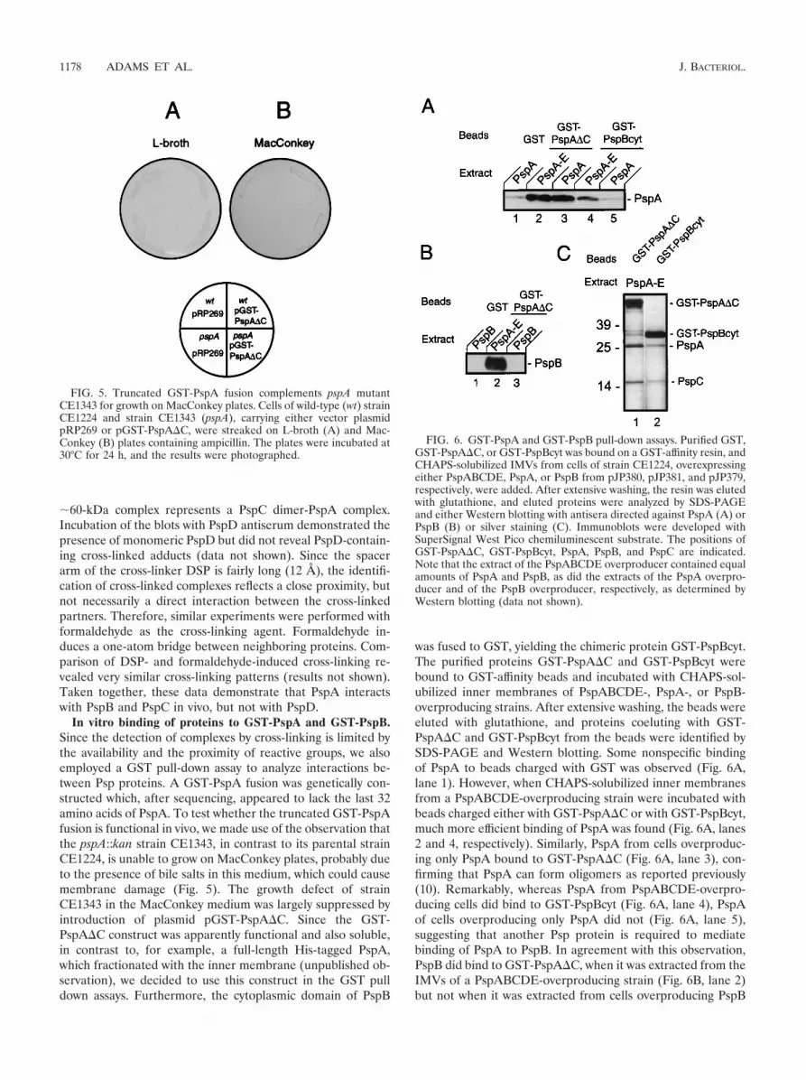
In vitro binding of proteins to GST-PspA and GST-PspB.Since the detection of complexes by cross-linking is limited bythe availability and the proximity of reactive groups, we alsoemployed a GST pull-down assay to analyze interactions be-tween Psp proteins. A GST-PspA fusion was genetically con-structed which, after sequencing, appeared to lack the last 32amino acids of PspA. To test whether the truncated GST-PspAfusion is functional in vivo, we made use of the observation thatthe pspA::kan strain CE1343, in contrast to its parental strainCE1224, is unable to grow on MacConkey plates, probably dueto the presence of bile salts in this medium, which could causemembrane damage (Fig. 5). The growth defect of strainCE1343 in the MacConkey medium was largely suppressed byintroduction of plasmid pGST-PspA�C. Since the GST-PspA�C construct was apparently functional and also soluble,in contrast to, for example, a full-length His-tagged PspA,which fractionated with the inner membrane (unpublished ob-servation), we decided to use this construct in the GST pulldown assays. Furthermore, the cytoplasmic domain of PspB
was fused to GST, yielding the chimeric protein GST-PspBcyt.The purified proteins GST-PspA�C and GST-PspBcyt werebound to GST-affinity beads and incubated with CHAPS-sol-ubilized inner membranes of PspABCDE-, PspA-, or PspB-overproducing strains. After extensive washing, the beads wereeluted with glutathione, and proteins coeluting with GST-PspA�C and GST-PspBcyt from the beads were identified bySDS-PAGE and Western blotting. Some nonspecific bindingof PspA to beads charged with GST was observed (Fig. 6A,lane 1). However, when CHAPS-solubilized inner membranesfrom a PspABCDE-overproducing strain were incubated withbeads charged either with GST-PspA�C or with GST-PspBcyt,much more efficient binding of PspA was found (Fig. 6A, lanes2 and 4, respectively). Similarly, PspA from cells overproduc-ing only PspA bound to GST-PspA�C (Fig. 6A, lane 3), con-firming that PspA can form oligomers as reported previously(10). Remarkably, whereas PspA from PspABCDE-overpro-ducing cells did bind to GST-PspBcyt (Fig. 6A, lane 4), PspAof cells overproducing only PspA did not (Fig. 6A, lane 5),suggesting that another Psp protein is required to mediatebinding of PspA to PspB. In agreement with this observation,PspB did bind to GST-PspA�C, when it was extracted from theIMVs of a PspABCDE-overproducing strain (Fig. 6B, lane 2)but not when it was extracted from cells overproducing PspB
FIG. 5. Truncated GST-PspA fusion complements pspA mutantCE1343 for growth on MacConkey plates. Cells of wild-type (wt) strainCE1224 and strain CE1343 (pspA), carrying either vector plasmidpRP269 or pGST-PspA�C, were streaked on L-broth (A) and Mac-Conkey (B) plates containing ampicillin. The plates were incubated at30°C for 24 h, and the results were photographed.
FIG. 6. GST-PspA and GST-PspB pull-down assays. Purified GST,GST-PspA�C, or GST-PspBcyt was bound on a GST-affinity resin, andCHAPS-solubilized IMVs from cells of strain CE1224, overexpressingeither PspABCDE, PspA, or PspB from pJP380, pJP381, and pJP379,respectively, were added. After extensive washing, the resin was elutedwith glutathione, and eluted proteins were analyzed by SDS-PAGEand either Western blotting with antisera directed against PspA (A) orPspB (B) or silver staining (C). Immunoblots were developed withSuperSignal West Pico chemiluminescent substrate. The positions ofGST-PspA�C, GST-PspBcyt, PspA, PspB, and PspC are indicated.Note that the extract of the PspABCDE overproducer contained equalamounts of PspA and PspB, as did the extracts of the PspA overpro-ducer and of the PspB overproducer, respectively, as determined byWestern blotting (data not shown).
1178 ADAMS ET AL. J. BACTERIOL.

only (Fig. 6B, lane 3). Considering the localization of PspE inthe periplasm, we suspect that PspC is the additional Pspprotein required for PspA-PspB binding, but we cannot ex-clude the possibility that PspD is required for this binding. Insupport of this hypothesis, silver staining of the gels revealed aband with an Mr of 16,000 when extracts of IMVs of PspABCDE-overproducing cells were bound to GST-PspA�C andGST-PspBcyt-charged beads (Fig. 6C, lanes 1 and 2, respec-tively). N-terminal sequencing of the protein identified theband as PspC. In conclusion, these data confirm interactionsbetween PspA and PspB and between PspA and PspC andsuggest that PspA binds to PspB only in the presence of an-other Psp protein, probably PspC.
DISCUSSION
Induction of the psp operon is regulated in a complex man-ner, requiring PspF and either PspB or PspC, or both, depen-dent on the specific inducing conditions. Because the operon isalso under negative control of PspA (32), induction can beviewed as overcoming the negative regulatory role of PspA.Consistent with this view, overproduction of PspC (32) or ofPspF (18) is sufficient to induce psp expression, indicatingtitration of PspA by protein-protein interactions. Sequenceanalysis suggests also that PspA and the cytoplasmic domain ofPspB have coiled-coil properties (24), whereas PspC has aleucine zipper motif in its periplasmic domain, all indicative forprotein-protein interactions. Recently, it has been demon-strated that PspA is the negative regulator of �54-dependenttranscription of the psp operon, which is probably based oninteraction with PspF (10, 11). Taken together, these datasuggested that regulation of the psp operon occurs via protein-protein interactions.
In this report, we have used an in vivo cross-linking ap-proach to analyze protein-protein interactions between Pspproteins. The use of the chemical cross-linker DSP allowed forthe identification of specific interactions between PspA, PspB,and PspC in intact cells. Western blotting revealed interactionsof PspA with PspB and with PspC, but not with PspD. Inaddition, the cross-linking experiments revealed interactions ofPspA with other proteins, resulting in cross-linked adductswith Mrs of �45,000 and a smear of �52,000, although theinteracting components were not identified. A PspA-PspF ad-duct, with an expected molecular mass of �63 kDa, could bepresent within the smear, but could not be identified due tounavailability of PspF-specific antiserum. Since evidence for aninteraction between PspA and PspF has recently been reported(11), we did not pursue demonstrating such interaction. Theinteractions between PspA and PspB and between PspA andPspC were confirmed by in vitro affinity experiments. Interest-ingly, the in vitro interaction between PspA and PspB couldonly be demonstrated when all Psp proteins were overex-pressed, indicating that another Psp protein is required. Wespeculate that PspC, which also interacted with PspB andPspA, is necessary to induce an efficient complex formationbetween PspA and PspB. An interaction between PspA andPspF was not revealed in the pull-down assays, probably be-cause PspF, which is expected to be a soluble cytoplasmicprotein, was not present in the solubilized inner membranesused in these experiments. Alternatively or in addition, PspF
might bind to the C-terminal fragment of PspA, which wasmissing in the GST-PspA fusion used. Previously, it has beenreported that a C-terminally truncated PspA failed to repressthe psp operon and, hence, was unable to bind PspF (7).
The Psp proteins protect the cell from dissipation of thePMF under stress conditions (20), probably by maintaining theintegrity of the inner membrane. A tempting model for theregulatory pathway is that the periplasmically exposed C-ter-minal part of PspC, which contains a leucine zipper motif,dimerizes in response to a PMF. Upon dissipation of the PMF,the dimer would dissociate, allowing the cytoplasmic part ofPspC to interact with PspB. This would induce binding of PspAto PspB, resulting in PspF release from a PspA-PspF complex.Subsequently, PspF activates the �54-dependent transcriptionof the pspABCDE operon. Such a PMF-sensing protein hasbeen described previously in mitochondria. It was demon-strated that dimerization of Tim23, a component of the pro-tein-import machinery of mitochondria, is dependent on aleucine-zipper motif, and it was found that Tim23 dimers wereformed in response to the membrane potential (5). The pos-tulated PMF-dependent dimerization of PspC will be a subjectfor further studies, although such a study will be a difficult taskconsidering the extremely stable PspC dimers that were al-ready detected in the present study.
ACKNOWLEDGMENTS
We thank Ria van Boxtel for technical assistance.This work was supported by grant HPRN-CT-2000-00075 of the
European Community.
REFERENCES
1. Adams, H., W. Teertstra, M. Koster, and J. Tommassen. 2002. PspE (phage-shock protein E) of Escherichia coli is a rhodanese. FEBS Lett. 518:173–176.
2. Agterberg, M., H. Adriaanse, A. van Bruggen, M. Karperien, and J. Tom-massen. 1990. Outer-membrane PhoE protein of Escherichia coli K-12 as anexposure vector: possibilities and limitations. Gene 88:37–45.
3. Akrim, M., M. Bally, G. Ball, J. Tommassen, H. Teerink, A. Filloux, and A.Lazdunski. 1993. Xcp-mediated protein secretion in Pseudomonas aerugi-nosa: identification of two additional genes and evidence for regulation ofxcp gene expression. Mol. Microbiol. 10:431–443.
4. Ansorge, W. 1985. Fast and sensitive detection of protein and DNA bands bytreatment with potassium permanganate. J. Biochem. Biophys. Methods11:13–20.
5. Bauer, M. F., C. Sirrenberg, W. Neupert, and M. Brunner. 1996. Role ofTim23 as voltage sensor and presequence receptor in protein import intomitochondria. Cell 87:33–41.
6. Bosch, D., J. Leunissen, J. Verbakel, M. de Jong, H. van Erp, and J. Tom-massen. 1986. Periplasmic accumulation of truncated forms of outer-mem-brane PhoE protein of Escherichia coli K-12. J. Mol. Biol. 189:449–455.
7. Brissette, J. L., M. Russel, L. Weiner, and P. Model. 1990. Phage-shockprotein, a stress protein of Escherichia coli. Proc. Natl. Acad. Sci. USA87:862–866.
8. Brissette, J. L., L. Weiner, T. L. Ripmaster, and P. Model. 1991. Character-ization and sequence of the Escherichia coli stress-induced psp operon. J.Mol. Biol. 220:35–48.
9. de Cock, H., S. van Blokland, and J. Tommassen. 1996. In vitro insertion andassembly of outer-membrane protein PhoE of Escherichia coli K-12 into theouter membrane. Role of Triton X-100. J. Biol. Chem. 271:12885–12890.
10. Dworkin, J., G. Jovanovic, and P. Model. 2000. The PspA protein of Esch-erichia coli is a negative regulator of �54-dependent transcription. J. Bacte-riol. 182:311–319.
11. Elderkin, S., S. Jones, J. Schumacher, D. Studholme, and M. Buck. 2002.Mechanism of action of the Escherichia coli Phage-shock protein PspA inrepression of the AAA family transcription factor PspF. J. Mol. Biol. 320:23–37.
12. El Khattabi, M., P. Van Gelder, W. Bitter, and J. Tommassen. 2000. Role ofthe lipase-specific foldase of Burkholderia glumae as a steric chaperone.J. Biol. Chem. 275:26885–26891.
13. Furste, J. P., W. Pansegrau, R. Frank, H. Blocker, P. Scholz, M. Bagdasar-ian, and E. Lanka. 1986. Molecular cloning of the plasmid RP4 primaseregion in a multi-host-range tacP expression vector. Gene 48:119–131.
VOL. 185, 2003 INTERACTIONS BETWEEN Psp PROTEINS 1179

14. Grant, S. G., J. Jessee, F. R. Bloom, and D. Hanahan. 1990. Differentialplasmid rescue from transgenic mouse DNAs into Escherichia coli methyla-tion-restriction mutants. Proc. Natl. Acad. Sci. USA 87:4645–4649.
15. Hardie, K. R., S. Lory, and A. P. Pugsley. 1996. Insertion of an outer-membrane protein in Escherichia coli requires a chaperone-like protein.EMBO J. 15:978–988.
16. Jovanovic, G., J. Dworkin, and P. Model. 1997. Autogenous control of PspF,a constitutively active enhancer-binding protein of Escherichia coli. J. Bac-teriol. 179:5232–5237.
17. Jovanovic, G., J. Rakonjac, and P. Model. 1999. In vivo and in vitro activitiesof the Escherichia coli sigma54 transcription activator, PspF, and its DNA-binding mutant, PspF�HTH. J. Mol. Biol. 285:469–483.
18. Jovanovic, G., L. Weiner, and P. Model. 1996. Identification, nucleotidesequence, and characterization of PspF, the transcriptional activator of theEscherichia coli stress-induced psp operon. J. Bacteriol. 178:1936–1945.
19. Kazmierczak, B. I., D. L. Mielke, M. Russel, and P. Model. 1994. pIV, afilamentous phage protein that mediates phage export across the bacterialcell envelope, forms a multimer. J. Mol. Biol. 238:187–198.
20. Kleerebezem, M., W. Crielaard, and J. Tommassen. 1996. Involvement ofstress protein PspA (phage-shock protein A) of Escherichia coli in mainte-nance of the proton-motive force under stress conditions. EMBO J. 15:162–171.
21. Kleerebezem, M., and J. Tommassen. 1993. Expression of the pspA genestimulates efficient protein export in Escherichia coli. Mol. Microbiol. 7:947–956.
22. Kobayashi, H., M. Yamamoto, and R. Aono. 1998. Appearance of a stress-response protein, phage-shock protein A, in Escherichia coli exposed tohydrophobic organic solvents. Microbiology 144:353–359.
23. Koster, M., W. Bitter, H. de Cock, A. Allaoui, G. R. Cornelis, and J. Tom-massen. 1997. The outer-membrane component, YscC, of the Yop secretionmachinery of Yersinia enterocolitica forms a ring-shaped multimeric complex.Mol. Microbiol. 26:789–797.
24. Lupas, A. 1996. Prediction and analysis of coiled-coil structures. MethodsEnzymol. 266:513–525.
25. Mielke, D. L., and M. Russel. 1992. A modified TnphoA useful for single-stranded DNA sequencing. Gene 118:93–95.
26. Model, P., G. Jovanovic, and J. Dworkin. 1997. The Escherichia coli phage-shock protein (psp) operon. Mol. Microbiol. 24:255–261.
27. Pettersson, A., P. van der Ley, J. T. Poolman, and J. Tommassen. 1993.Molecular characterization of the 98-kilodalton iron-regulated outer-mem-brane protein of Neisseria meningitidis. Infect. Immun. 61:4724–4733.
28. Reitzer, L., and B. L. Schneider. 2001. Metabolic context and possible phys-iological themes of �54-dependent genes in Escherichia coli. Microbiol. Mol.Biol. Rev. 65:422–444.
29. Smith, D. B., and K. S. Johnson. 1988. Single-step purification of polypep-tides expressed in Escherichia coli as fusions with glutathione S-transferase.Gene 67:31–40.
30. Tommassen, J., and B. Lugtenberg. 1980. Outer-membrane protein e ofEscherichia coli K-12 is coregulated with alkaline phosphatase. J. Bacteriol.143:151–157.
31. Tommassen, J., H. van Tol, and B. Lugtenberg. 1983. The ultimate localiza-tion of an outer-membrane protein of Escherichia coli K-12 is not deter-mined by the signal sequence. EMBO J. 2:1275–1279.
32. Weiner, L., J. L. Brissette, and P. Model. 1991. Stress-induced expression ofthe Escherichia coli phage shock protein operon is dependent on sigma 54and modulated by positive and negative feedback mechanisms. Genes Dev.5:1912–1923.
33. Weiner, L., J. L. Brissette, N. Ramani, and P. Model. 1995. Analysis of theproteins and cis-acting elements regulating the stress-induced phage-shockprotein operon. Nucleic Acids Res. 23:2030–2036.
34. Weiner, L., and P. Model. 1994. Role of an Escherichia coli stress-responseoperon in stationary-phase survival. Proc. Natl. Acad. Sci. USA 91:2191–2195.
35. Xu, H., and T. R. Hoover. 2001. Transcriptional regulation at a distance inbacteria. Curr. Opin. Microbiol. 4:138–144.
1180 ADAMS ET AL. J. BACTERIOL.


