Embed Size (px)
Citation preview

Interactions between UvrA and UvrB: the roleof UvrB’s domain 2 in nucleotide excision repair
James J Truglio1,5, Deborah L Croteau2,5,Milan Skorvaga2,3,5, Matthew JDellaVecchia2,5, Karsten Theis1,4,Bhaskar S Mandavilli2, Bennett VanHouten2,* and Caroline Kisker1,*1Department of Pharmacological Sciences, State University of New Yorkat Stony Brook, Stony Brook, NY, USA, 2Laboratory of MolecularGenetics, National Institute of Environmental Health Sciences, NationalInstitutes of Health, Research Triangle Park, NC, USA, 3Department ofMolecular Genetics, Cancer Research Institute, Slovak Academy ofSciences, Bratislava, Slovakia and 4Department of Biochemistry andMolecular Biology, University of Massachusetts Amherst, Amherst, MA,USA
Nucleotide excision repair (NER) is a highly conserved
DNA repair mechanism present in all kingdoms of life.
UvrB is a central component of the bacterial NER system,
participating in damage recognition, strand excision and
repair synthesis. None of the three presently available
crystal structures of UvrB has defined the structure of
domain 2, which is critical for the interaction with UvrA.
We have solved the crystal structure of the UvrB Y96A
variant, which reveals a new fold for domain 2 and
identifies highly conserved residues located on its surface.
These residues are restricted to the face of UvrB important
for DNA binding and may be critical for the interaction of
UvrB with UvrA. We have mutated these residues to study
their role in the incision reaction, formation of the pre-
incision complex, destabilization of short duplex regions
in DNA, binding to UvrA and ATP hydrolysis. Based on the
structural and biochemical data, we conclude that domain
2 is required for a productive UvrA–UvrB interaction,
which is a pre-requisite for all subsequent steps in nucleo-
tide excision repair.
The EMBO Journal (2004) 23, 2498–2509. doi:10.1038/
sj.emboj.7600263; Published online 10 June 2004
Subject Categories: structural biology; genome stability
& dynamics
Keywords: crystallography; DNA damage; DNA repair
nucleotide excision repair; UvrB
Introduction
Among the DNA repair mechanisms available to the cell,
nucleotide excision repair (NER) stands out because of its
broad substrate specificity (Van Houten, 1990; Friedberg et al,
1995; Lloyd and Van Houten, 1995; Sancar, 1996; Goosen and
Moolenaar, 2001). This repair mechanism is unique in its
versatility to repair substrates, including carcinogenic
cyclobutane pyrimidine dimers induced by UV radiation,
benzo[a]pyrene-guanine adducts caused by smoking and
guanine-cisplatinum adducts formed during chemotherapy
(Sancar, 1994). NER in bacteria, one of the first repair
mechanisms discovered (Boyce and Howard-Flanders, 1964;
Setlow and Carrier, 1964), is mediated by the products of the
uvrA, uvrB and uvrC genes. UvrA is involved in damage
recognition and was generally believed to form a heterotri-
meric (UvrA2UvrB) complex with UvrB (Theis et al, 2000).
Recently, however, the formation of a heterotetrameric
(UvrA2UvrB2) complex was suggested (Verhoeven et al,
2002). The UvrAUvrB complex has helicase-like properties
and specifically identifies conformational perturbations in-
duced by DNA lesions (Oh and Grossman, 1987, 1989; Koo
et al, 1991). After the damage has been identified, UvrA
dissociates, while UvrB remains bound to the DNA in a stable
pre-incision complex (Orren and Sancar, 1990). UvrC binds to
this complex and mediates the incision four nucleotides 30 of
the damaged site, followed by an incision seven nucleotides
50 of the damaged site (Sancar and Rupp, 1983; Lin and
Sancar, 1992; Lin et al, 1992; Verhoeven et al, 2000). UvrD and
DNA polymerase I are required for turnover of the UvrABC
proteins (Caron et al, 1985; Husain et al, 1985). UvrD removes
UvrC and the oligonucleotide containing the lesion, while
UvrB remains bound to the gapped DNA until it is filled by
DNA polymerase I (Husain et al, 1985). The reaction is
completed by DNA ligase, closing the nicked DNA. This
multi-step process of DNA recognition and repair ensures
discrimination between damaged and nondamaged DNA.
UvrB plays a central role in this repair cascade. It forms a
productive complex with UvrA that recognizes the damage
and guides the DNA from recognition to incision by complex
formation with UvrC. Finally, it is involved in repair synthesis,
ensuring that no gapped DNA intermediates are released
before the repair pathway is completed. Sequence analysis of
UvrB revealed that residues 154–251 share homology with the
transcription repair-coupling factor Mfd. Since both Mfd and
UvrB interact with UvrA, it was proposed that these homo-
logous regions are involved in interactions with UvrA (Selby
and Sancar, 1993). Biochemical studies using either residues
115–250 or the C-terminal residues 547–673 of Escherichia
coli UvrB fused to a maltose-binding protein suggest that
the first region interacts specifically with UvrA and the C-
terminal region interacts with UvrA and UvrC (Hsu et al,
1995). Both interactions are salt-dependent, indicating
that ionic interactions are important for UvrAUvrB complex
formation.Received: 27 January 2004; accepted: 11 May 2004; published online:10 June 2004
*Corresponding authors. C Kisker, Department of PharmacologicalSciences, State University of New York at Stony Brook, Stony Brook,NY 11794-5115, USA. Tel.: þ 1 631 632 1465; Fax: þ 1 631 632 1555;E-mail: [email protected] and B Van Houten, Laboratory ofMolecular Genetics, National Institute of Environmental HealthSciences, National Institutes of Health, Research Triangle Park,NC 27709, USA. Tel.: þ 1 919 541 2799;E-mail: [email protected] authors contributed equally to this work
The EMBO Journal (2004) 23, 2498–2509 | & 2004 European Molecular Biology Organization | All Rights Reserved 0261-4189/04
www.embojournal.org
The EMBO Journal VOL 23 | NO 13 | 2004 &2004 European Molecular Biology Organization
EMBO
THE
EMBOJOURNAL
THE
EMBOJOURNAL
2498
We solved the three-dimensional structure of the Bacillus
caldotenax UvrB variant Y96A. Despite the presence of three
independently determined UvrB structures (Machius et al,
1999; Nakagawa et al, 1999; Theis et al, 1999), no structural
information was obtained with respect to the putative UvrA-
interacting domains. The 60 C-terminal amino acids as well
as domain 2 harboring residues 151–251 are highly flexible in
all three structures. The structure of the C-terminal domain
was solved independently, revealing a coiled coil conforma-
tion (Sohi et al, 2000; Alexandrovich et al, 2001). Domain 2
was partially modeled as a poly-alanine model in two of the
three structures (Nakagawa et al, 1999; Theis et al, 1999),
although the structural details of this domain have remained
elusive. The structure of the Y96A variant allowed, for the
first time, a detailed atomic analysis of domain 2. We
identified several highly conserved residues on the surface
of this domain, which may be critical for the interaction with
UvrA. These residues define a patch that is located in close
proximity to the proposed DNA-binding site of UvrB.
Analyses of mutants within this patch support the conclusion
that the crosstalk between UvrA and UvrB, leading to damage
recognition, requires UvrA-interacting residues on the surface
of domain 2 of UvrB. These interactions are critical since the
formation of a productive complex allows damage recogni-
tion and repair, the first step in the reaction cascade of NER.
Results
Structure of the Y96A variant
The crystal structure of the UvrB Y96A variant from B.
caldotenax was solved by molecular replacement using the
wild-type (WT) structure (PDB code 1D9X) as a search
model. The protein crystallized in space group P3221 with
two molecules in the asymmetric unit, which are related to
each other by a translation of B0.5 along the z-axis. This is in
contrast to WT UvrB, which crystallizes in space group P3121
and contains a single molecule in the asymmetric unit (Theis
et al, 1999). In both structures, the functional unit of the
protein is a monomer. The structure was refined at 2.6 A
resolution to an R factor of 0.230 and Rfree of 0.286 (Table I).
The two Y96A molecules have nearly identical conforma-
tions, with r.m.s. deviations of 0.92 A for all Ca atoms.
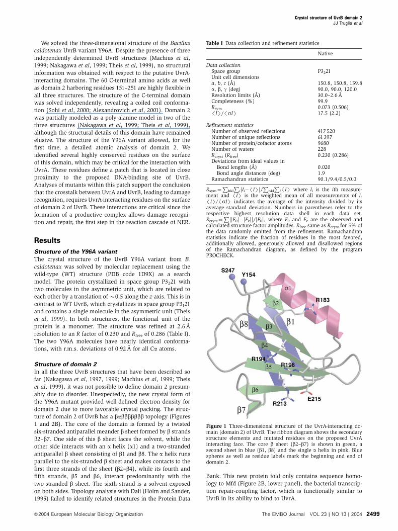
Structure of domain 2
In all the three UvrB structures that have been described so
far (Nakagawa et al, 1997, 1999; Machius et al, 1999; Theis
et al, 1999), it was not possible to define domain 2 presum-
ably due to disorder. Unexpectedly, the new crystal form of
the Y96A mutant provided well-defined electron density for
domain 2 due to more favorable crystal packing. The struc-
ture of domain 2 of UvrB has a babbbbbbb topology (Figures
1 and 2B). The core of the domain is formed by a twisted
six-stranded antiparallel meander b sheet formed by b strands
b2–b7. One side of this b sheet faces the solvent, while the
other side interacts with an a helix (a1) and a two-stranded
antiparallel b sheet consisting of b1 and b8. The a helix runs
parallel to the six-stranded b sheet and makes contacts to the
first three strands of the sheet (b2–b4), while its fourth and
fifth strands, b5 and b6, interact predominantly with the
two-stranded b sheet. The sixth strand is a solvent exposed
on both sides. Topology analysis with Dali (Holm and Sander,
1995) failed to identify related structures in the Protein Data
Bank. This new protein fold only contains sequence homo-
logy to Mfd (Figure 2B, lower panel), the bacterial transcrip-
tion repair-coupling factor, which is functionally similar to
UvrB in its ability to bind to UvrA.
Figure 1 Three-dimensional structure of the UvrA-interacting do-main (domain 2) of UvrB. The ribbon diagram shows the secondarystructure elements and mutated residues on the proposed UvrAinteracting face. The core b sheet (b2–b7) is shown in green, asecond sheet in blue (b1, b8) and the single a helix in pink. Bluespheres as well as residue labels mark the beginning and end ofdomain 2.
Table I Data collection and refinement statistics
Native
Data collectionSpace group P3221Unit cell dimensionsa, b, c (A) 150.8, 150.8, 159.8a, b, g (deg) 90.0, 90.0, 120.0Resolution limits (A) 30.0–2.6 ACompleteness (%) 99.9Rsym 0.073 (0.506)/IS//sIS 17.5 (2.2)
Refinement statisticsNumber of observed reflections 417 520Number of unique reflections 61 397Number of protein/cofactor atoms 9680Number of waters 228Rcryst (Rfree) 0.230 (0.286)Deviations from ideal values in
Bond lengths (A) 0.020Bond angle distances (deg) 1.9
Ramachandran statistics 90.1/9.4/0.5/0.0
Rsym¼P
hkl
Pi|Ii�/IS|/
Phkl
Pi/IS where Ii is the ith measure-
ment and /IS is the weighted mean of all measurements of I./IS//sIS indicates the average of the intensity divided by itsaverage standard deviation. Numbers in parentheses refer to therespective highest resolution data shell in each data set.Rcryst¼
P||F0|�|Fc||/|F0|, where F0 and Fc are the observed and
calculated structure factor amplitudes. Rfree same as Rcryst for 5% ofthe data randomly omitted from the refinement. Ramachandranstatistics indicate the fraction of residues in the most favored,additionally allowed, generously allowed and disallowed regionsof the Ramachandran diagram, as defined by the programPROCHECK.
Crystal structure of UvrB domain 2JJ Truglio et al
&2004 European Molecular Biology Organization The EMBO Journal VOL 23 | NO 13 | 2004 2499

Figure 2 Sequence conservation of domain 2 in UvrB. (A) Surface representation of domain 2 of UvrB (gray) with conserved residues labeledand color-coded (red: strictly conserved, dark blue: very highly conserved, cyan: highly conserved, green: moderate to highly conserved).Conservation is based on 56 domain 2 sequences aligned using ClustalX and analyzed by the ConSurf server. The upper panel shows the front(DNA-binding) side of UvrB and the lower panel is a 1801 rotation showing the back. The remainder of UvrB is drawn as a Ca trace and color-coded according to domain architecture with domain 1a in yellow, 1b in green, 3 in red and the b hairpin in cyan. (B) Sequence alignment ofUvrB domain 2 (first block) and the homologous domain in Mfd (second block). UvrB sequences from Bacillus burgdorferi (gi:8134783),Helicobacter pylori (gi:15645728), T. thermophilus (gi:2499102), Mycoplasma genitalium (gi:12044925), Staphylococcus aureus subsp. aureusMW2 (gi:21282449), E. coli (gi 137190), Salmonella typhimurium species LT2 (gi 16764161) and Mycobacterium tuberculosis (gi 3122992), andMfd sequences from E. coli strain 0157:H7 (gi:15830746), Yersinia pestis (gi:16121893), B. burgdorferi (gi:3914012), Corynebacteriumglutamicum species ATCC 13032 (gi:41325189), M. tuberculosis species H37Rv (gi:15608160), S. aureus subsp. aureus N315 (gi:15926180)and Chlamydia trachomatis (gi:15605481) are included. The secondary structure is indicated above the sequence with arrows for b-strands anda cylinder for the a-helix. Color coding of secondary structure was chosen to match that of Figure 1. Residues are highlighted according to theconservation shown in (A).
Crystal structure of UvrB domain 2JJ Truglio et al
The EMBO Journal VOL 23 | NO 13 | 2004 &2004 European Molecular Biology Organization2500

Interactions between domain 2 and the remainder
of UvrB
Domain 2 is built by a continuous stretch in the polypeptide
and connects domain 1a with domain 1b (Figure 2A). Apart
from these covalent connections, most of the interdomain
interactions involve an a helix C terminal to the b hairpin of
domain 1a, which encompasses residues 117–131 (helix 1a-
a4; Figure 3A), and mostly residues located in loop regions of
domain 2. Residues Phe 131 and His 124 at the C-terminal
half of the helix along with residue Tyr 328 form hydrophobic
and stacking interactions with residues His 248, Tyr 154 and
Pro 245. The hydrophobic core extends to residues Val 250,
Leu 157 and Ile 179. The center of helix 1a-a4 comes in close
contact to the main chain atoms of domain 2 (the distance
Figure 3 Comparison of the Y96A UvrB structure to WT UvrB. (A) Stereo view of the interface between domain 2 and the remainder of theUvrB molecule. Selected side chains are shown and labeled. Color coding is according to domain architecture as in Figure 2A and domain 2 inblue. Hydrogen bonds and salt bridges are indicated by red dotted lines. (B) Comparison of the overall structure of WT UvrB (cyan) and the twoNCS-related copies of UvrB Y96A (yellow and red) as a stereo view. Orientation is chosen as in Figure 2. For the superposition, domain 1a ofeach of the structures was used and the resulting transformations were applied to the entire molecule. (C) Superposition of UvrB Y96A (colorcoded as in Figure 2) and WT UvrB (gray). Side chains for Tyr 92, Asp 117 and Arg 190 are shown for both the WTand the UvrB Y96A structure.The side chain of Y96 is omitted from the native model since the electron density for this residue is insufficient. A sphere indicates the positionof the Ca atom of Y96 (A96 for the mutant).
Crystal structure of UvrB domain 2JJ Truglio et al
&2004 European Molecular Biology Organization The EMBO Journal VOL 23 | NO 13 | 2004 2501

between the Ca atom of residue 125 and the Cb atom of
residue 206 is 3.7 A). Two aspartate side chains from the N-
terminal part of helix 1a-a4, Asp 117 and Asp 120 form salt
bridges to Arg 190 of domain 2. Comparing the sequences of
B. caldotenax and Thermus thermophilus, Arg 190 is not
conserved in T. thermophilus, but replaced by Pro 187.
Likewise, Pro 114 from T. thermophilus replaces Asp 117.
Although domain 2 was resolved only partially in the struc-
ture of T. thermophilus UvrB solved by Nakagawa et al
(1999), the fragment of domain 2 that interacts with domain
1 was included in their model. Pro 114 and Pro 187 are
observed in van der Waals contact to each other, but this
interaction does not lead to a closer approach of domain 2
to domain 1a compared to the UvrB B. caldotenax structure.
Structural variation between WT UvrB and the Y96A
variant
After retracing and completing the model of domain 2 based
on the Y96A diffraction data, we re-examined the WT data
and model (Theis et al, 1999). The completed model of
domain 2 obtained from the Y96A mutant was placed into
the WT model. The refinement of the corrected WT model
against the original diffraction data did not improve the
density or the Rfree. Presumably, this is because domain 2
has a large range of motion in the WT crystal form and there
is very little density in this region. We compared the overall
structures of WT UvrB and the Y96A variant. Separate super-
positions of single domains show that there is little change
within the domains (1a: 0.66 A; 1b: 1.1 A; 2: 0.46 A; 3: 0.41 A
r.m.s. deviations between corresponding Ca atoms).
However, after superimposing residues of domain 1a only,
comparison of the entire structure shows some variations in
the orientation of the individual domains (Figure 3B). Similar
to the results when UvrB structures from different organisms
were compared (Theis et al, 2000), domain 3 and domain 1b
along with the tip of the b hairpin change their orientation
with respect to domain 1a. Apart from these domain motions,
local structural differences between the WT and the Y96A
variant are observed near the mutation in the b hairpin and in
helix 1a-a4 C terminal to the b hairpin. Upon mutation of Tyr
96 to alanine, the side chain of Tyr 92 changes its position and
forms a hydrogen bond to the backbone carbonyl group of
residues Lys 111 and Ala 113 on the opposite side of the bhairpin (Figure 3C). This conformational change may stabi-
lize the N-terminus of helix 1a-a4 including Asp 117, and in
turn stabilizes the interaction between Asp 117 and Arg 190
(Figure 3A). In WT UvrB, helix 1a-a4 is frayed at its N
terminal, resulting in a conformation of Asp 117 that points
away from Arg 190 and prevents formation of the salt bridge
observed in the structure of the Y96A variant (Figure 3C).
Thus, comparison of the structures of the Y96A variant and
WT UvrB suggests how a change in the conformation of the bhairpin might influence the interaction of domain 1a with
domain 2. The other interactions observed between domain 2
and the remainder of UvrB are unchanged between WT and
the Y96A variant.
Sequence conservation
In order to locate the UvrA-interaction interface on domain 2,
we mapped the sequence conservation of domain 2 of UvrB
from different organisms onto the surface (Figure 2A). The
sequence alignment for a randomly selected subset of these
organisms is shown in Figure 2B (upper panel). The only
solvent-exposed residue of domain 2 that is strictly conserved
is Arg 213, which is part of b strand b5 located at the center of
the six-stranded sheet. This arginine is also strictly conserved
in the homologous domain of Mfd (Figure 2B), which inter-
acts with UvrA as well. To quantify the level of conservation
of the other residues, we used the ConSurf server, which
bases conservation scores of single-residue positions on
phylogenetic trees calculated for the whole sequence.
The highly conserved and functionally important residues
of UvrB, including the ATPase active site and the b-hairpin
structure involved in DNA binding, are clustered on one face
of the molecule. The location of conserved surface residues in
domain 2 of UvrB follows this pattern, with a front side
(Figure 2A, upper panel) that contains highly conserved
patches and a backside (Figure 2A, lower panel) lacking
conservation. The largest conserved patch consists of a
band of residues including the strictly conserved Arg 213,
with Asp 228 on one end and Glu 220 on the other end. Some
of the conserved surface residues are hydrophobic (i.e. Leu
230), but most of the conserved residues are charged.
Calculation of the electrostatic potential showed neither
predominantly negative nor positive charged patches.
Instead, there is a pattern of positively charged residues,
mostly arginines, interspersed with the negatively charged
glutamates and aspartates, resulting in a net charge of zero.
Point mutants
The interaction between UvrA and domain 2 was shown to be
salt-labile, suggesting mostly electrostatic interactions (Hsu
et al, 1995). We therefore targeted charged residues as
candidates for single or double mutants in an attempt to
identify residues in domain 2 that are critical for the inter-
action with UvrA. We analyzed the point mutant R183E and
three double mutants R194A/R196A, R194E/R196E and
R213A/E215A (Figure 1). The R213A/E215A mutant changes
the strictly conserved Arg 213 without affecting the net charge
of the protein, because the nearby Glu 215 is mutated to
alanine as well. Arg 183 is conserved in UvrB. The R183E and
R194A/R196A, and R194E/R196E mutants decrease the net
charge of domain 2 by two and four units, respectively, which
is expected to affect the electrostatic interactions with UvrA
to different degrees.
DNA incision activity of UvrB domain 2 mutants
We first investigated the ability of the mutants to function in
the DNA incision assay. Using a model substrate of a 50 bp
duplexed DNA containing a single fluorescein-adducted thy-
mine (FldT), we determined that the incision activity for the
mutants varied significantly. The most severely compromised
UvrB mutant is the protein that lacks domain 2 entirely, the
D2 mutant. Little, if any, incision activity was detected when
this domain was deleted (Figure 4). Folding of this mutant
was confirmed by circular dichroism experiments (data not
shown). Upon our initial evaluation of the domain 2 point
variants, no significant differences were observed in the
incision assay compared to the WT protein (Figure 4D,
30 min). However, significant differences between WT UvrB
and domain 2 variants were apparent at shorter incubation
intervals. The double mutants R194A/R196A and R213A/
E215A display similar incision activities compared to WT
UvrB when using FldT as the substrate. The remaining two
Crystal structure of UvrB domain 2JJ Truglio et al
The EMBO Journal VOL 23 | NO 13 | 2004 &2004 European Molecular Biology Organization2502

mutants, R183E and R194E/R196E, possess significantly
lower activities than WT UvrB, with 10 and 18%, compared
to 50% of the substrate incised, respectively.
Since the UvrABC system repairs many different DNA
lesions varying in size and structure, we analyzed the UvrB
mutants on a second DNA substrate to determine if the
mutations affect only bulky substrates or lead to additional
defects not seen with the FldT substrate. We therefore chose
to evaluate the incision activity on a gapped heteroduplex
containing a single-nucleotide gap with a 30-hydroxyl and a
50-phosphate at the gap. The overall incision activity for all
proteins tested is reduced relative to that seen with the FldT
substrate (Figure 4C and E) and the D2 UvrB mutant is again
catalytically inactive. Curiously, with this substrate two of the
mutants, R194A/R196A and R213A/E215A, appeared to have
near WT levels of incision at the 5 min time point (25, 22 and
27% substrate incised, respectively), but failed to achieve the
full level of activity of the WT protein within 30 min (38, 35
and 60% substrate incised, respectively). The second sub-
strate clearly shows that the R183E and R194E/R196E mu-
tants are severely compromised in the incision assay (5 min,
9 and 12%; 30 min, 17 and 21%).
UvrA–UvrB protein–protein interaction
One possibility why the UvrB mutant proteins fail to incise
the DNA substrates to the same extent as WT UvrB may be
due to impaired protein–protein interactions between UvrA
and UvrB. To directly test whether UvrA is interacting with
UvrB, we performed a pull-down assay. We overexpressed
the B. caldotenax UvrA-chitin-binding domain fusion protein
(UvrA–CBD) in E. coli, and bound the UvrA–CBD protein to
chitin resin. The resin-bound UvrA was washed and then
incubated with different UvrB proteins. Figure 5 shows the
proteins that remain bound to the beads after they have been
washed. Even though B7 mg of UvrA were bound to the chitin
beads, nonspecific binding by BSA was not detected (panel C,
lane 2). Both WT UvrB and the Db-hairpin mutant possess an
intact domain 2 and are readily retained. In contrast, the D2
UvrB mutant is not retained at all. Mutants R183E and
R194E/R196E are retained by the UvrA beads to only 10
and 5% of WT UvrB, respectively, while mutants R194A/
R196A and R213A/E215A are bound at about 40 and 12%,
respectively. The decrease of incision by these mutants can
be attributed to UvrA’s reduced ability to recruit these UvrB
proteins.
Loading of UvrB domain 2 mutants onto DNA
While the pull-down assay demonstrates that all UvrB do-
main 2 mutants show impaired UvrA binding in the absence
of DNA, the relative affinity of UvrA for UvrB may be altered
in the presence of substrate DNA. In the course of the
UvrABC incision reaction, several of the protein–DNA inter-
mediates are sufficiently stable to be visualized on a native
gel: (1) UvrA2–DNA, (2) UvrAUvrB–DNA and (3) UvrB–DNA
Figure 4 Incision activity of domain 2 mutants. (A, B, C): The 50-end-labeled substrate was incubated with 20 nM UvrA, 50 nM UvrC and100 nM of the indicated UvrB protein for 5 min (A) or 30 min (B, C), at 551C in reaction buffer. The reactions were terminated with stop buffer,and the incision products were analyzed on a 10% denaturing polyacrylamide gel. (D, E): Comparison of the incision activity at 5 min (blackbars) and 30 min (white bars) using the indicated UvrB proteins. Data are reported as the mean7the standard deviation of the mean of two tofour incision assays per time point and substrate. Panels A, B and D: 50 mer dsDNA substrate containing a centrally located fluorescein (FldT).Panels C and E: 50 mer dsDNA substrate containing a centrally located single-nucleotide gap.
Crystal structure of UvrB domain 2JJ Truglio et al
&2004 European Molecular Biology Organization The EMBO Journal VOL 23 | NO 13 | 2004 2503

complexes (Zou et al, 1995). We employed a gel mobility shift
assay to evaluate how well the UvrB mutants are able to
generate the different complexes. In addition, we determined
how readily the various UvrB mutants are able to form
productive interactions with UvrA by the appearance of the
UvrB–DNA intermediate.
We tested complex formation after 5 and 20 min of incuba-
tion to detect minor differences between the mutants (Figure
6A and B, respectively). In the absence of UvrB, UvrA forms a
weak complex with the DNA, observed as a slowly migrating
species containing 6% of the total DNA. Upon adding WT
UvrB, an additional species with intermediate mobility ap-
pears (amounting to 60% of total DNA at both time points)
corresponding to the UvrB–DNA complex. Moreover, the
slowly migrating band increases to about 14% of total DNA
in the presence of UvrB. Previous experiments have shown
that this band contains two species with very similar mobi-
lities, the UvrA–DNA and UvrAUvrB–DNA complexes; how-
ever, for the Db-hairpin mutant the UvrAUvrB–DNA complex
has a clearly distinguishable mobility compared to the UvrA–
DNA complex (Skorvaga et al, 2002; Figure 6A, lanes 2 and
9), presumably because altering the b hairpin results in a
distinct protein–DNA complex.
The amount of the pre-incision complex formed with the
different domain 2 mutants varies considerably. Amounts
similar to those observed for WT UvrB (60%) after 20 min
incubation are detected for R194A/R196A (61%) and R213A/
E215A (58%), near WTamounts for R194E/R196E (47%) and
lower amounts for R183E (27%) and the D2 mutant (not
detectable). Mutants R194E/R196E and R183E show an ap-
proximately two-fold increase in the amount of pre-incision
complex formed at 20 min compared to 5 min (47 versus 22%
and 27 versus 13%, respectively), consistent with large
differences observed for the FldT incision activity at two
different incubation times; suggesting that these mutations
slow down the formation of the damage recognition complex.
Figure 5 UvrA pull-down assay. The UvrA-chitin beads were incubated with either WT UvrB or the mutants. After washing the beadsextensively, the bound proteins were analyzed on a 10% denaturing polyacrylamide gel. (A) Sample of all proteins used in the study. (B) One-twentieth of the reactions, the ‘inputs’. (C) Proteins that remained bound to the resin after extensive washing. (D) Quantitation of panel Creporting the percent of WT UvrB bound (data reported as the mean7the standard error of the mean n¼ 2). The asterisk (*) in panel C, lane 1indicates a nonspecific band observed in all reaction lanes, which migrates just above the band for UvrB. For quantitation, the area of this bandwas subtracted from all lanes except D2 and Db hairpin, whose proteins migrate faster.
Crystal structure of UvrB domain 2JJ Truglio et al
The EMBO Journal VOL 23 | NO 13 | 2004 &2004 European Molecular Biology Organization2504

Oligonucleotide-destabilizing activity of UvrB domain 2
mutants
The ability of the UvrAUvrB complex to locally unwind DNA
in the vicinity of the damage is critical for UvrB’s role in the
damage recognition/confirmation process (Skorvaga et al,
2002), and we therefore analyzed whether the mutants still
support separation of double-stranded DNA (Figure 7).
Consistent with the previous assays, the D2 UvrB protein is
catalytically inactive. The R194A/R196A and R213A/E215A
mutants display 37 and 48% reductions in activity, respec-
tively, at 10 min and barely significant differences compared
to WT UvrB at 30 min. The remaining two mutants, R183E
and R194E/R196E, have a reduced activity at both time
points, showing 73 and 51% reductions in activity at
30 min, respectively.
Effects of domain 2 mutations on ATPase/GTPase
activity
ATP binding and hydrolysis is required for the UvrABC
system to function (Oh and Grossman, 1987). As shown
above, the various UvrB mutants displayed different DNA
incision, complex formation and oligonucleotide-destabiliz-
ing activities compared to WT UvrB. Each of these steps is
believed to require ATP binding, hydrolysis or both. To test
whether specific mutations within domain 2 of the UvrB
protein result in altered ATP binding and/or hydrolysis by
UvrB, we measured the UV-irradiated damaged DNA (UV-
DNA) activation of the UvrAUvrB complex ATPase. To dis-
tinguish the activity contributed by each protein in the
UvrAUvrB complex, this experiment was repeated in the
presence of GTP, since UvrA can utilize both ATP and GTP
Figure 6 Protein–DNA complex formations by UvrA and WT UvrB or UvrB mutants. UvrA (20 nM) was incubated with the various UvrBproteins (120 nM) as indicated at 551C for 5 min (A) or 20 min (B) in the presence of 2 nM F26, 50 duplex DNA with the modified strand 50
terminally labeled. The reaction mixtures were analyzed on 4% polyacrylamide native gels in the presence of 1 mM ATP and 10 mM MgCl2. (C)Quantitation of EMSAs in panels A and B, reporting the percent of DNA bound to UvrA, WT UvrB or UvrB mutants at 5 and 20 min (data arereported as the mean7the standard deviation (n¼ 3) for each time point). White bars (solid or striped) indicate the percentage of DNA boundas the AB:DNA/A2:DNA or B:DNA complexes, respectively, at 5 min. Gray bars (solid or striped) indicate the percentage of DNA bound at20 min.
Crystal structure of UvrB domain 2JJ Truglio et al
&2004 European Molecular Biology Organization The EMBO Journal VOL 23 | NO 13 | 2004 2505

while UvrB specifically utilizes ATP (Thiagalingam and
Grossman, 1993).
In the presence of damaged DNA, UvrA exhibits similar
levels of ATPase and GTPase activity, whereas the ATPase
activity of WT UvrB by itself is barely above background
(Figure 8). When UvrA and UvrB are combined in the
presence of damaged DNA, an increase in ATPase activity is
observed compared to UvrA’s activity. Since UvrA’s GTPase
activity does not increase under similar conditions, this
increase in ATPase activity can be attributed to UvrB’s cryptic
ATPase activity, which is unlocked in the presence of UvrA,
specifically in the UvrAUvrB damage recognition complex.
All domain 2 mutants, except the R194E/R196E mutant,
tested in a UvrA/UvrB/UV-DNA reaction displayed ATPase
activities slightly higher than those observed for UvrA/UV-
DNA only, but much lower than the ATPase activity observed
in the presence of WT UvrB. The R194E/R196E mutant shows
ATPase activity similar to UvrA/UV-DNA only. In addition, all
domain 2 mutants demonstrate an elevated GTPase activity of
UvrA in the presence of UV-DNA. The ATPase data show that
for the domain 2 mutants, UvrB’s ATPase activity appears to
be muted in the presence of UvrA and UV-DNA. UvrA in the
presence of the Db-hairpin mutant and UV-DNA exhibited a
lower GTPase activity than UvrA/UV-DNA only, but a much
higher ATPase activity compared to the UvrA/WT-UvrB/UV-
DNA reactions.
Discussion
Functional role of domain 2
The UvrB Y96A variant crystallized in a different space group
compared to the WT protein, providing the first complete
view of domain 2. It was possible to assign all side chains
within domain 2, contrary to all previous UvrB structures that
lack sequence assignment in this domain. Conserved residues
on the surface of domain 2 are in proximity to the b hairpin,
which has been proposed to be key in the formation of the
pre-incision complex.
To study the functional role of these residues, we have
assayed UvrB mutants for their ability to participate in the
incision reaction, to form the pre-incision complex, to desta-
bilize short duplex regions in DNA, to bind to UvrA and to
hydrolyze ATP. The mutants can be divided into three classes,
with R213A/E215A and R194A/R196A showing the mildest
defects, R194E/R196E and R183E showing more severe de-
fects and the deletion mutant D2 being inactive in all the
assays performed. The degree to which UvrB mutants support
DNA incision (Figure 4) and duplex destabilization (Figure 7)
correlates well with the ability to form a UvrB–DNA pre-
incision complex (Figure 6), consistent with the suggestion
that formation of the pre-incision complex is the rate-limiting
step in UvrABC-mediated excision repair (Van Houten and
Snowden, 1993). Pull-down assays with UvrA (Figure 5)
indicate that the point mutants of UvrB do not interact as
Figure 7 Oligonucleotide-destabilizing activity of domain 2 mu-tants. The helicase substrate M13-F26/M13mp19(þ )(8 fmol) wasincubated with UvrA (50 nM) and UvrB WT or mutant (100 nM) at421C for 10 (black bars) or 30 min (white bars) (n¼mean of three,7s.d.).
Figure 8 ATP/GTP hydrolysis by UvrA, WT UvrB or UvrB mutants.(A) ATPase activity; (B) GTPase activity. Gray bars¼hydrolysis ofATP or GTP in the absence of UV-irradiated plasmid DNA (�DNA);white bars¼hydrolysis of ATP or GTP in the presence of UV-irradiated plasmid DNA (þDNA). The rate of hydrolysis wascalculated from the linear change in A340 nm over a 30 min period.The rates were determined three times and blank corrected for theoxidation of NADH (þATP or GTP) in the absence of the UvrA andUvrB (WT and mutant) proteins. The data are reported as themean7the standard error of the mean.
Crystal structure of UvrB domain 2JJ Truglio et al
The EMBO Journal VOL 23 | NO 13 | 2004 &2004 European Molecular Biology Organization2506

tightly with UvrA as WT UvrB. However, there is sufficient
interaction with UvrA in the presence of DNA (Figure 6) to
allow incision to occur at a reduced rate (Figure 4).
ATPase/GTPase measurements indicate that all UvrB
mutants have altered the crosstalk with UvrA
UvrA contains two ATPase active sites and their activity is
modulated when UvrA binds to undamaged or damaged
DNA. In contrast, UvrB contains only one ATPase active
site. While UvrB hydrolyzes only ATP, UvrA’s ATPase sites
can also hydrolyze GTP. By measuring ATPase and GTPase
activity side by side, we obtained separate hydrolysis rates
for UvrA and UvrB (Figure 8). The ATPase activity of WT
UvrB is activated upon binding to UvrA, and is dramatically
increased further if damaged DNA is present. This increase of
the UvrA/DNA damage-dependent ATPase activity of UvrB is
not observed for the domain 2 mutants. The altered ATPase
activity of the mutants could be interpreted as evidence that
they do not interact with UvrA. However, the incision activity
of some of the mutants suggests that at least a transient
complex of UvrA, UvrB and damaged DNA forms in the
incision assays, which are performed at similar enzyme
concentrations as the ATPase assay. Furthermore, recruitment
of UvrC by UvrB requires hydrolysis of ATP in the pre-
incision complex, that is, after UvrA dissociates, suggesting
that these mutants retain ATPase activity. Perhaps the muta-
tions disturb the crosstalk between UvrA and UvrB, leading
to a degree of activation and deactivation of UvrA’s and
UvrB’s ATPase activity different from that observed for WT
UvrB.
Arg 213 is the only surface residue of domain 2 that is
invariant among all the known UvrB and Mfd sequences. The
double mutant R213A/E215A retains some activity in the
pull-down assay and is almost fully active in the other in
vitro experiments described. However, mutating this residue
alters the ATPase activity of the UvrAUvrB–DNA complex,
which might affect the efficiency with which damaged DNA is
recognized. In our in vitro incision assay, one in 100 nucleo-
tides is damaged, whereas, in vivo, damaged DNA has to be
detected in the context of a large excess of undamaged DNA.
Altering the crosstalk between UvrA and UvrB might de-
crease the specificity or speed of damage recognition in the
context of the cell.
How do UvrA and UvrB communicate prior to formation
of the incision complex?
Intriguingly, UvrB lacking either the b hairpin, domain 2 or
the C-terminal coiled coil show a low basal ATPase activity
(Figure 8 and Hsu et al, 1995). Furthermore, the Db-hairpin
mutant in complex with UvrA exhibits a hyper-ATPase activ-
ity, nearly seven times greater than that of UvrA alone. We
attribute this hyper-ATPase activity to the fact that the UvrA
dimer can recruit the UvrB Db-hairpin protein to the lesion,
but the defective Db-hairpin protein cannot verify the damage
and UvrA cannot hand off the DNA to UvrB (Skorvaga et al,
2002). Thus, the UvrB mutant hydrolyzes ATP rapidly in an
attempt to engage the damage and no incision activity is
observed. In contrast, the GTPase activity of UvrA in complex
with the Db-hairpin mutant is suppressed compared to all
other proteins tested. Since the hydrolysis of ATP by the UvrA
dimer has been associated with monomerization, the de-
creased GTPase activity could be attributed to the proteins
being ‘stuck’ in a defective UvrAUvrB complex (the slowly
migrating species in lane 9, Figure 6A). This would lend
support to our model that the b hairpin binds to the separated
DNA strands in the region of the DNA lesion as part of the
damage-verification process (Theis et al, 2000; Skorvaga et al,
2002). In contrast, UvrA’s GTPase activity increases in the
presence of the UvrB D2 mutant and UV-DNA relative to
UvrA/UV-DNA only (Figure 8). This increase could be caused
by a decrease in stability of the UvrA dimer through the
interaction between the C-terminal domain of the D2 mutant
and UvrA. These results suggest that the b hairpin, domain 2
and the C-terminal coiled coil, are regulators of UvrB’s
ATPase activity and, through allosteric effects, may also
regulate UvrA’s ATPase activities.
The b hairpin of UvrB, which has a direct role in damage
recognition (Moolenaar et al, 2001; Skorvaga et al, 2002),
presumably changes conformation upon encountering the
lesion. The details of the interaction between domain 2 and
the remainder of UvrB observed in the structure of UvrB
Y96A suggest that structural changes in the b hairpin could
cause changes in the position of domain 2 relative to the
remainder of the protein, transmitted by conformational
changes in the a helix 1a-a4 C terminal to the b hairpin.
These events would be sensed by UvrA, which contacts both
the DNA and domain 2 of UvrB, and triggers its ATPase
activity, causing dissociation from the UvrB–DNA complex.
Our results suggest that the strength of the interactions
between UvrA and domain 2 of UvrB is critically important
for the role of UvrB in damage recognition.
Conclusion
Structural analysis of the UvrB variant Y96A provided for the
first time a detailed view of domain 2. Site-directed mutagen-
esis of highly conserved residues on the surface, namely Arg
183, Arg 194, Arg 196, Arg 213 and Glu 215, and a complete
deletion of this domain indicate that domain 2 plays a critical
role in the reaction pathway of nucleotide excision repair. Our
results suggest that the strength of the protein–protein inter-
action between UvrA and domain 2 of UvrB is critically
important for the proper functioning of UvrB as a damage
recognition component of the UvrABC system. Experiments
using site-directed mutants and protein–DNA crosslinking are
underway to understand the details of how domain 2 plays its
pivotal role in damage recognition and UvrA dissociation,
which lead to specific and efficient repair of damaged DNA.
Materials and methods
Expression and purification of the UvrB Y96A variantThe UvrB point mutant Y96A from B. caldotenax was purified usingthe T7 IMPACTtm system (New England Biolabs) and the proteinwas expressed in BL21(DE3)RIL cells by standard procedures,followed by gel filtration chromatography (Superdex XK 26/60column (Pharmacia Biotech)).
Crystallization and structure determinationCrystals of Y96A UvrB were grown by vapor diffusion, equilibratingequal volumes of protein solution (12.5 mg/ml) and precipitantsolution containing 16% PEG 6000, 30 mM ZnCl2, 100 mM bicine(pH 9.0), against a reservoir solution containing 20% PEG 6000,500 mM NaCl and 100 mM Tris–HCl (pH 8.5). The crystals weretransferred into precipitant solutions containing increasing amountsof glycerol until a final concentration of 30% was reached, andsubsequently cryocooled in liquid nitrogen. Data were collected at
Crystal structure of UvrB domain 2JJ Truglio et al
&2004 European Molecular Biology Organization The EMBO Journal VOL 23 | NO 13 | 2004 2507

beam line X26C at the National Synchrotron Light Source atBrookhaven National Laboratory, equipped with an ADSC Quantum4R detector. Diffraction data were processed using the HKL software(Supplementary data). Crystals belong to space group P3221 witha¼ 150.8 A and c¼ 159.8 A, and contain two Y96A UvrB moleculesper asymmetric unit. The structure was solved by molecularreplacement using the program COMO and the structure of UvrBfrom B. caldotenax (PDB code 1D9X) as a search model. Domain 2(residues 154–247) was rebuilt along with residues 253–299 ofdomain 1b using the program O. Two-fold noncrystallographicsymmetry (NCS) restraints were maintained throughout refinementusing REFMAC. TLS refinement was used in the final stages toaccount for overall anisotropic motion of the molecules. Two TLSgroups were defined corresponding to each monomer. The tightnessof constraints was chosen to minimize the free R-value.
Cloning of the Thermotoga maritima uvrC geneThe uvrC gene from T. maritima (Tma) was amplified by PCR usingTmC1 sense, 50-CACTCCCATATGAAAGAGAAGATCAGAAAGAAGA-30, TmC2 antisense 50-TTAGTCACGGCTCTTCCGCACAAAATATCCAGGACCCTTCG-30 oligonucleotides as primers, Tma genomicDNA as template, with Pfu DNA polymerase (Stratagene). The PCRconditions were: incubation at 941C for 3 min; 25 cycles: 941C for30 s, 551C for 30 s, 721C for 3 min and 30 s, followed by a 10 minincubation at 721C. The PCR product was column purified (Qiagen),digested with NdeI and SapI restriction endonucleases (NEB),isolated from SeaKem agarose and ligated into a pTXB1 vector(NEB). The cloned uvrCTma gene was sequenced to ensure nomutations.
Construction of the UvrB domain 2 mutantsThe construction of single amino-acid residue substitution anddeletion mutants of uvrBBca was performed with the QuickChangeSite-Directed Mutagenesis Kit from Stratagene using pUC18uvrBBca
as template, sense and antisense oligonucleotides specific for eachmutant as PCR primers, and Pfu-ultra DNA polymerase (Strata-gene). The sense PCR primers for single mutants are (all theantisense primers are complementary to the sense oligonucleotides;changed nucleotides are shown in bold): R183E mutant, 50-GACATCCAATACGACGAGAATGACATCGATTTT-30, R194A/R196A(R194E/R196E) double mutant, 50-GCCGCGGCACGTTTGCAGTAGCA(TGAAGTAGAA)GGCGATGTTGTCGAA-30, R213A/E215A doublemutant, 50-GATGAACATTGCATTGCTGTAGCGTTTTTCGGCGATGAA-30. The PCR conditions were: incubation at 951C for 2 min;25 cycles: 951C for 30 s, 551C for 30 s, 721C for 5 min and 30 s; 1cycle: 721C for 10 min.
The PCR primers for the UvrB domain 2 deletion mutant were:50-end antisense primer (1: 50-CCAATTACTAGTTCCCAGTTCGCGGTACTCTTCC-30) and 30-end sense primer (2: 50-AACCTTACTAGTGGCCCGGCGTCGCACTTCGTGAC-30). The nucleotides codingfor the Gly–Thr–Ser–Gly hinge segment introduced into the deletedsequence between Leu 157 and Pro 245 are underlined. PCRconditions for the D2 mutant were: incubation at 951C for 1 min; 16cycles: 951C for 30 s, 551C for 1 min, 681C for 10 min and 30 s.Mutagenized uvrB inserts were sequenced and subcloned into thepTYB1 vector (NEB).
DNA substratesDNA substrates were synthesized by Sigma-Genosys (Woodlands,TX). The DNA sequence of the 50mer double-stranded substratecontaining a single internal fluorescein (FldT) adduct was: F26, 50(50-GACTACGTACTGTTACGGCTCCATC[FldT]CTACCGCAATCAGGCCAGATCTGC-30), while the complementary strand was NDB(50-GCAGATCTGGCCTGATTGCGGTAGCGATGGAGCCGTAACAGTACGTAGTC-30). The F26, 50 strand was 50-end-labeled using T4polynucleotide kinase and [32P]g-ATP (3000 Ci/mmol, AmershamBiosciences) according to the manufacturer’s instructions. Thereaction was terminated by the addition of EDTA and the enzymewas heat denatured by incubation for 10 min at 651C. Unincorpo-rated radioactive nucleotides were removed by gel filtrationchromatography (Biospin-6, BioRad). The labeled oligonucleotidewas annealed with the complementary oligonucleotide usingequimolar amounts. The double-stranded character was analyzedon a native 12% polyacrylamide gel.
For the gapped heteroduplex, the 25mer oligonucleotide MJD1(50-GACTACGTACTGTTACGGCTCCATC-30) was 50-end-labeled withT4 polynucleotide kinase as described above. The reaction volume
was then passed through a Biospin 6 column (pre-washed fourtimes with 10 mM NH4OAc). The column eluent was evaporated todryness. The 50-labeled 25mer was resuspended in 1 mM Tris–HCl(pH 7.8)/0.1 mM EDTA and annealed at an equimolar ratio with a24mer, MJD4 (50-pCTACCGCAATCAGGCCAGATCTGC-30, the secondhalf of the top strand) and a 50mer, MJD3 (50-GCAGATCTGGCCTGATTGCGGTAGCGATGGAGCCGTAACAGTACGTAGTC-30, bottomstrand).
UvrABC incision assayThe 50-end-labeled duplex DNA (2 nM) was incised by the UvrABCenzymes (20 nM Bca UvrA, 50 nM Tma UvrC and 100 nM Bca UvrBor Bca UvrB mutant) in 20 ml of UvrABC buffer (50 mM Tris–HCl(pH 7.5), 50 mM KCl, 10 mM MgCl2, 1 mM ATP and 5 mM DTT) at551C for either 5 or 30 min. The reaction was terminated by additionof EDTA (20 mM). In all, 10% of the reaction was removed,denatured with formamide and heated to 851C for 5 min. Incisionproducts were resolved on a 10% denaturing polyacrylamide geland electrophoresis was performed at 400 V in Tris–borate–EDTAbuffer (1X TBE, 89 mM Tris, 89 mM boric acid and 2 mM EDTA).Gels were dried and exposed to a phosphorimager screen(Molecular Dynamics) overnight. The incision efficiency wascalculated using the Molecular Dynamics software ImageQuant.
Oligonucleotide-destabilizing assayThe reaction mixture containing 50 nM Bca UvrA, 100 nM Bca UvrB(or UvrB mutant) and B8 fmol (in ssDNA circles) of helicasesubstrate (M13-F26 oligonucleotide: 50-TAGATTTAGTT[FldT]GACCATTAGATACA-30 annealed to �M13mp19þ ) in buffer A (50 mMTris–HCl (pH 7.5), 150 mM KCl, 10 mM MgCl2, 2 mM ATP and 5 mMDTT) was incubated at 421C for 10 or 30 min, respectively. Thereaction was stopped by the addition of 5ml stop solution (50 v/vglycerol, 1% SDS, 0.1 mM EDTA, 0.25% orange G) and the samplewas loaded onto a 12% nondenaturing polyacrylamide gel in Tris–borate–EDTA buffer. Electrophoresis was conducted at 120–150 Vfor 1–2 h. The gels were analyzed as described above.
ATP/GTP hydrolysis assayThe conversion of ATP to ADP and GTP to GDP by the UvrABCsystem was monitored using a coupled enzyme assay systemconsisting of pyruvate kinase and lactate dehydrogenase to link thehydrolysis of ATP or GTP to the oxidation of NADH(e340 nm¼ 6220 M�1 cm�1). The assay mixture contained 50 mMTris–HCl (pH 7.5), 50 mM NaCl, 4 mM MgCl2, 1 mM DTT, 20 U/mllactate dehydrogenase, 20 U/ml pyruvate kinase, 2 mM phosphoe-nol pyruvate, 0.15 mM NADH, 100 nM Bca UvrA and 50 nM BcaUvrB (WT or mutants) in the presence of 10 ng of UV-irradiatedDNA substrate. The DNA substrate was prepared by exposure ofpUC18 DNA to 200 J/m2. The thermophilic proteins were preheatedto 551C for 10 min prior to adding them to the reaction. The reactionmixture (0.1 ml) equilibrated at 551C for 5 min followed by theaddition of ATP or GTP (0.1 mM). The rate of hydrolysis wascalculated from the linear change in the absorbance at 340 nm overa 30 min period, using a Beckman spectrophotometer. The rateswere determined three times and blank corrected for the oxidationof NADH (þATP or GTP) in the absence of the UvrA and/or UvrBproteins. The data are reported as the mean rate (M/min)7thestandard error of the mean.
Gel mobility shift assayBinding reactions were performed with 2 nM duplexed DNAsubstrate (50 32P-labeled F26, 50), 20 nM Bca UvrA and 120 nMUvrB or UvrB mutant in 20ml UvrABC buffer for 20 min at 551C.Glycerol was added to the reaction (8% v/v) and the reactionmixture was loaded onto a 4% native polyacrylamide gel (19:1).The gel and the buffer contained 0.5�TBE with 1 mM ATP and10 mM MgCl2. Electrophoresis was carried out for 1.5 h at 40 mAand 41C. The gels were analyzed as described above. Percentage ofDNA bound is reported as the mean7the standard deviation of themean (n¼ 3).
UvrB-pull-down assayThe plasmid pTYB1 uvrABca SD3 N5688 was transformed intoRosetta-gami (DE3) pLacI (Novagen) E. coli cells. Overexpression ofthe Bca UvrA protein was induced by the addition of 1 mM IPTG(Gold BioTechnology Inc, MO). Cells were harvested and stored at�801C. Cell paste from 2 l of culture was resuspended in 40 ml
Crystal structure of UvrB domain 2JJ Truglio et al
The EMBO Journal VOL 23 | NO 13 | 2004 &2004 European Molecular Biology Organization2508

buffer B containing 20 mM Tris (pH 8), 500 mM NaCl, 0.1 mMEDTA, 1 mM PMSF and 0.25% Triton X-100. Cells were lysed in aBranson Sonifier 450 for 5 min twice on ice. The lysate was clarifiedby centrifugation for 20 min at 10 000 g. The supernatant was mixedwith 1 ml chitin-binding resin (NEB, MA) and rotated end-over-endfor 2 h at 41C. The lysate-resin slurry was poured into a 1� 5BioRad column and washed overnight with 200 ml buffer B. Theamount of Bca UvrA on the beads was determined by boiling thebeads in SDS loading buffer and comparing the protein bandintensities with those of a known concentration of BSA. The beadscontained approximately 1.4 mg of Bca UvrA per ml of resin.
The Bca UvrA beads (5ml per sample) were diluted 10-fold withwater to reduce the salt concentration to 50 mM NaCl and washedonce with buffer C (50 mM Tris–HCl (pH 7.5), 50 mM KCl, 10 mMMgCl2, 1 mM ATP) to remove any unbound Bca UvrA. Followingcentrifugation and removal of the supernatant, the beads wereresuspended in buffer C, and 1 mM of the indicated protein in bufferC was added. After 20 min incubation at room temperature, one-twentieth of the reaction was removed and set aside as ‘input’. Theremaining samples were centrifuged (500 g) and the supernatants
discarded. The beads were washed three times with buffer C andthen resuspended in 2�NuPage SDS loading buffer, heated to 851Cfor 10 min, and loaded onto a precast 10% Tris–Bis gel (Invitrogen).Electrophoresis was performed in MOPS buffer (Invitrogen) for50 min at 200 V. The gels were removed and stained with SimplyBlue Safe Stain (Invitrogen).
Supplementary dataSupplementary data are available at The EMBO Journal Online.
Acknowledgements
This research was supported by grants from the DOE grant(DEFG02-01ER63073) and Pew Scholars Program in theBiomedical Sciences to CK. Beamline X26C at the NationalSynchrotron Light Source in Brookhaven is supported in part bythe State University of New York and its Research Foundation.Coordinates will be available from the Protein Data Bank (code1T5L) and can also be requested from the corresponding authors.
References
Alexandrovich A, Czisch M, Frenkiel TA, Kelly GP, Goosen N,Moolenaar GF, Chowdhry BZ, Sanderson MR, Lane AN(2001) Solution structure, hydrodynamics and thermodyna-mics of the UvrB C-terminal domain. J Biomol Struct Dyn 19:219–236
Boyce RP, Howard-Flanders P (1964) Release of ultra-violet light-induced thymine dimers from DNA in E. coli K-12. Proc Natl AcadSci USA 51: 293–300
Caron PR, Kushner SR, Grossman L (1985) Involvement of helicase-II (UvrD gene product) and DNA polymerase-I in excisionmediated by the UvrABC protein complex. Proc Natl Acad SciUSA 82: 4925–4929
Friedberg EC, Walker GC, Siede W (1995) DNA Repair andMutagenesis. Washington, DC: ASM Press
Goosen N, Moolenaar GF (2001) Role of ATP hydrolysis by UvrAand UvrB during nucleotide excision repair. Res Microbiol 152:401–409
Holm L, Sander C (1995) Dali: a network tool for protein structurecomparison. Trends Biochem Sci 20: 478–480
Hsu DS, Kim ST, Sun Q, Sancar A (1995) Structure and function ofthe UvrB protein. J Biol Chem 270: 8319–8327
Husain I, Houten BV, Thomas DC, Abdel-Monem M, Sancar A(1985) Effect of DNA polymerase I and DNA helicase II on theturnover rate of UvrABC excision nuclease. Proc Natl Acad SciUSA 82: 6774–6778
Koo HS, Claassen L, Grossman L, Liu LF (1991) ATP-dependentpartitioning of the DNA template into supercoiled domains byEscherichia coli UvrAB. Proc Natl Acad Sci USA 88: 1212–1216
Lin JJ, Phillips AM, Hearst JE, Sancar A (1992) Active site of (A)BCexcinuclease: II. Binding, bending and catalysis mutants of UvrBreveal a direct role in 30 and an indirect role in 50 incision. J BiolChem 267: 17693–17700
Lin J-J, Sancar A (1992) Active site of (A)BC excinuclease: I.Evidence for 50 incision by UvrC through a catalytic siteinvolving Asp399, Asp438, and His538 residues. J Biol Chem 267:17688–17692
Lloyd RS, Van Houten B (1995) DNA damage recognition. In DNARepair Mechanisms: Impact on Human Diseases and Cancer, VosJ-M (ed), pp 25–66. Austin, TX: R.G. Landes Company,Biomedical Publishers
Machius M, Henry L, Palnitkar M, Deisenhofer J (1999) Crystalstructure of the DNA nucleotide excision repair enzymeUvrB from Thermus thermophilus. Proc Natl Acad Sci USA 96:11717–11722
Moolenaar GF, Hoglund L, Goosen N (2001) Clue to damagerecognition by UvrB: residues in the beta-hairpin structure pre-vent binding to non-damaged DNA. EMBO J 20: 6140–6149
Nakagawa N, Masui R, Kato R, Kuramitsu S (1997) Domainstructure of Thermus thermophilus UvrB protein—similarity indomain structure to a helicase. J Biol Chem 272: 22703–22713
Nakagawa N, Sugahara M, Masui R, Kato R, Fukuyama K,Kuramitsu S (1999) Crystal structure of Thermus thermophilus
HB8 UvrB protein, a key enzyme of nucleotide excision repair.J Biochem 126: 986–990
Oh EY, Grossman L (1987) Helicase properties of the Escherichia coliUvrAB protein complex. Proc Natl Acad Sci USA 84: 3638–3642
Oh EY, Grossman L (1989) Characterization of the helicase activityof the Escherichia coli UvrAB protein complex. J Biol Chem 264:1336–1343
Orren DK, Sancar A (1990) Formation and enzymatic properties ofthe UvrB–DNA complex. J Biol Chem 265: 15796–15803
Sancar A (1994) Mechanisms of DNA excision repair. Science 266:1954–1956
Sancar A (1996) DNA excision repair. Annu Rev Biochem 65: 43–81Sancar A, Rupp WD (1983) A novel repair enzyme: UvrABC
excision nuclease of Escherichia coli cuts a DNA strand on bothsides of the damaged region. Cell 33: 249–260
Selby CP, Sancar A (1993) Molecular mechanism of transcription–repair coupling. Science 260: 53–58
Setlow RB, Carrier WL (1964) The disappearance of thymine dimersfrom DNA: an error-correcting mechanism. Proc Natl Acad SciUSA 51: 226–231
Skorvaga M, Theis K, Mandavilli BS, Kisker C, Van Houten B (2002)The beta-hairpin motif of UvrB is essential for DNA binding,damage processing, and UvrC-mediated incisions. J Biol Chem277: 1553–1559
Sohi M, Alexandrovich A, Moolenaar G, Visse R, Goosen N, VernedeX, Fontecilla-Camps JC, Champness J, Sanderson MR (2000)Crystal structure of Escherichia coli UvrB C-terminal domain,and a model for UvrB–uvrC interaction. FEBS Lett 465: 161–164
Theis K, Chen PJ, Skorvaga M, Houten BV, Kisker C (1999) Crystalstructure of UvrB, a DNA helicase adapted for nucleotide excisionrepair. EMBO J 18: 6899–6907
Theis K, Skorvaga M, Machius M, Nakagawa N, Van Houten B,Kisker C (2000) The nucleotide excision repair protein UvrB, ahelicase-like enzyme with a catch. Mutat Res 460: 277–300
Thiagalingam S, Grossman L (1993) The multiple roles for ATP inthe Escherichia coli UvrABC endonuclease-catalyzed incisionreaction. J Biol Chem 268: 18382–18389
Van Houten B (1990) Nucleotide excision repair in Escherichia coli.Microbiol Rev 54: 18–51
Van Houten B, Snowden A (1993) Mechanism of action of theEscherichia coli UvrABC nuclease: clues to the damage recogni-tion problem. BioEssays 15: 51–59
Verhoeven EE, Wyman C, Moolenaar GF, Goosen N (2002) Thepresence of two UvrB subunits in the UvrAB complex ensuresdamage detection in both DNA strands. EMBO J 21: 4196–4205
Verhoeven EEA, van Kesteren M, Moolenaar GF, Visse R, Goosen N(2000) Catalytic sites for 3’- and 50 incision of E. coli excisionrepair are both located in UvrC. J Biol Chem 275: 5120–5123
Zou Y, Liu T, Geacintov NE, Van Houten B (1995) Interaction of theUvrABC nuclease system with a DNA duplex containing a singlestereoisomer of dG-(+)- or dG-(�)-anti-BPDE. BioChemistry 34:13582–13593
Crystal structure of UvrB domain 2JJ Truglio et al
&2004 European Molecular Biology Organization The EMBO Journal VOL 23 | NO 13 | 2004 2509


![The Sequence Dependence of Human Nucleotide Excision Repair Efficiencies of Benzo[ a]pyrene-derived DNA Lesions: Insights into the Structural Factors that Favor Dual Incisions](https://img.pdfslide.net/doc/110x75/6310ffcac3611ef94d0c71db/the-sequence-dependence-of-human-nucleotide-excision-repair-efficiencies-of-benzo.jpg)


